Oxidative phosphorylation NADH transport Oxidative phosphorylation.

RESEARCH Open Access
Phosphorylation of different tau sitesduring progression of Alzheimer’s diseaseJoerg Neddens1, Magdalena Temmel1, Stefanie Flunkert1, Bianca Kerschbaumer1,2, Christina Hoeller1,3,Tina Loeffler1, Vera Niederkofler1, Guenther Daum2, Johannes Attems4 and Birgit Hutter-Paier1*
Abstract
Alzheimer’s disease is characterized by accumulation of amyloid plaques and tau aggregates in several cortical brainregions. Tau phosphorylation causes formation of neurofibrillary tangles and neuropil threads. Phosphorylation at tauSer202/Thr205 is well characterized since labeling of this site is used to assign Braak stage based on occurrence ofneurofibrillary tangles. Only little is known about the spatial and temporal phosphorylation profile of otherphosphorylated tau (ptau) sites. Here, we investigate total tau and ptau at residues Tyr18, Ser199, Ser202/Thr205, Thr231,Ser262, Ser396, Ser422 as well as amyloid-β plaques in human brain tissue of AD patients and controls. Allo- andisocortical brain regions were evaluated applying rater-independent automated quantification based on digital imageanalysis. We found that the level of ptau at several residues, like Ser199, Ser202/Thr205, and Ser422 was similar in healthycontrols and Braak stages I to IV but was increased in Braak stage V/VI throughout the entire isocortex and transentorhinalcortex. Quantification of ThioS-stained plaques showed a similar pattern. Only tau phosphorylation at Tyr18 and Thr231was already significantly increased in the transentorhinal region at Braak stage III/IV and hence showed a progressiveincrease with increasing Braak stages. Additionally, the increase in phosphorylation relative to controls was highest atTyr18, Thr231 and Ser199. By contrast, Ser396 tau and Ser262 tau showed only a weak phosphorylation in all analyzedbrain regions and only minor progression. Our results suggest that the ptau burden in the isocortex is comparablebetween all analyzed ptau sites when using a quantitative approach while levels of ptau at Tyr18 or Thr231 in thetransentorhinal region are different between all Braak stages. Hence these sites could be crucial in the pathogenesis of ADalready at early stages and therefore represent putative novel therapeutic targets.
Keywords: Microtubule-associated protein tau, Phosphorylation, Cingulate, Frontal, Occipital and temporal cortex,Transentorhinal region, Immunofluorescent labeling
IntroductionAlzheimer’s disease (AD) is neuropathologically char-acterized by two hallmark lesions, which are extracel-lular amyloid-β (Aβ) plaques and intracellularaccumulations of abnormally phosphorylated tau. Aβplaques initially develop in neocortical regions andthen progress to the limbic system, subcortical nucleiand reach the cerebellum at late stages of the disease[41]. Tau pathology manifests as neurofibrillary tan-gles (NFTs) and neuropil threads (NTs) and primarilyaccumulates in the entorhinal region and subse-quently progresses to the limbic system and
neocortical regions as reflected by NFT Braak stages[8]. Tau aggregation depends on several posttransla-tional modifications, including but not limited to,truncation, acetylation, ubiquitination, sumoylationand phosphorylation [13, 29, 34]. The best analyzedposttranslational modification in AD is abnormalphosphorylation of tau which in AD is referred to ashyperphosphorylation and that is characterized by anat least 3-fold increase of tau phosphorylation relativeto controls. Over 70 potential tau phosphorylation(ptau) sites spanning almost the entire protein struc-ture and including some phosphorylation sites are as-sumed to be pathologically relevant [40]. Some ofthese ptau sites are known to be abnormally phos-phorylated in paired helical filaments (PHFs), NFTs orNTs during progression of AD but are not
* Correspondence: [email protected] Austria GmbH, Neuropharmacology, Parkring 12, 8074 Grambach,AustriaFull list of author information is available at the end of the article
© The Author(s). 2018 Open Access This article is distributed under the terms of the Creative Commons Attribution 4.0International License (http://creativecommons.org/licenses/by/4.0/), which permits unrestricted use, distribution, andreproduction in any medium, provided you give appropriate credit to the original author(s) and the source, provide a link tothe Creative Commons license, and indicate if changes were made. The Creative Commons Public Domain Dedication waiver(http://creativecommons.org/publicdomain/zero/1.0/) applies to the data made available in this article, unless otherwise stated.
Neddens et al. Acta Neuropathologica Communications (2018) 6:52 https://doi.org/10.1186/s40478-018-0557-6

phosphorylated in healthy brains [10, 15, 22, 26, 28].Several of these ptau sites are also phosphorylated in thefetal brain and are thus associated with embryonic braindevelopmental processes [10, 15, 22]. In the diseasedbrain, ptau sites are commonly associated with tau aggre-gation processes such as incomplete binding anddestabilization of microtubules, causing the transitionfrom pre-tangles to NFT formation [12, 16, 21, 28, 32].The temporal and spatial phosphorylation pattern of
tau residue Ser202/Thr205 has already been well charac-terized since staging of AD cases is based on labeling ofptau Ser202/Thr205 using the specific antibody AT8 [1,9, 20, 46]. However, the phosphorylation profile of otherptau sites known to be involved in AD pathology is sofar strongly neglected and analyzed only by ELISA anddot blots of AD brain tissue [20, 46].Phosphorylation at several residues during disease pro-
gression has been repeatedly investigated in tau animalmodels but barely in AD brain tissue [4, 7, 14, 20, 46].Hence, the present study aims to evaluate the spatial
and temporal phosphorylation profile of different tausites by applying rater-independent automated quantifi-cation based on digital image analysis. We thereforequantified the expression of total tau, amyloid-β plaquesand the phosphorylation of seven different tau sites(Tyr18, Ser199, Ser202/Thr205, Thr231, Ser262, Ser396,Ser422) in iso- and allocortical brain regions of AD casesand controls.
Materials and methodsHuman brain samplesParaffin sections of 6 μm thickness and frozen samplesfrom AD patients of three different disease stages [Braakstages I/II, III/IV and V/VI; [9]] and control individualswith Braak stage 0 were provided by the Newcastle BrainTissue Resource (NBTR), Newcastle University, UK inaccordance with the approval of the joint Ethics Com-mittee of Newcastle and North Tyneside Health Author-ity and following NBTR brain banking procedures. Theneuropathological diagnosis was performed according to
Table 1 Individual case information. Demographic patient information
Case Age Sex Braakstage Post mortem delay tofixation in hours
Fixation time inweeks
Clinical information # on Westernblot
Controls 1 68 M 0 54 7 Cognitively normal 1
2 55 M 0 41 11 Cognitively normal 2
3 70 M 0 72 6 Cognitively normal –
4 78 F 0 34 8 Cognitively normal 4
5 73 M 0 25 9 Cognitively normal –
mean 68.8 0 45.2 8.2
Braak I/IILow
6 96 F 2 114 49 Mild dementia 6
7 77 M 2 83 15 Cognitively normal 7
8 94 F 2 15 9 Cognitively normal –
9 70 M 2 39 7 Multiple psychiatric and physicalproblems
9
10 74 F 1 49 10 Cognitively normal 10
mean 82.2 1.8 60 18
Braak III/IVMod.
11 75 M 4 82 23 Cognitively normal 11
12 79 M 3 13 15 Cognitively normal 12
13 81 M 3 82 8 Unspecified dementia 13
14 98 F 3 59 8 Cognitively normal 14
15 91 M 3 48 9 Moderate cognitive impairment andvascular disease
15
mean 84.8 3.2 56.8 12.6
Braak V/VIHigh
16 84 F 6 47 16 Severe dementia, anxiety anddepression
–
17 77 F 6 63 5 Dementia 17
18 80 F 6 32 16 Dementia 18
19 86 F 6 5 6 Dementia 19
20 89 F 6 85 8 Dementia –
mean 83.2 6 46.4 10.2
Neddens et al. Acta Neuropathologica Communications (2018) 6:52 Page 2 of 15
internationally accepted criteria [31] (Table 1). Tissue ofone patient of Braak stage V/VI yielded clearly strongerpathology compared to the other cases, representing anovershoot outlier. Data of this patient were excluded forpThr231 calculations. For histological analyses, five casesper group were investigated using sections of 5 brain re-gions per individual: temporal cortex (TeCtx), frontalcortex (FrCtx), cingulate cortex (CiCtx), occipital cortex(OcCtx), and transentorhinal region (TEntR). All histo-logical assessments were done blinded to clinical andneuropathological data.
Immunofluorescent labelingSections were deparaffinized for 10 min in Tissue Clear(Sakura, 1466, Netherlands) and 5 min in Tissue Clear/100% ethanol, washed for 5 min in 100% ethanol andthen subsequently rehydrated with decreasing alcoholconcentrations (96, 70 and 50% ethanol for 2 min each).Thereafter, sections were washed twice for 5 min in PBS.Sections for labeling of human total tau, ThioflavinS(ThioS), pThr231 tau, pSer199 tau, pTyr18 tau andpSer396 tau were treated for 15 min with citrate buffer(Thermo Scientific, AP-9003) at 95 °C in a steamer andcooled down to room temperature (RT) for another15 min. For ThioS staining, sections were then labeledwith 0.5% ThioS (Sigma-Aldrich, T1892) for 7 minunder light protection. Afterwards, sections were pre-treated with ice-cold sodium borohydride/PBS solution(1 mg/ml; Sigma-Aldrich, 213,462) for 4 min and tissuewas then permeabilized with 1% Triton® X-100/PBS(AppliChem, A1388) for 10 min. Non-specific labelingwas blocked by incubating sections for 30–60 min eitherwith 10% donkey serum/PBS or 10% horse serum/PBS.For antigen detection, sections were incubated with pri-mary antibodies (Table 2) in a damp chamber. Primaryantibody binding was visualized by incubating sectionswith secondary fluorophore conjugated antibodies (Table3) for 60 min. Furthermore, cell nuclei of tissue labeledfor pSer199 tau, pSer396 tau and pSer422 tau were visu-alized by counterstaining with 4′,6-Diamidin-2-pheny-lindol-working solution (DAPI, AppliChem, A1001) for
15 min, differentiated with 70% ethanol and washed for5 min in PBS. Finally, sections were washed in ddH2Oand covered with Moviol and coverslips.In order to use the human sections efficiently, double
labelings were performed: pSer202/Thr205 combinedwith pSer262 (Additional file 1: Online Source 9); ThioScombined with HT7; pSer199 combined with pSer396.Labelings against pTyr18, pThr231 and pSer422 wereperformed without co-labeling of another ptau residue.
Imaging and image analysisOf each labeled brain section two mosaic images with asize of approximately 3 mm2 including white and greymatter were captured at different z-levels and finallyprojected to 2D.Imaging of immunofluorescent labelings of pSer202/
Thr205 tau, pSer199 tau, pSer396 tau, pSer262 tau andpSer422 tau was performed using a Nikon Eclipse E400microscope with a high aperture lens (20× lens, numer-ical aperture 0.5, 1× optocoupler), equipped with aNikon DS-Qi1MC camera and controlled by the NikonNIS-Elements AR software. For quantification of totalhuman tau, pThr231 tau, pTyr18 tau and ThioS imageswere recorded using a Zeiss AxioImager.Z1 microscopewith a high aperture lens and an AxioVision 4.8software-driven AxioCam MRm digital camera (20×lens, numeric aperture 0.8, 1× optocoupler).Quantitative image analysis was performed using
Image-Pro Plus (version 6.2, Media Cybernetics, Inc.,Rockville, USA). Grey-scale single channel images werecorrected for background intensities using lowpass filter-ing, and signal from autofluorescent objects (mostlylipofuscin and erythrocytes) was subtracted from thechannel used for immunofluorescent labeling. Objectswere identified by a combination of Edge+ filter, ad-equate thresholding, and size and shape restrictions(Additional file 1: Online Source 10). After defining theparameters for detecting the targeted objects,macro-driven quantitative image analysis ran automatic-ally using the same parameters on all images. The
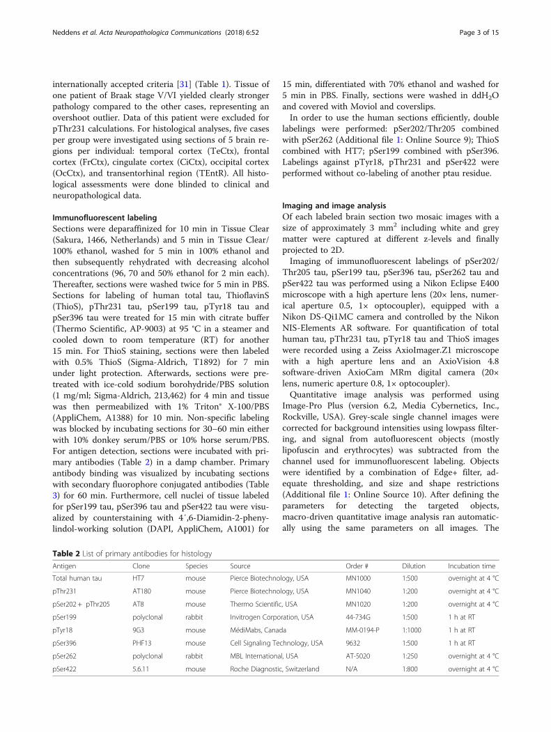
Table 2 List of primary antibodies for histology
Antigen Clone Species Source Order # Dilution Incubation time
Total human tau HT7 mouse Pierce Biotechnology, USA MN1000 1:500 overnight at 4 °C
pThr231 AT180 mouse Pierce Biotechnology, USA MN1040 1:200 overnight at 4 °C
pSer202 + pThr205 AT8 mouse Thermo Scientific, USA MN1020 1:200 overnight at 4 °C
pSer199 polyclonal rabbit Invitrogen Corporation, USA 44-734G 1:500 1 h at RT
pTyr18 9G3 mouse MédiMabs, Canada MM-0194-P 1:1000 1 h at RT
pSer396 PHF13 mouse Cell Signaling Technology, USA 9632 1:500 1 h at RT
pSer262 polyclonal rabbit MBL International, USA AT-5020 1:250 overnight at 4 °C
pSer422 5.6.11 mouse Roche Diagnostic, Switzerland N/A 1:800 overnight at 4 °C
Neddens et al. Acta Neuropathologica Communications (2018) 6:52 Page 3 of 15

quantitative results are therefore unbiased,rater-independent and fully reproducible.
Preparation of sarcosyl insoluble fraction from frozensamplesFrozen transentorhinal cortex tissue was homogenizedin 9 μl per mg of cold extraction buffer 1 (25 mM TrisHCl pH = 7.4, 150 mM NaCl, 1 mM EDTA, 1 mMEGTA, 10 mM ß-glycerophosphate, 30 mM NaF, 2 mMNa3VO4, protease and phosphatase inhibitor cocktails)and centrifuged at 80,000 g for 15 min at 4 °C. The pel-let was resuspended in extraction buffer 2 (10 mM TrisHCl pH = 7.4, 800 mM NaCl, 300 mM sucrose, 1 mMEGTA, protease and phosphatase inhibitor cocktails)and centrifuged at 4000 g for 10 min at 4 °C. The Super-natant was transferred to a fresh tube and sarcosyl (30%aqueous solution) was added to a final concentration of1% and incubated for 1.5 h at room temperature. Aftercentrifugation at 80,000 g for 30 min at 4 °C, the super-natant was discarded and the pellet resuspended in buf-fer 3 (50 mM Tris-HCl, pH = 7.4). The suspension wasaliquoted and frozen until used for Western blotting.
Western blot analysesEqual amounts of proteins of each brain extract were sep-arated by molecular weight on a SDS-PAGE polyacryl-amide gel. A protein marker visualized correct separationof the proteins and confirmed the correct protein bandsize. Subsequently, proteins were transferred onto a0.45 μm nitrocellulose membrane using a wet blot cham-ber (Bio-Rad, Hercules, USA), blocked with 5% non-fatdry milk in 1 × TBS and incubated with primary anti-bodies overnight. To blot for total tau, pTyr18, andpSer199 the same primary antibodies as used for histo-logical labeling were used in a concentration of 1:1000(Table 2). Antibodies AT180 and AT8 to label pThr231and pSer202/Thr205, respectively, did not give any signalin the Western blot and were thus substituted by a rabbitpolyclonal antibody against pThr231 (1:1000; Signalway,SAB1110–2, College Park, USA) and a rabbit monoclonal
antibody, clone EPR2402, against pSer202/Thr205(1:3000; abcam, ab108387, Cambridge, UK). All primaryantibodies were incubated for 2 h at room temperature.Afterwards, membranes were washed in TBS and incu-bated in horseradish peroxidase-coupled secondary anti-bodies for 1 h at room temperature (1:5000;donkey-anti-rabbit IgG: NA934/ GENA934;sheep-anti-mouse IgG: NXA931; GE-Healthcare, LittleChalfont, UK). Proteins were detected usingWester-Bright ECL spray (Advansta, Menlo Park, USA)and placed on a X-ray film. As loading control a rabbitpolyclonal GAPDH antibody (Sigma-Aldrich, G9545, St.Louis, USA) was co-labeled on each blot. To blot with 6different antibodies, three blots were used to prevent falsesignals due to stripping, reblotting or blocking of antibodybinding by previously used antibody. For each of the threeblots, GAPDH as loading control is shown. Densitometryof all bands was performed using Image J software. Cases3, 5, 8, 16 and 20 according to Table 1 are missing in thisanalysis since no frozen tissue was available from the cor-responding brain region.
Statistical analysesAll statistical analyses and preparation of graphs were con-ducted using Graph-Pad Prism (version 4.03, San Diego,CA, USA). Descriptive statistical analyses were performedon all evaluated parameters including the evaluation of nor-mal distribution using the Kolmogorov-Smirnov test.Group variances were calculated either by one-way or
two-way ANOVA. If a significant interaction amonggroups was detected, Newman-Keuls or Bonferroni’spost-hoc analysis was followed. A detailed description ofperformed statistical analyses is given in the appropriatefigure legend. Data were averaged and represented asmean + standard error of mean (SEM). An α-error level ofp < 0.05 was considered significant.
ResultsIn order to evaluate the overall human tau expressionlevels, brain tissues were labeled with the anti-human
Table 3 List of secondary antibodies
Antibody Conjugation Source Order # To visualize
Donkey Anti-Mouse IgG H&L AlexaFluor 555 abcam, UK ab150110 pSer422 tau
pSer396 tau
pSer202/ pThr205 tau
Donkey Anti-Rabbit IgG H&L DyLight 650 abcam, UK ab96922 pSer199 tau
pSer262 tau
Donkey Anti-Mouse IgG H&L DyLight 550 abcam, UK ab98795 pThr231 tau
Donkey Anti-Mouse IgG (H + L) Cyanine Cy3 Jackson ImmunoResearch, USA 715–165-151 Total human tau
Donkey Anti-Mouse IgG H&L DyLight 650 abcam, UK ab98797 pTyr18 tau
Dilution of all secondary antibodies was 1:500
Neddens et al. Acta Neuropathologica Communications (2018) 6:52 Page 4 of 15

total tau HT7 antibody. Quantification of the HT7 im-munoreactive area in the isocortex and transentorhinalregion (TEntR) revealed a progressive increase of totaltau with advancing Braak stages. This increase in totaltau signal was significant at Braak stage V/VI comparedto healthy controls but also compared to both lowerBraak stages I/II and III/IV (Fig. 1a). Data of the isocortexin Fig. 1a are the mean of four distinct isocortical regions,namely temporal, frontal, cingulate and occipital cortex.Separate presentation of these brain regions shows thattotal tau expression levels were very low in the cingulatecortex but relatively high in the occipital cortex of healthycontrols (Fig. 1b). In all isocortical regions total tau levelsstarted to increase at Braak stage III/IV and were signifi-cantly higher at Braak stage V/VI compared to controls orBraak stage I/II. At Braak stage V/VI total tau levels werehighest in the frontal and occipital cortex (Fig. 1b). Sinceamyloid-β plaques are, next to tau, a major pathologicalhallmark of AD, the ThioflavinS (ThioS) stained area wasquantified in the isocortex and TEntR of AD patients andhealthy controls (Fig. 1c). Threshold-based identificationof ThioS staining combined with a size restriction filterenabled automatic quantification of plaque cores while
excluding NFTs (Additional file 1: Online Source 11). Wefound that ThioS signal was significantly increased in theisocortex of Braak stage V/VI patients compared to con-trols and all other Braak stages; a first non-significant in-crease in the isocortical ThioS signal could be observed atBraak stage III/IV (Fig. 1c). In the TEntR, ThioS signalslightly increased at Braak stage III/IV but did not furtherincrease at Braak V/VI. Almost no ThioS signal could beobserved in the cingulate cortex. In the temporal andfrontal cortex the ThioS signal was very low while only inthe occipital cortex the ThioS signal was well measurableand progressively increasing starting at Braak stage III/IV(Fig. 1d).After evaluating the overall total tau and plaque load,
we measured the phosphorylation levels of ptau at sevendifferent residues. First, we show phosphorylation ofpSer202/Thr205 tau since this phosphorylation site isalready well characterized and thus can be utilized tovalidate the results.
pSer202/Thr205 tauPhosphorylation of tau at Ser202/Thr205 was low in allisocortical brain regions up to Braak stage III/IV. Only at
0.0
0.1
0.2
0.3
0.4
0.5
FrCtxTeCtx CiCtx OcCtx
*
*
****
***T
hio
S+
are
a [%
]
0.00
0.05
0.10
0.15
Isocortex TEntR
**
Th
ioS
+ a
rea
[%]
a b
c d
control
stage III/IVstage V/VI
stage I/II
0
1
2
3
4
5
6
Isocortex TEntR
***
***
HT
7 IR
are
a [%
]
01234
5678
9
FrCtxTeCtx CiCtx OcCtx
*
* ***
***
*
*********
HT
7 IR
are
a [%
]
Fig. 1 Quantification of total human tau and plaque load in the cortex of AD cases. a Total human tau (HT7 antibody) immunoreactive area inpercent in the isocortex and TEntR (allocortex) of different AD stages. c β sheet (ThioflavinS staining) positive area in percent in the isocortex andTEntR of different AD stages. Isocortical data in a and c present mean of different cortical regions that are shown separately in b and d,respectively. Two way ANOVA followed by Bonferroni’s posthoc test. Mean + SEM; N = 5.*p < 0.05; **p < 0.01; ***p < 0.001. c, d One outlier of Braakstage V/VI excluded for all ThioS labelings. Solid lines: Comparison of AD stages within a brain region; dotted lines: comparison of brain regionsof the same AD Braak stage. CiCtx: cingulate cortex; FrCtx: frontal cortex; OcCtx: occipital cortex; TeCtx: temporal cortex; TEntR:transentorhinal region
Neddens et al. Acta Neuropathologica Communications (2018) 6:52 Page 5 of 15

Braak stage V/VI Ser202/Thr205 tau phosphorylation sig-nificantly increased about 4- to 13-fold above controllevels (Fig. 2a, b). At Braak stage V/VI the highest Ser202/Thr205 tau phosphorylation could be observed in the tem-poral cortex while the lowest phosphorylation was in thecingulate cortex (Fig. 2b). Analysis of the allocorticalTEntR showed a first but not significant pSer202/Thr205tau increase in Braak stage III/IV that became significantat Braak stage V/VI (Fig. 2a).
pThr231 tauPhosphorylation of tau at Thr231 was also low in allisocortical brain regions up to Braak stage III/IV.Only at Braak stage V/VI Thr231 tau phosphorylationsignificantly increased about 30- to 160-fold abovecontrol levels (Fig. 2c, d). At Braak stage V/VI thehighest Thr231 tau phosphorylation could be ob-served in the temporal cortex while the lowest phos-phorylation was in the frontal cortex (Fig. 2d).Analysis of the allocortical TEntR showed a first butnot significant pThr231 tau increase at Braak stage I/II that became significant compared to healthy con-trols at Braak stage III/IV and further significantly in-creased at Braak stage V/VI compared to all othergroups (Fig. 2c).
pSer199 tauOverall, phosphorylation of tau at Ser199 was low com-pared to other phosphorylation sites. In all isocortical re-gions Ser199 tau phosphorylation started late, namely atBraak stage V/VI (Fig. 3a, b). At Braak stage V/VISer199 tau phosphorylation was at comparable levels inall isocortical regions and about 50- (occipital cortex) to1300-fold (cingulate cortex) increased compared tohealthy controls (Fig. 3b). Analysis of the allocorticalTEntR showed a progressive increase in Ser199 tauphosphorylation starting at Braak stage I/II but differ-ences between Braak stages became significant only atstage V/IV and were then about 3.5-fold compared toBraak stage III/IV and 160-fold compared to healthycontrols (Fig. 3a). Phosphorylation of Ser199 tau in theTEntR of late Braak stages was higher compared to allisocortical regions.
pTyr18 tauThe phosphorylation profile of pTyr18 tau in the isocor-tex and TEntR was very similar to pSer199 tau althoughthe final phosphorylation levels in Braak stage V/VI wereslightly higher and increased about 20- to 140-fold abovecontrol levels (Fig. 3c, d). The progression in the TEntRwas already significant at Braak stage III/IV compared to
0.0
2.5
5.0
7.5
10.0
FrCtxTeCtx CiCtx OcCtx
*
*
*** **
***
pS
er20
2/20
5 IR
are
a [%
]
0.0
2.5
5.0
7.5
10.0
12.5
Isocortex TEntR
**
***
0.0
2.5
5.0
7.5
10.0
FrCtxTeCtx CiCtx OcCtx
********
******
***p
Th
r231
IR
are
a [%
]
0.0
2.5
5.0
7.5
Isocortex TEntR
****
*
***
pT
hr2
31 I
R a
rea
[%]
a b
c d control
stage III/IVstage V/VI
stage I/II
pS
er20
2/T
hr2
05 IR
are
a[%
]
pS
er20
2/T
hr2
05 IR
are
a[%
]
Fig. 2 Quantification of tau phosphorylation at pSer202/Thr205 and pThr231 in the cortex of AD cases. a Human pSer202/Thr205 tauimmunoreactive area in percent in the isocortex and TEntR (allocortex) of different AD stages. c Human pThr231 tau immunoreactive area inpercent in the isocortex and TEntR of different AD stages. Isocortical data in a and c present mean of different cortical regions that are shownseparately in b and d, respectively. Two way ANOVA followed by Bonferroni’s posthoc test. Mean + SEM; N = 5.*p < 0.05; **p < 0.01; ***p < 0.001.Solid lines: Comparison of AD stages within a brain region; dotted lines: comparison of brain regions of the same AD Braak stage. CiCtx: cingulatecortex; FrCtx: frontal cortex; OcCtx: occipital cortex; TeCtx: temporal cortex; TEntR: transentorhinal region
Neddens et al. Acta Neuropathologica Communications (2018) 6:52 Page 6 of 15

stage I/II and controls. At Braak stage V/VI the highestpTyr18 tau could be observed in the TEntR and cingulatecortex while the lowest phosphorylation was measured inthe occipital cortex (Fig. 3c, d).
pSer396 tauPhosphorylation of tau at Ser396 shows higher vari-ability than other ptau sites. In the isocortex overallSer396 tau phosphorylation starts very late in Braakstage V/VI (Fig. 4b). When analyzing different isocor-tical regions separately, it seemed that pSer396 tauwas slightly increased in the temporal cortex ofhealthy controls and in the cingulate cortex at earlyBraak stage I/II but these variations were not signifi-cant (Fig. 4b). Otherwise, Ser396 tau phosphorylationin all investigated regions only increased about 2- to5-fold at Braak stage V/VI compared to control levels.Due to the high inter-individual variation this differencewas only significant in the temporal cortex compared toBraak stage I/II and III/IV (Fig. 4b). In the allocorticalTEntR pSer396 tau increased significantly at Braak stageV/VI compared to all other groups (Fig. 4a).
pSer262 tauPhosphorylation of tau at Ser262 was very low and showedalmost no progression in higher Braak stages (Fig. 4c). Only
in the TEntR a significant 8-fold increase could be notedbetween Braak stage I/II and V/VI (Fig. 4d).
pSer422 tauThe phosphorylation profile of tau at Ser422 was com-parable in all isocortical regions and also the allocorticalTEntR. In all analyzed regions phosphorylation signifi-cantly increased at Braak stage V/VI (Fig. 4e, f ) and wasabout 3- to 13-fold higher compared to the controlgroup.
Relative signal increase of ThioS and ptauWhen evaluating the signal increase relative tohealthy control brain tissue, it became evident thatThioS labeling in the TEntR increased already atBraak stage III/IV and reached a plateau at Braakstages III/IV and V/VI. The phosphorylation at tauTyr18 and tau Ser199 severely increased at the sametime at Braak stage V/VI with already a slight in-crease at Braak stage III/IV (Fig. 5a). Compared tothis tau phosphorylation the changes at other siteswere only minor. Analysis of the phosphorylationrelative to healthy control tissue in isocortical brainregions (Fig. 5b-e) showed a progressive increase ofThioS labeling in all regions with highest values inthe temporal cortex (Fig. 5b). Additionally, tau
0
1
2
3
FrCtxTeCtx CiCtx OcCtx
** *** ****
pS
er19
9 IR
are
a [%
]
0
1
2
3
4
5
FrCtxTeCtx CiCtx OcCtx
*****
***
**
***p
Tyr
18 I
R a
rea
[%]
0
1
2
3
4
5
Isocortex TEntR
***
*
***
pT
yr18
IR
are
a [%
]
0
1
2
3
4
Isocortex TEntR
***
***
pS
er19
9 IR
are
a [%
]
a b
c dcontrol
stage III/IVstage V/VI
stage I/II
Fig. 3 Quantification of tau phosphorylation at pSer199 and pTyr18 in the cortex of AD cases. a Human pSer199 tau immunoreactive area inpercent in the isocortex and TENtR (allocortex) of different AD stages. c Human pTyr18 tau immunoreactive area in percent in the isocortex andTEntR of different AD stages. Isocortical data in a and c present mean of different cortical regions that are shown separately in b and d,respectively. Two way ANOVA followed by Bonferroni’s posthoc test. Mean + SEM; N = 5.*p < 0.05; **p < 0.01; ***p < 0.001. Solid lines: Comparisonof AD stages within a brain region; dotted lines: comparison of brain regions of the same AD Braak stage. CiCtx: cingulate cortex; FrCtx: frontalcortex; OcCtx: occipital cortex; TeCtx: temporal cortex; TEntR: transentorhinal region
Neddens et al. Acta Neuropathologica Communications (2018) 6:52 Page 7 of 15

phosphorylation was severely increased at Thr231,Ser199 and Tyr18. Tau phosphorylation at Thr231was highest in the temporal cortex (Fig. 5b) whilephosphorylation at Ser199 and Tyr18 was highest inthe frontal cortex (Fig. 5c). Also in the isocortex tauphosphorylation changes at other sites are only minorcompared to Thr231, Ser199 and Tyr18.To validate results obtained by immunofluorescent la-
beling, Western blots of AD and control cases were per-formed (Fig. 6a). Cases showing an increase in total taualso showed a signal increase in all analyzed ptau resi-dues. Furthermore, the majority of human cases withstrong ptau signals were clinically diagnosed with milddementia (case 6 and 12) or dementia (case 17, 18, 19,Table 1), suggesting a correlation between phosphoryl-ation and cognitive status. Even that tissue of some caseswas missing the quantification of Western blot results
shows an overall conformity compared to immunofluor-escent labelings. Total tau by HT7 labeling (Fig. 6b),pSer202/205 (Fig. 6c), and pSer199 (Fig. 6e) also showeda late signal increase in Braak stage V/VI with only weaksignals in earlier Braak stages. Western blotting ofpThr231 (Fig. 6d) and pTyr18 (Fig. 6f ) also showed alate signal increase in Braak stage V/VI while signals ofimmunofluorescent labelings increased already in earlierBraak stages (Fig. 2c and Fig. 3c).Representative images of total tau (Fig. 7) and
pSer202/Thr205, pThr231, pSer199 and pTyr18 (Fig. 8)immunofluorescent labelings in the transentorhinal cor-tex of one case per Braak stage show the progressive in-crease of these ptau sites.In summary, our quantitative analysis of AD brain
tissue shows that a progressive increase of total tauand ptau expression can be observed in the
0.0
2.5
5.0
7.5
FrCtxTeCtx CiCtx OcCtx
*****
******
pS
er42
2 IR
are
a [%
]
0.0
2.5
5.0
7.5
Isocortex TEntR
***
***
pS
er42
2 IR
are
a [%
]
0.0
0.5
1.0
FrCtxTeCtx CiCtx OcCtx
* ***
pS
er26
2 IR
are
a [%
]
0.0
0.1
0.2
0.3
0.4
0.5
0.6
0.7
Isocortex TEntR
pS
er26
2 IR
are
a [%
]
0
1
2
3
4
FrCtxTeCtx CiCtx OcCtx
*
pS
er39
6 IR
are
a [%
]
0
1
2
3
4
5
6
Isocortex TEntR
*
*
pS
er39
6 IR
are
a [%
]
control
stage III/IVstage V/VI
stage I/II
a b
c d
e f
Fig. 4 Quantification of tau phosphorylation at pSer396, pSer262 and pSer422 in the cortex of AD cases. a Human pSer396 tau immunoreactivearea in percent in the isocortex and TEntR (allocortex) of different AD stages. c Human pSer422 tau immunoreactive area in percent in theisocortex and TEntR of different AD stages. e Human pSer262 tau immunoreactive area in percent in the isocortex and TEntR of different ADstages. Isocortical data in a, c and e present mean of different cortical regions that are shown separately in b, d and f, respectively. Two wayANOVA followed by Bonferroni’s posthoc test. Mean + SEM; N = 5.*p < 0.05; **p < 0.01; ***p < 0.001. Solid lines: Comparison of AD stages within abrain region; dotted lines: comparison of brain regions of the same AD Braak stage. CiCtx: cingulate cortex; FrCtx: frontal cortex; OcCtx: occipitalcortex; TeCtx: temporal cortex; TEntR: transentorhinal region
Neddens et al. Acta Neuropathologica Communications (2018) 6:52 Page 8 of 15

transentorhinal region and in most of the analyzedisocortical regions. Staining of tissue with ThioS re-sulted only in a weak signal in the transentorhinalregion and the occipital cortex. Labeling with anti-bodies against different tau phosphorylation sitesshowed that most ptau sites, namely pSer202/Thr205,pThr231, pSer199, pTyr18 and pSer422 were highlyincreased throughout the cortex at Braak stage V/VI.Only tau phosphorylation at Thr231 and Thy18 wasalready significantly increased at Braak stage III/IV.By contrast, pSer396 tau and pSer262 tau are onlyweakly expressed in all analyzed brain regions andonly minor progression was observed. When com-paring tau phosphorylation in percent relative tohealthy controls, phosphorylation is specifically in-creased at tau Thr231, Ser199 and Tyr18. These dataindicate that tau phosphorylation is a complex fea-ture of AD progression, involving many but not allpotential phosphorylation sites.
DiscussionThe current study was designed to analyze spatial pat-terns of tau phosphorylation at multiple residues indiscrete anatomical regions during AD progression. Ourdata revealed a very similar phosphorylation profile ofmost of the analyzed ptau sites in the allo- and isocortexwhile expression levels of phosphorylated tau at Tyr18and Thr231 was distinguishable between Braak stages.When normalized to controls, phosphorylation of tau atTyr18, Ser231 and also Ser199 was much more in-creased at Braak stage V/VI compared to other residues,suggesting a relevance of these sites for AD progressionand a crucial role in pathogenesis.Since tau phosphorylation is a main characteristic of
AD progression, several groups have already analyzedthe temporal phosphorylation pattern of different tausites by histological methods. According to Luna-Munozand colleagues the phosphorylation of Thr231 tau is anearly event in the neuronal pathology of AD [27]. The
a
e
dc
b*** **
T ra n s e n to rh in a l R e g io n
T h ioS
HT 7
p Se r2
0 2 /Th r2
0 5
p T h r23 1
p Se r1
9 9
p T y r18
p Se r3
9 6
p Se r2
6 2
p Se r4
2 2
0
5 0 0 0
1 0 0 0 0
1 5 0 0 0I / I I
I I I / IV
V /V I
***
***
***
***
Inc
rea
se
in%
rel.
toc
on
tro
ls
T e m p o ra l C o r te x
T h ioS
HT 7
p Se r2
0 2 /Th r2
0 5
p T h r23 1
p Se r1
9 9
p T y r18
p Se r3
9 6
p Se r2
6 2
p Se r4
2 2
0
1 0 0 0 0
2 0 0 0 0
3 0 0 0 0
***
***
*
**
**
***
***
Inc
rea
se
in%
rel .
toc
on
tro
ls
F ro n ta l C o r te x
T h ioS
HT 7
p Se r2
0 2 /Th r2
0 5
p T h r23 1
p Se r1
9 9
p T y r18
p Se r3
9 6
p Se r2
6 2
p Se r4
2 2
0
2 0 0 0 0
4 0 0 0 0
6 0 0 0 0**
****
Inc
rea
se
i n%
rel.
toc
on
tro
ls
C in g u la te C o r te x
T h ioS
HT 7
p Se r2
0 2 /Th r2
0 5
p T h r23 1
p Se r1
9 9
p T y r18
p Se r3
9 6
p Se r2
6 2
p Se r4
2 2
0
5 0 0 0 0
1 0 0 0 0 0
1 5 0 0 0 0
2 0 0 0 0 0
* **
***
Inc
rea
se
in%
r el.
toc
on
tro
ls
O c c ip ita l C o r te x
T h ioS
HT 7
p Se r2
0 2 /Th r2
0 5
p T h r23 1
p Se r1
9 9
p T y r18
p Se r3
9 6
p Se r2
6 2
p Se r4
2 2
0
5 0 0 0
1 0 0 0 0
1 5 0 0 0
2 0 0 0 0
* **
***
* *
*
Inc
rea
se
in%
rel.
toc
on
tro
ls
Fig. 5 Signal increase relative to control brain tissue in the cortex of AD cases. Values are calculated from means of absolute IR area from Figs. 1,2, 3 and 4 relative to control brain samples. Calculation is shown for ThioS, HT7, pSer202/Thr205, pThr231, pSer199, pTyr18, pSer396, pSer262 andpSer422 in the transentorhinal (a), temporal (b), frontal (c), cingulate (d) and occipital cortex (e) at Braak stages I/II; III/IV and V/VI. Two wayANOVA followed by Bonferroni’s posthoc test. Mean + SEM. White asterisks within bars mark significance compared to Braak stage I/II. *p < 0.05;**p < 0.01; ***p < 0.001
Neddens et al. Acta Neuropathologica Communications (2018) 6:52 Page 9 of 15

temporal analysis of pSer202/Thr205 tau and pSer396tau is very controversial. Simic and co-workers foundhigher phosphorylation of Ser396 and Ser202/Thr205tau in mild cognitive impairment (MCI) cases [38] sug-gesting a parallel phosphorylation of both residues.Temporal phosphorylation analyses by two other groupscontradict these results, while one group found an earl-ier phosphorylation of pSer202/Thr205 tau [39], theother reported an earlier phosphorylation of pSer396 tau[30]. Zhou and colleagues performed dot blots andELISA analyses of a whole series of ptau sites of ADmedial temporal cortex samples and found mostly asimilar temporal phosphorylation pattern as shown herefor the entorhinal cortex. Tau phosphorylation of Ser396for example, was a late event and only measurable inBraak stage V and VI [46]. Dot blot analyses of AD lat-eral temporal lobe samples by another group showedthat tau phosphorylation at Ser202/205 and Ser396
simultaneously increases with increasing Braak staging,but also that they observe the increase already in Braakstage III/IV compared to results shown here or by Zhouand colleagues [20, 46]. By quantitatively analyzing ptausites in different brain regions of AD cases we found thatmost analyzed ptau sites, pSer202/Thr205, pThr231,pSer199, pTyr18 and pSer422, have a very similar allo-and isocortical phosphorylation profile, suggesting thatpSer202/Thr205 tau analysis by AT8 antibody could bereplaced by any of these ptau sites when using this quan-titative histological approach. For neuropathological as-sessment of Braak stages ptau Ser202/Thr205immunopositivity is scored semi-quantitatively and is re-quired to be mild (+) in the transentorhinal cortex to as-sign Braak stage I, while for stages higher than Braakstage I, severity of ptau Ser202/Thr205 immunopositivityin the transentorhinal cortex is not decisive as otherareas, e.g. entorhinal cortex, occipitotemporal gyrus,
Fig. 6 Quantification of total tau and tau phosphorylation in the transentorhinal cortex of AD cases by Western blotting. a: Western blots of totaltau, pThr231, pSer202/Thr205, pTyr18, and pSer199. GAPDH was used as loading control. Quantification of Western blot for (b) total tau by HT7antibody, (c) pSer202/Thr205, (d) pThr231, (e) pSer199, and (f) pTyr18. All samples shown in (a) were used for quantification of (b-f). One wayANOVA followed by Tukey’s multiple comparisons test. Mean + SEM. *p < 0.05; **p < 0.01; n.s.: not significant
Neddens et al. Acta Neuropathologica Communications (2018) 6:52 Page 10 of 15

middle temporal gyrus, occipital cortex need to show atleast moderate (++) severity of immunopositivity [1].Therefore our findings of a lack of a significant increaseof ptau Ser202/Thr205 immunopositivity in the transen-torhinal cortex between Braak stages 0 to IV is not atvariance with neuropathological Braak staging. However,pSer202/Thr205 immunopositivity does indeed increasecontinuously in the transentorhinal cortex with increas-ing Braak stages. In our study the difference is only sta-tistically significant between Braak stages 0-IV and V/VI.The phosphorylation of the analyzed tau sites starts inthe isocortical regions late at Braak stage V/VI and con-sequently the analysis of the allocortical transentorhinalregion is preferable. Indeed, the quantitative analysis ofpTyr18 tau and pThr231 tau in the transentorhinal re-gion shows a distinct progression between Braak stages
that might be caused by highly increased phosphoryl-ation relative to control tissue that could also be ob-served for pSer199 tau. Since phosphorylation of tau atresidues Tyr18 and Thr231 occurs earlier compared toother sites, these phosphorylation sites might trigger dis-ease progression and thus would represent an earlytherapeutic target.Western blot analyses of total tau and the most
prominent phosphorylation residues pSer202/Thr205,pThr231, pSer199, and pTyr18 overall validated theresults obtained by immunofluorescent labeling. It hasto be mentioned that tissue of not all human caseswas available for blotting, so quantification and statis-tical analyses should be understood as preliminary.Blots for pSer202//Thr205 and pThr231 are not dir-ectly comparable, since different antibodies as used
Fig. 7 Representative images of total tau labeling in the transentorhinal cortex grey matter of AD cases. Labeling of total tau using HT7 antibody(a-d, a1-d1, a2-d2) and nuclei by DAPI staining (a-d, a1-d1,a3-d3). Autofluorescence is shown in white (a-d, a1-d1, a4-d4). Samples of healthycontrol tissue (a-a4, case 2), Braak stage I/II (b-b4, case 8), Braak stage III/IV (c-c4, case 11) and Braak stage V/VI (d-d4, case 16) are shown. Scalebar: 100 μm
Neddens et al. Acta Neuropathologica Communications (2018) 6:52 Page 11 of 15

for immunofluorescent labelings had to be used formethodical reasons.AD is known to affect women with a higher preva-
lence, which is among other reasons caused by hormonaldifferences. The higher frequency of AD cases in womenis reflected in this study by the fact that only tissue ofwomen was provided for Braak stage V/VI, while tissueof earlier Braak stages was predominantly from men. Al-though several in vitro and in vivo studies in rodentssuggest an impact of sex hormones (e.g. estrogens, pro-gesterone and prolactin) and therefore gender on tauphosphorylation, there are so far no studies in AD pa-tients validating these results in humans (for review see[33]). Future analyses of post mortem AD tissue and invivo imaging of AD patients should shed light on theimpact of gender on tau phosphorylation.In addition to the severe increases in tau phosphoryl-
ation also the ThioS signal, that includes amyloid plaques,was highly increased in several allo- and isocortical re-gions. The ThioS signal increase partially already startedat Braak stage III/IV and thus earlier than the increase ofptau. It has been shown that ptau residues, Tyr18, Thr231and Ser199 can be phosphorylated by Aβ via different ki-nases like Fyn [18, 35, 37, 42, 44, 45], GSK-3β [3, 23, 24]or CDK5 [6, 25]. The activation of tau by Aβ is furtherdemonstrated to be involved in the early formation ofneurofibrillary tangles, synaptic loss, neurodegeneration aswell as cognitive deficits [2, 3, 11, 25] and thus in the de-velopment of the most prominent AD pathologies. Theseresults were derived from AD cell and animal models butour study might suggest a similar effect of Aβ on tauphosphorylation at residues Tyr18, Thr231 and Ser199 inthe human disease. Further analyses are needed to validatethis hypothesis in humans. A valid tool to analyze suchevents in vivo might be the use of Pittsburgh compound B(PiB) analysis combined with tau tracer that are currentlyunder development [5, 17, 19, 36].Isocortical tau pathology is only sparse at early Braak
stages I to IV and hence use of higher magnification andmodified image analysis parameters may be necessary toreveal subtle changes in isocortical tau pathology at earlyBraak stages. Additionally, co-labeling of differentmarkers to quantify ptau only in distinct areas or cellpopulations combined with an increased number of in-vestigated brain regions may be helpful to gain a betterunderstanding of earliest isocortical tau pathology. A
Fig. 8 Representative images of ptau labelings in the transentorhinalcortex of control and AD cases. Labeling of pSer202/Thr205 (a),pThr231 (b), pSer199 (c), and pTyr18 (d) are shown. Tissues wereadditionally stained with DAPI to visualize nuclei. Autofluorescence isshown in white. Dotted lines indicate areas shown in grey scaleimages. Samples of healthy control tissue (case 2) and Braak stage V/VI (case 16) are shown. Scale bar: 100 μm
Neddens et al. Acta Neuropathologica Communications (2018) 6:52 Page 12 of 15

tissue microarray method that was developed recentlyallows to examine over 35 brain regions on one slidecould be used for such purposes [43]. However, ourstudy investigated only five brain regions in a limitednumber of cases and represents therefore proof ofprinciple. Our data need to be confirmed by includingmore cases and assessing a higher number of brainareas.
ConclusionWe show here for the first time the phosphorylationprofile of different tau sites in allocortical and isocorticalregions of human brains with tau pathology rangingfrom Braak stages 0 to VI using quantitativerater-independent immunofluorescent labeling. Our datasuggest that the profile of ptau in the isocortex is com-parable between all of the analyzed ptau sites while ex-pression levels of ptau at Thr231 and Tyr18 in thetransentorhinal region are distinguishable between Braakstages combined with the highest tau phosphorylationrelative to controls at these sites and Ser199.
Additional file
Additional file 1: Online Source 9 Double labeling of pSer262 andpSer202/Thr205 tau in the temporal cortex at Braak stage V/VI. Images showdifferent labeling pattern of pSer262 (arrows) and pS202 (white arrowheads)as well as their overlay (yellow arrowheads) (a1) and single fluorescenceimages (a2,3,4) of case 17. AF: autofluorescence. Scale bar: 20 μm. OnlineSource 10 Example of measurement procedure of tau pSer262. Objects inthe unlabeled autofluorescence channel were detected by thresholding(red in a1). The resulting mask images (a2) were then subtracted from taupSer262 images to remove autofluorescence (a3). The resulting imageswere Edge+ filtered (a4) to facilitate threshold-based detection of taupSer262-positive objects (red outline in a5). These outlines were then loadedonto the raw images to quantify original tau pSer262 signal (red outline ina6). AF: autofluorescence. Scale bar: 20 μm. Online Source 11 Example ofdetecting ThioS-positive amyloid-β but not NFTs. Image a displays theco-labeling of ThioS (green) and HT7 (red), while images b and c,respectively, show single channel images. ThioS shows intense labeling ofplaque-associated β-sheets (b, asterisk) whereas tangles are only weakly la-beled (c, arrows) (c). A combination of threshold-based identification ofThioS and size restriction (d‘, green rectangle) enables quantification ofThioS+ plaque labeling (red highlighted) but not tangles (d). ThioS:ThioflavinS. Scale bar: 20 μm. (PDF 599 kb)
AbbreviationsAD: Alzheimer’s disease; ANOVA: Analysis of variance; CiCtx: Cingulate cortex;FrCtx: Frontal cortex; NFT: Neurofibrillary tangle; NT: Neuropil threads;OcCtx: Occipital cortex; ptau: Phosphorylated tau; TeCtx: Temporal cortex;TEntR: Transentorhinal region
AcknowledgementsThe authors greatly thank the whole research team of QPS Austria GmbH fortheir technical support.
FundingThis work was supported by the Austrian Research Promotion Agency (FFG),R&D Projects (FFG# 844453, 851079, 855287).Tissue for this study was provided by the Newcastle Brain Tissue Resource,which is funded in part by a grant from the UK Medical Research Council(G0400074), by Brains for Dementia research, a joint venture betweenAlzheimer’s Society and Alzheimer’s Research UK and by the NIHR Newcastle
Biomedical Research Centre awarded to the Newcastle upon Tyne HospitalsNHS Foundation Trust and Newcastle University.
Availability of data and materialsThe datasets used and/or analyzed during the current study are availablefrom the corresponding author on reasonable request.
Authors’ contributionsJN designed the overall project, analyzed data, interpreted experiments andprepared the manuscript; MT analyzed data and prepared figures; SFinterpreted experiments and prepared figures and the manuscript; BK andCH performed experiments and analyzed data; VN edited the manuscript; JAprovided neuropathologically diagnoses and edited the manuscript; GDinterpreted experiments and edited the manuscript; BHP designed andinterpreted experiments and edited the manuscript. All authors read andapproved the final manuscript.
Ethics approval and consent to participateHuman tissue was provided by the Newcastle Brain Tissue Resource (NBTR),Newcastle University, UK in accordance with the approval of the joint EthicsCommittee of Newcastle and North Tyneside Health Authority and followingNBTR brain banking procedures.
Consent for publicationNot applicable.
Competing interestsJN, MT, SF, BK, CH, TL, VN and BHP are employees of QPS Austria GmbH.The authors declare that they have no other competing interests.
Publisher’s NoteSpringer Nature remains neutral with regard to jurisdictional claims inpublished maps and institutional affiliations.
Author details1QPS Austria GmbH, Neuropharmacology, Parkring 12, 8074 Grambach,Austria. 2Institute for Biochemistry, Graz University of Technology, Graz,Austria. 3Institute of Zoology, Karl Franzens University, Graz, Austria. 4Instituteof Neuroscience and Newcastle University Institute for Ageing Campus forAgeing and Vitality, Newcastle University, Newcastle upon Tyne NE4 5PL, UK.
Received: 24 May 2018 Accepted: 19 June 2018
References1. Alafuzoff I, Arzberger T, Al-Sarraj S, Bodi I, Bogdanovic N, Braak H, Bugiani O,
Del-Tredici K, Ferrer I, Gelpi E et al (2008) Staging of neurofibrillarypathology in Alzheimer's disease: a study of the BrainNet Europeconsortium. Brain Pathol 18: 484–496 doi 10.1111/j.1750-3639.2008.00147.x
2. Amadoro G, Corsetti V, Atlante A, Florenzano F, Capsoni S, Bussani R,Mercanti D, Calissano P (2012) Interaction between NH(2)-tau fragment andAbeta in Alzheimer's disease mitochondria contributes to the synapticdeterioration. Neurobiol Aging 33(833):e831–e825. https://doi.org/10.1016/j.neurobiolaging.2011.08.001
3. Amadoro G, Corsetti V, Ciotti MT, Florenzano F, Capsoni S, Amato G,Calissano P (2011) Endogenous Abeta causes cell death via early tauhyperphosphorylation. Neurobiol Aging 32:969–990. https://doi.org/10.1016/j.neurobiolaging.2009.06.005
4. Andorfer C, Kress Y, Espinoza M, de Silva R, Tucker KL, Barde YA, Duff K,Davies P (2003) Hyperphosphorylation and aggregation of tau in miceexpressing normal human tau isoforms. J Neurochem 86:582–590
5. Ariza M, Kolb HC, Moechars D, Rombouts F, Andres JI (2015) Tau positronemission tomography (PET) imaging: past, present, and future. J Med Chem58:4365–4382. https://doi.org/10.1021/jm5017544
6. Billingsley ML, Kincaid RL (1997) Regulated phosphorylation anddephosphorylation of tau protein: effects on microtubule interaction,intracellular trafficking and neurodegeneration. Biochem J 323(Pt 3):577–591
7. Boekhoorn K, Terwel D, Biemans B, Borghgraef P, Wiegert O, Ramakers GJ,de Vos K, Krugers H, Tomiyama T, Mori H et al (2006) Improved long-termpotentiation and memory in young tau-P301L transgenic mice before onset
Neddens et al. Acta Neuropathologica Communications (2018) 6:52 Page 13 of 15

of hyperphosphorylation and tauopathy. J Neurosci 26:3514–3523. https://doi.org/10.1523/JNEUROSCI.5425-05.2006
8. Braak H, Alafuzoff I, Arzberger T, Kretzschmar H, Del Tredici K (2006) Stagingof Alzheimer disease-associated neurofibrillary pathology using paraffinsections and immunocytochemistry. Acta Neuropathol 112:389–404. https://doi.org/10.1007/s00401-006-0127-z
9. Braak H, Braak E (1991) Neuropathological stageing of Alzheimer-relatedchanges. Acta Neuropathol 82:239–259
10. Bramblett GT, Goedert M, Jakes R, Merrick SE, Trojanowski JQ, Lee VM (1993)Abnormal tau phosphorylation at Ser396 in Alzheimer's diseaserecapitulates development and contributes to reduced microtubulebinding. Neuron 10:1089–1099
11. Chang KH, de Pablo Y, Lee HP, Lee HG, Smith MA, Shah K (2010) Cdk5 is amajor regulator of p38 cascade: relevance to neurotoxicity in Alzheimer'sdisease. J Neurochem 113:1221–1229. https://doi.org/10.1111/j.1471-4159.2010.06687.x
12. Cho JH, Johnson GV (2004) Primed phosphorylation of tau at Thr231 byglycogen synthase kinase 3beta (GSK3beta) plays a critical role in regulatingtau's ability to bind and stabilize microtubules. J Neurochem 88:349–358
13. Cook C, Stankowski JN, Carlomagno Y, Stetler C, Petrucelli L (2014)Acetylation: a new key to unlock tau's role in neurodegeneration.Alzheimers Res Ther 6:29. https://doi.org/10.1186/alzrt259
14. Flunkert S, Hierzer M, Loffler T, Rabl R, Neddens J, Duller S, Schofield EL,Ward MA, Posch M, Jungwirth H et al (2013) Elevated levels of soluble totaland hyperphosphorylated tau result in early behavioral deficits and distinctchanges in Brain Pathol in a new tau transgenic mouse model.Neurodegener Dis 11:194–205. https://doi.org/10.1159/000338152
15. Goedert M, Jakes R, Crowther RA, Six J, Lubke U, Vandermeeren M, Cras P,Trojanowski JQ, Lee VM (1993) The abnormal phosphorylation of tauprotein at Ser-202 in Alzheimer disease recapitulates phosphorylationduring development. Proc Natl Acad Sci U S A 90:5066–5070
16. Green KN, Steffan JS, Martinez-Coria H, Sun X, Schreiber SS, Thompson LM,LaFerla FM (2008) Nicotinamide restores cognition in Alzheimer's diseasetransgenic mice via a mechanism involving sirtuin inhibition and selectivereduction of Thr231-phosphotau. J Neurosci 28:11500–11510. https://doi.org/10.1523/JNEUROSCI.3203-08.2008
17. Hashimoto H, Kawamura K, Igarashi N, Takei M, Fujishiro T, Aihara Y, ShiomiS, Muto M, Ito T, Furutsuka K et al (2014) Radiosynthesis,photoisomerization, biodistribution, and metabolite analysis of 11C-PBB3 asa clinically useful PET probe for imaging of tau pathology. J Nucl Med 55:1532–1538. https://doi.org/10.2967/jnumed.114.139550
18. Ittner LM, Ke YD, Delerue F, Bi M, Gladbach A, van Eersel J, Wolfing H,Chieng BC, Christie MJ, Napier IA et al (2010) Dendritic function of taumediates amyloid-beta toxicity in Alzheimer's disease mouse models. Cell142:387–397. https://doi.org/10.1016/j.cell.2010.06.036
19. Kadir A, Nordberg A (2010) Target-specific PET probes forneurodegenerative disorders related to dementia. J Nucl Med 51:1418–1430.https://doi.org/10.2967/jnumed.110.077164
20. Koss DJ, Jones G, Cranston A, Gardner H, Kanaan NM, Platt B (2016)Soluble pre-fibrillar tau and beta-amyloid species emerge in earlyhuman Alzheimer's disease and track disease progression and cognitivedecline. Acta Neuropathol 132:875–895. https://doi.org/10.1007/s00401-016-1632-3
21. Lauckner J, Frey P, Geula C (2003) Comparative distribution of tauphosphorylated at Ser262 in pre-tangles and tangles. Neurobiol Aging24:767–776
22. Lee G, Thangavel R, Sharma VM, Litersky JM, Bhaskar K, Fang SM, DoLH, Andreadis A, Van Hoesen G, Ksiezak-Reding H (2004)Phosphorylation of tau by fyn: implications for Alzheimer's disease. JNeurosci 24:2304–2312. https://doi.org/10.1523/JNEUROSCI.4162-03.2004
23. Li T, Hawkes C, Qureshi HY, Kar S, Paudel HK (2006) Cyclin-dependentprotein kinase 5 primes microtubule-associated protein tau site-specificallyfor glycogen synthase kinase 3beta. Biochemistry 45:3134–3145. https://doi.org/10.1021/bi051635j
24. Li T, Paudel HK (2006) Glycogen synthase kinase 3beta phosphorylatesAlzheimer's disease-specific Ser396 of microtubule-associated proteintau by a sequential mechanism. Biochemistry 45:3125–3133. https://doi.org/10.1021/bi051634r
25. Liao X, Zhang Y, Wang Y, Wang J (2004) The effect of cdk-5overexpression on tau phosphorylation and spatial memory of rat.Sci China Ser C Life Sci 47:251–257
26. Lichtenberg-Kraag B, Mandelkow EM, Biernat J, Steiner B, Schroter C,Gustke N, Meyer HE, Mandelkow E (1992) Phosphorylation-dependentepitopes of neurofilament antibodies on tau protein and relationshipwith Alzheimer tau. Proc Natl Acad Sci U S A 89:5384–5388
27. Luna-Munoz J, Garcia-Sierra F, Falcon V, Menendez I, Chavez-Macias L, MenaR (2005) Regional conformational change involving phosphorylation of tauprotein at the Thr231, precedes the structural change detected by Alz-50antibody in Alzheimer's disease. J Alzheimer's Dis: JAD 8:29–41
28. Lund H, Cowburn RF, Gustafsson E, Stromberg K, Svensson A, Dahllund L,Malinowsky D, Sunnemark D (2013) Tau-tubulin kinase 1 expression,phosphorylation and co-localization with phospho-Ser422 tau in theAlzheimer's disease Brain. Brain Pathol 23:378–389. https://doi.org/10.1111/bpa.12001
29. Luo HB, Xia YY, Shu XJ, Liu ZC, Feng Y, Liu XH, Yu G, Yin G, Xiong YS, ZengK et al (2014) SUMOylation at K340 inhibits tau degradation throughderegulating its phosphorylation and ubiquitination. Proc Natl Acad Sci U SA 111:16586–16591. https://doi.org/10.1073/pnas.1417548111
30. Mondragon-Rodriguez S, Perry G, Zhu X, Moreira PI, Acevedo-Aquino MC,Williams S (2013) Phosphorylation of tau protein as the link betweenoxidative stress, mitochondrial dysfunction, and connectivity failure:implications for Alzheimer's disease. Oxidative Med Cell Longev 2013:940603. https://doi.org/10.1155/2013/940603
31. Montine TJ, Phelps CH, Beach TG, Bigio EH, Cairns NJ, Dickson DW,Duyckaerts C, Frosch MP, Masliah E, Mirra SS et al (2012) NationalInstitute on Aging-Alzheimer's Association guidelines for theneuropathologic assessment of Alzheimer's disease: a practicalapproach. Acta Neuropathologica 123:1–11. https://doi.org/10.1007/s00401-011-0910-3
32. Moszczynski AJ, Gohar M, Volkening K, Leystra-Lantz C, Strong W, Strong MJ(2015) Thr175-phosphorylated tau induces pathologic fibril formation viaGSK3beta-mediated phosphorylation of Thr231 in vitro. Neurobiol Aging 36:1590–1599. https://doi.org/10.1016/j.neurobiolaging.2014.12.001
33. Munoz-Mayorga D, Guerra-Araiza C, Torner L, Morales T (2018) Tauphosphorylation in female neurodegeneration: role of estrogens,progesterone, and prolactin. Front Endocrinol 9:133. https://doi.org/10.3389/fendo.2018.00133
34. Pevalova M, Filipcik P, Novak M, Avila J, Iqbal K (2006) Post-translationalmodifications of tau protein. Bratislavske Lekarske Listy 107:346–353
35. Roberson ED, Halabisky B, Yoo JW, Yao J, Chin J, Yan F, Wu T, Hamto P,Devidze N, Yu GQ et al (2011) Amyloid-beta/Fyn-induced synaptic, network,and cognitive impairments depend on tau levels in multiple mouse modelsof Alzheimer's disease. J Neurosci 31:700–711. https://doi.org/10.1523/JNEUROSCI.4152-10.2011
36. Rowe CC, Villemagne VL (2013) Amyloid imaging with PET in earlyAlzheimer disease diagnosis. Med Clin North Am 97:377–398. https://doi.org/10.1016/j.mcna.2012.12.017
37. Scales TM, Derkinderen P, Leung KY, Byers HL, Ward MA, Price C, Bird IN,Perera T, Kellie S, Williamson R et al (2011) Tyrosine phosphorylation of tauby the SRC family kinases lck and fyn. Mol Neurodegen 6:12. https://doi.org/10.1186/1750-1326-6-12
38. Simic G, Babic Leko M, Wray S, Harrington C, Delalle I, Jovanov-Milosevic N,Bazadona D, Buee L, de Silva R, Di Giovanni G et al (2016) Tau proteinhyperphosphorylation and aggregation in Alzheimer's disease and otherTauopathies, and possible neuroprotective strategies. Biomolecules 6: 6 doihttps://doi.org/10.3390/biom6010006
39. Su JH, Cummings BJ, Cotman CW (1994) Early phosphorylation of tau inAlzheimer's disease occurs at Ser-202 and is preferentially located withinneurites. Neuroreport 5:2358–2362
40. Tenreiro S, Eckermann K, Outeiro TF (2014) Protein phosphorylation inneurodegeneration: friend or foe? Front Mol Neurosci 7:42. https://doi.org/10.3389/fnmol.2014.00042
41. Thal DR, Rub U, Orantes M, Braak H (2002) Phases of a beta-deposition inthe human brain and its relevance for the development of AD. Neurology58:1791–1800
42. Usardi A, Pooler AM, Seereeram A, Reynolds CH, Derkinderen P,Anderton B, Hanger DP, Noble W, Williamson R (2011) Tyrosinephosphorylation of tau regulates its interactions with Fyn SH2 domains,but not SH3 domains, altering the cellular localization of tau. FEBS J278:2927–2937. https://doi.org/10.1111/j.1742-4658.2011.08218.x
43. Walker L, McAleese KE, Johnson M, Khundakar AA, Erskine D, Thomas AJ,McKeith IG, Attems J (2017) Quantitative neuropathology: an update on
Neddens et al. Acta Neuropathologica Communications (2018) 6:52 Page 14 of 15

automated methodologies and implications for large scale cohorts. J NeuralTransm (Vienna). https://doi.org/10.1007/s00702-017-1702-2
44. Williamson R, Scales T, Clark BR, Gibb G, Reynolds CH, Kellie S, Bird IN,Varndell IM, Sheppard PW, Everall I et al (2002) Rapid tyrosinephosphorylation of neuronal proteins including tau and focal adhesionkinase in response to amyloid-beta peptide exposure: involvement of Srcfamily protein kinases. J Neurosci 22:10–20
45. Williamson R, Usardi A, Hanger DP, Anderton BH (2008) Membrane-boundbeta-amyloid oligomers are recruited into lipid rafts by a fyn-dependentmechanism. FASEB J 22:1552–1559. https://doi.org/10.1096/fj.07-9766com
46. Zhou XW, Li X, Bjorkdahl C, Sjogren MJ, Alafuzoff I, Soininen H, Grundke-Iqbal I, Iqbal K, Winblad B, Pei JJ (2006) Assessments of the accumulationseverities of amyloid beta-protein and hyperphosphorylated tau in themedial temporal cortex of control and Alzheimer's brains. Neurobiol Dis 22:657–668. https://doi.org/10.1016/j.nbd.2006.01.006
Neddens et al. Acta Neuropathologica Communications (2018) 6:52 Page 15 of 15