Phenotypic distinctions between mosaic forms of tuberous ...
11
Phenotypic distinctions between mosaic forms of tuberous sclerosis complex Alison M. Treichel, BS 1,2 , Lana Hamieh, MD 3 , Neera R. Nathan, MD, MSHS 1,2 , Magdalena E. Tyburczy, PhD 3 , Ji-an Wang, AS 1 , Oyetewa Oyerinde, MD 1,2 , Sorana Raiciulescu, MSc 4 , Patricia Julien-Williams, NP 2 , Amanda M. Jones, NP 2 , Vissaagan Gopalakrishnan, BS 2 , Joel Moss, MD, PhD 2 , David J. Kwiatkowski, MD, PhD 3 and Thomas N. Darling, MD, PhD 1 Disclaimer: The opinions and assertions expressed herein are those of the author(s) and do not necessarily reflect the official policy or position of the Uniformed Services University, the Department of Defense or the National Institutes of Health. Purpose: To determine if mosaic tuberous sclerosis complex (TSC) can be stratified into subtypes that correspond with prognosis and extent of disease. Methods: Next-generation sequencing of skin tumor and other samples was used to identify patients with mosaic pathogenic variants in TSC1 or TSC2. Extent of disease, onset age, and family history of TSC were determined through retrospective analysis of patient records. Results: The median number of disease findings and age at penetrance differed between mosaic patients with asymmetrically distributed facial angiofibromas (4 findings, 24 years, n = 7), mosaic patients with bilaterally symmetric facial angiofibromas (8 findings, 10 years, n = 12), and germline TSC patients (10 findings, 4 years, n = 29). Cutaneous and internal organ involvement positively correlated in mosaic (R = 0.62, p = 0.005), but not germline (R = -0.24, p = 0.24) TSC. Variant allele fraction (VAF) in the blood (range: 0–19%) positively correlated with the number of major features (R = 0.55, p = 0.028). Five had a TSC2 variant identified in the skin that was below detection in the blood. One of 12 children from a mosaic parent had TSC. Conclusion: The phenotype of mosaic TSC ranged from mild to indistinguishable from germline disease. Patients with mosaicism and asymmetric facial angiofibromas exhibited fewer findings, later onset, and lower VAF in the blood. Genetics in Medicine (2019) 21:2594–2604; https://doi.org/10.1038/s41436- 019-0520-3 Keywords: tuberous sclerosis complex; mosaic; segmental; angiofibroma; genodermatosis INTRODUCTION Tuberous sclerosis complex (TSC) is an autosomal dominant neurocutaneous syndrome characterized by hamartomas in multiple organ systems. 1 TSC results from a pathogenic variant of TSC1 or, more commonly, TSC2. One-third of patients inherit the disease from a parent with a germline variant and the remaining two-thirds of cases result from a sporadic (de novo) variant. 2 Many of these sporadic cases represent mosaicism due to a postzygotic variant resulting in an individual comprised of wild-type and heterozygous cells. We have previously used next-generation sequencing (NGS) to identify mosaic TSC1/TSC2 variants in patients with TSC who had no variant identified (NVI) by conventional genetic testing. 3 About 15% of patients with a clinical TSC diagnosis, of whom half are mosaic, have NVI by conventional sequencing methods; 3 yet little is known about diagnosis or prognosis in this significant proportion of TSC patients. The spectrum of disease documented to date in those with genetically proven mosaic TSC overlaps extensively with that of individuals with germline TSC. As a group, patients with mosaic TSC tend to exhibit a lower overall severity, 3 but distinctive clinical features are incompletely defined. This is in contrast to neurofibromatosis type 1 (NF1), another auto- somal dominant neurocutaneous syndrome. Mosaicism in NF1 has historically centered on a phenotypically distinct form called segmental NF1, characterized by localized or asymmetric distribution of lesions, particularly in the skin. 4,5 Those with segmental NF1 tend to have a lower disease severity 4 and lower risk of disease transmission 6 than those with germline NF1. Submitted 16 November 2018; accepted: 12 April 2019 Published online: 22 May 2019 1 Department of Dermatology, Uniformed Services University of the Health Sciences, Bethesda, MD, USA; 2 Pulmonary Branch, National Heart, Lung, and Blood Institute, National Institutes of Health, Bethesda, MD, USA; 3 Division of Pulmonary Medicine and of Genetics, Brigham and Women’s Hospital, Harvard Medical School, Boston, MA, USA; 4 Department of Preventive Medicine and Biostatistics, Uniformed Services University of the Health Sciences, Bethesda, MD, USA. Correspondence: David J. Kwiatkowski (dk@rics. bwh.harvard.edu) or Thomas N. Darling ([email protected]) Denotes co-first authors: Alison M. Treichel, Lana Hamieh ARTICLE © American College of Medical Genetics and Genomics 2594 Volume 21 | Number 11 | November 2019 | GENETICS in MEDICINE
Transcript of Phenotypic distinctions between mosaic forms of tuberous ...

Phenotypic distinctions between mosaic forms of tuberoussclerosis complex
Alison M. Treichel, BS1,2, Lana Hamieh, MD3, Neera R. Nathan, MD, MSHS1,2,Magdalena E. Tyburczy, PhD3, Ji-an Wang, AS1, Oyetewa Oyerinde, MD1,2, Sorana Raiciulescu, MSc4,
Patricia Julien-Williams, NP2, Amanda M. Jones, NP2, Vissaagan Gopalakrishnan, BS2,Joel Moss, MD, PhD2, David J. Kwiatkowski, MD, PhD3 and Thomas N. Darling, MD, PhD 1
Disclaimer: The opinions and assertions expressed herein are those of the author(s) and do not necessarily reflect the official policy or position of the UniformedServices University, the Department of Defense or the National Institutes of Health.
Purpose: To determine if mosaic tuberous sclerosis complex (TSC)can be stratified into subtypes that correspond with prognosis andextent of disease.
Methods: Next-generation sequencing of skin tumor and othersamples was used to identify patients with mosaic pathogenicvariants in TSC1 or TSC2. Extent of disease, onset age, and familyhistory of TSC were determined through retrospective analysis ofpatient records.
Results: The median number of disease findings and age atpenetrance differed between mosaic patients with asymmetricallydistributed facial angiofibromas (4 findings, 24 years, n= 7),mosaic patients with bilaterally symmetric facial angiofibromas(8 findings, 10 years, n= 12), and germline TSC patients(10 findings, 4 years, n= 29). Cutaneous and internal organinvolvement positively correlated in mosaic (R= 0.62, p= 0.005),
but not germline (R=−0.24, p= 0.24) TSC. Variant allele fraction(VAF) in the blood (range: 0–19%) positively correlated with thenumber of major features (R= 0.55, p= 0.028). Five had a TSC2variant identified in the skin that was below detection in the blood.One of 12 children from a mosaic parent had TSC.
Conclusion: The phenotype of mosaic TSC ranged from mild toindistinguishable from germline disease. Patients with mosaicismand asymmetric facial angiofibromas exhibited fewer findings, lateronset, and lower VAF in the blood.
Genetics inMedicine (2019) 21:2594–2604; https://doi.org/10.1038/s41436-019-0520-3
Keywords: tuberous sclerosis complex; mosaic; segmental;angiofibroma; genodermatosis
INTRODUCTIONTuberous sclerosis complex (TSC) is an autosomal dominantneurocutaneous syndrome characterized by hamartomas inmultiple organ systems.1 TSC results from a pathogenicvariant of TSC1 or, more commonly, TSC2. One-third ofpatients inherit the disease from a parent with a germlinevariant and the remaining two-thirds of cases result from asporadic (de novo) variant.2 Many of these sporadic casesrepresent mosaicism due to a postzygotic variant resulting inan individual comprised of wild-type and heterozygous cells.We have previously used next-generation sequencing (NGS)to identify mosaic TSC1/TSC2 variants in patients with TSCwho had no variant identified (NVI) by conventional genetictesting.3 About 15% of patients with a clinical TSC diagnosis,of whom half are mosaic, have NVI by conventional
sequencing methods;3 yet little is known about diagnosis orprognosis in this significant proportion of TSC patients.The spectrum of disease documented to date in those with
genetically proven mosaic TSC overlaps extensively with thatof individuals with germline TSC. As a group, patients withmosaic TSC tend to exhibit a lower overall severity,3 butdistinctive clinical features are incompletely defined. This is incontrast to neurofibromatosis type 1 (NF1), another auto-somal dominant neurocutaneous syndrome. Mosaicism inNF1 has historically centered on a phenotypically distinctform called segmental NF1, characterized by localized orasymmetric distribution of lesions, particularly in the skin.4,5
Those with segmental NF1 tend to have a lower diseaseseverity4 and lower risk of disease transmission6 than thosewith germline NF1.
Submitted 16 November 2018; accepted: 12 April 2019Published online: 22 May 2019
1Department of Dermatology, Uniformed Services University of the Health Sciences, Bethesda, MD, USA; 2Pulmonary Branch, National Heart, Lung, and Blood Institute, NationalInstitutes of Health, Bethesda, MD, USA; 3Division of Pulmonary Medicine and of Genetics, Brigham and Women’s Hospital, Harvard Medical School, Boston, MA, USA;4Department of Preventive Medicine and Biostatistics, Uniformed Services University of the Health Sciences, Bethesda, MD, USA. Correspondence: David J. Kwiatkowski ([email protected]) or Thomas N. Darling ([email protected])Denotes co-first authors: Alison M. Treichel, Lana Hamieh
ARTICLE © American College of Medical Genetics and Genomics
2594 Volume 21 | Number 11 | November 2019 | GENETICS in MEDICINE

Patients with TSC have several types of skin lesions thatcould serve as markers of segmental disease, including facialangiofibromas (AFs), fibrous cephalic plaques (FCP), hypo-melanotic macules (HM), shagreen patches (SP), and ungualfibromas (UF). Among these, facial AFs have the greatestpotential to be markers of mosaicism because they occur inmost patients and are typically multiple and bilateral. Thissymmetrical pattern is not seen in rare individuals in whomthere is unexpected sparing of one side of the face.7 UnilateralAFs have been hypothesized to represent a segmental mosaicform of TSC, and this has been genetically confirmed in onepatient.8 Some of the patients with unilateral AFs reported todate exhibit low disease severity,7 but this has not beencharacterized in any detail.In our earlier study testing cultured skin tumor cells to
elucidate pathogenetic mechanisms underlying the formationof TSC skin tumors, we unexpectedly identified eightindividuals with TSC who had bilaterally symmetric AFsbut were nonetheless genetically mosaic.9 This prompted thecurrent study to look specifically for patients with unilateralor asymmetrically distributed AFs as a potential marker formosaic TSC. Skin lesions were again used as the target tissueto identify mosaicism, this time using mostly whole tissuerather than cultured tumor cells as the DNA source. Thegenetic findings of an additional 12 patients are reportedherein, together with extensive phenotyping of these and ourprevious patients, including those with germline TSC. Thesestudies broaden our understanding of the range of phenotypesdisplayed in mosaic TSC and improve our clinical and geneticdiagnostic capabilities.
MATERIALS AND METHODSPatients with TSC were recruited to participate in studies at theNational Institutes of Health Clinical Center in Bethesda,Maryland between 2005 and 2018. Written informed consentwas obtained according to institutional review board (IRB)-approved protocols 00-H-0051, 95-H-0186, 96-H-0100, and/or82-H-0032 (ClinicalTrials.gov identifiers NCT00001975,NCT00001465, NCT00001532, and NCT00001183, respectively).Patients seen under these protocols consented to skin biopsies ofcutaneous tumors for research purposes. NGS was performed onDNA isolated from fibroblast cultures, whole tumor tissue, orcomponents of whole tumor split by dispase (SupplementalMethods). Unaffected skin, blood, buccal cells, saliva, and/orurine were also collected. Our cohort of 112 patients with TSCwere screened for asymmetric or unilateral AFs, and thosesuspected of having mosaic TSC on this basis had NGSperformed on skin tumor and control tissue samples. Threepatients exhibited absence of tubers and subependymalnodules (SENs) as a potential marker for mosaicism.10
Mosaicism was defined as a variant allele fraction (VAF) ofless than 40%11. Results from newly identified patients withmosaic or germline TSC were combined with our previouslyreported cases for phenotype–genotype analysis.A retrospective review of clinical records from all patients
with mosaic or germline TSC was performed. This included a
review of history, patient photography, and radiographicimaging. The presence of major and minor features for theclinical diagnosis of TSC12 was determined, including eightmucocutaneous findings (AF, UF, FCP, SP, HM, confetti,dental pitting [DP], and oral fibromas [OF]), and six internalfindings (tuber, SEN, subependymal giant cell astrocytoma[SEGA], lymphangioleiomyomatosis [LAM], angiomyoli-poma [AML], and retinal hamartoma [RH]). The radiologistwas blinded to patient’s genotype. The term “features” is usedin analyses where the presence of AF or FCP is combined, asit comprises one major feature for diagnosis.12 The term“findings” is used herein when AF and FCP are countedseparately. Mucocutaneous findings that were present butinsufficient in number to meet diagnostic criteria (AF ≥3, HM≥3, UF ≥2, DP ≥3, and OF ≥2) were also recorded. AFdistribution was determined to be symmetric or asymmetric(estimated as more than three-quarters of lesions on one sideof the nose and cheeks) upon clinical examination, andquantified retrospectively using patient photography. Patientswith genetically proven mosaicism and similar numbers ofAFs on each side of the cheeks and nose were categorized ashaving symmetrical-AF (Sym-AF) mosaicism, and those withunilateral or asymmetrically distributed AFs were classified ashaving asymmetrical-AF (Asym-AF) mosaicism. Patient-reported ages of onset and diagnoses were recorded. Theage at first TSC finding was defined as the age when the firstTSC-associated lesion presented, and the age at TSCpenetrance was the age at which features were sufficient todiagnose TSC. In patients with LAM, baseline pulmonaryfunction testing (PFT) results were recorded; specificallythe percent predicted forced expiratory volume in one second(%FEV1) and diffusing capacity of the lungs for carbonmonoxide (%DLCO). Pulmonary cyst burden was quantifiedusing a computer-aided diagnostic system to measurepercentage of the total lung volume occupied with cysts(cyst score).13
Continuous characteristics of phenotypes were comparedusing two-sided t tests following a validation of normality andequal variances. Ages of onset and diagnosis variables, and thesum of findings present were assessed using two-sidedMann–Whitney test. Bivariate correlation was assessed usingPearson’s correlation coefficient. Due to the nature of thesedata lending to small observed cell counts, nominalcharacteristics of the phenotypes were assessed using Fisher’sexact test. The chi-squared test was supplemented whereappropriate. Means were compared across genotypes usingone-way analysis of variance. All analyses were conducted inIBM SPSS Statistics software and R statistical software. Type Ierror was controlled at 5%.
RESULTSPatient characteristicsA total of 52 patients were included in this study from ourcohort of 112 patients. Fifty were female and two male,reflecting enrichment of the cohort with patients diagnosedwith LAM, a TSC-associated lung disease that occurs
TREICHEL et al ARTICLE
1234
5678
90():,;
GENETICS in MEDICINE | Volume 21 | Number 11 | November 2019 2595

primarily in women. A flow diagram of all patients includedin this study, including those previously published,9,14,15 isprovided in Fig. S1. The average age at initial evaluation was38.0 ± 11.7 years, and did not differ between patients withgermline and mosaic TSC (p= 0.39).
Pathogenic variant identification using NGSSkin biopsies were obtained from 12 new patients and threepatients previously reported as NVI. NGS analysis identified11 with mosaicism, 1 with germline disease, and 3 with NVI(Table 1). Twenty-four skin tumor samples from these 11 newmosaic patients were evaluated by NGS (8 cultured fibroblast,15 whole tumor, 1 dermis). Mosaic variants were detected in21/24 (88%) samples, and the VAF ranged from 0.7% to23.7%. Second-hit variants were identified in 8/24 (33%)samples and 7/11 (64%) patients. They were identified assecond-hit variants due to presence in the skin tumor andabsence in control tissues, and two of these had a UVsignature variant, as we have previously observed.9 Once thevariant was identified in the affected skin, we looked for thesame variant in additional tissues utilizing amplicon NGS.These patients and our eight previously reported patients withmosaicism9 carried mosaic variants in TSC2, and the identicalvariant was confirmed in control and/or different skin tumorsamples. In one case (P49), the variant was identified only inthe skin tumor.
Angiofibroma distribution in Asym-AF and Sym-AFmosaicismBy definition, AF distribution was bilateral and symmetric inpatients with Sym-AF, and unilateral or asymmetric in thosewith Asym-AF (Fig. 1a, b). The side of AF predominancecontained 52 ± 1% of the AFs in Sym-AF (n= 12) and 87 ±9% in Asym-AF (n= 7), (p < 0.001) (Table S1). The totalnumber of AFs on the nose and cheeks ranged from 6 togreater than 600 in Sym-AF and 10 to 91 in Asym-AF. Onepatient (P26) selected for study based on asymmetric AFdistribution had germline disease. This patient had 127 AFson the nose and hundreds on the cheeks, which was muchgreater than the number observed in those with Asym-AFmosaicism. Because prior data indicate that AFs developthrough a two-hit mechanism with biallelic inactivatingvariants in TSC2 (or TSC1), it is possible this germlinepatient had a generalized second-hit variant affecting skinfibroblasts on one side of the face (type 2 segmentalmosaicism16), causing this asymmetric presentation.
The clinical phenotype of germline disease, Asym-AFmosaicism, and Sym-AF mosaicismThe overall range of clinical phenotypes observed in eachindividual with germline and mosaic TSC is summarized inFig. 2 and Fig. S2. Twenty-nine had germline disease,including those identified through NGS (TSC1: 1, TSC2:16), routine genetic testing (TSC1: 3), or parental transmissionwithout genetic testing (9). Of the 17 patients identified withgermline TSC by NGS, there were 5 with and 12 without a
family history of TSC in a parent or sibling. Nineteen(12 Sym-AF, 7 Asym-AF) had mosaicism. Many with Sym-AFwere indistinguishable from patients with germline disease,whereas those with Asym-AF were among the most mildlyaffected (Fig. 2, Fig. S2).Extent of disease was assessed by counting the number of
mucocutaneous, internal, and total findings. Patients withAsym-AF had fewer mucocutaneous (Fig. 3a), internal(Fig. 3b), and total findings (Fig. 3c) than those with germlinedisease or Sym-AF (Table S2). Those with Sym-AF hadsignificantly fewer mucocutaneous and total findings than inthose with germline TSC, but more than those with Asym-AF.There were 11/12 (92%) with Sym-AF and only 1/7 (14%)with Asym-AF with at least three of eight mucocutaneousfindings (p= 0.002). All but one patient with germline diseasehad at least three mucocutaneous findings. The number ofmajor internal (tuber, SEN, SEGA, LAM, AML) andcutaneous findings (AF, FCP, HM, UF, SP) correlatedsignificantly in mosaic (R= 0.62, n= 19, p= 0.005) but notgermline TSC (R=−0.24, n= 26, p= 0.24), (Fig. 3e, f).Many mucocutaneous findings were less frequent in mosaic
TSC (Table S3). UF, DP, and OF were less common in bothSym-AF and Asym-AF than in germline disease. Additionally,patients with Asym-AF less frequently had HM, FCP, and SPthan those with germline TSC and were less likely to haveHM, SP, and UF than those with Sym-AF.Tubers were common in all groups, but SENs were less
frequent in Asym-AF than in Sym-AF and germline disease(Table S3). The most common CNS phenotype was tuberswithout SENs in Asym-AF (6/7, 86%), (Fig. 3d). Thisphenotype was more frequent in patients with Asym-AFthan in those with germline TSC (6/26, 23%) or Sym-AF(2/12, 17%), (p= 0.005, p= 0.006). The most common CNSphenotype was tubers with SENs in Sym-AF (8/12, 67%) andgermline TSC (20/26, 77%) while this phenotype was not seenin any subject with Asym-AF (p < 0.001, p= 0.013). Therewere 2/12 (17%) with Sym-AF, and 1/7 (14%) with Asym-AFwithout tubers or SENs. None of the patients with germlinedisease had this phenotype, and all patients with SENs alsohad tubers.The prevalence of LAM and AMLs did not differ between
groups (Table S3). A past history of surgical interventions(embolization, renal transplant, or nephrectomy) also did notdiffer between germline and mosaic TSC. However, those withAsym-AF were more likely to have undergone renalembolization than those with germline disease (p= 0.012).Cyst burden and pulmonary function, assessed by %FEV1 and%DLCO, did not differ between groups (Table S4).Patients with Asym-AF were older than those with germline
disease at age of first TSC finding onset and TSC penetrance(p= 0.042, p= 0.043 respectively) (Table S5). The onset ofUF and AF tended to be later in patients with mosaic TSCcompared with those with germline TSC. UF onset was beforethe age of 15 years in 11/26 (42%) with germline and 2/13(15%) with mosaic TSC (p= 0.15). Likewise, AF onsetwas before the age of 5 years in 10/27 (37%) with germline
ARTICLE TREICHEL et al
2596 Volume 21 | Number 11 | November 2019 | GENETICS in MEDICINE

Table
1Variants
inTS
C2in
patients
withtuberoussclerosisco
mplex
ID#
Tumor
sample
bTS
C2va
rian
tdVariant
classificatione
Skin
tumor
Blood
Other
l
NGS
GREP
analysis
Ampliconva
lidation
VAF
WT
read
sVariant
read
sVAF
WT
read
sVariant
read
sVAFk
WTread
sVariant
read
sVAFk
Sample
type
WTread
sVariant
read
sVAFk
P14
AF-Cx2
c.26
02de
lG,
p.(Val86
8Cysfs*2
6)Del
[p]
3.4%
434
184.0%
43,791
1224
2.7%
671,61
471
,544
9.6%
Con
trol
skin
64,225
340
0.5%
Saliva
188,16
628
,111
13%
c.15
99+2T
>G,p.?
S[p]
3.0%
461
153.2%
77,252
2258
2.8%
2,24
6,27
324
090%
Con
trol
skin
94,605
390%
Saliva
808,18
569
20%
AF-W
c.26
02de
lG,
p.(Val86
8Cysfs*2
6)Del
[p]
24.3%
374
124
24.9%
14,582
4537
23.7%
c.15
99+2T
>G,p.?
S[p]
0%46
10
0%54
,843
390%
P20
AF-W
c.27
84_2
785d
elinsTT,
p.(Glu92
9*)
N[p]
2.9%
697
182.5%
22,704
776
3.3%
1,06
5,39
642
720.4%
Buccal
731,57
215
,385
2.1%
c.51
60+2d
elT,
p.?
S[p]
2.2%
655
172.5%
10,183
190
1.8%
926,12
30
0%Bu
ccal
751,36
70
0%P2
5AF-Cx1
c.26
27_2
633d
elCCAACCC,
p.(Thr87
6fs*16
)Del
[p]
48%
240
203
46%
NA#
NA#
50%
69,671
869
1.2%
Buccal
55,930
235
0.4%
c.36
11G>A,p.(Gly12
04Glu)
M[p]f
26%
111
356
24%
376,70
614
4,70
528
%40
,973
280%
AF-W1
c.26
27_2
633d
elCCAACCC,
p.(Thr87
6fs*16
)Del
[p]
3.3%
1050
534.8%
8023
142
1.7%
c.15
12_1
513d
elinsTT,
p.(Arg50
5*)
N[p]
3.2%
978
363.6%
36,683
1073
2.8%
92,267
00%
NippleAF-
W2
c.26
27_2
633d
elCCAACCC,
p.(Thr87
6fs*16
)Del
[p]
4.3%
1092
827.0%
15,921
266
1.6%
c.15
12_1
513d
elinsTT,
p.(Arg50
5*)
N[p]
0%11
460
0%43
,510
00%
P26a
AF-Cx1
c.32
30de
lC,p.(Thr10
87fs*4
)Del
[p]
97%
194
233,83
397
%71
,219
7164
350
%Con
trol
skin
103,72
210
3,47
650
%P2
9AF-Cx1
c.50
24C>T,
p.(Pro16
75Leu)
M[p]g
6.9%
361
195.0%
275,70
426
,341
8.7%
777,15
488
60%
c.47
86_4
787d
elinsA
A,
p.(Gly15
96Asn)
M[lp
]h2.4%
378
82.1%
44,881
1301
2.8%
682,72
660
30%
Urin
e10
3,05
90
0%
AF-Cx2
c.50
24C>T,
p.(Pro16
75Leu)
M[p]g
2.5%
1337
312.3%
17,390
469
2.6%
79,057
117
0%P3
0SP-W
cc.15
13C>T,
p.(Arg50
5*)
N[p]
1.1%
955
121.2%
110,11
111
231.0%
65,675
1019
1.5%
Con
trol
skin
1.3%
Urin
e14
0,20
581
855.8%
SP-D
cc.15
13C>T,
p.(Arg50
5*)
N[p]
1.4%
1520
221.4%
136,62
219
121.4%
P31
AF-W1
c.50
34C>G,p.(Tyr16
78*)
N[p]
1.3%
1112
131.2%
7578
103
1.3%
169,88
620
0%Con
trol
skin
132,55
419
60.2%
Buccal
102,95
527
522.6%
AF-W2
c.50
34C>G,p.(Tyr16
78*)
N[p]
1.8%
1098
191.7%
37,878
720
1.9%
P32
SP-W
1c.52
38_5
255d
el,
p.(His17
46_A
rg17
51de
l)I[p]
i9.4%
578
9814
.5%
130,51
259
744.4%
99,714
4126
4.0%
Buccal
134,44
710
520.8%
Urin
e44
,781
570
1.3%
UF-W2
c.52
38_5
255d
el,
p.(His17
46_A
rg17
51de
l)I[p]
i2.4%
989
302.9%
91,651
1777
1.9%
P33
AF-W1
c.97
6–15
G>A
p.(Ala32
6_Gln37
3del,
Met32
7Hisfs*5
)
S[p]j
3.0%
1116
272.4%
24,952
923
3.6%
c.34
12C>T,
p.(Arg11
38*)
N[p]
0%89
80
0%12
3,75
721
30%
1,33
4,83
217
340%
AF-W2
c.34
12C>T,
p.(Arg11
38*)
N[p]
1.7%
1120
161.4%
193,78
538
131.9%
Saliva
1,28
2,07
911
740%
Urin
e20
,395
200%
c.97
6–15
G>A
p.(Ala32
6_Gln37
3del,
Met32
7Hisfs*5
)
S[p]j
5.4%
1109
514.4%
16,420
1134
6.5%
65,787
3499
5.1
Saliva
111,25
028
362.5%
P39
AF-W1
c.11
08C>T,
p.(Gln37
0*)
N[p]
10.2%
1048
108
9.3%
185,04
723
,174
11.1%
1,47
9,09
510
320%
Saliva
1,59
2,18
811
010%
Buccal
1,01
3,19
859
60%
c.17
14C>T,
p.(Gln57
2*)
N[p]
0%13
410
0%17
0,83
517
60%
3,44
8,29
526
770%
Saliva
2,46
1,80
822
290%
Buccal
2,81
7,20
329
930%
AF-W2
c.11
08C>T,
p.(Gln37
0*)
N[p]
3.5%
984
313.1%
90,243
3772
4.0%
c.17
14C>T,
p.(Gln57
2*)
N[p]
3.1%
1135
312.7%
29,240
1082
3.6%
TREICHEL et al ARTICLE
GENETICS in MEDICINE | Volume 21 | Number 11 | November 2019 2597

Table
1continued
ID#
Tumor
sample
bTS
C2va
rian
tdVariant
classificatione
Skin
tumor
Blood
Other
l
NGS
GREP
analysis
Ampliconva
lidation
VAF
WT
read
sVariant
read
sVAF
WT
read
sVariant
read
sVAFk
WTread
sVariant
read
sVAFk
Sample
type
WTread
sVariant
read
sVAFk
P40
AF-W
c.43
75C>T,
p.(Arg14
59*)
N[p]
5.7%
1232
786.0%
288,62
216
,626
5.5%
3,03
7,47
615
,857
0.5%
Con
trol
skin
3,84
9,19
719
,660
0.5%
Saliva
3,07
3,75
922
,291
0.7%
c.22
51C>T,
p.(Arg75
1*)
N[p]
4.1%
1278
483.6%
39,195
1895
4.6%
363,67
557
10%
Con
trol
skin
731,92
087
60%
Saliva
405,57
550
20%
AF-Cx4
c.43
75C>T,
p.(Arg14
59*)
N[p]
1.3%
741
50.7%
360,51
473
932.0%
c.22
51C>T,
p.(Arg75
1*)
N[p]
0%84
70
0%23
,073
380%
AF-Cx2
c.43
75C>T,
p.(Arg14
59*)
N[p]
0%16
0,84
933
60%
c.22
51C>T,
p.(Arg75
1*)
N[p]
0%63
,437
910%
AF-Cx5
c.43
75C>T,
p.(Arg14
59*)
N[p]
0%33
3,18
096
30%
c.22
51C>T,
p.(Arg75
1*)
N[p]
0%30
,966
430%
P49
AF-Cx1
c.22
51C>T,
p.(Arg75
1*)
N[p]
21%
230
5419
%68
,007
19,219
22%
102,62
320
00%
Con
trol
skin
102,94
089
0%AF-Cx2
c.22
51C>T,
p.(Arg75
1*)
N[p]
0%17
0,02
922
60%
Patie
ntswith
NVIw
ereexclud
edfrom
thistable:
P16(AF-W),P4
1(AF-Cx1),an
dP7
0(AF-Cx1).Tw
oof
threepa
tientspreviously
repo
rted
asNVIh
adapa
thog
enicvaria
ntde
tected
inane
wlyan
alyzed
tissue(P14
,P2
0).P4
1remaine
dNVI.
AFan
giofibroma,
NA#confirm
ationdo
neby
Sang
ersequ
encing
,NVIn
ovaria
ntiden
tified,
NGSne
xt-gen
erationsequ
encing
,SP
shag
reen
patch,
UFun
gual
fibroma,
VAFvaria
ntallele
fractio
n,WTwild
type
.a Patient
with
germ
linetube
rous
sclerosiscomplex.
bTissue
was
processas
who
letissue(W
),cultu
redfib
roblast(Cx),or
split
with
dispaseto
isolatethede
rmis(D).
c Histologically,thelesion
exhibitedthickene
dan
dab
norm
allyarrang
edcollage
nfib
ers,an
doverlyingep
idermal
chan
gesreminiscent
ofep
idermal
nevus.
dTh
ereferencetran
scrip
tsused
arege
nomicRe
fSeq
IDNG_0
0589
5.1;
tran
scrip
tRe
fSeq
IDNM_0
0054
8.5.
e American
College
ofMed
ical
Gen
eticsan
dGen
omics(ACMG)interpretatio
nof
varia
ntclassificationwas
assessed
bystan
dard
crite
riaas
inGen
eticsin
Med
icine(201
5)17
:405
;with
referenceto
thetube
rous
sclerosis
complex
(TSC
)ge
nevaria
ntda
taba
seLO
VD
(http://chrom
ium.lo
vd.nl/LOVD2/TSC/hom
e.ph
p?select_d
b=TSC2).Variant
classificationwas
abbreviatedas
follows:
Del
deletio
n,Iin-fram
ede
letio
n,[lp
]likelypa
thog
enic,M
mis-
sense,
Nno
nsen
se,[p]pa
thog
enic,Ssplice.
f c.361
1GA,p.(Gly12
04Glu):Missensevaria
ntshow
nto
befunctio
nally
inactivating,
TSCge
nevaria
ntda
taba
seLO
VD
(http://chrom
ium.lo
vd.nl/LOVD2/TSC/hom
e.ph
p?select_d
b=TSC2).
gc.50
24C4T,
p.P1
675L:Highly
recurren
tmissense
varia
nt,kn
own
tooccurde
novo,an
dshow
nto
befunctio
nally
inactivating,
TSC
gene
varia
ntda
taba
seLO
VD
(http://chrom
ium.lo
vd.nl/LOVD2/TSC/hom
e.ph
p?select_d
b=TSC2).
hc.47
86_4
787G
G4AA,p.(Gly15
96Asn):Novel
missensevaria
nt,likelypa
thog
enic,ba
sedon
occurren
ceof
twode
novo
varia
nts,
p.(Gly15
96Asp)an
dp.(Gly15
96Val),repo
rted
atthispo
sitio
nin
theTSC
gene
varia
ntda
ta-
base
LOVD
(http://chrom
ium.lo
vd.nl/LOVD2/TSC/hom
e.ph
p?select_d
b=TSC2),on
eof
which
hasbe
enshow
nto
befunctio
nally
inactivating,
p.(Gly15
96Val).
i c.523
8_52
55de
lp.(His17
46_A
rg17
51de
l):Highlyrecurren
tin-framede
lvaria
nt,kn
ownto
occurde
novo,an
dshow
nto
beinactivating,
TSC
gene
varia
ntda
taba
seLO
VD
(http://chrom
ium.lo
vd.nl/LOVD2/TSC/hom
e.ph
p?select_d
b=TSC2).
j c.975
–15
G4A
(p.Ala32
6_Gln37
3del,Met32
7Hisfs*5
):Highlyrecurren
tsplicevaria
nt,kn
ownto
occurde
novo,an
dshow
nto
lead
toab
errant
splicing,
TSC
gene
varia
ntda
taba
seLO
VD
(http://chrom
ium.lo
vd.nl/LOVD2/
TSC/hom
e.ph
p?select_d
b=TSC2).
k 0%
means
that
thede
tected
VAFwas
similarto
that
seen
inun
relatedcontrolD
NA
samples,an
dhe
nceiseq
uivalent
to0%
.l Con
trol
skin
refers
tono
rmal
appe
aringskin.Con
trol
skin
was
processedas
cultu
redfib
roblasts
except
inpa
tientsP3
0an
dP3
1.
ARTICLE TREICHEL et al
2598 Volume 21 | Number 11 | November 2019 | GENETICS in MEDICINE

and 3/16 (19%) with mosaic TSC (p= 0.31). There was nodifference in the age at TSC diagnosis, LAM symptom onset,LAM diagnosis, or AML diagnosis between the three groups.
Fourteen germline patients had a total of 16/26 (62%)children with TSC. In contrast, one of 12 (8%) children from aparent with mosaic TSC developed germline TSC (p= 0.002).Five with Sym-AF had a total of seven children, one of whomwas diagnosed with TSC. Three with Asym-AF had a total offive unaffected children.
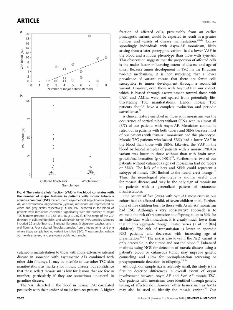
Correlation of VAF with the extent of organ involvementBlood samples from 16/19 patients with mosaicism wereavailable for NGS, and the median VAF was 1.35% (range:0–19%). The VAF correlated positively with the number ofmajor TSC features present (R= 0.55, p= 0.028) (Fig. 4a).The average VAF in the blood differed between Sym-AF(6.3 ± 6.1%) and Asym-AF (1.0 ± 1.8%), (p= 0.043). The VAFin the blood was also greater in those with mosaicism andSENs (9.1 ± 6.8%) than in those without (1.7 ± 2.2%), (p=0.005).
Variant detection is enhanced in lesion fibroblast culturesand in the dermisMosaic variants were identified in 31/36 (86%) samples (15whole tumor, 21 cultured fibroblast) derived from 19 patientswith mosaicism. Cultured fibroblast samples had a medianVAF of 10% (range 0–94%), whereas the median VAF inwhole tumor samples was 3.3% (range 0–24%), (Fig. 4b).Eight AFs from a patient with Sym-AF (P05) were analyzed todetermine if mutated cells were more prevalent within the
epidermis or dermis (Table S6). The VAF of TSC2 from fourwhole tumor samples was compared with that of four tumorssplit at the dermal–epidermal junction using dispase. Themean VAF was higher in the dermis (10.8%) than wholetumor (5.7%), (p= 0.013).The majority of blood and whole tissue skin tumor samples
evaluated from patients with mosaicism fell below the usualdetection limit of Sanger sequencing (20%) (Fig. S3). None ofthe 16 patients who underwent NGS of the blood had a VAFgreater than 20%, whereas NGS was able to detect variantsabove a frequency of 1% in 9/16 (56%). The VAF in the bloodwas <1% in 2/9 (22%) with Sym-AF and 5/7 (71%) withAsym-AF. Only 1/10 (10%) patients with whole tumorsamples exhibited a VAF greater than 20%, compared with7/14 (50%) of patients with skin tumors processed as culturedfibroblasts.
DISCUSSIONIn this cohort of germline and mosaic TSC patients, wepresent extensive information on the occurrence, geneticconsequences, and clinical importance of Asym-AF and Sym-AF mosaicism in TSC. Although earlier case reports hadnoted unilateral AFs in TSC, the clinical presentations andsignificance had not been reported in detail previously. Thosewith Asym-AF manifested the fewest median clinical findingsand oldest median age of TSC penetrance; while those withSym-AF were intermediate by both of these measures relativeto germline disease. This is remarkable considering the highvariability in disease expression among those with germlinedisease. One source of variability in germline disease is that
a b
aR bR bLaL
Fig. 1 The clinical picture of mosaicism in tuberous sclerosis complex. a Mosaicism with an asymmetric distribution of angiofibromas (AFs) on thenose and cheeks. aR,aL Right and left lateral views of the nose highlight the left-sided predominance of AFs. b Mosaicism with numerous AFs distributedsymmetrically on the nose and cheeks, indistinguishable from a patient with germline tuberous sclerosis complex (TSC). bR,bL Right and left lateral views ofthe nose reveal more numerous and symmetric distribution of AFs.
TREICHEL et al ARTICLE
GENETICS in MEDICINE | Volume 21 | Number 11 | November 2019 2599

individuals with variants in TSC1 tend to be less severe thanthose with variants in TSC2.1,17–19 Our germline patientsincluded four with TSC1 variants whereas all those withmosaicism had variants in TSC2, making the observation offewer manifestations in the mosaic group even moresurprising. A milder phenotype and/or later onset have alsobeen observed in mosaic NF1,20 NF2,21,22 and Turnersyndrome.23
In those with mosaic TSC, the number of cutaneousfindings correlated with the number of internal findings, aspreviously reported in a genetic condition that is always
mosaic: Proteus syndrome.24 External features may provideclues about the extent of internal organ involvement inmosaic TSC, but it should be cautioned that individuals withgermline disease may exhibit minimal or no skin manifesta-tions, particularly in early childhood.25 It is the unilateral orasymmetric pattern of AFs that is a marker for mosaicism,rather than the absence of cutaneous findings. Those withAsym-AF mosaicism had a minimum ratio of 3:1 for one sideof the nose and cheeks versus the other, and typically hadfewer AFs. There may a spectrum from the mildest disease inthose with a single patch of strictly unilateral AFs as the sole
P60 U
P#
Gen
e
Sum
maj
or M
CF
(A
)
Sum
maj
or &
min
or M
CF
(B
)
Sum
maj
or IF
(C
)
Sum
A +
C
Sum
B +
C
UF
ons
et a
ge g
roup
s
AF
ons
et a
ge g
roup
s
P#
Gen
e
Sum
maj
or M
CF
(A
)
VA
F b
lood
(%
)
Sum
maj
or &
min
or M
CF
(B
)
Sum
maj
or IF
(C
)
Sum
A +
C
Sum
B +
C
UF
ons
et a
ge g
roup
s
AF
ons
et a
ge g
roup
s
U
P04 U
U
U
U
U
U
NA
NA
NA
P09 TSC2
TSC2
TSC2
TSC2
TSC2
TSC2
TSC2
TSC2
TSC2
TSC2
TSC2
TSC2
TSC2
TSC2
TSC2
TSC2
TSC2
TSC2
TSC2
TSC2
TSC2
TSC2
TSC2
TSC2
TSC2
TSC2
TSC2
TSC2
TSC2
TSC2
TSC2
TSC2
TSC2
TSC2
P12 U
P28 P03 U
P08 P19 19
P01 P21 10
P61 U P14
P13 P05
P43 P10
P06 P33
P26 P32
P22 P30
P62 U P25
P63 U P40
P64 U U
U
UU
UUUUUU
U UUU
U
P20
P07 P11
P15 P29
0
0
0
0.4
0.5
1.2
1.5
4.0
5.1
6
7.0
9.6
P02 P39
0P37 P49
P31 0P27
P36
P65 TSC1
TSC1
TSC1
TSC2
TSC1
P67 U
P17
P50
P48 U
P35
P66 U
P68
P69 U
GERMLINE MOSAIC
KEY
A and C
5
5
5678910
678910
11 <15y<5y
5–10y
10–15y15–30y
>30y ornone at
>30y>15y or
none
12
4
4
4
3
3
3
5678
43
22
1
1
0 2
BSumA + C
SumB + C
UF onsetage
AF onsetage
Fig. 2 Phenotypic spectrum of tuberous sclerosis complex (TSC) patients with mosaic subtypes or germline disease. Patients were first sorted bythe variant allele fraction in the blood from highest to lowest, and then from the highest to lowest sum of total findings (major and minor mucocutaneousand major internal), color-coded from dark red to yellow. Those with germline disease tended to have a greater extent of disease than those with mosaicTSC. The phenotype of mosaic TSC ranged from very mild to indistinguishable from germline TSC. Bolded text indicates those with asymmetricalangiofibroma mosaicism. Patients with asymmetrical angiofibroma mosaicism have fewer findings (enriched toward the bottom), whereas those withsymmetrical angiofibroma mosaicism tended to have more findings (enriched towards the top). IF internal findings, MCF mucocutaneous findings, NA notapplicable (patients who lack UF but are under age 30 years), U unknown.
ARTICLE TREICHEL et al
2600 Volume 21 | Number 11 | November 2019 | GENETICS in MEDICINE

8
7
6
5
# M
ucoc
utan
eous
find
ings
(8
max
)#
Tota
l fin
ding
s (1
3 m
ax)
Per
cent
age
of p
atie
nts
with
eac
hC
NS
phe
noty
pe (
%)
# In
tern
al fi
ndin
gs (
5 m
ax)
# In
tern
al fi
ndin
gs
4
3
2
1
0
89
10111213
**
*
**
**
*
76543210
Asym-AFmosaic
Sym-AFmosaic
Germline
Asym-AFmosaic
Sym-AFmosaic
Mosaic TSC Germline TSC
GermlineAsym-AFmosaic
Sym-AFmosaic
Germline
Asym-AFmosaic
Sym-AFmosaic
Germline
5
4
3
2
1
0
5
5
4
4
3
3
# Cutaneous findings # Cutaneous findings
2
2
1
10
0 543210
# In
tern
al fi
ndin
gs
5
4
3
2
1
0
0– + +
+– –– + +
+– –– + +
+– –Tuber:SEN:
20
40
60
80
100
a b
c d
e f
Fig. 3 The number of tuberous sclerosis complex (TSC) findings and neurological tumor status differs between patients with asymmetricalangiofibroma mosaicism, symmetrical angiofibroma mosaicism, or germline disease. a The number of mucocutaneous findings increasedsequentially from asymmetrical angiofibroma (Asym-AF) mosaicism, to symmetrical angiofibroma (Sym-AF) mosaicism, to germline TSC. b The number ofinternal findings was significantly lower in patients with Asym-AF mosaicism than Sym-AF mosaicism and germline TSC. c The number of total findingsincreased sequentially from Asym-AF mosaicism, to Sym-AF mosaicism, to germline TSC. d The most common central nervous system (CNS) phenotypeswere tubers without subependymal nodules (SENs) in Asym-AF mosaicism, and tubers with SENs in Sym-AF mosaicism and germline TSC. e The number ofmajor internal and cutaneous findings correlated significantly in mosaic TSC (R= 0.62, n= 19, p= 0.005). Those with Asym-AF mosaicism (white) clusteredin the lower left quadrant whereas those with Sym-AF mosaicism (light gray) clustered in the upper right quadrant as observed in germline TSC. f In patientswith germline TSC, the number of internal and cutaneous findings did not correlate (R=−0.24, n= 26, p= 0.24). These patients clustered in the upperright quadrant of the figure.
TREICHEL et al ARTICLE
GENETICS in MEDICINE | Volume 21 | Number 11 | November 2019 2601
cutaneous manifestation to those with more extensive internaldisease in someone with asymmetric AFs combined withother skin findings. It may be possible to use other TSC skinmanifestations as markers for mosaic disease, but confidencethat these reflect mosaicism is low for lesions that are few innumber, particularly if they are sometimes unilateral ingermline disease.The VAF detected in the blood in mosaic TSC correlated
positively with the number of major features present. A higher
fraction of affected cells, presumably from an earlierpostzygotic variant, would be expected to result in a greaternumber and variety of disease manifestations.26,27 Corre-spondingly, individuals with Asym-AF mosaicism, likelyarising from a later postzygotic variant, had a lower VAF inthe blood and a milder phenotype than those with Sym-AF.This observation suggests that the proportion of affected cellsis the major factor influencing extent of disease and age ofonset. Because tumor development in TSC fits the Knudsontwo-hit mechanism, it is not surprising that a lowerprevalence of variant means that there are fewer cellssusceptible to tumor development through a second-hitvariant. However, even those with Asym-AF in our cohort,which is biased through ascertainment toward those withLAM and AMLs, were not spared from potentially life-threatening TSC manifestations. Hence, mosaic TSCpatients should have a complete evaluation and periodicsurveillance.28
A clinical feature enriched in those with mosaicism was theoccurrence of cortical tubers without SENs, seen in almost all(6/7) of our patients with Asym-AF. Mosaicism cannot beruled out in patients with both tubers and SENs because mostof our patients with Sym-AF mosaicism had this phenotype.Mosaic TSC patients who lacked SENs had a lower VAF inthe blood than those with SENs. Likewise, the VAF in theblood or buccal samples of patients with a mosaic PIK3CAvariant was lower in those without than with brain over-growth/malformation (p < 0.001)29. Furthermore, two of ourpatients without cutaneous signs of mosaicism had no tubersor SENs. The lack of tubers and SENs could represent asubtype of mosaic TSC limited to the neural crest lineage.10
Thus, the neurological phenotype is another useful cluefor mosaic disease, and may be the only sign of mosaicismin patients with a generalized pattern of cutaneousmanifestations.One patient of five (20%) with Sym-AF mosaicism in our
cohort had an affected child, of seven children total. Further,none of five children born to those with Asym-AF mosaicismhad TSC. Although a very conservative approach is toestimate the risk of transmission to offspring at up to 50% foran individual with mosaicism, it is clearly much lower thanthat in this aggregate though limited series (1 of 12 [8%]children). The risk of transmission is lower in sporadicNF2 patients, and decreases with increasing age atpresentation.30,31 The risk is also lower if the NF2 variant isonly detectable in the tumor and not the blood.32 Enhancedmethods using NGS for detection of mosaic disease using apatient’s blood or cutaneous tumor may improve geneticcounseling and allow for preimplantation screening orpresymptomatic detection in offspring.33,34
Although our sample size is relatively small, this study is thefirst to describe differences in overall extent of organinvolvement between Asym-AF and Sym-AF mosaic TSC.Our patients with mosaicism were identified through genetictesting of affected skin, however other tissues such as AMLsmay also be used to identify the mosaic variant.35 Our
20a
b
18
1614
12
10
8
6
4
2
00
100
75
50
VA
F ti
ssue
(%
)V
AF
blo
od (
%)
25
0
1 2 3 4Number of major criteria (9 max)
Cultured fibroblasts Whole tumorSample type
5 6 7 8 9
Fig. 4 The variant allele fraction (VAF) in the blood correlates withthe number of major features in patients with mosaic tuberoussclerosis complex (TSC). Patients with asymmetrical angiofibroma (Asym-AF) and symmetrical angiofibroma (Sym-AF) mosaicism are represented bywhite and gray circles respectively. a The VAF detected in the blood ofpatients with mosaicism correlated significantly with the number of majorTSC features present (R= 0.55, n= 16, p= 0.028). b The range of the VAFdetected in cultured fibroblast and whole skin tumor DNA samples. Samplesincluded 29 angiofibromas, 3 ungual fibromas, 3 shagreen patches, and 1oral fibroma. Four cultured fibroblast samples from three patients, and onewhole tissue sample had no variant identified (NVI). These samples includeour newly analyzed and previously published samples.
ARTICLE TREICHEL et al
2602 Volume 21 | Number 11 | November 2019 | GENETICS in MEDICINE
population consisted mostly of adult women with LAM, andtherefore may not reflect findings in the general TSCpopulation. Although LAM may not be as prevalent inmosaic TSC as within our cohort, this study shows thatpatients with Asym-AF may develop LAM. Future studies arewarranted to determine if the VAF in children with mosaicTSC predicts the extent of organ involvement in adult life,and to further investigate the severity of disease inmosaic TSC.We note that a parallel study on mosaicism in TSC, also
reported in this issue, came to similar observations on theprevalence and clinical significance of mosaicism but withsome distinct differences. In that report, individuals withmosaic TSC had a high prevalence of facial AFs and kidneyangiomyolipoma, but a low incidence of most other TSCclinical findings, and LAM was rare. These two sets of mosaicTSC patients have many similarities as well as somedifferences, which we suspect are due to the methods bywhich they were ascertained.In conclusion, the spectrum of mosaic TSC ranges from a
mild phenotype with localized cutaneous findings to a moregeneralized phenotype that is indistinguishable from patientswith inherited germline variants. Clinical clues suggestive oflow-level mosaicism in an adult with TSC include asymmetricdistribution of AFs, disease penetrance in adulthood, and lackof tubers and SENs. Patients with these features are likely tohave no pathogenic variant identified when non-NGSapproaches are used for genetic testing of the blood, andthus require NGS of their blood or affected skin to detect thedisease-causing variant. Patterns of mosaic disease observedin TSC provide clues about prognosis and risk of diseasetransmission to offspring that may hold true in many othermosaic genetic conditions.
SUPPLEMENTARY INFORMATIONThe online version of this article (https://doi.org/10.1038/s41436-019-0520-3) contains supplementary material, which is availableto authorized users.
ACKNOWLEDGEMENTSResearch reported in this publication was supported in part by theIntramural Research Program, National Institutes of Health (NIH),National Heart, Lung, and Blood Institute (NHLBI); the NIH,National Institute of Arthritis and Musculoskeletal and SkinDiseases, under award number R01AR062080; NIH, NHLBI underaward number 1U01HL131022; the Doris Duke CharitableFoundation Clinical Research Mentorship grants 2014088 and2018042; the Tuberous Sclerosis Alliance, Engles Fund forResearch in TSC and LAM. Additionally, this work was madepossible through the NIH Medical Research Scholars Program, apublic–private partnership supported jointly by the NIH andgenerous contributions to the Foundation for the NIH from theDoris Duke Charitable Foundation, the American Association forDental Research, the Colgate–Palmolive Company, Genentech,and other private donors. For a complete list, visit the foundation
website at http://www.fnih.org. Written consent was obtained topublish the photography of the patients included in Fig. 1.
DISCLOSUREThe authors declare no conflicts of interest.
Publisher’s note: Springer Nature remains neutral with regard tojurisdictional claims in published maps and institutionalaffiliations.
REFERENCES1. Peron A, Au KS, Northrup H. Genetics, genomics, and genotype-
phenotype correlations of TSC: insights for clinical practice. Am J MedGenet C Semin Med Genet. 2018;178:281–290.
2. Sampson JR, Scahill SJ, Stephenson JB, Mann L, Connor JM. Geneticaspects of tuberous sclerosis in the west of Scotland. J Med Genet.1989;26:28–31.
3. Tyburczy ME, Dies KA, Glass J, et al. Mosaic and intronic mutations inTSC1/TSC2 explain the majority of TSC patients with no mutationidentified by conventional testing. PLoS Genet. 2015;11:e1005637.
4. Ruggieri M, Huson SM. The clinical and diagnostic implications ofmosaicism in the neurofibromatoses. Neurology. 2001;56:1433–1443.
5. Messiaen L, Vogt J, Bengesser K, et al. Mosaic type-1 NF1 microdeletionsas a cause of both generalized and segmental neurofibromatosis type-1(NF1). Hum Mutat. 2011;32:213–219.
6. Garcia-Romero MT, Parkin P, Lara-Corrales I. Mosaic neurofibromatosistype 1: a systematic review. Pediatr Dermatol. 2016;33:9–17.
7. Hall MR, Kovach BT, Miller JL. Unilateral facial angiofibromas withoutother evidence of tuberous sclerosis: case report and review of theliterature. Cutis. 2007;80:284–288.
8. Bessis D, Malinge MC, Girard C. Isolated and unilateral facialangiofibromas revealing a type 1 segmental postzygotic mosaicism oftuberous sclerosis complex with c.4949_4982del TSC2 mutation. Br JDermatol. 2018;178:e53–e54.
9. Tyburczy ME, Wang JA, Li S, et al. Sun exposure causes somatic second-hit mutations and angiofibroma development in tuberous sclerosiscomplex. Hum Mol Genet. 2014;23:2023–2029.
10. Boronat S, Shaaya EA, Doherty CM, Caruso P, Thiele EA. Tuberoussclerosis complex without tubers and subependymal nodules: aphenotype-genotype study. Clin Genet. 2014;86:149–154.
11. Acuna-Hidalgo R, Bo T, Kwint MP, et al. Post-zygotic point mutations arean underrecognized source of de novo genomic variation. Am J HumGenet. 2015;97:67–74.
12. Northrup H, Krueger DA, International Tuberous Sclerosis ComplexConsensus Group. Tuberous sclerosis complex diagnostic criteria update:recommendations of the 2012 International Tuberous Sclerosis ComplexConsensus Conference. Pediatr Neurol. 2013;49:243–254.
13. Yao J, Taveira-DaSilva AM, Colby TV, Moss J. CT grading of lung disease inlymphangioleiomyomatosis. AJR Am J Roentgenol. 2012;199:787–793.
14. Nathan N, Tyburczy ME, Hamieh L, et al. Nipple angiofibromas with lossof TSC2 are associated with tuberous sclerosis complex. J InvestDermatol. 2016;136:535–538.
15. Cao J, Tyburczy ME, Moss J, Darling TN, Widlund HR, Kwiatkowski DJ.Tuberous sclerosis complex inactivation disrupts melanogenesis viamTORC1 activation. J Clin Invest. 2017;127:349–364.
16. Happle R. The categories of cutaneous mosaicism: a proposedclassification. Am J Med Genet A. 2016;170A:452–459.
17. Dabora SL, Jozwiak S, Franz DN, et al. Mutational analysis in a cohort of224 tuberous sclerosis patients indicates increased severity of TSC2,compared with TSC1, disease in multiple organs. Am J Hum Genet.2001;68:64–80.
18. Sancak O, Nellist M, Goedbloed M, et al. Mutational analysis of the TSC1and TSC2 genes in a diagnostic setting: genotype–phenotypecorrelations and comparison of diagnostic DNA techniques in tuberoussclerosis complex. Eur J Hum Genet. 2005;13:731–741.
19. Au KS, Williams AT, Roach ES, et al. Genotype/phenotype correlation in325 individuals referred for a diagnosis of tuberous sclerosis complex inthe United States. Genet Med. 2007;9:88–100.
20. Ben-Shachar S, Dubov T, Toledano-Alhadef H, et al. Predictingneurofibromatosis type 1 risk among children with isolated cafe-au-laitmacules. J Am Acad Dermatol. 2017;76:1077–83 e1073.
TREICHEL et al ARTICLE
GENETICS in MEDICINE | Volume 21 | Number 11 | November 2019 2603
21. Spyra M, Otto B, Schon G, Kehrer-Sawatzki H, Mautner VF.Determination of the mutant allele frequency in patients withneurofibromatosis type 2 and somatic mosaicism by means of deepsequencing. Genes Chromosomes Cancer. 2015;54:482–488.
22. Halliday D, Emmanouil B, Pretorius P, et al. Genetic Severity Scorepredicts clinical phenotype in NF2. J Med Genet. 2017;54:657–664.
23. Tuke MA, Ruth KS, Wood AR, et al. Mosaic Turner syndrome showsreduced penetrance in an adult population study. Genet Med.2019;21:877–886.
24. Nguyen D, Turner JT, Olsen C, Biesecker LG, Darling TN. Cutaneousmanifestations of proteus syndrome: correlations with general clinicalseverity. Arch Dermatol. 2004;140:947–953.
25. Jozwiak S, Schwartz RA, Janniger CK, Michalowicz R, Chmielik J. Skinlesions in children with tuberous sclerosis complex: their prevalence,natural course, and diagnostic significance. Int J Dermatol.1998;37:911–917.
26. Normand EA, Crandall SR, Thorn CA, et al. Temporal and mosaic Tsc1deletion in the developing thalamus disrupts thalamocortical circuitry,neural function, and behavior. Neuron. 2013;78:895–909.
27. Nathan N, Keppler-Noreuil KM, Biesecker LG, Moss J, Darling TN. MosaicDisorders of the PI3K/PTEN/AKT/TSC/mTORC1 signaling pathway.Dermatol Clin. 2017;35:51–60.
28. Krueger DA, Northrup H. International Tuberous Sclerosis ComplexConsensus Group. Tuberous sclerosis complex surveillance andmanagement: recommendations of the 2012 International
Tuberous Sclerosis Complex Consensus Conference. Pediatr Neurol.2013;49:255–265.
29. Kuentz P, St-Onge J, Duffourd Y, et al. Molecular diagnosis of PIK3CA-related overgrowth spectrum (PROS) in 162 patients and recommendationsfor genetic testing. Genet Med. 2017;19:989–997.
30. Evans DG, Ramsden RT, Shenton A, et al. Mosaicism in neurofibromatosistype 2: an update of risk based on uni/bilaterality of vestibularschwannoma at presentation and sensitive mutation analysis includingmultiple ligation-dependent probe amplification. J Med Genet.2007;44:424–428.
31. Evans DG, Wallace A. An update on age related mosaic and offspring riskin neurofibromatosis 2 (NF2). J Med Genet. 2009;46:792.
32. Moyhuddin A, Baser ME, Watson C, et al. Somatic mosaicism inneurofibromatosis 2: prevalence and risk of disease transmission tooffspring. J Med Genet. 2003;40:459–463.
33. Kluwe L, Friedrich RE, Tatagiba M, Mautner VF. Presymptomatic diagnosisfor children of sporadic neurofibromatosis 2 patients: a method based ontumor analysis. Genet Med. 2002;4:27–30.
34. Consoli C, Moss C, Green S, Balderson D, Cooper DN, Upadhyaya M.Gonosomal mosaicism for a nonsense mutation (R1947X) in the NF1gene in segmental neurofibromatosis type 1. J Invest Dermatol.2005;125:463–466.
35. Han MK, Tyburczy ME, Darling TN, et al. Apparent sporadiclymphangioleiomyomatosis in a man as a result of extreme mosaicismfor a TSC2 mutation. Ann Am Thorac Soc. 2017;14:1227–1229.
ARTICLE TREICHEL et al
2604 Volume 21 | Number 11 | November 2019 | GENETICS in MEDICINE