Phenol as an adjuvant for local control in the treatment of giant cell ...
9
European Journal of Surgical Oncology 1999; 25: 610–618 Phenol as an adjuvant for local control in the treatment of giant cell tumour of the bone H. R. Du ¨rr*, M. Maier*, V. Jansson*, A. Baur and H. J. Refior* *Department of Orthopedics and Orthopedic Surgery and Institute of Radiology, Ludwig-Maximilians-University Munich, Klinikum Großhadern, D-81366 Munich, Marchioninistr. 15, Germany Aims: Intralesional treatment of giant cell tumour (GCT) of the bone may result in a high rate of local recurrence. The introduction of local adjuvant therapy, such as cementation or phenolization, has lead to a significant reduction in recurrence rates. Due to the combined use of phenol and cementation in most studies, the e ect of phenol alone is described in this study. Methods: Twenty primary and nine recurrent surgical procedures in 26 patients with GCT of the bone with a median follow-up of 61 months were reviewed retrospectively. The mean age was 33.5 years (range 13.5–76.5 years). Eighteen curettages and 11 resections were performed. For the curettages, a large bone window was cut followed by high speed burring and bone graft reconstruction. In 11 of 18 curettages and three of 12 resections, phenol was additionally applied. Results: Four patients showed pulmonary metastasis. Three of these four cases also experienced local recurrences. Three patients died due to metastatic disease. In total, five patients developed local recurrence (17.2%); three in the first 2 years and one after 4 years. Four of 18 curettages recurred (22.2%), compared to one of 11 resections (9.1%). Only one of 11 patients (9.1%) treated with curettage and adjuvant phenol recurred, whereas three of seven patients (42.9%) treated with curettage alone recurred. Conclusion: Phenolization is an e ective and safe local adjuvant therapy for GCT. We did not observe any significant di erences in recurrence rates for curettage, phenolization and bone grafting compared to most published results using cryosurgery or cementation alone. We recommend adjuvant phenolization in the treatment of GCT of the bone after careful curettage in applicable cases, regardless of whether additional cementation is used. Key words: curettage; giant cell tumour; phenol. 1999 Harcourt Publishers Ltd Introduction the e ect of phenol alone is not well described. By comparing patients who were treated without adjuvant phenolization and bone grafting with those who were treated with adjuvant Despite various techniques in the surgical treatment of giant cell tumour (GCT) of bone, recurrence rates as high as 50% and bone grafting, we attempt here to overcome this void in the literature and describe the e ect of phenol in the have been reported. 1–5 There is a strong correlation between the surgical margins and the rate of recurrence, dependent on treatment of GCT. whether intralesional curettage, marginal or wide resection is used. 6 Due to the typical meta-epiphyseal location, however, Patients and methods wide resection may result in a major functional deficit. Hence, intralesional curettage has become the most From October 1981 to February 1997, 26 patients were recommended treatment. 7 surgically treated in our institution for GCT of the bone. The introduction of local adjuvant therapy, such as They consisted of 13 men and 13 women with a mean age cementation, cryosurgery or phenolization, in combination of 33.5 years (13.5–76.5 years). Six patients presented who with careful removal of the tumour using a large bone had already had local recurrence. As five patients window and high speed burrs has lead to a significant experienced recurrence after the initial treatment, a second reduction in recurrence rates. 7–12 However, due to the surgical procedure was performed in three of these patients combined use of phenol and cementation in most studies, in our institution.Two of the five recurrences were treated at di erent institutions. Therefore, 29 surgical procedures Correspondence to: Dr H. R. Du ¨rr, Orthopa ¨dische Klinik und in 26 patients are reviewed in this study. In total, 20 primary Poliklinik, Ludwig-Maximilians-Universita ¨t Mu ¨ nchen, Klinikum and nine recurrent lesions were treated. Follow-up for all Großhadern, D-81366 Mu ¨nchen, Marchioninistr. 15, Germany. Fax: +49 89/7095-8881; E-mail: [email protected]. patients included radiographical and clinical evaluations 0748–7983/99/060610+09 $12.00/0 1999 Harcourt Publishers Ltd
Transcript of Phenol as an adjuvant for local control in the treatment of giant cell ...

European Journal of Surgical Oncology 1999; 25: 610–618
Phenol as an adjuvant for local control in the treatment ofgiant cell tumour of the bone
H. R. Durr∗, M. Maier∗, V. Jansson∗, A. Baur† and H. J. Refior∗
∗Department of Orthopedics and Orthopedic Surgery and †Institute of Radiology,Ludwig-Maximilians-University Munich, Klinikum Großhadern, D-81366 Munich,
Marchioninistr. 15, Germany
Aims: Intralesional treatment of giant cell tumour (GCT) of the bone may result in a high rate of local recurrence.The introduction of local adjuvant therapy, such as cementation or phenolization, has lead to a significant reductionin recurrence rates. Due to the combined use of phenol and cementation in most studies, the effect of phenol aloneis described in this study.Methods: Twenty primary and nine recurrent surgical procedures in 26 patients with GCT of the bone with a medianfollow-up of 61 months were reviewed retrospectively. The mean age was 33.5 years (range 13.5–76.5 years). Eighteencurettages and 11 resections were performed. For the curettages, a large bone window was cut followed by highspeed burring and bone graft reconstruction. In 11 of 18 curettages and three of 12 resections, phenol was additionallyapplied.Results: Four patients showed pulmonary metastasis. Three of these four cases also experienced local recurrences.Three patients died due to metastatic disease. In total, five patients developed local recurrence (17.2%); three in thefirst 2 years and one after 4 years. Four of 18 curettages recurred (22.2%), compared to one of 11 resections (9.1%).Only one of 11 patients (9.1%) treated with curettage and adjuvant phenol recurred, whereas three of seven patients(42.9%) treated with curettage alone recurred.Conclusion: Phenolization is an effective and safe local adjuvant therapy for GCT. We did not observe any significantdifferences in recurrence rates for curettage, phenolization and bone grafting compared to most published resultsusing cryosurgery or cementation alone. We recommend adjuvant phenolization in the treatment of GCT of thebone after careful curettage in applicable cases, regardless of whether additional cementation is used.
Key words: curettage; giant cell tumour; phenol. 1999 Harcourt Publishers Ltd
Introduction the effect of phenol alone is not well described. By comparingpatients who were treated without adjuvant phenolizationand bone grafting with those who were treated with adjuvantDespite various techniques in the surgical treatment of giant
cell tumour (GCT) of bone, recurrence rates as high as 50% and bone grafting, we attempt here to overcome this voidin the literature and describe the effect of phenol in thehave been reported.1–5 There is a strong correlation between
the surgical margins and the rate of recurrence, dependent on treatment of GCT.whether intralesional curettage, marginal or wide resection isused.6 Due to the typical meta-epiphyseal location, however,
Patients and methodswide resection may result in a major functional deficit.Hence, intralesional curettage has become the most
From October 1981 to February 1997, 26 patients wererecommended treatment.7
surgically treated in our institution for GCT of the bone.The introduction of local adjuvant therapy, such asThey consisted of 13 men and 13 women with a mean agecementation, cryosurgery or phenolization, in combinationof 33.5 years (13.5–76.5 years). Six patients presented whowith careful removal of the tumour using a large bonehad already had local recurrence. As five patientswindow and high speed burrs has lead to a significantexperienced recurrence after the initial treatment, a secondreduction in recurrence rates.7–12 However, due to thesurgical procedure was performed in three of these patientscombined use of phenol and cementation in most studies,in our institution.Two of the five recurrences were treatedat different institutions. Therefore, 29 surgical procedures
Correspondence to: Dr H. R. Durr, Orthopadische Klinik und in 26 patients are reviewed in this study. In total, 20 primaryPoliklinik, Ludwig-Maximilians-Universitat Munchen, Klinikumand nine recurrent lesions were treated. Follow-up for allGroßhadern, D-81366 Munchen, Marchioninistr. 15, Germany.
Fax:+49 89/7095-8881; E-mail: [email protected]. patients included radiographical and clinical evaluations
0748–7983/99/060610+09 $12.00/0 1999 Harcourt Publishers Ltd

Phenol in giant cell tumour of the bone 611
12
011–20
Age (years)
Pat
ien
ts (
n)
10
8
6
4
2
21–30 31–40 41–50 51–60 61–70 71–80
Fig. 1. Age distribution of 26 patients with giant cell tumour ofthe bone.
Table 1. Surgical procedures performed in 29 lesions
Therapy
Curettage alone 1Curettage and autologous graft 16Curettage and homo-/autologous graft 1Total 18
Resection alone 4Resection and autologous graft 4Resection and prosthesis 1Resection and arthrodesis 2Total 11
Table 2. Surgical procedures and the use of phenol in primary andrecurrent cases
Fig. 2. Anatomical sites of the tumours in 29 surgical procedures.Procedure n Phenol used
(n/total)
impairment or a pathological fracture (Fig. 5) were foundPrimary Curettage 15 9/15in one case each. Two lesions were found incidentally. Thelesions (n=20) Resection 5 2/5median duration of symptoms was 3.4 months (range: 3Recurrent Curettage 3 2/3weeks to 52 months).lesions (n=9) Resection 6 1/6
Nine lesions (31%) showed joint involvement, whereas 15(52%) showed subchondral involvement. Fifteen lesionswere classified as active and 14 as aggressive, according tothe Enneking staging system. Initial diagnosis, based on theperfomed by a physician in our institution or in the patients
community. As MRI is much more sensitive for detecting typical radiographical findings of GCT, was confirmed bybiopsy in all cases.local recurrence, this method was preferably used in addition
to conventional radiographs. The median time to follow- Eighteen curettages and 11 resections were performed.Twenty-three were classified as intralesional, two as marginalup was 61 months (range: 6–178 months).and four as wide (Table 1). For curettages, a large windowwas cut into the bone followed by high-speed burring.Phenol was used in 11 of the 18 curettages and in three ofResultsthe 12 resections (Table 2). A solution of 50% phenol in75% of alcohol13 was administered on swabs to the lesionFig. 1 demonstrates that the highest incidence of GCT
occurs in the third decade of life. As for location, the distal for 1 min followed by irrigation with sodium bicarbonateand Ringer’s solution. The defect was then reconstructedfemur and the proximal tibia accounted for 14 of the 29
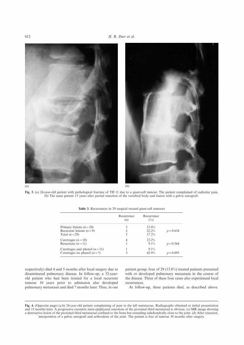
procedures (Fig. 2). Only three of the lesions involved the by autologous or homologous bone grafting (Fig. 6).On admission, three patients had pulmonary metastasestrunk, either the spine (n=2, Fig. 3) or the pelvis (n=1).
Of the four tumours found in the foot, two were in the (11.5%). Two of these patients also had local recurrentdisease. One 26-year-old patient underwent surgicaltalus and one each in the calcaneus and the proximal
metatarsal bone (Fig. 4). resection of the tumour in both lungs, as well as in thebone, and was tumour-free 53 months after surgeryIn 26 of the 29 lesions, the patients reported associated
pain. A swelling was found in nine cases, and neurological (Fig. 7). The two other patients (41 and 76 years old,
H. R. Durr et al.612
(a) (b)
Fig. 3. (a) 24-year-old patient with pathological fracture of TH 11 due to a giant-cell tumour. The patient complained of radicular pain.(b) The same patient 13 years after partial resection of the vertebral body and fusion with a pelvic autograft.
Table 3. Recurrences in 29 surgical treated giant-cell tumours
Recurrence Recurrence(n) (%)
Primary lesions (n=20) 3 15.0%Recurrent lesions (n=9) 2 22.2% p=0.634Total (n=29) 5 17.2%
Curettages (n=18) 4 22.2%Resections (n=11) 1 9.1% p=0.364
Curettages and phenol (n=11) 1 9.1%Curettages no phenol (n=7) 3 42.9% p=0.093
respectively) died 4 and 5 months after local surgery due to patient group, four of 29 (13.8%) treated patients presentedwith or developed pulmonary metastasis in the course ofdisseminated pulmonary disease. In follow-up, a 32-year-
old patient who had been treated for a local recurrent the disease. Three of these four cases also experienced localrecurrences.tumour 10 years prior to admission also developed
pulmonary metastasis and died 7 months later. Thus, in our At follow-up, three patients died, as described above.
Fig. 4. (Opposite page) (a,b) 24-year-old patient complaining of pain in the left metatarsus. Radiographs obtained at initial presentationand 15 months later. A progressive excentric meta-epiphyseal osteolysis of the proximal third metatarsal is obvious. (c) MR image showinga destructive lesion of the proximal third metatarsal confined to the bone but extending subchondrally close to the joint. (d) After resection,
interposition of a pelvic autograft and arthrodesis of the joint. The patient is free of tumour 30 months after surgery.

Phenol in giant cell tumour of the bone 613
(a) (b)
(c) (d)

H. R. Durr et al.614
(a) (b)
Fig. 5. (a) 16-year old girl with a pathological fracture of the distal femur due to a previously asymptomatic giant-cell tumour. Initialradiographs at presentation show the excentric large osteolytic destruction with a displaced fracture. (b) The same patient after curettage
of the lesion, bone grafting and osteosynthesis. The patient is free of tumour 14 years after surgery.
Two of them were initially treated outside our department of life, corresponding to our own findings. Only threepatients in our study group were outside this age range.with recurrences after local surgery. These treatments were
not included in the 29 procedures reviewed in our study Despite an equal male to female ratio in this study, genderseems to be unevenly distributed in larger series that showbecause they were not perfomed in our institution.
Of the 29 surgical procedures perfomed in our institution, a female preponderance for GCT.3,15 The most frequenttumour site in our study was the lower end of the femurfive patients developed local recurrences (17.2%). Recurrence
developed in a case of resection with intralesional margins and the proximal tibia, corresponding to the results ofother groups who have reported that approximately 50%without the use of phenol. Only one patient out of 11 (9.1%)
who had been treated with curettage and adjuvant phenol of tumours occur around the knee.14,16 The typical meta-and epiphyseal involvement was seen in all patients, whiledeveloped a reccurence, whereas three patients out of seven
(42.9%) who had been treated with curettage without phenol no patient showed the rare meta-diaphyseal extension.17
Conventional histological grading has been shown to bedeveloped a recurrence (Table 3). This was a clear tendencybut did not reach statistical significance. The time between of limited value in predicting clinical outcome.3,18–22 In recent
studies, the spindle-shaped stroma cells proved not only tothe initial surgical procedure and recurrence for all patientsis shown in Fig. 8. Four of the five lesions recurred in the be the major proliferating cells in the tumour,23 but also their
proliferation rate was associated with the radiographicalfirst 2 years and one after 4 years.aggressiveness of the tumour.24 Additional factors, such asexpression of metalloproteinases, may gain clinicalimportance,25 whereas others, such as DNA analysis, showDiscussionconflicting results.26–28
Pain was the most common clinical symptom (90%).Giant cell tumour of the bone accounts for approximately8.6% of all bone tumours.14 Interestingly, China seems to Swelling developed in only 31% of the patients. The duration
of complaints varied greatly, corresponding to thehave the highest incidence, at 20%.5 Most of the lesionsreportedly occur in patients in the third or fourth decade unpredictable course of the tumour.16

Phenol in giant cell tumour of the bone 615
(a) (b)Fig. 6. (a) 25-year old patient with giant-cell tumour of the distal femur. Radiograph showing a large epi-metaphyseal osteolysis extentingclose to the joint. (b) The same patient 3 years after curettage, phenolization and autologous bone grafting of the lesion. The patient is
free of tumour and symptoms.
Although GCT is classified as benign, pulmonary our cases, but due to the anatomical location of the tumour,a less destructive approach is often desirable. Severalmetastasis is found in approximately 3–10% of cases.20,29–36
The fact that our institution is a referral centre for inter- methods of adjuvant treatment have been advocated.Radiation therapy, first described by Ewing,39 is associateddisciplinary approaches in bone tumour may explain
the higher frequency of metastasis found in our institution. with a high rate of secondary malignancies and is no longerapplicable in routine cases. Cryosurgery, introduced in 1964,Histology has proven that GCT tends to infiltrate
periphereal veins, allowing the tumour to spread to distant showed recurrence rates of less than 10%, but was alsoassociated with considerable complications, such aslocations.37 Pulmonary metastasis develops preferrentially
in recurrent cases, as demonstrated in our series (three fractures and delayed bone and wound healing.8,40 In recentreports acceptable complication rates were described.41–43 Inof four patients).37 Although the overall survival of these
patients surpasses that of patients with other bone tumours reports by groups who used additional cementation, theresults were favourable.12,44,45 Acrylic cementation combineswith secondary pulmonary metastasis, in general 10–20%
of the patients die from their disease.20,29,35,36 Due to extensive the local effect of hyperthermia and the immediatestabilization of the defect without further surgery for bonetumour spread, three of our four cases died 4–7 months
after diagnosis. Because of the unpredictable nature of the graft harvesting.46–48 In a series of 38 consecutive patients,a recurrence rate of 8% was reported using this technique.10tumour, however, one may also observe a long asymptomatic
course of disease, despite extensive metastasis.32 A negative influence of acrylic cementation to the adjacentjoint cartilage was not observed.49 Another method basedThe remarkable local aggressiveness of GCT as a benign
bone tumour continues to be a surgical challenge. Until on the hyperthermic approach, CO2 laser cauterization, iscurrently being evaluated.501912, amputation was the preferred method of treatment,
at which time intralesional resection and bone grafting was Since its introduction for the elimination of recurrencesin benign tumours,51 a local application of phenol has beenintroduced.38 With no further treatment, local recurrence
may develop in 80% in such cases.3 There is no doubt that commonly used in the treatment of GCT.52 Most authorsuse both phenol and acrylic cementation in GCT of thewide resection reduces the risk of recurrence, as shown in

H. R. Durr et al.616
(a) (b)
(c) (d)
Fig. 7. (a,b) 26-year-old patient 7 years after initial surgery complaining of pain and swelling of the distal femur of 4 year’s standing.Radiographs obtained after angiography showing the large destructive giant-cell tumour. (c) Axial MR image showing the extent of thetumour infiltrating the popliteal vessels. An amputation had to be performed. (d) CT scan of the same patient showing multiple pulmonary
metastasis. After bilateral metastasectomy, the patient is free of tumour 4 years after surgery.
bone.53 Only two large studies by the same study group application of phenol (7%), whereas recurrence occured in26 of 55 patients without phenolization (47%). In a laterhave provided data on the use of phenolization alone in the
treatment of GCT.52,53 In the first study, 69 patients were series, recurrence was observed in 45% of 280 cases withoutfurther adjuvant treatment and in 17% of patients treatedtreated with adjuvant phenol applied with swabs and 14
patients were treated with autologous or homologous bone with either additional phenol (n=147), liquid nitrogen (n=20) or cement (n=187) without significant differences in thegrafting. In this series, one recurrence was seen after the

Phenol in giant cell tumour of the bone 617
Curettage and cement reconstruction. Clin Orthop 1995; 321:245–50.
11. Komiya S, Inoue A. Cementation in the treatment of giant celltumor of bone. Arch Orthop Trauma Surg 1993; 112: 51–5.
12. Pals SD, Wilkins RM. Giant cell tumor of bne treated bycurettage, cementation, and bone grafting. Orthopedics 1992;15: 703–8.
13. Lack W, Lang S, Brand G. Necrotizing effect of phenol onnormal tissues and on tumors. Acta Orthop Scand 1994; 65:351–4.
14. Schajowicz F. Giant-cell tumor (osteoclastoma). In: Tumorsand tumorlike lesions of bone. Berlin, Heidelberg, New York:Springer-Verlag, 1994: 257–99.
15. Dahlin DC. Caldwell Lecture. Giant cell tumor of bone:highlights of 407 cases. AJR 1985; 144: 955–60.
16. Campanacci M. Giant cell tumor. In: Bone and soft tissuetumors. Wien, New York: Springer-Verlag, 1990: 117–51.
17. Fain JS, Unni KK, Beabout JW, Rock MG. Nonepiphyseal
1.0
0.00
Time (months)
Rec
urr
ence
s (P
)
0.8
0.6
0.4
0.2
12 24 36 48 60 12072 84 96 108
giant cell tumor of the long bones. Clinical, radiologic, andpathologic study. Cancer 1993; 71: 3514–9.Fig. 8. Recurrences in 29 patients treated surgically for giant-cell
18. Larsson SE, Lorentzon R, Boquist L. Giant-cell tumor of bone.tumour of the bone.A demographic, clinical, and histopathological study of allcases recorded in the Swedish Cancer Registry for the years1958 through 1968. J Bone Joint Surg (Am) 1975; 57: 167–73.
19. Sanerkin NG. Malignancy, aggressiveness, and recurrence insubgroups. These data correspond very well with our own giant cell tumor of bone. Cancer 1980; 46: 1641–9.recurrence rate of 42% without phenol and 9% with phenol. 20. Rock MG, Pritchard DJ, Unni KK. Metastases fromRemarkably, in the study of Capanna et al., the combination histologically benign giant-cell tumor of bone. J Bone Joint
Surg (Am) 1984; 66: 269–74.of phenol and cementation reduced the recurrence rate to21. Schiodt T, Dissing I, Heerfordt J, Sneppen O. [Giant cell tumor3% in 33 patients.
of bone. Assessment of degree of malignancy in relationshipWe conclude that phenolization is an effective and safe to microscopic findings] Kaempecelletumor i knogle. En
local adjuvant therapy for GCT, comparable to other vurdering af maligniteten i relation til mikroskopifundet.Ugeskr Laeger 1978; 140: 1613–5.methods such as cryosurgery. Based on our data, there is
22. Lausten GS, Jesen PK, Schiodt T, Lund B. Local recurrencesno significant difference in recurrence rate for curettage,in giant cell tumour of bone. Long-term follow up of 31 cases.phenolization alone or bone grafting when compared to theInt Orthop 1996; 20: 172–6.
results of the studies using cementation. We recommend 23. Huang J, Hu S, Huang P, Shi N, Gao M, Saito M, Mori M.adjuvant phenolization in the treatment of GCT of bone Immunohistochemical evaluation of giant cell tumor of the
bone. Acta Histochem Cytochem 1996; 29: 107–13.after curettage in all applicable cases whether further24. Robinson D, Lewis MM, Nevo Z, Kenan S, Einhom TA. Thecementation is used or not.
radiographic stage of giant cell tumor related to stromal cells’proliferation. Tissue cultures in 13 cases. Acta Orthop Scand1997; 68: 294–7.
Acknowledgements 25. Schoedel KE, Greco MA, Stetler Stevenson WG, Ohori NP,Goswami S, Present D, Steiner GC. Expression ofmetalloproteinases and tissue inhibitors of metalloproteinasesThis report was supported by the Curt-Bohnewand-Fund.in giant cell tumor of bone: an immunohistochemical studywith clinical correlation. Hum Pathol 1996; 27: 1144–8.
26. Zhou L, Feng CH, Zhang HF. [Flow cytometric analysis ofDNA in giant cell tumor of bone] (in Chinese). Chung HuaReferencesWai Ko Tsa Chih 1994; 32: 367–70.
27. Scott SM, Pritchard DJ, Unni KK, Rainwater LM, Lieber1. McDonald DJ, Sim FH, McLeod RA, Dahlin DC. Giant-cellMM. ‘‘Benign’’ metastasizing giant cell tumor: evaluation oftumor of bone. J Bone Joint Surg (Am) 1986; 68: 235–42.nuclear DNA patterns by flow cytometry. J Orthop Res 1989;2. Dahlin DC, Cupps RE, Johnson EW, Jr. Giant-cell tumor: a7: 463–7.study of 195 cases. Cancer 1970; 25: 1061–70.
28. Helio H, Karaharju E, Bohling T, Nordling S. Giant cell3. Goldenberg RR, Campbell CJ, Bonfiglio M. Giant-cell tumortumours of bone. A DNA-flow cytometric study. Eur J Surgof bone. An analysis of two hundred and eighteen cases. JOncol 1994; 20: 200–6.Bone Joint Surg (Am) 1970; 52: 619–64.
29. Siebenrock KA, Unni KK, Rock MG. Giant-cell tumour of4. Mnaymneh WA, Ghandur-Mnaymneh L. Giant-cell tumor ofbone metastasising to the lungs. A long-term follow-up. J Bonebone. Prog Clin Cancer 1967; 3: 245–80.Joint Surg (Br) 1998; 80: 43–7.5. Sung HW, Kuo DP, Shu WP, Chai YB, Liu CC, Li SM. Giant-
30. Cheng JC, Johnston JO. Giant cell tumor of bone. Prognosiscell tumor of bone: analysis of two hundred and eight cases inand treatment of pulmonary metastases. Clin Orthop 1997; 338:Chinese patients. J Bone Joint Surg (Am) 1982; 64: 755–61.205–14.6. Campanacci M, Baldini N, Boriani S, Sudanese A. Giant-cell
31. Osaka S, Toriyama M, Taira K, Sano S, Saotome K. Analysistumor of bone. J Bone Joint Surg (Am) 1987; 69: 106–14.of giant cell tumor of bone with pulmonary metastases. Clin7. Eckardt JJ, Grogan TJ. Giant cell tumor of bone. Clin OrthopOrthop 1997; 335: 253–61.1986; 204: 45–58.
32. Kay RM, Eckardt JJ, Seeger LL, Mirra JM, Hak DJ.8. Jacobs PA, Clemency RE, Jr. The closed cryosurgical treatmentPulmonary metastasis of benign giant cell tumor of bone. Sixof giant cell tumor. Clin Orthop 1985; 192: 149–58.histologically confirmed cases, including one of spontaneous9. Pang Fu K, Pu Fan C, Hua Feng T, Tsai Wei S. Cryosurgeryregression. Clin Orthop 1994; 302: 219–30.in giant cell tumor of bone. Chinese Med J Engl 1979; 92:
33. Tubbs WS, Brown LR, Beabout JW, Rock MG, Unni KK.125–28.10. Bini SA, Gill K, Johnston JO. Giant cell tumor of bone. Benign giant-cell tumor of bone with pulmonary metastases:

H. R. Durr et al.618
clinical findings and radiologic appearance of metastases in 13 44. Freith R. Side effects of acrylic cement implanted into bone.Acta Orthop Scand 1975 (Suppl.) 161: 1–130.cases. AJR 1992; 158: 331–4.
34. Maloney WJ, Vaughan LM, Jones HH, Ross J, Nagel DA. 45. Malawer MM, Dunham WK. Cryosurgery and acryliccementation as surgical adjuncts in the treatment of aggressiveBenign metastasizing giant-cell tumor of bone. Report of three
cases and review of the literature. Clin Orthop 1989; 243: (benign) bone tumors: analysis of 25 patients below the age of21. Clin Orthop 1991; 262: 42–57.208–15.
35. Bertoni F, Present D, Sudanese A, Baldini N, Bacchini P, 46. Persson BM, Wouters HW. Curettage and acrylic cementationin surgery of giant cell tumors of bone. Clin Orthop 1976; 120:Campanacci M. Giant-cell tumor of bone with pulmonary
metastases. Six case reports and a review of the literature. Clin 125–33.47. Quint U. Recurrence of giant-cell tumour of bone after the useOrthop 1988; 237: 275–85.
36. Bertoi F, Present D, Enneking WF. Giant-cell tumor of bone of cment. J Bone Joint Surg (Br) 1998; 80: 370.48. Harle A, Wuisman P. [Curettage of giant cell tumors withwith pulmonary metastases. J Bone Joint Surg (Am) 1985; 67:
890–900. temporary implantation of bone cement] Die Kurettage vonRiesenzelltumoren mit temporarer Implantation von37. Caballes RL. The mechanism of metastasis in the so-calledKnochenzement. Z Orthop 1989; 127: 382–6.‘‘benign giant cell tumor of bone’’. Hum Pathol 1981; 12: 762–7.
49. Persson BM, Ekelund L, Lovdahl R, Gunterberg B. Favourable38. Bloodgood JC. The conservative treatment of giant-cellresults of acrylic cementation for giant cell tumors. Acta Orthopsarcoma with the study of bone transplantation. Ann SurgScand 1984; 55: 209–14.1912; 56: 210–39.
50. Kenan S, Kirby EJ, Buchalter J, Lewis MM. The potential39. McCarthy EF. Giant-cell tumor of bone: an historicalrole of the laser in marginal sterilization of giant cell tumorperspective. Clin Orthop 1980; 153: 14–25.following curettage. Bull Hosp Jt Dis Orthop Inst 1988; 48:40. Marcove RC, Weis LD, Vaghaiwalla MR, Pearson R.93–101.Cryosurgery in the treatment of giant cell tumors of bone: a
51. Bloodgood JC. Benign bone cysts, ostitis fibrosa, giant cellreport of 52 consecutive cases. Clin Orthop 1978; 134: 275–89.sarcoma and bone aneurysms of the long pipe bones. Ann Surg41. Malawer MM, Bickels J, Meller I, Buch RG, Henshaw RM,1910; 52: 145–85.Kollender Y. Cryosurgery in the treatment of giant cell tumor.
52. Capanna R, Sudanese A, Baldini N, Campanacci M. PhenolClin Orthop 1999; 359: 176–88.as an adjuvant in the control of local recurrence of benign42. Schreuder HWB, Veth RPH, Pruszczynski M, Lemmens JAM,neoplasms of bone treated by curettage. Ital J Orthop TraumatolKoops HS, Molenaar M. Aneurysmal bone cysts treated by1985; 11: 381–8.curettage, cryotherapy and bone grafting. J Bone Joint Surg
53. Capanna R, Fabbri N, Betelli G. Curettage of giant cell tumor(Br) 1997; 79-B: 20–5.of bone. The effect of surgical technique and adjuvants on local43. Schreuder HWB, Pruszczynski M, Veth RPH, Lemmens JAM.recurrence rate. Chir Organi Mov 1990; 75 (Suppl.): 206.Treatment of benign and low-grade malignant intramedullary
chondroid tumours with curettage and cryosurgery. Eur J SurgOncol 1998; 24: 120–6. Accepted for publication 6 May 1999