Pharmacology for Anaesthesia and Intensive Care 3rd Ed
390
Transcript of Pharmacology for Anaesthesia and Intensive Care 3rd Ed
P1: PCX Printer: Yet To Come
9780521704632pre CUFX213A/Peck 9780521618168 December 27, 2007 12:13
Pharmacology for Anaesthesia and Intensive Care
THIRD EDIT ION
The third edition of this market-leading book has been thoroughly updated andexpanded, with additional contributions from experts in the field, to include all newdrugs available to the anaesthetist and intensive care specialist. Basicpharmacological principles, vital to understanding how individual drugs actuallyhave their effects, are dealt with methodically and with many highly annotateddiagrams and tables. With hospital infections becoming increasingly prevalent, theimportant section on antibiotics has been further expanded. With the third edition,this well established title continues to provide its readers with the most concise yetcomprehensive coverage of all aspects of pharmacology. An ideal aid to study andpractice for junior and trainee anaesthetists, critical care nurses and all physiciansand healthcare professionals working in theatre, accident and emergencydepartments or intensive care units.
Tom Peck is a Consultant Anaesthetist at Royal Hampshire County Hospital,Winchester.
Sue Hill is a Consultant Neuroanaesthetist at Southampton General Hospital.
Mark Williams is a Consultant Anaesthetist at Royal Perth Hospital, Australia.
i

P1: PCX Printer: Yet To Come
9780521704632pre CUFX213A/Peck 9780521618168 December 27, 2007 12:13
ii
P1: PCX Printer: Yet To Come
9780521704632pre CUFX213A/Peck 9780521618168 December 27, 2007 12:13
Pharmacology for Anaesthesiaand Intensive Care
THIRD EDIT ION
Tom Peck, Sue Hill and Mark Williams
iii
P1: PCX Printer: Yet To Come
9780521704632pre CUFX213A/Peck 9780521618168 December 27, 2007 12:13
CAMBRIDGE UNIVERSITY PRESS
Cambridge, New York, Melbourne, Madrid, Cape Town, Singapore, Sao Paulo, Delhi
Cambridge University PressThe Edinburgh Building, Cambridge CB2 8RU, UKPublished in the United States of America by Cambridge University Press, New York
www.cambridge.orgInformation on this title: www.cambridge.org/9780521704632
C© Tom Peck Sue Hill and Mark Williams 2008
This publication is in copyright. Subject to statutory exceptionand to the provisions of relevant collective licensing agreements,no reproduction of any part may take place withoutthe written permission of Cambridge University Press.
First published 2000
Printed in the United Kingdom at the University Press, Cambridge
A catalogue record for this publication is available from the British Library.
Library of Congress Cataloging-in-Publication Data
Peck, T. E.Pharmacology for anaesthesia and intensive care / Tom Peck, S. Hill, Mark Williams. – 3rd ed.
p. ; cm.Includes bibliographical references and index.ISBN 978-0-521-70463-2 (pbk.)1. Pharmacology. 2. Anesthetics. 3. Anesthesia. 4. Critical care medicine.
I. Hill, S. A. (Sue A.) II. Williams, Mark (Mark Andrew) 1965- III. Title.[DNLM: 1. Anesthetics–pharmacology. 2. Cardiovascular Agents–pharmacology.
3. Intensive Care. QV 81 P367p 2007]
RM300.P396 2007615′.1–dc22 2007029048
ISBN 978-0-521-70463-2 paperback
Cambridge University Press has no responsibility for the persistence or accuracy of URLs forexternal or third-party internet websites referred to in this publication, and does not guaranteethat any content on such websites is, or will remain, accurate or appropriate.
iv
P1: PCX Printer: Yet To Come
9780521704632pre CUFX213A/Peck 9780521618168 December 27, 2007 12:13
CONTENTS
Preface page ixForeword xi
SECTION I Basic principles 11 Drug passage across the cell membrane 12 Absorption, distribution, metabolism and excretion 83 Drug action 244 Drug interaction 405 Isomerism 456 Mathematics and pharmacokinetics 507 Medicinal chemistry 85
SECTION II Core drugs in anaesthetic practice 998 General anaesthetic agents 999 Analgesics 135
10 Local anaesthetics 16311 Muscle relaxants and anticholinesterases 175
SECTION III Cardiovascular drugs 19712 Sympathomimetics 19713 Adrenoceptor antagonists 21714 Anti-arrhythmics 22815 Vasodilators 24616 Antihypertensives 258
SECTION IV Other important drugs 27017 Central nervous system 27018 Antiemetics and related drugs 28219 Drugs acting on the gut 29220 Intravenous fluids 29821 Diuretics 30522 Antimicrobials 31123 Drugs affecting coagulation 335
v

P1: PCX Printer: Yet To Come
9780521704632pre CUFX213A/Peck 9780521618168 December 27, 2007 12:13
vi Contents
24 Drugs used in Diabetes 34725 Corticosteroids and other
hormone preparations 353
Index 360

P1: PCX Printer: Yet To Come
9780521704632pre CUFX213A/Peck 9780521618168 December 27, 2007 12:13
CONTRIBUTOR
Dr. A.S. Grice, BSc, DCH, FRCA, Consultant Anaesthetist, Exeter
vii

P1: PCX Printer: Yet To Come
9780521704632pre CUFX213A/Peck 9780521618168 December 27, 2007 12:13
viii
P1: PCX Printer: Yet To Come
9780521704632pre CUFX213A/Peck 9780521618168 December 27, 2007 12:13
PREFACE
The third edition has seen further changes. The mathematics section has been over-hauled and expanded to give a better base for the kinetics. An additional chapterhas been added on intravenous fluids, and the chapters on intravenous and volatileanaesthetics have been combined with an expanded section on the molecular mech-anism of anaesthesia.
We have tried to maintain the style of the previous two editions with an emphasison clarity both in terms of presentation and content. In addition we have tried veryhard to eliminate those small errors that are disproportionately irritating. We hopethat this book will continue to be a helpful aid to the wide range of readers it appearsto have attracted.
Tom PeckSue Hill
Mark Williams
ix

P1: PCX Printer: Yet To Come
9780521704632pre CUFX213A/Peck 9780521618168 December 27, 2007 12:13
x
P1: PCX Printer: Yet To Come
9780521704632pre CUFX213A/Peck 9780521618168 December 27, 2007 12:13
FOREWORD
I can remember my first day as an anaesthetic senior house officer, back in August1985, and the advice that I received on that first day is still relevant for the twentyfirst century anaesthetist. “Anaesthesia is based on three basic sciences, physiology,physics and pharmacology,” said my first college tutor. The following weekend I tooka trip to Lewis’s book shop in Gower Street to buy the recommended texts of the day.None of those I bought are still in print, but the pharmacology text has certainly beenreplaced by “Pharmacology for Anaesthesia and Intensive Care.”
The first two editions established themselves as the core pharmacology text foraspiring anaesthetists. It has evolved with each edition and the third edition hascontinued this trend, with some interesting new features. The mathematics andpharmacokinetics chapter is particularly well presented, and explains the conceptsconcisely and clearly. This is core knowledge that appears in the examinations of theRoyal College of Anaesthetists but should be retained for all clinical anaesthetistsand intensive care specialists.
Perhaps the title of the book should be changed for future editions as there hasbeen a large change in medical education in the UK since the second edition, namelyModernising Medical Careers. This book is relevant to all foundation trainees whorotate through anaesthesia, intensive care and acute medical specialties. The newchapter on fluids is especially relevant to those at the start of their careers. Thecore knowledge presented should help to encourage good prescribing practice onall acute medical and surgical wards and avoid fluid management errors.
In summary the new edition is essential reading for those embarking on an anaes-thetic career and is a core text for the FRCA. The authors come from district gen-eral and teaching hospital backgrounds but this book offers concise common sensepharmacology knowledge to all grades, even those who started anaesthesia beforepropofol was introduced!!
Richard Griffiths MD FRCAConsultant in Anaesthesia & Intensive Care Medicine
Peterborough & Stamford Hospitals NHS Trust.Chairman of Structured Oral Examination I for the FRCA Pt I
xi

P1: PCX Printer: Yet To Come
9780521704632pre CUFX213A/Peck 9780521618168 December 27, 2007 12:13
xii
P1: PCX Printer: Yet To Come
9780521704632c01 CUFX213A/Peck 9780521618168 December 27, 2007 12:41
SECTION I Basic principles
1Drug passage across the cell membrane
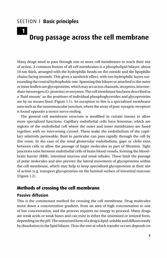
Many drugs need to pass through one or more cell membranes to reach their siteof action. A common feature of all cell membranes is a phospholipid bilayer, about10 nm thick, arranged with the hydrophilic heads on the outside and the lipophilicchains facing inwards. This gives a sandwich effect, with two hydrophilic layers sur-rounding the central hydrophobic one. Spanning this bilayer or attached to the outeror inner leaflets are glycoproteins, which may act as ion channels, receptors, interme-diate messengers (G-proteins) or enzymes. The cell membrane has been described asa ‘fluid mosaic’ as the positions of individual phosphoglycerides and glycoproteinsare by no means fixed (Figure 1.1). An exception to this is a specialized membranearea such as the neuromuscular junction, where the array of post-synaptic receptorsis found opposite a motor nerve ending.
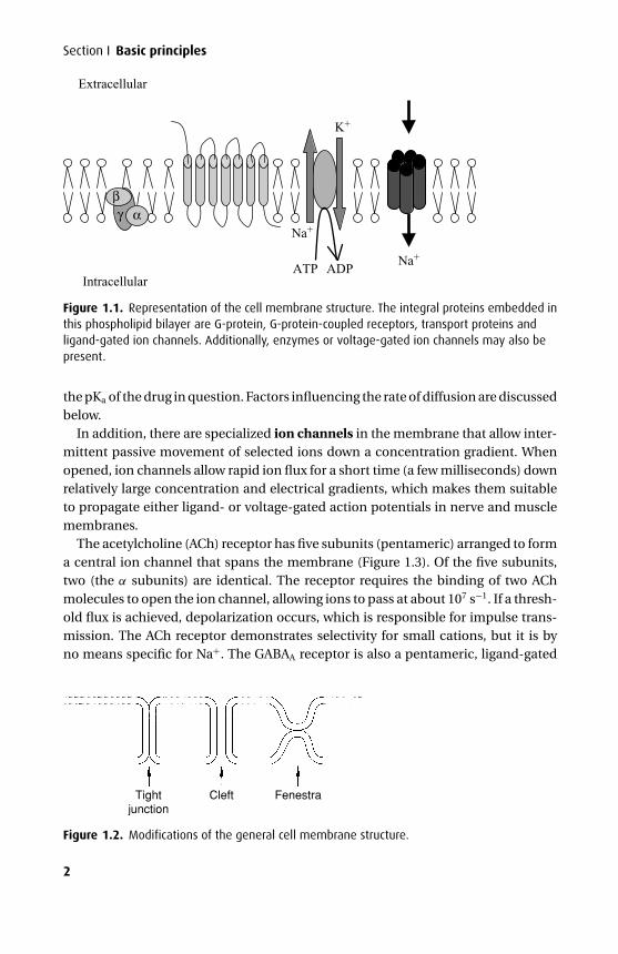
The general cell membrane structure is modified in certain tissues to allowmore specialized functions. Capillary endothelial cells have fenestrae, which areregions of the endothelial cell where the outer and inner membranes are fusedtogether, with no intervening cytosol. These make the endothelium of the capil-lary relatively permeable; fluid in particular can pass rapidly through the cell bythis route. In the case of the renal glomerular endothelium, gaps or clefts existbetween cells to allow the passage of larger molecules as part of filtration. Tightjunctions exist between endothelial cells of brain blood vessels, forming the blood–brain barrier (BBB), intestinal mucosa and renal tubules. These limit the passageof polar molecules and also prevent the lateral movement of glycoproteins withinthe cell membrane, which may help to keep specialized glycoproteins at their siteof action (e.g. transport glycoproteins on the luminal surface of intestinal mucosa)(Figure 1.2).
Methods of crossing the cell membrane
Passive diffusionThis is the commonest method for crossing the cell membrane. Drug moleculesmove down a concentration gradient, from an area of high concentration to oneof low concentration, and the process requires no energy to proceed. Many drugsare weak acids or weak bases and can exist in either the unionized or ionized form,depending on the pH. The unionized form of a drug is lipid-soluble and diffuses easilyby dissolution in the lipid bilayer. Thus the rate at which transfer occurs depends on
1
P1: PCX Printer: Yet To Come
9780521704632c01 CUFX213A/Peck 9780521618168 December 27, 2007 12:41
Section I Basic principles
Na+
Na+
K+
ATP ADP
βγ α
Intracellular
Extracellular
Figure 1.1. Representation of the cell membrane structure. The integral proteins embedded inthis phospholipid bilayer are G-protein, G-protein-coupled receptors, transport proteins andligand-gated ion channels. Additionally, enzymes or voltage-gated ion channels may also bepresent.
the pKa of the drug in question. Factors influencing the rate of diffusion are discussedbelow.
In addition, there are specialized ion channels in the membrane that allow inter-mittent passive movement of selected ions down a concentration gradient. Whenopened, ion channels allow rapid ion flux for a short time (a few milliseconds) downrelatively large concentration and electrical gradients, which makes them suitableto propagate either ligand- or voltage-gated action potentials in nerve and musclemembranes.
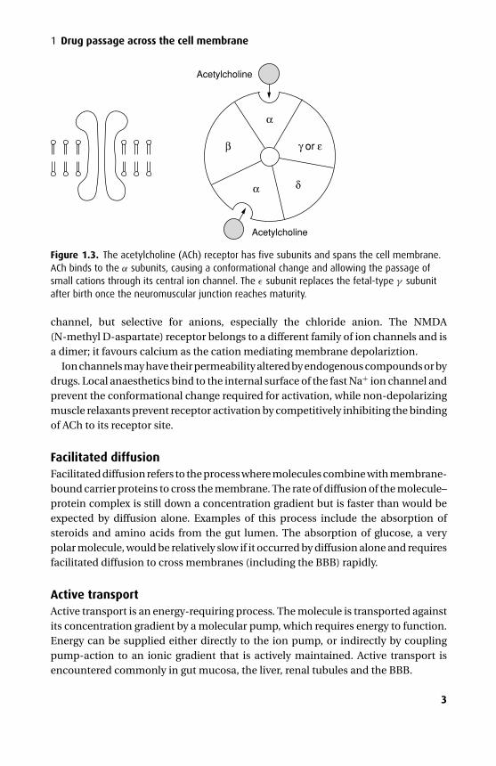
The acetylcholine (ACh) receptor has five subunits (pentameric) arranged to forma central ion channel that spans the membrane (Figure 1.3). Of the five subunits,two (the α subunits) are identical. The receptor requires the binding of two AChmolecules to open the ion channel, allowing ions to pass at about 107 s−1. If a thresh-old flux is achieved, depolarization occurs, which is responsible for impulse trans-mission. The ACh receptor demonstrates selectivity for small cations, but it is byno means specific for Na+. The GABAA receptor is also a pentameric, ligand-gated
Tightjunction
Cleft Fenestra
Figure 1.2. Modifications of the general cell membrane structure.
2
P1: PCX Printer: Yet To Come
9780521704632c01 CUFX213A/Peck 9780521618168 December 27, 2007 12:41
1 Drug passage across the cell membrane
Acetylcholine
Acetylcholine
β
α
α δ
γ or ε
Figure 1.3. The acetylcholine (ACh) receptor has five subunits and spans the cell membrane.ACh binds to the α subunits, causing a conformational change and allowing the passage ofsmall cations through its central ion channel. The ε subunit replaces the fetal-type γ subunitafter birth once the neuromuscular junction reaches maturity.
channel, but selective for anions, especially the chloride anion. The NMDA(N-methyl D-aspartate) receptor belongs to a different family of ion channels and isa dimer; it favours calcium as the cation mediating membrane depolariztion.
Ion channels may have their permeability altered by endogenous compounds or bydrugs. Local anaesthetics bind to the internal surface of the fast Na+ ion channel andprevent the conformational change required for activation, while non-depolarizingmuscle relaxants prevent receptor activation by competitively inhibiting the bindingof ACh to its receptor site.
Facilitated diffusionFacilitated diffusion refers to the process where molecules combine with membrane-bound carrier proteins to cross the membrane. The rate of diffusion of the molecule–protein complex is still down a concentration gradient but is faster than would beexpected by diffusion alone. Examples of this process include the absorption ofsteroids and amino acids from the gut lumen. The absorption of glucose, a verypolar molecule, would be relatively slow if it occurred by diffusion alone and requiresfacilitated diffusion to cross membranes (including the BBB) rapidly.
Active transportActive transport is an energy-requiring process. The molecule is transported againstits concentration gradient by a molecular pump, which requires energy to function.Energy can be supplied either directly to the ion pump, or indirectly by couplingpump-action to an ionic gradient that is actively maintained. Active transport isencountered commonly in gut mucosa, the liver, renal tubules and the BBB.
3
P1: PCX Printer: Yet To Come
9780521704632c01 CUFX213A/Peck 9780521618168 December 27, 2007 12:41
Section I Basic principles
ATP ADP
1° active transport
2° active transport (co-transport)
2° active transport (antiport)
Na
Na
Na
Ca
Glucose
K
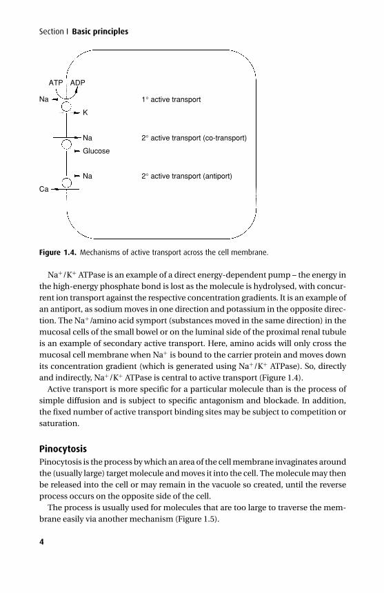
Figure 1.4. Mechanisms of active transport across the cell membrane.
Na+/K+ ATPase is an example of a direct energy-dependent pump – the energy inthe high-energy phosphate bond is lost as the molecule is hydrolysed, with concur-rent ion transport against the respective concentration gradients. It is an example ofan antiport, as sodium moves in one direction and potassium in the opposite direc-tion. The Na+/amino acid symport (substances moved in the same direction) in themucosal cells of the small bowel or on the luminal side of the proximal renal tubuleis an example of secondary active transport. Here, amino acids will only cross themucosal cell membrane when Na+ is bound to the carrier protein and moves downits concentration gradient (which is generated using Na+/K+ ATPase). So, directlyand indirectly, Na+/K+ ATPase is central to active transport (Figure 1.4).
Active transport is more specific for a particular molecule than is the process ofsimple diffusion and is subject to specific antagonism and blockade. In addition,the fixed number of active transport binding sites may be subject to competition orsaturation.
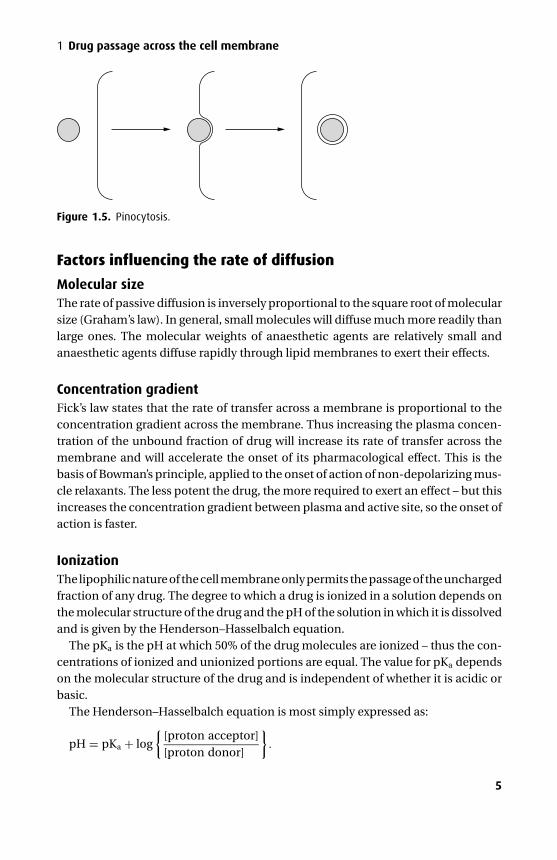
PinocytosisPinocytosis is the process by which an area of the cell membrane invaginates aroundthe (usually large) target molecule and moves it into the cell. The molecule may thenbe released into the cell or may remain in the vacuole so created, until the reverseprocess occurs on the opposite side of the cell.
The process is usually used for molecules that are too large to traverse the mem-brane easily via another mechanism (Figure 1.5).
4
P1: PCX Printer: Yet To Come
9780521704632c01 CUFX213A/Peck 9780521618168 December 27, 2007 12:41
1 Drug passage across the cell membrane
Figure 1.5. Pinocytosis.
Factors influencing the rate of diffusion
Molecular sizeThe rate of passive diffusion is inversely proportional to the square root of molecularsize (Graham’s law). In general, small molecules will diffuse much more readily thanlarge ones. The molecular weights of anaesthetic agents are relatively small andanaesthetic agents diffuse rapidly through lipid membranes to exert their effects.
Concentration gradientFick’s law states that the rate of transfer across a membrane is proportional to theconcentration gradient across the membrane. Thus increasing the plasma concen-tration of the unbound fraction of drug will increase its rate of transfer across themembrane and will accelerate the onset of its pharmacological effect. This is thebasis of Bowman’s principle, applied to the onset of action of non-depolarizing mus-cle relaxants. The less potent the drug, the more required to exert an effect – but thisincreases the concentration gradient between plasma and active site, so the onset ofaction is faster.
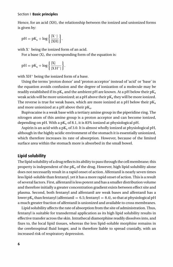
IonizationThe lipophilic nature of the cell membrane only permits the passage of the unchargedfraction of any drug. The degree to which a drug is ionized in a solution depends onthe molecular structure of the drug and the pH of the solution in which it is dissolvedand is given by the Henderson–Hasselbalch equation.
The pKa is the pH at which 50% of the drug molecules are ionized – thus the con-centrations of ionized and unionized portions are equal. The value for pKa dependson the molecular structure of the drug and is independent of whether it is acidic orbasic.
The Henderson–Hasselbalch equation is most simply expressed as:
pH = pKa + log{
[proton acceptor][proton donor]
}.
5
P1: PCX Printer: Yet To Come
9780521704632c01 CUFX213A/Peck 9780521618168 December 27, 2007 12:41
Section I Basic principles
Hence, for an acid (XH), the relationship between the ionized and unionized formsis given by:
pH = pKa + log{
[X−][XH]
},
with X− being the ionized form of an acid.For a base (X), the corresponding form of the equation is:
pH = pKa + log{
[X]
[X H+]
},
with XH+ being the ionized form of a base.Using the terms ‘proton donor’ and ‘proton acceptor’ instead of ‘acid’ or ‘base’ in
the equation avoids confusion and the degree of ionization of a molecule may bereadily established if its pKa and the ambient pH are known. At a pH below their pKa
weak acids will be more unionized; at a pH above their pKa they will be more ionized.The reverse is true for weak bases, which are more ionized at a pH below their pKa
and more unionized at a pH above their pKa.Bupivacaine is a weak base with a tertiary amine group in the piperidine ring. The
nitrogen atom of this amine group is a proton acceptor and can become ionized,depending on pH. With a pKa of 8.1, it is 83% ionized at physiological pH.
Aspirin is an acid with a pKa of 3.0. It is almost wholly ionized at physiological pH,although in the highly acidic environment of the stomach it is essentially unionized,which therefore increases its rate of absorption. However, because of the limitedsurface area within the stomach more is absorbed in the small bowel.
Lipid solubilityThe lipid solubility of a drug reflects its ability to pass through the cell membrane; thisproperty is independent of the pKa of the drug. However, high lipid solubility alonedoes not necessarily result in a rapid onset of action. Alfentanil is nearly seven timesless lipid-soluble than fentanyl, yet it has a more rapid onset of action. This is a resultof several factors. First, alfentanil is less potent and has a smaller distribution volumeand therefore initially a greater concentration gradient exists between effect site andplasma. Second, both fentanyl and alfentanil are weak bases and alfentanil has alower pKa than fentanyl (alfentanil = 6.5; fentanyl = 8.4), so that at physiological pHa much greater fraction of alfentanil is unionized and available to cross membranes.
Lipid solubility affects the rate of absorption from the site of administration. Thus,fentanyl is suitable for transdermal application as its high lipid solubility results ineffective transfer across the skin. Intrathecal diamorphine readily dissolves into, andfixes to, the local lipid tissues, whereas the less lipid-soluble morphine remains inthe cerebrospinal fluid longer, and is therefore liable to spread cranially, with anincreased risk of respiratory depression.
6
P1: PCX Printer: Yet To Come
9780521704632c01 CUFX213A/Peck 9780521618168 December 27, 2007 12:41
1 Drug passage across the cell membrane
Protein bindingOnly the unbound fraction of drug in plasma is free to cross the cell membrane; drugsvary greatly in the degree of plasma protein binding. In practice, the extent of thisbinding is of importance only if the drug is highly protein-bound (more than 90%).In these cases, small changes in the bound fraction produce large changes in theamount of unbound drug. In general, this increases the rate at which drug is metab-olized, so a new equilibrium is re-established with little change in free drug con-centration. For a very small number of highly protein-bound drugs where metabolicpathways are close to saturation (such as phenytoin) this cannot happen and plasmaconcentration of unbound drug will increase and possibly reach toxic levels.
Both albumin and globulins bind drugs, each has many binding sites, the numberand characteristics of which are determined by the pH of plasma. In general, albuminbinds neutral or acidic drugs (e.g. barbiturates), and globulins (in particular, α−1acid glycoprotein) bind basic drugs (e.g. morphine).
Albumin has two important binding sites: the warfarin and diazepam. Bindingis usually readily reversible, and competition for binding at any one site betweendifferent drugs can alter the active unbound fraction of each. Binding is also possi-ble at other sites on the molecule, which may cause a conformational change andindirectly influence binding at the diazepam and warfarin sites.
Although α−1 acid glycoprotein binds basic drugs, other globulins are importantin binding individual ions and molecules, particularly the metals. Thus, iron is boundto β−1 globulin and copper to α−2 globulin.
Protein binding is altered in a range of pathological conditions. Inflammationchanges the relative proportions of the different proteins and albumin concentrationfalls in any acute infective or inflammatory process. This effect is independent ofany reduction in synthetic capacity resulting from liver impairment and is not dueto protein loss. In conditions of severe hypoalbuminaemia (e.g. in end-stage livercirrhosis or burns), the proportion of unbound drug increases markedly such thatthe same dose will have a greatly exaggerated pharmacological effect. The magnitudeof these effects may be hard to estimate and drug dose should be titrated againstclinical effect.
7
P1: PCX Printer: Yet To Come
9780521704632c02 CUFX213A/Peck 9780521618168 December 27, 2007 14:4
2Absorption, distribution, metabolismand excretion
AbsorptionDrugs may be given by a variety of routes; the route chosen depends on the desiredsite of action and the type of drug preparations available. Routes used commonly bythe anaesthetist include inhalation, intravenous, oral, intramuscular, rectal, epiduraland intrathecal. Other routes, such as transdermal, subcutaneous and sublingual,also can be used. The rate and extent of absorption after a particular route of admin-istration depends on both drug and patient factors.
Drugs may be given orally for local as well as systemic effects, for example, oralvancomycin used to treat pseudomembranous colitis is acting locally; antacids alsoact locally in the stomach. In such cases, systemic absorption may result in unwantedside effects.
Intravenous administration provides a direct, and therefore more reliable, routeof systemic drug delivery. No absorption is required, so plasma levels are indepen-dent of such factors as gastrointestinal (GI) absorption and adequate skin or muscleperfusion. However, there are disadvantages in using this route. Pharmacologicalpreparations for intravenous therapy are generally more expensive than the corre-sponding oral medications, and the initially high plasma level achieved with somedrugs may cause undesirable side effects. In addition, if central venous access isused, this carries its own risks. Nevertheless, most drugs used in intensive care aregiven by intravenous infusion this way.
OralAfter oral administration, absorption must take place through the gut mucosa. Fordrugs without specific transport mechanisms, only unionized drugs pass readilythrough the lipid membranes of the gut. Because the pH of the GI tract varies alongits length, the physicochemical properties of the drug will determine from whichpart of the GI tract the drug is absorbed.
Acidic drugs (e.g. aspirin) are unionized in the highly acidic medium of the stomachand therefore are absorbed more rapidly than basic drugs. Although weak bases(e.g. propranolol) are ionized in the stomach, they are relatively unionized in theduodenum, so are absorbed from this site. The salts of permanently charged drugs(e.g. vecuronium, glycopyrrolate) remain ionized at all times and are therefore notabsorbed from the GI tract.
8
P1: PCX Printer: Yet To Come
9780521704632c02 CUFX213A/Peck 9780521618168 December 27, 2007 14:4
2 Absorption, distribution, metabolism and excretion
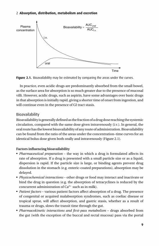
Plasmaconcentration
Bioavailability =AUCoral
AUCi.v.
i.v.
oral
Time
Figure 2.1. Bioavailability may be estimated by comparing the areas under the curves.
In practice, even acidic drugs are predominantly absorbed from the small bowel,as the surface area for absorption is so much greater due to the presence of mucosalvilli. However, acidic drugs, such as aspirin, have some advantages over basic drugsin that absorption is initially rapid, giving a shorter time of onset from ingestion, andwill continue even in the presence of GI tract stasis.
BioavailabilityBioavailability is generally defined as the fraction of a drug dose reaching the systemiccirculation, compared with the same dose given intravenously (i.v.). In general, theoral route has the lowest bioavailability of any route of administration. Bioavailabilitycan be found from the ratio of the areas under the concentration–time curves for anidentical bolus dose given both orally and intravenously (Figure 2.1).
Factors influencing bioavailability� Pharmaceutical preparation – the way in which a drug is formulated affects itsrate of absorption. If a drug is presented with a small particle size or as a liquid,dispersion is rapid. If the particle size is large, or binding agents prevent drugdissolution in the stomach (e.g. enteric-coated preparations), absorption may bedelayed.� Physicochemical interactions – other drugs or food may interact and inactivate orbind the drug in question (e.g. the absorption of tetracyclines is reduced by theconcurrent administration of Ca2+ such as in milk).� Patient factors – various patient factors affect absorption of a drug. The presenceof congenital or acquired malabsorption syndromes, such as coeliac disease ortropical sprue, will affect absorption, and gastric stasis, whether as a result oftrauma or drugs, slows the transit time through the gut.� Pharmacokinetic interactions and first-pass metabolism – drugs absorbed fromthe gut (with the exception of the buccal and rectal mucosa) pass via the portal
9
P1: PCX Printer: Yet To Come
9780521704632c02 CUFX213A/Peck 9780521618168 December 27, 2007 14:4
Section I Basic principles
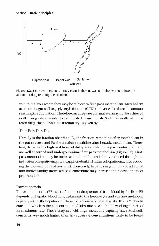
Liver
IVC
Hepatic vein Portal vein Gut lumenGut wall
Figure 2.2. First-pass metabolism may occur in the gut wall or in the liver to reduce theamount of drug reaching the circulation.
vein to the liver where they may be subject to first-pass metabolism. Metabolismat either the gut wall (e.g. glyceryl trinitrate (GTN)) or liver will reduce the amountreaching the circulation. Therefore, an adequate plasma level may not be achievedorally using a dose similar to that needed intravenously. So, for an orally adminis-tered drug, the bioavailable fraction (FB) is given by:
FB = FA × FG × FH.
Here FA is the fraction absorbed, FG the fraction remaining after metabolism inthe gut mucosa and FH the fraction remaining after hepatic metabolism. There-fore, drugs with a high oral bioavailability are stable in the gastrointestinal tract,are well absorbed and undergo minimal first-pass metabolism (Figure 2.2). First-pass metabolism may be increased and oral bioavailability reduced through theinduction of hepatic enzymes (e.g. phenobarbital induces hepatic enzymes, reduc-ing the bioavailability of warfarin). Conversely, hepatic enzymes may be inhibitedand bioavailability increased (e.g. cimetidine may increase the bioavailability ofpropranolol).
Extraction ratioThe extraction ratio (ER) is that fraction of drug removed from blood by the liver. ERdepends on hepatic blood flow, uptake into the hepatocyte and enzyme metaboliccapacity within the hepatocyte. The activity of an enzyme is described by its Michaelisconstant, which is the concentration of substrate at which it is working at 50% ofits maximum rate. Those enzymes with high metabolic capacity have Michaelisconstants very much higher than any substrate concentrations likely to be found
10
P1: PCX Printer: Yet To Come
9780521704632c02 CUFX213A/Peck 9780521618168 December 27, 2007 14:4
2 Absorption, distribution, metabolism and excretion
clinically; those with low capacity will have Michaelis constants close to clinicallyrelevant concentrations. Drugs fall into three distinct groups:
Drugs for which the hepatocyte has rapid uptake and a high metabolic capacity, forexample, propofol and lidocaine. Free drug is rapidly removed from plasma, bounddrug is released to maintain equilibrium and a concentration gradient is maintainedbetween plasma and hepatocyte because drug is metabolized very quickly. Becauseprotein binding has rapid equilibration, the total amount of drug metabolized willbe independent of protein binding but highly dependent on liver blood flow.
Drugs that have low metabolic capacity and high level of protein binding (>90%).This group includes phenytoin and diazepam. Their ER is limited by the metaboliccapacity of the hepatocyte and not by blood flow. If protein binding is altered (e.g. bycompetition) then the free concentration of drug increases significantly. This initiallyincreases uptake into the hepatocyte and rate of metabolism and plasma levels offree drug do not change significantly. However, if the intracellular concentrationexceeds maximum metabolic capacity (saturates the enzyme) drug levels withinthe cell remain high, so reducing uptake (reduced concentration gradient) and ER.Those drugs with a narrow therapeutic index may then show significant toxic effects;hence the need for regular checks on plasma concentration, particularly when othermedication is altered. Therefore for this group of drugs extraction is influenced bychanges in protein binding more than by changes in hepatic blood flow.
Drugs that have low metabolic capacity and low level of protein binding. The totalamount of drug metabolized for this group of drugs is unaffected by either by hepaticblood flow or by changes in protein binding.
SublingualThe sublingual, nasal and buccal routes have two advantages – they are rapid in onsetand, by avoiding the portal tract, have a higher bioavailability. This is advantageousfor drugs where a rapid effect is essential, for example, GTN spray for angina orsublingual nifedipine for the relatively rapid control of high blood pressure.
RectalThe rectal route can be used to avoid first-pass metabolism, and may be considered ifthe oral route is not available. Drugs may be given rectally for their local (e.g. steroidsfor inflammatory bowel disease), as well as their systemic effects (e.g. diclofenacsuppositories for analgesia). There is little evidence that the rectal route is more effi-cacious than the oral route; it provides a relatively small surface area, and absorptionmay be slow or incomplete.
IntramuscularThe intramuscular (i.m.) route avoids the problems associated with oral adminis-tration and the bioavailable fraction approaches 1.0. The speed of onset is generally
11
P1: PCX Printer: Yet To Come
9780521704632c02 CUFX213A/Peck 9780521618168 December 27, 2007 14:4
Section I Basic principles
more rapid compared with the oral route, and for some drugs approaches that forthe intravenous route.
The rate of absorption depends on local perfusion at the site of i.m. injection.Injection at a poorly perfused site may result in delayed absorption and for this reasonthe well-perfused muscles deltoid, quadriceps or gluteus are preferred. If muscleperfusion is poor as a result of systemic hypotension or local vasoconstriction thenan intramuscular injection will not be absorbed until muscle perfusion is restored.Delayed absorption will have two consequences. First, the drug will not be effectivewithin the expected time, which may lead to further doses being given. Second, ifperfusion is then restored, plasma levels may suddenly rise into the toxic range.For these reasons, the intravenous route is preferred if there is any doubt as to theadequacy of perfusion.
Not all drugs can be given i.m., for example, phenytoin. Intramuscular injectionsmay be painful (e.g. cyclizine) and may cause a local abscess or haematoma, soshould be avoided in the coagulopathic patient. There is also the risk of inadvertentintravenous injection of drug intended for the intramuscular route.
SubcutaneousCertain drugs are well absorbed from the subcutaneous tissues and this is thefavoured route for low-dose heparin therapy. A further indication for this route iswhere patient compliance is a problem and depot preparations may be useful. Anti-psychotic medication and some contraceptive formulations have been used in thisway. Co-preparation of insulin with zinc or protamine can produce a slow absorptionprofile lasting several hours after subcutaneous administration.
As with the intramuscular route, the kinetics of absorption is dependent on localand regional blood flow, and may be markedly reduced in shock. Again, this hasthe dual effect of rendering the (non-absorbed) drug initially ineffective, and thensubjecting the patient to a bolus once the perfusion is restored.
TransdermalDrugs may be applied to the skin either for local topical effect, such as steroids, butalso may be used to avoid first-pass metabolism and improve bioavailability. Thus,fentanyl and nitrates may be given transdermally for their systemic effects. Factorsfavouring transdermal absorption are high lipid solubility and a good regional bloodsupply to the site of application (therefore, the thorax and abdomen are preferred tolimbs). Special transdermal formulations (patches) are used to ensure slow, constantrelease of drug for absorption and provide a smoother pharmacokinetic profile. Onlysmall amounts of drug are released at a time, so potent drugs are better suited to thisroute of administration if systemic effects are required.
Local anaesthetics may be applied topically to anaesthetize the skin beforevenepuncture, skin grafts or minor surgical procedures. The two most commonpreparations are topical EMLA and topical amethocaine. The first is a eutectic
12
P1: PCX Printer: Yet To Come
9780521704632c02 CUFX213A/Peck 9780521618168 December 27, 2007 14:4
2 Absorption, distribution, metabolism and excretion
mixture (each agent lowers the boiling point of the other forming a gel-phase) oflidocaine and prilocaine. Amethocaine is an ester-linked local anaesthetic, whichmay cause mild, local histamine release producing local vasodilatation, in contrastto the vasoconstriction seen with eutectic mixture of local anaesthetic (EMLA). Ven-odilatation may be useful when anaesthetizing the skin prior to venepuncture.
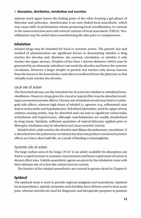
InhalationInhaled drugs may be intended for local or systemic action. The particle size andmethod of administration are significant factors in determining whether a drugreaches the alveolus and, therefore, the systemic circulation, or whether it onlyreaches the upper airways. Droplets of less than 1 micron diameter (which may begenerated by an ultrasonic nebulizer) can reach the alveolus and hence the systemiccirculation. However, a larger droplet or particle size reaches only airway mucosafrom the larynx to the bronchioles (and often is swallowed from the pharynx) so thatvirtually none reaches the alveolus.
Local site of actionThe bronchial airways are the intended site of action for inhaled or nebulized bron-chodilators. However, drugs given for a local or topical effect may be absorbed result-ing in unwanted systemic effects. Chronic use of inhaled steroids may lead to Cushin-goid side effects, whereas high doses of inhaled β2-agonists (e.g. salbutamol) maylead to tachycardia and hypokalaemia. Nebulized adrenaline, used for upper airwayoedema causing stridor, may be absorbed and can lead to significant tachycardia,arrhythmias and hypertension, although catecholamines are readily metabolizedby lung tissue. Similarly, sufficient quantities of topical lidocaine applied prior tofibreoptic intubation may be absorbed and cause systemic toxicity.
Inhaled nitric oxide reaches the alveolus and dilates the pulmonary vasculature. Itis absorbed into the pulmonary circulation but does not produce unwanted systemiceffects as it has a short half-life, as a result of binding to haemoglobin.
Systemic site of actionThe large surface area of the lungs (70 m2 in an adult) available for absorption canlead to a rapid increase in systemic concentration and hence rapid onset of action atdistant effect sites. Volatile anaesthetic agents are given by the inhalation route withtheir ultimate site of action the central nervous system.
The kinetics of the inhaled anaesthetics are covered in greater detail in Chapter 8.
EpiduralThe epidural route is used to provide regional analgesia and anaesthesia. Epidurallocal anaesthetics, opioids, ketamine and clonidine have all been used to treat acutepain, whereas steroids are used for diagnostic and therapeutic purposes in patients
13
P1: PCX Printer: Yet To Come
9780521704632c02 CUFX213A/Peck 9780521618168 December 27, 2007 14:4
Section I Basic principles
with chronic pain. Drug may be given as a single bolus or, through a catheter placedin the epidural space, as a series of boluses or by infusion.
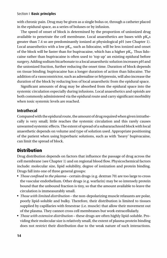
The speed of onset of block is determined by the proportion of unionized drugavailable to penetrate the cell membrane. Local anaesthetics are bases with pKasgreater than 7.4 so are predominantly ionized at physiological pH (see Chapter 1).Local anaesthetics with a low pKa, such as lidocaine, will be less ionized and onsetof the block will be faster than for bupivacaine, which has a higher pKa. Thus lido-caine rather than bupivacaine is often used to ‘top up’ an existing epidural beforesurgery. Adding sodium bicarbonate to a local anaesthetic solution increases pH andthe unionized fraction, further reducing the onset time. Duration of block dependson tissue binding; bupivacaine has a longer duration of action than lidocaine. Theaddition of a vasoconstrictor, such as adrenaline or felypressin, will also increase theduration of the block by reducing loss of local anaesthetic from the epidural space.
Significant amounts of drug may be absorbed from the epidural space into thesystemic circulation especially during infusions. Local anaesthetics and opioids areboth commonly administered via the epidural route and carry significant morbiditywhen toxic systemic levels are reached.
IntrathecalCompared with the epidural route, the amount of drug required when given intrathe-cally is very small; little reaches the systemic circulation and this rarely causesunwanted systemic effects. The extent of spread of a subararachnoid block with localanaesthetic depends on volume and type of solution used. Appropriate positioningof the patient when using hyperbaric solutions, such as with ‘heavy’ bupivacaine,can limit the spread of block.
DistributionDrug distribution depends on factors that influence the passage of drug across thecell membrane (see Chapter 1) and on regional blood flow. Physicochemical factorsinclude: molecular size, lipid solubility, degree of ionization and protein binding.Drugs fall into one of three general groups:� Those confined to the plasma – certain drugs (e.g. dextran 70) are too large to cross
the vascular endothelium. Other drugs (e.g. warfarin) may be so intensely proteinbound that the unbound fraction is tiny, so that the amount available to leave thecirculation is immeasurably small.� Those with limited distribution – the non-depolarizing muscle relaxants are polar,poorly lipid-soluble and bulky. Therefore, their distribution is limited to tissuessupplied by capillaries with fenestrae (i.e. muscle) that allow their movement outof the plasma. They cannot cross cell membranes but work extracellularly.� Those with extensive distribution – these drugs are often highly lipid-soluble. Pro-viding their molecular size is relatively small, the extent of plasma protein bindingdoes not restrict their distribution due to the weak nature of such interactions.
14
P1: PCX Printer: Yet To Come
9780521704632c02 CUFX213A/Peck 9780521618168 December 27, 2007 14:4
2 Absorption, distribution, metabolism and excretion
Other drugs are sequestered by tissues (amiodarone by fat; iodine by the thyroid;tetracyclines by bone), which effectively removes them from the circulation.
Those drugs that are not confined to the plasma are initially distributed to tissueswith the highest blood flow (brain, lung, kidney, thyroid, adrenal) then to tissues witha moderate blood flow (muscle), and finally to tissues with a very low blood flow (fat).These three groups of tissues provide a useful model when explaining how plasmalevels decline after drug administration.
Blood–brain barrier (BBB)The BBB is an anatomical and functional barrier between the circulation and thecentral nervous system (see Chapter 1).
Active transport and facilitated diffusion are the predominant methods of molec-ular transfer, which in health is tightly controlled. Glucose and hormones, such asinsulin, cross by active carrier transport, while only lipid-soluble, low molecularweight drugs can cross by simple diffusion. Thus inhaled and intravenous anaes-thetics can cross readily whereas the larger, polar muscle relaxants cannot andhave no central effect. Similarly, glycopyrrolate has a quaternary, charged nitro-gen and does not cross the BBB readily. This is in contrast to atropine, a tertiaryamine, which may cause centrally mediated effects such as confusion or paradoxicalbradycardia.
As well as providing an anatomical barrier, the BBB contains enzymes such asmonoamine oxidase. Therefore, monoamines are converted to non-active metabo-lites by passing through the BBB. Physical disruption of the BBB may lead to centralneurotransmitters being released into the systemic circulation and may help explainthe marked circulatory disturbance seen with head injury and subarachnoid haem-orrhage.
In the healthy subject penicillin penetrates the BBB poorly. However, in meningitis,the nature of the BBB alters as it becomes inflamed, and permeability to penicillin(and other drugs) increases, so allowing therapeutic access.
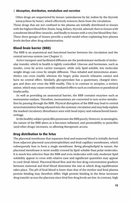
Drug distribution to the fetusThe placental membrane that separates fetal and maternal blood is initially derivedfrom adjacent placental syncytiotrophoblast and fetal capillary membranes, whichsubsequently fuse to form a single membrane. Being phospholipid in nature, theplacental membrane is more readily crossed by lipid-soluble than polar molecules.It is much less selective than the BBB and even molecules with only moderate lipidsolubility appear to cross with relative ease and significant quantities may appearin cord (fetal) blood. Placental blood flow and the free drug concentration gradientbetween maternal and fetal blood determine the rate at which drug equilibrationtakes place. The pH of fetal blood is lower than that of the mother and fetal plasmaprotein binding may therefore differ. High protein binding in the fetus increasesdrug transfer across the placenta since fetal free drug levels are low. In contrast, high
15
P1: PCX Printer: Yet To Come
9780521704632c02 CUFX213A/Peck 9780521618168 December 27, 2007 14:4
Section I Basic principles
protein binding in the mother reduces the rate of drug transfer since maternal freedrug levels are low. The fetus also may metabolize some drugs; the rate of metabolismincreases as the fetus matures.
The effects of maternal pharmacology on the fetus may be divided into thoseeffects that occur in pregnancy, especially the early first trimester when organogen-esis occurs, and at birth.
Drugs during pregnancyThe safety of any drug in pregnancy must be evaluated, but interspecies variationis great and animal models may not exclude the possibility of significant humanteratogenicity. In addition, teratogenic effects may not be apparent for some years;stilboestrol taken during pregnancy predisposes female offspring to ovarian cancer atpuberty. Wherever possible drug therapy should be avoided throughout pregnancy;if treatment is essential drugs with a long history of safety should be selected.
There are conditions, however, in which the risk of not taking medication out-weighs the theoretical or actual risk of teratogenicity. Thus, in epilepsy the risk ofhypoxic damage to the fetus secondary to fitting warrants the continuation of anti-epileptic medication during pregnancy. Similarly, the presence of an artificial heartvalve mandates the continuation of anticoagulation despite the attendant risks.
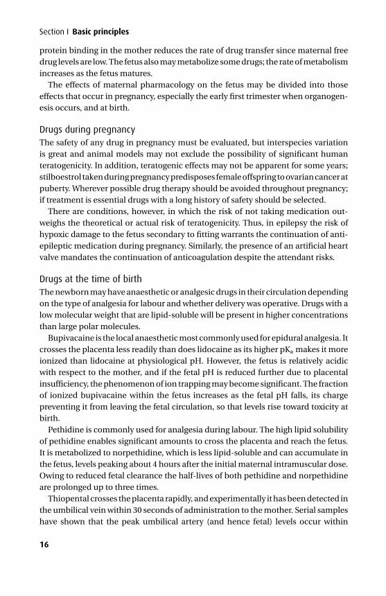
Drugs at the time of birthThe newborn may have anaesthetic or analgesic drugs in their circulation dependingon the type of analgesia for labour and whether delivery was operative. Drugs with alow molecular weight that are lipid-soluble will be present in higher concentrationsthan large polar molecules.
Bupivacaine is the local anaesthetic most commonly used for epidural analgesia. Itcrosses the placenta less readily than does lidocaine as its higher pKa makes it moreionized than lidocaine at physiological pH. However, the fetus is relatively acidicwith respect to the mother, and if the fetal pH is reduced further due to placentalinsufficiency, the phenomenon of ion trapping may become significant. The fractionof ionized bupivacaine within the fetus increases as the fetal pH falls, its chargepreventing it from leaving the fetal circulation, so that levels rise toward toxicity atbirth.
Pethidine is commonly used for analgesia during labour. The high lipid solubilityof pethidine enables significant amounts to cross the placenta and reach the fetus.It is metabolized to norpethidine, which is less lipid-soluble and can accumulate inthe fetus, levels peaking about 4 hours after the initial maternal intramuscular dose.Owing to reduced fetal clearance the half-lives of both pethidine and norpethidineare prolonged up to three times.
Thiopental crosses the placenta rapidly, and experimentally it has been detected inthe umbilical vein within 30 seconds of administration to the mother. Serial sampleshave shown that the peak umbilical artery (and hence fetal) levels occur within
16
P1: PCX Printer: Yet To Come
9780521704632c02 CUFX213A/Peck 9780521618168 December 27, 2007 14:4
2 Absorption, distribution, metabolism and excretion
3 minutes of maternal injection. There is no evidence that fetal outcome is affectedwith an‘injection to delivery’ time of up to 20 minutes after injection of a sleep doseof thiopental to the mother.
The non-depolarizing muscle relaxants are large polar molecules and essentiallydo not cross the placenta. Therefore, the fetal neuromuscular junction is not affected.Only very small amounts of suxamethonium cross the placenta, though again thisusually has little effect. However, if the mother has an inherited enzyme deficiencyand cannot metabolize suxamethonium, then maternal levels may remain high anda significant degree of transfer may occur. This may be especially significant if thefetus has also inherited the enzyme defect, in which case there may be a degree ofdepolarizing blockade at the fetal neuromuscular junction.
MetabolismWhile metabolism usually reduces the activity of a drug, activity may be designedto increase; a prodrug is defined as a drug that has no inherent activity beforemetabolism but that is converted by the body to an active moiety. Examples ofprodrugs are enalapril (metabolized to enaloprilat), diamorphine (metabolized to 6-monoacylmorphine), and parecoxib (metabolized to valdecoxib). Metabolites alsomay have equivalent activity to the parent compound, in which case duration ofaction is not related to plasma levels of the parent drug.
In general, metabolism produces a more polar (water soluble) molecule that canbe excreted in the bile or urine – the chief routes of drug excretion. There are twophases of metabolism, I and II.
Phase I (functionalization or non-synthetic)� Oxidation� Reduction� HydrolysisMany phase I reactions, particularly oxidative pathways, occur in the liver due to anon-specific mixed-function oxidase system in the endoplasmic reticulum. Theseenzymes form the cytochrome P450 system, named after the wavelength (in nm) oftheir maximal absorption of light when the reduced state is combined with carbonmonoxide. However, this cytochrome system is not unique to the liver; these enzymesare also found in gut mucosa, lung, brain and kidney. Methoxyflurane is metabolizedby CYP2E1 in the kidney, generating a high local concentration of fluoride ions, whichmay cause renal failure (see sevoflurane metabolism, p. 123).
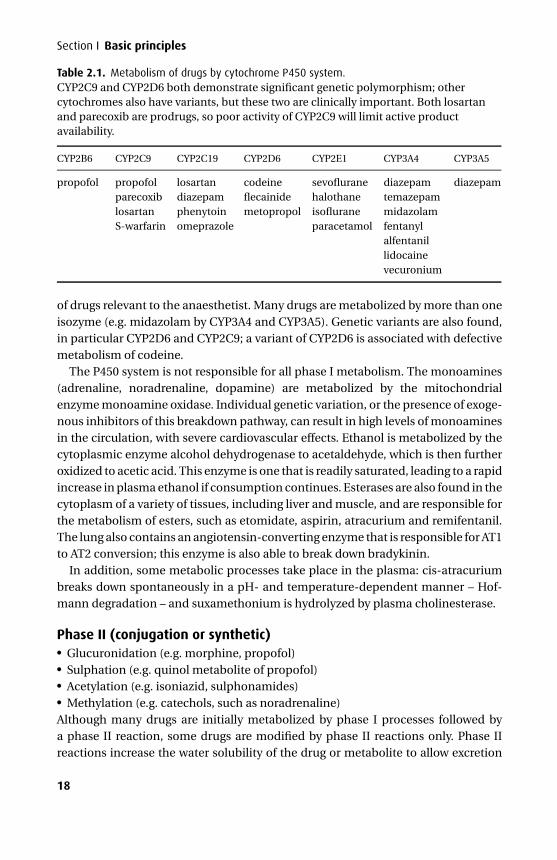
The enzymes of the cytochrome P450 system are classified into families and sub-families by their degree of shared amino acid sequences – families and subfamiliesshare 40% and 55% respectively of the amino acid sequence. In addition, the subfam-ilies are further divided into isoforms. Families are labelled CYP1, CYP2, and so on,the subfamilies CYP1A, CYP1B, and so on, and the isoforms CYP1A1, CYP1A2, andso on. Table 2.1 summarises isoenzymes of particular importance in the metabolism
17
P1: PCX Printer: Yet To Come
9780521704632c02 CUFX213A/Peck 9780521618168 December 27, 2007 14:4
Section I Basic principles
Table 2.1. Metabolism of drugs by cytochrome P450 system.CYP2C9 and CYP2D6 both demonstrate significant genetic polymorphism; othercytochromes also have variants, but these two are clinically important. Both losartanand parecoxib are prodrugs, so poor activity of CYP2C9 will limit active productavailability.
CYP2B6 CYP2C9 CYP2C19 CYP2D6 CYP2E1 CYP3A4 CYP3A5
propofol propofol losartan codeine sevoflurane diazepam diazepamparecoxib diazepam flecainide halothane temazepamlosartan phenytoin metopropol isoflurane midazolamS-warfarin omeprazole paracetamol fentanyl
alfentanillidocainevecuronium
of drugs relevant to the anaesthetist. Many drugs are metabolized by more than oneisozyme (e.g. midazolam by CYP3A4 and CYP3A5). Genetic variants are also found,in particular CYP2D6 and CYP2C9; a variant of CYP2D6 is associated with defectivemetabolism of codeine.
The P450 system is not responsible for all phase I metabolism. The monoamines(adrenaline, noradrenaline, dopamine) are metabolized by the mitochondrialenzyme monoamine oxidase. Individual genetic variation, or the presence of exoge-nous inhibitors of this breakdown pathway, can result in high levels of monoaminesin the circulation, with severe cardiovascular effects. Ethanol is metabolized by thecytoplasmic enzyme alcohol dehydrogenase to acetaldehyde, which is then furtheroxidized to acetic acid. This enzyme is one that is readily saturated, leading to a rapidincrease in plasma ethanol if consumption continues. Esterases are also found in thecytoplasm of a variety of tissues, including liver and muscle, and are responsible forthe metabolism of esters, such as etomidate, aspirin, atracurium and remifentanil.The lung also contains an angiotensin-converting enzyme that is responsible for AT1to AT2 conversion; this enzyme is also able to break down bradykinin.
In addition, some metabolic processes take place in the plasma: cis-atracuriumbreaks down spontaneously in a pH- and temperature-dependent manner – Hof-mann degradation – and suxamethonium is hydrolyzed by plasma cholinesterase.
Phase II (conjugation or synthetic)� Glucuronidation (e.g. morphine, propofol)� Sulphation (e.g. quinol metabolite of propofol)� Acetylation (e.g. isoniazid, sulphonamides)� Methylation (e.g. catechols, such as noradrenaline)Although many drugs are initially metabolized by phase I processes followed bya phase II reaction, some drugs are modified by phase II reactions only. Phase IIreactions increase the water solubility of the drug or metabolite to allow excretion
18
P1: PCX Printer: Yet To Come
9780521704632c02 CUFX213A/Peck 9780521618168 December 27, 2007 14:4
2 Absorption, distribution, metabolism and excretion
into the bile or urine. They occur mainly in the hepatic endoplasmic reticulum butother sites, such as the lung, may also be involved. This is especially true in the caseof acetylation, which also occurs in the lung and spleen.
In liver failure, phase I reactions are generally affected before phase II, so drugswith a predominantly phase II metabolism, such as lorazepam, are less affected.
Genetic polymorphismThere are inherited differences in enzyme structure that alter the way drugs aremetabolized in the body. The genetic polymorphisms of particular relevance toanaesthesia are those of plasma cholinesterase, those involved in acetylation andthe CYP2D6 variants mentioned above.
Suxamethonium is metabolized by hydrolysis in the plasma, a reaction that is catal-ysed by the relatively non-specific enzyme plasma cholinesterase. Certain individ-uals have an unusual variant of the enzyme and metabolize suxamethonium muchmore slowly. Several autosomal recessive genes have been identified, and these maybe distinguished by the degree of enzyme inhibition demonstrated in vitro by sub-stances such as fluoride and the local anaesthetic dibucaine. Muscle paralysis dueto suxamethonium may be prolonged in individuals with an abnormal form of theenzyme. This is discussed in greater detail in Chapter 11.
Acetylation is a phase II metabolic pathway in the liver. Drugs metabolized bythis route include hydralazine and isoniazid. There are genetically different isoen-zymes that acetylate at a slow or fast rate. The pharmacokinetic and hence phar-macodynamic profile seen with these drugs depends on the acetylator status of theindividual.
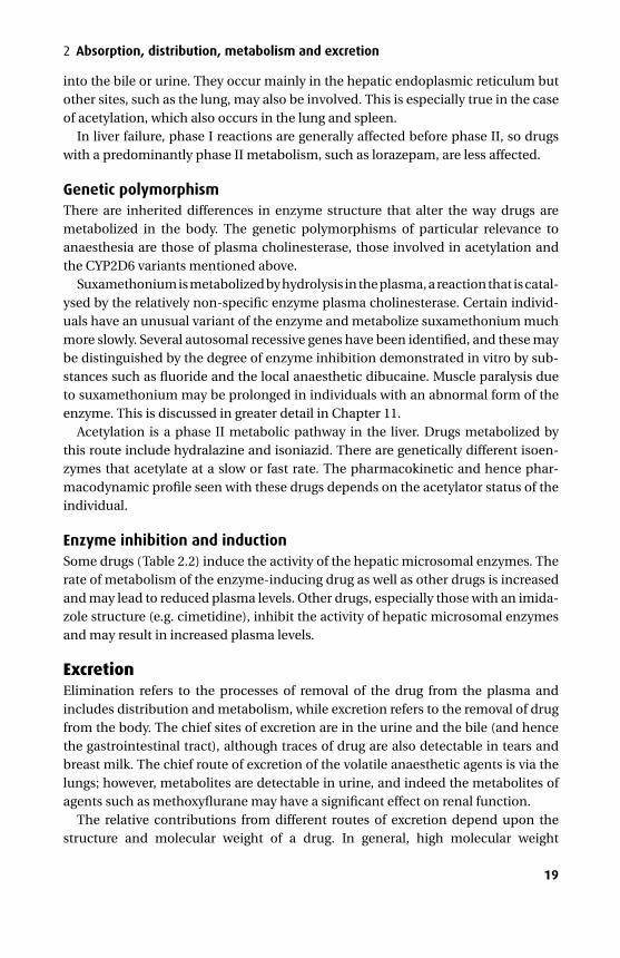
Enzyme inhibition and inductionSome drugs (Table 2.2) induce the activity of the hepatic microsomal enzymes. Therate of metabolism of the enzyme-inducing drug as well as other drugs is increasedand may lead to reduced plasma levels. Other drugs, especially those with an imida-zole structure (e.g. cimetidine), inhibit the activity of hepatic microsomal enzymesand may result in increased plasma levels.
ExcretionElimination refers to the processes of removal of the drug from the plasma andincludes distribution and metabolism, while excretion refers to the removal of drugfrom the body. The chief sites of excretion are in the urine and the bile (and hencethe gastrointestinal tract), although traces of drug are also detectable in tears andbreast milk. The chief route of excretion of the volatile anaesthetic agents is via thelungs; however, metabolites are detectable in urine, and indeed the metabolites ofagents such as methoxyflurane may have a significant effect on renal function.
The relative contributions from different routes of excretion depend upon thestructure and molecular weight of a drug. In general, high molecular weight
19
P1: PCX Printer: Yet To Come
9780521704632c02 CUFX213A/Peck 9780521618168 December 27, 2007 14:4
Section I Basic principles
Table 2.2. Effects of various drugs on hepatic microsomal enzymes.
Inducing Inhibiting
Antibiotics rifampicin metronidazole, isoniazid,chloramphenicol acute use
Alcohol chronic abuseInhaled anaesthetics enflurane, halothaneBarbiturates phenobarbital, thiopentalAnti-convulsants phenytoin, carbamazepineHormones glucocorticoidsMAOIs phenelzine, tranylcypromineH2 antagonists cimetidineOthers cigarette smoking amiodarone, grapefruit juice
compounds (>30 000) are not filtered or secreted by the kidney and are thereforepreferentially excreted in the bile. A significant fraction of a drug carrying a perma-nent charge, such as pancuronium, may be excreted unchanged in urine.
Renal excretion
Filtration at the glomerulusSmall, non-protein bound, poorly lipid-soluble but readily water-soluble drugs areexcreted into the glomerular ultrafiltrate. Only free drug present in that fraction ofplasma that is filtered is removed at the glomerulus. The remaining plasma willhave the same concentration of free drug as that fraction filtered and so there is nochange in the extent of plasma protein binding. Thus highly protein bound drugsare not extensively removed by filtration – but may be excreted by active secretorymechanisms in the tubule.
Secretion at the proximal tubulesThere are active energy-requiring processes in the proximal convoluted tubules bywhich a wide variety of molecules may be secreted into the urine against their con-centration gradients. Different carrier systems exist for acidic and basic drugs thatare each capacity-limited for their respective drug type (i.e. maximal clearance ofone acidic drug will result in a reduced clearance of another acidic drug but not ofa basic drug). Drug secretion also may be inhibited, for example, probenecid blocksthe secretion of penicillin.
Diffusion at the distal tubulesAt the distal tubule, passive diffusion may occur down the concentration gradient.Acidic drugs are preferentially excreted in an alkaline urine as this increases thefraction present in the ionized form, which cannot be re-absorbed. Conversely, basicdrugs are preferentially excreted in acidic urine where they are trapped as cations.
20
P1: PCX Printer: Yet To Come
9780521704632c02 CUFX213A/Peck 9780521618168 December 27, 2007 14:4
2 Absorption, distribution, metabolism and excretion
Biliary excretionHigh molecular weight compounds, such as the steroid-based muscle relaxants, areexcreted in bile. Secretion from the hepatocyte into the biliary canaliculus takesplace against a concentration gradient, and is therefore active and energy-requiring,and subject to inhibition and competition for transport. Certain drugs are excretedunchanged in bile (e.g. rifampicin), while others are excreted after conjugation (e.g.morphine metabolites are excreted as glucuronides).
Enterohepatic circulationDrugs excreted in the bile such as glucuronide conjugates may be hydrolyzed in thesmall bowel by glucuronidase secreted by bacteria. Lipid-soluble, active drug mayresult and be reabsorbed, passing into the portal circulation to the liver where theextracted fraction is reconjugated and re-excreted in the bile, and the rest passesinto the systemic circulation. This process may continue many times. Failure of theoral contraceptive pill while taking broad-spectrum antibiotics has been blamed ona reduced intestinal bacterial flora causing a reduced enterohepatic circulation ofoestrogen and progesterone.
Effect of disease
Renal diseaseIn the presence of renal disease, those drugs that are normally excreted via the renaltract may accumulate. This effect will vary according to the degree to which the drugis dependent upon renal excretion – in the case of a drug whose clearance is entirelyrenal a single dose may have a very prolonged effect. This was true of gallamine,a non-depolarizing muscle relaxant, which, if given in the context of renal failure,required dialysis or haemofiltration to reduce the plasma level and hence reverse thepharmacological effect.
If it is essential to give a drug that is highly dependent on renal excretion in thepresence of renal impairment, a reduction in dose must be made. If the apparentvolume of distribution remains the same, the loading dose also remains the same,but repeated doses may need to be reduced and dosing interval increased. However,due to fluid retention the volume of distribution is often increased in renal failure,so loading doses may be higher than in health.
Knowledge of a patient’s creatinine clearance is very helpful in estimating the dosereduction required for a given degree of renal impairment. As an approximation, thedose, D, required in renal failure is given by:
D = Usual dose × (impaired clearance/normal clearance).
Tables contained in the British National Formulary give an indication of the appro-priate reductions in mild, moderate and severe renal impairment.
21
P1: PCX Printer: Yet To Come
9780521704632c02 CUFX213A/Peck 9780521618168 December 27, 2007 14:4
Section I Basic principles
Liver diseaseHepatic impairment alters many aspects of the pharmacokinetic profile of a drug.Protein synthesis is decreased (hence decreased plasma protein levels and reducedprotein binding). Both phase I and II reactions are affected, and thus the metabolismof drugs is reduced. The presence of ascites increases the volume of distributionand the presence of portocaval shunts increases bioavailability by reducing hepaticclearance of drugs.
There is no analogous measure of hepatic function compared with creatinineclearance for renal function. Liver function tests in common clinical use may bedivided into those that measure the synthetic function of the liver – the internationalnormalized ratio (INR) or prothrombin time and albumin – and those that measureinflammatory damage of the hepatocyte. It is possible to have a markedly inflamedliver with high transaminase levels, with retention of reasonable synthetic function.In illness, the profile of protein synthesis shifts toward acute phase proteins; albuminis not an acute phase protein so levels are reduced in any acute illness.
Patients with severe liver failure may suffer hepatic encephalopathy as a result ofa failure to clear ammonia and other molecules. These patients are very susceptibleto the effects of benzodiazepines and opioids, which should therefore be avoided ifpossible. For patients requiring strong analgesia in the peri-operative period a co-existing coagulopathy will often rule out a regional technique, leaving few other anal-gesic options other than careful intravenous titration of opioid analgesics, acceptingthe risk of precipitating encephalopathy.
The extremes of age
Neonate and infantIn the newborn and young, the pharmacokinetic profiles of drugs are different for anumber of reasons. These are due to qualitative, as well as quantitative, differencesin the neonatal anatomy and physiology.
Fluid compartmentsThe volume and nature of the pharmacokinetic compartments is different, with thenewborn being relatively overhydrated and losing volume through diuresis in thehours and days after birth. As well as the absolute proportion of water being higher,the relative amount in the extracellular compartment is increased. The relative sizesof the organs and regional blood flows are also different from the adult; the neonatalliver is relatively larger than that of an adult although its metabolizing capacity islower and may not be as efficient.
DistributionPlasma protein levels and binding are less than in the adult. In addition, the pH ofneonatal blood tends to be a lower value, which alters the relative proportions of
22
P1: PCX Printer: Yet To Come
9780521704632c02 CUFX213A/Peck 9780521618168 December 27, 2007 14:4
2 Absorption, distribution, metabolism and excretion
ionized and unionized drug. Thus, both the composition and acid-base value of theblood affect plasma protein binding.
Metabolism and excretionWhile the neonate is born with several of the enzyme systems functioning at adultlevels, the majority of enzymes do not reach maturity for a number of months. Plasmalevels of cholinesterase are reduced, and in the liver the activity of the cytochromeP450 family of enzymes is markedly reduced. Newborns have a reduced rate of excre-tion via the renal tract. The creatinine clearance is less than 10% of the adult rateper unit body weight, with nephron numbers and function not reaching maturityfor some months after birth.
Though the implications of many of these differences may be predicted, the precisedoses of drugs used in the newborn has largely been determined clinically. Preferreddrugs should be those that have been used safely for a number of years, and in whichthe necessary dose adjustments have been derived empirically. In addition, there iswide variation between individuals of the same post-conceptual age.
ElderlyA number of factors contribute to pharmacokinetic differences observed in theelderly. The elderly have a relative reduction in muscle mass, with a consequentincrease in the proportion of fat, altering volume of distribution. This loss of musclemass is of great importance in determining the sensitivity of the elderly to remifen-tanil, which is significantly metabolized by muscle esterases. There is a reduction inthe activity of hepatic enzymes with increasing age, leading to a relative decrease inhepatic drug clearance. Creatinine clearance diminishes steadily with age, reflectingreduced renal function.
As well as physiological changes with increasing age, the elderly are more likely tohave multiple co-existing diseases. The implications of this are two-fold. First, thedisease processes may directly alter drug pharmacokinetics and second, polyphar-macy may produce drug interactions that alter both pharmacokinetics and pharma-codynamic response.
23
P1: PCX Printer: Yet To Come
9780521704632c03 CUFX213A/Peck 9780521618168 December 27, 2007 13:30
3Drug action
Mechanisms of drug actionDrugs may act in a number of ways to exert their effect. These range from relativelysimple non-specific actions that depend on the physicochemical properties of adrug to highly specific and stereoselective actions on proteins in the body, namelyenzymes, voltage-gated ion channels and receptors.
Actions dependent on chemical propertiesThe antacids exert their effect by neutralizing gastric acid. The chelating agentsare used to reduce the concentration of certain metallic ions within the body.Dicobalt edetate chelates cyanide ions and may be used in cyanide poisoning orfollowing a potentially toxic dose of sodium nitroprusside. The new reversal agent,γ -cyclodextrin, selectively chelates rocuronium and reversal is possible from deeperlevels of block than can be effected with the anticholinesterases.
EnzymesEnzymes are biological catalysts, and most drugs that interact with enzymesare inhibitors. The results are twofold: the concentration of the substrate nor-mally metabolized by the enzyme is increased and that of the product(s) of thereaction is decreased. Enzyme inhibition may be competitive (edrophonium foranticholinesterase), non-competitive or irreversible (aspirin for cyclo-oxygenaseand omeprazole for the Na+/H+ATPase). Angiotensin-converting enzyme (ACE)inhibitors such as captopril prevent the conversion of angiotensin I to II andbradykinin to various inactive fragments. Although reduced levels of angiotensinII are responsible for the therapeutic effects when used in hypertension and heartfailure, raised levels of bradykinin may cause an intractable cough.
Voltage-gated ion channelsVoltage-gated ion channels are involved in conduction of electrical impulses associ-ated with excitable tissues in muscle and nerve. Several groups of drugs have specificblocking actions at these ion channels. Local anaesthetics act by inhibiting Na+ chan-nels in nerve membrane, several anticonvulsants block similar channels in the brain,calcium channel blocking agents act on vascular smooth muscle ion channels and
24
P1: PCX Printer: Yet To Come
9780521704632c03 CUFX213A/Peck 9780521618168 December 27, 2007 13:30
3 Drug action
antiarrhythmic agents block myocardial ion channels. These actions are describedin the relevant chapters in Sections II and III.
ReceptorsA receptor is a protein, often integral to a membrane, containing a region to which anatural ligand binds specifically to bring about a response. A drug acting at a receptorbinds to a recognition site where it may elicit an effect (an agonist), prevent the actionof a natural ligand (an inhibitor), or reduce a constitutive effect of a receptor (aninverse agonist). Natural ligands may also bind to more than one receptor and havea different mechanism of action at each (e.g. ionotropic and metabotropic actionsof γ -aminobutyric acid (GABA) at GABAA and GABAB receptors).
Receptors are generally protein or glycoprotein in nature and may be associ-ated with or span the cell membrane, be present in the membranes of intracellularorganelles or be found in the cytosol or nucleus. Those in the membrane are gener-ally for ligands that do not readily penetrate the cell, whereas those within the cell arefor lipid-soluble ligands that can diffuse through the cell wall to their site of action,or for intermediary messengers generated within the cell itself.
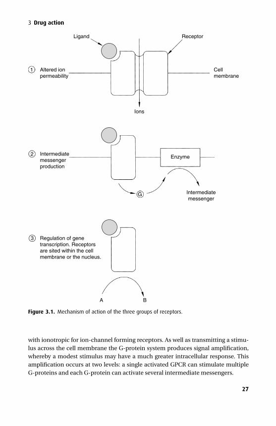
Receptors may be grouped into three classes depending on their mechanism ofaction: (1) altered ion permeability; (2) production of intermediate messengers and(3) regulation of gene transcription.
1) Altered ion permeability: ion channelsReceptors of this type are part of a membrane-spanning complex of protein subunitsthat have the potential to form a channel through the membrane. When opened,such a channel allows the passage of ions down their concentration and electricalgradients. Here, ligand binding causes a conformational change in the structure ofthis membrane protein complex allowing the channel to open and so increasing thepermeability of the membrane to certain ions (ionotropic). There are three importantligand-gated ion channel families: the pentameric, the ionotropic glutamate, and theionotropic purinergic receptors.
The pentameric familyThe pentameric family of receptors has five membrane-spanning subunits. The best-known example of this type of ion channel receptors is the nicotinic acetylcholinereceptor at the neuromuscular junction. It consists of one β, one ε, one γ and twoα subunits. Two acetylcholine molecules bind to the α subunits, resulting in a rapidincrease in Na+ flux through the ion channel formed, leading to membrane depo-larization.
Another familiar member of this family is the GABAA receptor, in which GABA is thenatural ligand. Conformational changes induced when the agonist binds causes achloride-selective ion channel to form, leading to membrane hyperpolarization. The
25
P1: PCX Printer: Yet To Come
9780521704632c03 CUFX213A/Peck 9780521618168 December 27, 2007 13:30
Section I Basic principles
benzodiazepines (BDZs) can influence GABA activity at this receptor but augmentchloride ion conductance by an allosteric mechanism (see below for explanation).
The 5-HT3 receptor is also a member of this pentameric family; it is the onlyserotonin receptor to act through ion-channel opening.
Ionotropic glutamateGlutamate is an excitatory neurotransmitter in the central nervous system (CNS)that works through several receptor types, of which NMDA, AMPA, and Kainate areligand-gated ion channels. The NMDA receptors are comprised of two subunits, onepore-forming (NR1) and one regulatory that binds the co-activator, glycine (NR2). Invivo, it is thought that the receptors dimerize, forming a complex with four subunits.Each NR1 subunit has three membrane-spanning helices, two of which are separatedby a re-entrant pore-forming loop. NMDA channels are equally permeable to Na+ andK+ but have a particularly high permeability to the divalent cation, Ca2+. Ketamine,xenon and nitrous oxide are non-competitive antagonists at these receptors.
Ionotropic purinergic receptorsThis family of receptors includes PX1 and PX2. Each has two membrane-spanninghelices and no pore-forming loops. They form cationic channels that are equallypermeable to Na+ and K+ but are also permeable to Ca2+. These purinergic receptorsare activated by ATP and are involved in mechanosensation and pain. These are not tobe confused with the two G-protein coupled receptor forms of purinergic receptors,which are distinguished by selectivity for adenosine or ATP.
2) Production of intermediate messengersThere are several membrane-bound systems that transduce a ligand-generated sig-nal presented on one side of the cell membrane into an intracellular signal trans-mitted by intermediate messangers. The most common is the G-protein coupledreceptor system but there are others including the tyrosine kinase and guanylylcyclase systems.
G-protein coupled receptors (GPCRs) and G-proteinsGPCRs are membrane-bound proteins with a serpentine structure consisting ofseven helical regions that traverse the membrane. G-proteins are a group of het-erotrimeric (three different subunits, α, β and γ ) proteins associated with the innerleaflet of the cell membrane that act as universal transducers involved in bringingabout an intracellular change from an extracellular stimulus. The GPCR binds a lig-and on its extracellular side and the resultant conformational change increases thelikelihood of coupling with a particular type of G-protein resulting in activation ofintermediate messengers at the expense of GTP (guanylyl triphosphate) breakdown.This type of receptor interaction is sometimes known as metabotropic in contrast
26
P1: PCX Printer: Yet To Come
9780521704632c03 CUFX213A/Peck 9780521618168 December 27, 2007 13:30
3 Drug action
Ligand Receptor
Ions
Enzyme
Intermediatemessenger
3
G
Cellmembrane
Altered ionpermeability
Intermediatemessengerproduction
Regulation of gene transcription. Receptors are sited within the cell membrane or the nucleus.
A B
1
2
Figure 3.1. Mechanism of action of the three groups of receptors.
with ionotropic for ion-channel forming receptors. As well as transmitting a stimu-lus across the cell membrane the G-protein system produces signal amplification,whereby a modest stimulus may have a much greater intracellular response. Thisamplification occurs at two levels: a single activated GPCR can stimulate multipleG-proteins and each G-protein can activate several intermediate messengers.
27
P1: PCX Printer: Yet To Come
9780521704632c03 CUFX213A/Peck 9780521618168 December 27, 2007 13:30
Section I Basic principles
G-proteins bind GDP and GTP, hence the name ‘G-protein’. In the inactive formGDP is bound to the α subunit but on interaction with an activated GPCR GTPreplaces GDP, giving a complex of α-GTP-βγ . The α-GTP subunit then dissociatesfrom the βγ dimer and activates or inhibits an effector protein, either an enzyme,such as adenylyl cyclase or phospholipase C (Figure 3.2) or an ion channel. Forexample, β-adrenergic agonists activate adenylyl cyclase and opioid receptor ago-nists, such as morphine, depress transmission of pain signals via inhibition of N-typeCa2+ channels through G-protein mechanisms. In some systems, the βγ dimer canalso activate intermediary mechanisms.
The α-subunit itself acts as a GTPase enzyme, splitting the GTP attached to itto regenerate an inactive α-GDP subunit. This then reforms the entire inactiveG-protein complex by recombination with another βγ dimer.
Theα subunit of the G-proteins shows marked variability, with at least 17 molecularvariants arranged into three main classes. Gs type G-proteins have α subunits thatactivate adenylyl cyclase, Gi have α subunits that inhibit adenylyl cyclase and Gq
have α subunits that activate phospholipase C. Each GPCR will act via a specifictype of G-protein complex and this determines the outcome from ligand-receptorcoupling. It is known that the ratio of G-protein to GPCR is in favour of the G-proteins in the order of about 100 to 1, permitting signal amplification. Regulation ofGPCR activity involves phosphorylation at the intracellular carboxyl-terminal thatencourages binding of a protein, β-arrestin, which is the signal for removal of thereceptor protein from the cell membrane. The binding of an agonist may increasephosphorylation and so regulate its own effect, accounting for tachyphylaxis seenwith β-andrenergic agonists.
Adenylyl cyclase catalyzes the formation of cAMP, which acts as a final commonpathway for a number of extracellular stimuli. All β-adrenergic effects are mediatedthrough Gs and opiate effects through Gi. The cAMP so formed acts by stimulatingprotein kinase A, which has two regulatory (R) and two catalytic (C) units. cAMP bindsto the R unit, revealing the active C unit, which is responsible for the biochemicaleffect, and it may cause either protein synthesis, gene activation or changes in ionicpermeability.
cAMP formed under the regulation of the G-proteins is broken down by the actionof the phosphodiesterases (PDEs). The PDEs are a family of five isoenzymes, ofwhich PDE III is the most important in heart muscle. PDE inhibitors, such as theo-phylline and enoximone, prevent the breakdown of cAMP so that intracellular levelsrise.
Therefore, in the heart, positive inotropy is possible by either increasing cAMPlevels (with a β-andrenergic agonist or a non-adrenergic inotrope such as glucagon),or by reducing the breakdown of cAMP (with a PDE III inhibitor such as milrinone).
Phospholipase C is also under the control of the G-proteins, but the α subunit is ofthe Gq type. Activation of the Gq-proteins by formation of an active ligand–receptorcomplex promotes the action of phospholipase C. This breaks down a membrane
28
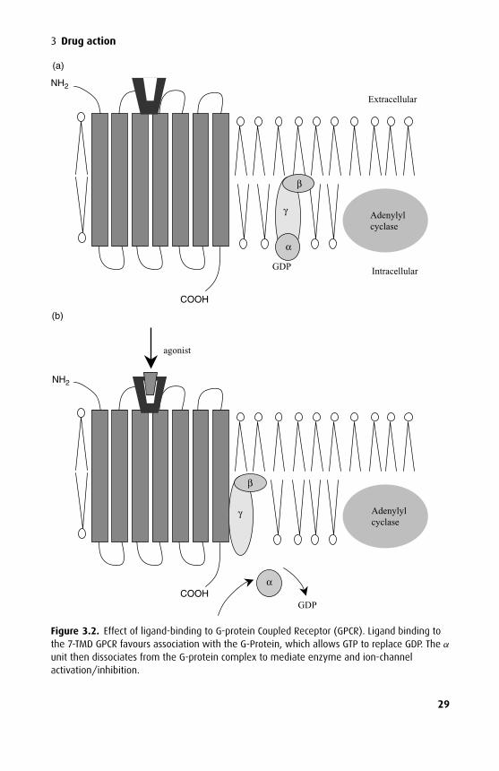
P1: PCX Printer: Yet To Come
9780521704632c03 CUFX213A/Peck 9780521618168 December 27, 2007 13:30
3 Drug action
TP
NH2
α
GDP COOH
γ
β
agonist
(b)
(a)
α
Adenylyl cyclase
GDP
COOH
γ
β
NH2
Extracellular
Intracellular
Adenylyl cyclase
Figure 3.2. Effect of ligand-binding to G-protein Coupled Receptor (GPCR). Ligand binding tothe 7-TMD GPCR favours association with the G-Protein, which allows GTP to replace GDP. The α
unit then dissociates from the G-protein complex to mediate enzyme and ion-channelactivation/inhibition.
29
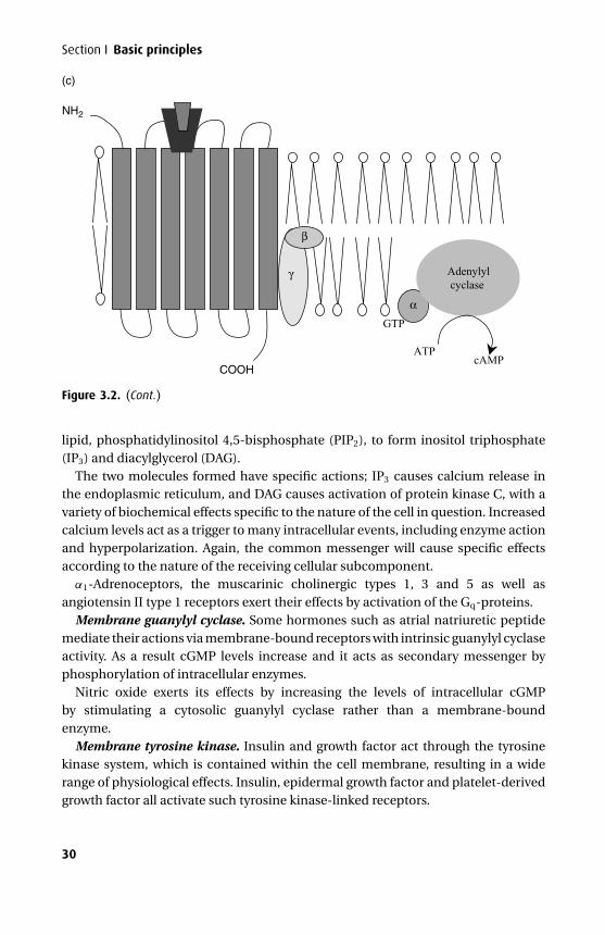
P1: PCX Printer: Yet To Come
9780521704632c03 CUFX213A/Peck 9780521618168 December 27, 2007 13:30
Section I Basic principles
GTP
ATP
NH2
α
Adenylyl cyclase
cAMP COOH
γ
β
(c)
Figure 3.2. (Cont.)
lipid, phosphatidylinositol 4,5-bisphosphate (PIP2), to form inositol triphosphate(IP3) and diacylglycerol (DAG).
The two molecules formed have specific actions; IP3 causes calcium release inthe endoplasmic reticulum, and DAG causes activation of protein kinase C, with avariety of biochemical effects specific to the nature of the cell in question. Increasedcalcium levels act as a trigger to many intracellular events, including enzyme actionand hyperpolarization. Again, the common messenger will cause specific effectsaccording to the nature of the receiving cellular subcomponent.
α1-Adrenoceptors, the muscarinic cholinergic types 1, 3 and 5 as well asangiotensin II type 1 receptors exert their effects by activation of the Gq-proteins.
Membrane guanylyl cyclase. Some hormones such as atrial natriuretic peptidemediate their actions via membrane-bound receptors with intrinsic guanylyl cyclaseactivity. As a result cGMP levels increase and it acts as secondary messenger byphosphorylation of intracellular enzymes.
Nitric oxide exerts its effects by increasing the levels of intracellular cGMPby stimulating a cytosolic guanylyl cyclase rather than a membrane-boundenzyme.
Membrane tyrosine kinase. Insulin and growth factor act through the tyrosinekinase system, which is contained within the cell membrane, resulting in a widerange of physiological effects. Insulin, epidermal growth factor and platelet-derivedgrowth factor all activate such tyrosine kinase-linked receptors.
30
P1: PCX Printer: Yet To Come
9780521704632c03 CUFX213A/Peck 9780521618168 December 27, 2007 13:30
3 Drug action
The insulin receptor consists of two α and two β-subunits, the latter span the cellmembrane. When a ligand binds to the α-subunits, intracellular tyrosine residueson its β-subunits are phosphorylated, so activating their tyrosine kinase activity. Theactivated enzyme catalyzes phosphorylation of other protein targets, which generatethe many effects of insulin. These effects include the intracellular metabolic effects,the insertion of glucose transport protein into the cell membrane as well as thoseactions involving gene transcription.
3) Regulation of gene transcriptionSteroids and thyroid hormones act through intracellular receptors to alter the expres-sion of DNA and RNA. They indirectly alter the production of cellular proteins sotheir effects are necessarily slow. These cytoplasmic receptors act as ligand-regulatedtranscription factors; they are normally held in an inactive form by association withinhibitory proteins. The binding of an appropriate hormone induces a conforma-tional change that activates the receptor and permits translation to the nucleolus,which leads to association with specific DNA promotor sequences and productionof mRNA.
Adrenosteroid hormonesThere are two types of corticosteroid receptor: the mineralocorticoid receptor, MR,and the glucocorticoid receptor, GR. The GR receptor is wide spread in cells, includ-ing the liver where corticosteroids alter the hepatic production of proteins duringstress to favour the so-called acute-phase reaction proteins. The MR is restrictedto epithelial tissue such as renal collecting tubules and colon, although these cellsalso contain GR receptors. Selective MR receptor activation occurs due to the pres-ence of 11-beta hydroxysteroid dehydrogenase, which converts cortisol to cortisone:cortisone is inactive at the GR receptor.
Other nuclear receptorsThe new antidiabetic drug rosiglitazone is an agonist at a nuclear receptor, perox-isome proliferator-activated receptor, which controls protein transcription associ-ated with increased sensitivity to insulin in adipose tissue.
Dynamics of drug–receptor bindingThe binding of a ligand (L) to its receptor (R) is represented by the equation:
L + R ↔ LR.
This reaction is reversible. The law of mass action states that the rate of a reactionis proportional to the concentrations of the reacting components. Thus, the velocityof the forward reaction is given by:
V1 = k1 · [L] · [R],
31
P1: PCX Printer: Yet To Come
9780521704632c03 CUFX213A/Peck 9780521618168 December 27, 2007 13:30
Section I Basic principles
where k1 is the rate constant for the forward reaction (square brackets indicate con-centration).
The velocity of the reverse reaction is given by:
V2 = k2 · [LR],
where k2 is the rate constant for the reverse reaction.At equilibrium, the reaction occurs at the same rate in both directions (V1 = V2),
and the equilibrium dissociation constant, KD, is given by the equation:
KD = [L] · [R]/[LR] = k2/k1.
Its reciprocal, KA, is the equilibrium association constant and is a reflection of thestrength of binding between the ligand and receptor. Note that these constants donot have the same units; the units for KD are mmol.l−1 whereas those for KA arel.mmol−1 (when reading pharmacology texts take careful note of which of these twoconstants are being described). It was Ariens who first suggested that response isproportional to receptor occupancy and this was the basis of pharmacodynamicmodelling. However, the situation is not as straightforward as this may imply and weneed to explain the existence of partial agonists and inverse agonists as well as thephenomenon of spare receptors.
Receptor proteins can exist in a number of conformations that are in equilib-rium, in particular the active and inactive forms; in the absence of an agonist theequilibrium favours the inactive form. Antagonists bind equally to both forms of thereceptor and do not alter the equilibrium. Agonists bind to the receptor and push thisequilibrium toward the active conformation. The active conformation then triggersthe series of molecular events that result in the observed effect.
Types of drug–receptor interactionThe two properties of a drug that determine the nature of its pharmacological effectare affinity and intrinsic activity.� Affinity refers to how well or avidly a drug binds to its receptor – in the analogy
of the lock and key, this is how well the key fits the lock. The avidity of binding isdetermined by the KD or KA of the drug.� Intrinsic activity (IA) or efficacy refers to the magnitude of effect the drug has oncebound; IA takes a value between 0 and 1, although inverse agonists can have an IAbetween −1 and 0.
It is important to distinguish these properties. A drug may have a high affinity, butno activity, and thus binding will produce no pharmacological response. If such adrug prevents the binding of a more active ligand, this ligand will be unable to exertits effect – so the drug is demonstrating receptor antagonism. However, if a drugbinds well but only induces a fractional response, never a full response, then the
32
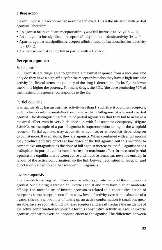
P1: PCX Printer: Yet To Come
9780521704632c03 CUFX213A/Peck 9780521618168 December 27, 2007 13:30
3 Drug action
maximum possible response can never be achieved. This is the situation with partialagonists. Therefore:� An agonist has significant receptor affinity and full intrinsic activity (IA = 1).� An antagonist has significant receptor affinity but no intrinsic activity (IA = 0).� A partial agonist has significant receptor affinity but only fractional intrinsic activity
(0 < IA <1).� An inverse agonist can be full or partial with −1 ≤ IA < 0.
Receptor agonism
Full agonistsFull agonists are drugs able to generate a maximal response from a receptor. Notonly do they have a high affinity for the receptor, but also they have a high intrinsicactivity. In clinical terms, the potency of the drug is determined by its KD; the lowerthe KD the higher the potency. For many drugs, the ED50 (the dose producing 50% ofthe maximum response) corresponds to the KD.
Partial agonistsIf an agonist drug has an intrinsic activity less than 1, such that it occupies receptors,but produces a submaximal effect compared with the full agonist, it is termed a partialagonist. The distinguishing feature of partial agonists is that they fail to achieve amaximal effect even in very high dose (i.e. with full receptor occupancy) (Figure3.3(c)C). An example of a partial agonist is buprenorphine acting at the µ-opioidreceptor. Partial agonists may act as either agonists or antagonists depending oncircumstances. If used alone, they are agonists. When combined with a full agonistthey produce additive effects at low doses of the full agonist, but this switches tocompetitive antagonism as the dose of full agonist increases; the full agonist needsto displace the partial agonist in order to restore maximum effect. In the case of partialagonists the equilibrium between active and inactive forms can never be entirely infavour of the active conformation, so the link between activation of receptor andeffect is only a fraction of that seen with full agonists.
Inverse agonistsIt is possible for a drug to bind and exert an effect opposite to that of the endogenousagonist. Such a drug is termed an inverse agonist and may have high or moderateaffinity. The mechanism of inverse agonism is related to a constitutive action ofreceptors; some receptors can show a low level of activity even in the absence of aligand, since the probability of taking up an active conformation is small but mea-surable. Inverse agonists bind to these receptors and greatly reduce the incidence ofthe active conformation responsible for this constitutive activity, as a result inverseagonists appear to exert an opposite effect to the agonist. The difference between
33
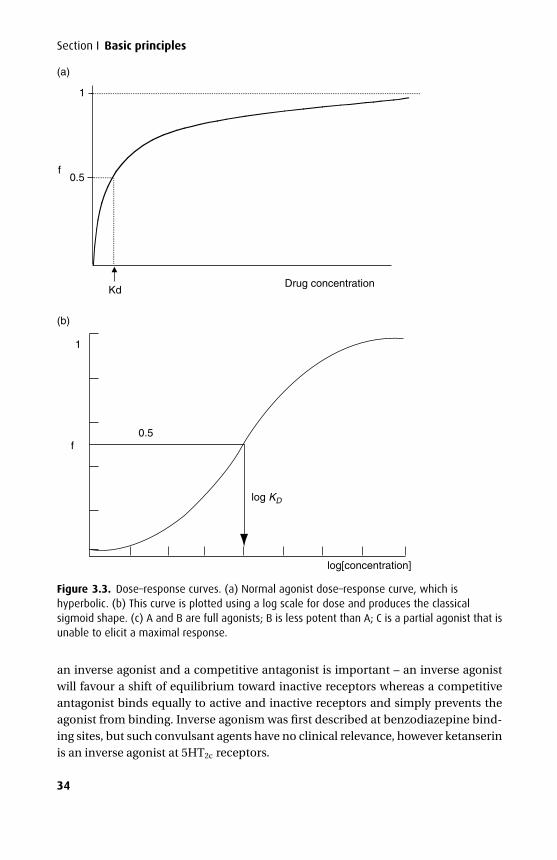
P1: PCX Printer: Yet To Come
9780521704632c03 CUFX213A/Peck 9780521618168 December 27, 2007 13:30
Section I Basic principles
1
0.5f
Drug concentrationKd
(a)
log[concentration]
1
f
(b)
0.5
log KD
Figure 3.3. Dose–response curves. (a) Normal agonist dose–response curve, which ishyperbolic. (b) This curve is plotted using a log scale for dose and produces the classicalsigmoid shape. (c) A and B are full agonists; B is less potent than A; C is a partial agonist that isunable to elicit a maximal response.
an inverse agonist and a competitive antagonist is important – an inverse agonistwill favour a shift of equilibrium toward inactive receptors whereas a competitiveantagonist binds equally to active and inactive receptors and simply prevents theagonist from binding. Inverse agonism was first described at benzodiazepine bind-ing sites, but such convulsant agents have no clinical relevance, however ketanserinis an inverse agonist at 5HT2c receptors.
34
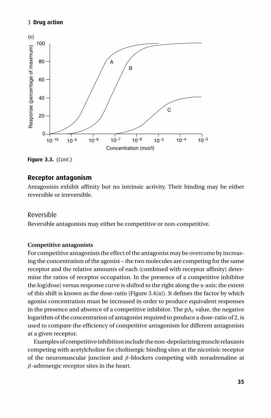
P1: PCX Printer: Yet To Come
9780521704632c03 CUFX213A/Peck 9780521618168 December 27, 2007 13:30
3 Drug action
(c)
A
100
80
60
40
20
010−10 10−9 10−8 10−7 10−6 10−5 10−4 10−3
Concentration (mol/l)
Res
pons
e (p
erce
ntag
e of
max
imum
)
B
C
Figure 3.3. (Cont.)
Receptor antagonismAntagonists exhibit affinity but no intrinsic activity. Their binding may be eitherreversible or irreversible.
ReversibleReversible antagonists may either be competitive or non-competitive.
Competitive antagonistsFor competitive antagonists the effect of the antagonist may be overcome by increas-ing the concentration of the agonist – the two molecules are competing for the samereceptor and the relative amounts of each (combined with receptor affinity) deter-mine the ratios of receptor occupation. In the presence of a competitive inhibitorthe log[dose] versus response curve is shifted to the right along the x-axis; the extentof this shift is known as the dose-ratio (Figure 3.4(a)). It defines the factor by whichagonist concentration must be increased in order to produce equivalent responsesin the presence and absence of a competitive inhibitor. The pA2 value, the negativelogarithm of the concentration of antagonist required to produce a dose-ratio of 2, isused to compare the efficiency of competitive antagonism for different antagonistsat a given receptor.
Examples of competitive inhibition include the non-depolarizing muscle relaxantscompeting with acetylcholine for cholinergic binding sites at the nicotinic receptorof the neuromuscular junction and β-blockers competing with noradrenaline atβ-adrenergic receptor sites in the heart.
35
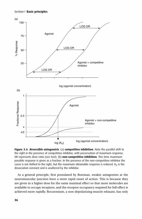
P1: PCX Printer: Yet To Come
9780521704632c03 CUFX213A/Peck 9780521618168 December 27, 2007 13:30
Section I Basic principles
x/2
Fra
ctio
nal R
espo
nse
1
0.5
Agonist
Agonist + non-competitive inhibitor
log (agonist concentration)log (KD)
x
(b)
Agonist
LOG DR
LOG DR
% R
espo
nse
100
75
50
25
LOG DR
log (agonist concentration)
Agonist + competitive inhibitor
(a)
Figure 3.4. Reversible antagonists. (a) competitive inhibition. Note the parallel shift tothe right in the presence of competitive inhibitor, with preservation of maximum response.DR represents dose-ratio (see text). (b) non-competitive inhibition. This time maximumpossible response is given as a fraction. In the presence of the non-competitive inhibitor thecurve is not shifted to the right, but the maximum obtainable response is reduced. KD is thedissociation constant and is unaltered by the inhibitor.
As a general principle, first postulated by Bowman, weaker antagonists at theneuromuscular junction have a more rapid onset of action. This is because theyare given in a higher dose for the same maximal effect so that more molecules areavailable to occupy receptors, and the receptor occupancy required for full effect isachieved more rapidly. Rocuronium, a non-depolarizing muscle relaxant, has only
36
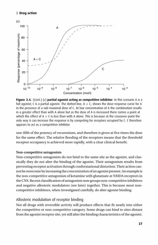
P1: PCX Printer: Yet To Come
9780521704632c03 CUFX213A/Peck 9780521618168 December 27, 2007 13:30
3 Drug action
(c)
A + C
C
A100
80
20
0
10−10 10−9 10−8 10−7 10−6 10−5 10−4 10−3
Concentration (mol/l)
Res
pons
e (p
erce
ntag
e of
max
imum
)
60
40
Figure 3.4. (Cont.) (c) partial agonist acting as competitive inhibitor. In this scenario A is afull agonist; C is a partial agonist. The dotted line, A + C, shows the dose-response curve for Ain the presence of a sub-maximal dose of C. At low concentration of A the combination resultsin a greater effect than with A alone but as the dose of A is increased there comes a point atwhich the effect of A + C is less than with A alone. This is because at the crossover point theonly way A can increase the response is by competing for receptors occupied by C. C thereforeappears to act as a competitive inhibitor.
one-fifth of the potency of vecuronium, and therefore is given at five times the dosefor the same effect. The relative flooding of the receptors means that the thresholdreceptor occupancy is achieved more rapidly, with a clear clinical benefit.
Non-competitive antagonistsNon-competitive antagonists do not bind to the same site as the agonist, and clas-sically they do not alter the binding of the agonist. Their antagonism results frompreventing receptor activation through conformational distortion. Their action can-not be overcome by increasing the concentration of an agonist present. An example isthe non-competitive antagonism of ketamine with glutamate at NMDA receptors inthe CNS. Recent classification of antagonists now groups non-competitive inhibitorsand negative allosteric modulators (see later) together. This is because most non-competitive inhibitors, when investigated carefully, do alter agonist binding.
Allosteric modulation of receptor bindingNot all drugs with reversible activity will produce effects that fit neatly into eitherthe competitive or non-competitive category. Some drugs can bind to sites distantfrom the agonist receptor site, yet still alter the binding characteristics of the agonist.
37
P1: PCX Printer: Yet To Come
9780521704632c03 CUFX213A/Peck 9780521618168 December 27, 2007 13:30
Section I Basic principles
Such drugs are called allosteric modulators and may either reduce (negative allostericmodulators) or enhance (positive allosteric modulators) the activity of a given dose ofagonist without having any discernible effects of their own. An example of a positiveallosteric modulator is the effect of benzodiazepines on the activity of GABA at theGABAA receptor complex.
IrreversibleIrreversible antagonists may either bind irreversibly to the same site as the ago-nist or at a distant site. Whatever the nature of the binding site, increasing agonistconcentration will not overcome the blockade. A clinical example is that of phenoxy-benzamine that irreversibly binds to and antagonizes the effects of catecholaminesat α-adrenoceptors. Aspirin is also an irreversible inhibitor; it causes cross-linkingof the COX-1 enzyme in platelets rendering it inactive for the lifetime of the platelet.A single 75 mg dose is sufficient to block all platelet COX-1 and so return of normalfunction requires new platelet formation. This is why aspirin should be stopped 7–10days before surgery where surgical bleeding must be minimized.
Spare receptorsThe neuromuscular junction contains ACh receptors, which when occupied by AChcause depolarization of the motor end plate. However, only a fraction of these recep-tors needs to be occupied to produce a maximal pharmacological effect. Occupancyof only a small proportion of the receptors ensures that a small quantity of AChproduces a maximal response. As a result there are ‘spare receptors’, which providesome protection against failure of transmission in the presence of toxins. This canbe demonstrated as follows: if a small dose of an irreversible inhibitor is given then aproportion of the receptors are bound by the inhibitor and rendered unavailable sothe log[dose] versus response curve for acetylcholine is shifted to the right becausea higher fraction of the remaining receptors must be occupied to produce the orig-inal response. If a further irreversible antagonist is used more receptors are madeunavailable and results in a further right-shift of the log[dose] versus response curve.When more than three-quarters of the receptors are occupied by irreversible antag-onist then whatever the dose of ACh, a maximum response cannot occur and theshape of the curve changes so that both the maximum response and the slope isreduced.
Tachyphylaxis, desensitization and toleranceRepeated doses of a drug may lead to a change in the pharmacological response,which may be increased or decreased for the same dose.
TachyphylaxisTachyphylaxis is defined as a rapid decrease in response to repeated doses over a shorttime period. The most common mechanism is the decrease of stores of a transmitter
38
P1: PCX Printer: Yet To Come
9780521704632c03 CUFX213A/Peck 9780521618168 December 27, 2007 13:30
3 Drug action
before resynthesis can take place. An example is the diminishing response to repeateddoses of ephedrine, an indirectly acting sympathomimetic amine, caused by thedepletion of noradrenaline.
DesensitizationDesensitization refers to a chronic loss of response over a longer period and maybe caused by a structural change in receptor morphology or by an absolute lossof receptor numbers. The term is often used synonymously with tachyphylaxis. Anexample is the loss of β-adrenergic receptors from the myocardial cell surface in thecontinued presence of adrenaline and dobutamine.
ToleranceTolerance refers to the phenomenon whereby larger doses are required to producethe same pharmacological effect, such as occurs in chronic opioid use or abuse. Thisreflects an altered sensitivity of the receptors of the central nervous system to opioids– the mechanism may be a reduction of receptor density or a reduction of receptoraffinity. Tolerance occurs if nitrates are given by continuous infusion for prolongedperiods as the sulphydryl groups on vascular smooth muscle become depleted. Adrug holiday of a few hours overnight when the need for vasodilatation is likely tobe at its lowest allows replenishment of the sulphydryl groups and restoration of thepharmacological effect.
39
P1: PCX Printer: Yet To Come
9780521704632c04 CUFX213A/Peck 9780521618168 December 27, 2007 13:35
4Drug interaction
Interactions occur when one drug modifies the action of another. This interactionmay either increase or decrease the second drug’s action. Sometimes these inter-actions result in unwanted effects, but some interactions are beneficial and can beexploited therapeutically.
Drug interaction can be described as physicochemical, relating to the propertiesof the drug or its pharmaceutical preparation, pharmacokinetic due to alterations inthe way the body handles the drug or pharmacodynamic where the activity of onedrug is affected. The chance of a significant interaction increases markedly with thenumber of drugs used and the effects of any interaction are often exaggerated in thepresence of disease or coexisting morbidity.
About one in six inpatient drug charts contain a significant drug interaction, one-third of which are potentially serious. An uncomplicated general anaesthetic for arelatively routine case may use ten or more different agents that may interact withone another or, more commonly, with the patient’s concurrent medication.
PharmaceuticalThese interactions occur because of a chemical or physical incompatibility betweenthe preparations being used. Sodium bicarbonate and calcium will precipitate outof solution as calcium carbonate when co-administered in the same giving set. How-ever, one agent may inactivate another without such an overt indication to theobserver; insulin may be denatured if prepared in solutions of dextrose and may,therefore, lose its pharmacological effect. Drugs also may react with the giving setor syringe and therefore need special equipment for delivery, such as a glass syringefor paraldehyde administration. Glyceryl trinitrate is absorbed by polyvinyl chloride;therefore, special polyethylene administration sets are preferred.
Pharmacokinetic
AbsorptionIn the case of drugs given orally, this occurs either as a result of one drug bind-ing another in the lumen of the gastrointestinal tract or by altering the functionof the gastrointestinal tract as a whole. Charcoal can adsorb drugs in the stomach,preventing absorption through the gastrointestinal tract (charcoal is activated bysteam to cause fissuring, thereby greatly increasing the surface area for adsorption).
40
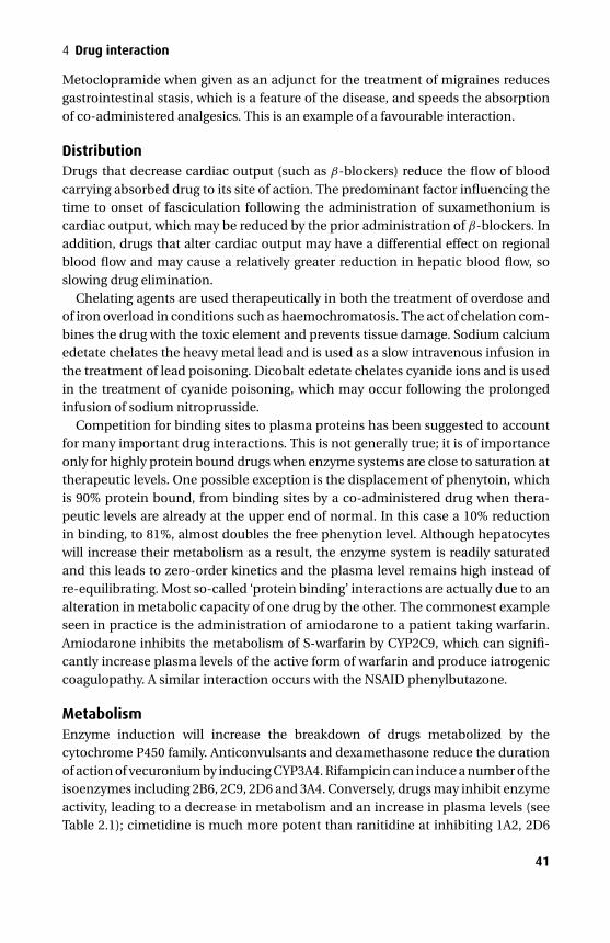
P1: PCX Printer: Yet To Come
9780521704632c04 CUFX213A/Peck 9780521618168 December 27, 2007 13:35
4 Drug interaction
Metoclopramide when given as an adjunct for the treatment of migraines reducesgastrointestinal stasis, which is a feature of the disease, and speeds the absorptionof co-administered analgesics. This is an example of a favourable interaction.
DistributionDrugs that decrease cardiac output (such as β-blockers) reduce the flow of bloodcarrying absorbed drug to its site of action. The predominant factor influencing thetime to onset of fasciculation following the administration of suxamethonium iscardiac output, which may be reduced by the prior administration of β-blockers. Inaddition, drugs that alter cardiac output may have a differential effect on regionalblood flow and may cause a relatively greater reduction in hepatic blood flow, soslowing drug elimination.
Chelating agents are used therapeutically in both the treatment of overdose andof iron overload in conditions such as haemochromatosis. The act of chelation com-bines the drug with the toxic element and prevents tissue damage. Sodium calciumedetate chelates the heavy metal lead and is used as a slow intravenous infusion inthe treatment of lead poisoning. Dicobalt edetate chelates cyanide ions and is usedin the treatment of cyanide poisoning, which may occur following the prolongedinfusion of sodium nitroprusside.
Competition for binding sites to plasma proteins has been suggested to accountfor many important drug interactions. This is not generally true; it is of importanceonly for highly protein bound drugs when enzyme systems are close to saturation attherapeutic levels. One possible exception is the displacement of phenytoin, whichis 90% protein bound, from binding sites by a co-administered drug when thera-peutic levels are already at the upper end of normal. In this case a 10% reductionin binding, to 81%, almost doubles the free phenytion level. Although hepatocyteswill increase their metabolism as a result, the enzyme system is readily saturatedand this leads to zero-order kinetics and the plasma level remains high instead ofre-equilibrating. Most so-called ‘protein binding’ interactions are actually due to analteration in metabolic capacity of one drug by the other. The commonest exampleseen in practice is the administration of amiodarone to a patient taking warfarin.Amiodarone inhibits the metabolism of S-warfarin by CYP2C9, which can signifi-cantly increase plasma levels of the active form of warfarin and produce iatrogeniccoagulopathy. A similar interaction occurs with the NSAID phenylbutazone.
MetabolismEnzyme induction will increase the breakdown of drugs metabolized by thecytochrome P450 family. Anticonvulsants and dexamethasone reduce the durationof action of vecuronium by inducing CYP3A4. Rifampicin can induce a number of theisoenzymes including 2B6, 2C9, 2D6 and 3A4. Conversely, drugs may inhibit enzymeactivity, leading to a decrease in metabolism and an increase in plasma levels (seeTable 2.1); cimetidine is much more potent than ranitidine at inhibiting 1A2, 2D6
41
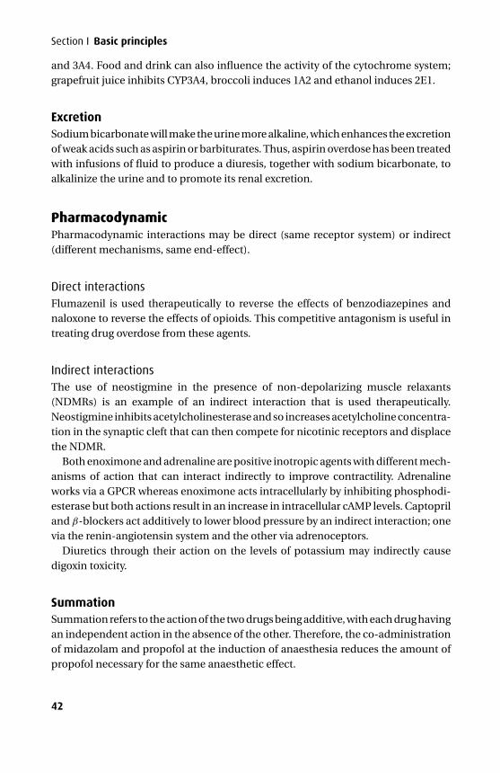
P1: PCX Printer: Yet To Come
9780521704632c04 CUFX213A/Peck 9780521618168 December 27, 2007 13:35
Section I Basic principles
and 3A4. Food and drink can also influence the activity of the cytochrome system;grapefruit juice inhibits CYP3A4, broccoli induces 1A2 and ethanol induces 2E1.
ExcretionSodium bicarbonate will make the urine more alkaline, which enhances the excretionof weak acids such as aspirin or barbiturates. Thus, aspirin overdose has been treatedwith infusions of fluid to produce a diuresis, together with sodium bicarbonate, toalkalinize the urine and to promote its renal excretion.
PharmacodynamicPharmacodynamic interactions may be direct (same receptor system) or indirect(different mechanisms, same end-effect).
Direct interactionsFlumazenil is used therapeutically to reverse the effects of benzodiazepines andnaloxone to reverse the effects of opioids. This competitive antagonism is useful intreating drug overdose from these agents.
Indirect interactionsThe use of neostigmine in the presence of non-depolarizing muscle relaxants(NDMRs) is an example of an indirect interaction that is used therapeutically.Neostigmine inhibits acetylcholinesterase and so increases acetylcholine concentra-tion in the synaptic cleft that can then compete for nicotinic receptors and displacethe NDMR.
Both enoximone and adrenaline are positive inotropic agents with different mech-anisms of action that can interact indirectly to improve contractility. Adrenalineworks via a GPCR whereas enoximone acts intracellularly by inhibiting phosphodi-esterase but both actions result in an increase in intracellular cAMP levels. Captopriland β-blockers act additively to lower blood pressure by an indirect interaction; onevia the renin-angiotensin system and the other via adrenoceptors.
Diuretics through their action on the levels of potassium may indirectly causedigoxin toxicity.
SummationSummation refers to the action of the two drugs being additive, with each drug havingan independent action in the absence of the other. Therefore, the co-administrationof midazolam and propofol at the induction of anaesthesia reduces the amount ofpropofol necessary for the same anaesthetic effect.
42
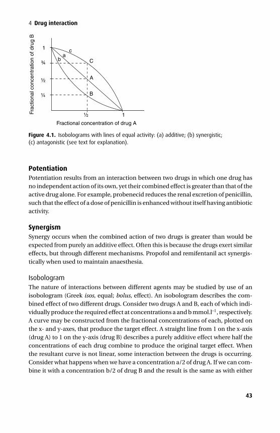
P1: PCX Printer: Yet To Come
9780521704632c04 CUFX213A/Peck 9780521618168 December 27, 2007 13:35
4 Drug interaction
c1a
b
B
Frac
tiona
l con
cent
ratio
n of
dru
g B
Fractional concentration of drug A
1
A
C
Figure 4.1. Isobolograms with lines of equal activity: (a) additive; (b) synergistic;(c) antagonistic (see text for explanation).
PotentiationPotentiation results from an interaction between two drugs in which one drug hasno independent action of its own, yet their combined effect is greater than that of theactive drug alone. For example, probenecid reduces the renal excretion of penicillin,such that the effect of a dose of penicillin is enhanced without itself having antibioticactivity.
SynergismSynergy occurs when the combined action of two drugs is greater than would beexpected from purely an additive effect. Often this is because the drugs exert similareffects, but through different mechanisms. Propofol and remifentanil act synergis-tically when used to maintain anaesthesia.
IsobologramThe nature of interactions between different agents may be studied by use of anisobologram (Greek isos, equal; bolus, effect). An isobologram describes the com-bined effect of two different drugs. Consider two drugs A and B, each of which indi-vidually produce the required effect at concentrations a and b mmol.l–1, respectively.A curve may be constructed from the fractional concentrations of each, plotted onthe x- and y-axes, that produce the target effect. A straight line from 1 on the x-axis(drug A) to 1 on the y-axis (drug B) describes a purely additive effect where half theconcentrations of each drug combine to produce the original target effect. Whenthe resultant curve is not linear, some interaction between the drugs is occurring.Consider what happens when we have a concentration a/2 of drug A. If we can com-bine it with a concentration b/2 of drug B and the result is the same as with either
43
P1: PCX Printer: Yet To Come
9780521704632c04 CUFX213A/Peck 9780521618168 December 27, 2007 13:35
Section I Basic principles
drug alone, then the point A ( 12 , 1
2 ) can be marked, and there is a straight line indi-cating additivity. If however we need to combine a concentration a/2 of drug A withonly a concentration b/4 of drug B to elicit the same response, the point ( 1
2 , 14 ) can
be marked, which describes a curve that is concave, suggesting synergism betweendrugs A and B. Similarly, if we needed a concentration greater than b/2 of drug B thecurve would be convex, indicating some antagonistic interaction between the twodrugs point C ( 1
2 , 34 ) (Figure 4.1).
44
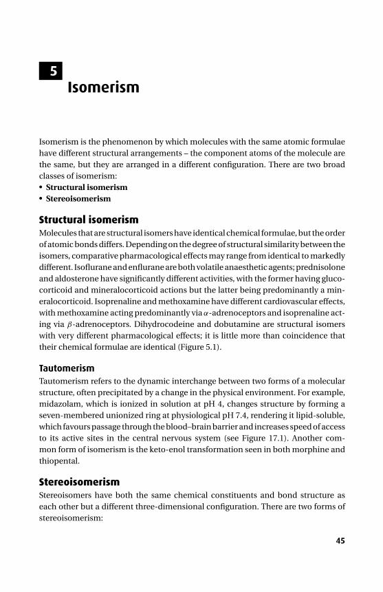
P1: PCX Printer: Yet To Come
9780521704632c05 CUFX213A/Peck 9780521618168 December 27, 2007 13:38
5Isomerism
Isomerism is the phenomenon by which molecules with the same atomic formulaehave different structural arrangements – the component atoms of the molecule arethe same, but they are arranged in a different configuration. There are two broadclasses of isomerism:� Structural isomerism� Stereoisomerism
Structural isomerismMolecules that are structural isomers have identical chemical formulae, but the orderof atomic bonds differs. Depending on the degree of structural similarity between theisomers, comparative pharmacological effects may range from identical to markedlydifferent. Isoflurane and enflurane are both volatile anaesthetic agents; prednisoloneand aldosterone have significantly different activities, with the former having gluco-corticoid and mineralocorticoid actions but the latter being predominantly a min-eralocorticoid. Isoprenaline and methoxamine have different cardiovascular effects,with methoxamine acting predominantly via α-adrenoceptors and isoprenaline act-ing via β-adrenoceptors. Dihydrocodeine and dobutamine are structural isomerswith very different pharmacological effects; it is little more than coincidence thattheir chemical formulae are identical (Figure 5.1).
TautomerismTautomerism refers to the dynamic interchange between two forms of a molecularstructure, often precipitated by a change in the physical environment. For example,midazolam, which is ionized in solution at pH 4, changes structure by forming aseven-membered unionized ring at physiological pH 7.4, rendering it lipid-soluble,which favours passage through the blood–brain barrier and increases speed of accessto its active sites in the central nervous system (see Figure 17.1). Another com-mon form of isomerism is the keto-enol transformation seen in both morphine andthiopental.
StereoisomerismStereoisomers have both the same chemical constituents and bond structure aseach other but a different three-dimensional configuration. There are two forms ofstereoisomerism:
45
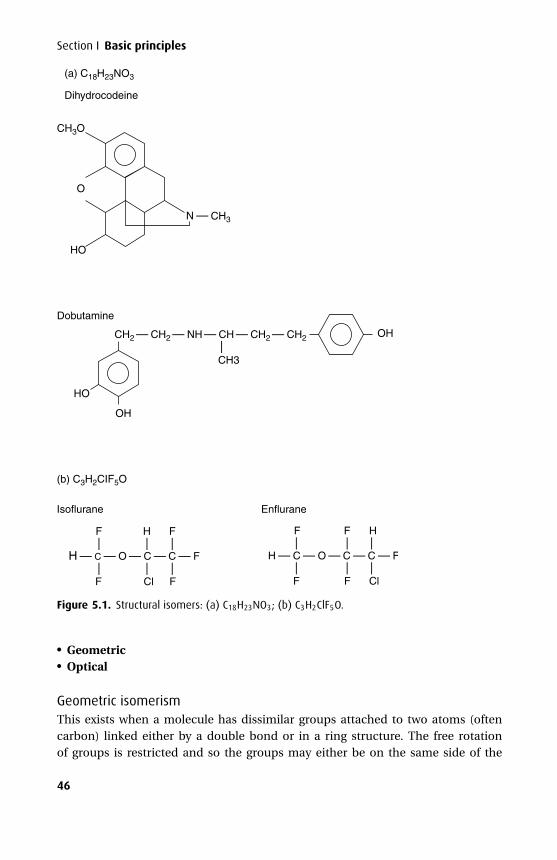
P1: PCX Printer: Yet To Come
9780521704632c05 CUFX213A/Peck 9780521618168 December 27, 2007 13:38
Section I Basic principles
CH2 — CH2 — NH — CH — CH2 — CH2
—
CH3
HO
Dobutamine
OH
OH
N —
CH3O
(a) C18H23NO3
Dihydrocodeine
HO
O
——
F
F
(b) C3H2CIF5O
EnfluraneIsoflurane
H — C — O — C — C — F
——
Cl
H
——
F
F
——
F
F
H — C — O — C — C — F
——
F
F
——
Cl
H
CH3
Figure 5.1. Structural isomers: (a) C18H23NO3; (b) C3H2ClF5O.
� Geometric� Optical
Geometric isomerismThis exists when a molecule has dissimilar groups attached to two atoms (oftencarbon) linked either by a double bond or in a ring structure. The free rotationof groups is restricted and so the groups may either be on the same side of the
46
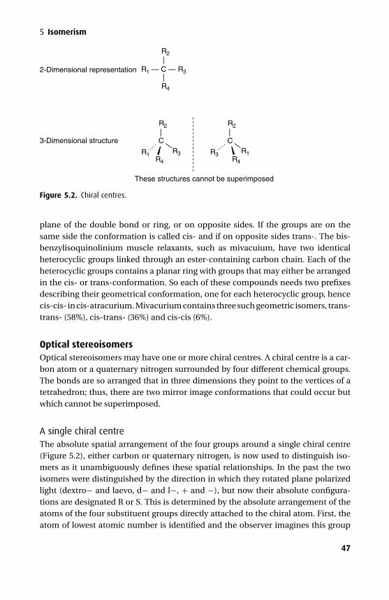
P1: PCX Printer: Yet To Come
9780521704632c05 CUFX213A/Peck 9780521618168 December 27, 2007 13:38
5 Isomerism
——
R4
R2
R1 — C — R3
—
—R4
R1 R3
R2
2-Dimensional representation
3-Dimensional structure
These structures cannot be superimposed
—
—
R4
R3 R1
R2
CC
Figure 5.2. Chiral centres.
plane of the double bond or ring, or on opposite sides. If the groups are on thesame side the conformation is called cis- and if on opposite sides trans-. The bis-benzylisoquinolinium muscle relaxants, such as mivacuium, have two identicalheterocyclic groups linked through an ester-containing carbon chain. Each of theheterocyclic groups contains a planar ring with groups that may either be arrangedin the cis- or trans-conformation. So each of these compounds needs two prefixesdescribing their geometrical conformation, one for each heterocyclic group, hencecis-cis- in cis-atracurium. Mivacurium contains three such geometric isomers, trans-trans- (58%), cis-trans- (36%) and cis-cis (6%).
Optical stereoisomersOptical stereoisomers may have one or more chiral centres. A chiral centre is a car-bon atom or a quaternary nitrogen surrounded by four different chemical groups.The bonds are so arranged that in three dimensions they point to the vertices of atetrahedron; thus, there are two mirror image conformations that could occur butwhich cannot be superimposed.
A single chiral centreThe absolute spatial arrangement of the four groups around a single chiral centre(Figure 5.2), either carbon or quaternary nitrogen, is now used to distinguish iso-mers as it unambiguously defines these spatial relationships. In the past the twoisomers were distinguished by the direction in which they rotated plane polarizedlight (dextro− and laevo, d− and l−, + and −), but now their absolute configura-tions are designated R or S. This is determined by the absolute arrangement of theatoms of the four substituent groups directly attached to the chiral atom. First, theatom of lowest atomic number is identified and the observer imagines this group
47
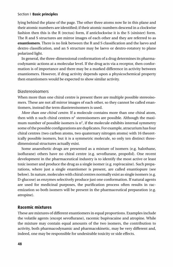
P1: PCX Printer: Yet To Come
9780521704632c05 CUFX213A/Peck 9780521618168 December 27, 2007 13:38
Section I Basic principles
lying behind the plane of the page. The other three atoms now lie in this plane andtheir atomic numbers are identified; if their atomic numbers descend in a clockwisefashion then this is the R (rectus) form, if anticlockwise it is the S (sinister) form.The R and S structures are mirror images of each other and they are referred to asenantiomers. There is no link between the R and S classification and the laevo anddextro classification, and an S structure may be laevo or dextro-rotatory to planepolarized light.
In general, the three-dimensional conformation of a drug determines its pharma-codynamic actions at a molecular level. If the drug acts via a receptor, then confor-mation is of importance and there may be a marked difference in activity betweenenantiomers. However, if drug activity depends upon a physicochemical propertythen enantiomers would be expected to show similar activity.
DiastereoisomersWhen more than one chiral centre is present there are multiple possible stereoiso-mers. These are not all mirror images of each other, so they cannot be called enan-tiomers, instead the term diastereoisomers is used.
More than one chiral centre. If a molecule contains more than one chiral atom,then with n such chiral centres n2 stereoisomers are possible. Although the maxi-mum number of possible isomers is n2, if the molecule exhibits internal symmetrysome of the possible configurations are duplicates. For example, atracurium has fourchiral centres (two carbon atoms, two quaternary nitrogen atoms) with 16 theoret-ically possible isomers, but it is a symmetric molecule, so only ten distinct three-dimensional structures actually exist.
Some anaesthetic drugs are presented as a mixture of isomers (e.g. halothane,isoflurane) others have no chiral centre (e.g. sevoflurane, propofol). One recentdevelopment in the pharmaceutical industry is to identify the most active or leasttoxic isomer and produce the drug as a single isomer (e.g. ropivacaine). Such prepa-rations, where just a single enantiomer is present, are called enantiopure (seebelow). In nature, molecules with chiral centres normally exist as single isomers (e.g.D-glucose) as enzymes selectively produce just one conformation. If natural agentsare used for medicinal purposes, the purification process often results in rac-emization so both isomers will be present in the pharmaceutical preparation (e.g.atropine).
Racemic mixturesThese are mixtures of different enantiomers in equal proportions. Examples includethe volatile agents (except sevoflurane), racemic bupivacaine and atropine. Whilethe mixture may contain equal amounts of the two isomers, the contribution toactivity, both pharmacodynamic and pharmacokinetic, may be very different and,indeed, one may be responsible for undesirable toxicity or side effects.
48
P1: PCX Printer: Yet To Come
9780521704632c05 CUFX213A/Peck 9780521618168 December 27, 2007 13:38
5 Isomerism
Enantiopure preparationsThere may be advantages in selecting the more desirable moiety from a racemicmixture and producing it as a single isomer, known as an enantiopure preparation.The amide local anaesthetics of the mepivacaine series have enantiomeric forms.The R-form has a more toxic profile than the S-form so that both ropivacaine andbupivacaine are now available as enantiopure preparations – S-ropivacaine and S-bupivacaine. Other enantiopure preparations such as S-ketamine, dexketoprofenand dexmedetomidine are also available.
49
P1: PCX Printer: Yet To Come
9780521704632c06 CUFX213A/Peck 9780521618168 December 27, 2007 14:11
6Mathematics and pharmacokinetics
Pharmacokinetics is the study of the way in which the body handles administereddrugs. The use of mathematical models allows us to predict how plasma concentra-tion changes with time when the dose and interval between doses are changed, orwhen infusions of a drug are used. Because there is an association between plasmaconcentration of a drug and its pharmacodynamic effect, models allow us to predictthe extent and duration of clinical effects. Mathematical models may therefore beused to program computers to deliver a variable rate infusion to achieve a predeter-mined plasma level and hence a desired therapeutic effect.
It should be remembered that these pharmacokinetic models make a number ofassumptions. Compartmental models make general assumptions based on virtualvolumes without attempting to model ‘real-life’ volumes such as plasma or extra-cellular fluid volumes. Therefore, although convenient and useful to associate thevirtual compartments with various tissue groups such as ‘well perfused’ or ‘poorlyperfused’, this remains only an approximation of the physiological state.
MathematicsCompartmental models are mathematical equations used to predict plasma con-centrations of drugs based on experimental observations. Mathematical functionsof importance in the understanding of these models are linear, logarithmic and expo-nential functions. Predicted behaviour and calculation of the parameters that definethe model require manipulation of exponential functions, the logarithmic functionand calculus. The following sections will cover all these concepts, starting with func-tions, particularly the exponential function, logarithms and finally calculus. In eachsection we will relate these to their use in pharmacokinetics, particularly the simpleone-compartment model.
FunctionsA function defines a unique value for y given a value for x. We write this as:
y = f(x).
For pharmacokinetics, we are interested in drug concentration (C) as a function oftime (t). We can write:
C = f(t).
50
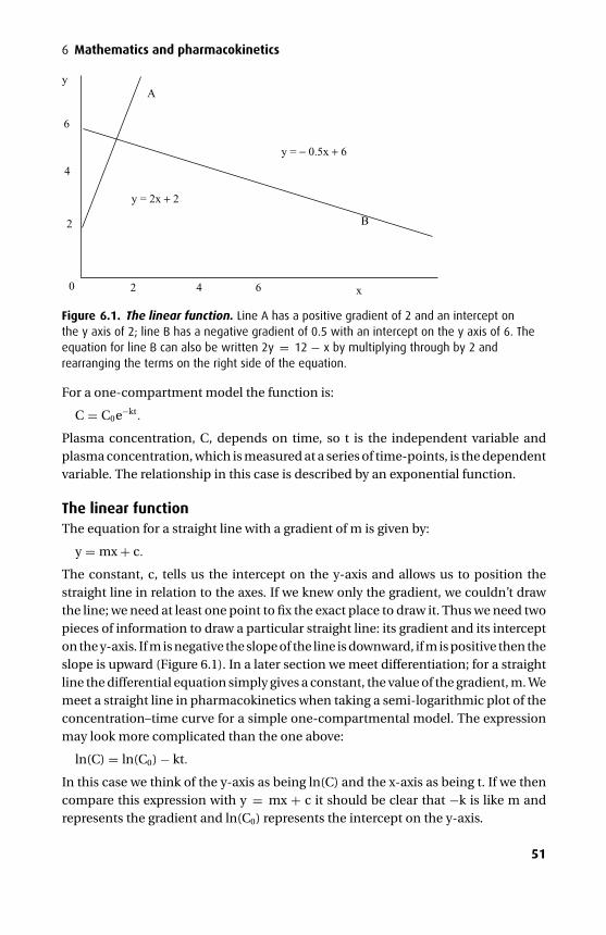
P1: PCX Printer: Yet To Come
9780521704632c06 CUFX213A/Peck 9780521618168 December 27, 2007 14:11
6 Mathematics and pharmacokinetics
y = 2x + 2
2
6
4
y = − 0.5x + 6
x
yA
B
2 4 60
Figure 6.1. The linear function. Line A has a positive gradient of 2 and an intercept onthe y axis of 2; line B has a negative gradient of 0.5 with an intercept on the y axis of 6. Theequation for line B can also be written 2y = 12 − x by multiplying through by 2 andrearranging the terms on the right side of the equation.
For a one-compartment model the function is:
C = C0e−kt.
Plasma concentration, C, depends on time, so t is the independent variable andplasma concentration, which is measured at a series of time-points, is the dependentvariable. The relationship in this case is described by an exponential function.
The linear functionThe equation for a straight line with a gradient of m is given by:
y = mx + c.
The constant, c, tells us the intercept on the y-axis and allows us to position thestraight line in relation to the axes. If we knew only the gradient, we couldn’t drawthe line; we need at least one point to fix the exact place to draw it. Thus we need twopieces of information to draw a particular straight line: its gradient and its intercepton the y-axis. If m is negative the slope of the line is downward, if m is positive then theslope is upward (Figure 6.1). In a later section we meet differentiation; for a straightline the differential equation simply gives a constant, the value of the gradient, m. Wemeet a straight line in pharmacokinetics when taking a semi-logarithmic plot of theconcentration–time curve for a simple one-compartmental model. The expressionmay look more complicated than the one above:
ln(C) = ln(C0) − kt.
In this case we think of the y-axis as being ln(C) and the x-axis as being t. If we thencompare this expression with y = mx + c it should be clear that −k is like m andrepresents the gradient and ln(C0) represents the intercept on the y-axis.
51
P1: PCX Printer: Yet To Come
9780521704632c06 CUFX213A/Peck 9780521618168 December 27, 2007 14:11
Section I Basic principles
The exponential functionAn exponential function takes the form:
y = Anax.
In this relationship n is the base and x the exponent; A and a are constants. Althoughit is possible to use any base for our exponential function, the natural number eis chosen for its mathematical properties. The exponential function, y = ex, is theonly function that integrates and differentiates to itself, making manipulation ofrelationships involving exponentials much easier than if another base were chosen.The number e is irrational, it cannot be expressed as a fraction, and takes the value2.716 . . . where there is an infinite number of digits following the decimal point.Exponentials are positive if the rate of change of y increases or negative if the rateof change decreases as x increases; in the example above ‘a’ is positive for a posi-tive exponential and negative for a negative exponential. Bacterial cell growth is anexample of a positive exponential relationship between the number of bacteria andtime; compound interest is a further example relating to the growth of an investmentwith time. For a negative exponential we write:
y = Be−bx.
For the two examples above, A and B are constants that relate to the intercept on the y-axis and a and b are rate constants that determine how steep the exponential curve is.In pharmacokinetics, we consider time, t, to be our independent variable (equivalentto x) and plasma concentration, C, to be the dependent variable (equivalent to y).The simple wash-out curve (where plasma concentration declines and approacheszero) describes a negative exponential. This equation was given above:
C = C0e−kt.
The ‘steepness’ of the wash-out curve depends on the rate constant, k. If k is halvedand everything else remained constant, then the plasma concentration would taketwice as long to reach any given level; if k is doubled then the time for the plasmaconcentration to reach a given level would halve. The wash-in curve also describes anegative exponential; although plasma concentration increases with time, the rate atwhich it approaches its maximum value is decreasing with time, making it a negativeexponential (Figure 6.2).
Asymptote. Theoretically, a negative exponential process approaches its steady-state value evermore closely without actually reaching it. This steady-state value istermed the asymptote. For the wash-out curve this asymptote is zero; for the wash-incurve during a constant infusion it is the steady-state concentration reached, whichis determined by the infusion rate and the clearance of the drug (Figure 6.2). If weconsider the wash-out curve, after one half-life plasma concentration has fallen by50%, after two half-lives it has fallen another (50/2)%, that is, a further 25% andafter five half-lives the process will be 50 + 25 + 12.5 + 6.25 + 3.125 = 96.875%complete, thus in practice five half-lives represents the approximate time needed to
52
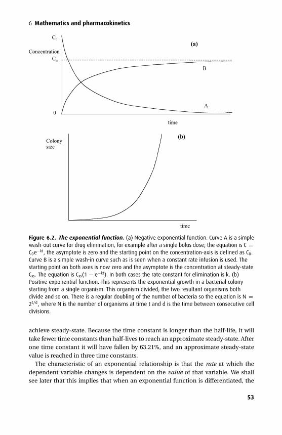
P1: PCX Printer: Yet To Come
9780521704632c06 CUFX213A/Peck 9780521618168 December 27, 2007 14:11
6 Mathematics and pharmacokinetics
Concentration
C0
time
A
Css
B
0
Colonysize
time
(a)
(b)
Figure 6.2. The exponential function. (a) Negative exponential function. Curve A is a simplewash-out curve for drug elimination, for example after a single bolus dose; the equation is C =C0e−kt, the asymptote is zero and the starting point on the concentration-axis is defined as C0.Curve B is a simple wash-in curve such as is seen when a constant rate infusion is used. Thestarting point on both axes is now zero and the asymptote is the concentration at steady-stateCss. The equation is Css(1 − e−kt). In both cases the rate constant for elimination is k. (b)Positive exponential function. This represents the exponential growth in a bacterial colonystarting from a single organism. This organism divided; the two resultant organisms bothdivide and so on. There is a regular doubling of the number of bacteria so the equation is N =2t/d, where N is the number of organisms at time t and d is the time between consecutive celldivisions.
achieve steady-state. Because the time constant is longer than the half-life, it willtake fewer time constants than half-lives to reach an approximate steady-state. Afterone time constant it will have fallen by 63.21%, and an approximate steady-statevalue is reached in three time constants.
The characteristic of an exponential relationship is that the rate at which thedependent variable changes is dependent on the value of that variable. We shallsee later that this implies that when an exponential function is differentiated, the
53
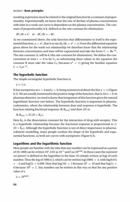
P1: PCX Printer: Yet To Come
9780521704632c06 CUFX213A/Peck 9780521618168 December 27, 2007 14:11
Section I Basic principles
resulting expression must be related to the original function by a constant of propor-tionality. Experimentally we know that the rate of decline of plasma concentrationwith time in a wash-out curve is dependent on the plasma concentration. The con-stant of proportionality is k, defined as the rate constant for elimination:
dC/dt ∝ C or dC/dt = −kC.
As we commented above, the only function that differentiates to itself is the expo-nential function, y = ex, that is to say dy/dx = ex = y. From the differential equationgiven above for the wash-out relationship we therefore know that the relationshipbetween concentration and time will be exponential and take the form: C = Be−bt.The rate constant, b, will be k (the rate constant for elimination). We define the con-centration at time t = 0 to be C0 so substituting these values in the equation theconstant B must take the value C0 (because e0 = 1) giving the familiar equationC = C0e−kt.
The hyperbolic functionThe simple rectangular hyperbolic function is:
y = 1/x.
It has asymptotes at x = 0 and y = 0, being symmetrical about the line y = x (Figure6.3). We are usually interested in the positive range of the function, that is, for x > 0. Inpharmacokinetics, we need to know that integration of this function gives the naturallogarithmic function (see below). The hyperbolic function is important in pharma-codynamics, where the relationship between dose and response is hyperbolic. Thefunction relating fractional response (R/Rmax) and dose (D) is:
R/Rmax = D/(D + KD).
Here KD is the dissociation constant for the interaction of drug with receptor. Thisis a hyperbolic relationship because the fractional response is proportional to 1/
(D + KD). Although the hyperbolic function is not of direct importance to pharma-cokinetic modelling, many people confuse the shape of the hyperbolic and expo-nential functions, as both are curves with asymptotes (Figure 6.4).
Logarithms and the logarithmic functionMost people are familiar with the idea that any number can be expressed as a powerof 10; 1000 can be written 103, 0.01 as 10−2 and 5 as 100.699. In these cases the exponent(or power) is defined as the logarithm to the base 10 (simply written as log) of eachnumber. Thus the log of 1000 is 3, which can be written log(1000) = 3, with log(0.01)= −2 and log(5) = 0.699. Note that log(10) = 1 because 101 = 10 and that log(1) =0 because 100 = 1. Any number can be written in this way so that for any positivevalue of x
x = 10log(x).
54
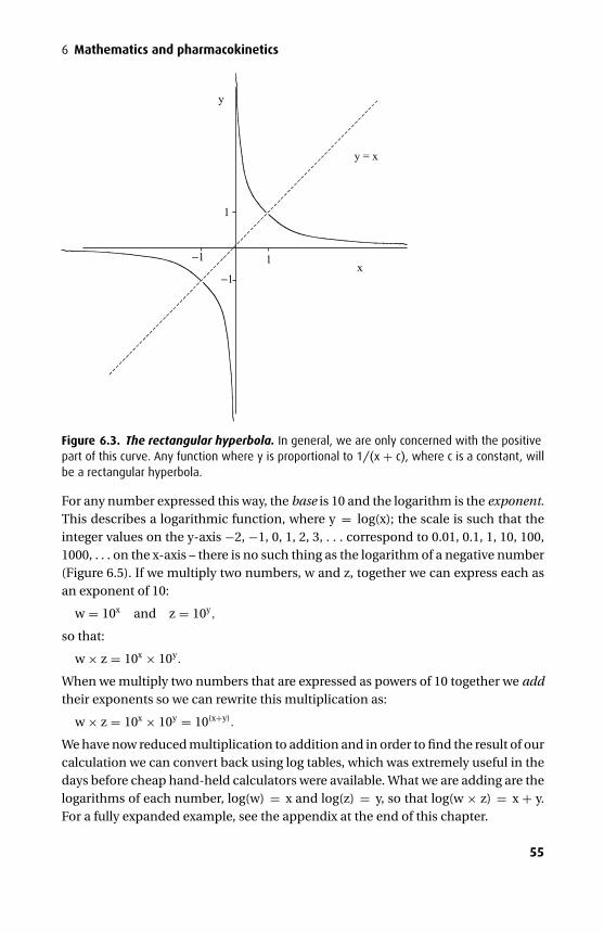
P1: PCX Printer: Yet To Come
9780521704632c06 CUFX213A/Peck 9780521618168 December 27, 2007 14:11
6 Mathematics and pharmacokinetics
y = x
y
x1
1
−1
−1
Figure 6.3. The rectangular hyperbola. In general, we are only concerned with the positivepart of this curve. Any function where y is proportional to 1/(x + c), where c is a constant, willbe a rectangular hyperbola.
For any number expressed this way, the base is 10 and the logarithm is the exponent.This describes a logarithmic function, where y = log(x); the scale is such that theinteger values on the y-axis −2, −1, 0, 1, 2, 3, . . . correspond to 0.01, 0.1, 1, 10, 100,1000, . . . on the x-axis – there is no such thing as the logarithm of a negative number(Figure 6.5). If we multiply two numbers, w and z, together we can express each asan exponent of 10:
w = 10x and z = 10y,
so that:
w × z = 10x × 10y.
When we multiply two numbers that are expressed as powers of 10 together we addtheir exponents so we can rewrite this multiplication as:
w × z = 10x × 10y = 10(x+y).
We have now reduced multiplication to addition and in order to find the result of ourcalculation we can convert back using log tables, which was extremely useful in thedays before cheap hand-held calculators were available. What we are adding are thelogarithms of each number, log(w) = x and log(z) = y, so that log(w × z) = x + y.For a fully expanded example, see the appendix at the end of this chapter.
55
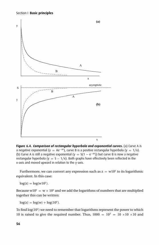
P1: PCX Printer: Yet To Come
9780521704632c06 CUFX213A/Peck 9780521618168 December 27, 2007 14:11
Section I Basic principles
A
B
x
y
B
Ay
x
asymptoteS
(a)
(b)
Figure 6.4. Comparison of rectangular hyperbola and exponential curves. (a) Curve A isa negative exponential (y = Ae−ax), curve B is a positive rectangular hyperbola (y = 1/x).(b) Curve A is still a negative exponential (y = S(1 − e−ax)) but curve B is now a negativerectangular hyperbola (y = S − 1/x). Both graphs have effectively been reflected in thex-axis and moved upward in relation to the y-axis.
Furthermore, we can convert any expression such as z = w10y to its logarithmicequivalent. In this case:
log(z) = log(w10y).
Because w10y = w × 10y and we add the logarithms of numbers that are multipliedtogether this can be written:
log(z) = log(w) + log(10y).
To find log(10y) we need to remember that logarithms represent the power to which10 is raised to give the required number. Thus, 1000 = 103 = 10 ×10 ×10 and
56
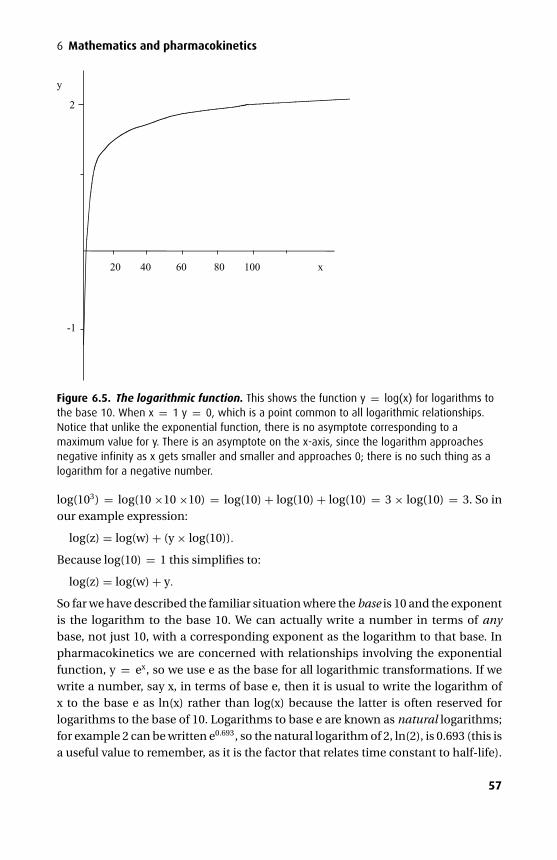
P1: PCX Printer: Yet To Come
9780521704632c06 CUFX213A/Peck 9780521618168 December 27, 2007 14:11
6 Mathematics and pharmacokinetics
20 40 60 80 100
2
y
x
-1
Figure 6.5. The logarithmic function. This shows the function y = log(x) for logarithms tothe base 10. When x = 1 y = 0, which is a point common to all logarithmic relationships.Notice that unlike the exponential function, there is no asymptote corresponding to amaximum value for y. There is an asymptote on the x-axis, since the logarithm approachesnegative infinity as x gets smaller and smaller and approaches 0; there is no such thing as alogarithm for a negative number.
log(103) = log(10 ×10 ×10) = log(10) + log(10) + log(10) = 3 × log(10) = 3. So inour example expression:
log(z) = log(w) + (y × log(10)).
Because log(10) = 1 this simplifies to:
log(z) = log(w) + y.
So far we have described the familiar situation where the base is 10 and the exponentis the logarithm to the base 10. We can actually write a number in terms of anybase, not just 10, with a corresponding exponent as the logarithm to that base. Inpharmacokinetics we are concerned with relationships involving the exponentialfunction, y = ex, so we use e as the base for all logarithmic transformations. If wewrite a number, say x, in terms of base e, then it is usual to write the logarithm ofx to the base e as ln(x) rather than log(x) because the latter is often reserved forlogarithms to the base of 10. Logarithms to base e are known as natural logarithms;for example 2 can be written e0.693, so the natural logarithm of 2, ln(2), is 0.693 (this isa useful value to remember, as it is the factor that relates time constant to half-life).
57
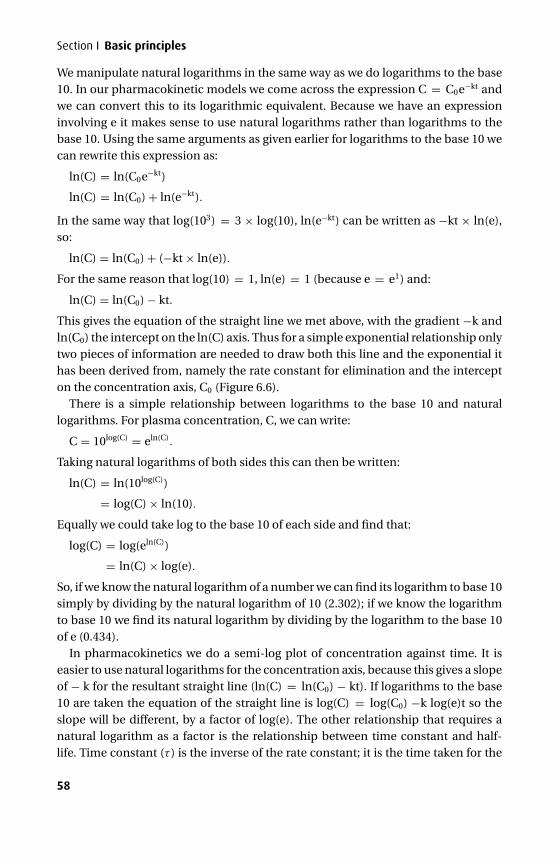
P1: PCX Printer: Yet To Come
9780521704632c06 CUFX213A/Peck 9780521618168 December 27, 2007 14:11
Section I Basic principles
We manipulate natural logarithms in the same way as we do logarithms to the base10. In our pharmacokinetic models we come across the expression C = C0e−kt andwe can convert this to its logarithmic equivalent. Because we have an expressioninvolving e it makes sense to use natural logarithms rather than logarithms to thebase 10. Using the same arguments as given earlier for logarithms to the base 10 wecan rewrite this expression as:
ln(C) = ln(C0e−kt)
ln(C) = ln(C0) + ln(e−kt).
In the same way that log(103) = 3 × log(10), ln(e−kt) can be written as −kt × ln(e),so:
ln(C) = ln(C0) + (−kt × ln(e)).
For the same reason that log(10) = 1, ln(e) = 1 (because e = e1) and:
ln(C) = ln(C0) − kt.
This gives the equation of the straight line we met above, with the gradient −k andln(C0) the intercept on the ln(C) axis. Thus for a simple exponential relationship onlytwo pieces of information are needed to draw both this line and the exponential ithas been derived from, namely the rate constant for elimination and the intercepton the concentration axis, C0 (Figure 6.6).
There is a simple relationship between logarithms to the base 10 and naturallogarithms. For plasma concentration, C, we can write:
C = 10log(C) = eln(C).
Taking natural logarithms of both sides this can then be written:
ln(C) = ln(10log(C))
= log(C) × ln(10).
Equally we could take log to the base 10 of each side and find that:
log(C) = log(eln(C))
= ln(C) × log(e).
So, if we know the natural logarithm of a number we can find its logarithm to base 10simply by dividing by the natural logarithm of 10 (2.302); if we know the logarithmto base 10 we find its natural logarithm by dividing by the logarithm to the base 10of e (0.434).
In pharmacokinetics we do a semi-log plot of concentration against time. It iseasier to use natural logarithms for the concentration axis, because this gives a slopeof − k for the resultant straight line (ln(C) = ln(C0) − kt). If logarithms to the base10 are taken the equation of the straight line is log(C) = log(C0) −k log(e)t so theslope will be different, by a factor of log(e). The other relationship that requires anatural logarithm as a factor is the relationship between time constant and half-life. Time constant (τ ) is the inverse of the rate constant; it is the time taken for the
58
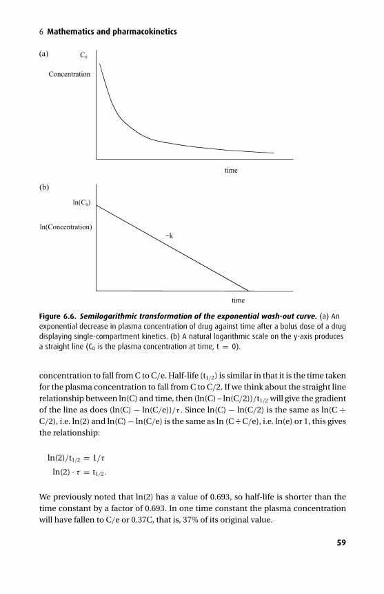
P1: PCX Printer: Yet To Come
9780521704632c06 CUFX213A/Peck 9780521618168 December 27, 2007 14:11
6 Mathematics and pharmacokinetics
Concentration
C0
time
time
ln(Concentration)
ln(C0)
−k
(a)
(b)
Figure 6.6. Semilogarithmic transformation of the exponential wash-out curve. (a) Anexponential decrease in plasma concentration of drug against time after a bolus dose of a drugdisplaying single-compartment kinetics. (b) A natural logarithmic scale on the y-axis producesa straight line (C0 is the plasma concentration at time, t = 0).
concentration to fall from C to C/e. Half-life (t1/2) is similar in that it is the time takenfor the plasma concentration to fall from C to C/2. If we think about the straight linerelationship between ln(C) and time, then (ln(C) – ln(C/2))/t1/2 will give the gradientof the line as does (ln(C) − ln(C/e))/τ . Since ln(C) − ln(C/2) is the same as ln(C ÷C/2), i.e. ln(2) and ln(C) − ln(C/e) is the same as ln (C ÷ C/e), i.e. ln(e) or 1, this givesthe relationship:
ln(2)/t1/2 = 1/τ
ln(2) · τ = t1/2.
We previously noted that ln(2) has a value of 0.693, so half-life is shorter than thetime constant by a factor of 0.693. In one time constant the plasma concentrationwill have fallen to C/e or 0.37C, that is, 37% of its original value.
59
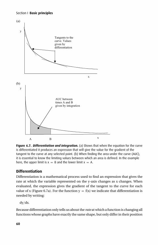
P1: PCX Printer: Yet To Come
9780521704632c06 CUFX213A/Peck 9780521618168 December 27, 2007 14:11
Section I Basic principles
y
Tangents to thecurve. Valuesgiven bydifferentiation
y
xA B
AUC betweentimes A and Bgiven by integration
x
(a)
(b)
Figure 6.7. Differentiation and integration. (a) Shows that when the equation for the curveis differentiated it produces an expression that will give the value for the gradient of thetangent to the curve at any selected point. (b) When finding the area under the curve (AUC),it is essential to know the limiting values between which an area is defined. In the examplehere, the upper limit is x = B and the lower limit x = A.
DifferentiationDifferentiation is a mathematical process used to find an expression that gives therate at which the variable represented on the y-axis changes as x changes. Whenevaluated, the expression gives the gradient of the tangent to the curve for eachvalue of x (Figure 6.7a). For the function y = f(x) we indicate that differentiation isneeded by writing:
dy/dx.
Because differentiation only tells us about the rate at which a function is changing allfunctions whose graphs have exactly the same shape, but only differ in their position
60
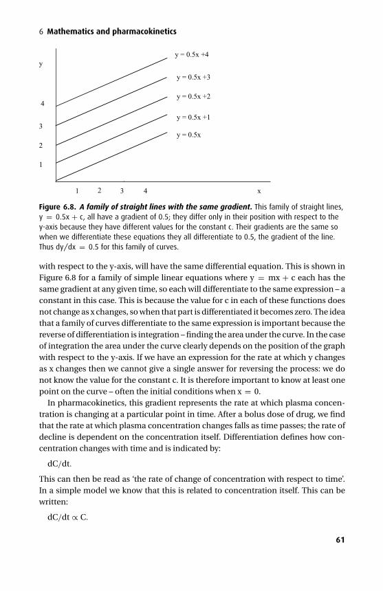
P1: PCX Printer: Yet To Come
9780521704632c06 CUFX213A/Peck 9780521618168 December 27, 2007 14:11
6 Mathematics and pharmacokinetics
y
x
1
2
3
4
2 4
y = 0.5x
y = 0.5x +1
y = 0.5x +2
y = 0.5x +3
y = 0.5x +4
1 3
Figure 6.8. A family of straight lines with the same gradient. This family of straight lines,y = 0.5x + c, all have a gradient of 0.5; they differ only in their position with respect to they-axis because they have different values for the constant c. Their gradients are the same sowhen we differentiate these equations they all differentiate to 0.5, the gradient of the line.Thus dy/dx = 0.5 for this family of curves.
with respect to the y-axis, will have the same differential equation. This is shown inFigure 6.8 for a family of simple linear equations where y = mx + c each has thesame gradient at any given time, so each will differentiate to the same expression – aconstant in this case. This is because the value for c in each of these functions doesnot change as x changes, so when that part is differentiated it becomes zero. The ideathat a family of curves differentiate to the same expression is important because thereverse of differentiation is integration – finding the area under the curve. In the caseof integration the area under the curve clearly depends on the position of the graphwith respect to the y-axis. If we have an expression for the rate at which y changesas x changes then we cannot give a single answer for reversing the process: we donot know the value for the constant c. It is therefore important to know at least onepoint on the curve – often the initial conditions when x = 0.
In pharmacokinetics, this gradient represents the rate at which plasma concen-tration is changing at a particular point in time. After a bolus dose of drug, we findthat the rate at which plasma concentration changes falls as time passes; the rate ofdecline is dependent on the concentration itself. Differentiation defines how con-centration changes with time and is indicated by:
dC/dt.
This can then be read as ‘the rate of change of concentration with respect to time’.In a simple model we know that this is related to concentration itself. This can bewritten:
dC/dt ∝ C.
61
P1: PCX Printer: Yet To Come
9780521704632c06 CUFX213A/Peck 9780521618168 December 27, 2007 14:11
Section I Basic principles
By using a constant of proportionality (k) and observing that as time passes theconcentration falls but more importantly the rate of change also falls, that is, theconstant of proportionality must be negative, we can write:
dC/dt = −kC.
This describes a first-order differential equation, as it is dependent on C raised tothe power one. If we now want an expression for C, we would need to integrate thisexpression. We saw earlier that this describes an exponential relationship. It can beshown that dC/dt = −kC when integrated, gives the expression:
C = C0e−kt.
To give the exact equation we use the condition that when t = 0, C = C0. Note thata zero-order differential equation is dependent on C raised to the power zero, whichis just one. A zero-order differential equation can therefore be written:
dC/dt = −k.
This tells us that the gradient is constant, which is true only for a straight line.
IntegrationIntegration can be thought of as a way in which we can find the area under a curve(AUC) if we know the equation that has been used to draw that curve. To find an areawe need to define the starting and ending points of interest on the x-axis (Figure 6.7b).In pharmacokinetics the area we are interested in is that under the concentration,C (equivalent to the y-axis), against time, t (equivalent to the x-axis), curve. Usuallywe want to know the entire area under the curve, which starts at time t = 0 and runsto infinity; occasionally we choose other limits – for example between t = 0 andt = t1/2 (where t1/2 is the half-life). Although knowledge of how to integrate functionsis not required, it is useful to know some important integrals related to pharmaco-kinetics.
Integration is indicated by the symbol∫
with the limiting values written above andbelow it. If no limits are given it is assumed that integration is taking place over theentire range possible. After the symbol for integration we write the function that isto be integrated together with an indication of the axis along which the limits havebeen given. For our concentration/time curve we integrate over time (rather thanconcentration) so we are integrating with respect to time, which is indicated using dt,just as for differentiation. So for a simple concentration against time curve modelledusing one compartment we can write:
AUC =∫
C0e−ktdt.
This has omitted the limits, which are usually t = 0 and t = infinity. Effectively wefind the expression that gives the area under the curve, then evaluate it for the lowerlimit (t = 0) and subtract this from the value for the upper limit (t = infinity); anyconstants will cancel out. We do not need to know how to do this particular integral,
62
P1: PCX Printer: Yet To Come
9780521704632c06 CUFX213A/Peck 9780521618168 December 27, 2007 14:11
6 Mathematics and pharmacokinetics
although we can note that it simplifies to:
AUC = C0/k.
We mentioned in the previous section that integration can be thought of as theopposite of differentiation, but we need a little more information before integratinga differential equation back to the original relationship between variables. The infor-mation required is usually the initial conditions. For example, taking the differentialequation we met previously:
dC/dt = −k · C.
We can rearrange this and write:
(1/C) · dC = −k · dt.
If we then integrate both sides, knowing that the integral of 1/x = ln(x) and theintegral of the constant k is kx + c, where c is a different constant, we end up with anexpression:
ln(C) = −kt + c.
When t = 0, C = C0 so putting in these initial conditions we find the value for c:
ln(C0) = c.
We can now substitute for c and write:
ln(C) = −kt + ln(C0).
This is the linear relationship we have met before. We can now simplify this to get arelationship that does not involve logarithms; first re-arrange:
ln(C) − ln(C0) = −kt.
We saw in a previous section that subtracting the logarithm of two numbers is thesame as dividing one by the other, so we can write:
ln(C/C0) = −kt.
We can now convert back to the exponent form:
C/C0 = e−kt.
Re-arranging this now gives the familiar equation for the one-compartmentalmodel:
C = C0e−kt.
Pharmacokinetic modelsModelling involves fitting a mathematical equation to experimental observationsof plasma concentration following drug administration to a group of volunteers orpatients. These models can then be used to predict plasma concentration undera variety of conditions and because for many drugs there is a close relationship
63
P1: PCX Printer: Yet To Come
9780521704632c06 CUFX213A/Peck 9780521618168 December 27, 2007 14:11
Section I Basic principles
between plasma concentration and drug activity pharmacodynamic effects can alsobe predicted. There is a variety of models that can be used; most commonly we usecompartmental models. In compartmental models we assume a single, central com-partment is connected to one or two peripheral compartments. Drug is assumed toenter and leave only through the central compartment, although it can distributeto and re-distribute from the peripheral compartments. For each peripheral com-partment we use an exponential term to model its volume and inter-compartmentalclearance; the central compartment is also represented by an exponential term, butdrug can be removed from the model entirely, so clearance from this compartmentreflects removal from the body. There are also complex physiological models thatcan more closely predict drug concentrations in different organs as well as non-compartmental models based on the statistical concept of mean residence time. Inthis text we will concentrate on compartmental models.
Single bolus dose
The one-compartment modelThe simplest model is that of a single, well-stirred, homogenous compartment. If asingle dose of drug is given, then the model predicts that it instantaneously dispersesevenly throughout this compartment and is eliminated in an exponential fashionwith a single rate constant for elimination (Figure 6.9a). This is the one-compartmentmodel that we have discussed in the mathematics section. Although such a modelis not directly relevant to clinical practice, it is important to understand because itintroduces the concepts that are further developed in more complex compartmentalmodels. The pharmacokinetic parameters introduced are volume of distribution,clearance, rate constant for elimination, time constant and half-life. If we take C asdrug concentration and t as time since administration of drug then this model isdescribed by an equation with a single exponential term:
C = C0e−kt,
where C0 is the concentration at time t = 0 and k is the rate constant for elimination.The volume of the single compartment is the volume of distribution, Vd, and theproportion of plasma from which drug is removed per minute is the rate constant forelimination, k. For example, if k is 0.1, then every minute a tenth of the compartmentwill have drug completely removed from it. The total volume cleared of drug everyminute must therefore be the product of k and the compartment volume (k × Vd)(Figure 6.10). This is known as the clearance (Cl) of drug from the compartment;clearance has units of ml.min−1:
Cl = k × Vd.
Because the time constant τ is the inverse of the rate constant, clearance also can beexpressed as the ratio of the volume of distribution and the time constant:
Cl = Vd/τ .
64
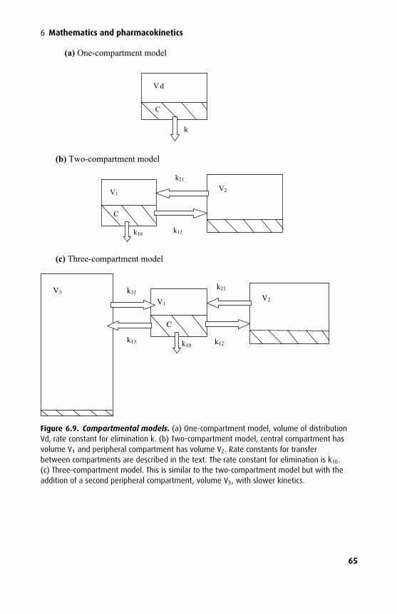
P1: PCX Printer: Yet To Come
9780521704632c06 CUFX213A/Peck 9780521618168 December 27, 2007 14:11
6 Mathematics and pharmacokinetics
V d
k
C
(a) One-compartment model
k10
V1V2
k12
k21
(b) Two-compartment model
(c) Three-compartment model
C
k10
V1V2
k12
k21
C
k13
k31V3
Figure 6.9. Compartmental models. (a) One-compartment model, volume of distributionVd, rate constant for elimination k. (b) Two-compartment model, central compartment hasvolume V1 and peripheral compartment has volume V2. Rate constants for transferbetween compartments are described in the text. The rate constant for elimination is k10.(c) Three-compartment model. This is similar to the two-compartment model but with theaddition of a second peripheral compartment, volume V3, with slower kinetics.
65
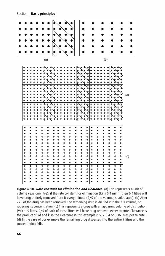
P1: PCX Printer: Yet To Come
9780521704632c06 CUFX213A/Peck 9780521618168 December 27, 2007 14:11
Section I Basic principles
(a) (b)
(d)
(c)
Figure 6.10. Rate constant for elimination and clearance. (a) This represents a unit ofvolume (e.g. one litre); if the rate constant for elimination (k) is 0.4 min−1 then 0.4 litres willhave drug entirely removed from it every minute (2/5 of the volume, shaded area). (b) After2/5 of the drug has been removed, the remaining drug is diluted into the full volume, soreducing its concentration. (c) This represents a drug with an apparent volume of distribution(Vd) of 9 litres, 2/5 of each of those litres will have drug removed every minute. Clearance isthe product of Vd and k so the clearance in this example is 9 × 0.4 or 0.36 litres per minute.(d) In the case of our example the remaining drug disperses into the entire 9 litres and theconcentration falls.
66
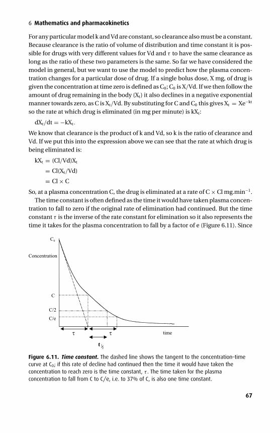
P1: PCX Printer: Yet To Come
9780521704632c06 CUFX213A/Peck 9780521618168 December 27, 2007 14:11
6 Mathematics and pharmacokinetics
For any particular model k and Vd are constant, so clearance also must be a constant.Because clearance is the ratio of volume of distribution and time constant it is pos-sible for drugs with very different values for Vd and τ to have the same clearance aslong as the ratio of these two parameters is the same. So far we have considered themodel in general, but we want to use the model to predict how the plasma concen-tration changes for a particular dose of drug. If a single bolus dose, X mg, of drug isgiven the concentration at time zero is defined as C0; C0 is X/Vd. If we then follow theamount of drug remaining in the body (Xt) it also declines in a negative exponentialmanner towards zero, as C is Xt/Vd. By substituting for C and C0 this gives Xt = Xe−kt
so the rate at which drug is eliminated (in mg per minute) is kXt:
dXt/dt = −kXt.
We know that clearance is the product of k and Vd, so k is the ratio of clearance andVd. If we put this into the expression above we can see that the rate at which drug isbeing eliminated is:
kXt = (Cl/Vd)Xt
= Cl(Xt/Vd)
= Cl × C
So, at a plasma concentration C, the drug is eliminated at a rate of C × Cl mg.min−1.The time constant is often defined as the time it would have taken plasma concen-
tration to fall to zero if the original rate of elimination had continued. But the timeconstant τ is the inverse of the rate constant for elimination so it also represents thetime it takes for the plasma concentration to fall by a factor of e (Figure 6.11). Since
Concentration
time
C
C/e
C0
C/2
t
τ τ
12
Figure 6.11. Time constant. The dashed line shows the tangent to the concentration–timecurve at C0; if this rate of decline had continued then the time it would have taken theconcentration to reach zero is the time constant, τ . The time taken for the plasmaconcentration to fall from C to C/e, i.e. to 37% of C, is also one time constant.
67
P1: PCX Printer: Yet To Come
9780521704632c06 CUFX213A/Peck 9780521618168 December 27, 2007 14:11
Section I Basic principles
τ has units of time, usually minutes, then k has units of time−1, usually min−1. In thesection above we saw that the time constant τ is longer than the half-life; there is asimple inverse relationship between k and τ , whereas k = ln(2)/t1/2, so it is easier touse time constants rather than half-lives when discussing models.
Multi-compartment modelsMulti-compartment models make allowance for the uptake of drug by different tis-sues within the body, and for the different blood flow rates to these tissues. Differ-ent tissues that share pharmacokinetic properties form compartments. Convenientlabels include ‘vessel rich’ and ‘vessel poor’ compartments. The number of theoret-ical compartments that may be included in any model is limitless, but more thanthree compartments become experimentally indistinguishable. In these models it isimportant to realize that elimination can occur only from the central compartmentand that the ‘effect compartment’ is in equilibrium with the central compartment.The volume of this effect compartment is very small so it does not contribute tothe total volume, but is useful for predicting the onset and offset of the responsewhen the observed effect is proportional to effect site concentration. Models forindividual drugs differ in the volume of compartments and in transfer rates betweencompartments; the values for these pharmacokinetic parameters can vary enor-mously and depend on the physicochemical properties of a drug as well as the siteand rate of drug metabolism.
Two compartmentsIn the two-compartment model the central compartment connects with a secondcompartment; the volume of the central compartment is V1 and that of the peripheralcompartment V2 (Figure 6.9b). The total volume of distribution is the sum of thesetwo volumes. Unlike the single compartment model, there are now two pathways fordrug elimination from plasma: an initial rapid transfer from the central to peripheralcompartment and removal from the central compartment. The latter removes drugfrom the system, whereas after distribution to the second compartment, the drugcan re-distribute to the central compartment when conditions allow. Inspection ofthe concentration–time curve shows that the initial rate of decline in plasma con-centration is much faster than would be expected from a single compartment model;this represents rapid distribution to the second compartment. A semi-logarithmicplot of ln (C) against time is now a curve, rather than a straight line, which is the sumof two straight lines representing the exponential processes with rate constants α
and β (Figure 6.12).Transfer between compartments is assumed to occur in an exponential fashion at
a rate depending on the concentration difference between compartments and theequilibrium constants for transfer. Rate constants for transfer in each direction aredescribed: k12 is the rate constant for the transfer from the central to peripheral com-partment and k21 is the rate constant for the transfer from the peripheral to central
68
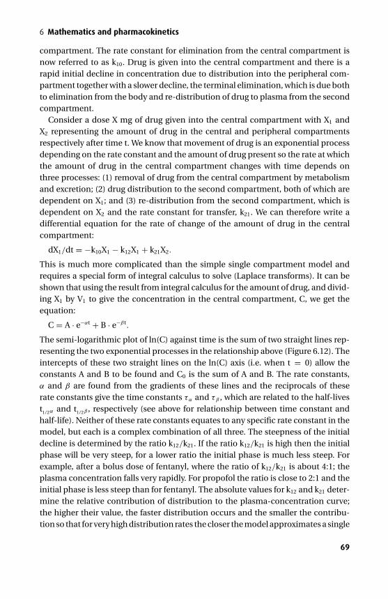
P1: PCX Printer: Yet To Come
9780521704632c06 CUFX213A/Peck 9780521618168 December 27, 2007 14:11
6 Mathematics and pharmacokinetics
compartment. The rate constant for elimination from the central compartment isnow referred to as k10. Drug is given into the central compartment and there is arapid initial decline in concentration due to distribution into the peripheral com-partment together with a slower decline, the terminal elimination, which is due bothto elimination from the body and re-distribution of drug to plasma from the secondcompartment.
Consider a dose X mg of drug given into the central compartment with X1 andX2 representing the amount of drug in the central and peripheral compartmentsrespectively after time t. We know that movement of drug is an exponential processdepending on the rate constant and the amount of drug present so the rate at whichthe amount of drug in the central compartment changes with time depends onthree processes: (1) removal of drug from the central compartment by metabolismand excretion; (2) drug distribution to the second compartment, both of which aredependent on X1; and (3) re-distribution from the second compartment, which isdependent on X2 and the rate constant for transfer, k21. We can therefore write adifferential equation for the rate of change of the amount of drug in the centralcompartment:
dX1/dt = −k10X1 − k12X1 + k21X2.
This is much more complicated than the simple single compartment model andrequires a special form of integral calculus to solve (Laplace transforms). It can beshown that using the result from integral calculus for the amount of drug, and divid-ing X1 by V1 to give the concentration in the central compartment, C, we get theequation:
C = A · e−αt + B · e−βt.
The semi-logarithmic plot of ln(C) against time is the sum of two straight lines rep-resenting the two exponential processes in the relationship above (Figure 6.12). Theintercepts of these two straight lines on the ln(C) axis (i.e. when t = 0) allow theconstants A and B to be found and C0 is the sum of A and B. The rate constants,α and β are found from the gradients of these lines and the reciprocals of theserate constants give the time constants τα and τβ , which are related to the half-livest1/2α and t1/2β , respectively (see above for relationship between time constant andhalf-life). Neither of these rate constants equates to any specific rate constant in themodel, but each is a complex combination of all three. The steepness of the initialdecline is determined by the ratio k12/k21. If the ratio k12/k21 is high then the initialphase will be very steep, for a lower ratio the initial phase is much less steep. Forexample, after a bolus dose of fentanyl, where the ratio of k12/k21 is about 4:1; theplasma concentration falls very rapidly. For propofol the ratio is close to 2:1 and theinitial phase is less steep than for fentanyl. The absolute values for k12 and k21 deter-mine the relative contribution of distribution to the plasma-concentration curve;the higher their value, the faster distribution occurs and the smaller the contribu-tion so that for very high distribution rates the closer the model approximates a single
69
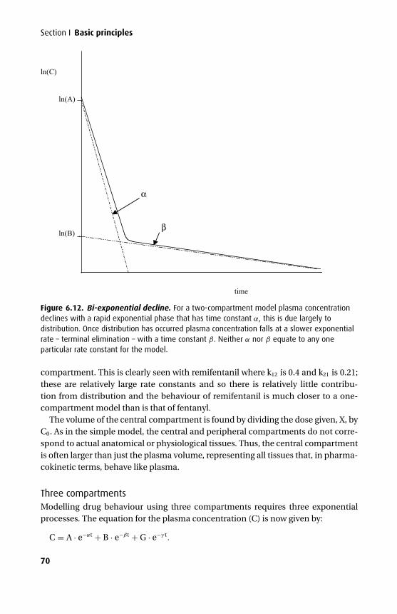
P1: PCX Printer: Yet To Come
9780521704632c06 CUFX213A/Peck 9780521618168 December 27, 2007 14:11
Section I Basic principles
ln(C)
ln(A)
ln(B)
time
α
β
Figure 6.12. Bi-exponential decline. For a two-compartment model plasma concentrationdeclines with a rapid exponential phase that has time constant α, this is due largely todistribution. Once distribution has occurred plasma concentration falls at a slower exponentialrate – terminal elimination – with a time constant β. Neither α nor β equate to any oneparticular rate constant for the model.
compartment. This is clearly seen with remifentanil where k12 is 0.4 and k21 is 0.21;these are relatively large rate constants and so there is relatively little contribu-tion from distribution and the behaviour of remifentanil is much closer to a one-compartment model than is that of fentanyl.
The volume of the central compartment is found by dividing the dose given, X, byC0. As in the simple model, the central and peripheral compartments do not corre-spond to actual anatomical or physiological tissues. Thus, the central compartmentis often larger than just the plasma volume, representing all tissues that, in pharma-cokinetic terms, behave like plasma.
Three compartmentsModelling drug behaviour using three compartments requires three exponentialprocesses. The equation for the plasma concentration (C) is now given by:
C = A · e−αt + B · e−βt + G · e−γ t.
70
P1: PCX Printer: Yet To Come
9780521704632c06 CUFX213A/Peck 9780521618168 December 27, 2007 14:11
6 Mathematics and pharmacokinetics
This is similar to the relationship for the two-compartment model, but with theaddition of a third exponential process resulting from the presence of an additionalcompartment; the kinetic constants G and γ describe that additional process. Themodel consists of a central compartment into which a drug is infused and fromwhich excretion can occur, together with two peripheral compartments with whichdrug can be exchanged (Figure 6.9c). These may typically represent well-perfused(the second compartment) and poorly perfused (the third compartment) tissues,respectively, with the central compartment representing plasma. Distribution intothe second compartment is always faster than into the third compartment. This isa reasonable model for the majority of anaesthetic agents, where drug reaches theplasma and is distributed to muscle and fat. The volume of distribution at steady-state is the sum of the volumes of the three compartments. The mathematics issimilar to that for two compartments, but more complicated as transfer into and outof the third compartment also must be taken into account.
As in the two-compartment model, there is final phase that can be described bya single exponential and the half-life associated with this phase is known as theterminal elimination half-life, which reflects both elimination from the body andre-distribution from the peripheral compartments. The rate constant for eliminationis therefore not the inverse of the terminal elimination time constant; it can becalculated once the model parameters have been found. Clearance of drug out ofthe body is still defined as the product of V1 and the rate constant for elimination;as discussed below clearance is usually found using a non-compartmental methodand calculating the ratio of dose to the AUC.
Non-compartmental modelsNon-compartmental models make no assumptions about specific volumes but useinformation from the AUC, as this reflects the removal of drug from plasma. The areaunder the concentration–time curve can be used to find clearance because AUC isthe ratio of dose to clearance. Clearance (Cl) is therefore:
Cl = dose/AUC.
If we plot the product of concentration and time on the y-axis against time we producewhat is known as the first moment curve. The area under this curve (AUMC) can beused to find a parameter known as mean residence time (MRT). Mean residence timeis a measure of how long the drug stays in the body and is similar to a time constantin the compartmental models.
MRT = AUMC/AUC.
The product of clearance and MRT is the steady-state volume of distribution (Vss)according to this model. So volume of distribution is:
Vss = Cl × MRT.
71
P1: PCX Printer: Yet To Come
9780521704632c06 CUFX213A/Peck 9780521618168 December 27, 2007 14:11
Section I Basic principles
Definition and measurement of pharmacokinetic parametersThe parameters described in the compartmental models are useful for comparingthe persistence of different drugs in the body namely: volume of distribution (Vd),clearance (Cl) and time constants (τ in the single compartment and α, β and γ inmulti-compartment models). Half-lives (t1/2) are related to their corresponding timeconstants by a factor of ln(2), which is 0.693.
Volume of distributionVolume of distribution (Vd) is defined as the apparent volume into which a drugdisperses in order to produce the observed plasma concentrations. It does not cor-respond to any particular physiological volume and can be much larger than totalbody water. The physicochemical properties of a drug including its molecular size,lipid solubility and charge characteristics all influence the volume of distribution.Propofol is a very lipid-soluble drug and has a large volume of distribution of about250 litres; pancuronium is a highly charged molecule and has a relatively small vol-ume of distribution of about 17.5 litres. Tissue binding of drug, particularly intracel-lular sequestration, can account for extremely high volumes of distribution; the anti-malarial chloroquine has a volume of distribution in excess of 10,000 litres. Pathologyalso influences the kinetic parameters; in hepatic and renal disease volumes of dis-tribution are increased as the relative volumes of body fluid compartments change.
In the simple one-compartmental model, Vd is a constant that relates dose admin-istered to the plasma concentration at time zero. It has the units of volume (e.g. litres)but can be indexed to bodyweight and expressed as litres.kg−1. In the simple model,Vd is the initial dose divided by the plasma concentration occurring immediatelyafter administration:
Vd = dose/plasma concentration = X/C0.
In the multi-compartment models, the central compartment volume is the initialvolume into which the drug disperses (Vintial). The volume of this compartmentdepends partly on the degree of protein binding; a highly protein bound drug willhave a larger central compartment volume than a drug that is poorly bound. Propofolis 98% protein bound and has a central compartment volume of about 16 litres,compared with an actual plasma volume of about 3 litres. The volume of this centralcompartment can be estimated from the rapid distribution phase; if the dose givenwas X and the intercept on the y-axis is A then:
Vintial = X/A.
The total volume of distribution is the sum of all the volumes that comprise the model.There are several methods available that attempt to estimate this volume: Vextrap,Varea and Vss. The first of these simply ignores the contribution made by any volumeapart from that associated with the terminal phase of elimination; the interceptof the line representing terminal elimination is extrapolated back to its intercept
72
P1: PCX Printer: Yet To Come
9780521704632c06 CUFX213A/Peck 9780521618168 December 27, 2007 14:11
6 Mathematics and pharmacokinetics
on the ln(concentration) axis (ln(B) for the two compartment model) this gives aconcentration which, when divided into X (dose given), will give Vextrap:
Vextrap = X/B.
It greatly overestimates the total volume of distribution for many drugs, particularlywhen the distribution phase contributes significantly to drug dispersion. The secondmethod, Varea, is of more use because it is related both to clearance and the termi-nal elimination constant. This uses the non-compartmental method of calculatingclearance from AUC and assumes that an ‘average’ rate constant for removal of drugfrom plasma can be approximated by the inverse of the terminal elimination timeconstant (β for the two-compartment model):
Varea = Clearance/β = X/(AUC × β).
This gives a better estimate of volume of distribution than Vextrap, but is still anoverestimate; using β as the ‘average’ rate constant is an underestimate, particularlyif there is significant distribution and re-distribution to and from compartments.However, it has the advantage of being easily calculated from experimental data.The final method Vss is entirely based on non-compartment models (see above) andis calculated from the product of clearance and mean residence time:
Vss = (dose/AUC) × (AUMC/AUC) = dose × AUMC/AUC2.
This gives an estimate of volume of distribution that is independent of elimination,which can be useful. The estimate of volume of distribution using this method issmaller than for either of the other methods, but is usually close to the area method:Vextrap > Varea > Vss.
ClearanceClearance (Cl) is defined as that volume of plasma from which drug is completelyremoved per unit time – the usual units are ml.min−1. For the one-compartmentmodel we saw that clearance is related to the rate constant for elimination; a highrate constant reflects rapid removal of drug since a large fraction of the distributionvolume is cleared of drug. Clearance is simply the product of this rate constant andthe volume of distribution (Figure 6.10). Clearance relates plasma concentration ofdrug at a given time to the actual rate of drug elimination (Rateel in mg.min−1) atthat time:
Rateel = C × Cl.
As we saw above, for the one-compartment model the clearance (Cl) of drug fromthe compartment is the product of the rate constant for elimination, k, and Vd,the volume of distribution. We can find k from the slope of the linear ln(C)-timegraph and Vd from C0, the concentration at t = 0, since we know the dose of druggiven.
In the multi-compartment model, we can talk about inter-compartmental clear-ances as well as a clearance that describes loss of drug from the model. Quoted values
73
P1: PCX Printer: Yet To Come
9780521704632c06 CUFX213A/Peck 9780521618168 December 27, 2007 14:11
Section I Basic principles
for clearance define the removal of drug from the body, which is the product of therate constant for elimination k10 and V1, the volume of distribution of the centralcompartment. For example, propofol has a k10 of approximately 0.12, so about 1/8of the plasma-equivalent volume has drug removed by elimination in unit time. Forpropofol, V1 is about 16 litres and k10 is approximately 0.12, so the clearance of propo-fol is 16 × 0.12, which is about 2 litres per minute. Remifentanil has a much smallercentral compartment volume but a higher k10, so the clearance of remifentanil is 5.1× 0.5, which is 2.5 litres, quite similar to that of propofol. If we do not know V1 wecould use Vintial as an estimate but it is often inaccurate due to sampling artefacts atearly times when mixing has not taken place. Instead, clearance is usually calculatedfrom the area under the concentration–time curve:
Clearance = Dose/AUC.
Elimination of drug from the model represents both metabolism and excretion ofunchanged drug. Metabolism may occur in many sites and be organ-dependentor independent; a clearance that is greater than hepatic blood flow suggests thathepatic elimination is not the only route of elimination – either there are other sitesof metabolism or the drug is excreted unchanged, for example through renal orpulmonary routes. Remifentanil has a very high clearance because it is eliminatedby non-specific esterase in both plasma and tissue.
Inter-compartmental clearance relates to the movement of drug betweencompartments; C12 and C13 define drug transfer between compartments 1 and 2 and1 and 3, respectively. For drugs with comparable compartmental volumes, the higherthe inter-compartmental clearance the more rapidly distribution and re-distributiontakes place.
Time constant and half-life (t1/2)In the single-compartment model there is just a single exponential relationshipbetween plasma concentration and time. The time constant defines how quicklythe plasma concentration falls with time and is defined as the time it would havetaken plasma concentration to fall to zero if the original rate of elimination had con-tinued (Figure 6.11). Time constant τ has units of time, usually minutes. The half-lifeis the time taken for the plasma concentration to fall to 50% of its initial value. In themathematics section we saw that half-life is related to time constant by a constant ofproportionality and half-life is ln2.τ , that is, half-life is 0.693.τ ; the half-life is shorterthan the time constant. Note that either half-life or time constant may be used to rep-resent the time dependency of an exponential process. In the multi-compartmentmodels there are several hybrid time constants each of which relates to one of thedistinct exponential phases of drug elimination. As mentioned above, these do notrelate directly to any one of the individual rate constants in the model.
74
P1: PCX Printer: Yet To Come
9780521704632c06 CUFX213A/Peck 9780521618168 December 27, 2007 14:11
6 Mathematics and pharmacokinetics
Relationship between constantsThe rate constant for elimination (kel or k10), clearance (Cl) and volume of distributionof the central compartment (Vd for one-compartment and V1 for two-and three-compartment models) are all constant values for any one model and are closelyrelated:
kel = Cl/V1 or Cl = V1 · kel.
In the one compartment model the time constant for elimination, τ , is given by:
τ = 1/kel.
The relationship between time constant and clearance is therefore:
τ = V1/Cl.
Clearance is determined both by V1 and τ ; drugs with the same clearance can havevery different volumes of distribution and time constants, it is the ratio of the twothat is important. In non-compartment models, the mean residence time is equiv-alent to time constant and clearance is the ratio of dose given to the area under theconcentration time curve.
Multiple doses and infusions
Loading dose, infusion rate and dose intervalIf the initial volume of distribution (V1) and the required plasma concentration areknown then the dose needed to give a particular plasma concentration can be cal-culated:
Dose = V1 × required concentration.
If we want to maintain this particular plasma level, this first dose is known as theloading dose; repeated doses must be then given or the drug can be given by aconstant infusion. The frequency of drug dosing depends on the rate at which drugis removed from plasma either by distribution or elimination. After one half-life,the concentration will have fallen to half of the initial value. If this concentration isacceptable as the minimum therapeutic concentration, then the dose frequency isequal to one elimination half-life (Figure 6.13). If the rate of removal is high, then thedosing interval must be short and doses given frequently; if the rate of removal is slow,then the dosing interval can be longer and doses given less frequently. The frequencyand size of dose at the start of treatment influences the rate at which a steady statecan be reached; for certain drugs, such as amiodarone, frequent initial dosing isreplaced by less frequent and lower doses as the peripheral compartments becomesaturated with drug. Although many drugs have a high therapeutic index and largedoses are well tolerated, some drugs have a narrow therapeutic index and maximumdose is restricted by adverse side effects. Thus dosing schedules are determined bothby the pharmacokinetics and the pharmacodynamics of a drug.
75
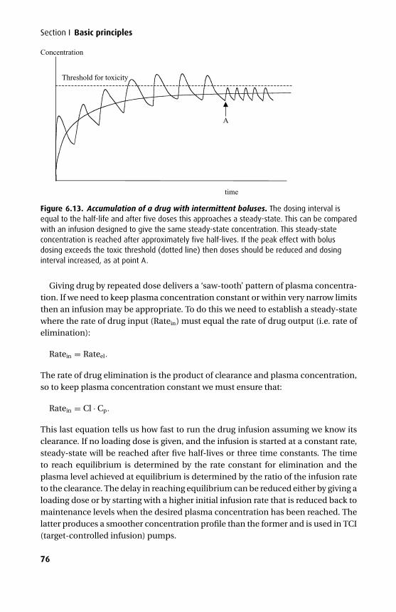
P1: PCX Printer: Yet To Come
9780521704632c06 CUFX213A/Peck 9780521618168 December 27, 2007 14:11
Section I Basic principles
Threshold for toxicity
time
A
Concentration
Figure 6.13. Accumulation of a drug with intermittent boluses. The dosing interval isequal to the half-life and after five doses this approaches a steady-state. This can be comparedwith an infusion designed to give the same steady-state concentration. This steady-stateconcentration is reached after approximately five half-lives. If the peak effect with bolusdosing exceeds the toxic threshold (dotted line) then doses should be reduced and dosinginterval increased, as at point A.
Giving drug by repeated dose delivers a ‘saw-tooth’ pattern of plasma concentra-tion. If we need to keep plasma concentration constant or within very narrow limitsthen an infusion may be appropriate. To do this we need to establish a steady-statewhere the rate of drug input (Ratein) must equal the rate of drug output (i.e. rate ofelimination):
Ratein = Rateel.
The rate of drug elimination is the product of clearance and plasma concentration,so to keep plasma concentration constant we must ensure that:
Ratein = Cl · Cp.
This last equation tells us how fast to run the drug infusion assuming we know itsclearance. If no loading dose is given, and the infusion is started at a constant rate,steady-state will be reached after five half-lives or three time constants. The timeto reach equilibrium is determined by the rate constant for elimination and theplasma level achieved at equilibrium is determined by the ratio of the infusion rateto the clearance. The delay in reaching equilibrium can be reduced either by giving aloading dose or by starting with a higher initial infusion rate that is reduced back tomaintenance levels when the desired plasma concentration has been reached. Thelatter produces a smoother concentration profile than the former and is used in TCI(target-controlled infusion) pumps.
76
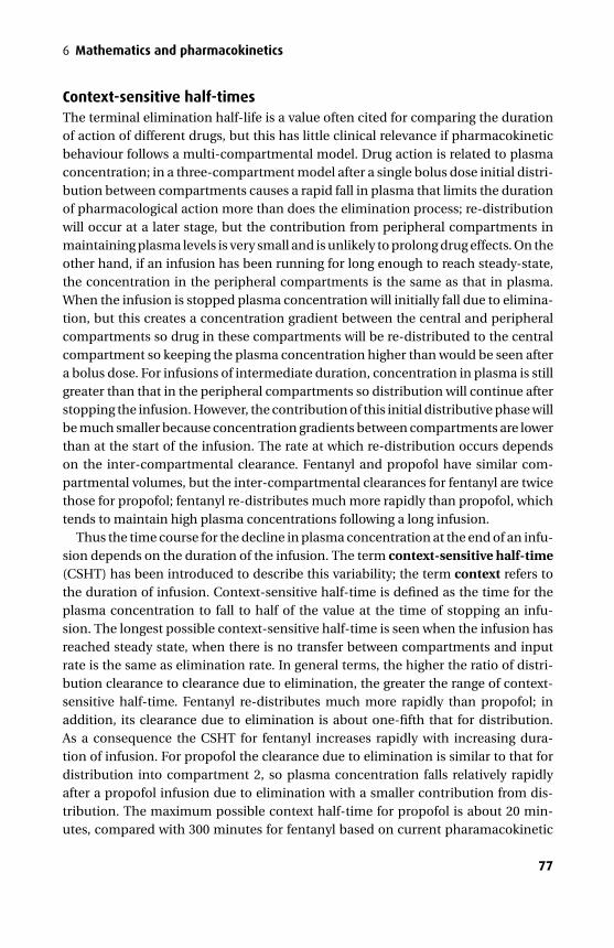
P1: PCX Printer: Yet To Come
9780521704632c06 CUFX213A/Peck 9780521618168 December 27, 2007 14:11
6 Mathematics and pharmacokinetics
Context-sensitive half-timesThe terminal elimination half-life is a value often cited for comparing the durationof action of different drugs, but this has little clinical relevance if pharmacokineticbehaviour follows a multi-compartmental model. Drug action is related to plasmaconcentration; in a three-compartment model after a single bolus dose initial distri-bution between compartments causes a rapid fall in plasma that limits the durationof pharmacological action more than does the elimination process; re-distributionwill occur at a later stage, but the contribution from peripheral compartments inmaintaining plasma levels is very small and is unlikely to prolong drug effects. On theother hand, if an infusion has been running for long enough to reach steady-state,the concentration in the peripheral compartments is the same as that in plasma.When the infusion is stopped plasma concentration will initially fall due to elimina-tion, but this creates a concentration gradient between the central and peripheralcompartments so drug in these compartments will be re-distributed to the centralcompartment so keeping the plasma concentration higher than would be seen aftera bolus dose. For infusions of intermediate duration, concentration in plasma is stillgreater than that in the peripheral compartments so distribution will continue afterstopping the infusion. However, the contribution of this initial distributive phase willbe much smaller because concentration gradients between compartments are lowerthan at the start of the infusion. The rate at which re-distribution occurs dependson the inter-compartmental clearance. Fentanyl and propofol have similar com-partmental volumes, but the inter-compartmental clearances for fentanyl are twicethose for propofol; fentanyl re-distributes much more rapidly than propofol, whichtends to maintain high plasma concentrations following a long infusion.
Thus the time course for the decline in plasma concentration at the end of an infu-sion depends on the duration of the infusion. The term context-sensitive half-time(CSHT) has been introduced to describe this variability; the term context refers tothe duration of infusion. Context-sensitive half-time is defined as the time for theplasma concentration to fall to half of the value at the time of stopping an infu-sion. The longest possible context-sensitive half-time is seen when the infusion hasreached steady state, when there is no transfer between compartments and inputrate is the same as elimination rate. In general terms, the higher the ratio of distri-bution clearance to clearance due to elimination, the greater the range of context-sensitive half-time. Fentanyl re-distributes much more rapidly than propofol; inaddition, its clearance due to elimination is about one-fifth that for distribution.As a consequence the CSHT for fentanyl increases rapidly with increasing dura-tion of infusion. For propofol the clearance due to elimination is similar to that fordistribution into compartment 2, so plasma concentration falls relatively rapidlyafter a propofol infusion due to elimination with a smaller contribution from dis-tribution. The maximum possible context half-time for propofol is about 20 min-utes, compared with 300 minutes for fentanyl based on current pharamacokinetic
77
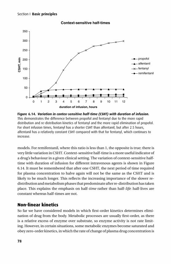
P1: PCX Printer: Yet To Come
9780521704632c06 CUFX213A/Peck 9780521618168 December 27, 2007 14:11
Section I Basic principles
Context-sensitive half-times
0
50
100
150
200
250
300
350
0 1 2 3 4 5 6 7 8 9 10 11 12
duration of infusion, hours
CS
HT,
min propofol
alfentanil
fentanyl
remifentanil
Figure 6.14. Variation in contex-sensitive half-time (CSHT) with duration of infusion.This demonstrates the difference between propofol and fentanyl due to the more rapiddistribution and re-distribution kinetics of fentanyl and the more rapid elimination of propofol.For short infusion times, fentanyl has a shorter CSHT than alfentanil, but after 2.5 hours,alfentanil has a relatively constant CSHT compared with that for fentanyl, which continues toincrease.
models. For remifentanil, where this ratio is less than 1, the opposite is true; there isvery little variation in CSHT. Context-sensitive half-time is a more useful indicator ofa drug’s behaviour in a given clinical setting. The variation of context-sensitive half-time with duration of infusion for different intravenous agents is shown in Figure6.14. It must be remembered that after one CSHT, the next period of time requiredfor plasma concentration to halve again will not be the same as the CSHT and islikely to be much longer. This reflects the increasing importance of the slower re-distribution and metabolism phases that predominate after re-distribution has takenplace. This explains the emphasis on half-time rather than half-life: half-lives areconstant whereas half-times are not.
Non-linear kineticsSo far we have considered models in which first-order kinetics determines elimi-nation of drug from the body. Metabolic processes are usually first-order, as thereis a relative excess of enzyme over substrate, so enzyme activity is not rate limit-ing. However, in certain situations, some metabolic enzymes become saturated andobey zero-order kinetics, in which the rate of change of plasma drug concentration is
78
P1: PCX Printer: Yet To Come
9780521704632c06 CUFX213A/Peck 9780521618168 December 27, 2007 14:11
6 Mathematics and pharmacokinetics
constant rather than being dependent upon the concentration of drug. This is alsoknown as saturation kinetics and indicates that enzyme activity is maximal, so cannotbe influenced by increasing substrate concentration. An example is the metabolismof ethanol, which proceeds at a relatively constant rate after the ingestion of a mod-erate amount of alcohol. This is because the rate-limiting step in its metabolismby alcohol dehydrogenase is the presence of a co-factor for the reaction, which ispresent only in small quantities.
Certain processes obey first-order kinetics at low dose, but zero-order at higherdoses. For example, the metabolism of phenytoin becomes saturable within theupper limit of the normal range, and the pharmacokinetics of thiopental obeys zero-order kinetics when used by infusion for prolonged periods, such as in the treatmentof status epilepticus. There are two important implications of a process obeyingzero-order kinetics within a normal dose range.
First, during zero-order kinetics a small increase in dose may cause a large increasein plasma level. If this occurs at a level near the upper limit of the therapeutic range,toxicity may be experienced after a modest dose increase. Checking plasma con-centration is essential to avoid toxic levels when prescribing drugs where this is aproblem, such as occurs with phenytoin.
Second, during zero-order kinetics there is no steady-state. If the rate of drug deliv-ery exceeds the rate of drug excretion, plasma levels will continue to rise inexorablyuntil ingestion stops or toxicity leads to death (Figure 6.15).
B
time
concentration
Zero-order kinetics
First-order kinetics
A
Figure 6.15. Transition to non-linear (zero-order) kinetics. If an infusion is started at aconstant rate at point A, initially plasma concentration rises in a negative exponential fashionaccording to first-order kinetics. At point B the elimination process is overwhelmed and theplasma concentration continues to rise in a linear fashion, according to zero-order kinetics. Thisusually occurs because hepatic enzymes become saturated and work at maximum capacity.
79
P1: PCX Printer: Yet To Come
9780521704632c06 CUFX213A/Peck 9780521618168 December 27, 2007 14:11
Section I Basic principles
Applied pharmacokinetics
PropofolThe intravenous anaesthetic propofol has become more popular than thiopental forinduction of anaesthesia as many of its properties approximate to those of the idealanaesthetic agent. It can be used both for induction and maintenance of anaesthesia,and it has become accepted as the anaesthetic agent of choice in daycase surgerydue to its favourable ‘wake up’ profile. Although it is possible to give a bolus dose ofpropofol without understanding its pharmacokinetic properties, to use it sensiblyby infusion requires some understanding of its disposition in the body.
StructurePropofol is a sterically hindered phenol: the potentially active hydroxyl (-OH) groupis shielded by the electron clouds surrounding the attached isopropyl ((CH3)2CH-)groups, which reduces its reactivity. So, unlike phenol, propofol is insoluble in waterbut highly soluble in fat and requires preparation as a lipid emulsion.
PharmacokineticsThe usual induction dose of propofol is 1.5–2.5 mg.kg−1, which when given intra-venously is extensively bound to plasma proteins (98% bound to albumin). Followingan induction dose there is rapid loss of consciousness as the lipid tissue of the cen-tral nervous system (CNS) takes up the highly lipophilic drug. Over the next fewminutes propofol then distributes to peripheral tissues and the concentration in theCNS falls, such that in the absence of further doses or another anaesthetic agentthe patient will wake up. Its distribution half-life is 1–2 minutes (with distribution tothe lipid tissues) and accounts for the rapid fall in plasma levels with a short durationof action. The terminal elimination half-life is very much longer (5–12 hours) andplays little or no role in offset of clinical action following a bolus dose.
Propofol causes peripheral vasodilatation, possibly through nitric oxide release ina similar manner to nitrates, so that when used in the elderly or hypovolaemic patientit may cause profound hypotension. This may be avoided by giving the drug as a slowinfusion, however this results in the plasma concentration rising more slowly andonset of anaesthesia is delayed.
Context-sensitive half-timeContext-sensitive half-time is discussed above. When propofol is used by infusionit steadily loads the peripheral compartments; the longer the duration of the infu-sion, the more peripheral compartment loading will occur. Therefore, following aprolonged infusion, there will be more propofol to re-distribute from the peripheralcompartments back into the central compartment, which will tend to maintain theplasma concentration and duration of action. In practice, the longer the durationof the infusion, the longer the time required before the plasma level falls below that
80
P1: PCX Printer: Yet To Come
9780521704632c06 CUFX213A/Peck 9780521618168 December 27, 2007 14:11
6 Mathematics and pharmacokinetics
required for anaesthesia. Although the CSHT has a maximum value of about 20 min-utes, during long, stimulating surgery infusion rates will be high and the plasmaconcentration when wake-up occurs may be much less than half the plasma con-centration at the end of the infusion. Thus time to awakening using propofol alonemay be as long as 1 hour.
RemifentanilUnlike most agents used in anaesthesia, remifentanil has a relatively constantcontext-sensitive half-time. Remifentanil is a fentanyl derivative that is a pure µ
agonist. It has an ester linkage, which is very rapidly broken down by plasma andnon-specific tissue esterases, particularly in muscle. The metabolites have mini-mal pharmacodynamic activity. With increasing age, muscle bulk decreases and thissignificantly influences the rate of remifentanil metabolism. The context-sensitivehalf-time of remifentanil is relatively constant over a wide range of contexts (infusionduration), from 3 to 8 minutes. Therefore a patient may be maintained on a remifen-tanil infusion for a long period, without significant drug accumulation as seen withother opioids. The advantage of this pharmacokinetic profile is that a patient maybe given prolonged infusions of remifentanil for analgesia during surgery, with rapidoffset of action when this is no longer required. The potential disadvantage is thatanalgesic effects wear off so rapidly that pain may be a significant problem imme-diately post-operatively. This must then be anticipated, either by using a regionaltechnique or by the administration of a longer acting opioid shortly before the endof surgery.
Total intravenous anaesthesia (TIVA) and target-controlledinfusion (TCI)When using an infusion of propofol or remifentanil there is no ‘point-of-delivery’measure of the target concentration comparable to the end-tidal monitoring ofinhalational agents. A target-controlled infusion will display a calculated value forplasma concentration based upon the software model used and the information ithas been given, usually patient weight and, for remifentanil, patient age.
TIVA: the Bristol modelThe Bristol algorithm, based on a three-compartment model, provides a simple infu-sion scheme for propofol to achieve a target blood concentration of about 3 µg.ml−1
within 2 minutes and to maintain this level for the duration of surgery. This algo-rithm was based on plasma concentrations obtained from fit patients premedicatedwith temazepam and induced with fentanyl 3 µg.kg−1 followed by a propofol bolusof just 1 mg.kg−1. Induction was followed by a variable-rate infusion of propofol at10 mg.kg−1.h−1 for 10 minutes, 8 mg.kg−1.h−1 for 10 minutes then 6 mg.kg−1.h−1
thereafter. This ‘10-8-6’ infusion scheme was supplemented with nitrous oxide and
81
P1: PCX Printer: Yet To Come
9780521704632c06 CUFX213A/Peck 9780521618168 December 27, 2007 14:11
Section I Basic principles
patients were ventilated. The more usual induction dose of propofol is 2 mg.kg−1,which is much higher than described in the Bristol regime and can produce adversecardiovascular depression. This infusion regime may need adjusting according tothe nature of the surgery either by giving boluses of propofol or by reducing or stop-ping the infusion altogether to allow the blood level of propofol to fall. Clearly, ifhigher plasma concentrations of propofol are needed to maintain adequate anaes-thesia, then the 10-8-6 algorithm should be adjusted appropriately or an additionalintravenous supplement used, such as a remifentanil infusion.
TCITCI is a technique that uses a microprocessor-controlled infusion pump pro-grammed with a three-compartment model of propofol pharmacokinetics. Thereare two types of TCI: one that targets plasma concentration and, more recently intro-duced, one that targets the effect site. If the target concentration and the patient’sweight are entered, the pump will infuse propofol at varying rates calculated to keepthat target level constant. For remifentanil it is also important to enter the patient’sage, since metabolism of remifentanil is significantly reduced in the elderly. There arealso two different pharmacokinetic models for propofol infusion. The better knownis the Marsh model, whereas the newer one is the Schnider model. They differ partic-ularly in terms of the half-life reflecting equilibration with the effect compartment,known as the t1/2keo. In the original pharmacokinetic model this constant was notdefined, but experimentally it has been suggested that propofol has a t1/2keo of about2.6 minutes; with the Schnider model, this has been reduced to about 1.5 minutes.The choice of model depends on the target chosen; in general the Schnider modelis better where effect compartment is targeted whereas the Marsh model appearsto perform better for targeting plasma concentration, particularly for longer proce-dures.
At induction, when the plasma concentration is the target, propofol is delivered at1200 ml.h−1, giving a bolus calculated to fill the central compartment. The infusionthen continues at a diminishing rate, calculated to match the exponential transferand uptake of drug to different compartments. If a higher blood level is required, forexample to cover a highly stimulating point of surgery, the new target is entered anda small bolus is automatically delivered to reach the desired level. Similarly, if a lowerlevel is required, the infusion automatically stops to allow a multi-exponential fallto the new level, at which point the infusion restarts at a lower rate.
The blood concentration targeted is titrated to clinical effect. For propofol thetarget concentration for an adult generally varies between 4 and 8 µg.ml−1. In theunpremedicated patient an initial target of 5–6 µg.ml−1 can be used, whereas inthe premedicated patient 3–4 µg.ml−1 may be more appropriate. These targets willbe lower if remifentanil is co-infused, with propofol targets as low as 2–2.5 µg.ml−1
commonly used. The target for remifentanil is around 6–10 ng.ml−1 for induction,which can be reduced for maintenance to somewhere between 3 and 8 ng.ml−1.
82
P1: PCX Printer: Yet To Come
9780521704632c06 CUFX213A/Peck 9780521618168 December 27, 2007 14:11
6 Mathematics and pharmacokinetics
Again, it is important to realize that the actual blood level achieved by TCI pumpin any individual patient is not necessarily the exact target level; there are pharma-cokinetic variations between patients. In addition, this target level may or may notbe appropriate for the stage of surgery. While a convenient aid to the anaesthetist,therefore, the infusion must be adjusted to effect, just as the vaporizer setting isadjusted during volatile anaesthesia.
Some TCI pumps have a decrement time displayed for propofol, which is a cal-culated value that is the predicted time for the plasma level to fall to a value of 1.2µg.ml−1 (by default). At this level the patient is expected to awaken. However, thereis a number of reasons why this time may be longer, particularly the use of hypnoticsother than propofol (especially opioids).
Safety of TIVA and TCI in clinical practiceDelivering anaesthesia by the inhalational route to a spontaneously breathing patienthas an inherent feedback that provides some degree of autoregulation for depth ofanaesthesia. If the patient is too deep, the minute volume falls and delivery of theinhaled anaesthetic is reduced. Conversely, if the patient is too light, more volatile isinhaled and anaesthesia deepens. No such protection occurs during volatile anaes-thesia in a paralyzed patient and this is also the situation for all patients anaes-thetized with TIVA and TCI. Inadvertent discontinuation of the infusion will resultin the patient waking up. This technique must therefore be used carefully to avoidawareness. Various measures may be taken to reduce this:� The infusion should be either via a dedicated intravenous cannula or by a dedicated
lumen of a multi-lumen central line, and it should be in view at all times so thata disconnection may be noticed. Ideally a non-refluxing valve should be used foreach infusion.� The use of midazolam in a small dose (2 mg) as an adjunct to anaesthesia reducesthe incidence of awareness. Co-induction with propofol and midazolam allows alower initial propofol target to be set for induction.� Using oxygen in nitrous oxide rather than air to give an additional analgesic andanaesthetic effect (not strictly TIVA).The pumps used for TCI infusions have two duplicate sets of circuitry to calculate
the infusion rate and predicted effect site levels. If the two independent calculationsdo not agree, the pump will alarm. One advantage of using TIVA/TCI over con-ventional intravenous induction followed by volatile anaesthesia is that there is no‘twilight period’ between the offset of intravenous anaesthetic effects and the onsetof volatile anaesthesia. This period characteristically occurs at the time of intubation.
It should always be remembered that the effect site and/or plasma concentrationsdisplayed on a TCI /TIVA pump remain a guide to the anaesthetist. More recentwork has led to the development of a TCI pump for children and, as was mentionedabove, a TCI pump that targets effect-site concentration rather than plasma concen-tration. Although targeting the effect site seems more appropriate, it is important to
83
P1: PCX Printer: Yet To Come
9780521704632c06 CUFX213A/Peck 9780521618168 December 27, 2007 14:11
Section I Basic principles
understand the different pharmacokinetic models available to choose one that suitsthe individual patient and their surgical intervention.
AppendixRemember:
w × z = 10x × 10y = 10(x+y)
w ÷ z = 10x ÷ 10y = 10(x−y).�For example, multiply 13 by 257 using their logarithms:
[NB log(13) = 1.1139; i.e. 13 = 101.1139 and log(257) = 2.4100; i.e. 257 = 102.4100]
13 × 257 = 101.1139 × 102.4100
= 10(1.1139+2.4100)
= 103.5239.
To convert back to a numerical value we need to find the antilog of 3.5239 in theantilog table. However, only the numbers 0.0001 to 0.9999 are contained withinantilog tables so we have to split the exponent into an integer part and a positivedecimal part; for the example we used this gives 103 × 100.5239. We know this is 1000× antilog(0.5239); we can look up 0.5239 in the body of the logarithm tables and findit corresponds to 3.341.
So the result of multiplying 13 by 257 is 1000 × 3.341, which is 3341.
�For example, divide 13 by 257 using their logarithms:
[NB log(13) = 1.1139; i.e. 13 = 101.1139 and log(257) = 2.4100; i.e. 257 = 102.4100]
13 ÷ 257 = 101.1139 ÷ 102.4100
= 10(1.1139−2.4100)
= 10(−1.2961).
Although this can be written 10−1 × 10−0.2961, which is 0.1 × antilog(−0.2961), thereare no tables of negative logarithms!
Therefore, we actually write it as 10−2 × 100.7039 because −2 + 0.7039 = −1.2961.We now have 0.01 × antilog(0.7039) and the antilog of 0.7039 is 5.057, so the result
of dividing 13 by 257 is 0.01 × 5.057, which is 0.05057.Of course, this is a very old-fashioned way of finding logs and antilogs – a calculator
can do both with much greater accuracy and without the need to change negativevalues to the sum of an integer part and a positive decimal part as we did in thesecond calculation. In fact, these days we would never dream of using logarithms forsuch calculations – that is what a calculator is for! However, it has introduced us tothe idea of logarithms and shown that the logarithm to the base 10 of a number isthe same as its exponent when it is written as a power of 10.
84
P1: PCX Printer: Yet To Come
9780521704632c07 CUFX213A/Peck 9780521618168 December 28, 2007 8:51
7Medicinal chemistry
Structure-activity relationships (SAR) describe how the structure of related drugsinfluences their behaviour, for example whether they are agonists or antagonists. Inorder to understand how differences in drug structure can affect activity it is nec-essary to appreciate drug development methods and some basic organic chemistry.Once the properties of the contributing groups are understood, then it becomes eas-ier to predict the likely behaviour of a drug molecule compared with the parent drug.In addition, knowledge of the structural properties of a drug may help us appreciatesome of their physicochemical properties, such as their solubility in oil and water,their pKa values and whether they are weak acids or bases. These in turn help usunderstand the pharmacokinetic behaviour of a drug.
Drug design starts with a lead compound that has the required action in an animalmodel, but is not necessarily ideal; for example, the drug may resemble a neuro-transmitter or be an enzyme inhibitor. By adding various functional groups to thiscompound it is possible to develop a more specific drug to target the required sys-tem. Once a compound with the most favourable pharmacodynamic effects is found,further modifications may be made to make the drug’s pharmacokinetic behaviourmore desirable.
In this chapter we will introduce some basic organic chemistry and identify thestructures associated with drugs commonly used in anaesthesia. Those basic struc-tures that should be readily identified are mentioned briefly below, together withdiagrams of their structures and examples relevant to anaesthesia. These should beused in conjunction with a description of drug activity in Sections II–IV.
Organic chemistry is the study of carbon-based compounds. The position of car-bon in the middle of the periodic table (Group 4) gives it an atomic structure thatcan form covalent bonds with elements from either end of the table. This contrastswith inorganic chemistry, where ionic bonds are most common. Covalent bondsare stronger than ionic bonds and do not interact readily with water, making manyorganic molecules insoluble in water. By the addition of functional groups (such ashydroxyl –OH or amine –NH2) these organic compounds can become water-soluble.Organic molecules are the basis of life, from DNA to structural proteins and chemi-cal messengers. Knowledge of these basic building blocks and signalling systems iscrucial to an understanding of how therapeutic agents modify existing physiologicalprocesses at the molecular level.
85
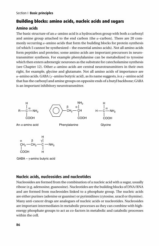
P1: PCX Printer: Yet To Come
9780521704632c07 CUFX213A/Peck 9780521618168 December 28, 2007 8:51
Section I Basic principles
Building blocks: amino acids, nucleic acids and sugars
Amino acidsThe basic structure of an α-amino acid is a hydrocarbon group with both a carboxyland amine group attached to the end carbon (the α-carbon). There are 20 com-monly occurring α-amino acids that form the building blocks for protein synthesis(of which 5 cannot be synthesized – the essential amino acids). Not all amino acidsform peptides and proteins; some amino acids are important precursors in neuro-transmitter synthesis. For example phenylalanine can be metabolized to tyrosinewhich then enters adrenergic neurones as the substrate for catecholamine synthesis(see Chapter 12). Other α-amino acids are central neurotransmitters in their ownright, for example, glycine and glutamate. Not all amino acids of importance areα-amino acids. GABA (γ -amino butyric acid), as its name suggests, is a γ -amino acidthat has the carboxyl and amine groups on opposite ends of a butyl backbone; GABAis an important inhibitory neurotransmitter.
R NH2 CH2
CH2
COOH
CH2 NH2C
H
H
CH
NH2
α
α β γ
β αC
H
COOH
An α-amino acid
GABA – γ-amino butyric acid
Phenylalanine Glycine
H NH2
αC
H
COOHCOOH
Nucleic acids, nucleosides and nucleotidesNucleosides are formed from the combination of a nucleic acid with a sugar, usuallyribose (e.g. adenosine, guanosine). Nucleotides are the building blocks of DNA/RNAand are formed from nucleosides linked to a phosphate group. The nucleic acidsare either purines (adenine or guanine) or pyrimidines (cytosine, uracil or thymine).Many anti-cancer drugs are analogues of nucleic acids or nucleotides. Nucleosidesare important intermediates in metabolic processes as they can combine with high-energy phosphate groups to act as co-factors in metabolic and catabolic processeswithin the cell.
86
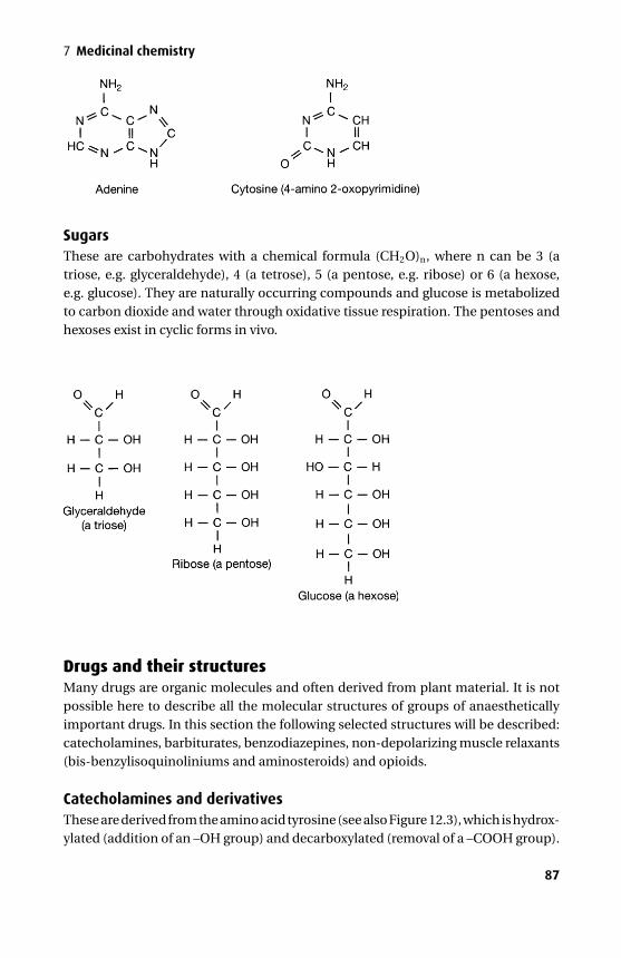
P1: PCX Printer: Yet To Come
9780521704632c07 CUFX213A/Peck 9780521618168 December 28, 2007 8:51
7 Medicinal chemistry
SugarsThese are carbohydrates with a chemical formula (CH2O)n, where n can be 3 (atriose, e.g. glyceraldehyde), 4 (a tetrose), 5 (a pentose, e.g. ribose) or 6 (a hexose,e.g. glucose). They are naturally occurring compounds and glucose is metabolizedto carbon dioxide and water through oxidative tissue respiration. The pentoses andhexoses exist in cyclic forms in vivo.
Drugs and their structuresMany drugs are organic molecules and often derived from plant material. It is notpossible here to describe all the molecular structures of groups of anaestheticallyimportant drugs. In this section the following selected structures will be described:catecholamines, barbiturates, benzodiazepines, non-depolarizing muscle relaxants(bis-benzylisoquinoliniums and aminosteroids) and opioids.
Catecholamines and derivativesThese are derived from the amino acid tyrosine (see also Figure 12.3), which is hydrox-ylated (addition of an –OH group) and decarboxylated (removal of a –COOH group).
87
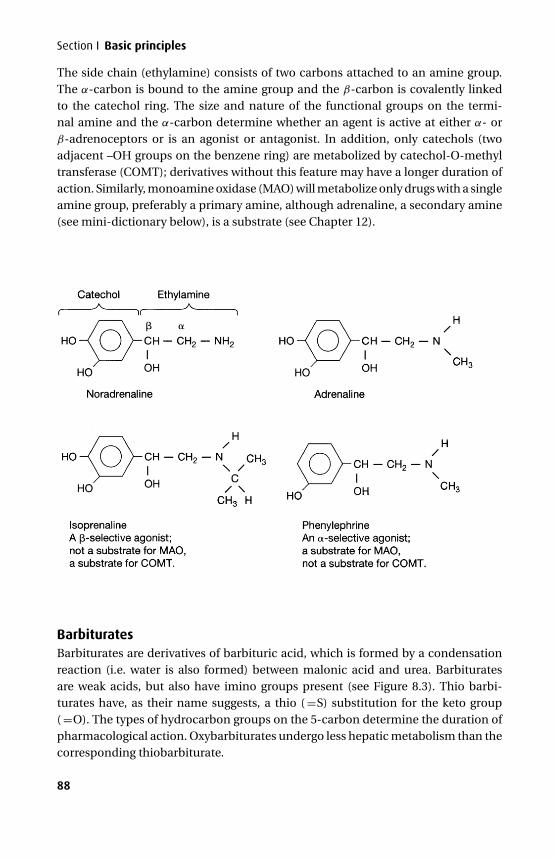
P1: PCX Printer: Yet To Come
9780521704632c07 CUFX213A/Peck 9780521618168 December 28, 2007 8:51
Section I Basic principles
The side chain (ethylamine) consists of two carbons attached to an amine group.The α-carbon is bound to the amine group and the β-carbon is covalently linkedto the catechol ring. The size and nature of the functional groups on the termi-nal amine and the α-carbon determine whether an agent is active at either α- orβ-adrenoceptors or is an agonist or antagonist. In addition, only catechols (twoadjacent –OH groups on the benzene ring) are metabolized by catechol-O-methyltransferase (COMT); derivatives without this feature may have a longer duration ofaction. Similarly, monoamine oxidase (MAO) will metabolize only drugs with a singleamine group, preferably a primary amine, although adrenaline, a secondary amine(see mini-dictionary below), is a substrate (see Chapter 12).
BarbituratesBarbiturates are derivatives of barbituric acid, which is formed by a condensationreaction (i.e. water is also formed) between malonic acid and urea. Barbituratesare weak acids, but also have imino groups present (see Figure 8.3). Thio barbi-turates have, as their name suggests, a thio ( =S) substitution for the keto group( =O). The types of hydrocarbon groups on the 5-carbon determine the duration ofpharmacological action. Oxybarbiturates undergo less hepatic metabolism than thecorresponding thiobarbiturate.
88
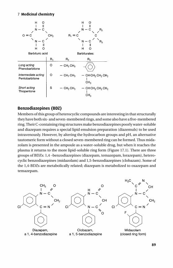
P1: PCX Printer: Yet To Come
9780521704632c07 CUFX213A/Peck 9780521618168 December 28, 2007 8:51
7 Medicinal chemistry
Benzodiazepines (BDZ)Members of this group of heterocyclic compounds are interesting in that structurallythey have both six- and seven-membered rings, and some also have a five-memberedring. Their C-containing ring structures make benzodiazepines poorly water-solubleand diazepam requires a special lipid emulsion preparation (diazemuls) to be usedintravenously. However, by altering the hydrocarbon groups and pH, an alternativetautomeric form without a closed seven-membered ring can be formed. Thus mida-zolam is presented in the ampoule as a water-soluble drug, but when it reaches theplasma it returns to the more lipid-soluble ring form (Figure 17.1). There are threegroups of BDZs: 1,4 –benzodiazepines (diazepam, temazepam, lorazepam), hetero-cyclic benzodiazepines (midazolam) and 1,5-benzodiazepines (clobazam). Some ofthe 1,4-BDZs are metabolically related; diazepam is metabolized to oxazepam andtemazepam.
89
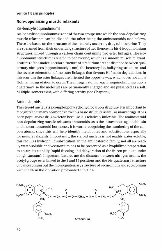
P1: PCX Printer: Yet To Come
9780521704632c07 CUFX213A/Peck 9780521618168 December 28, 2007 8:51
Section I Basic principles
Non-depolarizing muscle relaxants
Bis-benzylisoquinoliniumsBis-benzylisoquinoliniums is one of the two groups into which the non-depolarizingmuscle relaxants can be divided, the other being the aminosteroids (see below).These are based on the structure of the naturally occurring drug tubocurarine. Theyare so named from their underlying structure of two (hence the bis-) isoquinoliniumstructures, linked through a carbon chain containing two ester linkages. The iso-quinolinium structure is related to papaverine, which is a smooth muscle relaxant.Features of the moleculecular structure of atracurium are the distance between qua-ternary nitrogens (approximately 1 nm), the heterocyclic, bulky ring structures andthe reverse orientation of the ester linkages that favours Hofmann degradation. Inmivacurium the ester linkages are oriented the opposite way, which does not allowHofmann degradation to occur. The nitrogen atom in each isoquinolinium group isquaternary, so the molecules are permanently charged and are presented as a salt.Multiple isomers exist, with differing activity (see Chapter 5).
AminosteroidsThe steroid nucleus is a complex polycyclic hydrocarbon structure. It is important torecognize that many hormones have this basic structure as well as many drugs. It hasbeen popular as a drug skeleton because it is relatively inflexible. The aminosteroidnon-depolarizing muscle relaxants are steroids, as is the intravenous agent althesinand the corticosteroid hormones. It is worth recognizing the numbering of the car-bon atoms, since this will help identify metabolites and substitutions especiallyfor muscle relaxants. Importantly, the steroid nucleus is not readily water-soluble;this requires hydrophilic substitution. In the aminosteroid family, not all are read-ily water-soluble and vecuronium has to be presented as a lyophilized preparationto ensure its stability (rapid freezing and dehydration of the frozen product undera high vacuum). Important features are the distance between nitrogen atoms, theacetyl groups ester linked to the 3 and 17 positions and the bis-quaternary structureof pancuronium but the monoquaternary structure of vecuronium and rocuroniumwith the N- in the 2 position protonated at pH 7.4.
90
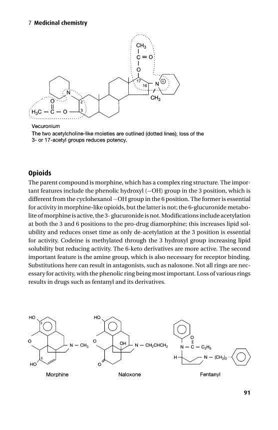
P1: PCX Printer: Yet To Come
9780521704632c07 CUFX213A/Peck 9780521618168 December 28, 2007 8:51
7 Medicinal chemistry
OpioidsThe parent compound is morphine, which has a complex ring structure. The impor-tant features include the phenolic hydroxyl (−OH) group in the 3 position, which isdifferent from the cyclohexanol −OH group in the 6 position. The former is essentialfor activity in morphine-like opioids, but the latter is not; the 6-glucuronide metabo-lite of morphine is active, the 3- glucuronide is not. Modifications include acetylationat both the 3 and 6 positions to the pro-drug diamorphine; this increases lipid sol-ubility and reduces onset time as only de-acetylation at the 3 position is essentialfor activity. Codeine is methylated through the 3 hydroxyl group increasing lipidsolubility but reducing activity. The 6-keto derivatives are more active. The secondimportant feature is the amine group, which is also necessary for receptor binding.Substitutions here can result in antagonists, such as naloxone. Not all rings are nec-essary for activity, with the phenolic ring being most important. Loss of various ringsresults in drugs such as fentanyl and its derivatives.
91
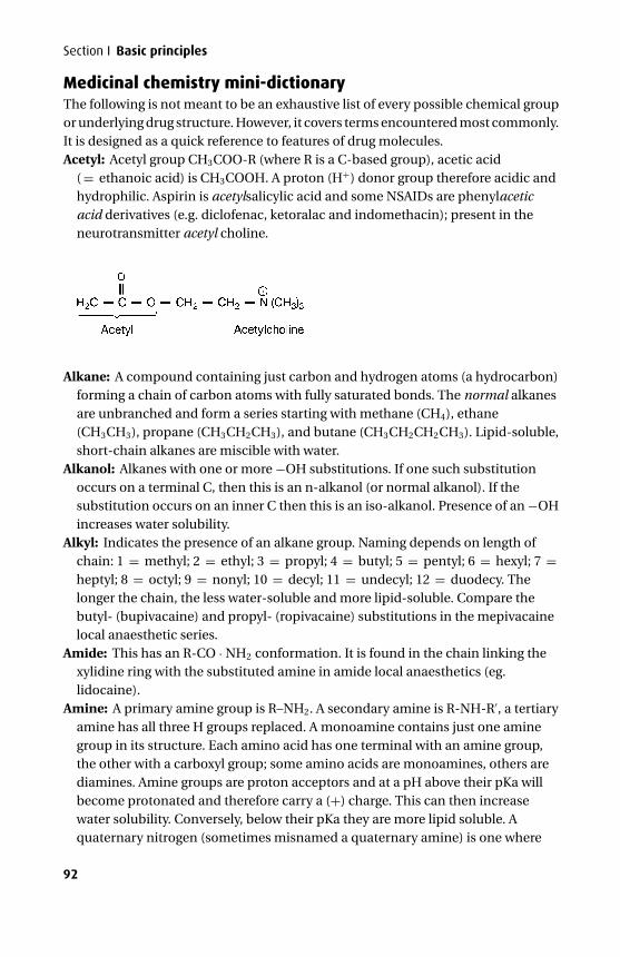
P1: PCX Printer: Yet To Come
9780521704632c07 CUFX213A/Peck 9780521618168 December 28, 2007 8:51
Section I Basic principles
Medicinal chemistry mini-dictionaryThe following is not meant to be an exhaustive list of every possible chemical groupor underlying drug structure. However, it covers terms encountered most commonly.It is designed as a quick reference to features of drug molecules.Acetyl: Acetyl group CH3COO-R (where R is a C-based group), acetic acid
( = ethanoic acid) is CH3COOH. A proton (H+) donor group therefore acidic andhydrophilic. Aspirin is acetylsalicylic acid and some NSAIDs are phenylaceticacid derivatives (e.g. diclofenac, ketoralac and indomethacin); present in theneurotransmitter acetyl choline.
Alkane: A compound containing just carbon and hydrogen atoms (a hydrocarbon)forming a chain of carbon atoms with fully saturated bonds. The normal alkanesare unbranched and form a series starting with methane (CH4), ethane(CH3CH3), propane (CH3CH2CH3), and butane (CH3CH2CH2CH3). Lipid-soluble,short-chain alkanes are miscible with water.
Alkanol: Alkanes with one or more −OH substitutions. If one such substitutionoccurs on a terminal C, then this is an n-alkanol (or normal alkanol). If thesubstitution occurs on an inner C then this is an iso-alkanol. Presence of an −OHincreases water solubility.
Alkyl: Indicates the presence of an alkane group. Naming depends on length ofchain: 1 = methyl; 2 = ethyl; 3 = propyl; 4 = butyl; 5 = pentyl; 6 = hexyl; 7 =heptyl; 8 = octyl; 9 = nonyl; 10 = decyl; 11 = undecyl; 12 = duodecy. Thelonger the chain, the less water-soluble and more lipid-soluble. Compare thebutyl- (bupivacaine) and propyl- (ropivacaine) substitutions in the mepivacainelocal anaesthetic series.
Amide: This has an R-CO · NH2 conformation. It is found in the chain linking thexylidine ring with the substituted amine in amide local anaesthetics (eg.lidocaine).
Amine: A primary amine group is R–NH2. A secondary amine is R-NH-R′, a tertiaryamine has all three H groups replaced. A monoamine contains just one aminegroup in its structure. Each amino acid has one terminal with an amine group,the other with a carboxyl group; some amino acids are monoamines, others arediamines. Amine groups are proton acceptors and at a pH above their pKa willbecome protonated and therefore carry a (+) charge. This can then increasewater solubility. Conversely, below their pKa they are more lipid soluble. Aquaternary nitrogen (sometimes misnamed a quaternary amine) is one where
92
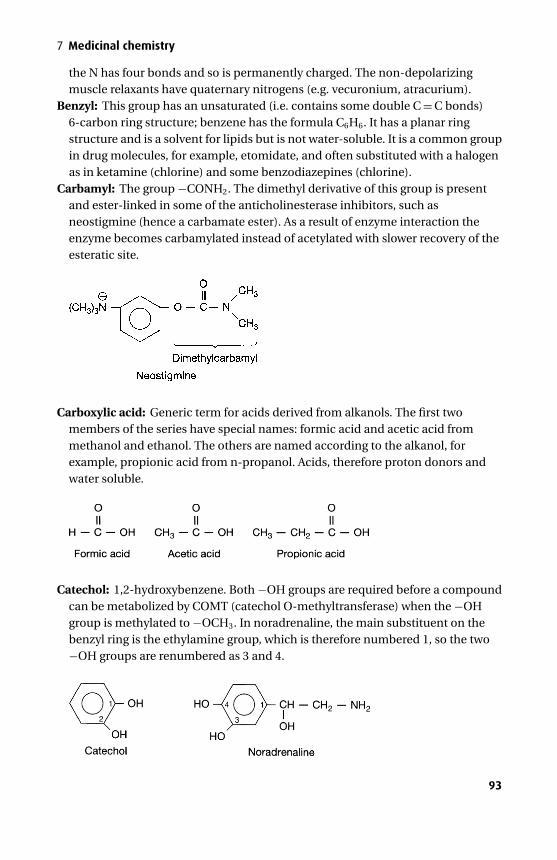
P1: PCX Printer: Yet To Come
9780521704632c07 CUFX213A/Peck 9780521618168 December 28, 2007 8:51
7 Medicinal chemistry
the N has four bonds and so is permanently charged. The non-depolarizingmuscle relaxants have quaternary nitrogens (e.g. vecuronium, atracurium).
Benzyl: This group has an unsaturated (i.e. contains some double C = C bonds)6-carbon ring structure; benzene has the formula C6H6. It has a planar ringstructure and is a solvent for lipids but is not water-soluble. It is a common groupin drug molecules, for example, etomidate, and often substituted with a halogenas in ketamine (chlorine) and some benzodiazepines (chlorine).
Carbamyl: The group −CONH2. The dimethyl derivative of this group is presentand ester-linked in some of the anticholinesterase inhibitors, such asneostigmine (hence a carbamate ester). As a result of enzyme interaction theenzyme becomes carbamylated instead of acetylated with slower recovery of theesteratic site.
Carboxylic acid: Generic term for acids derived from alkanols. The first twomembers of the series have special names: formic acid and acetic acid frommethanol and ethanol. The others are named according to the alkanol, forexample, propionic acid from n-propanol. Acids, therefore proton donors andwater soluble.
Catechol: 1,2-hydroxybenzene. Both −OH groups are required before a compoundcan be metabolized by COMT (catechol O-methyltransferase) when the −OHgroup is methylated to −OCH3. In noradrenaline, the main substituent on thebenzyl ring is the ethylamine group, which is therefore numbered 1, so the two−OH groups are renumbered as 3 and 4.
93
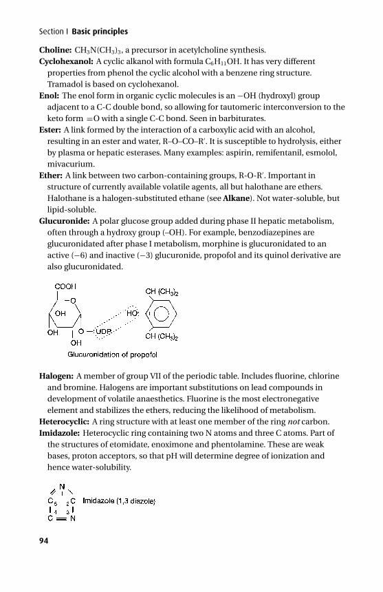
P1: PCX Printer: Yet To Come
9780521704632c07 CUFX213A/Peck 9780521618168 December 28, 2007 8:51
Section I Basic principles
Choline: CH3N(CH3)3, a precursor in acetylcholine synthesis.Cyclohexanol: A cyclic alkanol with formula C6H11OH. It has very different
properties from phenol the cyclic alcohol with a benzene ring structure.Tramadol is based on cyclohexanol.
Enol: The enol form in organic cyclic molecules is an −OH (hydroxyl) groupadjacent to a C-C double bond, so allowing for tautomeric interconversion to theketo form =O with a single C-C bond. Seen in barbiturates.
Ester: A link formed by the interaction of a carboxylic acid with an alcohol,resulting in an ester and water, R–O–CO–R′. It is susceptible to hydrolysis, eitherby plasma or hepatic esterases. Many examples: aspirin, remifentanil, esmolol,mivacurium.
Ether: A link between two carbon-containing groups, R-O-R′. Important instructure of currently available volatile agents, all but halothane are ethers.Halothane is a halogen-substituted ethane (see Alkane). Not water-soluble, butlipid-soluble.
Glucuronide: A polar glucose group added during phase II hepatic metabolism,often through a hydroxy group (–OH). For example, benzodiazepines areglucuronidated after phase I metabolism, morphine is glucuronidated to anactive (−6) and inactive (−3) glucuronide, propofol and its quinol derivative arealso glucuronidated.
Halogen: A member of group VII of the periodic table. Includes fluorine, chlorineand bromine. Halogens are important substitutions on lead compounds indevelopment of volatile anaesthetics. Fluorine is the most electronegativeelement and stabilizes the ethers, reducing the likelihood of metabolism.
Heterocyclic: A ring structure with at least one member of the ring not carbon.Imidazole: Heterocyclic ring containing two N atoms and three C atoms. Part of
the structures of etomidate, enoximone and phentolamine. These are weakbases, proton acceptors, so that pH will determine degree of ionization andhence water-solubility.
94
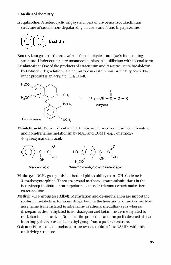
P1: PCX Printer: Yet To Come
9780521704632c07 CUFX213A/Peck 9780521618168 December 28, 2007 8:51
7 Medicinal chemistry
Isoquinoline: A heterocyclic ring system, part of bis-benzylisoquinoliniumstructure of certain non-depolarizing blockers and found in papaverine.
Keto: A keto group is the equivalent of an aldehyde group ( =O) but in a ringstructure. Under certain circumstances it exists in equilibrium with its enol form.
Laudanosine: One of the products of atracurium and cis-atracurium breakdownby Hofmann degradation. It is neurotoxic in certain non-primate species. Theother product is an acrylate (CH2CH–R).
Mandelic acid: Derivatives of mandelic acid are formed as a result of adrenalineand noradrenaline metabolism by MAO and COMT, e.g. 3-methoxy-4-hydroxymandelic acid.
Methoxy: –OCH3 group, this has better lipid solubility than –OH. Codeine is3-methoxymorphine. There are several methoxy- group substitutions in thebenzylisoquinolinium non-depolarizing muscle relaxants which make themwater-soluble.
Methyl: –CH3 group (see Alkyl). Methylation and de-methylation are importantroutes of metabolism for many drugs, both in the liver and in other tissues. Nor-adrenaline is methylated to adrenaline in adrenal medullary cells whereasdiazepam is de-methylated to nordiazepam and ketamine de-methylated tonorketamine in the liver. Note that the prefix nor- and the prefix desmethyl- canboth imply the removal of a methyl group from a parent structure.
Oxicam: Piroxicam and meloxicam are two examples of the NSAIDs with thisunderlying structure.
95
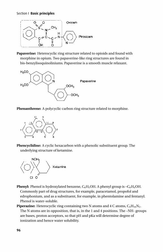
P1: PCX Printer: Yet To Come
9780521704632c07 CUFX213A/Peck 9780521618168 December 28, 2007 8:51
Section I Basic principles
Papaverine: Heterocyclic ring structure related to opioids and found withmorphine in opium. Two papaverine-like ring structures are found inbis-benzylisoquinoliniums. Papaverine is a smooth muscle relaxant.
Phenanthrene: A polycyclic carbon ring structure related to morphine.
Phencyclidine: A cyclic hexacarbon with a phenolic substituent group. Theunderlying structure of ketamine.
Phenyl: Phenol is hydroxylated benzene, C6H5OH. A phenyl group is –C6H4OH.Commonly part of drug structures, for example, paracetamol, propofol andedrophonium, and as a substituent, for example, in phentolamine and fentanyl.Phenol is water-soluble.
Piperazine: Heterocyclic ring containing two N atoms and 4 C atoms, C4H10N2.The N atoms are in opposition, that is, in the 1 and 4 positions. The –NH- groupsare bases, proton acceptors, so that pH and pKa will determine degree ofionization and hence water solubility.
96
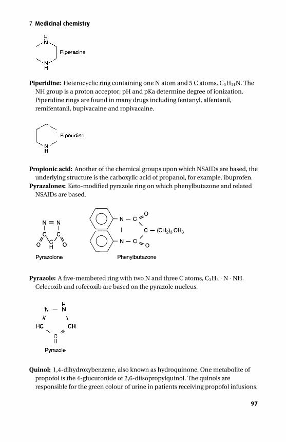
P1: PCX Printer: Yet To Come
9780521704632c07 CUFX213A/Peck 9780521618168 December 28, 2007 8:51
7 Medicinal chemistry
Piperidine: Heterocyclic ring containing one N atom and 5 C atoms, C5H11N. TheNH group is a proton acceptor; pH and pKa determine degree of ionization.Piperidine rings are found in many drugs including fentanyl, alfentanil,remifentanil, bupivacaine and ropivacaine.
Propionic acid: Another of the chemical groups upon which NSAIDs are based, theunderlying structure is the carboxylic acid of propanol, for example, ibuprofen.
Pyrazalones: Keto-modified pyrazole ring on which phenylbutazone and relatedNSAIDs are based.
Pyrazole: A five-membered ring with two N and three C atoms, C3H3 · N · NH.Celecoxib and rofecoxib are based on the pyrazole nucleus.
Quinol: 1,4-dihydroxybenzene, also known as hydroquinone. One metabolite ofpropofol is the 4-glucuronide of 2,6-diisopropylquinol. The quinols areresponsible for the green colour of urine in patients receiving propofol infusions.
97
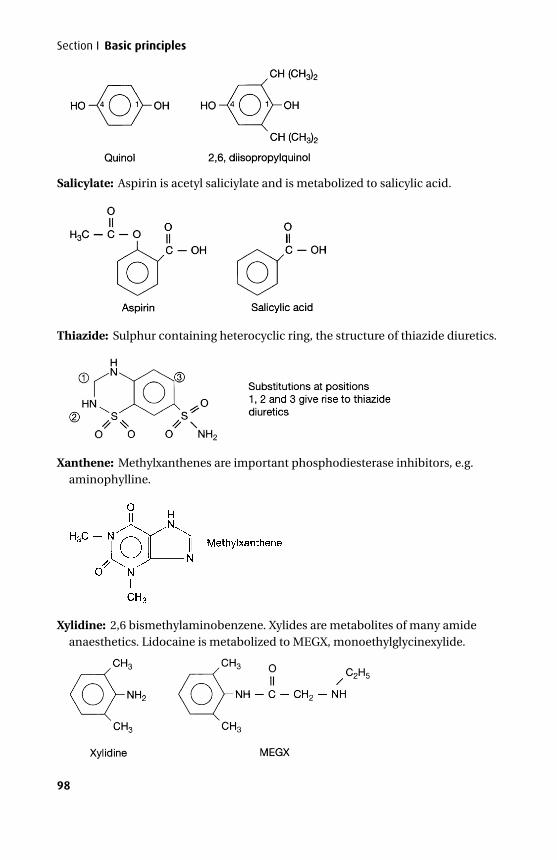
P1: PCX Printer: Yet To Come
9780521704632c07 CUFX213A/Peck 9780521618168 December 28, 2007 8:51
Section I Basic principles
Salicylate: Aspirin is acetyl saliciylate and is metabolized to salicylic acid.
Thiazide: Sulphur containing heterocyclic ring, the structure of thiazide diuretics.
Xanthene: Methylxanthenes are important phosphodiesterase inhibitors, e.g.aminophylline.
Xylidine: 2,6 bismethylaminobenzene. Xylides are metabolites of many amideanaesthetics. Lidocaine is metabolized to MEGX, monoethylglycinexylide.
98
P1: PCX Printer: Yet To Come
9780521704632c08 CUFX213A/Peck 9780521618168 December 28, 2007 10:38
SECTION II Core drugs in anaesthetic practice
8General anaesthetic agents
Our understanding of the mechanisms involved in the action of general anaestheticshas increased considerably in recent times and is discussed below. This is followedby sections discussing intravenous and inhaled anaesthetic agents.
Mechanisms of general anaesthetic actionAny mechanism of general anaesthetic action must be able to explain: loss of con-scious awareness, loss of response to noxious stimuli (anti-nociceptive effect) andperhaps most important of all, reversibility.
Anatomical sites of actionGeneral anaesthetic agents affect both brain and spinal cord to account for phys-iological responses to nociception, loss of consciousness and inhibition of explicitmemory. Auditory and sensory evoked potential data implicate the thalamus as themost likely primary target, but secondary sites such as the limbic system (associatedwith memory) and certain cortical areas are also important. Halogenated volatileanaesthetics appear to have a greater influence on spinal cord than do the intra-venous agents.
Molecular theoriesAt the beginning of the 19th century, Overton and Meyer independently describedthe linear correlation between the lipid solubility of anaesthetic agents and theirpotency (Figure 8.1). This correlation was so impressive, given the great variationin structure of these agents, that it suggested a non-specific mechanism of actionbased on this physicochemical property. Later interpretation pointed out that anyhighly lipophilic area was a potential site of action, with cell membranes being themost likely contender given the high concentration of lipids. There are problemswith a unified theory based on lipid interactions: some general anaesthetics, such asketamine, are extreme outliers, and the stereoisomers R-etomidate and S-etomidatehave identical lipid solubility but only R-etomidate has anaesthetic properties.
Membrane lipidsThere are several potential lipophilic sites in cell membranes, including the lipidbilayer itself and the annular lipids surrounding ionic channels.
99
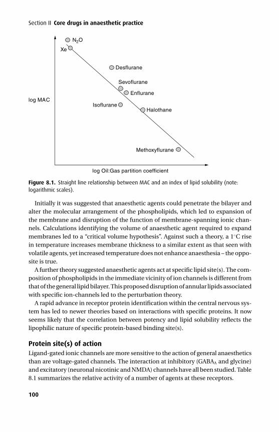
P1: PCX Printer: Yet To Come
9780521704632c08 CUFX213A/Peck 9780521618168 December 28, 2007 10:38
Section II Core drugs in anaesthetic practice
log MAC
Xe
N2O
Desflurane
Sevoflurane
Enflurane
IsofluraneHalothane
Methoxyflurane
log Oil:Gas partition coefficient
Figure 8.1. Straight line relationship between MAC and an index of lipid solubility (note:logarithmic scales).
Initially it was suggested that anaesthetic agents could penetrate the bilayer andalter the molecular arrangement of the phospholipids, which led to expansion ofthe membrane and disruption of the function of membrane-spanning ionic chan-nels. Calculations identifying the volume of anaesthetic agent required to expandmembranes led to a “critical volume hypothesis”. Against such a theory, a 1◦C risein temperature increases membrane thickness to a similar extent as that seen withvolatile agents, yet increased temperature does not enhance anaesthesia – the oppo-site is true.
A further theory suggested anaesthetic agents act at specific lipid site(s). The com-position of phospholipids in the immediate vicinity of ion channels is different fromthat of the general lipid bilayer. This proposed disruption of annular lipids associatedwith specific ion-channels led to the perturbation theory.
A rapid advance in receptor protein identification within the central nervous sys-tem has led to newer theories based on interactions with specific proteins. It nowseems likely that the correlation between potency and lipid solubility reflects thelipophilic nature of specific protein-based binding site(s).
Protein site(s) of actionLigand-gated ionic channels are more sensitive to the action of general anaestheticsthan are voltage-gated channels. The interaction at inhibitory (GABAA and glycine)and excitatory (neuronal nicotinic and NMDA) channels have all been studied. Table8.1 summarizes the relative activity of a number of agents at these receptors.
100
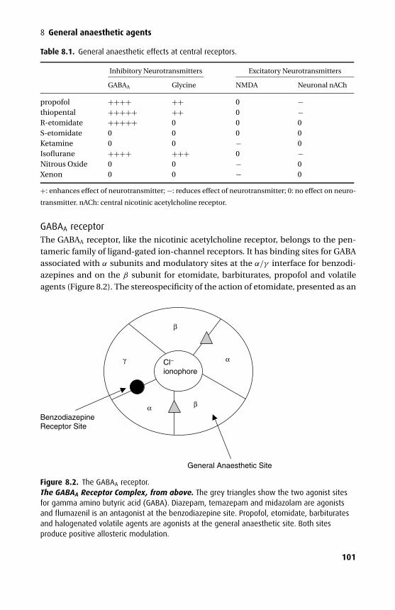
P1: PCX Printer: Yet To Come
9780521704632c08 CUFX213A/Peck 9780521618168 December 28, 2007 10:38
8 General anaesthetic agents
Table 8.1. General anaesthetic effects at central receptors.
Inhibitory Neurotransmitters Excitatory Neurotransmitters
GABAA Glycine NMDA Neuronal nACh
propofol ++++ ++ 0 −thiopental +++++ ++ 0 −R-etomidate +++++ 0 0 0S-etomidate 0 0 0 0Ketamine 0 0 − 0Isoflurane ++++ +++ 0 −Nitrous Oxide 0 0 − 0Xenon 0 0 − 0
+: enhances effect of neurotransmitter; −: reduces effect of neurotransmitter; 0: no effect on neuro-
transmitter. nACh: central nicotinic acetylcholine receptor.
GABAA receptorThe GABAA receptor, like the nicotinic acetylcholine receptor, belongs to the pen-tameric family of ligand-gated ion-channel receptors. It has binding sites for GABAassociated with α subunits and modulatory sites at the α/γ interface for benzodi-azepines and on the β subunit for etomidate, barbiturates, propofol and volatileagents (Figure 8.2). The stereospecificity of the action of etomidate, presented as an
β
γ Cl− ionophore
General Anaesthetic Site
Benzodiazepine Receptor Site
α
α
β
Figure 8.2. The GABAA receptor.The GABAA Receptor Complex, from above. The grey triangles show the two agonist sitesfor gamma amino butyric acid (GABA). Diazepam, temazepam and midazolam are agonistsand flumazenil is an antagonist at the benzodiazepine site. Propofol, etomidate, barbituratesand halogenated volatile agents are agonists at the general anaesthetic site. Both sitesproduce positive allosteric modulation.
101
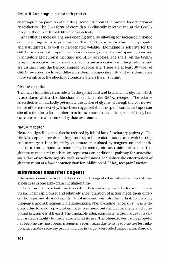
P1: PCX Printer: Yet To Come
9780521704632c08 CUFX213A/Peck 9780521618168 December 28, 2007 10:38
Section II Core drugs in anaesthetic practice
enantiopure preparation of the R(+) isomer, supports the protein-based action ofanaesthetics. The S(−) form of etomidate is clinically inactive and at the GABAA
receptor there is a 30-fold difference in activity.Anaesthetics increase channel opening time, so allowing for increased chloride
entry resulting in hyperpolarisation. The effect is seen for etomidate, propofoland barbiturates, as well as halogenated volatiles. Etomidate is selective for theGABAA receptor but propofol will also increase glycine channel opening time andis inhibitory at neuronal nicotinic and 5HT3 receptors. The site(s) on the GABAA
receptor associated with anaesthetic action are associated with the β subunit andare distinct from the benzodiazepine receptor site. There are at least 30 types ofGABAA receptor, each with different subunit composition; β2 and β3 subunits aremore sensitive to the effects of etomidate than is the β1 subunit.
Glycine receptorThe major inhibitory transmitter in the spinal cord and brainstem is glycine, whichis associated with a chloride channel similar to the GABAA receptor. The volatileanaesthetics all markedly potentiate the action of glycine, although there is no evi-dence of stereoselectivity. It has been suggested that the spinal cord is an importantsite of action for volatile rather than intravenous anaesthetic agents. Efficacy herecorrelates more with immobility than awareness.
NMDA receptorNeuronal signalling may also be reduced by inhibition of excitatory pathways. TheNMDA receptor is involved in long-term signal potentiation associated with learningand memory; it is activated by glutamate, modulated by magnesium and inhib-ited in a non-competitive manner by ketamine, nitrous oxide and xenon. Thisglutamate-mediated mechanism represents an additional pathway for anaesthe-sia. Other anaesthetic agents, such as barbiturates, can reduce the effectiveness ofglutamate but at a lower potency than for inhibition of GABAA receptor function.
Intravenous anaesthetic agentsIntravenous anaesthetics have been defined as agents that will induce loss of con-sciousness in one arm–brain circulation time.
The introduction of barbiturates in the 1930s was a significant advance in anaes-thesia. Their rapid onset and relatively short duration of action made them differ-ent from previously used agents. Hexobarbitone was introduced first, followed bythiopental and subsequently methohexitone. Phencyclidine (angel dust) was with-drawn due to serious psychotomimetic reactions, but the chemically related com-pound ketamine is still used. The imidazole ester, etomidate, is useful due to its car-diovascular stability but side-effects limit its use. The phenolic derivative propofolhas become the most popular agent in recent years due to its ready-to-use formula-tion, favourable recovery profile and use in target-controlled anaesthesia. Steroidal
102
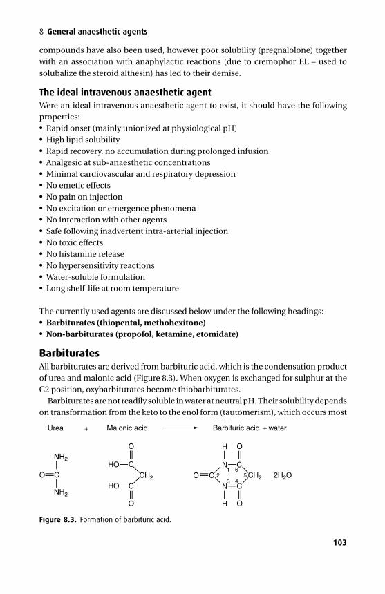
P1: PCX Printer: Yet To Come
9780521704632c08 CUFX213A/Peck 9780521618168 December 28, 2007 10:38
8 General anaesthetic agents
compounds have also been used, however poor solubility (pregnalolone) togetherwith an association with anaphylactic reactions (due to cremophor EL – used tosolubalize the steroid althesin) has led to their demise.
The ideal intravenous anaesthetic agentWere an ideal intravenous anaesthetic agent to exist, it should have the followingproperties:� Rapid onset (mainly unionized at physiological pH)� High lipid solubility� Rapid recovery, no accumulation during prolonged infusion� Analgesic at sub-anaesthetic concentrations� Minimal cardiovascular and respiratory depression� No emetic effects� No pain on injection� No excitation or emergence phenomena� No interaction with other agents� Safe following inadvertent intra-arterial injection� No toxic effects� No histamine release� No hypersensitivity reactions� Water-soluble formulation� Long shelf-life at room temperature
The currently used agents are discussed below under the following headings:� Barbiturates (thiopental, methohexitone)� Non-barbiturates (propofol, ketamine, etomidate)
BarbituratesAll barbiturates are derived from barbituric acid, which is the condensation productof urea and malonic acid (Figure 8.3). When oxygen is exchanged for sulphur at theC2 position, oxybarbiturates become thiobarbiturates.
Barbiturates are not readily soluble in water at neutral pH. Their solubility dependson transformation from the keto to the enol form (tautomerism), which occurs most
Urea
NH2
NH2
N
N
H
H
CH2 2 CH2 2H2OCOHO
HO
O
C
CO
C
O O
O
C
C
Malonic acid Barbituric acid + water
—— ——
— — — —— —— —
——
— —
—
—
—
——
——
—
—
—65
43
1
+
Figure 8.3. Formation of barbituric acid.
103
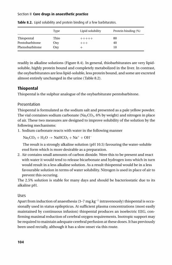
P1: PCX Printer: Yet To Come
9780521704632c08 CUFX213A/Peck 9780521618168 December 28, 2007 10:38
Section II Core drugs in anaesthetic practice
Table 8.2. Lipid solubility and protein binding of a few barbiturates.
Type Lipid solubility Protein binding (%)
Thiopental Thio +++++ 80Pentobarbitone Oxy +++ 40Phenobarbitone Oxy + 10
readily in alkaline solutions (Figure 8.4). In general, thiobarbiturates are very lipid-soluble, highly protein bound and completely metabolized in the liver. In contrast,the oxybarbiturates are less lipid-soluble, less protein bound, and some are excretedalmost entirely unchanged in the urine (Table 8.2).
ThiopentalThiopental is the sulphur analogue of the oxybarbiturate pentobarbitone.
PresentationThiopental is formulated as the sodium salt and presented as a pale yellow powder.The vial containes sodium carbonate (Na2CO3, 6% by weight) and nitrogen in placeof air. These two measures are designed to improve solubility of the solution by thefollowing mechanisms:1. Sodium carbonate reacts with water in the following manner
Na2CO3 + H2O → NaHCO3 + Na+ + OH−
The result is a strongly alkaline solution (pH 10.5) favouring the water-solubleenol form which is more desirable as a preparation.
2. Air contains small amounts of carbon dioxide. Were this to be present and reactwith water it would tend to release bicarbonate and hydrogen ions which in turnwould result in a less alkaline solution. As a result thiopental would be in a lessfavourable solution in terms of water solubility. Nitrogen is used in place of air toprevent this occuring.
The 2.5% solution is stable for many days and should be bacteriostatic due to itsalkaline pH.
UsesApart from induction of anaesthesia (3–7 mg.kg−1 intravenously) thiopental is occa-sionally used in status epilepticus. At sufficient plasma concentrations (most easilymaintained by continuous infusion) thiopental produces an isoelectric EEG, con-firming maximal reduction of cerebral oxygen requirements. Inotropic support maybe required to maintain adequate cerebral perfusion at these doses. It has previouslybeen used rectally, although it has a slow onset via this route.
104
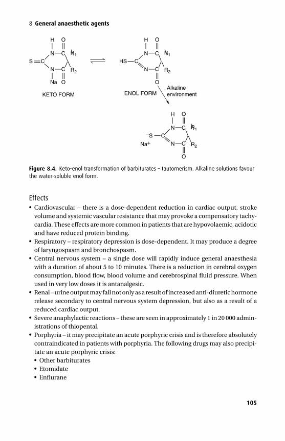
P1: PCX Printer: Yet To Come
9780521704632c08 CUFX213A/Peck 9780521618168 December 28, 2007 10:38
8 General anaesthetic agents
N
N
H
CHS
O
O
C
C
R1
R2
—
— —— —
——
—
—
—
—
——
— —N
N
Na
N
N
H
C−S
O
O
C
C
R1
R2
—
— —— —
——
—
—
—
—
——
— —
Na+
H
CS
KETO FORM ENOL FORMAlkalineenvironment
O
O
C
C
R1
R2
— —— ——
———
—
—
——
——
— —
Figure 8.4. Keto-enol transformation of barbiturates – tautomerism. Alkaline solutions favourthe water-soluble enol form.
Effects� Cardiovascular – there is a dose-dependent reduction in cardiac output, strokevolume and systemic vascular resistance that may provoke a compensatory tachy-cardia. These effects are more common in patients that are hypovolaemic, acidoticand have reduced protein binding.� Respiratory – respiratory depression is dose-dependent. It may produce a degreeof laryngospasm and bronchospasm.� Central nervous system – a single dose will rapidly induce general anaesthesiawith a duration of about 5 to 10 minutes. There is a reduction in cerebral oxygenconsumption, blood flow, blood volume and cerebrospinal fluid pressure. Whenused in very low doses it is antanalgesic.� Renal – urine output may fall not only as a result of increased anti-diuretic hormonerelease secondary to central nervous system depression, but also as a result of areduced cardiac output.� Severe anaphylactic reactions – these are seen in approximately 1 in 20 000 admin-istrations of thiopental.� Porphyria – it may precipitate an acute porphyric crisis and is therefore absolutelycontraindicated in patients with porphyria. The following drugs may also precipi-tate an acute porphyric crisis:� Other barbiturates� Etomidate� Enflurane
105
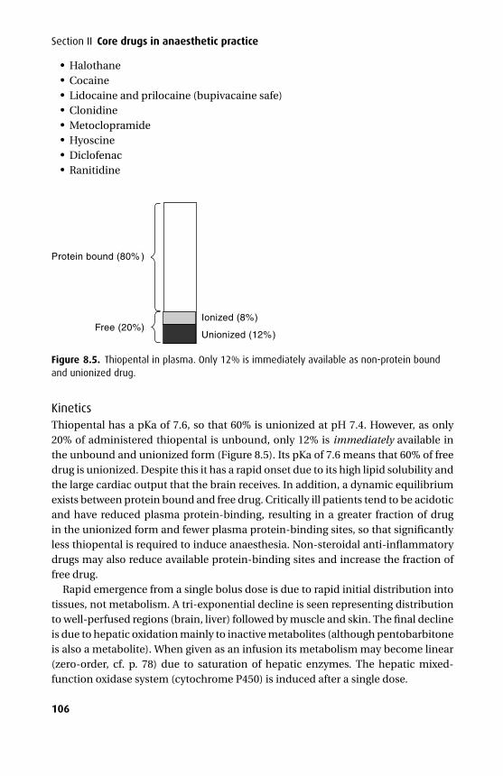
P1: PCX Printer: Yet To Come
9780521704632c08 CUFX213A/Peck 9780521618168 December 28, 2007 10:38
Section II Core drugs in anaesthetic practice� Halothane� Cocaine� Lidocaine and prilocaine (bupivacaine safe)� Clonidine� Metoclopramide� Hyoscine� Diclofenac� Ranitidine
Protein bound (80% )
Free (20%)Ionized (8%)
Unionized (12%)
Figure 8.5. Thiopental in plasma. Only 12% is immediately available as non-protein boundand unionized drug.
KineticsThiopental has a pKa of 7.6, so that 60% is unionized at pH 7.4. However, as only20% of administered thiopental is unbound, only 12% is immediately available inthe unbound and unionized form (Figure 8.5). Its pKa of 7.6 means that 60% of freedrug is unionized. Despite this it has a rapid onset due to its high lipid solubility andthe large cardiac output that the brain receives. In addition, a dynamic equilibriumexists between protein bound and free drug. Critically ill patients tend to be acidoticand have reduced plasma protein-binding, resulting in a greater fraction of drugin the unionized form and fewer plasma protein-binding sites, so that significantlyless thiopental is required to induce anaesthesia. Non-steroidal anti-inflammatorydrugs may also reduce available protein-binding sites and increase the fraction offree drug.
Rapid emergence from a single bolus dose is due to rapid initial distribution intotissues, not metabolism. A tri-exponential decline is seen representing distributionto well-perfused regions (brain, liver) followed by muscle and skin. The final declineis due to hepatic oxidation mainly to inactive metabolites (although pentobarbitoneis also a metabolite). When given as an infusion its metabolism may become linear(zero-order, cf. p. 78) due to saturation of hepatic enzymes. The hepatic mixed-function oxidase system (cytochrome P450) is induced after a single dose.
106
P1: PCX Printer: Yet To Come
9780521704632c08 CUFX213A/Peck 9780521618168 December 28, 2007 10:38
8 General anaesthetic agents
Intra-arterial injectionAs 2.5% thiopental at pH 10.5 is injected into arterial blood with a pH of 7.4 thetautomeric equilibrium swings away from the enol towards the keto form resultingin a less water-soluble solution. This in turn leads to the precipitation of thiopentalcrystalls which become wedged into small blood vessels leading to ischaemia andpain. Thiopental does not precipitate when injected intravenously as it is continuallydiluted by more venous blood. Treatment should begin immediately and may includeintra-arterial injection of papaverine or procaine, analgesia, sympathetic block of thelimb and anticoagulation.
Peri-vascular injection is painful and may cause serious tissue necrosis if largedoses extravasate.
MethohexitoneMethohexitone is a methylated oxybarbiturate and is no longer available in the UK.
PresentationMethohexitone was produced as the sodium salt with sodium carbonate (6% byweight) which was readily soluble in water forming an alkaline solution (pH 11.0).It had a pKa = 7.9 so that 75% of unbound drug was unionized at pH 7.4. However,60% of an administered dose was protein bound. There are four optically activeisomers but the preparation used clinically was a racemic mixture of α-d and α-lmethohexitone.
UsesMethohexitone was used as a 1% solution at 1–2 mg.kg−1 for the induction of anaes-thesia where excitatory phenomena were of little concern (notably for electrocon-vulsive therapy).
EffectsMethohexitone has a similar pharmacological profile to thiopental, producing rapidloss of consciousness, rapid emergence due to distribution and exerts similar effectson the cardiovascular and hepatic systems. It may also precipitate a porphyriccrisis.
Differences from thiopentalMethohexitone sometimes caused an excitatory phase before loss of conscious-ness, with muscle twitching, increased tone and hiccup. It occasionally precipitatedconvulsions in those with a history of epilepsy and its use in this setting was con-troversial. Recovery was more rapid after methohexitone due to a higher hepaticclearance. When injected intra-arterially or subcutaneously there were fewer vascu-lar complications and there was less tissue damage. This was probably due to the
107
P1: PCX Printer: Yet To Come
9780521704632c08 CUFX213A/Peck 9780521618168 December 28, 2007 10:38
Section II Core drugs in anaesthetic practice
lower concentrations used. Methohexitone was associated with a greater incidenceof hypersensitivity reactions although these did not appear to be as severe. Its mainmetabolite hydroxymethohexitone had only limited hypnotic activity (Table 8.3).
Non-barbiturates
Propofol
PresentationThis phenolic derivative (2,6 diisopropylphenol) is highly lipid-soluble and is pre-sented as a 1% or 2% lipid–water emulsion (containing soya bean oil and purifiedegg phosphatide) due to poor solubility in water. It is a weak organic acid with apKa = 11 so that it is almost entirely unionized at pH 7.4.
UsesPropofol is used for the induction and maintenance of general anaesthesia and forsedation of ventilated patients in intensive care. The induction dose is 1–2 mg.kg−1
while a plasma concentration of 4–8 µg.ml−1 will maintain anaesthesia.
Effects� Cardiovascular – the systemic vascular resistance falls resulting in a drop in bloodpressure. A reflex tachycardia is rare and propofol is usually associated with abradycardia especially if administered with fentanyl or alfentanil. Sympatheticactivity and myocardial contractility are also reduced.� Respiratory – respiratory depression leading to apnoea is common. It is rare toobserve cough or laryngospasm following its use and so it is often used in anaes-thesia for ease of placement of a laryngeal mask.� Central nervous system – excitatory effects have been associated with propofolin up to 10% of patients. They probably do not represent true cortical seizureactivity; rather they are the manifestation of subcortical excitatory–inhibitory cen-tre imbalance. The movements observed are dystonic with choreiform elementsand opisthotonos. Propofol has been used to control status epilepticus.� Gut – some evidence exists to suggest that propofol possesses anti-emetic prop-erties following its use for induction, maintenance or in subhypnotic doses post-operatively. Anatagonism of the dopamine D2 receptor is a possible mechanism.� Pain – injection into small veins is painful but may be reduced if lidocaine is mixedwith propofol or if a larger vein is used.� Metabolic – a fat overload syndrome, with hyperlipidaemia, and fatty infiltrationof heart, liver, kidneys and lungs can follow prolonged infusion.� Miscellaneous – it may turn urine and hair green.
108
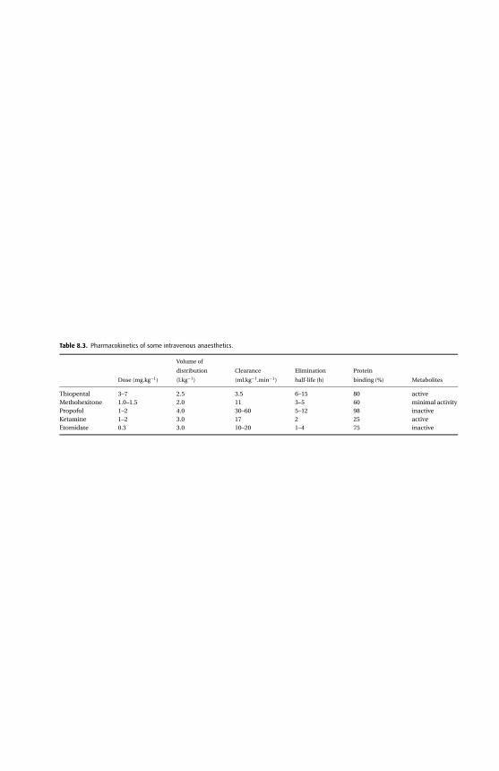
P1:P
CX
Prin
ter:YetToC
om
e
9780521704632c08C
UF
X213A
/Peck9780521618168
Decem
ber
28,200710:38
Table 8.3. Pharmacokinetics of some intravenous anaesthetics.
Dose (mg.kg−1)
Volume of
distribution
(l.kg−1)
Clearance
(ml.kg−1.min−1)
Elimination
half-life (h)
Protein
binding (%) Metabolites
Thiopental 3–7 2.5 3.5 6–15 80 activeMethohexitone 1.0–1.5 2.0 11 3–5 60 minimal activityPropofol 1–2 4.0 30–60 5–12 98 inactiveKetamine 1–2 3.0 17 2 25 activeEtomidate 0.3 3.0 10–20 1–4 75 inactive
109
P1: PCX Printer: Yet To Come
9780521704632c08 CUFX213A/Peck 9780521618168 December 28, 2007 10:38
Section II Core drugs in anaesthetic practice
KineticsPropofol is 98% protein bound to albumin and has the largest volume of distributionof all the induction agents at 4 l.kg−1. Following bolus administration, its durationof action is short due to the rapid decrease in plasma levels as it is distributed towell-perfused tissues.
Metabolism is largely hepatic; about 40% undergoing conjugation to a glu-curonide and 60% metabolized to a quinol, which is excreted as a glucuronideand sulphate, all of which are inactive and excreted in the urine. Its clearanceexceeds hepatic blood flow suggesting some extra-hepatic metabolism. Owing tothis high clearance, plasma levels fall more rapidly than those of thiopental fol-lowing the initial distribution phase. Its terminal elimination half-life is 5–12 hoursalthough it has been suggested that when sampling is performed for longer than24 hours the figure approaches 60 hours and may reflect the slow release of propo-fol from fat. During prolonged infusion its context-sensitive half-time increases,although where the infusion has been titrated carefully, waking may still be relativelyrapid.
ToxicityPropofol has been associated with the unexpected deaths of a small number ofchildren being ventilated for respiratory tract infection in intensive care. Progressivemetabolic acidosis and unresponsive bradycardia lead to death. The serum wasnoted to be lipaemic. While further small studies failed to demonstrate a worseoutcome when propofol was used as sedation for critically ill children, a more recentstudy showed a higher overall death rate and the Committee on Safety of Medicines(CSM) has advised that propofol should be contraindicated for sedation in intensivecare units in children 16 years and younger.
Patients allergic to eggs are usually allergic to egg protein or albumin. The eggcomponent of the propofol preparation is lecethin, which is a phosphatide, thereforeit is unlikely that allergic reactions are due to these components of its preparation.Propofol does not appear to be allergenic in patients who are sensitive to soya beansbecause all protein within the soya bean oil is removed.
It does not appear to cause any adverse effects when given intra-arterially, althoughonset of anaesthesia is delayed.
KetamineKetamine is a phencyclidine derivative.
Presentation and usesKetamine is presented as a racemic mixture or as the single S(+) enantiomer, whichis 2 to 3 times as potent as the R(–) enantiomer. It is soluble in water formingan acidic solution (pH 3.5–5.5). Three concentrations are available: 10, 50 and
110
P1: PCX Printer: Yet To Come
9780521704632c08 CUFX213A/Peck 9780521618168 December 28, 2007 10:38
8 General anaesthetic agents
100 mg.ml−1, and it may be given intravenously (1–2 mg.kg−1) or intramuscularly(5–10 mg.kg−1) for induction of anaesthesia. Intravenous doses of 0.2–0.5 mg.kg−1
may be used to provide analgesia during vaginal delivery and to facilitate the posi-tioning of patients with fractures before regional anaesthetic techniques are per-formed. It has been used via the oral and rectal route for sedation and also byintrathecal and epidural routes for analgesia. However, its use has been limited byunpleasant side effects.
Effects� Cardiovascular – ketamine is unlike other induction agents in that it pro-duces sympathetic nervous system stimulation, increasing circulating levels ofadrenaline and noradrenaline. Consequently heart rate, cardiac output, bloodpressure and myocardial oxygen requirements are all increased. However, itdoes not appear to precipitate arrhythmias. This indirect stimulation masks themild direct myocardial depressant effects that racemic ketamine would oth-erwise exert on the heart. S(+) ketamine produces less direct cardiac depres-sion in vitro compared with R(−) ketamine. In addition while racemic ketaminehas been shown to block ATP-sensitive potassium channels (the key mecha-nism of ischaemic myocardial preconditioning), S(+) ketamine does not, whichtherefore must be considered advantageous for patients with ischaemic heart dis-ease.� Respiratory – the respiratory rate may be increased and the laryngeal reflexes rela-tively preserved. A patent airway is often, but not always, maintained, and increasedmuscle tone associated with the jaw may precipitate airway obstruction. It causesbronchodilation and may be useful for patients with asthma.� Central nervous system – it produces a state of dissociative anaesthesia that isdemonstrated on EEG by dissociation between the thalamocortical and limbicsystems. In addition, intense analgesia and amnesia are produced. The α rhythmis replaced by θ and δ wave activity. Ketamine is different from other intravenousanaesthetics as it does not induce anaesthesia in one arm–brain circulation time –central effects becoming evident 90 seconds after an intravenous dose. Vivid andunpleasant dreams, hallucinations and delirium may follow its use. These emer-gence phenomena may be reduced by the concurrent use of benzodiazepines oropioids. S(+) ketamine produces less intense although no less frequent emergencephenomena. They are less common in the young and elderly and also in those leftto recover undisturbed. Cerebral blood flow, oxygen consumption and intracra-nial pressure are all increased. Muscle tone is increased and there may be jerkingmovements of the limbs.� Gut – nausea and vomiting occur more frequently than after propofol or thiopental.Salivation is increased requiring anticholinergic premedication.
111
P1: PCX Printer: Yet To Come
9780521704632c08 CUFX213A/Peck 9780521618168 December 28, 2007 10:38
Section II Core drugs in anaesthetic practice
KineticsFollowing an intravenous dose the plasma concentration falls in a bi-exponentialfashion. The initial fall is due to distribution across lipid membranes while the slowerphase is due to hepatic metabolism. Ketamine is the least protein bound (about 25%)of the intravenous anaesthetics and is demethylated to the active metabolite norke-tamine by hepatic P450 enzymes. Norketamine (which is 30% as potent as ketamine)is further metabolized to inactive glucuronide metabolites. The conjugated metabo-lites are excreted in the urine.
EtomidateEtomidate is an imidazole derivative and an ester. While it continues to be usedinfrequently in the UK it has been withdrawn in North America and Australia.
PresentationEtomidate is prepared as a 0.2% solution at pH of 4.1 and contains 35% v/v propyleneglycol to improve stability and reduce its irritant properties on injection. A lipidformulation is now also available.
UsesEtomidate is used for the induction of general anaesthesia at a dose of 0.3 mg.kg−1.
EffectsAt first glance etomidate would appear to have some desirable properties, but dueto its side effects its place in anaesthesia has remained limited.� Cardiovascular – of the commonly used intravenous anaesthetics it produces
the least cardiovascular disturbance. The peripheral vascular resistance may fallslightly (but less so than with other induction agents), while myocardial oxygensupply, contractility and blood pressure remain largely unchanged. Hypersensitiv-ity reactions are less common following etomidate and histamine release is rare.� Metabolic – it suppresses adrenocortical function by inhibition of the enzymes11β-hydroxylase and 17α-hydroxylase, resulting in inhibition of cortisol and aldos-terone synthesis. It was associated with an increase in mortality when used as aninfusion to sedate septic patients in intensive care. Single doses can influenceadrenocortical function but are probably of little clinical significance in otherwisefit patients. However it is it unlikely to be used to induce elective patients. In otherwords the situation in which it has the best cardiovascular profile is the unwellpatient in whom the consequences of steroid inhibition are likely to be the mostdetrimental.
112

P1: PCX Printer: Yet To Come
9780521704632c08 CUFX213A/Peck 9780521618168 December 28, 2007 10:38
8 General anaesthetic agents
——
——
—
—— —
—
C2H5
CH�C3H7
CH3
CH3
H
S
O
Thiopental
Methohexitone
Etomidate
N
N
N
N
N
O
O
O
H
N
H O
O
CH2�CH CH2——
CH3
CH�C C�C2H5
C2H5O — C
H — C — CH3
(CH3)2�CH CH�(CH3)2 Ketamine
O
Cl
NH�CH3
OH
Propofol
———
Figure 8.6. Chemical structure of some intravenous anaesthetics.� Miscellaneous – unpleasant side effects relate to pain on injection in up to 25% ofpatients, excitatory movements and nausea and vomiting. It may also precipitatea porphyric crisis.
KineticsEtomidate is 75% bound to albumin. Its actions are terminated by rapid distributioninto tissues, while its elimination from the body depends on hepatic metabolism and
113
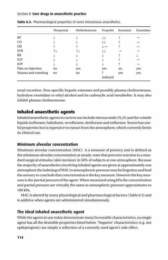
P1: PCX Printer: Yet To Come
9780521704632c08 CUFX213A/Peck 9780521618168 December 28, 2007 10:38
Section II Core drugs in anaesthetic practice
Table 8.4. Pharmacological properties of some intravenous anaesthetics.
Thiopental Methohexitone Propofol Ketamine Etomidate
BP ↓ ↓ ↓↓ ↑ →CO ↓ ↓ ↓↓ ↑ →HR ↑ ↑ ↓→ ↑ →SVR ↑↓ ↑↓ ↓↓ → →RR ↓ ↓ ↓ ↑ ↓ICP ↓ ↓ ↓ ↑ →IOP ↓ ↓ ↓ ↑ →Pain on injection no yes yes no yesNausea and vomiting no no ?
reducedyes yes
renal excretion. Non-specific hepatic esterases and possibly plasma cholinesterase,hydrolyse etomidate to ethyl alcohol and its carboxylic acid metabolite. It may alsoinhibit plasma cholinesterase.
Inhaled anaesthetic agentsInhaled anaesthetic agents in current use include nitrous oxide (N2O) and the volatileliquids isoflurane, halothane, sevoflurane, desflurane and enflurane. Xenon has use-ful properties but is expensive to extract from the atmosphere, which currently limitsits clinical use.
Minimum alveolar concentrationMinimum alveolar concentration (MAC) is a measure of potency and is defined asthe minimum alveolar concentration at steady-state that prevents reaction to a stan-dard surgical stimulus (skin incision) in 50% of subjects at one atmosphere. Becausethe majority of anaesthetics involving inhaled agents are given at approximately oneatmosphere the indexing of MAC to atmosphereic pressure may be forgotten and leadthe unwary to conclude that concentration is the key measure. However the key mea-sure is the partial pressure of the agent. When measured using kPa the concentrationand partial pressure are virtually the same as atmsopheric pressure approximates to100 kPa.
MAC is altered by many physiological and pharmacological factors (Table 8.5) andis additive when agents are administered simultaneously.
The ideal inhaled anaesthetic agentWhile the agents in use today demonstrate many favourable characteristics, no singleagent has all the desirable properties listed below. ‘Negative’ characteristics (e.g. notepileptogenic) are simply a reflection of a currently used agent’s side effect.
114
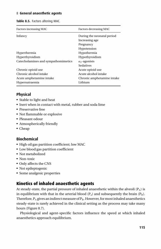
P1: PCX Printer: Yet To Come
9780521704632c08 CUFX213A/Peck 9780521618168 December 28, 2007 10:38
8 General anaesthetic agents
Table 8.5. Factors altering MAC.
Factors increasing MAC Factors decreasing MAC
Infancy During the neonatal periodIncreasing agePregnancyHypotension
Hyperthermia HypothermiaHyperthyroidism HypothyroidismCatecholamines and sympathomimetics α2-agonists
SedativesChronic opioid use Acute opioid useChronic alcohol intake Acute alcohol intakeAcute amphetamine intake Chronic amphetamine intakeHypernatraemia Lithium
Physical� Stable to light and heat� Inert when in contact with metal, rubber and soda lime� Preservative free� Not flammable or explosive� Pleasant odour� Atmospherically friendly� Cheap
Biochemical� High oil:gas partition coefficient; low MAC� Low blood:gas partition coefficient� Not metabolized� Non-toxic� Only affects the CNS� Not epileptogenic� Some analgesic properties
Kinetics of inhaled anaesthetic agentsAt steady-state, the partial pressure of inhaled anaesthetic within the alveoli (PA) isin equilibrium with that in the arterial blood (Pa) and subsequently the brain (PB).Therefore, PA gives an indirect measure of PB. However, for most inhaled anaestheticssteady-state is rarely achieved in the clinical setting as the process may take manyhours (Figure 8.7).
Physiological and agent-specific factors influence the speed at which inhaledanaesthetics approach equilibrium.
115
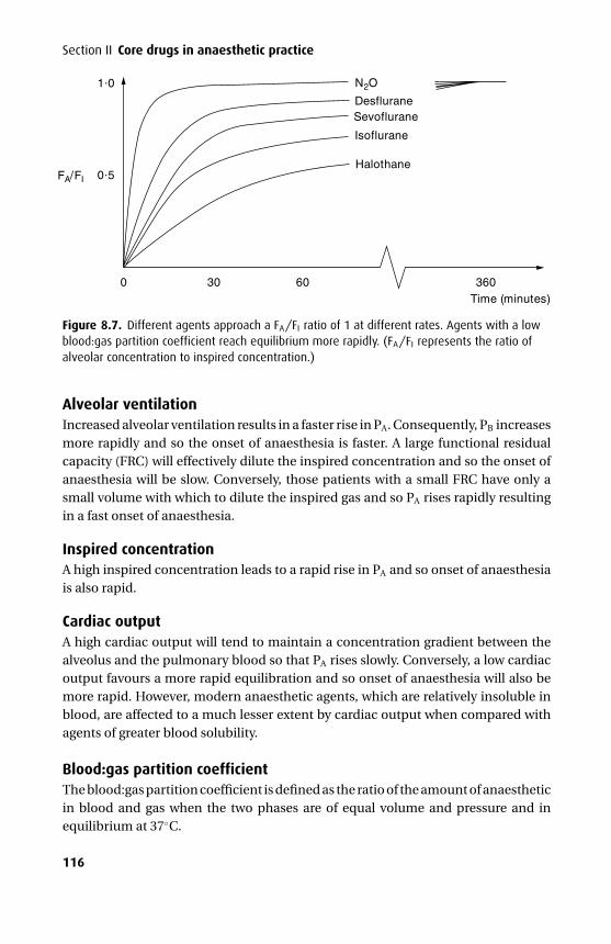
P1: PCX Printer: Yet To Come
9780521704632c08 CUFX213A/Peck 9780521618168 December 28, 2007 10:38
Section II Core drugs in anaesthetic practice
1 0
0 5FA/FI
0 30 60
N2O
DesfluraneSevoflurane
Isoflurane
Halothane
360Time (minutes)
Figure 8.7. Different agents approach a FA/FI ratio of 1 at different rates. Agents with a lowblood:gas partition coefficient reach equilibrium more rapidly. (FA/FI represents the ratio ofalveolar concentration to inspired concentration.)
Alveolar ventilationIncreased alveolar ventilation results in a faster rise in PA. Consequently, PB increasesmore rapidly and so the onset of anaesthesia is faster. A large functional residualcapacity (FRC) will effectively dilute the inspired concentration and so the onset ofanaesthesia will be slow. Conversely, those patients with a small FRC have only asmall volume with which to dilute the inspired gas and so PA rises rapidly resultingin a fast onset of anaesthesia.
Inspired concentrationA high inspired concentration leads to a rapid rise in PA and so onset of anaesthesiais also rapid.
Cardiac outputA high cardiac output will tend to maintain a concentration gradient between thealveolus and the pulmonary blood so that PA rises slowly. Conversely, a low cardiacoutput favours a more rapid equilibration and so onset of anaesthesia will also bemore rapid. However, modern anaesthetic agents, which are relatively insoluble inblood, are affected to a much lesser extent by cardiac output when compared withagents of greater blood solubility.
Blood:gas partition coefficientThe blood:gas partition coefficient is defined as the ratio of the amount of anaestheticin blood and gas when the two phases are of equal volume and pressure and inequilibrium at 37◦C.
116
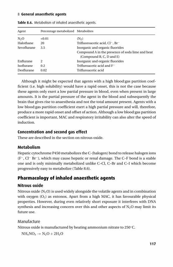
P1: PCX Printer: Yet To Come
9780521704632c08 CUFX213A/Peck 9780521618168 December 28, 2007 10:38
8 General anaesthetic agents
Table 8.6. Metabolism of inhaled anaesthetic agents.
Agent Percentage metabolized Metabolites
N2O <0.01 (N2)Halothane 20 Trifluoroacetic acid, Cl−, Br−
Sevoflurane 3.5 Inorganic and organic fluoridesCompound A in the presence of soda lime and heat
(Compound B, C, D and E)Enflurane 2 Inorganic and organic fluoridesIsoflurane 0.2 Trifluroacetic acid and F−
Desflurane 0.02 Trifluroacetic acid
Although it might be expected that agents with a high blood:gas partition coef-ficient (i.e. high solubility) would have a rapid onset, this is not the case becausethese agents only exert a low partial pressure in blood, even when present in largeamounts. It is the partial pressure of the agent in the blood and subsequently thebrain that gives rise to anaesthesia and not the total amount present. Agents with alow blood:gas partition coefficient exert a high partial pressure and will, therefore,produce a more rapid onset and offset of action. Although a low blood:gas partitioncoefficient is important, MAC and respiratory irritability can also alter the speed ofinduction.
Concentration and second gas effectThese are described in the section on nitrous oxide.
MetabolismHepatic cytochrome P450 metabolizes the C-(halogen) bond to release halogen ions(F−, Cl− Br−), which may cause hepatic or renal damage. The C–F bond is a stableone and is only minimally metabolized unlike C–Cl, C–Br and C–I which becomeprogressively easy to metabolize (Table 8.6).
Pharmacology of inhaled anaesthetic agents
Nitrous oxideNitrous oxide (N2O) is used widely alongside the volatile agents and in combinationwith oxygen (O2) as entonox. Apart from a high MAC, it has favourable physicalproperties. However, during even relatively short exposure it interferes with DNAsynthesis and increasing concern over this and other aspects of N2O may limit itsfuture use.
ManufactureNitrous oxide is manufactured by heating ammonium nitrate to 250◦C.
NH4NO3 → N2O + 2H2O
117
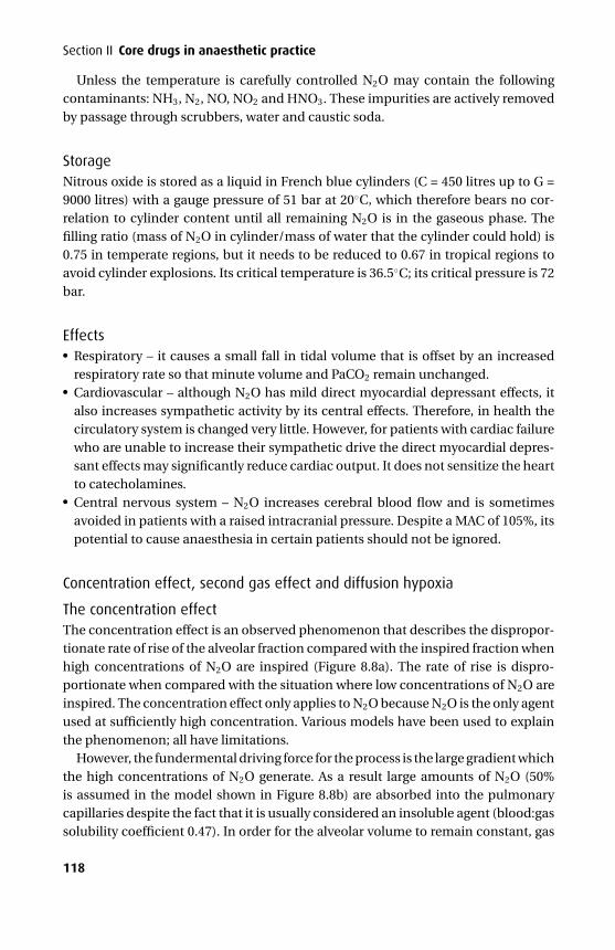
P1: PCX Printer: Yet To Come
9780521704632c08 CUFX213A/Peck 9780521618168 December 28, 2007 10:38
Section II Core drugs in anaesthetic practice
Unless the temperature is carefully controlled N2O may contain the followingcontaminants: NH3, N2, NO, NO2 and HNO3. These impurities are actively removedby passage through scrubbers, water and caustic soda.
StorageNitrous oxide is stored as a liquid in French blue cylinders (C = 450 litres up to G =9000 litres) with a gauge pressure of 51 bar at 20◦C, which therefore bears no cor-relation to cylinder content until all remaining N2O is in the gaseous phase. Thefilling ratio (mass of N2O in cylinder/mass of water that the cylinder could hold) is0.75 in temperate regions, but it needs to be reduced to 0.67 in tropical regions toavoid cylinder explosions. Its critical temperature is 36.5◦C; its critical pressure is 72bar.
Effects� Respiratory – it causes a small fall in tidal volume that is offset by an increasedrespiratory rate so that minute volume and PaCO2 remain unchanged.� Cardiovascular – although N2O has mild direct myocardial depressant effects, italso increases sympathetic activity by its central effects. Therefore, in health thecirculatory system is changed very little. However, for patients with cardiac failurewho are unable to increase their sympathetic drive the direct myocardial depres-sant effects may significantly reduce cardiac output. It does not sensitize the heartto catecholamines.� Central nervous system – N2O increases cerebral blood flow and is sometimesavoided in patients with a raised intracranial pressure. Despite a MAC of 105%, itspotential to cause anaesthesia in certain patients should not be ignored.
Concentration effect, second gas effect and diffusion hypoxia
The concentration effectThe concentration effect is an observed phenomenon that describes the dispropor-tionate rate of rise of the alveolar fraction compared with the inspired fraction whenhigh concentrations of N2O are inspired (Figure 8.8a). The rate of rise is dispro-portionate when compared with the situation where low concentrations of N2O areinspired. The concentration effect only applies to N2O because N2O is the only agentused at sufficiently high concentration. Various models have been used to explainthe phenomenon; all have limitations.
However, the fundermental driving force for the process is the large gradient whichthe high concentrations of N2O generate. As a result large amounts of N2O (50%is assumed in the model shown in Figure 8.8b) are absorbed into the pulmonarycapillaries despite the fact that it is usually considered an insoluble agent (blood:gassolubility coefficient 0.47). In order for the alveolar volume to remain constant, gas
118
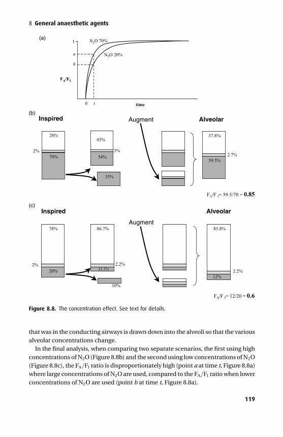
P1: PCX Printer: Yet To Come
9780521704632c08 CUFX213A/Peck 9780521618168 December 28, 2007 10:38
8 General anaesthetic agents
(b)
(c)
FA/F I= 12/20 = 0.6
FA/F I= 59.5/70 = 0.85
Augment
Augment
28%
70%
37.8%
59.5%
43%
54%2% 3%
2.7%
35%
Inspired Alveolar
78%
20%
86.7%
2% 2.2%
10%
11.1%
85.8%
12%2.2%
Alveolar Inspired
(a)N2O 70%
N2O 20%
FA/FI
1
0 time
a
b
t
Figure 8.8. The concentration effect. See text for details.
that was in the conducting airways is drawn down into the alveoli so that the variousalveolar concentrations change.
In the final analysis, when comparing two separate scenarios, the first using highconcentrations of N2O (Figure 8.8b) and the second using low concentrations of N2O(Figure 8.8c), the FA/FI ratio is disproportionately high (point a at time t, Figure 8.8a)where large concentrations of N2O are used, compared to the FA/FI ratio when lowerconcentrations of N2O are used (point b at time t, Figure 8.8a).
119
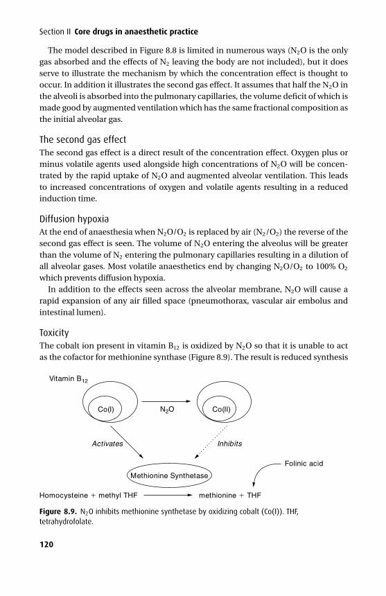
P1: PCX Printer: Yet To Come
9780521704632c08a CUFX213A/Peck 9780521618168 December 28, 2007 10:40
Section II Core drugs in anaesthetic practice
The model described in Figure 8.8 is limited in numerous ways (N2O is the onlygas absorbed and the effects of N2 leaving the body are not included), but it doesserve to illustrate the mechanism by which the concentration effect is thought tooccur. In addition it illustrates the second gas effect. It assumes that half the N2O inthe alveoli is absorbed into the pulmonary capillaries, the volume deficit of which ismade good by augmented ventilation which has the same fractional composition asthe initial alveolar gas.
The second gas effectThe second gas effect is a direct result of the concentration effect. Oxygen plus orminus volatile agents used alongside high concentrations of N2O will be concen-trated by the rapid uptake of N2O and augmented alveolar ventilation. This leadsto increased concentrations of oxygen and volatile agents resulting in a reducedinduction time.
Diffusion hypoxiaAt the end of anaesthesia when N2O/O2 is replaced by air (N2/O2) the reverse of thesecond gas effect is seen. The volume of N2O entering the alveolus will be greaterthan the volume of N2 entering the pulmonary capillaries resulting in a dilution ofall alveolar gases. Most volatile anaesthetics end by changing N2O/O2 to 100% O2
which prevents diffusion hypoxia.In addition to the effects seen across the alveolar membrane, N2O will cause a
rapid expansion of any air filled space (pneumothorax, vascular air embolus andintestinal lumen).
ToxicityThe cobalt ion present in vitamin B12 is oxidized by N2O so that it is unable to actas the cofactor for methionine synthase (Figure 8.9). The result is reduced synthesis
Vitamin B12
N2O
Methionine Synthetase
Co(I)
Folinic acid
Homocysteine methyl THF methionine THF
Co(II)
Activates Inhibits
Figure 8.9. N2O inhibits methionine synthetase by oxidizing cobalt (Co(I)). THF,tetrahydrofolate.
120
P1: PCX Printer: Yet To Come
9780521704632c08a CUFX213A/Peck 9780521618168 December 28, 2007 10:40
8 General anaesthetic agents
of methionine, thymidine, tetrahydrofolate and DNA. Methionine synthetase alsoappears to be directly inhibited by N2O. Exposure of only a few hours may result inmegaloblastic changes in bone marrow but more prolonged exposure (i.e. days) mayresult in agranulocytosis. Recovery is governed by synthesis (taking a few days) ofnew methionine synthase, but may be helped by the administration of folinic acid,which provides a different source of tetrahydrofolate.
In a properly scavenged environment where N2O concentrations are less than 50ppm there is no effect on DNA synthesis. However, in unscavenged dental surgerieswhere large amounts are used, chronic exposure may result in neurological syn-dromes that resemble subacute combined degeneration of the cord, as a result ofchronic vitamin B12 inactivation.
In experimental conditions N2O has been shown to be teratogenic to rats but thiseffect is prevented by folinic acid. While this has never been unequivocally demon-strated in humans, N2O is often not used in the first trimester when anaesthesia isrequired.
EntonoxEntonox is a 50:50 mixture of N2O and O2. The two gases effectively dissolve intoeach other and do not behave in a way that would be predicted from their individualproperties. This phenomenon is called the Poynting effect.
UsesEntonox is widely used for analgesia during labour and other painful procedures.
StorageEntonox is stored as a gas in French blue cylinders (G = 3200 litres; J = 6400 litres)with white and blue checked shoulders at 137 bar. It separates into its constituentparts below its pseudo-critical temperature, which is about –7◦C and is most likelyto occur at 117 bar. Higher or lower pressures reduce the likelihood of separation.When delivered via pipeline at 4.1 bar the pseudo-critical temperature is less than–30◦C. If a cylinder is used following separation, the inspired gas will initially producelittle analgesia as it contains mainly O2, but as the cylinder empties the mixture willbecome progressively potent and hypoxic as it approaches 100% N2O.
IsofluraneThis halogenated ethyl methyl ether is a structural isomer of enflurane. It is widelyused to maintain anaesthesia. Its physical properties are summarized in Table 8.7.
Effects� Respiratory – isoflurane depresses ventilation more than halothane but lessthan enflurane. Minute volume is decreased while respiratory rate and PaCO2
are increased. It is rarely used to induce anaesthesia due to its pungent smell,
121
P1: PCX Printer: Yet To Come
9780521704632c08a CUFX213A/Peck 9780521618168 December 28, 2007 10:40
Section II Core drugs in anaesthetic practice
which may cause upper airway irritability, coughing and breath-holding. How-ever, despite its pungent smell it causes some bronchodilation.� Cardiovascular – its main effect is to reduce systemic vascular resistance. The result-ing reflex tachycardia suggests that the carotid sinus reflex is preserved. It causesonly a small decrease in myocardial contractility and cardiac output. It has beensuggested that isoflurane may cause coronary steal whereby normally responsivecoronary arterioles are dilated and divert blood away from areas supplied by unre-sponsive diseased vessels, resulting in ischaemia. However more recent work hassuggested that as long as coronary perfusion is maintained coronary steal doesnot occur. In addition isofluane may have myocardial protective properties via itseffects on ATP-dependent potassium channels.� Central nervous system – of all the volatile agents isoflurane produces the bestbalance of reduced cerebral oxygen requirement and minimal increase in cerebralblood flow. At concentrations up to 1 MAC cerebral autoregulation is preserved.
MetabolismOnly 0.2% is metabolized and none of the products has been linked to toxicity.
ToxicityOwing to the presence of a –CHF2 group in its structure it may react with dry soda lime(or baralyme) producing carbon monoxide. Reports of this relate to circle systemsthat have been left with dry gas circulating over a weekend so that subsequent use ofisoflurane causes release of carbon monoxide. Enflurane and desflurane also possess–CHF2 groups and may react in a similar manner.
SevofluraneSevoflurane is a polyfluorinated isopropyl methyl ether and has the favourable com-bination of a relatively low blood:gas partition coefficient (0.7), pleasant odour andrelatively low MAC (1.8). However during storage where the concentration of addedwater is below 100 ppm it is susceptible to attack by Lewis acids at its ether and halo-gen bonds, releasing the highly toxic hydrofluoric acid (HF). (A Lewis acid is definedas any substance that can accept an electron pair and includes many metal oxidesbut also H+; glass is a source of Lewis acids.) Hydrofluoric acid corrodes glass expos-ing sevoflurane to further Lewis acids. As a result sevoflurane is formulated with300 ppm of water, which acts as a Lewis acid inhibitor. In addition it is stored in poly-thylene napthalate bottles, rather than glass. A dry formulation containing less than130 ppm water is now available and is presented in an aluminium bottle lined with anepoxyphenolic resin lacquer. Unlike the other volatile agents sevoflurane is achiral.
Manufacture� The one pot method – all the ingrediants are added together to produce sevofluraneand then water is added to 300 ppm.
122
P1: PCX Printer: Yet To Come
9780521704632c08a CUFX213A/Peck 9780521618168 December 28, 2007 10:40
8 General anaesthetic agents� The chloro-fluoro method – here the basic molecular architecture is manufac-tured but with chlorine attached. This is then substituted with flourine to producesevoflurane.
Effects� Respiratory – sevoflurane is a useful agent for induction of anaesthesia due toits pleasant odour and favourable physical properties. It does, however, depressventilation in a predictable fashion with a reduction in minute volume and a risein PaCO2.� Cardiovascular – the systemic vascular resistance falls and, due to an unchangedheart rate, the blood pressure falls. Cardiac contractility is unaffected and the heartis not sensitized to catecholamines. Vascular resistance to both cerebral and coro-nary circulations is decreased.� Central Nervous System – compared with halothane, there is evidence that childrenexhibit a higher incidence of post-operative agitation and delirium, which mayextend beyond the initial recovery period.
MetabolismSevoflurane undergoes hepatic metabolism by cytochrome P450 (isoform 2E1) to agreater extent than all the other commonly used volatile agents except halothane(Table 8.6). Hexafluoroisopropanol and inorganic F− (known to cause renal toxic-ity) are produced. The now obsolete volatile anaesthetic methoxyflurane was alsometabolized by hepatic P450 releasing F−, and when plasma levels rose above 50µmol.l−1 renal toxicity was observed. However, renal toxicity is not observed fol-lowing sevoflurane administration even when plasma levels reach 50 µmol.l−1. Apossible explanation lies in the additional metabolism of methoxyflurane by renalP450 to F−, which generates a high local concentration while sevoflurane undergoeslittle or no renal metabolism.
ToxicityCompounds A, B, C, D and E have all been identified when sevoflurane is used inthe pressence of carbon dioxide absorbents. Only compounds A and B (which is lesstoxic) are present in sufficient amount to make analysis feasible. Their formationis favoured in the presence of potassium hydroxide rather than sodium hydroxidebased absorbents particularly when dry. The reaction releases heat and consumessevoflurane both of which are readily detectible.
The lethal concentration of compound A in 50% of rats is 300–400 ppm after 3hours exposure. Extrapolation of these and other animal studies suggest a humannephrotoxic threshold of 150–200 ppm. Recent work suggests that even with flowrates of 0.25 l.min−1 for 5 hours the level of compound A peaks at less than 20 ppmand is not associated with abnormal tests of renal function.
123
P1: PCX Printer: Yet To Come
9780521704632c08a CUFX213A/Peck 9780521618168 December 28, 2007 10:40
Section II Core drugs in anaesthetic practice
HalothaneThis halogenated hydrocarbon is unstable when exposed to light, and corrodes cer-tain metals. It is stored with 0.01% thymol to prevent the liberation of free bromine.It dissolves into rubber and may leach out into breathing circuits after the vaporizeris turned off. Its physical properties are summarized in Table 8.7.
Effects� Respiratory – the minute ventilation is depressed largely due to a decreased tidalvolume. The normal responses to hypoxia and hypercarbia are also blunted andthese effects are more pronounced above 1 MAC. Bronchiolar tone is reduced andit is useful in asthmatic patients. Owing to its sweet non-irritant odour it may beused to induce anaesthesia.� Cardiovascular – halothane has significant effects on the heart. Bradycardia is pro-duced by increased vagal tone, depressed sino-atrial and atrioventricular activ-ity. It has direct myocardial depressant properties that reduce cardiac output. Itsensitizes the heart to catecholamines, which may lead to arrhythmias (especiallyventricular and bradyarrhythmias) and are more common than compared to otheragents. Where adrenaline is infiltrated to improve the surgical field less than 100µg per 10 minutes should be given. Drugs that specifically reduce atrioventricu-lar conductivity (e.g. verapamil) should be used with caution alongside halothane.The systemic vascular resistance is reduced resulting in increased cutaneous bloodflow. However, due to a reduced cardiac output blood flow to the liver and kidneysis reduced.� Central nervous system – halothane increases cerebral blood flow more than anyother volatile agent leading to significant increases in intracranial pressure above0.6 MAC. Cerebral oxygen requirements are reduced.
MetabolismUp to 25% of inhaled halothane undergoes oxidative metabolism by hepaticcytochrome P450 to produce trifluoroacetic acid, Br− and Cl−. However, reductivemetabolism producing F− and other reduced metabolites predominate when theliver becomes hypoxic. While these reduced metabolites are toxic it is thought thatthey are not involved in halothane hepatitis.
ToxicityHepatic damage may take one of two forms:� A reversible form that is often subclinical and associated with a rise in hepatic
transaminases. This is probably due to hepatic hypoxia.� Fulminant hepatic necrosis (halothane hepatitis). Trifluoroacetyl chloride (anoxidative metabolite of halothane) may behave as a hapten, binding covalentlywith hepatic proteins, inducing antibody formation. The diagnosis of halothane
124
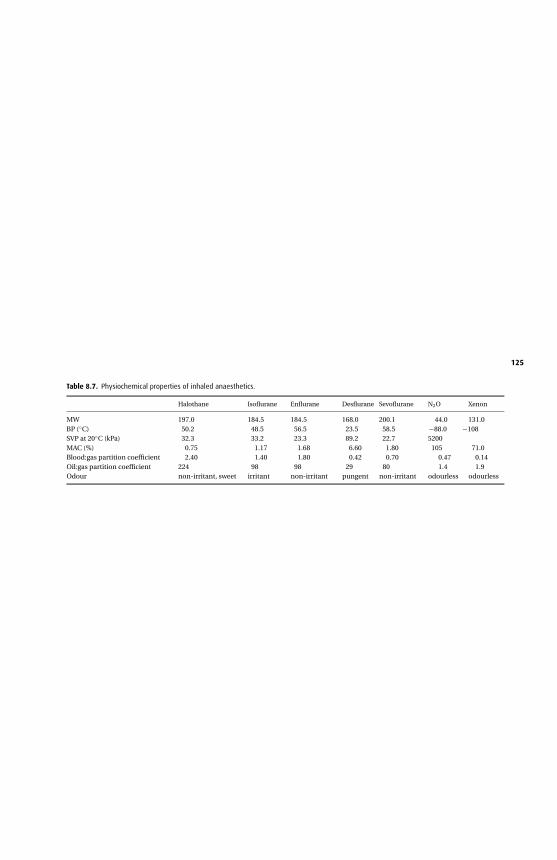
P1:P
CX
Prin
ter:YetToC
om
e
9780521704632c08aC
UF
X213A
/Peck
9780521618168D
ecemb
er28,2007
10:40
Table 8.7. Physiochemical properties of inhaled anaesthetics.
Halothane Isoflurane Enflurane Desflurane Sevoflurane N2O Xenon
MW 197.0 184.5 184.5 168.0 200.1 44.0 131.0BP (◦C) 50.2 48.5 56.5 23.5 58.5 −88.0 −108SVP at 20◦C (kPa) 32.3 33.2 23.3 89.2 22.7 5200MAC (%) 0.75 1.17 1.68 6.60 1.80 105 71.0Blood:gas partition coefficient 2.40 1.40 1.80 0.42 0.70 0.47 0.14Oil:gas partition coefficient 224 98 98 29 80 1.4 1.9Odour non-irritant, sweet irritant non-irritant pungent non-irritant odourless odourless
125
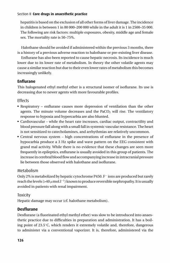
P1: PCX Printer: Yet To Come
9780521704632c08a CUFX213A/Peck 9780521618168 December 28, 2007 10:40
Section II Core drugs in anaesthetic practice
hepatitis is based on the exclusion of all other forms of liver damage. The incidencein children is between 1 in 80 000–200 000 while in the adult it is 1 in 2500–35 000.The following are risk factors: multiple exposures, obesity, middle age and femalesex. The mortality rate is 50–75%.
Halothane should be avoided if administered within the previous 3 months, thereis a history of a previous adverse reaction to halothane or pre-existing liver disease.
Enflurane has also been reported to cause hepatic necrosis. Its incidence is muchlower due to its lower rate of metabolism. In theory the other volatile agents maycause a similar reaction but due to their even lower rates of metabolism this becomesincreasingly unlikely.
EnfluraneThis halogenated ethyl methyl ether is a structural isomer of isoflurane. Its use isdecreasing due to newer agents with more favourable profiles.
Effects� Respiratory – enflurane causes more depression of ventilation than the otheragents. The minute volume decreases and the PaCO2 will rise. The ventilatoryresponse to hypoxia and hypercarbia are also blunted.� Cardiovascular – while the heart rate increases, cardiac output, contractility andblood pressure fall along with a small fall in systemic vascular resistance. The heartis not sensitized to catecholamines, and arrhythmias are relatively uncommon.� Central nervous system – high concentrations of enflurane in the presence ofhypocarbia produce a 3 Hz spike and wave pattern on the EEG consistent withgrand mal activity. While there is no evidence that these changes are seen morefrequently in epileptics, enflurane is usually avoided in this group of patients. Theincrease in cerebral blood flow and accompanying increase in intracranial pressurelie between those observed with halothane and isoflurane.
MetabolismOnly 2% is metabolized by hepatic cytochrome P450. F− ions are produced but rarelyreach the levels (>40 µmol.l−1) known to produce reversible nephropathy. It is usuallyavoided in patients with renal impairment.
ToxicityHepatic damage may occur (cf. halothane metabolism).
DesfluraneDesflurane (a fluorinated ethyl methyl ether) was slow to be introduced into anaes-thetic practice due to difficulties in preparation and administration. It has a boil-ing point of 23.5◦C, which renders it extremely volatile and, therefore, dangerousto administer via a conventional vaporizer. It is, therefore, administered via the
126
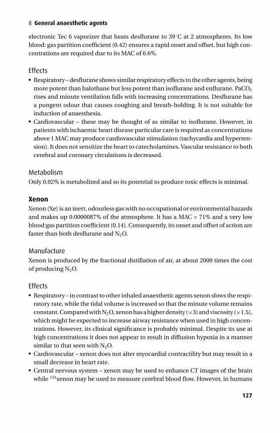
P1: PCX Printer: Yet To Come
9780521704632c08a CUFX213A/Peck 9780521618168 December 28, 2007 10:40
8 General anaesthetic agents
electronic Tec 6 vaporizer that heats desflurane to 39◦C at 2 atmospheres. Its lowblood: gas partition coefficient (0.42) ensures a rapid onset and offset, but high con-centrations are required due to its MAC of 6.6%.
Effects� Respiratory – desflurane shows similar respiratory effects to the other agents, beingmore potent than halothane but less potent than isoflurane and enflurane. PaCO2
rises and minute ventilation falls with increasing concentrations. Desflurane hasa pungent odour that causes coughing and breath-holding. It is not suitable forinduction of anaesthesia.� Cardiovascular – these may be thought of as similar to isoflurane. However, inpatients with ischaemic heart disease particular care is required as concentrationsabove 1 MAC may produce cardiovascular stimulation (tachycardia and hyperten-sion). It does not sensitize the heart to catecholamines. Vascular resistance to bothcerebral and coronary circulations is decreased.
MetabolismOnly 0.02% is metabolized and so its potential to produce toxic effects is minimal.
XenonXenon (Xe) is an inert, odourless gas with no occupational or environmental hazardsand makes up 0.0000087% of the atmosphere. It has a MAC = 71% and a very lowblood:gas partition coefficient (0.14). Consequently, its onset and offset of action arefaster than both desflurane and N2O.
ManufactureXenon is produced by the fractional distillation of air, at about 2000 times the costof producing N2O.
Effects� Respiratory – in contrast to other inhaled anaesthetic agents xenon slows the respi-ratory rate, while the tidal volume is increased so that the minute volume remainsconstant. Compared with N2O, xenon has a higher density (×3) and viscosity (×1.5),which might be expected to increase airway resistance when used in high concen-trations. However, its clinical significance is probably minimal. Despite its use athigh concentrations it does not appear to result in diffusion hypoxia in a mannersimilar to that seen with N2O.� Cardiovascular – xenon does not alter myocardial contractility but may result in asmall decrease in heart rate.� Central nervous system – xenon may be used to enhance CT images of the brainwhile 133xenon may be used to measure cerebral blood flow. However, in humans
127
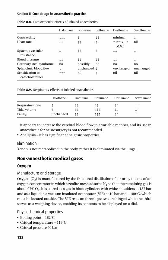
P1: PCX Printer: Yet To Come
9780521704632c08a CUFX213A/Peck 9780521618168 December 28, 2007 10:40
Section II Core drugs in anaesthetic practice
Table 8.8. Cardiovascular effects of inhaled anaesthetics.
Halothane Isoflurane Enflurane Desflurane Sevoflurane
Contractility ↓↓↓ ↓ ↓↓ minimal ↓Heart rate ↓↓ ↑↑ ↑ ↑ (↑↑ > 1.5
MAC)nil
Systemic vascularresistance
↓ ↓↓ ↓ ↓↓ ↓
Blood pressure ↓↓ ↓↓ ↓↓ ↓↓ ↓Coronary steal syndrome no possibly no no noSplanchnic blood flow ↓ unchanged ↓ unchanged unchangedSensitization to
catecholamines↑↑↑ nil ↑ nil nil
Table 8.9. Respiratory effects of inhaled anaesthetics.
Halothane Isoflurane Enflurane Desflurane Sevoflurane
Respiratory Rate ↑ ↑↑ ↑↑ ↑↑ ↑↑Tidal volume ↓ ↓↓ ↓↓↓ ↓↓ ↓PaCO2 unchanged ↑↑ ↑↑↑ ↑↑ ↑
it appears to increase the cerebral blood flow in a variable manner, and its use inanaesthesia for neurosurgery is not recommended.� Analgesia – it has significant analgesic properties.
EliminationXenon is not metabolized in the body, rather it is eliminated via the lungs.
Non-anaesthetic medical gases
Oxygen
Manufacture and storageOxygen (O2) is manufactured by the fractional distillation of air or by means of anoxygen concentrator in which a zeolite mesh adsorbs N2 so that the remaining gas isabout 97% O2. It is stored as a gas in black cylinders with white shoulders at 137 barand as a liquid in a vacuum insulated evaporator (VIE) at 10 bar and −180◦C, whichmust be located outside. The VIE rests on three legs; two are hinged while the thirdserves as a weighing device, enabling its contents to be displayed on a dial.
Physiochemical properties� Boiling point −182◦C� Critical temperature −119◦C� Critical pressure 50 bar
128
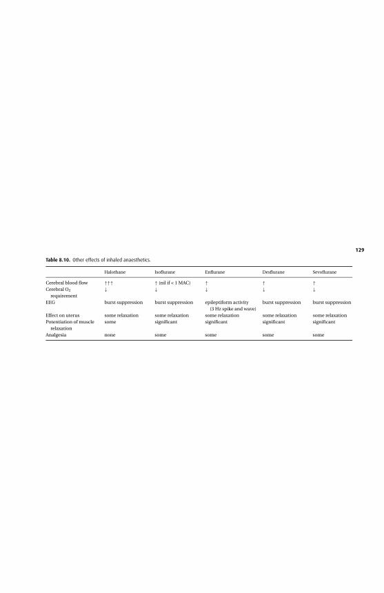
P1:P
CX
Prin
ter:YetToC
om
e
9780521704632c08aC
UF
X213A
/Peck
9780521618168D
ecemb
er28,2007
10:40
Table 8.10. Other effects of inhaled anaesthetics.
Halothane Isoflurane Enflurane Desflurane Sevoflurane
Cerebral blood flow ↑↑↑ ↑ (nil if < 1 MAC) ↑ ↑ ↑Cerebral O2
requirement↓ ↓ ↓ ↓ ↓
EEG burst suppression burst suppression epileptiform activity(3 Hz spike and wave)
burst suppression burst suppression
Effect on uterus some relaxation some relaxation some relaxation some relaxation some relaxationPotentiation of muscle
relaxationsome significant significant significant significant
Analgesia none some some some some
129
P1: PCX Printer: Yet To Come
9780521704632c08a CUFX213A/Peck 9780521618168 December 28, 2007 10:40
Section II Core drugs in anaesthetic practice
UsesIt is used to prevent hypoxaemia.
MeasurementDepending on the sample type, various means are used to measure O2. In a mixtureof gases a mass spectrometer, paramagnetic analyzer or fuel cell may be used; whendissolved in blood a Clarke electrode, transcutaneous electrode or pulse oximetrymay be used; in vitro blood samples may be analyzed by bench or co-oximetry.
Effects� Cardiovascular – if O2 is being used to correct hypoxaemia then an improvementin all cardiovascular parameters will be seen. However, prolonged administrationof 100% O2 will directly reduce cardiac output slightly and cause coronary arteryvasoconstriction. It causes a fall in pulmonary vascular resistance and pulmonaryartery pressure.� Respiratory – in healthy subjects, a high concentration causes mild respiratorydepression. However, in those patients who are truly dependent on a hypoxic driveto maintain respiration, even a modest concentration of O2 may prove fatal.
ToxicityO2 toxicity is caused by free radicals. They affect the CNS resulting in anxiety, nauseaand seizures when the partial pressure exceeds 200 kPa. The alveolar capillary mem-brane undergoes lipid peroxidation and regions of lung may collapse. Neonates aresusceptible to retrolental fibroplasia, which may be a result of vasoconstriction ofdeveloping retinal vessels during development.
Nitric oxideNitric oxide (NO) is an endogenous molecule but it is potentially a contaminantin nitrous oxide cylinders. It was formerly known as endothelium-derived relaxingfactor (EDRF).
SynthesisNitric oxide is synthesized from one of the terminal guanidino nitrogen atoms ofl-arginine in a process catalyzed by nitric oxide synthase (NOS), which is present intwo forms:� Constitutive – which is normally present in endothelial, neuronal, skeletal mus-
cle, cardiac tissue and platelets. Here NOS is Ca2+/calmodulin-dependent and isstimulated by cGMP.� Inducible – which is seen only after exposure to endotoxin or certain cytokines inendothelium, vascular smooth muscle, myocytes, macrophages and neutrophils.
130
P1: PCX Printer: Yet To Come
9780521704632c08a CUFX213A/Peck 9780521618168 December 28, 2007 10:40
8 General anaesthetic agents
Following induction large quantities of NO are produced, which may be cytotoxic.In addition, it may form radicals leading to cellular damage and capillary leakage.
Effects� Cardiovascular – vasodilator tone in small arteries and arterioles is dependenton a continuous supply of locally synthesized NO. Shear stresses in these ves-sels increase NO production and may account for flow-dependent vasodilatation.Nitric oxide derived from the endothelium inhibits platelet aggregation. In septicshock there is overproduction of NO resulting in hypotension and capillary leakage.� Respiratory – endogenous NO provides an important basal vasodilator tone in pul-monary and bronchial vessels, which may be reversed in hypoxia. When inhaled inconcentrations of up to 40 ppm it may reduce V/Q mismatching in acute respira-tory distress syndrome (ARDS) and reduce pulmonary hypertension in neonates.Inhaled NO has no effect on the systemic circulation due to its rapid inactivationwithin red blood cells. Its affinity for haemoglobin is 1500 times that of carbonmonoxide. It has no bronchodilator properties.� Immune – NO synthesized in macrophages and neutrophils can be toxic to certainpathogens and may be an important host defence mechanism.� Haematological – NO inhibits platelet aggregation.� Neuronal – nerves containing NO are widely distributed throughout the centralnervous system. Proposed roles include modulation of the state of arousal, painperception, programmed cell death and long-term neuronal depression and exci-tation whereby neurones may ‘remember’ previous signals. Peripheral neuronescontaining NO control regional blood flow in the corpus cavernosum.
N-monomethyl-l-arginine (l-NMMA) is a guanidino substituted analogue ofl-arginine, which inhibits NOS. While l-NMMA has been used to antagonize NOS,resulting in an increased blood pressure in septic shock, it does not alter the courseof the underlying pathology and has not been shown to alter survival.
Sodium nitroprusside and the organic nitrates (e.g. glyceryl trinitrate) exert theireffect by the spontaneous release of nitric oxide or metabolism to nitric oxide insmooth muscle cells.
UK guidelines for the use of inhaled NO in adult intensive care unitsAn expert group of physicians and representatives from the department of healthand industry issued the following guidelines in 1997:
Indications: severe ARDS (optimally ventilated, PaO2 < 12 kPa with FIO2 = 1), orright-sided cardiac failure.
Dose: maximum = 40 ppm, but use minimum effective dose.Equipment: a synchronized inspiratory injection system is considered optimal.
If a continuous delivery system is used it must be through a calibratedflowmeter. Stainless steel pressure regulators and connectors should be used.
131
P1: PCX Printer: Yet To Come
9780521704632c08a CUFX213A/Peck 9780521618168 December 28, 2007 10:40
Section II Core drugs in anaesthetic practice
Monitoring: chemiluminescence or electrochemical analyzers should be usedand are accurate to 1 ppm. Methaemoglobinaemia is only very rarelysignificant and is more likely in paediatric patients or those withmethaemoglobin reductase deficiency but levels should be checked beforeand after starting NO, and daily thereafter.
Exposure: Environmental NO levels should not exceed 25 ppm for 8 hours(time-weighted average). Scavenging is not required in a well ventilated unit.
Contraindications: Methaemoglobinaemia (bleeding diathesis, intracranialhaemorrhage, severe left ventricular failure).
HeliumHelium (He) is an inert gas presented as either Heliox (79% He, 21% O2) in browncylinders with white shoulders or as 100% helium in brown cylinders at 137 bar. Itdoes not support combustion.
Its key physical characteristic is its lower density (and hence specific gravity) thanboth air and oxygen.
Helium Heliox Oxygen Air
Specific gravity 0.178 0.337 1.091 1
Therefore, during turbulent flow the velocity will be higher when Heliox is used.This will reduce the work of breathing and improve oxygenation in patients with anupper airway obstruction such as a tumour. Helium/oxygen mixtures are also usedfor deep water diving to avoid nitrogen narcosis. The lower density of helium/oxygenmixtures produces higher frequency vocal sounds, giving the typical squeaky voice.
Carbon dioxideCarbon dioxide (CO2) is a colourless gas with a pungent odour at high concentrations.It is stored as a liquid at 51 bar at 20◦C in grey cylinders (C = 450 litres up to E = 1800litres).
Physiochemical properties� Boiling point −78.5◦C� Critical temperature 31◦C� Critical pressure 73.8 bar
UsesIt is used as the insufflating gas during laparoscopic procedures and occasionally tostimulate respiration following general anaesthesia. It is also used in cryotherapy.
132
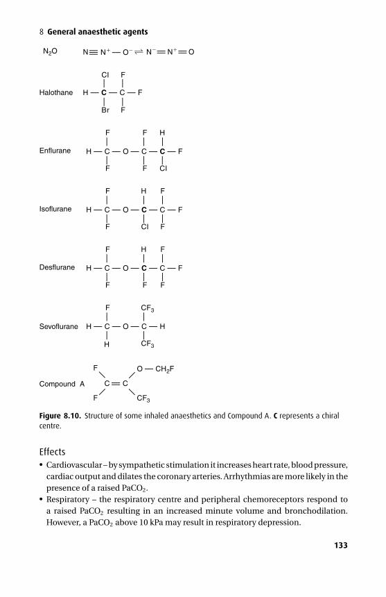
P1: PCX Printer: Yet To Come
9780521704632c08a CUFX213A/Peck 9780521618168 December 28, 2007 10:40
8 General anaesthetic agents
N2O N N N ON — O——— —— ——
Halothane
CI
Br F
F
H — C — C — F
— —
— —
Enflurane
F
F
F
F
H
CI
H — C — O — C — C — F— —
—
—— —
Isoflurane
F
F
H
CI
F
F
H — C — O — C — C — F
— —
—
—— —
Desflurane
F
F
H
F
F
F
H — C — O — C — C — F
— —
—
—— —
Sevoflurane
F
H
CF3
CF3
H — C — O — C — H
——
——
Compound A ——
F
F CF3
——
O — CH2F
C C—
—
Figure 8.10. Structure of some inhaled anaesthetics and Compound A. C represents a chiralcentre.
Effects� Cardiovascular – by sympathetic stimulation it increases heart rate, blood pressure,cardiac output and dilates the coronary arteries. Arrhythmias are more likely in thepresence of a raised PaCO2.� Respiratory – the respiratory centre and peripheral chemoreceptors respond toa raised PaCO2 resulting in an increased minute volume and bronchodilation.However, a PaCO2 above 10 kPa may result in respiratory depression.
133
P1: PCX Printer: Yet To Come
9780521704632c08a CUFX213A/Peck 9780521618168 December 28, 2007 10:40
Section II Core drugs in anaesthetic practice� Central nervous system – as PaCO2 rises so does cerebral blood flow and intracranialpressure. Beyond 10 kPa narcosis may ensue.
Carbon dioxide absorbentsAbsorbents consume CO2 to prevent rebreathing in a circle system. In the UK theyare mainly sodium hydroxide (sodalime) based while in the US they are potassiumhydroxide based (baralyme). There are three steps in the chemical reaction:
H2O + CO2 → H2CO3
H2CO3 + 2NaOH → Na2CO3 + 2H2ONa2CO3 + Ca(OH)2 → CaCO3 + 2NaOH.
134
P1: PCX Printer: Yet To Come
9780521704632c09 CUFX213A/Peck 9780521618168 December 28, 2007 11:33
9Analgesics
Pain is defined as an unpleasant sensory and emotional experience associated withactual or potential tissue damage. Since pain is so highly subjective, it may also bedescribed as being what the patient says it is.
Pain may be classified according to its presumed aetiology. Nociceptive pain isthe result of the stimulation of nociceptors by noxious stimuli, whilst neuropathicpain is the result of dysfunction of the nervous system. These may exist together asmixed pain. There is also visceral pain, the clearest example being that associatedwith gallstones.
An alternative classification is based on chronicity. The point at which acute painbecomes chronic has been suggested at about 12 weeks or when the pain is no longerthought to be due to the initial insult.
PhysiologyNociceptive impulses are triggered by the stimulation of nociceptors that respondto chemical, mechanical or thermal damage. The chemical mediators that initi-ate (H+, K+, acetylcholine, histamine, serotonin (5-HT), bradykinin), and sensitize(prostaglandins, leukotrienes, substance P, neurokinin A, calcitonin gene-relatedpeptide) the nociceptors are legion. Two types of primary afferent fibres exist:� small myelinated Aδ fibres (diameter 2–5 µm) that conduct sharp pain rapidly
(40 m.s−1)� unmyelinated C fibres (diameter < 2 µm) that conduct dull pain slowly (2 m.s−1)These fibres enter the dorsal horn of the spinal cord and synapse at different sites (Aδ
at Rexed laminae II and V; C at Rexed laminae II). The substantia gelatinosa (laminaII) integrates these inputs, from where second-order neurones form the ascendingspinothalamic and spinoreticular pathways on the contralateral side. Descendingpathways and the larger Aβ fibres conducting ‘touch’ stimulate inhibitory interneu-rones within the substantia gelatinosa and inhibit C fibre nociceptive inputs. Thisforms the basis of the ‘gate theory’ of pain (Figure 9.1).
Pain may be modified by altering the neural pathway from its origin at the noci-ceptor to its interpretation within the central nervous system. The commonly usedagents are discussed below under the following headings:� Opioids and related drugs� Non-steroidal anti-inflammatory drugs (NSAIDs)
135
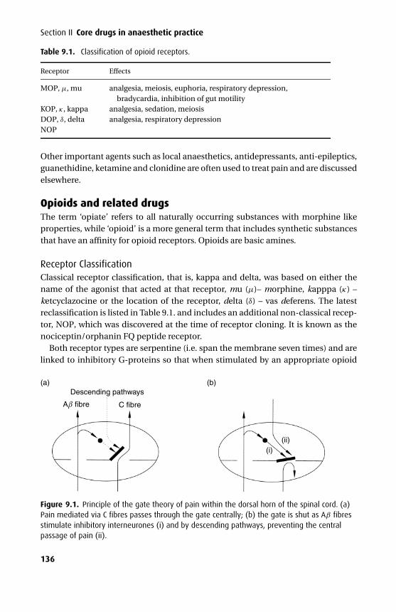
P1: PCX Printer: Yet To Come
9780521704632c09 CUFX213A/Peck 9780521618168 December 28, 2007 11:33
Section II Core drugs in anaesthetic practice
Table 9.1. Classification of opioid receptors.
Receptor Effects
MOP, µ, mu analgesia, meiosis, euphoria, respiratory depression,bradycardia, inhibition of gut motility
KOP, κ , kappa analgesia, sedation, meiosisDOP, δ, delta analgesia, respiratory depressionNOP
Other important agents such as local anaesthetics, antidepressants, anti-epileptics,guanethidine, ketamine and clonidine are often used to treat pain and are discussedelsewhere.
Opioids and related drugsThe term ‘opiate’ refers to all naturally occurring substances with morphine likeproperties, while ‘opioid’ is a more general term that includes synthetic substancesthat have an affinity for opioid receptors. Opioids are basic amines.
Receptor ClassificationClassical receptor classification, that is, kappa and delta, was based on either thename of the agonist that acted at that receptor, mu (µ)– morphine, kapppa (κ) –ketcyclazocine or the location of the receptor, delta (δ) – vas deferens. The latestreclassification is listed in Table 9.1. and includes an additional non-classical recep-tor, NOP, which was discovered at the time of receptor cloning. It is known as thenociceptin/orphanin FQ peptide receptor.
Both receptor types are serpentine (i.e. span the membrane seven times) and arelinked to inhibitory G-proteins so that when stimulated by an appropriate opioid
(a)Descending pathways
Aβ fibre C fibre
(ii)(i)
(b)
Figure 9.1. Principle of the gate theory of pain within the dorsal horn of the spinal cord. (a)Pain mediated via C fibres passes through the gate centrally; (b) the gate is shut as Aβ fibresstimulate inhibitory interneurones (i) and by descending pathways, preventing the centralpassage of pain (ii).
136
P1: PCX Printer: Yet To Come
9780521704632c09 CUFX213A/Peck 9780521618168 December 28, 2007 11:33
9 Analgesics
Table 9.2. Opioid receptor subtypes and their ligands.
Receptor
Ligand µ κ δ NOP
Endorphins +++ +++ +++Enkephalins + +++Dynorphins + +++N/OFQ +++Morphine +++ + +Fentanyl +++ +Naloxone +++ ++ ++Note: + represents receptor affinity, blank represents no receptor affinity.
agonist (e.g. morphine to µ) the following sequence occurs: voltage sensitive Ca2+
channels are closed, hyperpolarization by K+ efflux and adenylase cyclase inhibitionleading to reduced cAMP. These processes result in inhibition of transmitter releasebetween nerve cells.
MOP or µ-receptorThe µ-receptor is located thoughout the CNS including the cerebral cortex, the basalganglia, the spinal cord (presynaptically on primary afferent neurons within the dor-sal horn) and the periaqueductal grey (as the origin of the descending inhibitory con-trol pathway). Apart from analgesia µ-receptor stimulation produces wide-rangingeffects including respiratory depression (by reducing chemoreceptor sensitivity tocarbon dioxide), constipation (reduced secretions and peristalsis) and cardiovascu-lar depression.
KOP or κ-receptorThe original κ-receptor agonist was ketocyclazacine, which demonstrated a differentset of effects when compared with µ-receptor stimulation. The main advantage ofκ-receptor stimulation relates to a lack of respiratory depression, althoughκ-agonistsdo seem to have µ-antagonist effects thus limiting their use.
DOP or δ-receptorThe δ-receptor was the first to be cloned and is less widely spread throughout thecentral nervous system. Like µ-receptors, when stimulated they inhibit neurotrans-mitter release. It may also be involved in regulating mood and movement.
NOP-receptorWhen stimulated by nociceptin/orphanin FQ the NOP-receptor produces effectssimilar to µ-receptor stimulation. It acts at both spinal and supraspinal levels to
137
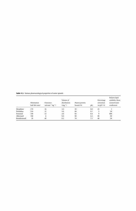
P1:P
CX
Prin
ter:YetToC
om
e
9780521704632c09C
UF
X213A
/Peck9780521618168
Decem
ber
28,200711:33
Table 9.3. Various pharmacological properties of some opioids.
Elimination
half-life (min)
Clearance
(ml.min−1.kg−1)
Volume of
distribution
(l.kg−1)
Plasma protein
bound (%) pKa
Percentage
unionized
(at pH 7.4)
Relative lipid
solubility (from
octanol:water
coefficient)
Morphine 170 16 3.5 35 8.0 23 1Pethidine 210 12 4.0 60 8.7 5 30Fentanyl 190 13 4.0 83 8.4 9 600Alfentanil 100 6 0.6 90 6.5 89 90Remifentanil 10 40 0.3 70 7.1 68 20
138
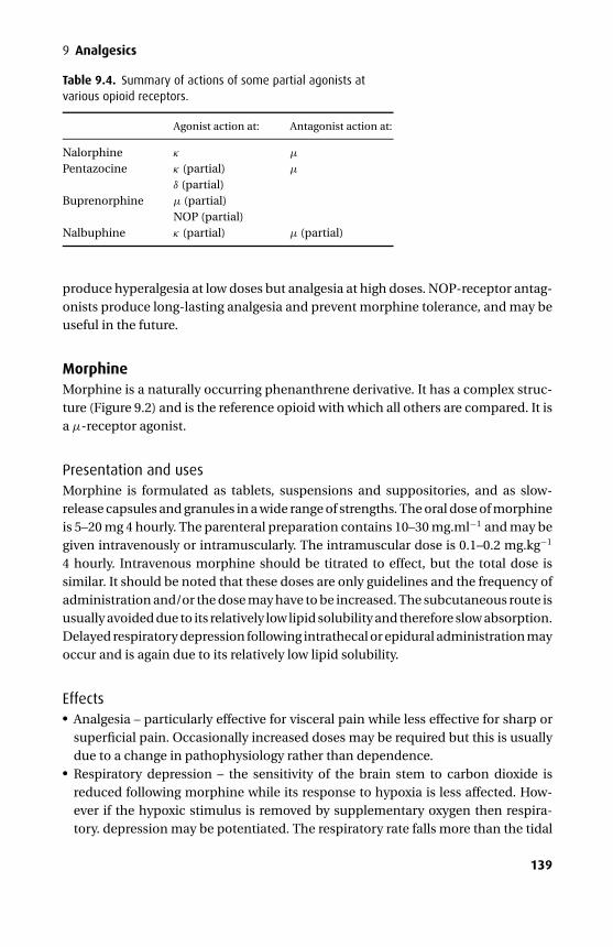
P1: PCX Printer: Yet To Come
9780521704632c09 CUFX213A/Peck 9780521618168 December 28, 2007 11:33
9 Analgesics
Table 9.4. Summary of actions of some partial agonists atvarious opioid receptors.
Agonist action at: Antagonist action at:
Nalorphine κ µ
Pentazocine κ (partial)δ (partial)
µ
Buprenorphine µ (partial)NOP (partial)
Nalbuphine κ (partial) µ (partial)
produce hyperalgesia at low doses but analgesia at high doses. NOP-receptor antag-onists produce long-lasting analgesia and prevent morphine tolerance, and may beuseful in the future.
MorphineMorphine is a naturally occurring phenanthrene derivative. It has a complex struc-ture (Figure 9.2) and is the reference opioid with which all others are compared. It isa µ-receptor agonist.
Presentation and usesMorphine is formulated as tablets, suspensions and suppositories, and as slow-release capsules and granules in a wide range of strengths. The oral dose of morphineis 5–20 mg 4 hourly. The parenteral preparation contains 10–30 mg.ml−1 and may begiven intravenously or intramuscularly. The intramuscular dose is 0.1–0.2 mg.kg−1
4 hourly. Intravenous morphine should be titrated to effect, but the total dose issimilar. It should be noted that these doses are only guidelines and the frequency ofadministration and/or the dose may have to be increased. The subcutaneous route isusually avoided due to its relatively low lipid solubility and therefore slow absorption.Delayed respiratory depression following intrathecal or epidural administration mayoccur and is again due to its relatively low lipid solubility.
Effects� Analgesia – particularly effective for visceral pain while less effective for sharp orsuperficial pain. Occasionally increased doses may be required but this is usuallydue to a change in pathophysiology rather than dependence.� Respiratory depression – the sensitivity of the brain stem to carbon dioxide isreduced following morphine while its response to hypoxia is less affected. How-ever if the hypoxic stimulus is removed by supplementary oxygen then respira-tory. depression may be potentiated. The respiratory rate falls more than the tidal
139
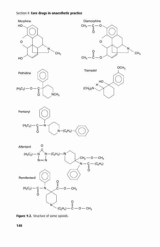
P1: PCX Printer: Yet To Come
9780521704632c09 CUFX213A/Peck 9780521618168 December 28, 2007 11:33
Section II Core drugs in anaesthetic practice
Morphine
HO
O
O
HO
Pethidine
Fentanyl
Alfentanil
Remifentanil
(H5C2) — O — C (CH3)2N
CH3 — C — O
OCH3
CH3
HHO
Tramadol
(H5C2) — C — N
CH2 — O — CH3
C — O — CH3
N — C — (C2H5)
(H5C2) — N
(H5C2) — C — N
(C2H4) — C — O — CH3
N (C2H4)
CH3
N
— —
O
— —
CH3 — C — O
O
O
N
Diamorphine
— —
——
O
— —
O
— —
O
O
N
— —
— —
O
— —
— —
N N
N—(C2H4)—N
O
NCH3
Figure 9.2. Structure of some opioids.
140
P1: PCX Printer: Yet To Come
9780521704632c09 CUFX213A/Peck 9780521618168 December 28, 2007 11:33
9 Analgesics
volume Morphine is anti-tussive. It may precipitate histamine release and bron-chospasm.� Nausea and vomiting – the chemoreceptor trigger zone is stimulated via 5-HT3
and dopamine receptors. The cells within the vomiting centre are depressed bymorphine and do not stimulate vomiting.� Central nervous system – sedation, euphoria and dysphoria occur with increasingdoses.� Circulatory – morphine may induce a mild bradycardia and hypotension secondaryto histamine release and a reduction in sympathetic tone. It has no direct myocar-dial depressant effects.� Gut – morphine constricts the sphincters of the gut. Constipation results from astate of spastic immobility of the bowel. Whilst the sphincter of Oddi is contractedby morphine thereby raising the pressure within the biliary tree the clinical signif-icance of this is unknown.� Histamine release – reducing the rate of administration will help tolimit histamine-induced bronchospasm and hypotension. Histamine releasemay result in a rash and pruritus but this may be reversed by naloxone.� Pruritus – most marked following intrathecal or epidural use. However, this doesnot appear to be due to histamine release and is generally not associated with a rash.Paradoxically, antihistamines may be effective treatment for pruritus, possibly asa result of their sedative effects.� Muscle rigidity – occasionally, morphine (and other opioids) can precipitate chestwall rigidity, which is thought to be due to opioid receptor interaction withdopaminergic and GABA pathways in the substantia nigra and striatum.� Meiosis – due to stimulation of the Edinger–Westphal nucleus, which can bereversed by atropine.� Endocrine – morphine inhibits the release of adrenocorticotrophic hormone(ACTH), prolactin and gonadotrophic hormones. Antidiuretic hormone (ADH)secretion is increased and may cause impaired water excretion and hyponatraemia.� Urinary – the tone of the bladder detrusor and vesical sphincter is increased andmay precipitate urinary retention. Ureteric tone is also increased.
KineticsWhen given orally morphine is ionized in the acidic gastric environment (becauseit is a weak base, pKa = 8.0) so that absorption is delayed until it reaches the rela-tively alkaline environment of the small bowel where it becomes unionized. Its oralbioavailability of 30% is due to hepatic first pass metabolism. Its peak effects fol-lowing intravenous or intramuscular injection are reached after 10 and 30 minutes,respectively, and it has a duration of action of 3–4 hours. It has been given by theepidural (2–4 mg) and intrathecal (0.2–1.0 mg) routes but this has been associatedwith delayed respiratory depression.
141
P1: PCX Printer: Yet To Come
9780521704632c09 CUFX213A/Peck 9780521618168 December 28, 2007 11:33
Section II Core drugs in anaesthetic practice
Morphine concentration in the brain falls slowly due to its low lipid solubility, andconsequently plasma concentrations do not correlate with its effects.
Morphine metabolism occurs mainly in the liver but also in the kidneys. Up to 70%is metabolized to morphine 3-glucuronide which appears to have effects on arousaland is possibly a µ-receptor antagonist. The other major metabolite is morphine 6-glucuronide, which is 13 times more potent than morphine and has a similar durationof action. They are both excreted in urine and accumulate in renal failure. Morphineis also N-demethylated. Neonates are more sensitive than adults to morphine dueto reduced hepatic conjugating capacity, and in the elderly peak plasma levels arehigher due to a reduced volume of distribution.
DiamorphineDiamorphine is a diacetylated morphine derivative with no affinity for opioid recep-tors. It is a prodrug whose active metabolites are responsible for its effects. It is saidto be approximately two times as potent as morphine.
Presentation and usesDiamorphine is available as 10 mg tablets and as a white powder for injection con-taining 5, 10, 30, 100 or 500 mg diamorphine hydrochloride, which is readily dis-solved before administration. It is used parenterally for the relief of severe pain anddyspnoea associated with pulmonary oedema at 2.5–10 mg. It is used intrathecally(0.1–0.4 mg) and via the epidural route (1–3 mg) for analgesia where, due to a higherlipid solubility, it is theoretically less likely to cause delayed respiratory depressionwhen compared with morphine.
KineticsOwing to its high lipid solubility it is well absorbed from the gut but has a low oralbioavailability due to an extensive first-pass metabolism. Its high lipid solubilityenables it to be administered effectively by the subcutaneous route. Once in theplasma it is 40% protein bound. It has a pKa = 7.6 so that 37% is in the unionizedform at pH 7.4. Metabolism occurs rapidly in the liver, plasma and central nervoussystem by ester hydrolysis to 6-monoacetylmorphine and morphine, which confer itsanalgesic and other effects. The plasma half-life of diamorphine itself is aproximatelyfive minutes.
It produces the greatest degree of euphoria of the opioids and subsequently hasbecome a drug of abuse.
PapaveretumPapaveretum is a semi-synthetic mixture of the anhydrous hydrochlorides of thealkaloids of opium. It contains morphine, codeine and papaverine. Noscapine wasremoved from its formulation after it had been shown to be teratogenic in animalstudies resulting in a standard dose of 15.4 mg, which is approximately equivalent
142
P1: PCX Printer: Yet To Come
9780521704632c09 CUFX213A/Peck 9780521618168 December 28, 2007 11:33
9 Analgesics
to 10 mg morphine. It is not given via the intrathecal or epidural route due to preser-vatives. Its effects are essentially the same as morphine and are antagonized bynaloxone.
MethadoneThe notable features of methadone are its relatively low first-pass metabolism result-ing in a relatively high oral bioavailability of 75% and a long plasma half-life. Thusit may be used orally, and as such it is used to treat those addicted to intravenousopioids, that is, diamorphine, by means of slow weaning programs. It is less sedativethan morphine.
Methadone may also act as an antagonist at the NMDA receptor and this is thoughtto be especially beneficial in the treatment of certain neuropathic pain that wouldbe otherwise resistant to typical opioids.
KineticsMethadone is 90% plasma protein bound and metabolism occurs in the liver to anumber of inactive metabolites. Its plasma half-life is 18–36 hours. Up to 40% isexcreted as unchanged drug in the urine, which is enhanced in acidic conditions.
CodeineCodeine (methylmorphine) is 10 times less potent than morphine and not suitablefor severe pain. The oral and intramuscular adult dose is 30–60 mg, the paediatricdose is 0.5–1 mg.kg−1 The intravenous route tends to cause hypotension probablyvia histamine release and is therefore avoided. It has been suggested codeine acts asno more than a prodrug for morphine.
KineticsThe presence of a methyl group reduces hepatic conjugation resulting in an oralbioavailability of 50%. Post-operative oral bioavailability is much more variable andranges from 20% to 80%.
A small proportion (5–15%) of codeine is eliminated unchanged in the urine whilethe remainder is eliminated via one of three metabolic pathways in the liver. The pre-dominant metabolic pathway is 6-hydroxy glucuronidation, although 10–20% under-goes N-demethylation to norcodeine, and 5–15% undergoes O-demethylation tomorphine. A number of other metabolites, such as normorphine and hydrocodone,have also been identified. Of these metabolites only morphine has significant activ-ity at µ-receptors. O-demethylation is dependent on the non-inducible cytochromeP450 (CYP2D6), which exhibits genetic polymorphism so that poor metabolizersexperience little pain relief. The frequency of poor metabolizers varies and is esti-mated at 9% of the UK population but 30% in the Hong Kong Chinese population.
143
P1: PCX Printer: Yet To Come
9780521704632c09 CUFX213A/Peck 9780521618168 December 28, 2007 11:33
Section II Core drugs in anaesthetic practice
DihydrocodeineDihydrocodeine is a synthetic opioid, which is structurally similar to codeine butis approximately twice as potent. Like codeine its metabolism is subject to geneticpolymorphism by virtue of the cytochrome P450 (CYP2D6).
PethidinePethidine is a synthetic phenylpiperidine derivative originally designed as an anti-cholinergic agent but was subsequently shown to have analgesic properties.
PresentationPethidine is available as tablets and as a solution for injection containing 10–50mg.ml−1. The intravenous and intramuscular dose is 0.5–1.0 mg.kg−1 and may berepeated 2–3 hourly. In common with all opioids the dose should be titrated toeffect.
UsesPethidine is often used during labour. Its high lipid solubility enables significantamounts to cross the placenta and reach the foetus. Following its metabolism, the lesslipid-soluble norpethidine accumulates in the foetus, levels peaking about 4 hoursafter the initial maternal intramuscular dose. Owing to reduced foetal clearance,the half-lives of both pethidine and norpethidine are prolonged by a factor ofthree.
EffectsPethidine shares the common opioid effects with morphine. However, differencesare seen:� Anticholinergic effects – it produces less marked meiosis and possibly a degree of
mydriasis, a dry mouth and sometimes tachycardia.� Gut – it is said to produce less biliary tract spasm than morphine but the clinicalsignificance of this is unclear.� Interactions – pethidine may produce a serious interaction if administered withmonoamine oxidase inhibitors (MAOI). This is probably due to central sero-toninenergic hyperactivity caused by pethidine inhibition of serotonin re-uptakein combination with an MAOI induced reduction in amine breakdown. Effectsinclude coma, labile circulation, convulsions and hyperpyrexia. Other opioids aresafe.
KineticsPethidine is more lipid-soluble than morphine resulting in a faster onset of action. Ithas an oral bioavailability of 50%. It is metabolized in the liver by ester hydrolysis tothe inactive pethidinic acid and by N-demethylation to norpethidine, which has halfthe analgesic activity of pethidine. Norpethidine has a longer elimination half-life
144
P1: PCX Printer: Yet To Come
9780521704632c09 CUFX213A/Peck 9780521618168 December 28, 2007 11:33
9 Analgesics
(14–21 hours) than pethidine and accumulates in renal failure. It has been associatedwith hallucinations and grand mal seizures following its accumulation. Its effectsare not reversed by naloxone. Norpethidine and pethidinic acid are excreted in theurine along with small amounts of unchanged pethidine. The duration of action ofpethidine is 120–150 minutes.
FentanylFentanyl is a synthetic phenylpiperidine derivative with a rapid onset of action. It isa µ-receptor agonist and as such shares morphine’s effects. However, it is less likelyto precipitate histamine release. High doses (50–150 µg.kg−1) significantly reduceor even eliminate the metabolic stress response to surgery but are associated withbradycardia and chest wall rigidity.
PresentationFentanyl is prepared as a colourless solution for injection containing 50 µg.ml−1, astransdermal patches that release between 25 and 100 µg per hour for 72 hours andas lozenges releasing 200 µg – 1.6 mg over 15 minutes.
UsesDoses vary enormously depending on the duration of analgesia and sedationrequired. For pain associated with minor surgery, 1–2 µg.kg−1 is used intravenouslyand has a duration of about 30 minutes. Higher doses are generally required to obtundthe stimulation of laryngoscopy. High doses (50–100 µg.kg−1) are used for an opioid-based anaesthetic (although a hypnotic is also required), and here its duration ofaction is extended to about 6 hours. Following prolonged administration by contin-uous infusion only its elimination half-life is apparent, leading to a more prolongedduration of action.
Fentanyl has also been used to augment the effects of local anaesthetics in spinaland epidural anaesthesia at 10–25 µg and 25–100 µg, respectively. Its high lipid sol-ubility ensures that a typical intrathecal dose does not cause delayed respiratorydepression as it diffuses rapidly from cerebrospinal fluid (CSF) into the spinal cord.This contrasts with morphine, which enters the spinal cord slowly leaving some to betransported in the CSF by bulk flow up to the midbrain. However, respiratory depres-sion is observed when epidural fentanyl is administered by continuous infusion oras repeated boluses.
KineticsIts onset of action is rapid following intravenous administration due to its high lipidsolubility (nearly 600 times more lipid-soluble than morphine). However, follow-ing the application of a transdermal patch, plasma levels take 12 hours to reachequilibrium. At low doses (<3 µg.kg−1 intravenous) its short duration of action isdue solely to distribution. However, following prolonged administration or with
145
P1: PCX Printer: Yet To Come
9780521704632c09 CUFX213A/Peck 9780521618168 December 28, 2007 11:33
Section II Core drugs in anaesthetic practice
high doses, its duration of action is significantly prolonged as tissues become sat-urated. Its clearance is similar to and its elimination half-life is longer than thatof morphine, reflecting its higher lipid solubility and volume of distribution. Fen-tanyl may become trapped in the acidic environment of the stomach where morethan 99.9% is ionized. As it passes into the alkaline environment of the small bowelit becomes unionized and, therefore, available for systemic absorption. However,this is unlikely to raise systemic levels significantly due to a rapid hepatic first-pass metabolism, where it is N-demethylated to norfentanyl, which along withfentanyl is further hydroxylated. These inactive metabolites are excreted in theurine.
AlfentanilAlfentanil is a synthetic phenylpiperidine derivative. It is a µ-receptor agonist butwith some significant differences from fentanyl.
Presentation and usesAlfentanil is presented as a colourless solution containing 500 µg or 5 mg.ml−1. Forshort-term analgesia it is used in boluses of 5–25 µg.kg−1. It is also used by infusionfor sedation where its duration of action is significantly prolonged.
KineticsAlfentanil has a pKa = 6.5; at a pH of 7.4, 89% is present in the unionized form andis, therefore, available to cross lipid membranes. Fentanyl has a pKa = 8.4, so only9% is unionized at a pH of 7.4. So despite a significantly lower lipid solubility thanfentanyl, it has a faster onset of action (when given in equipotent doses). Alfentanilhas a much smaller initial volume of distribution so that despite a smaller clearanceits elimination half-life is also shorter.
Metabolism occurs in the liver by N-demethylation to noralfentanil. This andother metabolites are conjugated and excreted in the urine. Midazolam is metabo-lized by the same hepatic enzymes (CYP3A3/4) so when administered concurrentlyboth elimination half-lives are significantly increased. Erythromycin may prolongalfentanil’s activity by inhibiting hepatic CYP450.
RemifentanilThe pure µ-receptor agonist remifentanil is a synthetic phenylpiperidine derivativeof fentanyl with a similar potency. While it shares many of the effects associated withthe opioids its metabolism makes it unique in this class of drug. It is not a controlleddrug.
146
P1: PCX Printer: Yet To Come
9780521704632c09 CUFX213A/Peck 9780521618168 December 28, 2007 11:33
9 Analgesics
PresentationRemifentanil is presented as a crystalline white powder in glass vials containing 1,2 or 5 mg remifentanil hydrochloride. The preparation also contains glycine and isnot licensed for spinal or epidural administration.
UsesRemifentanil is administered intravenously by infusion. It should be diluted beforeuse with 5% dextrose, 0.9 or 0.45% saline, in which it is stable for 24 hours. It is notrecommended for use as a sole induction agent and is given as an initial bolus of1 µg.kg−1 over not less than 30 seconds, followed by an infusion that will vary accord-ing to the choice of supplemental anaesthesia. The usual dose range is 0.05–2.00µg.kg−1.min−1.
While it is capable of producing intense analgesia during administration, it shouldbe remembered that additional post-operative analgesia is required following painfulprocedures due to its short duration of action.
EffectsRemifentanil shares many of morphine’s effects including respiratory depressionand chest wall rigidity. However, due to its ultra-short duration of action nausea andvomiting seem to be less common. It characteristically causes a fall in heart rate andblood pressure, which may be reversed by glycopyrrolate. Its analgesic effects arereversed by naloxone.
KineticsRemifentanil is rapidly broken down by non-specific plasma and tissue esterasesresulting in an elimination half-life of 3–10 minutes. Its duration of action is, there-fore, determined by metabolism and not distribution (cf. alfentanil and fentanyl).Owing to the abundance of these esterases the duration of administration doesnot effect the duration of action, that is, the context-sensitive half-time (c.f. p77)does not change. This is in contrast to the other opioids whose half-time is context-sensitive, being dependent on the duration of infusion. It is a poor substrate forplasma cholinesterases and as such is unaffected by cholinesterase deficiency. Anti-cholinesterase drugs do not alter its metabolism. An essentially inactive carboxylicacid metabolite (1/4600th as potent) is excreted in the urine. The half-life of thismetabolite in the healthy adult is 2 hours. Impaired hepatic and renal function donot prolong its effects.
TramadolTramadol is a cyclohexanol derivative. It is a racemic mixture, each enantiomerproducing specific actions.
147
P1: PCX Printer: Yet To Come
9780521704632c09 CUFX213A/Peck 9780521618168 December 28, 2007 11:33
Section II Core drugs in anaesthetic practice
PresentationTramadol is available as tablets, capsules or sachets in a variety of strengths (50–400 mg modified release) and as a solution for intravenous or intramuscular injectioncontaining 100 mg in 2 ml. Its analgesic potency is one-fifth to one-tenth that ofmorphine.
Mechanism of actionTramadol has agonist properties at all opioid receptors but particularly atµ-receptorsbut has not been classified as a controlled drug. It also inhibits the re-uptake ofnoradrenaline and 5-HT, and stimulates presynaptic 5-HT release, which providesan alternative pathway for analgesia involving the descending inhibitory pathwayswithin the spinal cord.
EffectsIn equi-analgesic doses to morphine, tramadol produces less respiratory depressionand constipation. In other respects it has similar actions to morphine. Respiratorydepression and analgesia are reversed by naloxone.
InteractionsTramadol has the potential to interact with drugs that inhibit central 5-HT or nora-drenaline re-uptake, that is, the tricyclic antidepressants and selective serotoninre-uptake inhibitors, resulting in seizures. It should not be used in patients withepilepsy.
KineticsTramadol is well absorbed from the gut with an oral bioavailability of 70% whichincreases to more than 90% after repeated doses. It is metabolized in the liver bydemethylation and subsequent glucuronidation to a number of metabolites, onlyone of which (O-desmethyltramadol) has been shown to have analgesic activity.These products are excreted in the urine. Its volume of distribution is 4 l.kg−1 and itselimination half-life is 5–6 hours.
NaloxoneNaloxone is a pure opioid antagonist and will reverse opioid effects at µ-, κ- andδ-receptors, although its affinity is highest for µ-receptors. Other occasional effectsinclude hypertension, pulmonary oedema and cardiac arrhythmias and antanalge-sia in opioid naive subjects.
At 1–4µg.kg−1 intravenously it is the drug of choice in opioid overdose. However, itsduration of action at 30–40 minutes is shorter than morphine and high-dose fentanylso that supplementary doses or an infusion of naloxone may be required.
148
P1: PCX Printer: Yet To Come
9780521704632c09 CUFX213A/Peck 9780521618168 December 28, 2007 11:33
9 Analgesics
Opioid partial agonistsThis group of drugs has been used to control pain and to reverse opioid-inducedrespiratory depression with variable success. They are not widely used.
Nalorphine was the first partial agonist to be introduced as a morphine antagonistbut was subsequently found to have analgesic effects of its own. It produced a highincidence of psychomimetic effects at analgesic doses and is no longer available inthe UK.
Pentazocine produces analgesia with little respiratory depression. However, sideeffects including nausea, vomiting, hallucinations and dysphoria have meant that itis rarely used.
Buprenorphine is structurally similar to and more potent than morphine with aduration of up to 10 hours due to receptor binding. Due to its receptor-binding profileit produces analgesia at low concentrations (µ-receptor) but with increasing dosesthe NOP effects take over to produce anti-analgesic effects. Nausea and vomiting aresevere and prolonged.
Nalbuphine is equipotent to morphine but appears to have a ceiling effect withrespect to its respiratory depression. Unfortunately it also appears to have a ceilingeffect with respect to its analgesic actions. Its actions may be reversed with naloxone.
Non-steroidal anti-inflammatory drugs (NSAIDs)Non-steroidal anti-inflammatory drugs are used widely to treat mild to moderatepain and also to reduce opioid consumption in the peri-operative period.
The route of administration is usually oral or rectal although some agents may beadministered intravenously (tenoxicam, ketorolac, parecoxib). Absorption is rapidthrough the small bowel. NSAIDs are highly protein bound in the plasma and havelow volumes of distribution. The effects of other highly protein bound drugs (e.g.warfarin) may be potentiated as they become displaced. Characteristically thesedrugs are metabolized in the liver and excreted in an inactive form in the urine andbile.
Mechanism of actionNon-steroidal anti-inflammatory drugs inhibit the enzyme cyclo-oxy-genase thereby preventing the production of both prostaglandins (includingprostacyclin) and thromboxanes from membrane phospholipids (Figure 9.3).Thromboxane is produced by platelets when activated by exposure to adenosine,collagen or adrenaline, and promotes haemostasis by vasoconstriction and plateletaggregation. Conversely, endothelial prostacyclin promotes vasodilatation andinhibits platelet aggregation.
Low-dose aspirin prevents arterial thromboembolism by selectively inhibitingplatelet thromboxane production. Platelets have no nuclei and are therefore notable to regenerate new cyclo-oxygenase so that aspirin’s effects last for the life span
149
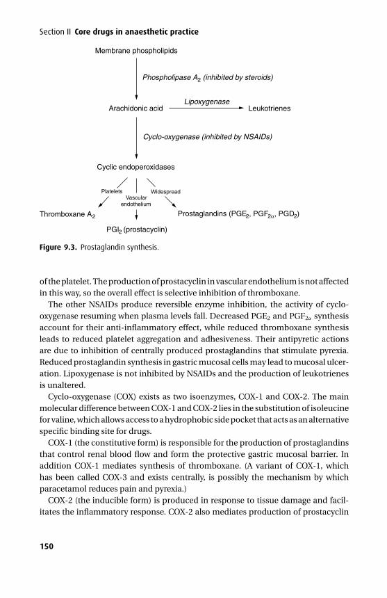
P1: PCX Printer: Yet To Come
9780521704632c09 CUFX213A/Peck 9780521618168 December 28, 2007 11:33
Section II Core drugs in anaesthetic practice
Membrane phospholipids
Arachidonic acid
Cyclic endoperoxidases
Platelets WidespreadVascular
endothelium
Leukotrienes
Phospholipase A2 (inhibited by steroids)
Thromboxane A2
PGI2 (prostacyclin)
Prostaglandins (PGE2, PGF2α, PGD2)
Cyclo-oxygenase (inhibited by NSAIDs)
Lipoxygenase
Figure 9.3. Prostaglandin synthesis.
of the platelet. The production of prostacyclin in vascular endothelium is not affectedin this way, so the overall effect is selective inhibition of thromboxane.
The other NSAIDs produce reversible enzyme inhibition, the activity of cyclo-oxygenase resuming when plasma levels fall. Decreased PGE2 and PGF2α synthesisaccount for their anti-inflammatory effect, while reduced thromboxane synthesisleads to reduced platelet aggregation and adhesiveness. Their antipyretic actionsare due to inhibition of centrally produced prostaglandins that stimulate pyrexia.Reduced prostaglandin synthesis in gastric mucosal cells may lead to mucosal ulcer-ation. Lipoxygenase is not inhibited by NSAIDs and the production of leukotrienesis unaltered.
Cyclo-oxygenase (COX) exists as two isoenzymes, COX-1 and COX-2. The mainmolecular difference between COX-1 and COX-2 lies in the substitution of isoleucinefor valine, which allows access to a hydrophobic side pocket that acts as an alternativespecific binding site for drugs.
COX-1 (the constitutive form) is responsible for the production of prostaglandinsthat control renal blood flow and form the protective gastric mucosal barrier. Inaddition COX-1 mediates synthesis of thromboxane. (A variant of COX-1, whichhas been called COX-3 and exists centrally, is possibly the mechanism by whichparacetamol reduces pain and pyrexia.)
COX-2 (the inducible form) is produced in response to tissue damage and facil-itates the inflammatory response. COX-2 also mediates production of prostacyclin
150
P1: PCX Printer: Yet To Come
9780521704632c09 CUFX213A/Peck 9780521618168 December 28, 2007 11:33
9 Analgesics
(PGI2) in vascular endothelium. As a result, COX-2 inhibitors may alter the delicatethromboxane/prostacyclin balance in favour of platelet aggregation, vasoconstric-tion and thromboembolism.
Other effects� Gastric irritation – intestinal erosions not limited to the stomach are commonlyencountered during prolonged administration of NSAIDs. These lesions result ina spectrum of adverse effects from mild pain to iron deficiency anaemia and fatalhaemorrhage. Many elements (mucous layer, bicarbonate secretion, rapid cellturnover and an abundant blood supply) are involved in the protection of theintestinal mucosa against acid and enzyme attack. Prostaglandins are involved inmany of these elements so that when their synthesis is inhibited, protection isreduced. During aspirin therapy acetylsalicylate and salicylate ions are trapped inthe alkaline environment of the mucosal cells thereby increasing their potential forside-effects. The potential for haemorrhage is increased due to its effect on plateletfunction. Meloxicam is more selective towards COX-2 and has been associated withreduced gastric ulceration although a similar renal toxicity profile when comparedwith other NSAIDs. Diclofenac blocks both forms equally while indomethacin andaspirin have a much higher affinity for COX-1.� NSAID sensitive asthma – acute severe asthma may be precipitated in up to 20%of asthmatics when given NSAIDs and is associated with chronic rhinitis or nasalpolyps. Those affected are usually middle-aged – children are relatively spared. Byinhibiting cyclo-oxygenase, more arachidonic acid is converted to leukotrieneswhich are known to cause bronchospasm. Aspirin also causes an abnormalreaction to the platelets of susceptible patients causing the release of cytotoxicmediators.� Renal function – renally produced prostaglandins (PGE2 and PGI2) are essential inmaintaining adequate renal perfusion when the level of circulating vasoconstric-tors (renin, angiotensin, noradrenaline) is high. Aspirin and other NSAIDs may alterthis delicate balance by inhibiting their production, reducing renal perfusion andpotentially leading to acute renal failure. At low doses of aspirin (<2 g.day−1), urateis retained as its tubular secretion is inhibited. At higher doses (>5 g.day−1), aspirinbecomes uricosuric as re-absorption of urates is inhibited to a greater degree. It israrely used for this purpose as the side effects at higher doses are unacceptable.Analgesic nephropathy may develop after prolonged use of aspirin. The featuresare papillary necrosis and interstitial fibrosis. NSAIDs may precipitate fluid reten-tion and this may become significant in those with heart failure. COX-2 inhibitorsappear to lead to a greater degree of fluid retention and hypertension.� Platelet function – while altered platelet function may be advantageous in certaincircumstances (acute myocardial infarction and prevention of stroke), during theperi-operative period it may cause increased blood loss. The reduced production
151
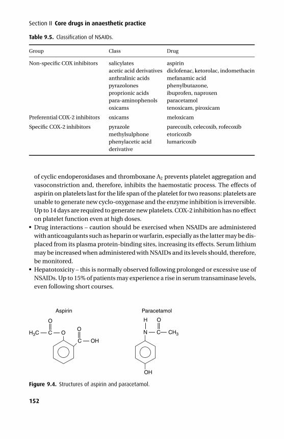
P1: PCX Printer: Yet To Come
9780521704632c09 CUFX213A/Peck 9780521618168 December 28, 2007 11:33
Section II Core drugs in anaesthetic practice
Table 9.5. Classification of NSAIDs.
Group Class Drug
Non-specific COX inhibitors salicylatesacetic acid derivativesanthralinic acidspyrazolonesproprionic acidspara-aminophenolsoxicams
aspirindiclofenac, ketorolac, indomethacinmefanamic acidphenylbutazone,ibuprofen, naproxenparacetamoltenoxicam, piroxicam
Preferential COX-2 inhibitors oxicams meloxicam
Specific COX-2 inhibitors pyrazolemethylsulphonephenylacetic acidderivative
parecoxib, celecoxib, rofecoxibetoricoxiblumaricoxib
of cyclic endoperoxidases and thromboxane A2 prevents platelet aggregation andvasoconstriction and, therefore, inhibits the haemostatic process. The effects ofaspirin on platelets last for the life span of the platelet for two reasons: platelets areunable to generate new cyclo-oxygenase and the enzyme inhibition is irreversible.Up to 14 days are required to generate new platelets. COX-2 inhibition has no effecton platelet function even at high doses.� Drug interactions – caution should be exercised when NSAIDs are administeredwith anticoagulants such as heparin or warfarin, especially as the latter may be dis-placed from its plasma protein-binding sites, increasing its effects. Serum lithiummay be increased when administered with NSAIDs and its levels should, therefore,be monitored.� Hepatotoxicity – this is normally observed following prolonged or excessive use ofNSAIDs. Up to 15% of patients may experience a rise in serum transaminase levels,even following short courses.
O
OH
Paracetamol
N — C— CH3
H— ——
Aspirin
C — OH
— —
OH3C — C — O
— —
O
Figure 9.4. Structures of aspirin and paracetamol.
152
P1: PCX Printer: Yet To Come
9780521704632c09 CUFX213A/Peck 9780521618168 December 28, 2007 11:33
9 Analgesics
Non-specific COX inhibitors
Salicylates
Aspirin
UsesAspirin (acetylsalicylic acid) is widely used for its analgesic and anti-inflammatoryeffects. It is also used for its effects on platelet function in acute myocardial infarctionand the prevention of stroke.
Mechanism of actionAt low dose, aspirin selectively inhibits platelet cyclo-oxygenase while preservingvessel wall cyclo-oxygenase. This has the effect of reducing TXA2-induced vaso-constriction and platelet aggregation while leaving vessel wall synthesis of prostag-landins unaltered and, therefore, dilated.
Other effects� Metabolic – aspirin also has effects on the metabolic state, which are usually oflittle significance, but in overdose these become significant. It uncouples oxida-tive phosphorylation, thereby increasing oxygen consumption and carbon dioxideproduction. Initially minute ventilation is increased to keep PaCO2 static. However,when aspirin levels are increased significantly the respiratory centre is stimulateddirectly causing a respiratory alkalosis. The picture is complicated in the premor-bid state by a metabolic acidosis. However, in children the respiratory centre isdepressed by rising aspirin levels, and a metabolic acidosis occurs earlier so that amixed respiratory and metabolic acidosis is more common.
Features of aspirin overdose
Common� Usually conscious – only unconscious in massive overdose� Sweaty� Tinnitus� Blurred vision� Tachycardia� Pyrexia� Hyperventilation� Respiratory alkalosis (subsequently complicated by metabolic acidosis)
Rare� Nausea and vomiting, epigastric pain� Oliguria� Gastrointestinal bleed
153
P1: PCX Printer: Yet To Come
9780521704632c09 CUFX213A/Peck 9780521618168 December 28, 2007 11:33
Section II Core drugs in anaesthetic practice� Pulmonary oedema (due to increased capillary permeability)� Coagulopathy� Hypokalaemia� Hypo- or hyperglycaemia� Encephalopathic, unconscious
Treatment� Activated charcoal� Forced alkaline diuresis� Haemofiltration/haemodialysis
Reye’s syndrome is uncommon and affects mainly children. Its aetiology has beenlinked to aspirin. It causes widespread mitochondrial damage, fatty changes in theliver progressing to hepatic failure, encephalopathy with cerebral oedema and hasa mortality rate of up to 40%. Therefore, aspirin is only recommended for childrenbelow 12 years of age when specifically indicated, for example, for juvenile arthritis(Still’s disease).
KineticsAspirin is a weak acid with a pKa = 3 and is present essentially in the unionized formin the stomach allowing gastric absorption, but due to the relatively alkaline natureof the mucosal cells salicylate ions may become trapped and unable to reach thesystemic circulation. However, due to total surface area the small bowel absorbs moredrug. Once in the systemic circulation, 85% is protein bound, mainly by albumin. It israpidly hydrolyzed by intestinal and hepatic esterases to salicylate, which undergoesfurther hepatic metabolism to salicyluric acid and glucuronide derivatives. Salicylateand its metabolites are excreted in the urine (enhanced under alkaline conditions).The elimination half-life varies because glycine conjugation (converting salicylate tosalicyluric acid) may become saturated in overdose resulting in zero-order kinetics.
Para-aminophenols
ParacetamolWhile paracetamol has essentially no effect on cyclo-oxygenase in vitro it has beenclassified as a NSAID because of its moderate analgesic and antipyretic properties. Ithas been proposed that its antipyretic actions are due to inhibition of prostaglandinsynthesis within the central nervous system, by inhibition of COX-3, a COX-1variant.
Presentation and usesParacetamol is presented as 500 mg tablets alone and in combination with weakopioids. Suppositories contain 125 mg and 1 g and the paediatric elixir contains
154
P1: PCX Printer: Yet To Come
9780521704632c09 CUFX213A/Peck 9780521618168 December 28, 2007 11:33
9 Analgesics
120 mg in 5 ml. A preparation containing 100 mg methionine and 500 mg parac-etamol is available but at increased cost. A solution of 1 g in 100 mls is available forintravenous use. The adult dose is 4 g.day−1 in divided doses. The initial paediatricdose is 15–30 mg.kg−1 which is then reduced to 10–15 mg.kg−1 every 4 hours with amaximum dose of 90 mg.kg−1.day−1.
KineticsParacetamol is well absorbed from the small bowel and has an oral bioavailability of80%. Unlike the other NSAIDs it does not cause gastric irritation, is less protein bound(10%) and has a larger volume of distribution. Paracetamol is metabolized by the livermainly to glucuronide conjugates but also to sulphate and cysteine conjugates. Theseare actively excreted in the urine, only a small fraction being excreted unchanged. N-acetyl-p-amino-benzoquinoneimine is a highly toxic metabolite of paracetamol thatis produced in small amounts following therapeutic doses. It is rapidly conjugatedwith hepatic glutathione to render it harmless.
ToxicityFollowing a toxic dose, the normal hepatic conjugation pathways become satu-rated so that more N-acetyl-p-amino-benzoquinoneimine is produced which rapidlyexhausts hepatic glutathione. It is then free to form covalent bonds with sulphydrylgroups on hepatocytes resulting in cell death and centrilobular hepatic necrosis.Treatment with oral methionine and oral or intravenous acetylcysteine is directedat replenishing hepatic glutathione. Methionine enhances glutathione synthesiswhile acetylcysteine is hydrolyzed to cysteine, which is a glutathione precursor.Intravenous acetylcysteine is preferred as vomiting is common in paracetamoloverdose.
Features of paracetamol overdose� Normally remain conscious� Nausea and vomiting� Epigastric pain� Sweating� Erythema, urticaria, mucosal lesions� Acute haemolytic anaemia� Peripheral vasodilatation and shock following massive overdose� Delayed hyperglycaemia� Hepatic failure after 48 hours� LFT and clotting (INR) worst at 3–5 days� Cholestasis� Fulminant hepatic failure at 3–7 days
155
P1: PCX Printer: Yet To Come
9780521704632c09 CUFX213A/Peck 9780521618168 December 28, 2007 11:33
Section II Core drugs in anaesthetic practice
Treatment� Activated charcoal� Intravenous glucose� Acetylcysteine or methionine� Early referral to specialist centre
Acetic acid derivatives
DiclofenacDiclofenac is a phenylacetic acid derivative.
PresentationDiclofenac is available in a parenteral as well as an oral and rectal formulation.The intravenous preparation should be diluted and administered over a minimumof 30 minutes. It is also available in combination with misoprostil, which providesprophylaxis against gastric and duodenal ulceration. The maximum adult dose is150 mg.day−1 in divided doses. The paediatric dose is 1 mg.kg−1 tds for pain associ-ated with minor surgery (tonsillectomy, inguinal herniotomy).
UsesDiclofenac may be used alone to treat mild to moderate post-operative pain or toreduce opioid consumption when treating severe pain. It is particularly useful intreating renal colic. Owing to its effects on cyclo-oxygenase it may also precipitategastric irritation, acute renal impairment and reduced platelet function, which oftenprevents it from being used in major surgery.
Other effects� Gut – diclofenac produces less gastric irritation than both indomethacin andaspirin.� Pain – the parenteral formulation is highly irritant and intramuscular injectionmay be very painful and is associated with muscle damage. Intravenous injectioncauses local thrombosis.� Interactions – plasma concentrations of lithium and digoxin may be increased. Ingeneral it does not effect either oral anticoagulants or oral hypoglycaemic agents,but isolated reports would suggest that close monitoring is used.
KineticsIn keeping with other drugs in its class diclofenac is well absorbed from thegut, highly plasma protein bound (99%) and has a small volume of distribution(0.15 l.kg−1). It undergoes hepatic hydroxylation and conjugation to inactivemetabolites that are excreted in the urine (60%) and bile (40%).
156
P1: PCX Printer: Yet To Come
9780521704632c09 CUFX213A/Peck 9780521618168 December 28, 2007 11:33
9 Analgesics
Table 9.6. Clinical and kinetic data for some NSAIDs.
Drug
Maximum
daily dose
Elimination
half-life (h)
Plasma
protein-
binding
(%)
Analgesic and
antipyretic
activity
Anti-
inflammatory
activity
Aspirin 4 g variable* 85 +++ ++Paracetamol 4 g 2 10 +++ ++Diclofenac 150 mg 1–2 99 + +++Ketorolac 40 mg 5 99 ++ +Indomethacin 200 mg 6 95 + +++Phenylbutazone 300 mg 50–100 98 + ++++Tenoxicam 20 mg 72 99 + ++Meloxicam 15 mg 20 99 + ++Ibuprofen 1.8 g 2–3 99 + +* When obeying first-order kinetics the t1/2 elimination of aspirin is short (15–30 min). However, this issignificantly prolonged when enzyme systems become saturated and its kinetics become zero-order.
KetorolacKetorolac is an acetic acid derivative with potent analgesic activity but limited anti-inflammatory activity. It is also a potent antipyretic. It may be given orally or par-enterally and has a duration of action of up to 6 hours. It shares the side-effect profilecommon to all NSAIDs.
IndomethacinIndomethacin is a potent anti-inflammatory agent but is a less effective analgesic.Rectal use has been associated not only with a reduced peri-operative opioid require-ment, but also with reduced platelet function resulting in wound haematoma andincreased blood loss. It is used to promote closure of the ductus arteriosus in thepremature infant by inhibiting prostaglandin synthesis. It shares the other effectscommon to NSAIDs but has been particularly linked with headache. It may alsoimpair hepatic function and antagonize the effects of diuretics and angiotensin-converting enzyme inhibitors.
KineticsIndomethacin is well absorbed from the gut with an oral bioavailability of 80%. It ismore than 99% bound by plasma proteins and is metabolized to inactive metabolitesthat are excreted in the urine and bile. Five percent is excreted unchanged.
Pyrazolones
PhenylbutazonePhenylbutazone is a potent anti-inflammatory agent. Its use has been limited tohospital patients with ankylosing spondylitis due to serious haematological side
157
P1: PCX Printer: Yet To Come
9780521704632c09 CUFX213A/Peck 9780521618168 December 28, 2007 11:33
Section II Core drugs in anaesthetic practice
effects including agranulocytosis and aplastic anaemia. It is significantly bound byplasma proteins and will interact with other highly bound drugs. It may also impairhepatic function, produce a rash, and cause sodium and water retention.
Proprionic acids
IbuprofenIbuprofen is prepared as 200–600 mg tablets and as a paediatric elixir containing20 mg.ml−1. It is not recommended for children below 1 year of age. The paediatricdose is 20 mg.kg−1.day−1 in divided doses. It has mild anti-inflammatory and anal-gesic effects but has the lowest incidence of side effects of the most commonly usedNSAIDs.
Oxicams
TenoxicamTenoxicam exhibits many of the features common with other NSAIDs. Two specificfeatures make it particularly useful in the peri-operative period:� It may be given intravenously resulting in a rapid onset of action.� It has a long elimination half-life (72 hours) resulting in a long duration of action
and allowing once-daily dosage.However, these advantages may become disadvantages if side effects become sig-nificant.
KineticsTenoxicam is well absorbed from the gut and has a high oral bioavailability. It ishighly plasma protein bound (99%). Clearance from the body is due to metabolismto an inactive metabolite that is excreted in the urine (66%) and in bile (33%). Thedose is 20 mg.day−1.
Preferential COX–2 inhibitors
MeloxicamMeloxicam is available as tablets and suppositories and the initial dose is 7.5mg.day−1, which may be doubled.
Meloxicam has limited preferential selectivity for COX-2 and is quoted as beingbetween 3 to 50 times as potent against COX-2. At a dose of 7.5 mg.day−1 ithas a reduced gastrointestinal side-effect profile when compared with diclofenac,although its renal side-effect profile appears to be equivalent to other NSAIDs.
KineticsMeloxicam is slowly but almost completely absorbed from the gut with an oralbioavailability of 90%. It is 99% protein bound, essentially to albumin. Metabolismoccurs in the liver to inactive metabolites that are excreted in the urine (50%) and bile
158
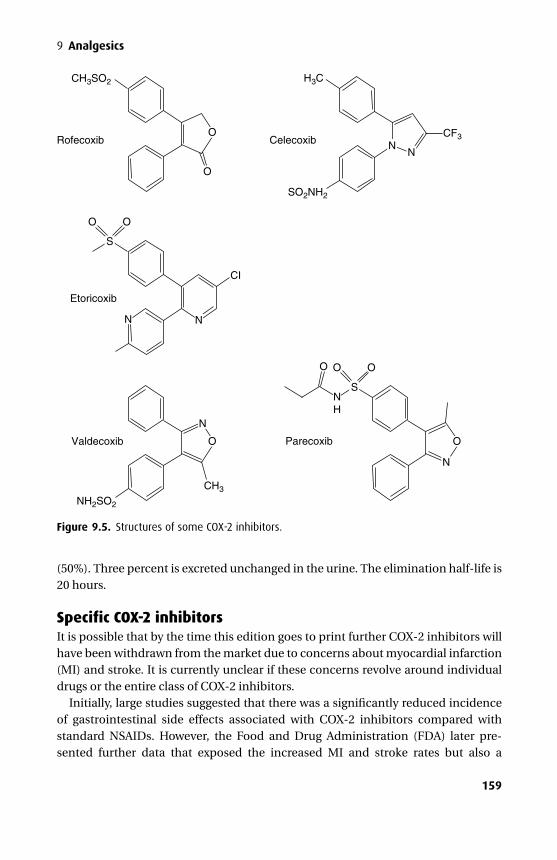
P1: PCX Printer: Yet To Come
9780521704632c09 CUFX213A/Peck 9780521618168 December 28, 2007 11:33
9 Analgesics
O
O
O
Rofecoxib
Etoricoxib
Celecoxib
O
CI
NN
S
OOO
S
H3CCH3SO2
O
N
CH3
Valdecoxib
CF3
SO2NH2
NH2SO2
OParecoxib
NN
N
N H
Figure 9.5. Structures of some COX-2 inhibitors.
(50%). Three percent is excreted unchanged in the urine. The elimination half-life is20 hours.
Specific COX-2 inhibitorsIt is possible that by the time this edition goes to print further COX-2 inhibitors willhave been withdrawn from the market due to concerns about myocardial infarction(MI) and stroke. It is currently unclear if these concerns revolve around individualdrugs or the entire class of COX-2 inhibitors.
Initially, large studies suggested that there was a significantly reduced incidenceof gastrointestinal side effects associated with COX-2 inhibitors compared withstandard NSAIDs. However, the Food and Drug Administration (FDA) later pre-sented further data that exposed the increased MI and stroke rates but also a
159
P1: PCX Printer: Yet To Come
9780521704632c09 CUFX213A/Peck 9780521618168 December 28, 2007 11:33
Section II Core drugs in anaesthetic practice
gastrointestinal side-effect rate equal to standard NSAIDs after 12 months therapyas well as a number of other side-effects – hypertension, heart failure, hepatotoxicityand oedema.
The Committee on Safety of Medicines have advised that COX-2 use is contraindi-cated in patients with ischaemic heart disease, cerebrovascular disease, mild heartfailure and peripheral vascular disease. Careful consideration should be given beforetheir use in patients with risk factors for cardiovascular events (hypertension, hyper-lipidaemia, diabetes, smoking).
RofecoxibRofecoxib (withdrawn September 2004) is presented as 12.5 mg tablets and as asuspension containing 12.5 mg per 5 mls. The daily dose is 12.5–25 mg. It has aCOX-1:COX-2 inhibitory ratio of 1:276.
The VIGOR study demonstrated that rofecoxib significantly reduces the number ofclinically important gastric complications when compared to a non-specific NSAID.However, more comprehensive analysis of further data beyond 12 months, not pub-lished with the original article, revealed a much less favourable picture. The incidenceof MI was 1.7% in those taking rofecoxib compared with 0.7% in the control groupwhich prompted its withdrawl from the market.
KineticsRofecoxib is well absorbed from the gut (oral bioavailability >90%) with plasma con-centration peaking at 3 hours. It is 85% protein bound and the plasma half-life isaround 17 hours allowing once-daily administration. Its volume of distribution is1.2 l.kg. It is metabolized in the liver by cytosolic reduction (not CYP450) to inactiveproducts that are excreted in the urine. As rofecoxib is not metabolized by CYP450it has fewer potential interactions than celecoxib. Rofecoxib appears to be safe insubjects with previous adverse cutaneous reactions (erythema, urticariaangioedema) to non-specific NSAIDs.
CelecoxibCelecoxib is presented as 100 mg tablets and used to a maximum dose of 200 mg bdfor osteoarthritis and rheumatoid arthritis. It has a COX-1:COX-2 inhibitory ratio of1:30.
The CLASS trial demonstrated the incidence of ulcer complications in patientstreated with celecoxib was similar to those treated with non-specific NSAIDs,although this may have been confused by concurrent aspirin therapy. Also the inci-dence of stroke and MI was not increased by celecoxib. Other studies designed toassess the ability of celecoxib to prevent colon cancer have confused the pictureregarding stroke and MI because on this point they did not agree.
160
P1: PCX Printer: Yet To Come
9780521704632c09 CUFX213A/Peck 9780521618168 December 28, 2007 11:33
9 Analgesics
KineticsCelecoxib reaches peak plasma concentration after 2–3 hours and has an eliminationhalf-life of 8–12 hours. It is 97% protein bound and has a volume of distribution of5.7 l.kg. It is metabolized by hepatic CYP2C9 to inactive metabolites so that only asmall amount appears unchanged in the urine. Drugs that inhibit (omeprazole) orinduce (carbamazepine) CYP2C9 will increase or decrease plasma concentrations.
It has a sulphonamide group and therefore should not be used in patients with asulphonamide allergy.
Valdecoxib and parecoxibParecoxib is a prodrug, which is converted to the active moiety valdecoxib. Its half-lifeis 45 minutes and has no actions of its own. Valdecoxib has been withdrawn (April2005) due to serious dermatological side effects (see below). At the time of writingparacoxib remains in use and has not been associated with similar dermatologicalside effects probably due to its short-term use. It has a COX-1:COX-2 inhibitory ratioof 1:61.
Parecoxib is a parenteral COX-2 antagonist and should be reconsituted with 0.9%saline prior to administration. The initial dose is 40 mg (i.m. or i.v.) followed by 20–40mg 6–12 hourly up to a maximum dose of 80 mg per day.
KineticsParecoxib is converted to valdecoxib by enzymatic hydrolysis in the liver. Valdecoxibundergoes hepatic metabolism by CYP2C9, CYP3A4 and glucuronidation to manymetabolites, one of which antagonizes COX-2. Due to its hepatic elimination, renalimpairment does not influence its kinetics, but it is not recommended in severehepatic impairment.
The dose should be reduced when coadministered with fluconazole due to inhi-bition of CYP2C9, although no adjustment is necessary when coadministered withketoconazole or midazolam which is metabolized by CYP3A4. Valdecoxib appears toinhibit other cytochrome P450 enzymes and may increase the plasma concentrationof flecanide and metoprolol (CYP2D6 inhibition), and omeprazole, phenytoin anddiazepam (CYP2C19 inhibition).
Parecoxib has a plasma half-life of 20 minutes while valdecoxib has an eliminationhalf-life of 8 hours.
Hypersensitivity reactions including exfoliative dermatitis, Stevens–Johnson syn-drome, toxic dermal necrolysis and angiodema have been reported following valde-coxib in those who also have sulphonamide sensitivity. The overall reporting rate isin the order of 8 per million patients. It is, therefore, contraindicated in this group.However, parecoxib has a non-aromatic sulphonamide group similar to frusemideand tolbutamide, neither of which is contraindicated in sulphonamide sensitivity.
161
P1: PCX Printer: Yet To Come
9780521704632c09 CUFX213A/Peck 9780521618168 December 28, 2007 11:33
Section II Core drugs in anaesthetic practice
EtoricoxibEtoricoxib is a highly selective COX-2 inhibitor that has a UK license but is awaitingFDA approval. It is a methylsulphone.
It has been shown to be efficacious in providing effective pain control when com-pared to COX-1 inhibitors. It has a COX-1:COX-2 inhibitory ratio of 1:344.
KineticsEtoricoxib is absorbed efficiently producing an oral bioavailability of > 95%. It has avolume of distribution of 1.5 l.kg–1 and is 90% plasma protein bound. Its eliminationhalf-life is 22 hours. It is metabolized in the liver through cytochrome P450 oxidation,CYP3A4 being the predominant isoenzyme. However, other isoenzymes are involvedand may become more involved with specific CYP3A4 inhibition. It is a weak inhibitorof CYP-2D6, -3A, and -2C9. Its elimination half-life increases in hepatic failure, butin isolated renal failure no change is seen.
Its side effects (or lack of them) are predictable. Its rate of gastric irritation is sim-ilar to placebo and significantly lower than COX-1 inhibitors, although this effectdecreases beyond 9–12 months therapy. There is currently insufficient data to con-firm that etoricoxib will be free from the cardiovascular effects that caused rofecoxibto be withdrawn from the market.
LumaricoxibLumaricoxib is a phenylacetic acid derivative and as such is structurally differentfrom the other COX-2 inhibitors, bearing a closer resemblance to diclofenac. It hasa COX-1:COX-2 inhibitory ratio of 1:700. Due to its structure it has a lower volume ofdistribution compared with the other COX-2 inhibitors.
Following reports of serious hepatoxicity including cases resulting in death or livertransplantation, lumaricoxib is now contraindicated in patients with any currentliver disease or a history of drug induced elevation of transaminases. In additionliver function tests are required during treatment.
KineticsIt has the shortest elimination half-life of 5 hours.
162
P1: PCX Printer: Yet To Come
9780521704632c10 CUFX213A/Peck 9780521618168 December 28, 2007 11:41
10Local anaesthetics
PhysiologyIndividual nerve fibres are made up of a central core (axoplasm) and a phospholipidmembrane containing integral proteins, some of which function as ion channels.
The resting membrane potentialThe neuronal membrane contains the enzyme Na+/K+ ATPase that actively main-tains a thirty fold K+ concentration gradient (greater concentration inside) and a tenfold Na+ concentration gradient (greater concentration outside). K+ tends to flowdown its concentration gradient out of the cell due to the selective permeability ofthe membrane. However, intracellular anionic proteins tend to oppose this ionicflux, and the balance of these processes results in the resting membrane potentialof −80 mV (negative inside). It can, therefore, be seen that the ratio of intracellularto extracellular K+ alters the resting membrane potential. Hypokalaemia increases(makes more negative) the resting membrane potential while the Na+ concentra-tion has little effect, as the membrane is essentially impermeable to Na+ when in theresting state.
The action potentialThe action potential is generated by altered Na+ permeability across the phos-pholipid membrane and lasts only 1–2 milliseconds. Electrical or chemical trig-gers initially cause a slow rise in membrane potential until the threshold potential(about −50 mV) is reached. Voltage sensitive Na+ channels then open, increasingNa+ permeability dramatically and the membrane potential briefly reaches +30 mV(approaching the Na+ equilibrium potential of+67 mV) at which point the Na+ chan-nels close. The membrane potential returns to its resting value with an increasedefflux of K+. The Na+/K+ ATPase restores the concentration gradients althoughthe total number of ions moving across the membrane is small. Conduction alongunmyelinated fibres is relatively slow compared with myelinated fibres where cur-rent jumps from one node of Ranvier to another (saltatory conduction) and reaches120 m.s−1. Retrograde conduction is not possible under normal circumstances dueto inactive Na+ channels following the action potential.
163
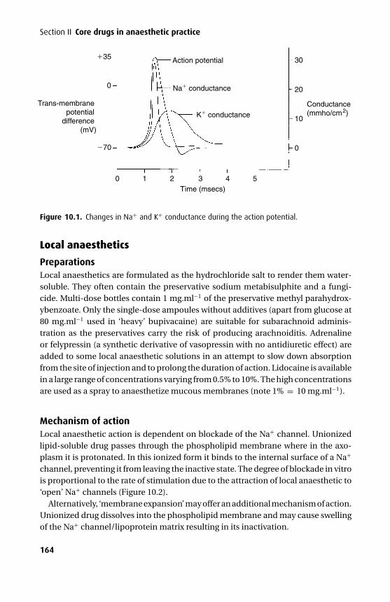
P1: PCX Printer: Yet To Come
9780521704632c10 CUFX213A/Peck 9780521618168 December 28, 2007 11:41
Section II Core drugs in anaesthetic practice
35
Na conductance
K conductance
Action potential
70
0
Trans-membranepotential
difference(mV)
0 1 2 3 4 5Time (msecs)
0
10
20
30
Conductance(mmho/cm2)
Figure 10.1. Changes in Na+ and K+ conductance during the action potential.
Local anaesthetics
PreparationsLocal anaesthetics are formulated as the hydrochloride salt to render them water-soluble. They often contain the preservative sodium metabisulphite and a fungi-cide. Multi-dose bottles contain 1 mg.ml−1 of the preservative methyl parahydrox-ybenzoate. Only the single-dose ampoules without additives (apart from glucose at80 mg.ml−1 used in ‘heavy’ bupivacaine) are suitable for subarachnoid adminis-tration as the preservatives carry the risk of producing arachnoiditis. Adrenalineor felypressin (a synthetic derivative of vasopressin with no antidiuretic effect) areadded to some local anaesthetic solutions in an attempt to slow down absorptionfrom the site of injection and to prolong the duration of action. Lidocaine is availablein a large range of concentrations varying from 0.5% to 10%. The high concentrationsare used as a spray to anaesthetize mucous membranes (note 1% = 10 mg.ml−1).
Mechanism of actionLocal anaesthetic action is dependent on blockade of the Na+ channel. Unionizedlipid-soluble drug passes through the phospholipid membrane where in the axo-plasm it is protonated. In this ionized form it binds to the internal surface of a Na+
channel, preventing it from leaving the inactive state. The degree of blockade in vitrois proportional to the rate of stimulation due to the attraction of local anaesthetic to‘open’ Na+ channels (Figure 10.2).
Alternatively, ‘membrane expansion’ may offer an additional mechanism of action.Unionized drug dissolves into the phospholipid membrane and may cause swellingof the Na+ channel/lipoprotein matrix resulting in its inactivation.
164
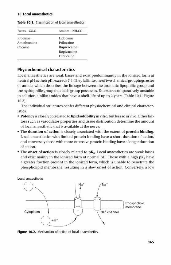
P1: PCX Printer: Yet To Come
9780521704632c10 CUFX213A/Peck 9780521618168 December 28, 2007 11:41
10 Local anaesthetics
Table 10.1. Classification of local anaesthetics.
Esters −CO.O− Amides −NH.CO−
Procaine LidocaineAmethocaine PrilocaineCocaine Bupivacaine
RopivacaineDibucaine
Physiochemical characteristicsLocal anaesthetics are weak bases and exist predominantly in the ionized form atneutral pH as their pKa exceeds 7.4. They fall into one of two chemical groupings, esteror amide, which describes the linkage between the aromatic lipophilic group andthe hydrophilic group that each group possesses. Esters are comparatively unstablein solution, unlike amides that have a shelf-life of up to 2 years (Table 10.1, Figure10.3).
The individual structures confer different physiochemical and clinical character-istics.� Potency is closely correlated to lipid solubility in vitro, but less so in vivo. Other fac-
tors such as vasodilator properties and tissue distribution determine the amountof local anaesthetic that is available at the nerve.� The duration of action is closely associated with the extent of protein binding.Local anaesthetics with limited protein binding have a short duration of action,and conversely those with more extensive protein binding have a longer durationof action.� The onset of action is closely related to pKa. Local anaesthetics are weak basesand exist mainly in the ionized form at normal pH. Those with a high pKa havea greater fraction present in the ionized form, which is unable to penetrate thephospholipid membrane, resulting in a slow onset of action. Conversely, a low
Local anaesthetic
Cytoplasm
H
Na Na
Phospholipidmembrane
Na channel
Figure 10.2. Mechanism of action of local anaesthetics.
165
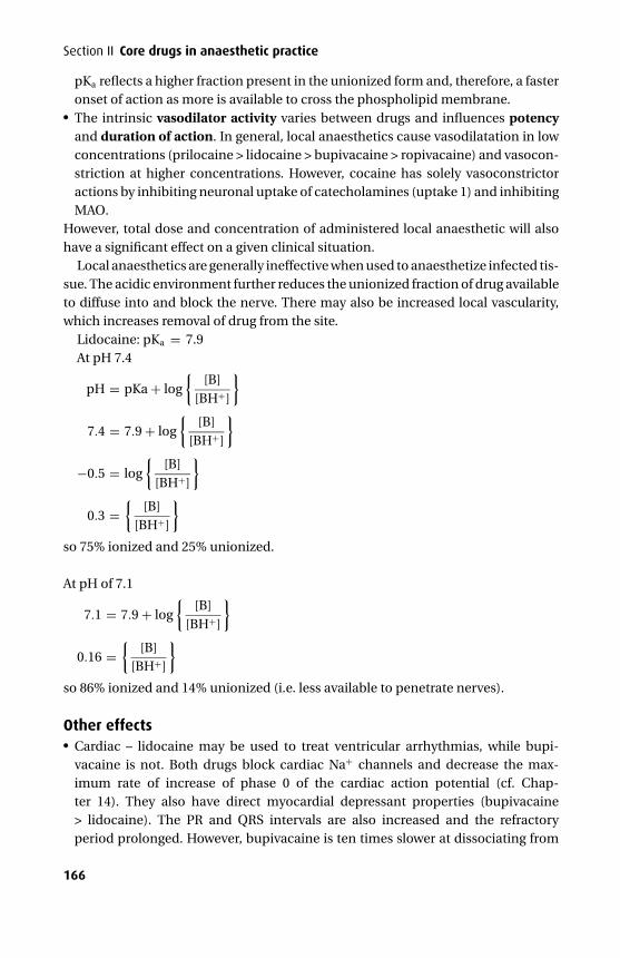
P1: PCX Printer: Yet To Come
9780521704632c10 CUFX213A/Peck 9780521618168 December 28, 2007 11:41
Section II Core drugs in anaesthetic practice
pKa reflects a higher fraction present in the unionized form and, therefore, a fasteronset of action as more is available to cross the phospholipid membrane.� The intrinsic vasodilator activity varies between drugs and influences potencyand duration of action. In general, local anaesthetics cause vasodilatation in lowconcentrations (prilocaine > lidocaine > bupivacaine > ropivacaine) and vasocon-striction at higher concentrations. However, cocaine has solely vasoconstrictoractions by inhibiting neuronal uptake of catecholamines (uptake 1) and inhibitingMAO.
However, total dose and concentration of administered local anaesthetic will alsohave a significant effect on a given clinical situation.
Local anaesthetics are generally ineffective when used to anaesthetize infected tis-sue. The acidic environment further reduces the unionized fraction of drug availableto diffuse into and block the nerve. There may also be increased local vascularity,which increases removal of drug from the site.
Lidocaine: pKa = 7.9At pH 7.4
pH = pKa + log{
[B][BH+]
}
7.4 = 7.9 + log{
[B][BH+]
}
−0.5 = log{
[B][BH+]
}
0.3 ={
[B][BH+]
}
so 75% ionized and 25% unionized.
At pH of 7.1
7.1 = 7.9 + log{
[B][BH+]
}
0.16 ={
[B][BH+]
}
so 86% ionized and 14% unionized (i.e. less available to penetrate nerves).
Other effects� Cardiac – lidocaine may be used to treat ventricular arrhythmias, while bupi-vacaine is not. Both drugs block cardiac Na+ channels and decrease the max-imum rate of increase of phase 0 of the cardiac action potential (cf. Chap-ter 14). They also have direct myocardial depressant properties (bupivacaine> lidocaine). The PR and QRS intervals are also increased and the refractoryperiod prolonged. However, bupivacaine is ten times slower at dissociating from
166
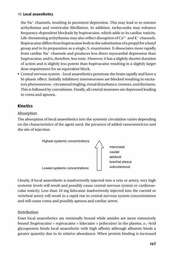
P1: PCX Printer: Yet To Come
9780521704632c10 CUFX213A/Peck 9780521618168 December 28, 2007 11:41
10 Local anaesthetics
the Na+ channels, resulting in persistent depression. This may lead to re-entrantarrhythmias and ventricular fibrillation. In addition, tachycardia may enhancefrequency-dependent blockade by bupivacaine, which adds to its cardiac toxicity.Life-threatening arrhythmias may also reflect disruption of Ca2+ and K+ channels.Ropivacaine differs from bupivacaine both in the substitution of a propyl for a butylgroup and in its preparation as a single, S, enantiomer. It dissociates more rapidlyfrom cardiac Na+ channels and produces less direct myocardial depression thanbupivacaine, and is, therefore, less toxic. However, it has a slightly shorter durationof action and is slightly less potent than bupivacaine resulting in a slightly largerdose requirement for an equivalent block.� Central nervous system – local anaesthetics penetrate the brain rapidly and have abi-phasic effect. Initially inhibitory interneurones are blocked resulting in excita-tory phenomenon – circumoral tingling, visual disturbance, tremors, and dizziness.This is followed by convulsions. Finally, all central neurones are depressed leadingto coma and apnoea.
Kinetics
AbsorptionThe absorption of local anaesthetics into the systemic circulation varies dependingon the characteristics of the agent used, the presence of added vasoconstrictor andthe site of injection.
Lowest systemic concentrations
Highest systemic concentrations
intercostal
caudal
epidural
brachial plexus
subcutaneous
Clearly, if local anaesthetic is inadvertently injected into a vein or artery, very highsystemic levels will result and possibly cause central nervous system or cardiovas-cular toxicity. Less than 10 mg lidocaine inadvertently injected into the carotid orvertebral artery will result in a rapid rise in central nervous system concentrationsand will cause coma and possibly apnoea and cardiac arrest.
DistributionEster local anaesthetics are minimally bound while amides are more extensivelybound (bupivacaine > ropivacaine > lidocaine > prilocaine) in the plasma. α1-Acidglycoprotein binds local anaesthetic with high affinity although albumin binds agreater quantity due to its relative abundance. When protein binding is increased
167
P1: PCX Printer: Yet To Come
9780521704632c10 CUFX213A/Peck 9780521618168 December 28, 2007 11:41
Section II Core drugs in anaesthetic practice
(pregnancy, myocardial infarction, renal failure, post-operatively and in infancy) thefree fraction of drug is reduced.
The degree of protein binding will affect the degree of placental transfer. Bupiva-caine is more highly bound than lidocaine, so less crosses the placenta. If the foetusbecomes acidotic there will be an increase in the ionized fraction and local anaes-thetic will accumulate in the foetus (ion trapping). Ester local anaesthetics do notcross the placenta in significant amounts due to their rapid metabolism.
Metabolism and eliminationEsters are hydrolyzed rapidly by plasma cholinesterases and other esterases to inac-tive compounds. Para-aminobenzoate is one of the main metabolites and has beenassociated with hypersensitivity reactions especially in the atopic patient. This rapidhydrolysis results in a short elimination half-life. Cocaine is the exception, undergo-ing hepatic hydrolysis to water-soluble metabolites that are excreted in the urine.
Amides undergo hepatic metabolism by amidases. Amidase metabolism is muchslower than plasma hydrolysis and so amides are more prone to accumulation whenadministered by continuous infusion. Reduced hepatic blood flow or hepatic dys-function can decrease amide metabolism.
Toxic dosesRaised systemic blood levels of local anaesthetic lead initially to the central ner-vous system and then to cardiovascular toxicity. However, the absorption of localanaesthetic varies widely depending on the site of administration and presence ofvasoconstrictors. Therefore, the concept of a toxic dose without regard to the site ofadministration is meaningless. The toxic plasma levels are given in Table 10.2.
Intravenous regional anaesthesiaBupivacaine has been used for intravenous regional techniques, but following a num-ber of deaths attributed to cardiac toxicity it is no longer used in this way. Prilocaine(0.5%) is commonly used in this setting although lidocaine may also be used.
LidocaineLidocaine is an amide local anaesthetic that is also used to control ventricular tach-yarrhythmias. It has class Ib anti-arrhythmic actions (see Chapter 14).
PreparationsLidocaine is formulated as the hydrochloride and is presented as a colourless solution(0.5–2%) with or without adrenaline (1 in 80–200 000); a 2% gel; a 5% ointment; a spraydelivering 10 mg.dose−1 and a 4% solution for topical use on mucous membranes.
KineticsLidocaine is 70% protein bound to α1-acid glycoprotein. It is extensively metabo-lized in the liver by dealkylation to monoethylglycine-xylidide and acetaldehyde.
168
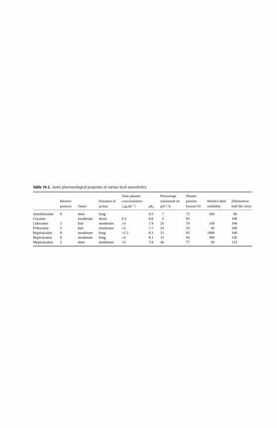
P1:P
CX
Prin
ter:YetToC
om
e
9780521704632c10C
UF
X213A
/Peck9780521618168
Decem
ber
28,200711:41
Table 10.2. Some pharmacological properties of various local anaesthetics.
Relative
potency Onset
Duration of
action
Toxic plasma
concentration
(µg.ml−1) pKa
Percentage
unionized (at
pH 7.4)
Plasma
protein
bound (%)
Relative lipid
solubility
Elimination
half-life (min)
Amethocaine 8 slow long 8.5 7 75 200 80Cocaine moderate short 0.5 8.6 5 95 100Lidocaine 2 fast moderate >5 7.9 25 70 150 100Prilocaine 2 fast moderate >5 7.7 33 55 50 100Bupivacaine 8 moderate long >1.5 8.1 15 95 1000 160Ropivacaine 8 moderate long >4 8.1 15 94 300 120Mepivacaine 2 slow moderate >5 7.6 40 77 50 115
169
P1: PCX Printer: Yet To Come
9780521704632c10 CUFX213A/Peck 9780521618168 December 28, 2007 11:41
Section II Core drugs in anaesthetic practice
The former is further hydrolyzed while the latter is hydroxylated to 4-hydroxy–2,6-xylidine forming the main metabolite, which is excreted in the urine. Some of themetabolic products of lidocaine have anti-arrhythmic properties while others maypotentiate lidocaine-induced seizures.
Clearance is reduced in the presence of hepatic or cardiac failure.
Eutectic mixture of local anaesthetic (EMLA)When two compounds are mixed to produce a substance that behaves with a sin-gle set of physical characteristics, it is said to be eutectic. Eutectic mixture of localanaesthetic (5%) contains a mixture of crystalline bases of 2.5% lidocaine and 2.5%prilocaine in a white oil:water emulsion. The mixture has a lower melting point, beingan oil at room temperature, while the individual components would be crystallinesolids.
Presentation and usesEutectic mixture of local anaesthetic is presented as an emulsion in tubes containing5 g or 30 g. It is used to anaesthetize skin before vascular cannulation or harvestingfor skin grafts. It should be applied to intact skin under an occlusive dressing for atleast 60 minutes to ensure adequate anaesthesia.
CautionsEMLA cream should be avoided in patients with congenital or idiopathicmethaemoglobinaemia, or in infants less than 12 months of age who are receiv-ing treatment with methaemoglobin-inducing drugs. Patients taking drugs associ-ated with methaemoglobinaemia (e.g. sulphonamides or phenytoin) are at greaterrisk of developing methaemoglobinaemia if concurrently treated with EMLA cream.Methaemoglobinaemia is caused by o-toluidine, a metabolite of prilocaine.
EMLA should not be used on mucous membranes due to rapid systemic absorp-tion. EMLA should be used with caution in patients receiving class I anti-arrhythmicdrugs (e.g. tocainide, mexiletine) because the toxic effects are additive and poten-tially synergistic.
Bupivacaine
Presentation and usesBupivacaine is prepared as a 0.25% and 0.5% (with or without 1:200 000 adrenaline)solution. A 0.5% preparation containing 80 mg.ml−1 glucose (specific gravity 1.026)is available for subarachnoid block.
It remains the mainstay of epidural infusions in labour and post-operativelydespite concerns regarding its potential cardiac toxicity and the availability of newerdrugs (ropivacaine and levobupivacaine). The maximum dose is said to be 2 mg.kg–1.
170
P1: PCX Printer: Yet To Come
9780521704632c10 CUFX213A/Peck 9780521618168 December 28, 2007 11:41
10 Local anaesthetics
KineticsThe onset of action is intermediate or slow and significantly slower than that of lido-caine. It is the most highly protein bound amide local anaesthetic and is metabolizedin the liver by dealkylation to pipecolic acid and pipecolylxylidine.
LevobupivacaineLevobupivacaine is the S-enantiomer of bupivacaine, which is the racemic mixtureof the S- and R-enantiomer.
Presentation and usesLevobupivacaine is prepared as a 2.5, 5 and 7.5 mg.ml–1 solution. It is used in a mannersimilar to that of bupivacaine. A maximum single dose of 150 mg is recommendedwith a maximum dose over 24 hours of 400 mg.
Toxicity profileThe single advantage of levobupivacaine over bupivacaine and other local anaes-thetics is its potential for reduced toxicity. While extrapolation of research from ani-mal models to humans may be confusing, combined with limited human volunteerwork it appears that levobupivacaine has two potentially useful properties. First, thedose required to produce myocardial depression (by blocking cardiac K+ channels)is higher for levobupivacaine compared with bupivacaine, and second, excitatorycentral nervous system effects or convulsions occur at lower doses with bupivacainethan levobupivacaine.
Ropivacaine
Presentation and usesThe amide local anaesthetic ropivacaine is prepared in three concentrations (2, 7.5and 10 mg.ml−1), in two volumes (10 and 100 ml) and as the pure S-enantiomer. It isnot prepared in combination with a vasoconstrictor as this does not alter its durationof action or uptake from tissues. The R-enantiomer is less potent and more toxic. Ithas a propyl group on its piperidine nitrogen in contrast to the butyl group presentin bupivacaine and the methyl group present in mepivacaine (Figure 10.3).
The main differences from bupivacaine lie in its pure enantiomeric formulation,improved toxic profile and lower lipid solubility. Its lower lipid solubility may resultin reduced penetration of the large myelinated Aβ motor fibres, so that initially thesefibres are relatively spared from local anaesthetic. However, during continuous infu-sion they too will become blocked by local anaesthetic resulting in similar degreesof block between Aβ fibres and the smaller unmyelinated C fibres. Therefore, themotor block produced by ropivacaine is slower in onset, less dense and of shorterduration when compared with an equivalent dose of bupivacaine. Theoretically it
171
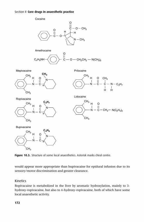
P1: PCX Printer: Yet To Come
9780521704632c10 CUFX213A/Peck 9780521618168 December 28, 2007 11:41
Section II Core drugs in anaesthetic practice
CH3
CH3
CH3
CH3CH3
CH3
CH3 CH3CH3
CH2
CH3
CH3
C4H9NH
C3H7
N(C2H5)2
C3H7
C — O — CH2CH2 — N(CH3)2
C
C O
O
O
O
N
N C
O N
N
OH
H
N C
OH
CH3
CH3
C4H9
N
N C
OH
H O
N C
H O
N C
C
H H
N
H
Mepivacaine
Cocaine
Amethocaine
Prilocaine
LidocaineRopivacaine
Bupivacaine
*
*
*
Figure 10.3. Structure of some local anaesthetics. Asterisk marks chiral centre.
would appear more appropriate than bupivacaine for epidural infusion due to itssensory/motor discrimination and greater clearance.
KineticsRopivacaine is metabolized in the liver by aromatic hydroxylation, mainly to 3-hydroxy-ropivacaine, but also to 4-hydroxy-ropivacaine, both of which have somelocal anaesthetic activity.
172
P1: PCX Printer: Yet To Come
9780521704632c10 CUFX213A/Peck 9780521618168 December 28, 2007 11:41
10 Local anaesthetics
Prilocaine
Presentation and usesPrilocaine is presented as a 0.5–2.0% solution. It is also available as a 3% solution withfelypressin (0.03 unit.ml−1) for dental use. It has similar indications to lidocaine butis most frequently used for intravenous regional anaesthesia. The maximum dose is6 mg.kg–1 or 8 mg.kg–1 when administered with felypressin.
KineticsPrilocaine is the most rapidly metabolized amide local anaesthetic, metabolismoccurring not only in the liver, but also the kidney and lung. When given in large dosesone of its metabolites, o-toluidine, may precipitate methaemoglobinaemia. This mayrequire treatment with ascorbic acid or methylene blue, which act as reducing agents.The neonate is at special risk as its red blood cells are deficient in methaemoglobinreductase. EMLA cream may precipitate the same reaction.
Cocaine
Presentation and usesCocaine is an ester local anaesthetic derived from the leaves of Erythroxylon coca,a plant indigenous to Bolivia and Peru. It is used for topical anaesthesia and localvasoconstriction. Moffatt’s solution (2 ml 8% cocaine, 2 ml 1% sodium bicarbonate,1 ml 1:1000 adrenaline) has been used in the nasal cavities, although its potential forside effects has rendered it less popular. Cocaine is also formulated as a paste rangingfrom 1% to 4%. A maximum dose of 1.5 mg.kg –1 or 100 mg is currently recommended.
Mechanism of actionCocaine blocks uptake 1 and MAO while also stimulating the central nervous system.These combined effects increase the likelihood of precipitating hypertension andarrhythmias. Its use also provokes hyperthermia.
Side effectsWhen taken or administered in high doses it can cause confusion, hallucinations,seizures, arrhythmias and cardiac rupture.
KineticsCocaine is absorbed well from mucous membranes and is highly protein bound(about 95%). Unlike other esters it undergoes significant hepatic hydrolysis to inac-tive products, which are excreted in the urine.
AmethocaineAmethocaine is an ester local anaesthetic used for topical anaesthesia. It is presentedas 0.5% and 1% drops for topical use before local anaesthetic block or as a sole agent
173
P1: PCX Printer: Yet To Come
9780521704632c10 CUFX213A/Peck 9780521618168 December 28, 2007 11:41
Section II Core drugs in anaesthetic practice
for lens surgery. It may produce a burning sensation on initial instillation. It is alsoavailable as a 4% cream for topical anaesthesia to the skin and is used in a similarfashion to EMLA cream. However, it has a faster onset of action, producing goodtopical anaesthesia by 30 minutes, following which it may be removed. Its effects lastfor 4–6 hours. It produces some local vasodilatation and erythema, which may assistvenous cannulation.
174
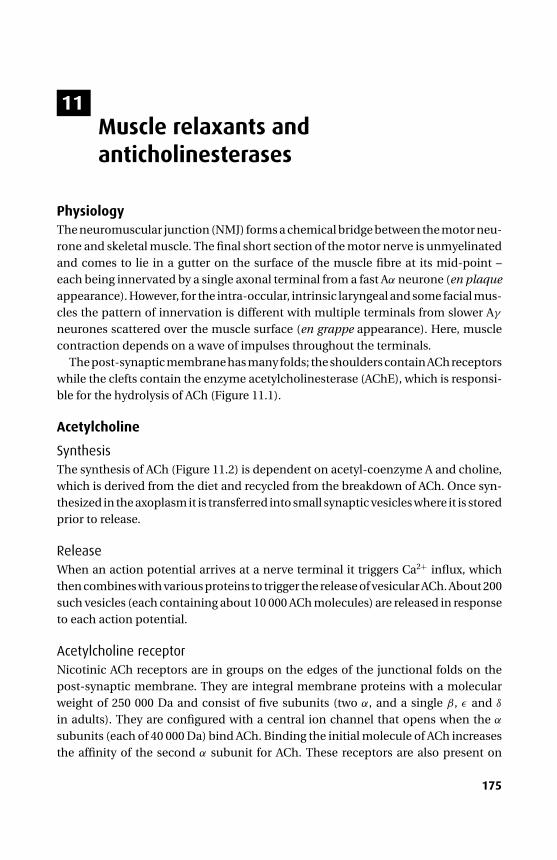
P1: PSB/JDO/OZN P2: PSB Printer: Yet To Come
9780521704632c11 CUFX213A/Peck 9780521618168 December 28, 2007 12:5
11Muscle relaxants andanticholinesterases
PhysiologyThe neuromuscular junction (NMJ) forms a chemical bridge between the motor neu-rone and skeletal muscle. The final short section of the motor nerve is unmyelinatedand comes to lie in a gutter on the surface of the muscle fibre at its mid-point –each being innervated by a single axonal terminal from a fast Aα neurone (en plaqueappearance). However, for the intra-occular, intrinsic laryngeal and some facial mus-cles the pattern of innervation is different with multiple terminals from slower Aγ
neurones scattered over the muscle surface (en grappe appearance). Here, musclecontraction depends on a wave of impulses throughout the terminals.
The post-synaptic membrane has many folds; the shoulders contain ACh receptorswhile the clefts contain the enzyme acetylcholinesterase (AChE), which is responsi-ble for the hydrolysis of ACh (Figure 11.1).
Acetylcholine
SynthesisThe synthesis of ACh (Figure 11.2) is dependent on acetyl-coenzyme A and choline,which is derived from the diet and recycled from the breakdown of ACh. Once syn-thesized in the axoplasm it is transferred into small synaptic vesicles where it is storedprior to release.
ReleaseWhen an action potential arrives at a nerve terminal it triggers Ca2+ influx, whichthen combines with various proteins to trigger the release of vesicular ACh. About 200such vesicles (each containing about 10 000 ACh molecules) are released in responseto each action potential.
Acetylcholine receptorNicotinic ACh receptors are in groups on the edges of the junctional folds on thepost-synaptic membrane. They are integral membrane proteins with a molecularweight of 250 000 Da and consist of five subunits (two α, and a single β, ε and δ
in adults). They are configured with a central ion channel that opens when the α
subunits (each of 40 000 Da) bind ACh. Binding the initial molecule of ACh increasesthe affinity of the second α subunit for ACh. These receptors are also present on
175
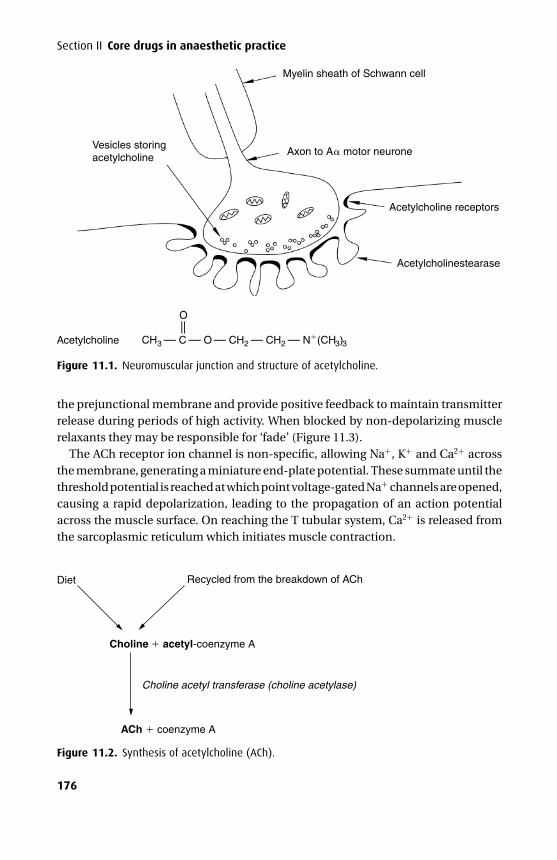
P1: PSB/JDO/OZN P2: PSB Printer: Yet To Come
9780521704632c11 CUFX213A/Peck 9780521618168 December 28, 2007 12:5
Section II Core drugs in anaesthetic practice
Vesicles storingacetylcholine
Myelin sheath of Schwann cell
Acetylcholine receptors
Acetylcholinestearase
Acetylcholine
Axon to A motor neurone
CH3 — C — O — CH2 — CH2 — N (CH3)3
— —
O
Figure 11.1. Neuromuscular junction and structure of acetylcholine.
the prejunctional membrane and provide positive feedback to maintain transmitterrelease during periods of high activity. When blocked by non-depolarizing musclerelaxants they may be responsible for ‘fade’ (Figure 11.3).
The ACh receptor ion channel is non-specific, allowing Na+, K+ and Ca2+ acrossthe membrane, generating a miniature end-plate potential. These summate until thethreshold potential is reached at which point voltage-gated Na+ channels are opened,causing a rapid depolarization, leading to the propagation of an action potentialacross the muscle surface. On reaching the T tubular system, Ca2+ is released fromthe sarcoplasmic reticulum which initiates muscle contraction.
Diet Recycled from the breakdown of ACh
Choline acetyl transferase (choline acetylase)
Choline acetyl-coenzyme A
ACh coenzyme A
Figure 11.2. Synthesis of acetylcholine (ACh).
176
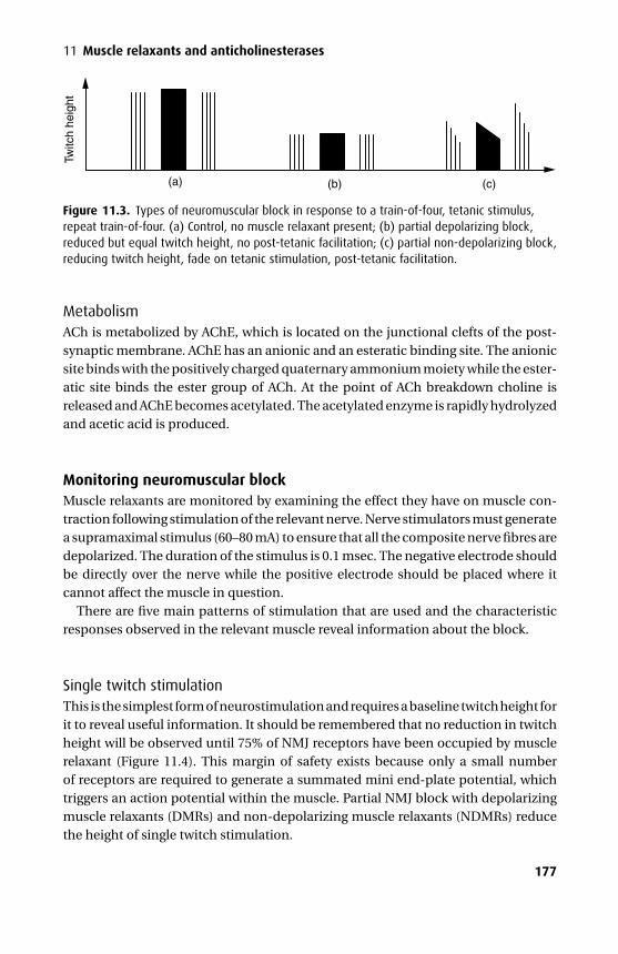
P1: PSB/JDO/OZN P2: PSB Printer: Yet To Come
9780521704632c11 CUFX213A/Peck 9780521618168 December 28, 2007 12:5
11 Muscle relaxants and anticholinesterasesTw
itch
heig
ht
(a) (b) (c)
Figure 11.3. Types of neuromuscular block in response to a train-of-four, tetanic stimulus,repeat train-of-four. (a) Control, no muscle relaxant present; (b) partial depolarizing block,reduced but equal twitch height, no post-tetanic facilitation; (c) partial non-depolarizing block,reducing twitch height, fade on tetanic stimulation, post-tetanic facilitation.
MetabolismACh is metabolized by AChE, which is located on the junctional clefts of the post-synaptic membrane. AChE has an anionic and an esteratic binding site. The anionicsite binds with the positively charged quaternary ammonium moiety while the ester-atic site binds the ester group of ACh. At the point of ACh breakdown choline isreleased and AChE becomes acetylated. The acetylated enzyme is rapidly hydrolyzedand acetic acid is produced.
Monitoring neuromuscular blockMuscle relaxants are monitored by examining the effect they have on muscle con-traction following stimulation of the relevant nerve. Nerve stimulators must generatea supramaximal stimulus (60–80 mA) to ensure that all the composite nerve fibres aredepolarized. The duration of the stimulus is 0.1 msec. The negative electrode shouldbe directly over the nerve while the positive electrode should be placed where itcannot affect the muscle in question.
There are five main patterns of stimulation that are used and the characteristicresponses observed in the relevant muscle reveal information about the block.
Single twitch stimulationThis is the simplest form of neurostimulation and requires a baseline twitch height forit to reveal useful information. It should be remembered that no reduction in twitchheight will be observed until 75% of NMJ receptors have been occupied by musclerelaxant (Figure 11.4). This margin of safety exists because only a small numberof receptors are required to generate a summated mini end-plate potential, whichtriggers an action potential within the muscle. Partial NMJ block with depolarizingmuscle relaxants (DMRs) and non-depolarizing muscle relaxants (NDMRs) reducethe height of single twitch stimulation.
177
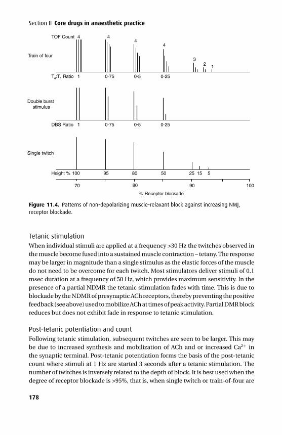
P1: PSB/JDO/OZN P2: PSB Printer: Yet To Come
9780521704632c11 CUFX213A/Peck 9780521618168 December 28, 2007 12:5
Section II Core drugs in anaesthetic practice
TOF Count 4 4
Train of four
T4:T1 Ratio 1 0 75 0 5 0 25
Double burststimulus
DBS Ratio 1 0 75 0 5 0 25
Single twitch
Height % 100 95 80 50 25 15 5
% Receptor blockade
70 80 90 100
44
23
1
Figure 11.4. Patterns of non-depolarizing muscle-relaxant block against increasing NMJ,receptor blockade.
Tetanic stimulationWhen individual stimuli are applied at a frequency >30 Hz the twitches observed inthe muscle become fused into a sustained muscle contraction – tetany. The responsemay be larger in magnitude than a single stimulus as the elastic forces of the muscledo not need to be overcome for each twitch. Most stimulators deliver stimuli of 0.1msec duration at a frequency of 50 Hz, which provides maximum sensitivity. In thepresence of a partial NDMR the tetanic stimulation fades with time. This is due toblockade by the NDMR of presynaptic ACh receptors, thereby preventing the positivefeedback (see above) used to mobilize ACh at times of peak activity. Partial DMR blockreduces but does not exhibit fade in response to tetanic stimulation.
Post-tetanic potentiation and countFollowing tetanic stimulation, subsequent twitches are seen to be larger. This maybe due to increased synthesis and mobilization of ACh and or increased Ca2+ inthe synaptic terminal. Post-tetanic potentiation forms the basis of the post-tetaniccount where stimuli at 1 Hz are started 3 seconds after a tetanic stimulation. Thenumber of twitches is inversely related to the depth of block. It is best used when thedegree of receptor blockade is >95%, that is, when single twitch or train-of-four are
178
P1: PSB/JDO/OZN P2: PSB Printer: Yet To Come
9780521704632c11 CUFX213A/Peck 9780521618168 December 28, 2007 12:5
11 Muscle relaxants and anticholinesterases
unable to evoke muscle twitches. It should be remembered that the effects of tetanicstimulation may last for up to 6 minutes and may therefore give a false impression ofinadequate block to single twitch or train-of-four analysis. Partial DMR block doesnot exhibit post tetanic potentiation.
Train-of-fourThe train-of-four (TOF) is four 0.1 msec stimuli delivered at 2 Hz. The ratio of thefourth twitch height to the first twitch height (T4:T1), or the number of twitches maybe recorded leading to the TOF ratio or the TOF count respectively (Figure 11.4). Itdoes not require a baseline twitch height.
In a manner similar to that seen with tetanic stimulation the rapid stimuli lead toa reducing twitch height in the presence of partial non-depolarizing muscle relaxantblockade, that is, T4 < T1. As receptor occupancy rises above 70% T4 will start todecrease in size. When T4 has decreased by 25% T1 starts to decrease, correspondingto 75–80% receptor occupancy. T4 disappears when T1 is approximately 25% of itsoriginal height. TOF ratio is difficult to assess in practise.
The TOF count records the number of twitches in response to a TOF. As recep-tor ocupancy exceeds 90% T4 disappears and only T1 is present at 95% receptorocupancy. So the TOF count assesses the degree of deep NDMR block.
TOF ratio in the presence of partial DMR block is 1.
Double burst stimulationDouble burst stimulation (DBS) describes the delivery of two bursts of stimulationseparated by 0.75 sec. Each burst consists of three 0.2 msec stimuli separated by20 msec (i.e. at a 50 Hz). Double burst stimulation was developed to allow easy manualdetection of small amounts of residual NMJ blockade so that when the magnitudeof the two stmuli are equal clinically significant residual NMJ blockade does notexist. However, when assessed mechanically DBS is no more sensitive than TOF(Figure 11.4).
Depolarizing muscle relaxants
Suxamethonium
CH2 CH2 CH2 N+(CH3)3
N+(CH3)3
C— — — — —O
CH2 CH2C
O
O
OCH2 — —— —
——
—
— — —
Suxamethonium was first introduced in 1952 and provided a significant advantageover tubocurarine as profound muscle relaxation of short duration was achieved
179
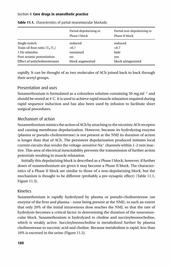
P1: PSB/JDO/OZN P2: PSB Printer: Yet To Come
9780521704632c11 CUFX213A/Peck 9780521618168 December 28, 2007 12:5
Section II Core drugs in anaesthetic practice
Table 11.1. Characteristics of partial neuromuscular blockade.
Partial depolarizing or
Phase I block
Partial non-depolarizing or
Phase II block
Single twitch reduced reducedTrain-of-four ratio (T4:T1) >0.7 <0.71 Hz stimulus sustained fadePost-tetanic potentiation no yesEffect of anticholinesterases block augmented block antagonized
rapidly. It can be thought of as two molecules of ACh joined back to back throughtheir acetyl groups.
Presentation and usesSuxamethonium is formulated as a colourless solution containing 50 mg.ml−1 andshould be stored at 4◦C. It is used to achieve rapid muscle relaxation required duringrapid sequence induction and has also been used by infusion to facilitate shortsurgical procedures.
Mechanism of actionSuxamethonium mimics the action of ACh by attaching to the nicotinic ACh receptorand causing membrane depolarization. However, because its hydrolyzing enzyme(plasma or pseudo-cholinesterase) is not present at the NMJ its duration of actionis longer than that of ACh. The persistent depolarization produced initiates localcurrent circuits that render the voltage-sensitive Na+ channels within 1–2 mm inac-tive. This area of electrical inexcitability prevents the transmission of further actionpotentials resulting in muscle relaxation.
Initially this depolarizing block is described as a Phase I block; however, if furtherdoses of suxamethonium are given it may become a Phase II block. The character-istics of a Phase II block are similar to those of a non-depolarizing block, but themechanism is thought to be different (probably a pre-synaptic effect) (Table 11.1,Figure 11.3).
KineticsSuxamethonium is rapidly hydrolyzed by plasma or pseudo-cholinesterase (anenzyme of the liver and plasma – none being present at the NMJ), to such an extentthat only 20% of the initial intravenous dose reaches the NMJ, so that the rate ofhydrolysis becomes a critical factor in determining the duration of the neuromus-cular block. Suxamethonium is hydrolyzed to choline and succinylmonocholine,which is weakly active. Succinylmonocholine is metabolized further by plasmacholinesterase to succinic acid and choline. Because metabolism is rapid, less than10% is excreted in the urine (Figure 11.5).
180
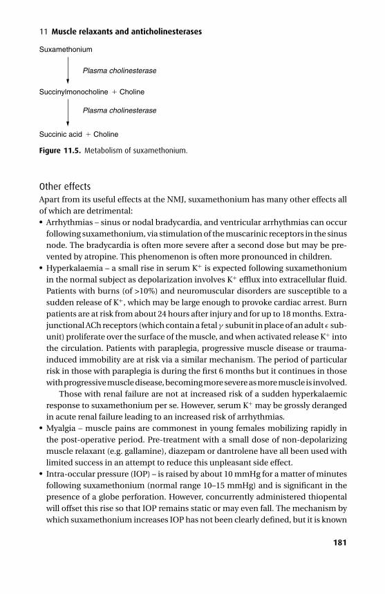
P1: PSB/JDO/OZN P2: PSB Printer: Yet To Come
9780521704632c11 CUFX213A/Peck 9780521618168 December 28, 2007 12:5
11 Muscle relaxants and anticholinesterases
Suxamethonium
Succinylmonocholine Choline
Plasma cholinesterase
Plasma cholinesterase
Succinic acid Choline
Figure 11.5. Metabolism of suxamethonium.
Other effectsApart from its useful effects at the NMJ, suxamethonium has many other effects allof which are detrimental:� Arrhythmias – sinus or nodal bradycardia, and ventricular arrhythmias can occur
following suxamethonium, via stimulation of the muscarinic receptors in the sinusnode. The bradycardia is often more severe after a second dose but may be pre-vented by atropine. This phenomenon is often more pronounced in children.� Hyperkalaemia – a small rise in serum K+ is expected following suxamethoniumin the normal subject as depolarization involves K+ efflux into extracellular fluid.Patients with burns (of >10%) and neuromuscular disorders are susceptible to asudden release of K+, which may be large enough to provoke cardiac arrest. Burnpatients are at risk from about 24 hours after injury and for up to 18 months. Extra-junctional ACh receptors (which contain a fetal γ subunit in place of an adult ε sub-unit) proliferate over the surface of the muscle, and when activated release K+ intothe circulation. Patients with paraplegia, progressive muscle disease or trauma-induced immobility are at risk via a similar mechanism. The period of particularrisk in those with paraplegia is during the first 6 months but it continues in thosewith progressive muscle disease, becoming more severe as more muscle is involved.
Those with renal failure are not at increased risk of a sudden hyperkalaemicresponse to suxamethonium per se. However, serum K+ may be grossly derangedin acute renal failure leading to an increased risk of arrhythmias.� Myalgia – muscle pains are commonest in young females mobilizing rapidly inthe post-operative period. Pre-treatment with a small dose of non-depolarizingmuscle relaxant (e.g. gallamine), diazepam or dantrolene have all been used withlimited success in an attempt to reduce this unpleasant side effect.� Intra-occular pressure (IOP) – is raised by about 10 mmHg for a matter of minutesfollowing suxamethonium (normal range 10–15 mmHg) and is significant in thepresence of a globe perforation. However, concurrently administered thiopentalwill offset this rise so that IOP remains static or may even fall. The mechanism bywhich suxamethonium increases IOP has not been clearly defined, but it is known
181
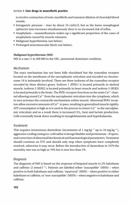
P1: PSB/JDO/OZN P2: PSB Printer: Yet To Come
9780521704632c11 CUFX213A/Peck 9780521618168 December 28, 2007 12:5
Section II Core drugs in anaesthetic practice
to involve contraction of tonic myofibrils and transient dilation of choroidal bloodvessels.� Intragastric pressure – rises by about 10 cmH2O, but as the lower oesophagealsphincter tone increases simultaneously there is no increased risk of reflux.� Anaphylaxis – suxamethonium makes up a significant proportion of the cases ofanaphylaxis caused by muscle relaxants.� Malignant hyperthermia (see below).� Prolonged neuromuscular block (see below).
Malignant hyperthermia (MH)MH is a rare (1 in 200 000 in the UK), autosomal-dominant condition.
MechanismThe exact mechanism has not been fully elucidated but the ryanodine receptorlocated on the membrane of the sarcoplasmic reticulum and encoded on chromo-some 19 is intimately involved. There are three isoforms of the ryanodine receptorencoded by three distinct genes. Isoform 1 (RYR1) is located primarily in skeletalmuscle, isoform 2 (RYR2) is located primarily in heart muscle and isoform 3 (RYR3)is located primarily is the brain. The RYR1 receptor functions as the main Ca2+ chan-nel allowing stored Ca2+ from the sarcoplasmic reticulum into the cytoplasm, whichin turn activates the contractile mechanisms within muscle. Abnormal RYR1 recep-tors allow excessive amounts of Ca2+ to pass, resulting in generalized muscle rigidity.ATP consumption is high as it is used in the process to return Ca2+ to the sarcolplas-mic reticulum and as a result there is increased CO2, heat and lactate production.Cells eventually break down resulting in myoglobinaemia and hyperkalaemia.
TreatmentThis requires intravenous dantrolene (increments of 1 mg.kg−1 up to 10 mg.kg−1),aggressive cooling (using ice-cold saline to lavage bladder and peritoneum – if open),and correction of abnormal biochemical and haematological parameters. Treatmentshould continue on ITU and should only stop when symptoms have completelyresolved, otherwise it may recur. Before the introduction of dantrolene in 1979 themortality rate was as high as 70% but is now less than 5%.
DiagnosisThe diagnosis of MH is based on the response of biopsied muscle to 2% halothaneand caffeine (2 mmol.l−1). Patients are labelled either ‘susceptible’ (MHS) – whenpositive to both halothane and caffeine, ‘equivocal’ (MHE) – when positive to eitherhalothane or caffeine, or ‘non-susceptible’ (MHN) – when negative to halothane andcaffeine.
182
P1: PSB/JDO/OZN P2: PSB Printer: Yet To Come
9780521704632c11 CUFX213A/Peck 9780521618168 December 28, 2007 12:5
11 Muscle relaxants and anticholinesterases
Safe drugsThese include opioids, thiopental, propofol, etomidate, ketamine, benzodiazepines,atropine, local anaesthetics and N2O.
Patients suspected of having MH should be referred to the UK MH investigationunit in Leeds.
Dantrolene is used in the treatment (and prophylaxis) of MH, neuroleptic malig-nant syndrome, chronic spasticity of voluntary muscle and ecstacy intoxication. Itis available as capsules and in vials as an orange powder containing dantrolene20 mg, mannitol 3 g and sodium hydroxide. Each vial should be reconstituted with60 ml water producing a solution of pH 9.5. It is highly irritant when extravasatedand a diuresis follows intravenous administration reflecting its formulation withmannitol. Chronic use is associated with hepatitis and pleural effusion.
Mechanism of actionDantrolene uncouples the excitation contraction process by binding to the ryanodinereceptor thereby preventing the release of Ca2+ from the sarcoplasmic reticulum instriated muscle. As vascular smooth muscle and cardiac muscle are not primarilydependent on Ca2+ release for contraction, they are not usually affected. It has noeffect on the muscle action potential and usually has little effect on the clinical dura-tion of the non-depolarizing muscle relaxants. It may, however, produce respiratoryfailure secondary to skeletal muscle weakness.
KineticsOral bioavailability is variable and it is approximately 85% bound in the plasma toalbumin. It is metabolized in the liver and excreted in the urine.
Prolonged block (suxamethonium apnoea)Plasma cholinesterase activity may be reduced due to genetic variability or acquiredconditions, leading to prolonged neuromuscular block. Single amino acid sub-stitutions are responsible for genetically altered enzymatic activity. Four alleles –usual (normal), atypical (dibucaine-resistant), silent (absent) and fluoride-resistant– have been identified at a single locus of chromosome 3 and make up the 10 geno-types.
Ninety-six percent of the population is homozygous for the normal Eu gene andmetabolize suxamethonium rapidly. Up to 4% may be heterozygotes resulting in amildly prolonged block of up to 10 minutes while a very small fraction may havea genotype that confers a block of a few hours. This prolonged block may bereversed by administration of fresh frozen plasma, which provides a source of plasmacholinesterase. Alternatively the patient may be sedated and ventilated while theblock wears off naturally.
183
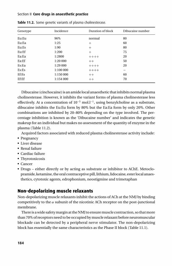
P1: PSB/JDO/OZN P2: PSB Printer: Yet To Come
9780521704632c11 CUFX213A/Peck 9780521618168 December 28, 2007 12:5
Section II Core drugs in anaesthetic practice
Table 11.2. Some genetic variants of plasma cholinesterase.
Genotype Incidence Duration of block Dibucaine number
Eu:Eu 96% normal 80Eu:Ea 1:25 + 60Eu:Es 1:90 + 80Eu:Ef 1:200 + 75Ea:Ea 1:2800 ++++ 20Ea:Ef 1:20 000 ++ 50Es:Ea 1:29 000 ++++ 20Es:Es 1:100 000 ++++ −Ef:Es 1:150 000 ++ 60Ef:Ef 1:154 000 ++ 70
Dibucaine (cinchocaine) is an amide local anaesthetic that inhibits normal plasmacholinesterase. However, it inhibits the variant forms of plasma cholinesterase lesseffectively. At a concentration of 10−5 mol.l−1, using benzylcholine as a substrate,dibucaine inhibits the Eu:Eu form by 80% but the Ea:Ea form by only 20%. Othercombinations are inhibited by 20–80% depending on the type involved. The per-centage inhibition is known as the ‘Dibucaine number’ and indicates the geneticmakeup for an individual but makes no assessment of the quantity of enzyme in theplasma (Table 11.2).
Acquired factors associated with reduced plasma cholinesterase activity include:� Pregnancy� Liver disease� Renal failure� Cardiac failure� Thyrotoxicosis� Cancer� Drugs – either directly or by acting as substrate or inhibitor to AChE. Metoclo-pramide, ketamine, the oral contraceptive pill, lithium, lidocaine, ester local anaes-thetics, cytotoxic agents, edrophonium, neostigmine and trimetaphan
Non-depolarizing muscle relaxantsNon-depolarizing muscle relaxants inhibit the actions of ACh at the NMJ by bindingcompetitively to the α subunit of the nicotinic ACh receptor on the post-junctionalmembrane.
There is a wide safety margin at the NMJ to ensure muscle contraction, so that morethan 70% of receptors need to be occupied by muscle relaxant before neuromuscularblockade can be detected by a peripheral nerve stimulator. The non-depolarizingblock has essentially the same characteristics as the Phase II block (Table 11.1).
184
P1: PSB/JDO/OZN P2: PSB Printer: Yet To Come
9780521704632c11 CUFX213A/Peck 9780521618168 December 28, 2007 12:5
11 Muscle relaxants and anticholinesterases
Non-depolarizing muscle relaxants fall into one of two chemical groupings:� Aminosteroidal compounds – vecuronium, rocuronium, pancuronium� Benzylisoquinolinium compounds – atracurium, mivacurium, tubocurarineAcross the two chemical groups the drugs can be divided according to their durationof action:� Short – mivacurium� Intermediate – atracurium� Long – pancuroniumOwing to their relatively polar nature, non-depolarizing drugs are unable to crosslipid membranes resulting in a small volume of distribution. Some are hydrolyzedin the plasma (atracurium, mivacurium) while others undergo a degree of hepaticmetabolism (pancuronium, vecuronium). The unmetabolized fraction is excreted inthe urine or bile.
Muscle relaxants are never given in isolation and so their potential for drug inter-action should be considered (Table 11.3).
VecuroniumVecuronium is a ‘clean’ drug, so called because it does not affect the cardiovascularsystem or precipitate the release of histamine. Its chemical structure differs frompancuronium by a single methyl group making it the monoquaternary analogue.
Presentation and usesVecuronium is potentially unstable in solution and so is presented as 10 mg freeze-dried powder containing mannitol and sodium hydroxide and is dissolved in 5 mlwater before administration. At 0.1 mg.kg−1 satisfactory intubating conditions arereached in about 90–120 seconds. It has a medium duration of action.
Other effects� Cardiovascular – vecuronium has no cardiac effects but, unlike pancuronium ortubocurarine, it may leave unchecked the bradycardias associated with fentanyland propofol.� Critical illness myopathy – this is seen most frequently in critically ill patientsin association with corticosteroids and/or muscle relaxants and may involve aprolonged recovery.
KineticsLike pancuronium, vecuronium is metabolized in the liver by de-acetylation to 3-and 17-hydroxy and 3,17-dihydroxy-vecuronium. Again the 3-hydroxy metabolitecarries significant muscle-relaxant properties, but unlike 3-hydroxypancuroniumit has a very short half-life and is of little clinical significance with normal renal
185
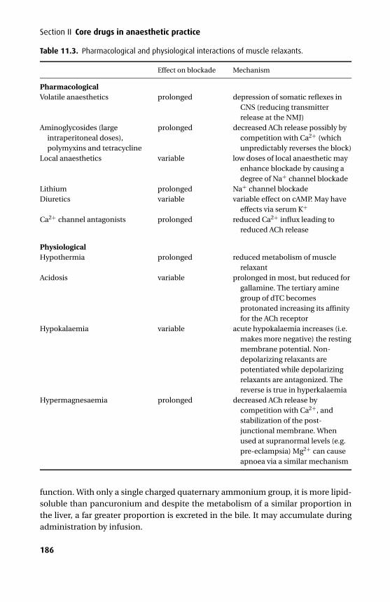
P1: PSB/JDO/OZN P2: PSB Printer: Yet To Come
9780521704632c11 CUFX213A/Peck 9780521618168 December 28, 2007 12:5
Section II Core drugs in anaesthetic practice
Table 11.3. Pharmacological and physiological interactions of muscle relaxants.
Effect on blockade Mechanism
PharmacologicalVolatile anaesthetics prolonged depression of somatic reflexes in
CNS (reducing transmitterrelease at the NMJ)
Aminoglycosides (largeintraperitoneal doses),polymyxins and tetracycline
prolonged decreased ACh release possibly bycompetition with Ca2+ (whichunpredictably reverses the block)
Local anaesthetics variable low doses of local anaesthetic mayenhance blockade by causing adegree of Na+ channel blockade
Lithium prolonged Na+ channel blockadeDiuretics variable variable effect on cAMP. May have
effects via serum K+
Ca2+ channel antagonists prolonged reduced Ca2+ influx leading toreduced ACh release
PhysiologicalHypothermia prolonged reduced metabolism of muscle
relaxantAcidosis variable prolonged in most, but reduced for
gallamine. The tertiary aminegroup of dTC becomesprotonated increasing its affinityfor the ACh receptor
Hypokalaemia variable acute hypokalaemia increases (i.e.makes more negative) the restingmembrane potential. Non-depolarizing relaxants arepotentiated while depolarizingrelaxants are antagonized. Thereverse is true in hyperkalaemia
Hypermagnesaemia prolonged decreased ACh release bycompetition with Ca2+, andstabilization of the post-junctional membrane. Whenused at supranormal levels (e.g.pre-eclampsia) Mg2+ can causeapnoea via a similar mechanism
function. With only a single charged quaternary ammonium group, it is more lipid-soluble than pancuronium and despite the metabolism of a similar proportion inthe liver, a far greater proportion is excreted in the bile. It may accumulate duringadministration by infusion.
186
P1: PSB/JDO/OZN P2: PSB Printer: Yet To Come
9780521704632c11 CUFX213A/Peck 9780521618168 December 28, 2007 12:5
11 Muscle relaxants and anticholinesterases
RocuroniumThis aminosteroidal drug was developed from vecuronium and is structurally differ-ent at only four positions. Its main advantage is its rapid onset (intubating conditionswithin 60–90 seconds), which in turn is due to its low potency.
A muscle relaxant with a low potency must be given at a higher dose to achievea clinically significant effect. A higher number of molecules result in a greater con-centration gradient from the plasma to the NMJ so that diffusion is faster and onsettime is reduced.
Presentation and usesRocuronium is prepared as a colourless solution containing 50 mg in 5 ml. At0.6 mg.kg−1 intubating conditions are reached within 100–120 seconds, althoughthis may be reduced to 60 seconds with higher doses (0.9–1.2 mg.kg−1). Its durationof action is similar to that of vecuronium.
Other effects� Cardiovascular – like vecuronium it has minimal cardiovascular effects, althoughat high doses when used to facilitate a more rapid tracheal intubation, it may causean increase in heart rate.
KineticsRocuronium is mainly excreted unchanged in the bile and to a lesser extent in theurine, although some de-acetylated metabolites may be produced. Its duration ofaction may be prolonged in hepatic and renal failure.
PancuroniumPancuronium is a bisquaternary aminosteroidal compound.
Presentation and usesPancuronium is presented as a colourless solution containing 4 mg in 2 ml andshould be stored at 4◦C. At 0.1 mg.kg−1 intubating conditions are reached within90–150 seconds. Its duration of action is about 45 minutes.
Other effects� Cardiovascular – pancuronium causes a tachycardia by blocking cardiac mus-carinic receptors. It may also have indirect sympathomimetic actions by preventingthe uptake of noradrenaline at post-ganglionic nerve endings.
KineticsBetween 10% and 40% is plasma protein bound, and in keeping with other drugs inits class pancuronium has a low volume of distribution. About 35% is metabolized in
187
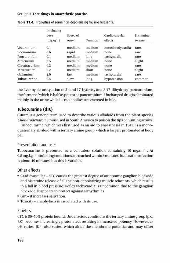
P1: PSB/JDO/OZN P2: PSB Printer: Yet To Come
9780521704632c11 CUFX213A/Peck 9780521618168 December 28, 2007 12:5
Section II Core drugs in anaesthetic practice
Table 11.4. Properties of some non-depolarizing muscle relaxants.
Intubating
dose
(mg.kg−1)
Speed of
onset Duration
Cardiovascular
effects
Histamine
release
Vecuronium 0.1 medium medium none/bradycardia rareRocuronium 0.6 rapid medium none rarePancuronium 0.1 medium long tachycardia rareAtracurium 0.5 medium medium none slightCis-atracurium 0.2 medium medium none rareMivacurium 0.2 medium short none slightGallamine 2.0 fast medium tachycardia rareTubocurarine 0.5 slow long hypotension common
the liver by de-acetylation to 3- and 17-hydroxy and 3,17-dihydroxy-pancuronium,the former of which is half as potent as pancuronium. Unchanged drug is eliminatedmainly in the urine while its metabolites are excreted in bile.
Tubocurarine (dTC)Curare is a generic term used to describe various alkaloids from the plant speciesChondrodendron. It was used in South America to poison the tips of hunting arrows.
Tubocurarine, which was first used as an aid to anaesthesia in 1942, is a mono-quaternary alkaloid with a tertiary amine group, which is largely protonated at bodypH.
Presentation and usesTubocurarine is presented as a colourless solution containing 10 mg.ml−1. At0.5 mg.kg−1 intubating conditions are reached within 3 minutes. Its duration of actionis about 40 minutes, but this is variable.
Other effects� Cardiovascular – dTC causes the greatest degree of autonomic ganglion blockadeand histamine release of all the non-depolarizing muscle relaxants, which resultsin a fall in blood pressure. Reflex tachycardia is uncommon due to the ganglionblockade. It appears to protect against arrhythmias.� Gut – it increases salivation.� Toxicity – anaphylaxis is associated with its use.
KineticsdTC is 30–50% protein bound. Under acidic conditions the tertiary amine group (pKa
8.0) becomes increasingly protonated, resulting in increased potency. However, aspH varies, [K+] also varies, which alters the membrane potential and may offset
188
P1: PSB/JDO/OZN P2: PSB Printer: Yet To Come
9780521704632c11a CUFX213A/Peck 9780521618168 December 28, 2007 11:59
11 Muscle relaxants and anticholinesterases
altered potency. Elimination is independent of metabolism with 70% excreted inthe urine and 30% in bile as unchanged drug. It accumulates in patients with renalfailure in whom a greater proportion is excreted in the bile.
AtracuriumAtracurium is a benzylisoquinolinium compound that is formulated as a mixture of10 stereoisomers, resulting from the presence of 4 chiral centres.
Presentation and usesAtracurium is presented as a colourless solution containing 10 mg.ml−1 in 2.5, 5 and25 ml vials and should be stored at 4◦C. At 0.5 mg.kg−1 intubating conditions arereached within 90–120 seconds.
Other effects� Cardiorespiratory – following rapid administration it may precipitate the releaseof histamine, which may be localized to the site of injection but may be gener-alized resulting in bronchospasm and hypotension. Slow intravenous injectionminimizes these effects.� Myopathy – in a manner similar to that of vecuronium, atracurium is associatedwith critical illness myopathy.
KineticsAtracurium has a unique metabolic pathway, undergoing ester hydrolysis and Hof-mann elimination.� Ester hydrolysis – non-specific esterases unrelated to plasma cholinesterase are
responsible for hydrolysis and account for 60% of atracurium’s metabolism. Thebreakdown products are a quaternary alcohol, a quaternary acid and laudanosine.Unlike Hofmann elimination, acidic conditions accelerate this metabolic pathway.However, pH changes in the clinical range probably do not alter the rate of esterhydrolysis of atracurium.� Hofmann elimination – while atracurium is stable at pH 4 and at 4◦C, Hofmannelimination describes its spontaneous breakdown to laudanosine and a quaternarymonoacrylate when placed at normal body temperature and pH. Acidosis andhypothermia will slow the process. Both breakdown products have been shown tohave potentially serious side effects (i.e. seizures), but at concentrations in excessof those encountered clinically. Laudanosine, while being a glycine antagonist, hasno neuromuscular-blocking properties and is cleared by the kidneys.These metabolic pathways result in a drug whose elimination is independent of
hepatic or renal function, which in certain clinical situations is advantageous.
Cis-atracuriumCis-atracurium is one of the ten stereoisomers present in atracurium.
189
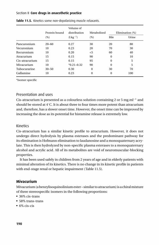
P1: PSB/JDO/OZN P2: PSB Printer: Yet To Come
9780521704632c11a CUFX213A/Peck 9780521618168 December 28, 2007 11:59
Section II Core drugs in anaesthetic practice
Table 11.5. Kinetics some non-depolarizing muscle relaxants.
Elimination (%)Protein bound
(%)
Volume of
distribution
(l.kg−1)
Metabolized
(%) Bile Urine
Pancuronium 20–60 0.27 30 20 80Vecuronium 10 0.23 20 70 30Rocuronium 10 0.20 <5 60 40Atracurium 15 0.15 90 0 10Cis-atracurium 15 0.15 95 0 5Mivacurium 10 *0.21–0.32 90 0 5Tubocurarine 30–50 0.30 0 30 70Gallamine 10 0.23 0 0 100
*Isomer specific
Presentation and usesCis-atracurium is presented as a colourless solution containing 2 or 5 mg.ml−1 andshould be stored at 4◦C. It is about three to four times more potent than atracuriumand, therefore, has a slower onset time. However, the onset time can be improved byincreasing the dose as its potential for histamine release is extremely low.
KineticsCis-atracurium has a similar kinetic profile to atracurium. However, it does notundergo direct hydrolysis by plasma esterases and the predominant pathway forits elimination is Hofmann elimination to laudanosine and a monoquaternary acry-late. This is then hydrolyzed by non-specific plasma esterases to a monoquaternaryalcohol and acrylic acid. All of its metabolites are void of neuromuscular-blockingproperties.
It has been used safely in children from 2 years of age and in elderly patients withminimal alteration of its kinetics. There is no change in its kinetic profile in patientswith end-stage renal or hepatic impairment (Table 11.5).
MivacuriumMivacurium (a benzylisoquinolinium ester – similar to atracurium) is a chiral mixtureof three stereospecific isomers in the following proportions:� 36% cis-trans� 58% trans-trans� 6% cis-cis
190
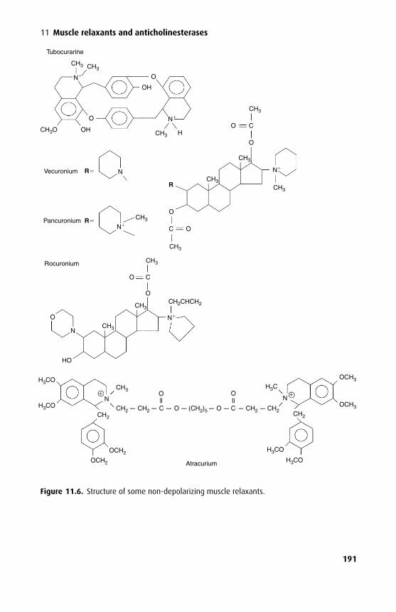
P1: PSB/JDO/OZN P2: PSB Printer: Yet To Come
9780521704632c11a CUFX213A/Peck 9780521618168 December 28, 2007 11:59
11 Muscle relaxants and anticholinesterases
Tubocurarine
Vecuronium
Pancuronium
CH3 CH3
CH3O HCH3
CH3
CH3
CH3
CH3
CH3
OH
OH
O
O
N
R
RO
O
CO
C O
R
N
Rocuronium CH3
CH3
CH3
CH2CHCH2
CH3
O
O
O
C
N
HO
N
N
N
N
Atracurium
H3COCH3
CH2 CH2 (CH2)5C O O C CH2 CH2 CH2
OCH3
OCH3
H3C
H3CO
H3CO
ON
O
OCH2
OCH2
CH2
NH3CO
+ +
Figure 11.6. Structure of some non-depolarizing muscle relaxants.
191
P1: PSB/JDO/OZN P2: PSB Printer: Yet To Come
9780521704632c11a CUFX213A/Peck 9780521618168 December 28, 2007 11:59
Section II Core drugs in anaesthetic practice
The cis-cis isomer has about 10% of the potency of the other two isomers andis not metabolized enzymatically. Its half-life is ten times that of the other twoisomers.
The main advantage of mivacurium is its short duration of action. Routine rever-sal of mivacurium with neostigmine may not be required due to its rapid enzymaticmetabolism. In addition, neostigmine inhibits plasma cholinesterase and may pre-vent its metabolism. Edrophonium may be a more suitable agent for the reversal ofneuromuscular block secondary to mivacurium.
Presentation and usesMivacurium is presented as an acidic (pH 3.5–5.0) aqueous solution containing2 mg.ml−1 in 5 and 10 ml ampoules. It has a shelf-life of 18 months when storedbelow 25◦C.
Effects� Cardiorespiratory – high doses may release histamine, resulting in a fall in bloodpressure and bronchospasm.
KineticsPlasma cholinesterase is responsible for the metabolism of the cis-trans and trans-trans isomers, so those patients with genetically low plasma cholinesterase levels(cf. suxamethonium apnoea) are subject to prolonged neuromuscular blockade. Itsduration of action is significantly prolonged in patients with end-stage liver disease,mainly due to reduced plasma cholinesterase activity.
GallamineGallamine was introduced into anaesthesia in 1947 as the first synthetic musclerelaxant. It currently has a limited role in anaesthesia, being used to reduce musclefasciculations induced by suxamethonium.
It selectively blocks cardiac muscarinic receptors causing a tachycardia and mayalso activate the sympathetic nervous system. As it is excreted unchanged by thekidneys, renal failure significantly prolongs its half-life. Unlike other muscle relaxantsan alkalosis prolongs its duration of action while an acidosis shortens it.
AnticholinesterasesThe enzyme AChE hydrolyzes ACh, terminating its effects. Anticholinesterases antag-onize AChE so that more ACh is available at the NMJ. However, the actions of anti-cholinesterases are not specific to the NMJ and autonomic cholinergic effects (brady-cardia, salivation) are also seen. For this reason they are often given with an anti-cholinergic (atropine or glycopyrrolate).
192
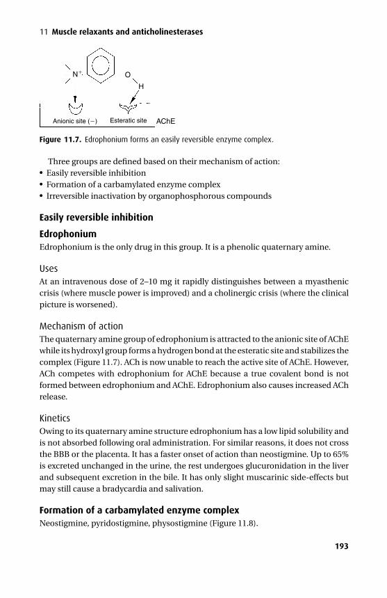
P1: PSB/JDO/OZN P2: PSB Printer: Yet To Come
9780521704632c11a CUFX213A/Peck 9780521618168 December 28, 2007 11:59
11 Muscle relaxants and anticholinesterases
N O
H
Anionic site ( ) Esteratic site AChE
Figure 11.7. Edrophonium forms an easily reversible enzyme complex.
Three groups are defined based on their mechanism of action:� Easily reversible inhibition� Formation of a carbamylated enzyme complex� Irreversible inactivation by organophosphorous compounds
Easily reversible inhibition
EdrophoniumEdrophonium is the only drug in this group. It is a phenolic quaternary amine.
UsesAt an intravenous dose of 2–10 mg it rapidly distinguishes between a myastheniccrisis (where muscle power is improved) and a cholinergic crisis (where the clinicalpicture is worsened).
Mechanism of actionThe quaternary amine group of edrophonium is attracted to the anionic site of AChEwhile its hydroxyl group forms a hydrogen bond at the esteratic site and stabilizes thecomplex (Figure 11.7). ACh is now unable to reach the active site of AChE. However,ACh competes with edrophonium for AChE because a true covalent bond is notformed between edrophonium and AChE. Edrophonium also causes increased AChrelease.
KineticsOwing to its quaternary amine structure edrophonium has a low lipid solubility andis not absorbed following oral administration. For similar reasons, it does not crossthe BBB or the placenta. It has a faster onset of action than neostigmine. Up to 65%is excreted unchanged in the urine, the rest undergoes glucuronidation in the liverand subsequent excretion in the bile. It has only slight muscarinic side-effects butmay still cause a bradycardia and salivation.
Formation of a carbamylated enzyme complexNeostigmine, pyridostigmine, physostigmine (Figure 11.8).
193
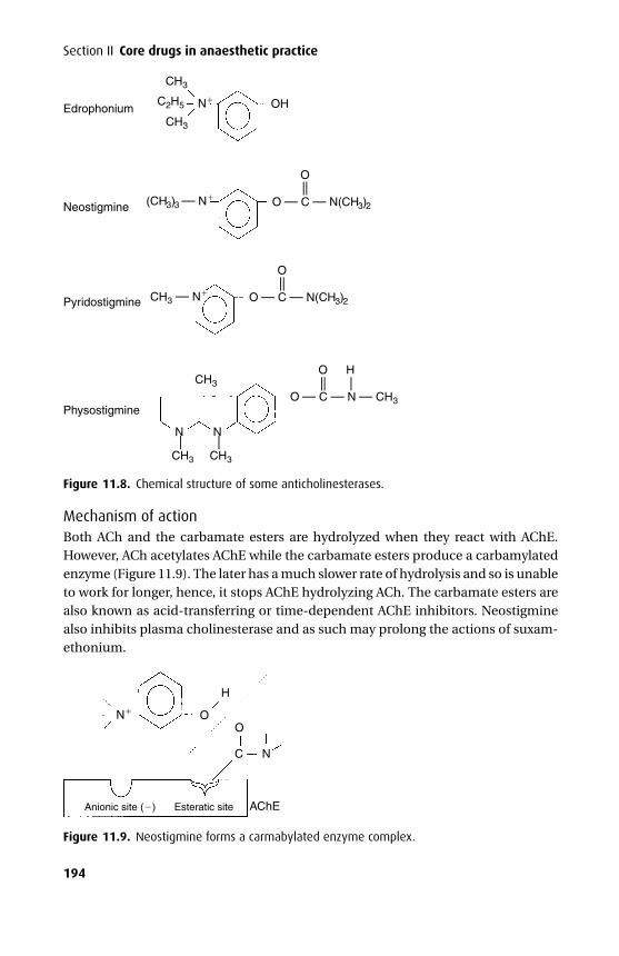
P1: PSB/JDO/OZN P2: PSB Printer: Yet To Come
9780521704632c11a CUFX213A/Peck 9780521618168 December 28, 2007 11:59
Section II Core drugs in anaesthetic practice
Edrophonium N
Neostigmine
Pyridostigmine
Physostigmine
CH3
CH3
CH3
N
CH3 — N
CH3
(CH3)3 — N O — C — N(CH3)2
C2H5 OH
O
— —O — C — N(CH3)2
O— —
O — C — N — CH3
O H
— — —
—
CH3
N
—
Figure 11.8. Chemical structure of some anticholinesterases.
Mechanism of actionBoth ACh and the carbamate esters are hydrolyzed when they react with AChE.However, ACh acetylates AChE while the carbamate esters produce a carbamylatedenzyme (Figure 11.9). The later has a much slower rate of hydrolysis and so is unableto work for longer, hence, it stops AChE hydrolyzing ACh. The carbamate esters arealso known as acid-transferring or time-dependent AChE inhibitors. Neostigminealso inhibits plasma cholinesterase and as such may prolong the actions of suxam-ethonium.
NO
C N
O
H
Anionic site ( ) Esteratic site AChE
Figure 11.9. Neostigmine forms a carmabylated enzyme complex.
194
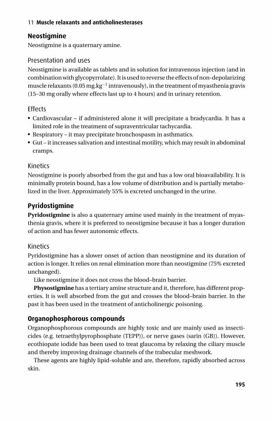
P1: PSB/JDO/OZN P2: PSB Printer: Yet To Come
9780521704632c11a CUFX213A/Peck 9780521618168 December 28, 2007 11:59
11 Muscle relaxants and anticholinesterases
NeostigmineNeostigmine is a quaternary amine.
Presentation and usesNeostigmine is available as tablets and in solution for intravenous injection (and incombination with glycopyrrolate). It is used to reverse the effects of non-depolarizingmuscle relaxants (0.05 mg.kg−1 intravenously), in the treatment of myasthenia gravis(15–30 mg orally where effects last up to 4 hours) and in urinary retention.
Effects� Cardiovascular – if administered alone it will precipitate a bradycardia. It has alimited role in the treatment of supraventricular tachycardia.� Respiratory – it may precipitate bronchospasm in asthmatics.� Gut – it increases salivation and intestinal motility, which may result in abdominalcramps.
KineticsNeostigmine is poorly absorbed from the gut and has a low oral bioavailability. It isminimally protein bound, has a low volume of distribution and is partially metabo-lized in the liver. Approximately 55% is excreted unchanged in the urine.
PyridostigminePyridostigmine is also a quaternary amine used mainly in the treatment of myas-thenia gravis, where it is preferred to neostigmine because it has a longer durationof action and has fewer autonomic effects.
KineticsPyridostigmine has a slower onset of action than neostigmine and its duration ofaction is longer. It relies on renal elimination more than neostigmine (75% excretedunchanged).
Like neostigmine it does not cross the blood–brain barrier.Physostigmine has a tertiary amine structure and it, therefore, has different prop-
erties. It is well absorbed from the gut and crosses the blood–brain barrier. In thepast it has been used in the treatment of anticholinergic poisoning.
Organophosphorous compoundsOrganophosphorous compounds are highly toxic and are mainly used as insecti-cides (e.g. tetraethylpyrophosphate (TEPP)), or nerve gases (sarin (GB)). However,ecothiopate iodide has been used to treat glaucoma by relaxing the ciliary muscleand thereby improving drainage channels of the trabecular meshwork.
These agents are highly lipid-soluble and are, therefore, rapidly absorbed acrossskin.
195
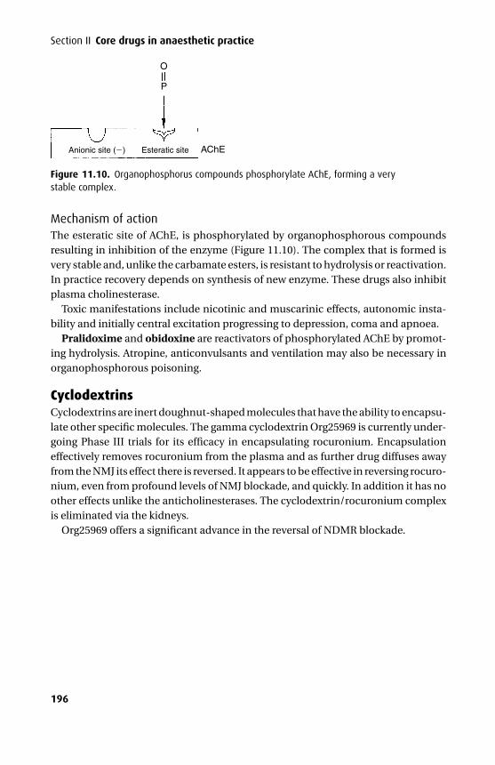
P1: PSB/JDO/OZN P2: PSB Printer: Yet To Come
9780521704632c11a CUFX213A/Peck 9780521618168 December 28, 2007 11:59
Section II Core drugs in anaesthetic practice
O
Anionic site ( ) Esteratic site AChE
P
Figure 11.10. Organophosphorus compounds phosphorylate AChE, forming a verystable complex.
Mechanism of actionThe esteratic site of AChE, is phosphorylated by organophosphorous compoundsresulting in inhibition of the enzyme (Figure 11.10). The complex that is formed isvery stable and, unlike the carbamate esters, is resistant to hydrolysis or reactivation.In practice recovery depends on synthesis of new enzyme. These drugs also inhibitplasma cholinesterase.
Toxic manifestations include nicotinic and muscarinic effects, autonomic insta-bility and initially central excitation progressing to depression, coma and apnoea.
Pralidoxime and obidoxine are reactivators of phosphorylated AChE by promot-ing hydrolysis. Atropine, anticonvulsants and ventilation may also be necessary inorganophosphorous poisoning.
CyclodextrinsCyclodextrins are inert doughnut-shaped molecules that have the ability to encapsu-late other specific molecules. The gamma cyclodextrin Org25969 is currently under-going Phase III trials for its efficacy in encapsulating rocuronium. Encapsulationeffectively removes rocuronium from the plasma and as further drug diffuses awayfrom the NMJ its effect there is reversed. It appears to be effective in reversing rocuro-nium, even from profound levels of NMJ blockade, and quickly. In addition it has noother effects unlike the anticholinesterases. The cyclodextrin/rocuronium complexis eliminated via the kidneys.
Org25969 offers a significant advance in the reversal of NDMR blockade.
196
P1: PSB Printer: Yet To Come
9780521704632c12 CUFX213A/Peck 9780521618168 December 28, 2007 12:15
SECTION III Cardiovascular drugs
12Sympathomimetics
Physiology
Autonomic nervous system (ANS)The ANS is a complex system of neurones that controls the body’s internal milieu. It isnot under voluntary control and is anatomically distinct from the somatic nervoussystem. Its efferent limb controls individual organs and smooth muscle, while itsafferent limb relays information (occasionally in somatic nerves) concerning visceralsensation and may result in reflex arcs.
The hypothalamus is the central point of integration of the ANS, but is itselfunder the control of the neocortex. However, not all autonomic activity involvesthe hypothalamus: locally, the gut coordinates its secretions; some reflex activity isprocessed within the spinal cord; and the control of vital functions by baroreceptorsis processed within the medulla. The ANS is divided into the parasympathetic andsympathetic nervous systems.
Parasympathetic nervous system (PNS)The PNS is made up of pre- and post-ganglionic fibres. The pre-ganglionic fibresarise from two locations (Figure 12.1):� Cranial nerves (III, VII, IX, X) – which supply the eye, salivary glands, heart, bronchi,
upper gastrointestinal tract (to the splenic flexure) and ureters� Sacral fibres (S2,3,4) – which supply distal bowel, bladder and genitalsAll these fibres synapse within ganglia that are close to, or within, the effector organ.The post-ganglionic neurone releases acetylcholine, which acts via nicotinic recep-tors.
The PNS may be modulated by anticholinergics (see Chapter 18) and anti-cholinesterases (see Chapter 11).
Sympathetic nervous system (SNS)The SNS is also made up of pre- and post-ganglionic fibres. The pre-ganglionicfibres arise within the lateral horns of the spinal cord at the thoracic and upperlumbar levels (T1–L2) and pass into the anterior primary rami, and via the whiterami communicans into the sympathetic chain or ganglia where they may eithersynapse at that or an adjacent level, or pass anteriorly through a splanchnic nerve tosynapse in a prevertebral ganglion (Figure 12.2). The unmyelinated post-ganglionic
197
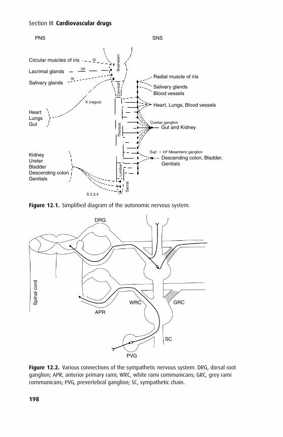
P1: PSB Printer: Yet To Come
9780521704632c12 CUFX213A/Peck 9780521618168 December 28, 2007 12:15
Section III Cardiovascular drugs
PNS
Circular muscles of iris
Radial muscle of iris
Salivary glandsBlood vessels
Gut and Kidney
Descending colon, Bladder, Genitals
Heart, Lungs, Blood vessels
Lacrimal glands
Salivary glands
HeartLungsGut
KidneyUreterBladderDescending colonGenitals
III
VII
IX
X (vagus)
S 2,3,4
Bra
inst
emC
ervi
cal
Tho
raci
cLu
mba
r
Sac
ral
Coeliac ganglion
Sup' Inf' Mesenteric ganglion
SNS
Figure 12.1. Simplified diagram of the autonomic nervous system.
DRG
APR
Spi
nal c
ord
PVG
SC
GRCWRC
Figure 12.2. Various connections of the sympathetic nervous system. DRG, dorsal rootganglion; APR, anterior primary rami; WRC, white rami communicans; GRC, grey ramicommunicans; PVG, prevertebral ganglion; SC, sympathetic chain.
198

P1: PSB Printer: Yet To Come
9780521704632c12 CUFX213A/Peck 9780521618168 December 28, 2007 12:15
12 Sympathomimetics
Table 12.1. Summary of transmitters within the autonomic nervous system.
Pre-ganglionic Post-ganglionic
PNS acetylcholine acetylcholineSNS acetylcholine noradrenalineAdrenal medulla acetylcholine —Sweat glands acetylcholine acetylcholine
fibres then pass into the adjacent spinal nerve via the grey rami communicans. Theyrelease noradrenaline, which acts via adrenoceptors.
The adrenal medulla receives presynaptic fibres that synapse directly with its chro-maffin cells using acetylcholine as the transmitter. It releases adrenaline into thecirculation, which, therefore, acts as a hormone, not a transmitter.
Post-ganglionic sympathetic fibres release acetylcholine to innervate sweatglands.
All pre-ganglionic ANS fibres are myelinated and release acetylcholine, which actsvia nicotinic receptors (Table 12.1).
SympathomimeticsSympathomimetics exert their effects via adrenoceptors or dopamine receptorseither directly or indirectly. Direct-acting sympathomimetics attach to and actdirectly via these receptors, while indirect-acting sympathomimetics cause therelease of noradrenaline to produce their effects via these receptors.
The structure of sympathomimetics is based on a benzene ring with variousamine side chains attached at the C1 position. Where a hydroxyl group is presentat the C3 and C4 positions the agent is known as a catecholamine (because 3,4-dihydroxybenzene is otherwise known as ‘catechol’).
Sympathomimetic and other inotropic agents will be discussed under the follow-ing headings:� Naturally occurring catecholamines� Synthetic agents� Other inotropic agents
Naturally occurring catecholaminesAdrenaline, noradrenaline and dopamine are the naturally occurring cate-cholamines and their synthesis is interrelated (Figure 12.3). They act via adrenergicand dopaminergic receptors, which are summarized in Table 12.2.
Adrenaline
Presentation and usesAdrenaline is presented as a clear solution containing 0.1–1 mg.ml−1 for admin-istration as a bolus in asystole or anaphylaxis or by infusion (dose range
199
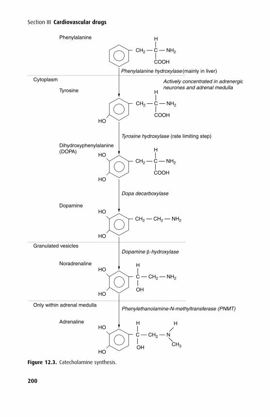
P1: PSB Printer: Yet To Come
9780521704632c12 CUFX213A/Peck 9780521618168 December 28, 2007 12:15
Section III Cardiovascular drugs
Phenylalanine hydroxylase (mainly in liver)
Tyrosine hydroxylase (rate limiting step)
Dopa decarboxylase
Granulated vesicles
Only within adrenal medulla
Dopamine -hydroxylase
Phenylethanolamine-N-methyltransferase (PNMT)
Actively concentrated in adrenergicneurones and adrenal medulla
CH2 — CH2 — NH2
HO
Cytoplasm
HO
HO
HO
HO
HO
HO
HO
HO
Phenylalanine
Tyrosine
Dihydroxyphenylalanine(DOPA)
Dopamine
Noradrenaline
Adrenaline
CH2 — C — NH2
H
COOH
——
CH2 — C — NH2
H
COOH
——
CH2 — C — NH2
H
COOH
——
H
C — CH2 — NH2
OH
——
CH3
C — CH2 — N
OH
H H
—
—
— —
Figure 12.3. Catecholamine synthesis.
200
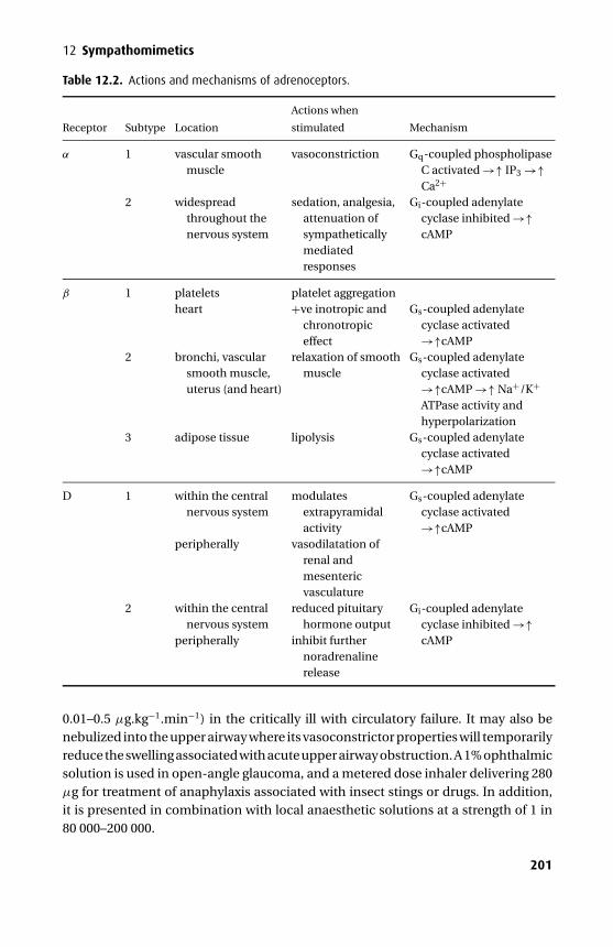
P1: PSB Printer: Yet To Come
9780521704632c12 CUFX213A/Peck 9780521618168 December 28, 2007 12:15
12 Sympathomimetics
Table 12.2. Actions and mechanisms of adrenoceptors.
Receptor Subtype Location
Actions when
stimulated Mechanism
α 1 vascular smoothmuscle
vasoconstriction Gq-coupled phospholipaseC activated →↑ IP3 →↑Ca2+
2 widespreadthroughout thenervous system
sedation, analgesia,attenuation ofsympatheticallymediatedresponses
Gi-coupled adenylatecyclase inhibited →↑cAMP
β 1 platelets platelet aggregationheart +ve inotropic and
chronotropiceffect
Gs-coupled adenylatecyclase activated→↑cAMP
2 bronchi, vascularsmooth muscle,uterus (and heart)
relaxation of smoothmuscle
Gs-coupled adenylatecyclase activated→↑cAMP →↑ Na+/K+
ATPase activity andhyperpolarization
3 adipose tissue lipolysis Gs-coupled adenylatecyclase activated→↑cAMP
D 1 within the centralnervous system
modulatesextrapyramidalactivity
Gs-coupled adenylatecyclase activated→↑cAMP
peripherally vasodilatation ofrenal andmesentericvasculature
2 within the centralnervous system
reduced pituitaryhormone output
Gi-coupled adenylatecyclase inhibited →↑
peripherally inhibit furthernoradrenalinerelease
cAMP
0.01–0.5 µg.kg−1.min−1) in the critically ill with circulatory failure. It may also benebulized into the upper airway where its vasoconstrictor properties will temporarilyreduce the swelling associated with acute upper airway obstruction. A 1% ophthalmicsolution is used in open-angle glaucoma, and a metered dose inhaler delivering 280µg for treatment of anaphylaxis associated with insect stings or drugs. In addition,it is presented in combination with local anaesthetic solutions at a strength of 1 in80 000–200 000.
201
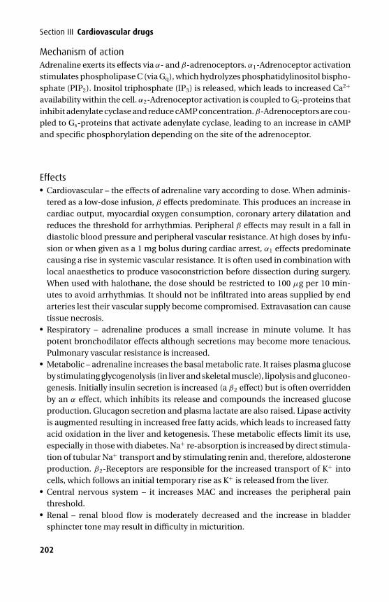
P1: PSB Printer: Yet To Come
9780521704632c12 CUFX213A/Peck 9780521618168 December 28, 2007 12:15
Section III Cardiovascular drugs
Mechanism of actionAdrenaline exerts its effects via α- and β-adrenoceptors. α1-Adrenoceptor activationstimulates phospholipase C (via Gq), which hydrolyzes phosphatidylinositol bispho-sphate (PIP2). Inositol triphosphate (IP3) is released, which leads to increased Ca2+
availability within the cell. α2-Adrenoceptor activation is coupled to Gi-proteins thatinhibit adenylate cyclase and reduce cAMP concentration. β-Adrenoceptors are cou-pled to Gs-proteins that activate adenylate cyclase, leading to an increase in cAMPand specific phosphorylation depending on the site of the adrenoceptor.
Effects� Cardiovascular – the effects of adrenaline vary according to dose. When adminis-tered as a low-dose infusion, β effects predominate. This produces an increase incardiac output, myocardial oxygen consumption, coronary artery dilatation andreduces the threshold for arrhythmias. Peripheral β effects may result in a fall indiastolic blood pressure and peripheral vascular resistance. At high doses by infu-sion or when given as a 1 mg bolus during cardiac arrest, α1 effects predominatecausing a rise in systemic vascular resistance. It is often used in combination withlocal anaesthetics to produce vasoconstriction before dissection during surgery.When used with halothane, the dose should be restricted to 100 µg per 10 min-utes to avoid arrhythmias. It should not be infiltrated into areas supplied by endarteries lest their vascular supply become compromised. Extravasation can causetissue necrosis.� Respiratory – adrenaline produces a small increase in minute volume. It haspotent bronchodilator effects although secretions may become more tenacious.Pulmonary vascular resistance is increased.� Metabolic – adrenaline increases the basal metabolic rate. It raises plasma glucoseby stimulating glycogenolysis (in liver and skeletal muscle), lipolysis and gluconeo-genesis. Initially insulin secretion is increased (a β2 effect) but is often overriddenby an α effect, which inhibits its release and compounds the increased glucoseproduction. Glucagon secretion and plasma lactate are also raised. Lipase activityis augmented resulting in increased free fatty acids, which leads to increased fattyacid oxidation in the liver and ketogenesis. These metabolic effects limit its use,especially in those with diabetes. Na+ re-absorption is increased by direct stimula-tion of tubular Na+ transport and by stimulating renin and, therefore, aldosteroneproduction. β2-Receptors are responsible for the increased transport of K+ intocells, which follows an initial temporary rise as K+ is released from the liver.� Central nervous system – it increases MAC and increases the peripheral painthreshold.� Renal – renal blood flow is moderately decreased and the increase in bladdersphincter tone may result in difficulty in micturition.
202
P1: PSB Printer: Yet To Come
9780521704632c12 CUFX213A/Peck 9780521618168 December 28, 2007 12:15
12 Sympathomimetics
KineticsAdrenaline is not given orally due to inactivation. Subcutaneous absorption is lessrapid than intramuscular. Tracheal absorption is erratic but may be used in emer-gencies where intravenous access is not available.
It is metabolized by mitochondrial MAO and catechol O-methyl trans-ferase (COMT) within the liver, kidney and blood to the inactive 3-methoxy-4-hydroxymandelic acid (vanillylmandelic acid or VMA) and metadrenaline, whichis conjugated with glucuronic acid or sulphates, both of which are excreted in theurine. It has a short half-life (about 2 minutes) due to rapid metabolism.
Noradrenaline
Presentation and usesNoradrenaline is presented as a clear solution containing 0.2–2 mg.ml−1 nora-drenaline acid tartrate, which is equivalent to 0.1–1 mg.ml−1 respectively of nora-drenaline base, and contains the preservative sodium metabisulphite. It is used as anintravenous infusion (dose range 0.05–0.5 µg.kg−1.min−1) to increase the systemicvascular resistance.
Mechanism of actionIts actions are mediated mainly via stimulation of α1-adrenoceptors but alsoβ-adrenoceptors.
Effects� Cardiovascular – the effects of systemically infused noradrenaline are slightlydifferent from those of endogenous noradrenaline. Systemically infused nora-drenaline causes peripheral vasoconstriction, increases systolic and diastolicblood pressure and may cause a reflex bradycardia. Cardiac output may fall andmyocardial oxygen consumption is increased. A vasodilated coronary circulationcarries an increased coronary blood flow. Pulmonary vascular resistance may beincreased and venous return is increased by venoconstriction. In excess it produceshypertension, bradycardia, headache and excessive peripheral vasoconstriction,occasionally leading to ischaemia and gangrene of extremities. Extravazation cancause tissue necrosis. Endogenously released noradrenaline causes tachycardiaand a rise in cardiac output.� Splanchnic – renal and hepatic blood flow falls due to vasoconstriction.� Uterus – blood flow to the pregnant uterus is reduced and may result in foetalbradycardia. It may also exert a contractile effect and cause foetal asphyxia.� Interactions – despite being a direct-acting sympathomimetic amine it should beused with caution in patients taking monoamine oxidase inhibitors (MAOI) as itseffects may be exaggerated and prolonged.
203
P1: PSB Printer: Yet To Come
9780521704632c12 CUFX213A/Peck 9780521618168 December 28, 2007 12:15
Section III Cardiovascular drugs
KineticsFor endogenously released noradrenaline, Uptake 1 describes its active uptake backinto the nerve terminal where it is metabolized by MAO (COMT is not present in sym-pathetic nerves) or recycled. It forms the main mechanism by which noradrenaline isinactivated. Uptake 2 describes the diffusion away from the nerve and is less impor-tant. Noradrenaline reaches the circulation in this way and is metabolized by COMTto the inactive 3-methoxy–4-hydroxymandelic acid (vanillylmandelic acid or VMA)and normetadrenaline, which is conjugated with glucuronic acid or sulphates, bothof which are excreted in the urine. It has a short half-life (about 2 minutes) dueto rapid metabolism. Unlike adrenaline and dopamine, up to 25% is taken up as itpasses through the lungs.
DopamineIn certain cells within the brain and interneurones of the autonomic ganglia, do-pamine is not converted to noradrenaline and is released as a neurotransmitter.
Presentation and usesDopamine is presented as a clear solution containing 200 or 800 mg in 5 ml waterwith sodium metabisulphite. It is used to improve haemodynamic parameters andurine output.
Mechanism of actionIn addition to its effects on α and β adrenoceptors, dopamine also acts via dopamine(D1 and D2) receptors via Gs and Gi coupled adenylate cyclase leading to increasedor decreased levels of cAMP.
Effects� Cardiovascular – these depend on its rate of infusion and vary between patients.At lower rates (up to 10 µg.kg−1.min−1) β1 effects predominate leading toincreased contractility, heart rate, cardiac output and coronary blood flow. Inaddition to its direct effects, it also stimulates the release of endogenous nora-drenaline. At higher rates (>10 µg.kg−1.min−1) α effects tend to predominateleading to increased systemic vascular resistance and venous return. In keep-ing with other inotropes an adequate preload is essential to help control tachy-cardia. It is less arrhythmogenic than adrenaline. Extravasation can cause tissuenecrosis.� Respiratory – infusions of dopamine attenuate the response of the carotid body tohypoxaemia. Pulmonary vascular resistance is increased.� Splanchnic – dopamine has been shown to vasodilate mesenteric vessels via D1
receptors. However, the improvement in urine output may be entirely due to
204
P1: PSB Printer: Yet To Come
9780521704632c12 CUFX213A/Peck 9780521618168 December 28, 2007 12:15
12 Sympathomimetics
inhibition of proximal tubule Na+ reabsorption and an improved cardiac outputand blood pressure.� Central nervous system – dopamine modulates extra-pyramidal movement andinhibits the secretion of prolactin from the pituitary gland. It cannot cross theblood–brain barrier, although its precursor l-dopa can.� Miscellaneous – owing to stimulation of the chemoreceptor trigger zone it causesnausea and vomiting. Gastric transit time is also increased.� Interactions – despite being a direct-acting sympathomimetic amine the effects ofdopamine may be significantly exaggerated and prolonged during MAOI therapy.
KineticsDopamine is only administered intravenously and preferably via a central vein. It actswithin 5 minutes and has a duration of 10 minutes. Metabolism is via MAO and COMTin the liver, kidneys and plasma to inactive compounds (3,4-dihydroxyphenylaceticacid and homovanillic acid (HVA)) which are excreted in the urine as sulphate andglucuronide conjugates. About 25% of an administered dose is converted to nora-drenaline in sympathetic nerve terminals. Its half-life is about 3 minutes.
Synthetic agentsOf the synthetic agents, only isoprenaline, dobutamine and dopexamine are classi-fied as catecholamines as only they contain hydroxyl groups on the 3- and 4-positionsof the benzene ring.
α1-Agonists
PhenylephrinePhenylephrine is a direct-acting sympathomimetic amine with potent α1-agonistactions. It causes a rapid rise in systemic vascular resistance and blood pressure. Ithas no effect on β-adrenoceptors.
Presentation and usesPhenylephrine is presented as a clear solution containing 10 mg in 1 ml. Phenyle-phrine is presented as a clear solution containing 10 mg in 1 ml. Bolus doses of50–100 µg are used intravenously although 2–5 mg may be administered intramus-cularly or subcutaneously for a more prolonged duration. It is used to increase alow systemic vascular resistance associated with spinal anaesthesia or systemicallyadministered drugs. In certain patients, general anaesthesia may drop the systemicvascular resistance and reverse a left-to-right intracardiac shunt, this may be reversedby phenylephrine. It is also available for use as a nasal decongestant and mydriaticagent. It may have a limited use in the treatment of supraventricular tachycardiaassociated with hypotension.
205
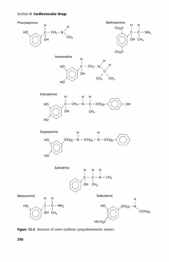
P1: PSB Printer: Yet To Come
9780521704632c12 CUFX213A/Peck 9780521618168 December 28, 2007 12:15
Section III Cardiovascular drugs
HO
HO
HO
Phenylephrine
Isoprenaline
Methoxamine
Dobutamine
Dopexamine
Ephedrine
SalbutamolMetaraminol
HO
HO
HO
OH
HO
NCH2C
HH
OHCH3
C
H
OH
C
H
N
N N
H
H
C
OH
HO HO N
H
HO H2C
C N
H H
H
H
C
H
OH
OH
C C
H H
(CH2)2
(CH2)2
(CH2)2C(CH3)3
(CH2)6 (CH2)2
CH3
NH2
CH3
H
C
OH
C
H
CH3
C
CH3
NH2
CH3
CH3O
CH3O
CH3
CH2
CH2
CH3
HH
N
Figure 12.4. Structure of some synthetic sympathomimetic amines.
206
P1: PSB Printer: Yet To Come
9780521704632c12 CUFX213A/Peck 9780521618168 December 28, 2007 12:15
12 Sympathomimetics
Effects� Cardiovascular – phenylephrine raises the systemic vascular resistance and bloodpressure and may result in a reflex bradycardia, all of which results in a drop incardiac output. It is not arrhythmogenic.� Central nervous system – it has no stimulatory effects.� Renal – blood flow falls in a manner similar to that demonstrated by noradrenaline.� Uterus – while its use in obstetrics results in a more favorable cord gas profileit has not yet gained widespread acceptance due to the possibility of accidentaloverdose.
KineticsIntravenous administration results in a rapid rise in blood pressure, which lasts5–10 minutes, while intramuscular or subcutaneous injection takes 15 minutes towork but lasts up to 1 hour. It is metabolized in the liver by MAO. The products ofmetabolism and their route of elimination have not been identified.
MethoxamineMethoxamine (no longer in production) is a direct-acting sympathomimetic aminewith specificα1-agonist actions. Consequently it has similar effects to phenylephrine.It is presented as a clear solution containing 20 mg.ml−1.
An intravenous bolus of 1 mg usually produces a rapid rise in systemic vascularresistance and, therefore, blood pressure, often with an accompanying reflex brady-cardia. It may also be given intramuscularly or subcutaneously at a higher dose fora more prolonged duration of action.
β-Agonists
IsoprenalineIsoprenaline is a highly potent synthetic catecholamine with actions at β1- andβ2-adrenoceptors. It has no α effects. It is not available in the UK any longer.
Presentation and usesIsoprenaline is presented as a clear solution containing 1 mg.ml−1 for intravenousinfusion and as a metered dose inhaler delivering 80 or 400 µg. It is no longer used totreat reversible airway obstruction as this was associated with an increased mortality.More specific β2-agonists are now used (e.g. salbutamol). The 30 mg tablets arevery rarely used. It is used intravenously to treat severe bradycardia associated withatrioventricular (AV) block or β-blockers (dose range 0.5–10 µg.min−1).
Effects� Cardiovascular – stimulation of β1-adrenoceptors increases heart rate, myocardialcontractility, automaticity and cardiac output. The effects on blood pressure are
207
P1: PSB Printer: Yet To Come
9780521704632c12 CUFX213A/Peck 9780521618168 December 28, 2007 12:15
Section III Cardiovascular drugs
varied. Theβ2 effects may drop the systemic vascular resistance so that the increasein cardiac output is insufficient to maintain blood pressure. Myocardial oxygendelivery may decrease significantly when tachycardia reduces diastolic coronaryfilling time and the reduced diastolic blood pressure reduces coronary perfusion.Some coronary vasodilatation occurs to attenuate this.� Respiratory – it is a potent bronchodilator and inhibits histamine release in thelungs, improving mucous flow. Anatomical dead space and ventilation perfusionmismatching increases, which may lead to systemic hypoxaemia.� Central nervous system – isoprenaline has stimulant effects on the CNS.� Splanchnic – mesenteric and renal blood flow is increased.� Metabolic – its β effects lead to a raised blood glucose and free fatty acids.
KineticsWhen administered orally it is well absorbed but extensive first-pass metabolismresults in a low oral bioavailability, being rapidly metabolized by COMT within theliver. A significant fraction is excreted unchanged in the urine along with conjugatedmetabolites.
DobutamineDobutamine is a direct-acting synthetic catecholamine derivative of isoprenaline.β1 effects predominate but it retains a small effect at β2-adrenoceptors.
Presentation and usesDobutamine is presented in 20 ml water containing 250 mg dobutamine and sodiummetabisulphite or in 5 ml water containing 250 mg dobutamine and ascorbic acid. Itis used to augment low cardiac output states associated with myocardial infarction,cardiac surgery and cardiogenic shock (dose range 0.5–20 µg.kg−1.min−1). It is alsoused in cardiac stress testing as an alternative to exercise.
Effects� Cardiovascular – its main actions are direct stimulation of β1-receptors resulting inincreased contractility, heart rate and myocardial oxygen requirement. The bloodpressure is usually increased despite a limited fall in systemic vascular resistancevia β2 stimulation. It may precipitate arrhythmias including an increased ventric-ular response rate in patients with atrial fibrillation or flutter, due to increased AVconduction. It should be avoided in patients with cardiac outflow obstruction (e.g.aortic stenosis, cardiac tamponade).� Splanchnic – it has no effect on the splanchnic circulation although urine outputmay increase following a rise in cardiac output.
208
P1: PSB Printer: Yet To Come
9780521704632c12 CUFX213A/Peck 9780521618168 December 28, 2007 12:15
12 Sympathomimetics
KineticsDobutamine is only administered intravenously. It is rapidly metabolized by COMTto inactive metabolites that are conjugated and excreted in the urine. It has a half-lifeof 2 minutes.
DopexamineDopexamine is a synthetic analogue of dopamine.
Presentation and usesDopexamine is presented as 50 mg in 5 ml (at pH 2.5) for intravenous use. It isused to improve cardiac output and improve mesenteric perfusion (dose range0.5–6 µg.kg−1.min−1).
Mechanism of actionDopexamine stimulates β2-adrenoceptors and dopamine (D1) receptors and mayalso inhibit the re-uptake of noradrenaline. It has only minimal effect on D2 andβ1-adrenoceptors, and no effect on α-adrenoceptors.
Effects� Cardiovascular – while it has positive inotropic effects (due to cardiacβ2-receptors),improvements in cardiac output are aided by a reduced afterload due to peripheralβ2 stimulation, which may reduce the blood pressure. It produces a small increasein coronary blood flow and there is no change in myocardial oxygen extraction.The alterations in heart rate are varied and it only rarely precipitates arrhythmias.� Mesenteric and renal – blood flow to the gut and kidneys increases due to anincreased cardiac output and reduced regional vascular resistance. Urine outputincreases. It may cause nausea and vomiting.� Respiratory – bronchodilation is mediated via β2 stimulation.� Miscellaneous – tremor and headache have been reported.
KineticsDopexamine is cleared rapidly from the blood and has a half-life of 7 minutes.
SalbutamolSalbutamol is a synthetic sympathomimetic amine with actions mainly atβ2-adreno-ceptors.
Presentation and usesSalbutamol is presented as a clear solution containing 50–500 µg.ml−1 for intra-venous infusion after dilution, a metered dose inhaler (100 µg) and a dry powder(200–400 µg) for inhalation, a solution containing 2.5–5 mg.ml−1 for nebulization,
209
P1: PSB Printer: Yet To Come
9780521704632c12 CUFX213A/Peck 9780521618168 December 28, 2007 12:15
Section III Cardiovascular drugs
and oral preparations (syrup 0.4 mg.ml−1 and 2, 4 or 8 mg tablets). It is used inthe treatment of reversible lower airway obstruction and occasionally in prematurelabour.
Effects� Respiratory – its main effects are relaxation of bronchial smooth muscle. It reverseshypoxic pulmonary vasoconstriction, increasing shunt, and may lead to hypox-aemia. Adequate oxygen should, therefore, be administered with nebulized salbu-tamol.� Cardiovascular – the administration of high doses, particularly intravenously, cancause stimulation of β1-adrenoceptors resulting in tachycardia, which may limitthe dose. Lower doses are sometimes associated with β2-mediated vasodilatation,which may reduce the blood pressure. It may also precipitate arrhythmias, espe-cially in the presence of hypokalaemia.� Metabolic – Na+/K+ ATPase is stimulated and transports K+ into cells resulting inhypokalaemia. Blood sugar rises especially in diabetic patients and is exacerbatedby concurrently administered steroids.� Uterus – it relaxes the gravid uterus. A small amount crosses the placenta to reachthe foetus.� Miscellaneous – a direct effect on skeletal muscle may produce tremor.
KineticsThe absorption of salbutamol from the gut is incomplete and is subject to a significanthepatic first-pass metabolism. Following inhalation or intravenous administration,it has a rapid onset of action. It is 10% protein bound and has a half-life of 4–6 hours.It is metabolized in the liver to the inactive 4-O-sulphate, which is excreted alongwith salbutamol in the urine.
SalmeterolSalmeterol is a long-acting β2-agonist used in the treatment of nocturnal andexercise-induced asthma. It should not be used during acute attacks due to a rela-tively slow onset.
It has a long non-polar side chain, which binds to the β2-adrenoceptor giving it along duration of action (about 12 hours). It is 15 times more potent than salbutamolat the β2-adrenoceptor, but four times less potent at the β1-adrenoceptor. It preventsthe release of histamine, leukotrienes and prostaglandin D2 from mast cells, and alsohas additional anti-inflammatory effects that differ from those induced by steroids.
Its effects are similar to those of salbutamol.
210
P1: PSB Printer: Yet To Come
9780521704632c12 CUFX213A/Peck 9780521618168 December 28, 2007 12:15
12 Sympathomimetics
RitodrineRitodrine is a β2-agonist that is used to treat premature labour. Tachycardia (β1
effect) is often seen during treatment. It crosses the placenta and may result in foetaltachycardia.
Ritodrine has been associated with fatal maternal pulmonary oedema. It alsocauses hypokalaemia, hyperglycaemia and, at higher levels, vomiting, restlessnessand seizures.
TerbutalineTerbutaline is a β2-agonist with some activity at β1-adrenoceptors. It is used in thetreatment of asthma and uncomplicated preterm labour. It has a similar side-effectprofile to other drugs in its class.
Mixed (α and β)
EphedrineEphedrine is found naturally in certain plants but is synthesized for medical use.
Presentation and usesEphedrine is formulated as tablets, an elixir, nasal drops and as a solution for injectioncontaining 30 mg.ml−1. It can exist as four isomers but only the l-isomer is active.It is used intravenously to treat hypotension associated with regional anaesthesia.In the obstetric setting this is now known to result in a poorer cord gas pH whencompared to purer α agonists, but its widespread use persists due to the potentialfor the α agonists to cause a significant maternal hypertension. It is also used to treatbronchospasm, nocturnal enuresis and narcolepsy.
Mechanism of actionEphedrine has both direct and indirect sympathomimetic actions. It also inhibits theactions of MAO on noradrenaline.
Owing to its indirect actions it is prone to tachyphylaxis as noradrenaline storesin sympathetic nerves become depleted.
Effects� Cardiovascular – it increases the cardiac output, heart rate, blood pressure, coro-nary blood flow and myocardial oxygen consumption. Its use may precipitatearrhythmias.� Respiratory – it is a respiratory stimulant and causes bronchodilation.� Renal – renal blood flow is decreased and the glomerular filtration rate falls.� Interactions – it should be used with extreme caution in those patients takingMAOI.
211
P1: PSB Printer: Yet To Come
9780521704632c12 CUFX213A/Peck 9780521618168 December 28, 2007 12:15
Section III Cardiovascular drugs
KineticsEphedrine is well absorbed orally, intramuscularly and subcutaneously. Unlikeadrenaline it is not metabolized by MAO or COMT and, therefore, has a longer dura-tion of action and an elimination half-life of 4 hours. Some is metabolized in the liverbut 65% is excreted unchanged in the urine.
MetaraminolMetaraminol is a synthetic amine with both direct and indirect sympathomimeticactions. It acts mainly via α1-adrenoceptors but also retains some β-adrenoceptoractivity.
Presentation and usesMetaraminol is presented as a clear solution containing 10 mg.ml−1. It is used tocorrect hypotension associated with spinal or epidural anaesthesia. An intravenousbolus of 0.5–2 mg is usually sufficient.
Effects� Cardiovascular – its main actions are to increase systemic vascular resistance,which leads to an increased blood pressure. Despite its activity at β-adrenoceptorsthe cardiac output often drops in the face of the raised systemic vascular resistance.Coronary artery flow increases by an indirect mechanism. Pulmonary vascularresistance is also increased leading to raised pulmonary artery pressure.
Other inotropic agents
Non-selective phosphodiesterase inhibitors
AminophyllineAminophylline is a methylxanthine derivative. It is a complex of 80% theophyllineand 20% ethylenediamine (which has no therapeutic effect but improves solubility).
Presentation and usesAminophylline is available as tablets and as a solution for injection containing25 mg.ml−1. Oral preparations are often formulated as slow release due to its half-life of about 6 hours. It is used in the treatment of asthma where the dose rangesfrom 450 to 1250 mg daily. When given intravenously during acute severe asthmaa loading dose of 6 mg.kg−1 over 20 minutes is given, followed by an infusion of0.5 mg.kg−1.h−1. It may also be used to reduce the frequency of episodes of centralapnoea in premature neonates. It is very occasionally used in the treatment of heartfailure.
212
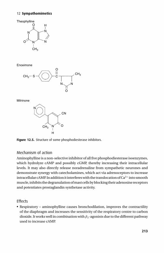
P1: PSB Printer: Yet To Come
9780521704632c12 CUFX213A/Peck 9780521618168 December 28, 2007 12:15
12 Sympathomimetics
Theophylline
Enoximone
Milrinone
O
N
S
O
C
N
N
N
H
CN
O
O
N
N N
N
H
CH3
CH3
CH3
CH3
O
Figure 12.5. Structure of some phosphodiesterase inhibitors.
Mechanism of actionAminophylline is a non-selective inhibitor of all five phosphodiesterase isoenzymes,which hydrolyze cAMP and possibly cGMP, thereby increasing their intracellularlevels. It may also directly release noradrenaline from sympathetic neurones anddemonstrate synergy with catecholamines, which act via adrenoceptors to increaseintracellular cAMP. In addition it interferes with the translocation of Ca2+ into smoothmuscle, inhibits the degranulation of mast cells by blocking their adenosine receptorsand potentiates prostaglandin synthetase activity.
Effects� Respiratory – aminophylline causes bronchodilation, improves the contractilityof the diaphragm and increases the sensitivity of the respiratory centre to carbondioxide. It works well in combination with β2-agonists due to the different pathwayused to increase cAMP.
213
P1: PSB Printer: Yet To Come
9780521704632c12 CUFX213A/Peck 9780521618168 December 28, 2007 12:15
Section III Cardiovascular drugs� Cardiovascular – it has mild positive inotropic and chronotropic effects and causessome coronary and peripheral vasodilatation. It lowers the threshold for arrhyth-mias (particularly ventricular) especially in the presence of halothane.� Central nervous system – the alkyl group at the 1-position (also present in caffeine)is responsible for its central nervous system stimulation, resulting in a reducedseizure threshold.� Renal – the alkyl group at the 1-position is also responsible for its weak diureticeffects. Inhibition of tubular Na+ reabsorption leads to a naturesis and may pre-cipitate hypokalaemia.� Interactions – co-administration of drugs that inhibit hepatic cytochrome P450(cimetidine, erythromycin, ciprofloxacin and oral contraceptives) tend to delay theelimination of aminophylline and a reduction in dose is recommended. The use ofcertain selective serotonin re-uptake inhibitors (fluvoxamine) should be avoidedwith aminophylline as levels of the latter may rise sharply. Drugs that induce hep-atic cytochrome P450 (phenytoin, carbamazepine, barbiturates and rifampicin)increase aminophylline clearance and the dose may need to be increased.
KineticsAminophylline is well absorbed from the gut with a high oral bioavailability (>90%).About 50% is plasma protein bound. It is metabolized in the liver by cytochromeP450 to inactive metabolites and interacts with the metabolism of other drugsundergoing metabolism by a similar route. Owing to its low hepatic extraction ratioits metabolism is independent of liver blood flow. Approximately 10% is excretedunchanged in the urine. The effective therapeutic plasma concentration is 10–20µg.ml−1. Cigarette smoking increases the clearance of aminophylline.
ToxicityAbove 35 µg.ml−1, hepatic enzymes become saturated and its kinetics changefrom first- to zero-order resulting in toxicity. Cardiac toxicity manifests itself astachyarrhythmias including ventricular fibrillation. Central nervous system toxic-ity includes tremor, insomnia and seizures (especially following rapid intravenousadministration). Nausea and vomiting are also a feature, as is rhabdomyolysis.
Selective phosphodiesterase inhibitors
EnoximoneThe imidazolone derivative enoximone is a selective phosphodiesterase III inhibitor.
Presentation and usesEnoximone is available as a yellow liquid (pH 12) for intravenous use containing 5mg.ml−1. It is supplied in propyl glycol and ethanol and should be stored between5◦C and 8◦C. It is used to treat congestive heart failure and low cardiac output states
214
P1: PSB Printer: Yet To Come
9780521704632c12 CUFX213A/Peck 9780521618168 December 28, 2007 12:15
12 Sympathomimetics
associated with cardiac surgery. It should be diluted with an equal volume of water or0.9% saline in plastic syringes (crystal formation is seen when mixed in glass syringes)and administered as an infusion of 5–20 µg.kg−1.min−1, which may be preceded bya loading dose of 0.5 mg.kg−1, and can be repeated up to a maximum of 3 mg.kg−1.Unlike catecholamines it may take up to 30 minutes to act.
Mechanism of actionEnoximone works by preventing the degradation of cAMP and possibly cGMP incardiac and vascular smooth muscle. By effectively increasing cAMP within themyocardium, it increases the slow Ca2+ inward current during the cardiac actionpotential. This produces an increase in Ca2+ release from intracellular stores andan increase in the Ca2+ concentration in the vicinity of the contractile proteins, andhence to a positive inotropic effect. By interfering with Ca2+ flux into vascular smoothmuscle it causes vasodilatation.
Effects� Cardiovascular – enoximone has been termed an ‘inodilator’ due to its positiveinotropic and vasodilator effects on the heart and vascular system. In patientswith heart failure the cardiac output increases by about 30% while end diastolicfilling pressures decrease by about 35%. The myocardial oxygen extraction ratioremains unchanged by virtue of a reduced ventricular wall tension and improvedcoronary artery perfusion. The blood pressure may remain unchanged or fall, theheart rate remains unchanged or rises slightly and arrhythmias occur only rarely. Itshortens atrial, AV node and ventricular refractoriness. When used in patients withischaemic heart disease, a reduction in coronary perfusion pressure and a rise inheart rate may outweigh the benefits of improved myocardial blood flow so thatfurther ischaemia ensues.� Miscellaneous – agranulocytosis has been reported.
KineticsWhile enoximone is well absorbed from the gut an extensive first-pass metabolismrenders it useless when given orally. About 70% is plasma protein bound andmetabolism occurs in the liver to a renally excreted active sulphoxide metabolitewith 10% of the activity of enoximone and a terminal half-life of 7.5 hours. Onlysmall amounts are excreted unchanged in the urine and by infusion enoximone hasa terminal half-life of 4.5 hours. It has a wide therapeutic ratio and the risks of toxicityare low. The dose should be reduced in renal failure.
MilrinoneMilrinone is a bipyridine derivative and a selective phosphodiesterase III inhibitorwith similar effects to enoximone. However, it has been associated with an increasedmortality rate when administered orally to patients with severe heart failure.
215
P1: PSB Printer: Yet To Come
9780521704632c12 CUFX213A/Peck 9780521618168 December 28, 2007 12:15
Section III Cardiovascular drugs
Preparation and usesMilrinone is formulated as a yellow solution containing 1 mg.ml−1 and may be storedat room temperature. It should be diluted before administration and should only beused intravenously for the short-term management of cardiac failure.
KineticsApproximately 70% is plasma protein bound. It has an elimination half-life of 1–2.5hours and is 80% excreted in the urine unchanged. The dose should be reduced inrenal failure.
AmrinoneAmrinone is not available in the UK. It has a similar pharmacological profile tothe other selective phosphodiesterase inhibitors. Approximately 40% is excretedunchanged in the urine. One in 40 patients suffers a reversible dose-related throm-bocytopenia.
GlucagonWithin the pancreas, α-cells secrete the polypeptide glucagon. The activation ofglucagon receptors, via G-protein mediated mechanisms, stimulates adenylatecyclase and increases intracellular cAMP. It has only a limited role in cardiac failure,occasionally being used in the treatment of β-blocker overdose by an initial bolus of10 mg followed by infusion of up to 5 mg.hr−1. Hyperglycaemia and hyperkalaemiamay complicate its use.
Ca2+
While intravenously administered Ca2+ salts often improve blood pressure for a fewminutes, their use should be restricted to circulatory collapse due to hyperkalaemiaand Ca2+ channel antagonist overdose.
T3
Thyroxine (T4) and triiodothyronine (T3) have positive inotropic and chronotropiceffects via intracellular mechanisms. They are only used to treat hypothyroidism andare discussed in more detail in Chapter 25.
216
P1: PSB Printer: Yet To Come
9780521704632c13 CUFX213A/Peck 9780521618168 December 28, 2007 12:27
13Adrenoceptor antagonists
� α-Adrenoceptor antagonists� β-Adrenoceptor antagonists� Combined α- and β-adrenoceptor antagonists
α-Adrenoceptor antagonistsα-Adrenoceptor antagonists (α-blockers) prevent the actions of sympathomimeticagents on α-adrenoceptors. Certainα-blockers (phentolamine, phenoxybenzamine)are non-specific and inhibit both α1- and α2-receptors, whereas others selectivelyinhibit α1-receptors (prazosin) or α2-receptors (yohimbine). The actions of specificα-adrenoceptor stimulation are shown in Table 13.1.
Non-selective α-blockade
PhentolaminePhentolamine (an imidazolone) is a competitive non-selective α-blocker. Its affinityfor α1-adrenoceptors is three times that for α2-adrenoceptors.
PresentationIt is presented as 10 mg phentolamine mesylate in 1 ml clear pale-yellow solution.The intravenous dose is 1–5 mg and should be titrated to effect. The onset of actionis 1–2 minutes and its duration of action is 5–20 minutes.
UsesPhentolamine is used in the treatment of hypertensive crises due to excessive sym-pathomimetics, MAOI reactions with tyramine and phaeochromocytoma, especiallyduring tumour manipulation. It has a role in the assessment of sympathetically medi-ated chronic pain and has previously been used to treat pulmonary hypertension.Injection into the corpus cavernosum has been used to treat impotence due to erec-tile failure.
Effects� Cardiovascular – α1-blockade results in vasodilatation and hypotension while α2-blockade facilitates noradrenaline release leading to tachycardia and a raised
217
P1: PSB Printer: Yet To Come
9780521704632c13 CUFX213A/Peck 9780521618168 December 28, 2007 12:27
Section III Cardiovascular drugs
Table 13.1. Actions of specific α-adrenoceptor stimulation.
Receptor type Action
Post-synapticα1-Receptors vasoconstriction
mydriasiscontraction of bladder sphincter
α2-Receptors platelet aggregationhyperpolarization of some CNS neurones
Presynapticα2-Receptors inhibit noradrenaline release
cardiac output. Pulmonary artery pressure is also reduced. Vasodilatation of vesselsin the nasal mucosa leads to marked nasal congestion.� Respiratory – the presence of sulphites in phentolamine ampoules may lead tohypersensitivity reactions, which are manifest as acute bronchospasm in suscep-tible asthmatics.� Gut – phentolamine increases secretions and motility of the gastrointestinal tract.� Metabolic – it may precipitate hypoglycaemia secondary to increased insulin secre-tion.
KineticsThe oral route is rarely used and has a bioavailability of 20%. It is 50% plasma proteinbound and extensively metabolized, leaving about 10% to be excreted unchanged inthe urine. Its elimination half-life is 20 minutes.
PhenoxybenzaminePhenoxybenzamine is a long-acting non-selective α-blocker. It has a high affinity forα1-adrenoceptors.
PresentationIt is presented as capsules containing 10 mg and as a clear, faintly straw-colouredsolution for injection containing 100 mg/2 ml phenoxybenzamine hydrochloridewith ethyl alcohol, hydrochloric acid and propylene glycol.
UsesPhenoxybenzamine is used in the preoperative management of phaeochromocy-toma (to allow expansion of the intravascular compartment), peri-operative man-agement of some neonates undergoing cardiac surgery, hypertensive crises andoccasionally as an adjunct to the treatment of severe shock. The oral dose startsat 10 mg and is increased daily until hypertension is controlled, the usual dose is 1–2mg.kg−1.day−1. Intravenous administration should be via a central cannula and the
218
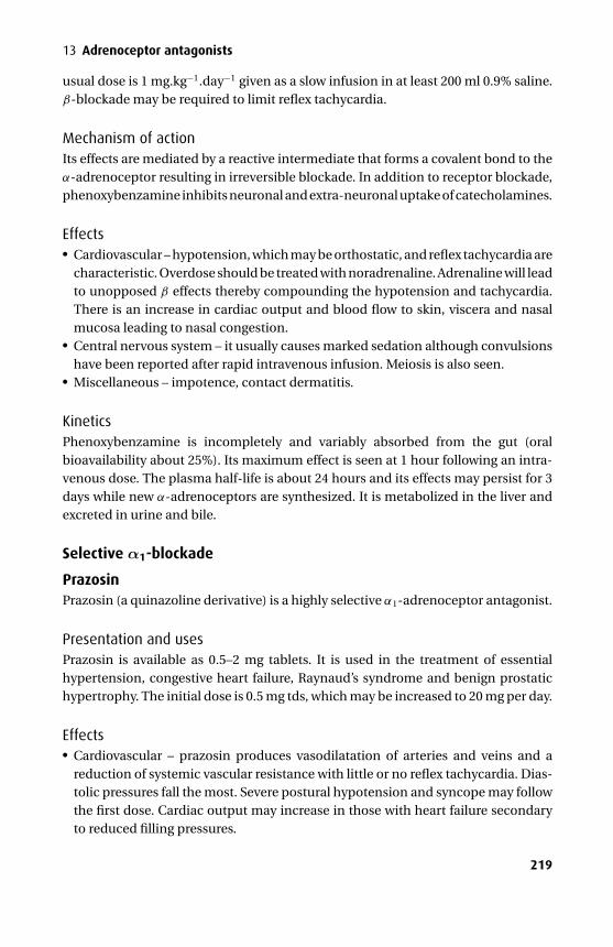
P1: PSB Printer: Yet To Come
9780521704632c13 CUFX213A/Peck 9780521618168 December 28, 2007 12:27
13 Adrenoceptor antagonists
usual dose is 1 mg.kg−1.day−1 given as a slow infusion in at least 200 ml 0.9% saline.β-blockade may be required to limit reflex tachycardia.
Mechanism of actionIts effects are mediated by a reactive intermediate that forms a covalent bond to theα-adrenoceptor resulting in irreversible blockade. In addition to receptor blockade,phenoxybenzamine inhibits neuronal and extra-neuronal uptake of catecholamines.
Effects� Cardiovascular – hypotension, which may be orthostatic, and reflex tachycardia arecharacteristic. Overdose should be treated with noradrenaline. Adrenaline will leadto unopposed β effects thereby compounding the hypotension and tachycardia.There is an increase in cardiac output and blood flow to skin, viscera and nasalmucosa leading to nasal congestion.� Central nervous system – it usually causes marked sedation although convulsionshave been reported after rapid intravenous infusion. Meiosis is also seen.� Miscellaneous – impotence, contact dermatitis.
KineticsPhenoxybenzamine is incompletely and variably absorbed from the gut (oralbioavailability about 25%). Its maximum effect is seen at 1 hour following an intra-venous dose. The plasma half-life is about 24 hours and its effects may persist for 3days while new α-adrenoceptors are synthesized. It is metabolized in the liver andexcreted in urine and bile.
Selective α1-blockade
PrazosinPrazosin (a quinazoline derivative) is a highly selective α1-adrenoceptor antagonist.
Presentation and usesPrazosin is available as 0.5–2 mg tablets. It is used in the treatment of essentialhypertension, congestive heart failure, Raynaud’s syndrome and benign prostatichypertrophy. The initial dose is 0.5 mg tds, which may be increased to 20 mg per day.
Effects� Cardiovascular – prazosin produces vasodilatation of arteries and veins and areduction of systemic vascular resistance with little or no reflex tachycardia. Dias-tolic pressures fall the most. Severe postural hypotension and syncope may followthe first dose. Cardiac output may increase in those with heart failure secondaryto reduced filling pressures.
219
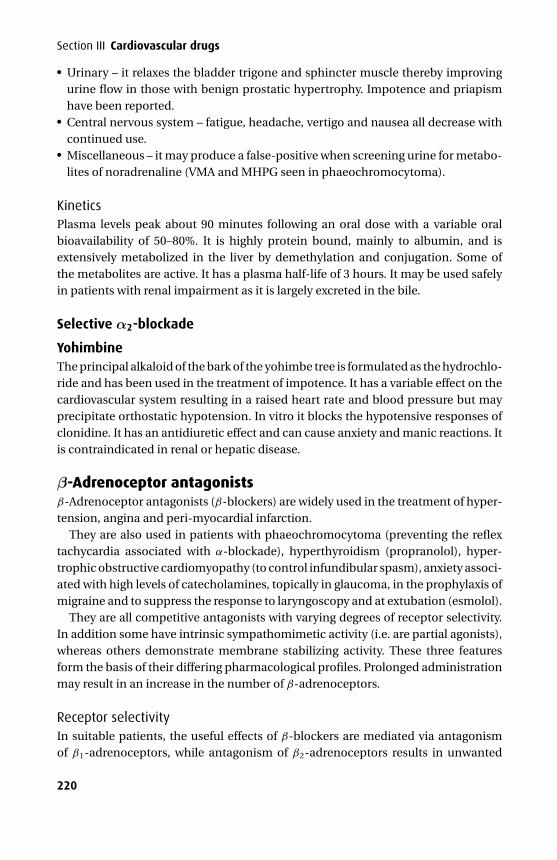
P1: PSB Printer: Yet To Come
9780521704632c13 CUFX213A/Peck 9780521618168 December 28, 2007 12:27
Section III Cardiovascular drugs� Urinary – it relaxes the bladder trigone and sphincter muscle thereby improvingurine flow in those with benign prostatic hypertrophy. Impotence and priapismhave been reported.� Central nervous system – fatigue, headache, vertigo and nausea all decrease withcontinued use.� Miscellaneous – it may produce a false-positive when screening urine for metabo-lites of noradrenaline (VMA and MHPG seen in phaeochromocytoma).
KineticsPlasma levels peak about 90 minutes following an oral dose with a variable oralbioavailability of 50–80%. It is highly protein bound, mainly to albumin, and isextensively metabolized in the liver by demethylation and conjugation. Some ofthe metabolites are active. It has a plasma half-life of 3 hours. It may be used safelyin patients with renal impairment as it is largely excreted in the bile.
Selective α2-blockade
YohimbineThe principal alkaloid of the bark of the yohimbe tree is formulated as the hydrochlo-ride and has been used in the treatment of impotence. It has a variable effect on thecardiovascular system resulting in a raised heart rate and blood pressure but mayprecipitate orthostatic hypotension. In vitro it blocks the hypotensive responses ofclonidine. It has an antidiuretic effect and can cause anxiety and manic reactions. Itis contraindicated in renal or hepatic disease.
β-Adrenoceptor antagonistsβ-Adrenoceptor antagonists (β-blockers) are widely used in the treatment of hyper-tension, angina and peri-myocardial infarction.
They are also used in patients with phaeochromocytoma (preventing the reflextachycardia associated with α-blockade), hyperthyroidism (propranolol), hyper-trophic obstructive cardiomyopathy (to control infundibular spasm), anxiety associ-ated with high levels of catecholamines, topically in glaucoma, in the prophylaxis ofmigraine and to suppress the response to laryngoscopy and at extubation (esmolol).
They are all competitive antagonists with varying degrees of receptor selectivity.In addition some have intrinsic sympathomimetic activity (i.e. are partial agonists),whereas others demonstrate membrane stabilizing activity. These three featuresform the basis of their differing pharmacological profiles. Prolonged administrationmay result in an increase in the number of β-adrenoceptors.
Receptor selectivityIn suitable patients, the useful effects of β-blockers are mediated via antagonismof β1-adrenoceptors, while antagonism of β2-adrenoceptors results in unwanted
220
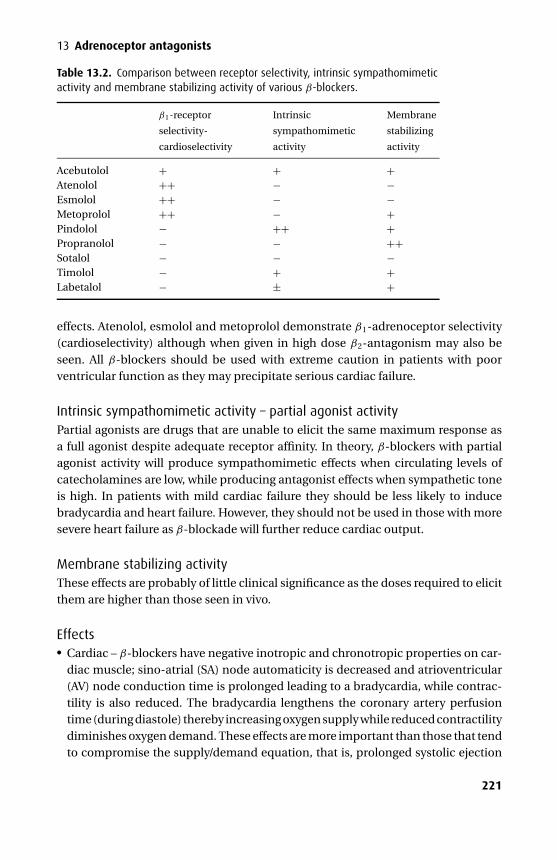
P1: PSB Printer: Yet To Come
9780521704632c13 CUFX213A/Peck 9780521618168 December 28, 2007 12:27
13 Adrenoceptor antagonists
Table 13.2. Comparison between receptor selectivity, intrinsic sympathomimeticactivity and membrane stabilizing activity of various β-blockers.
β1-receptor
selectivity-
cardioselectivity
Intrinsic
sympathomimetic
activity
Membrane
stabilizing
activity
Acebutolol + + +Atenolol ++ − −Esmolol ++ − −Metoprolol ++ − +Pindolol − ++ +Propranolol − − ++Sotalol − − −Timolol − + +Labetalol − ± +
effects. Atenolol, esmolol and metoprolol demonstrate β1-adrenoceptor selectivity(cardioselectivity) although when given in high dose β2-antagonism may also beseen. All β-blockers should be used with extreme caution in patients with poorventricular function as they may precipitate serious cardiac failure.
Intrinsic sympathomimetic activity – partial agonist activityPartial agonists are drugs that are unable to elicit the same maximum response asa full agonist despite adequate receptor affinity. In theory, β-blockers with partialagonist activity will produce sympathomimetic effects when circulating levels ofcatecholamines are low, while producing antagonist effects when sympathetic toneis high. In patients with mild cardiac failure they should be less likely to inducebradycardia and heart failure. However, they should not be used in those with moresevere heart failure as β-blockade will further reduce cardiac output.
Membrane stabilizing activityThese effects are probably of little clinical significance as the doses required to elicitthem are higher than those seen in vivo.
Effects� Cardiac – β-blockers have negative inotropic and chronotropic properties on car-diac muscle; sino-atrial (SA) node automaticity is decreased and atrioventricular(AV) node conduction time is prolonged leading to a bradycardia, while contrac-tility is also reduced. The bradycardia lengthens the coronary artery perfusiontime (during diastole) thereby increasing oxygen supply while reduced contractilitydiminishes oxygen demand. These effects are more important than those that tendto compromise the supply/demand equation, that is, prolonged systolic ejection
221
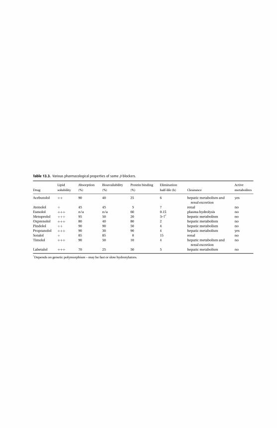
P1:P
SBP
rinter:YetTo
Co
me
9780521704632c13C
UF
X213A
/Peck9780521618168
Decem
ber
28,200712:27
Table 13.3. Various pharmacological properties of some β-blockers.
Drug
Lipid
solubility
Absorption
(%)
Bioavailability
(%)
Protein binding
(%)
Elimination
half-life (h) Clearance
Active
metabolites
Acebutolol ++ 90 40 25 6 hepatic metabolism andrenal excretion
yes
Atenolol + 45 45 5 7 renal noEsmolol +++ n/a n/a 60 0.15 plasma hydrolysis noMetoprolol +++ 95 50 20 3–7* hepatic metabolism noOxprenolol +++ 80 40 80 2 hepatic metabolism noPindolol ++ 90 90 50 4 hepatic metabolism noPropranolol +++ 90 30 90 4 hepatic metabolism yesSotalol + 85 85 0 15 renal noTimolol +++ 90 50 10 4 hepatic metabolism and
renal excretionno
Labetalol +++ 70 25 50 5 hepatic metabolism no
*Depends on genetic polymorphism – may be fast or slow hydroxylators.
222
P1: PSB Printer: Yet To Come
9780521704632c13 CUFX213A/Peck 9780521618168 December 28, 2007 12:27
13 Adrenoceptor antagonists
time, dilation of the ventricles and increased coronary vascular resistance (dueto antagonism of the vasodilatory β2 coronary receptors). The improvement inthe balance of oxygen supply/demand forms the basis for their use in anginaand peri-myocardial infarction. However, in patients with poor left ventric-ular function β-blockade may lead to cardiac failure. β-blockers are class IIanti-arrhythmic agents and are mainly used to treat arrhythmias associated withhigh levels of catecholamines (see Chapter 14).� Circulatory – the mechanism by which β-blockers control blood pressure is notyet fully elucidated but probably includes a reduced heart rate and cardiac output,and inhibition of the renin-angiotensin system. Inhibition of β1-receptors at thejuxtaglomerular apparatus reduces renin release leading ultimately to a reductionin angiotensin II and its effects (vasoconstriction and augmenting aldosteroneproduction). In addition, the baroreceptors may be set at a lower level, presynapticβ2-receptors may inhibit noradrenaline release and some β-blockers may havecentral effects. However, due to antagonism of peripheral β2-receptors there willbe an element of vasoconstriction, which appears to have little hypertensive effectbut may result in poor peripheral circulation and cold hands.� Respiratory – all β-blockers given in sufficient dose will precipitate bronchospasmvia β2-antagonism. The relatively cardioselective drugs (atenolol, esmolol andmetoprolol) are preferred but should still be used with extreme caution in patientswith asthma.� Metabolic – the control of blood sugar is complicated involving different tissuetypes (liver, pancreas, adipose), receptors (α-, β-adrenoceptors) and hormones(insulin, glucagon, catecholamines). Non-selective β-blockade may obtund thenormal blood sugar response to exercise and hypoglycaemia although it mayalso increase the resting blood sugar levels in diabetics with hypertension. There-fore, non-selective β-blockers should not be used with hypoglycaemic agents. Inaddition, β-blockade may mask the normal symptoms of hypoglycaemia. Lipidmetabolism may be altered resulting in increased triglycerides and reduced highdensity lipoproteins.� Central nervous system – the more lipid-soluble β-blockers (metoprolol, propra-nolol) are more likely to produce CNS side effects. These include depression, hal-lucination, nightmares, paranoia and fatigue.� Occular – intra-occular pressure is reduced, probably as a result of decreased pro-duction of aqueous humour.� Gut – dry mouth and gastrointestinal disturbances.
KineticsVarying lipid solubility confers the main differences seen in the kinetics ofβ-blockers.Those with low lipid solubility (atenolol) are poorly absorbed from the gut, undergolittle hepatic metabolism and are excreted largely unchanged in the urine. However,those with high lipid solubility are well absorbed from the gut and are extensively
223
P1: PSB Printer: Yet To Come
9780521704632c13 CUFX213A/Peck 9780521618168 December 28, 2007 12:27
Section III Cardiovascular drugs
metabolized in the liver. They have a shorter half-life and consequently need morefrequent administration. In addition, they cross the blood–brain barrier resulting insedation and nightmares. Protein binding is variable.
Individual β-blockers
AcebutololAcebutolol is a relatively cardioselective β-blocker that is only available orally. It haslimited intrinsic sympathomimetic activity and some membrane stabilizing prop-erties. The adult dose is 400 mg bd but may be increased to 1.2 g.day−1 if required.
KineticsAcebutolol is well absorbed from the gut due to its moderately high lipid solubility, butdue to a high first-pass metabolism its oral bioavailability is only 40%. Despite its lipidsolubility it does not cross the BBB to any great extent. Hepatic metabolism producesthe active metabolite diacetol, which has a longer half-life, and is less cardioselectivethan acebutolol. Both are excreted in bile and may undergo enterohepatic recycling.They are also excreted in urine and the dose should be reduced in the presence ofrenal impairment.
AtenololAtenolol is a relatively cardioselectiveβ-blocker that is available as 25–100 mg tablets,a syrup containing 5 mg.ml−1 and as a colourless solution for intravenous use con-taining 5 mg in 10 ml. The oral dose is 50–100 mg.day−1 while the intravenous doseis 2.5 mg slowly, repeated up to a maximum of 10 mg, which may then be followedby an infusion.
KineticsAtenolol is incompletely absorbed from the gut. It is not significantly metabolizedand has an oral bioavailability of 45%. Only 5% is protein bound. It is excretedunchanged in the urine and, therefore, the dose should be reduced in patients withrenal impairment. It has an elimination half-life of 7 hours but its actions appear topersist for longer than this would suggest.
EsmololEsmolol is a highly lipophilic, cardioselective β-blocker with a rapid onset and offset.It is presented as a clear liquid with either 2.5 g or 100 mg in 10 ml. The former shouldbe diluted before administration as an infusion (dose range 50–200 µg.kg−1.min−1),while the latter is titrated in 10 mg boluses to effect. It is used in the short-termmanagement of tachycardia and hypertension in the peri-operative period, and foracute supraventricular tachycardia. It has no intrinsic sympathomimetic activity ormembrane stabilizing properties.
224
P1: PSB Printer: Yet To Come
9780521704632c13 CUFX213A/Peck 9780521618168 December 28, 2007 12:27
13 Adrenoceptor antagonists
KineticsEsmolol is only available intravenously and is 60% protein bound. Its volume ofdistribution is 3.5 l.kg−1. It is rapidly metabolized by red blood cell esterases to anessentially inactive acid metabolite (with a long half-life) and methyl alcohol. Itsrapid metabolism ensures a short half-life of 10 minutes. The esterases responsiblefor its hydrolysis are distinct from plasma cholinesterase so that it does not prolongthe actions of suxamethonium.
Like other β-blockers it may also precipitate heart failure and bronchospasm,although its short duration of action limits these side effects.
It is irritant to veins and extravasation may lead to tissue necrosis.
MetoprololMetoprolol is a relatively cardioselective β-blocker with no intrinsic sympath-omimetic activity. Early use of metoprolol in myocardial infarction reduces infarctsize and the incidence of ventricular fibrillation. It is also used in hypertension, as anadjunct in thyrotoxicosis and for migraine prophylaxis. The dose is 50–200 mg daily.Up to 5 mg may be given intravenously for arrhythmias and in myocardial infarction.
KineticsAbsorption is rapid and complete, but due to hepatic first-pass metabolism, its oralbioavailability is only 50%. However, this increases to 70% during continuous admin-istration and is also increased when given with food. Hepatic metabolism may exhibitgenetic polymorphism resulting in two different half-life profiles of 3 and 7 hours. Itshigh lipid solubility enables it to cross the blood–brain barrier and also into breastmilk. Only 20% is plasma protein bound.
PropranololPropranolol is a non-selectiveβ-blocker without intrinsic sympathomimetic activity.It exhibits the full range of effects described above at therapeutic concentrations. It isa racemic mixture, the S-isomer conferring most of its effects, although the R-isomeris responsible for preventing the peripheral conversion of T4 to T3.
UsesPropranolol is used to treat hypertension, angina, essential tremor and in the pro-phylaxis of migraine. It is the β-blocker of choice in thyrotoxicosis as it not onlyinhibits the effects of the thyroid hormones, but also prevents the peripheral con-version of T4 to T3. Intravenous doses of 0.5 mg (up to 10 mg) are titrated to effect.The oral dose ranges from 160 mg to 320 mg daily, but due to increased clearance inthyrotoxicosis even higher doses may be required.
225
P1: PSB Printer: Yet To Come
9780521704632c13 CUFX213A/Peck 9780521618168 December 28, 2007 12:27
Section III Cardiovascular drugs
KineticsOwing to its high lipid solubility it is well absorbed from the gut but a high first-passmetabolism reduces its oral bioavailability to 30%. It is highly protein bound althoughthis may be reduced by heparin. Hepatic metabolism of the R-isomer is more rapidthan the S-isomer and one of their metabolites, 4-hydroxypropranolol, retains someactivity. Its elimination is dependent on hepatic metabolism but is impaired in renalfailure by an unknown mechanism. The duration of action is longer than its half-lifeof 4 hours would suggest.
SotalolSotalol is a non-selective β-blocker with no intrinsic sympathomimetic properties.It also has class III anti-arrhythmic properties (see Chapter 14).
It is a racemic mixture, the d-isomer conferring the class III activity while thel-isomer has both class III and class II (β-blocking) actions.
UsesSotalol is used to treat ventricular tachyarrhythmias and for the prophylaxis of parox-ysmal supraventricular tachycardias following direct current (DC) cardioversion. Theventricular rate is also well controlled if sinus rhythm degenerates back into atrialfibrillation. The CSM states that sotalol should not be used for angina, hypertension,thyrotoxicosis or peri-myocardial infarction. The oral dose is 80–160 mg bd and theintravenous dose is 50–100 mg over 20 minutes.
Other effectsThe most serious side effect is precipitation of torsades de pointes, which is rare,occurring in less than 2% of those being treated for sustained ventricular tachycardiaor fibrillation. It is more common with higher doses, a prolonged QT interval andelectrolyte imbalance. It may precipitate heart failure.
KineticsSotalol is completely absorbed from the gut and its oral bioavailability exceeds 90%.It is not protein bound or metabolized. Approximately 90% is excreted unchanged inurine while the remainder is excreted in bile. Renal impairment significantly reducesclearance.
Combined α- and β-adrenoceptor antagonists
LabetalolLabetalol, as its name indicates, is an α- and β-adrenoceptor antagonist; α-blockadeis specific to α1-receptors while β-blockade is non-specific. It contains two asym-metric centres and exists as a mixture of four stereoisomers present in equal propor-tions. The (SR)-stereoisomer is probably responsible for the α1 effects while the
226
P1: PSB Printer: Yet To Come
9780521704632c13 CUFX213A/Peck 9780521618168 December 28, 2007 12:27
13 Adrenoceptor antagonists
(RR)-stereoisomer probably confers the β-blockade. The ratio of α1:β-blockingeffects is dependent on the route of administration: 1:3 for oral, 1:7 for intravenous.
Presentation and usesLabetalol is available as 50–400 mg tablets and as a colourless solution containing5 mg.ml−1. It is used to treat hypertensive crises and to facilitate hypotension dur-ing anaesthesia. The intravenous dose is 5–20 mg titrated up to a maximum of200 mg. The oral form is used to treat hypertension associated with angina and dur-ing pregnancy where the dose is 100–800 mg bd but may be increased to a maximumof 2.4 g daily.
Mechanism of actionSelectiveα1-blockade produces peripheral vasodilatation while β-blockade preventsreflex tachycardia. Myocardial afterload and oxygen demand are decreased providingfavourable conditions for those with angina.
KineticsLabetalol is well absorbed from the gut but due to an extensive hepatic first-passmetabolism its oral bioavailability is only 25%. However, this may increase markedlywith increasing age and when administered with food. It is 50% protein bound.Metabolism occurs in the liver and produces several inactive conjugates.
227
P1: PSB Printer: Yet To Come
9780521704632c14 CUFX213A/Peck 9780521618168 December 28, 2007 12:32
14Anti-arrhythmics
Physiology
Cardiac action potentialThe heart is composed of pacemaker, conducting and contractile tissue. Each has adifferent action potential morphology allowing the heart to function as a coordinatedunit.
The SA node is in the right atrium, and of all cardiac tissue it has the fastest rateof spontaneous depolarization so that it sets the heart rate. The slow spontaneousdepolarization (pre-potential or pacemaker potential) of the membrane potentialis due to increased Ca2+ conductance (directed inward). At −40 mV, slow voltage-gated Ca2+ channels (L channels) open resulting in membrane depolarization. Na+
conductance changes very little. Repolarization is due to increased K+ conductancewhile Ca2+ channels close (Figure 14.1a).
Contractile cardiac tissue has a more stable resting potential at −80 mV. Its actionpotential has been divided into five phases (Figure 14.1b):� Phase 0 – describes the rapid depolarization (duration <1 ms) of the membrane,
resulting from increased Na+ (and possibly some Ca2+) conductance throughvoltage-gated Na+ channels.� Phase 1 – represents closure of the Na+ channels while Cl− is expelled.� Plateau phase 2 – due to Ca2+ influx via voltage-sensitive type-L Ca2+ channelsand lasts up to 150 ms. This period is also known as the absolute refractory periodin which the myocyte cannot be further depolarized. This prevents myocardialtetany.� Phase 3 – commences when the Ca2+ channels are inactivated and there is anincrease in K+ conductance that returns the membrane potential to its rest-ing value. This period is also known as the relative refractory period in whichthe myocyte requires a greater than normal stimulus to provoke a contrac-tion.� Phase 4 – during this the Na+/K+ ATPase maintains the ionic concentration gra-dient at about −80 mV, although there will be variable spontaneous ‘diastolic’depolarization.
228
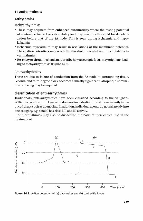
P1: PSB Printer: Yet To Come
9780521704632c14 CUFX213A/Peck 9780521618168 December 28, 2007 12:32
14 Anti-arrhythmics
Arrhythmias
Tachyarrhythmias� These may originate from enhanced automaticity where the resting potentialof contractile tissue loses its stability and may reach its threshold for depolari-zation before that of the SA node. This is seen during ischaemia and hypo-kalaemia.� Ischaemic myocardium may result in oscillations of the membrane potential.These after-potentials may reach the threshold potential and precipitate tach-yarrhythmias.� Re-entry or circus mechanisms describe how an ectopic focus may originate, lead-ing to tachyarrhythmias (Figure 14.2).
BradyarrhythmiasThese are due to failure of conduction from the SA node to surrounding tissue.Second- and third-degree block becomes clinically significant. Atropine, β stimula-tion or pacing may be required.
Classification of anti-arrhythmicsTraditionally anti-arrhythmics have been classified according to the Vaughan–Williams classification. However, it does not include digoxin and more recently intro-duced drugs such as adenosine. In addition, individual agents do not fall neatly intoone category, e.g. sotalol has class I, II and III activity.
Anti-arrhythmics may also be divided on the basis of their clinical use in thetreatment of:
0
30
60
90
0 100 200 300 400 Time (msec)
4
3
2
1
0
(a) (b)
Mem
bran
e po
tent
ial (
mV
)
Figure 14.1. Action potentials of (a) pacemaker and (b) contractile tissue.
229
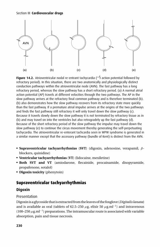
P1: PSB Printer: Yet To Come
9780521704632c14 CUFX213A/Peck 9780521618168 December 28, 2007 12:32
Section III Cardiovascular drugs
AVN
(a) (b) (d) (e)(c)
Figure 14.2. Atrioventricular nodal re-entrant tachycardia ( action potential followed byrefractory period). In this situation, there are two anatomically and physiologically distinctconduction pathways within the atrioventricular node (AVN). The fast pathway has a longrefractory period, whereas the slow pathway has a short refractory period. (a) A normal atrialaction potential (AP) travels at different velocities through the two pathways. The AP in theslow pathway arrives at the refractory final common pathway and is therefore terminated (b).(b) also demonstrates how the slow pathway recovers from its refractory state more quicklythan the fast pathway. If a premature atrial impulse arrives at the origins of the two pathwaysand finds the fast pathway still refractory it will only travel down the slow pathway (c).Because it travels slowly down the slow pathway it is not terminated by refractory tissue as in(b) and may travel on into the ventricles but also retrogradely up the fast pathway (d).Because of the short refractory period of the slow pathway the impulse may travel down theslow pathway (e) to continue the circus movement thereby generating the self-perpetuatingtachycardia. The atrioventricular re-enterant tachcardia seen in WPW syndrome is generated ina similar manner except that the accessory pathway (bundle of Kent) is distinct from the AVN.
� Supraventricular tachyarrhythmias (SVT) (digoxin, adenosine, verapamil, β-blockers, quinidine)� Ventricular tachyarrhythmias (VT) (lidocaine, mexiletine)� Both SVT and VT (amiodarone, flecainide, procainamide, disopyramide,propafenone, sotalol)� Digoxin toxicity (phenytoin)
Supraventricular tachyarrhythmias
Digoxin
PresentationDigoxin is a glycoside that is extracted from the leaves of the foxglove (Digitalis lanata)and is available as oral (tablets of 62.5–250 µg, elixir 50 µg.ml−1) and intravenous(100–250 µg.ml−1) preparations. The intramuscular route is associated with variableabsorption, pain and tissue necrosis.
230
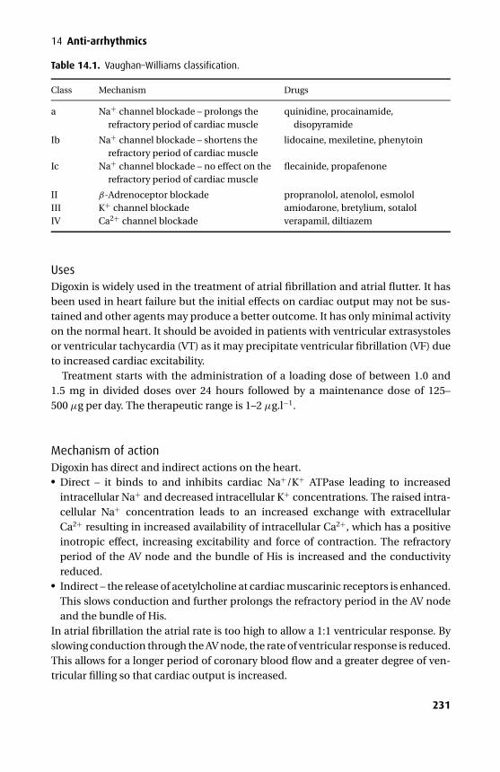
P1: PSB Printer: Yet To Come
9780521704632c14 CUFX213A/Peck 9780521618168 December 28, 2007 12:32
14 Anti-arrhythmics
Table 14.1. Vaughan–Williams classification.
Class Mechanism Drugs
a Na+ channel blockade – prolongs therefractory period of cardiac muscle
quinidine, procainamide,disopyramide
Ib Na+ channel blockade – shortens therefractory period of cardiac muscle
lidocaine, mexiletine, phenytoin
Ic Na+ channel blockade – no effect on therefractory period of cardiac muscle
flecainide, propafenone
II β-Adrenoceptor blockade propranolol, atenolol, esmololIII K+ channel blockade amiodarone, bretylium, sotalolIV Ca2+ channel blockade verapamil, diltiazem
UsesDigoxin is widely used in the treatment of atrial fibrillation and atrial flutter. It hasbeen used in heart failure but the initial effects on cardiac output may not be sus-tained and other agents may produce a better outcome. It has only minimal activityon the normal heart. It should be avoided in patients with ventricular extrasystolesor ventricular tachycardia (VT) as it may precipitate ventricular fibrillation (VF) dueto increased cardiac excitability.
Treatment starts with the administration of a loading dose of between 1.0 and1.5 mg in divided doses over 24 hours followed by a maintenance dose of 125–500 µg per day. The therapeutic range is 1–2 µg.l−1.
Mechanism of actionDigoxin has direct and indirect actions on the heart.� Direct – it binds to and inhibits cardiac Na+/K+ ATPase leading to increased
intracellular Na+ and decreased intracellular K+ concentrations. The raised intra-cellular Na+ concentration leads to an increased exchange with extracellularCa2+ resulting in increased availability of intracellular Ca2+, which has a positiveinotropic effect, increasing excitability and force of contraction. The refractoryperiod of the AV node and the bundle of His is increased and the conductivityreduced.� Indirect – the release of acetylcholine at cardiac muscarinic receptors is enhanced.This slows conduction and further prolongs the refractory period in the AV nodeand the bundle of His.
In atrial fibrillation the atrial rate is too high to allow a 1:1 ventricular response. Byslowing conduction through the AV node, the rate of ventricular response is reduced.This allows for a longer period of coronary blood flow and a greater degree of ven-tricular filling so that cardiac output is increased.
231
P1: PSB Printer: Yet To Come
9780521704632c14 CUFX213A/Peck 9780521618168 December 28, 2007 12:32
Section III Cardiovascular drugs
Side effectsDigoxin has a low therapeutic ratio and side effects are not uncommon:� Cardiac – these include various arrhythmias and conduction disturbances – prema-
ture ventricular contractions, bigemini, all forms of AV block including third-degreeblock, junctional rhythm and atrial or ventricular tachycardia. Hypokalaemia,hypercalcaemia or altered pH may precipitate side effects. The ECG signs of pro-longed PR interval, characteristic ST segment depression, T wave flattening andshortened QT interval are not signs of toxicity.
DC cardioversion – severe ventricular arrhythmias may be precipitated inpatients with toxic levels and it is recommended to withhold digoxin for 24 hoursbefore elective cardioversion.� Non-cardiac – anorexia, nausea and vomiting, diarrhoea and lethargy. Visualdisturbances (including deranged red–green colour perception) and headacheare common while gynaecomastia occurs during long-term administration. Skinrashes are rarely seen and may be accompanied by an eosinophilia.� Interactions – plasma levels are increased by amiodarone, captopril, erythromycinand carbenoxolone. They are reduced by antacids, cholestyramine, phenytoin andmetoclopramide. Ca2+ channel antagonists produce variable effects, verapamilwill increase, while nifedipine and diltiazem may have no effect or produce a smallrise in plasma levels.
KineticsThe absorption of digoxin from the gut is variable depending on the specific formu-lation used, but the oral bioavailability is greater than 70%. It is about 25% plasmaand has a volume of distribution of 5–10 l.kg−1. Its volume of distribution is signif-icantly increased in thyrotoxicosis and decreased in hypothyroidism. It undergoesonly minimal hepatic metabolism, being excreted mainly in the unchanged form byfiltration at the glomerulus and active tubular secretion. The elimination half-life isapproximately 35 hours but is increased significantly in the presence of renal failure.
ToxicityPlasma concentrations exceeding 2.5 µg.l−1 are associated with toxicity althoughserious problems are unusual at levels below 10 µg.l−1. Despite these figures theseverity of toxicity does not correlate well with plasma levels. However, a dose ofmore than 30 mg is invariably associated with death unless digoxin-specific antibodyfragments (Fab) are used.
Treatment of digoxin toxicityGastric lavage should be used with caution as any increase in vagal tone may precipi-tate further bradycardia or cardiac arrest. Owing to Na+/K+ ATPase inhibition, hyper-kalaemia may be a feature and should be corrected. Hypokalaemia will exacerbate
232
P1: PSB Printer: Yet To Come
9780521704632c14 CUFX213A/Peck 9780521618168 December 28, 2007 12:32
14 Anti-arrhythmics
cardiac toxicity and should also be corrected. Where bradycardia is symptomaticatropine or pacing is preferred to infusions of catecholamines, which may precipi-tate further arrhythmias. Ventricular arrhythmias may be treated with lidocaine orphenytoin.
If plasma levels rise above 20 µg.l−1, there are life-threatening arrhythmias orhyperkalaemia becomes uncontrolled, digoxin-specific Fab are indicated. These areIgG fragments. Digoxin is bound more avidly by Fab than by its receptor so that itis effectively removed from its site of action. The inactive digoxin–Fab complex isremoved from the circulation by the kidneys. There is a danger of hypersensitivity oranaphylaxis on re-exposure to digoxin-specific Fab.
AdenosineAdenosine is a naturally occurring purine nucleoside consisting of adenine (thepurine base) and d-ribose (the pentose sugar), which is present in all cells.
PresentationAdenosine is presented as a colourless solution in vials containing 3 mg.ml−1. Itshould be stored at room temperature.
UsesAdenosine is used to differentiate between SVT, where the rate is at least transientlyslowed and VT, where the rate does not slow. Where SVT is due to re-entry circuitsthat involve the AV node, adenosine may covert the rhythm to sinus. Atrial fibrillationand flutter are not converted by adenosine to sinus rhythm as they are not generatedby re-entry circuits involving the AV node, although its use in this setting will slowthe ventricular response and aid ECG diagnosis.
Mechanism of actionAdenosine has specific actions on the SA and AV node mediated by adenosine A1
receptors that are not found elsewhere within the heart. These adenosine-sensitiveK+ channels are opened, causing membrane hyperpolarization, and Gi-proteinscause a reduction in cAMP. This results in a dramatic negative chronotropic effectwithin the AV node.
Side effectsBecause of its short half-life its side effects are also short lived but for the patientmay be very distressing.� Cardiac – it may induce atrial fibrillation or flutter as it decreases the atrial refractory
period. It is contraindicated in those with second- or third-degree AV-block or withsick sinus syndrome.
233
P1: PSB Printer: Yet To Come
9780521704632c14 CUFX213A/Peck 9780521618168 December 28, 2007 12:32
Section III Cardiovascular drugs� Non-cardiac – these include chest discomfort, shortness of breath and facial flush-ing. It should be used with caution in asthmatics as it may precipitate bron-chospasm.� Drug interactions – its effects may be enhanced by dipyridamole (by blocking itsuptake) and antagonized by the methylxanthines especially aminophylline.
KineticsAdenosine is given in incremental doses from 3 to 12 mg as an intravenous bolus,preferably via a central cannula. It is rapidly de-aminated in the plasma and takenup by red blood cells so that its half-life is less than 10 seconds.
VerapamilVerapamil is a competitive Ca2+ channel antagonist.
PresentationIt is presented as film-coated and modified-release tablets and as a solution forintravenous injection containing 2.5 mg.ml−1.
UsesVerapamil is used to treat SVT, atrial fibrillation or flutter, which it may slow or convertto a sinus rhythm. It is also used in the prophylaxis of angina and the treatment ofhypertension.
Mechanism of actionVerapamil prevents the influx of Ca2+ through voltage-sensitive slow (L) channels inthe SA and AV node, thereby reducing their automaticity. It has a much less markedeffect on the contractile tissue of the heart, but does reduce Ca2+ influx during theplateau phase 2. Antagonism of these Ca2+ channels results in a reduced rate ofconduction through the AV node and coronary artery dilatation.
Side effects� Cardiac – if used to treat SVT complicating Wolff–Parkinson–White (WPW)syndrome, verapamil may precipitate VT due to increased conduction across theaccessory pathway. In patients with poor left ventricular function it may precip-itate cardiac failure. When administered concurrently with agents that also slowAV conduction (digoxin, β-blockers, halothane) it may precipitate serious brady-cardia and AV block. It may increase the serum levels of digoxin. Grapefruit juicehas been reported to increase serum levels and should be avoided during verapamiltherapy.
Although its effects are relatively specific to cardiac tissue it may alsoprecipitate hypotension through vascular smooth muscle relaxation.
234
P1: PSB Printer: Yet To Come
9780521704632c14 CUFX213A/Peck 9780521618168 December 28, 2007 12:32
14 Anti-arrhythmics� Non-cardiac – cerebral artery vasodilatation occurs after the administration ofverapamil.
KineticsVerapamil is used orally and intravenously. Although almost 90% is absorbed fromthe gut a high first-pass metabolism reduces its oral bioavailability to about 25%.Approximately 90% is bound to plasma proteins. It is metabolized in the liver to atleast 12 inactive metabolites that are excreted in the urine. Its volume of distributionis 3–5 l.kg−1. The elimination half-life of 3–7 hours is prolonged with higher doses ashepatic enzymes become saturated.
β-BlockersThe effects of catecholamines are antagonized by β-blockers. Therefore, they inducea bradycardia (by prolonging ‘diastolic’ depolarization – phase 4), depress myocar-dial contractility and prolong AV conduction. In addition, some β-blockers exhibita degree of membrane stabilizing activity (class I) although this probably has lit-tle clinical significance. Sotalol also demonstrates class III activity by blocking K+
channels and prolonging repolarization.β-Blockers are used in the treatment of hypertension, angina, myocardial infarc-
tion, tachyarrhythmias, thyrotoxicosis, anxiety states, the prophylaxis of migraineand topically in glaucoma. Their use as an anti-arrhythmic is limited to treatment ofparoxysmal SVT and sinus tachycardia due to increased levels of catecholamines.They have a role following acute myocardial infarction where they may reducearrhythmias and prevent further infarction. Owing to their negative inotropic effectsthey should be avoided in those with poor ventricular function for fear of precipitat-ing cardiac failure.
EsmololEsmolol is a relatively cardioselective β-blocker with a rapid onset and offset.
PresentationIt is presented as a clear liquid with either 2.5 g or 100 mg in 10 ml. The former shouldbe diluted before administration as an infusion (dose range 50–200 µg.kg−1.min−1),while the latter is titrated in 10 mg boluses to effect.
UsesEsmolol is used in the short-term management of tachycardia and hypertension inthe peri-operative period, and for acute SVT. It has no intrinsic sympathomimeticactivity or membrane-stabilizing properties.
235
P1: PSB Printer: Yet To Come
9780521704632c14 CUFX213A/Peck 9780521618168 December 28, 2007 12:32
Section III Cardiovascular drugs
Side effectsAlthough esmolol is relatively cardioselective it does demonstrate β2-adrenoceptorantagonism at high doses and should therefore be used with caution in asthmatics.Like other β-blockers it may also precipitate heart failure. However, due to its shortduration of action these side effects are also limited in time.
It is irritant to veins and extravasation may lead to tissue necrosis.
KineticsEsmolol is only available intravenously and is 60% plasma protein bound. Its volumeof distribution is 3.5 l.kg−1. It is rapidly metabolized by red blood cell esterases toan essentially inactive acid metabolite (with a long half-life) and methyl alcohol. Itsrapid metabolism ensures a short half-life of 10 minutes. The esterases responsiblefor its hydrolysis are distinct from plasma cholinesterase so that it does not prolongthe actions of suxamethonium.
QuinidineThe use of quinidine has declined as alternative treatments have become availablewith improved side-effect profiles. However, it may still be used to treat SVT, includingatrial fibrillation and flutter, and ventricular ectopic beats.
Mechanism of actionQuinidine is a class Ia anti-arrhythmic and as such reduces the rate of rise of phase 0of the action potential by blocking Na+ channels. In addition, it raises the thresholdpotential and prolongs the refractory period without affecting the duration of theaction potential. It also antagonizes vagal tone.
Side effectsThese are common and become unacceptable in up to 30% of patients.� Cardiac – quinidine may provoke other arrhythmias including heart block, sinus
tachycardia (vagolytic action) and ventricular arrhythmias. The following ECGchanges may be seen: prolonged PR interval, widened QRS and prolonged QT inter-val. When used to treat atrial fibrillation or flutter the patient should be pretreatedwith β-blockers, Ca2+ channel antagonists or digoxin to slow AV conduction, whichmay otherwise become enhanced leading to a ventricular rate equivalent to theatrial rate. Hypotension may result from α-blockade or direct myocardial depres-sion, which is exacerbated by hyperkalaemia.� Non-cardiac – central nervous system toxicity known as ‘cinchonism’ ischaracterized by tinnitus, blurred vision, impaired hearing, headache andconfusion.� Drug interactions – digoxin is displaced from its binding sites so that its serum con-centration is increased. Phenytoin will reduce quinidine levels (hepatic enzyme
236
P1: PSB Printer: Yet To Come
9780521704632c14 CUFX213A/Peck 9780521618168 December 28, 2007 12:32
14 Anti-arrhythmics
induction) while cimetidine will increase quinidine levels (hepatic enzyme inhi-bition). The effects of depolarizing and non-depolarizing muscle relaxants areincreased.
KineticsQuinidine is well absorbed from the gut and has an oral bioavailability of about75%. It is highly protein bound (about 90%) and is metabolized by the liver to activemetabolites, which are excreted mainly in the urine. The elimination half-life is 5–9hours.
Ventricular tachyarrhythmias
LidocaineLidocaine is a class Ib anti-arrhythmic agent.
PresentationThe 1% or 2% solutions (10–20 mg.ml−1) are the preparations used in this setting.
UsesLidocaine is used to treat sustained ventricular tachyarrhythmias especially whenassociated with ischaemia (where inactivated Na+ channels predominate) or re-entry pathways. An initial intravenous bolus of 1 mg.kg−1 is followed by an intra-venous infusion of 1–3 mg.min−1 for an adult. This infusion rate should be slowedwhere hepatic blood flow is reduced as hepatic metabolism will also be reduced.
Mechanism of actionLidocaine reduces the rate of rise of phase 0 of the action potential by blockinginactivated Na+ channels and raising the threshold potential. The duration of theaction potential and the refractory period are decreased as the repolarization phase3 is shortened.
Side effects� Cardiac – cardiovascular toxicity becomes apparent as plasma levels exceed10 µg.ml−1 and are manifest as AV block and unresponsive hypotension due tomyocardial depression. Some of the cardiac effects may be due to central medullarydepression.� Non-cardiac – these become apparent only when the plasma levels exceed4 µg.ml−1. Initially central nervous system toxicity is manifest as circumoral tin-gling, dizziness and parasthesia. This progresses to confusion, coma and seizuresas plasma levels rise above 5 µg.ml−1.
237
P1: PSB Printer: Yet To Come
9780521704632c14 CUFX213A/Peck 9780521618168 December 28, 2007 12:32
Section III Cardiovascular drugs
KineticsWhen used for the treatment of arrhythmias lidocaine is only given intravenously. Itis 33% unionized and 70% protein bound. It is metabolized by hepatic amidases toproducts that are eliminated in the urine. Its elimination half-life is about 90 minutesso that in the presence of normal hepatic function a steady-state would be reachedafter about 6 hours in the absence of a loading dose. Its clearance is reduced incardiac failure due to reduced hepatic blood flow.
MexiletineMexiletine is an analogue of lidocaine with similar effects on ventricular tach-yarrhythmias.
PresentationIt is presented as a colourless solution containing 250 mg mexiletine hydrochloridein 10 ml. The oral formulation is also available as modified release.
UsesMexiletine has similar indications to lidocaine particularly when arrhythmias areassociated with ischaemia or digoxin.
Mechanism of actionMexiletine reduces the rate of rise of phase 0 of the action potential by blocking Na+
channels and raising the threshold potential. The duration of the action potentialand the refractory period are decreased as the repolarization phase 3 is shortened.
Side effectsMexiletine has a low therapeutic ratio and side effects are common.� Cardiac – it may precipitate sinus bradycardia, supraventricular and ventricular
tachyarrhythmias.� Non-cardiac – up to 40% of patients have unacceptable nausea and vomiting andaltered bowel habit. Confusion, diplopia, seizures, tremor and ataxia are also seen.Thrombocytopenia, rash and jaundice have also been reported.
KineticsIts oral bioavailability of 90% reflects good absorption from the upper part of thesmall bowel and minimal first-pass metabolism (about 10%). It is 65% plasma pro-tein bound and has a volume of distribution of 6–13 l.kg−1. It undergoes hepaticmetabolism to a number of inactive metabolites. Up to 20% is excreted unchangedin the urine.
238
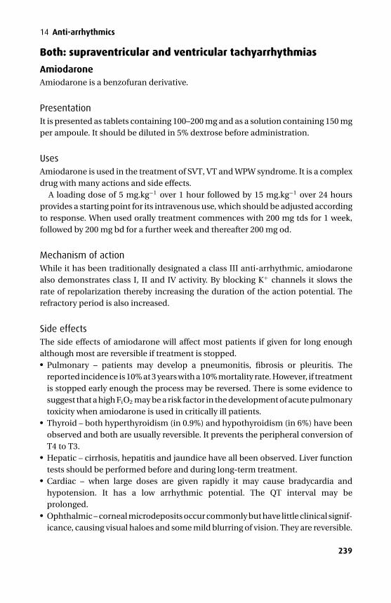
P1: PSB Printer: Yet To Come
9780521704632c14 CUFX213A/Peck 9780521618168 December 28, 2007 12:32
14 Anti-arrhythmics
Both: supraventricular and ventricular tachyarrhythmias
AmiodaroneAmiodarone is a benzofuran derivative.
PresentationIt is presented as tablets containing 100–200 mg and as a solution containing 150 mgper ampoule. It should be diluted in 5% dextrose before administration.
UsesAmiodarone is used in the treatment of SVT, VT and WPW syndrome. It is a complexdrug with many actions and side effects.
A loading dose of 5 mg.kg−1 over 1 hour followed by 15 mg.kg−1 over 24 hoursprovides a starting point for its intravenous use, which should be adjusted accordingto response. When used orally treatment commences with 200 mg tds for 1 week,followed by 200 mg bd for a further week and thereafter 200 mg od.
Mechanism of actionWhile it has been traditionally designated a class III anti-arrhythmic, amiodaronealso demonstrates class I, II and IV activity. By blocking K+ channels it slows therate of repolarization thereby increasing the duration of the action potential. Therefractory period is also increased.
Side effectsThe side effects of amiodarone will affect most patients if given for long enoughalthough most are reversible if treatment is stopped.� Pulmonary – patients may develop a pneumonitis, fibrosis or pleuritis. The
reported incidence is 10% at 3 years with a 10% mortality rate. However, if treatmentis stopped early enough the process may be reversed. There is some evidence tosuggest that a high FiO2 may be a risk factor in the development of acute pulmonarytoxicity when amiodarone is used in critically ill patients.� Thyroid – both hyperthyroidism (in 0.9%) and hypothyroidism (in 6%) have beenobserved and both are usually reversible. It prevents the peripheral conversion ofT4 to T3.� Hepatic – cirrhosis, hepatitis and jaundice have all been observed. Liver functiontests should be performed before and during long-term treatment.� Cardiac – when large doses are given rapidly it may cause bradycardia andhypotension. It has a low arrhythmic potential. The QT interval may beprolonged.� Ophthalmic – corneal microdeposits occur commonly but have little clinical signif-icance, causing visual haloes and some mild blurring of vision. They are reversible.
239
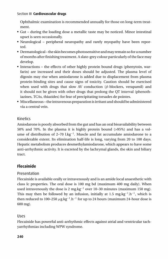
P1: PSB Printer: Yet To Come
9780521704632c14 CUFX213A/Peck 9780521618168 December 28, 2007 12:32
Section III Cardiovascular drugs
Ophthalmic examination is recommended annually for those on long-term treat-ment.� Gut – during the loading dose a metallic taste may be noticed. Minor intestinalupset is seen occasionally.� Neurological – peripheral neuropathy and rarely myopathy have been repor-ted.� Dermatological – the skin becomes photosensitive and may remain so for a numberof months after finishing treatment. A slate-grey colour particularly of the face maydevelop.� Interactions – the effects of other highly protein bound drugs (phenytoin, war-farin) are increased and their doses should be adjusted. The plasma level ofdigoxin may rise when amiodarone is added due to displacement from plasmaprotein-binding sites and cause signs of toxicity. Caution should be exercisedwhen used with drugs that slow AV conduction (β-blockers, verapamil) andit should not be given with other drugs that prolong the QT interval (phenoth-iazines, TCAs, thiazides) for fear of precipitating torsades de pointes.� Miscellaneous – the intravenous preparation is irritant and should be administeredvia a central vein.
KineticsAmiodarone is poorly absorbed from the gut and has an oral bioavailability between50% and 70%. In the plasma it is highly protein bound (>95%) and has a vol-ume of distribution of 2–70 l.kg−1. Muscle and fat accumulate amiodarone to aconsiderable extent. Its elimination half-life is long, varying from 20 to 100 days.Hepatic metabolism produces desmethylamiodarone, which appears to have someanti-arrhythmic activity. It is excreted by the lachrymal glands, the skin and biliarytract.
Flecainide
PresentationFlecainide is available orally or intravenously and is an amide local anaesthetic withclass Ic properties. The oral dose is 100 mg bd (maximum 400 mg daily). Whenused intravenously the dose is 2 mg.kg−1 over 10–30 minutes (maximum 150 mg).This may then be followed by an infusion, initially at 1.5 mg.kg−1.h−1, which isthen reduced to 100–250 µg.kg−1.h−1 for up to 24 hours (maximum 24-hour dose is600 mg).
UsesFlecainide has powerful anti-arrhythmic effects against atrial and ventricular tach-yarrhythmias including WPW syndrome.
240
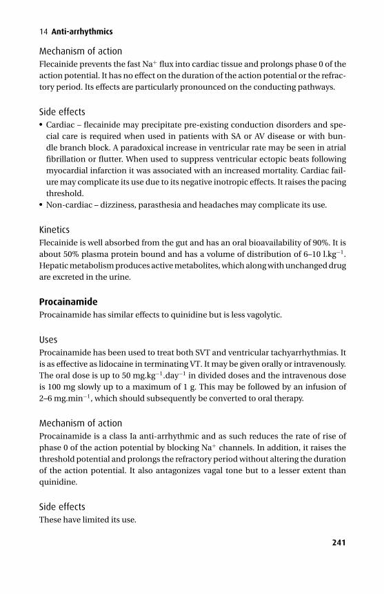
P1: PSB Printer: Yet To Come
9780521704632c14 CUFX213A/Peck 9780521618168 December 28, 2007 12:32
14 Anti-arrhythmics
Mechanism of actionFlecainide prevents the fast Na+ flux into cardiac tissue and prolongs phase 0 of theaction potential. It has no effect on the duration of the action potential or the refrac-tory period. Its effects are particularly pronounced on the conducting pathways.
Side effects� Cardiac – flecainide may precipitate pre-existing conduction disorders and spe-cial care is required when used in patients with SA or AV disease or with bun-dle branch block. A paradoxical increase in ventricular rate may be seen in atrialfibrillation or flutter. When used to suppress ventricular ectopic beats followingmyocardial infarction it was associated with an increased mortality. Cardiac fail-ure may complicate its use due to its negative inotropic effects. It raises the pacingthreshold.� Non-cardiac – dizziness, parasthesia and headaches may complicate its use.
KineticsFlecainide is well absorbed from the gut and has an oral bioavailability of 90%. It isabout 50% plasma protein bound and has a volume of distribution of 6–10 l.kg−1.Hepatic metabolism produces active metabolites, which along with unchanged drugare excreted in the urine.
ProcainamideProcainamide has similar effects to quinidine but is less vagolytic.
UsesProcainamide has been used to treat both SVT and ventricular tachyarrhythmias. Itis as effective as lidocaine in terminating VT. It may be given orally or intravenously.The oral dose is up to 50 mg.kg−1.day−1 in divided doses and the intravenous doseis 100 mg slowly up to a maximum of 1 g. This may be followed by an infusion of2–6 mg.min−1, which should subsequently be converted to oral therapy.
Mechanism of actionProcainamide is a class Ia anti-arrhythmic and as such reduces the rate of rise ofphase 0 of the action potential by blocking Na+ channels. In addition, it raises thethreshold potential and prolongs the refractory period without altering the durationof the action potential. It also antagonizes vagal tone but to a lesser extent thanquinidine.
Side effectsThese have limited its use.
241
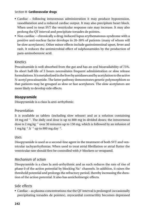
P1: PSB Printer: Yet To Come
9780521704632c14 CUFX213A/Peck 9780521618168 December 28, 2007 12:32
Section III Cardiovascular drugs� Cardiac – following intravenous administration it may produce hypotension,vasodilatation and a reduced cardiac output. It may also precipitate heart block.When used to treat SVT the ventricular response rate may increase. It may alsoprolong the QT interval and precipitate torsades de pointes.� Non-cardiac – chronically a drug-induced lupus erythematosus syndrome with apositive anti-nuclear factor develops in 20–30% of patients (many of whom willbe slow acetylators). Other minor effects include gastrointestinal upset, fever andrash. It reduces the antimicrobial effect of sulphonamides by the production ofpara-aminobenzoic acid.
KineticsProcainamide is well absorbed from the gut and has an oral bioavailability of 75%.Its short half-life of 3 hours necessitates frequent administration or slow releaseformulations. It is metabolized in the liver by amidases and by acetylation to the activeN-acetyl procainamide. The latter pathway demonstrates genetic polymorphism sothat patients may be grouped as slow or fast acetylators. The slow acetylators aremore likely to develop side effects.
DisopyramideDisopyramide is a class Ia anti-arrhythmic.
PresentationIt is available as tablets (including slow release) and as a solution containing10 mg.ml−1. The daily oral dose is up to 800 mg in divided doses; the intravenousdose is 2 mg.kg−1 over 30 minutes up to 150 mg, which is followed by an infusion of1 mg.kg−1.h−1 up to 800 mg.day−1.
UsesDisopyramide is used as a second-line agent in the treatment of both SVT and ven-tricular tachyarrhythmias. When used to treat atrial fibrillation or atrial flutter theventricular rate should first be controlled with β-blockers or verapamil.
Mechanism of actionDisopyramide is a class Ia anti-arrhythmic and as such reduces the rate of rise ofphase 0 of the action potential by blocking Na+ channels. In addition, it raises thethreshold potential and prolongs the refractory period, thereby increasing the dura-tion of the action potential. It also has anticholinergic effects.
Side effects� Cardiac – as plasma concentrations rise the QT interval is prolonged (occasionallyprecipitating torsades de pointes), myocardial contractility becomes depressed
242
P1: PSB Printer: Yet To Come
9780521704632c14 CUFX213A/Peck 9780521618168 December 28, 2007 12:32
14 Anti-arrhythmics
while ventricular excitability is increased and may predispose to re-entry arrhyth-mias. Cardiac failure and cardiogenic shock occur rarely.� Non-cardiac – anticholinergic effects (blurred vision, dry mouth and occasionallyurinary retention) often prove unacceptable.
KineticsDisopyramide is well absorbed from the gut and has an oral bioavailability of 75%.It is only partially metabolized in the kidney, the majority of the drug being excretedin the urine unchanged. Its elimination half-life is about 5 hours but this increasessignificantly in patients with renal or cardiac failure.
PropafenonePropafenone is similar in many respects to flecainide.
PresentationIt is available only as film-coated tablets in the UK although it has been used intra-venously at a dose of 1–2 mg.kg−1. The oral dose is initially 600–900 mg followed by150–300 mg bd or tds.
UsesPropafenone is used as second-line therapy for resistant SVT, including atrial fibril-lation and flutter, and also for ventricular tachyarrhythmias.
Mechanism of actionPropafenone prevents the fast Na+ flux into cardiac tissue and prolongs phase 0 ofthe action potential. The duration of the action potential and refractory period is pro-longed especially in the conducting tissue. The threshold potential is increased andcardiac excitability reduced by an increase in the ventricular fibrillation threshold.At higher doses it may exhibit some β-blocking properties.
Side effectsPropafenone is generally well tolerated.� Cardiac – owing to its weak β-blocking actions it should be used with caution in
those with heart failure.� Non-cardiac – it may produce minor nervous system effects and at higher dosesgastrointestinal side effects may become more prominent. It may worsen myas-thenia gravis. Propafenone increases the plasma levels of concurrently admin-istered digoxin and warfarin. It may precipitate asthma due to its β-blockingproperties.
243
P1: PSB Printer: Yet To Come
9780521704632c14 CUFX213A/Peck 9780521618168 December 28, 2007 12:32
Section III Cardiovascular drugs
KineticsAbsorption from the gut is nearly complete and initially oral bioavailability is 50%.However, this increases disproportionately to nearly 100% as the enzymes involved infirst-pass metabolism become saturated. It is more than 95% protein bound. Hepaticmetabolism ensures that only tiny amounts are excreted unchanged. However, theenzyme responsible for its metabolism demonstrates genetic polymorphism so thataffected patients may have an increased response.
SotalolSotalol is a β-blocker but also has class I and III anti-arrhythmic activity.
PresentationIt is available as tablets and as a solution containing 40 mg in 4 ml. It is a racemicmixture, the d-isomer conferring the class III activity while the l-isomer has bothclass III and β-blocking actions.
UsesSotalol is used to treat ventricular tachyarrhythmias and in the prophylaxis of parox-ysmal SVT. The oral dose is 80–160 mg bd and the intravenous dose is 50–100 mgover 20 minutes.
Mechanism of actionSotalol prolongs the duration of the action potential so that the effective refractoryperiod is prolonged in the conducting tissue. It is also a non-selective β-blocker andis more effective at maintaining sinus rhythm following DC cardioversion for atrialfibrillation than other β-blockers. The ventricular rate is also well controlled if therhythm degenerates back into atrial fibrillation.
Side effects� Cardiac – the most serious side effect is precipitation of torsades de pointes, whichoccurs in less than 2% of those being treated for sustained VT or VF. It is morecommon with higher doses, a prolonged QT interval and electrolyte imbalance. Itmay precipitate heart failure.� Non-cardiac – bronchospasm, masking of symptoms of hypoglycaemia, visual dis-turbances and sexual dysfunction are all rare.
KineticsSotalol is completely absorbed from the gut and its oral bioavailability is greaterthan 90%. It is not plasma protein-bound and is not metabolized. Approximately90% is excreted unchanged in the urine while the remainder is excreted in bile. Renalimpairment significantly reduces clearance.
244

P1: PSB Printer: Yet To Come
9780521704632c14 CUFX213A/Peck 9780521618168 December 28, 2007 12:32
14 Anti-arrhythmics
Digoxin toxicity
PhenytoinAlthough phenytoin is mainly used for its antiepileptic activity, it has a limited rolein the treatment of arrhythmias associated with digoxin toxicity.
It depresses normal pacemaker activity while augmenting conduction throughthe conducting system especially when this has become depressed by digoxin. Italso demonstrates class I anti-arrhythmic properties by blocking Na+ channels.
245
P1: PSB Printer: Yet To Come
9780521704632c15 CUFX213A/Peck 9780521618168 December 28, 2007 13:25
15Vasodilators
� Sodium nitroprusside� Nitrates� Potassium channel activators� Calcium channel antagonsis� Miscellaneous
Sodium nitroprusside (SNP)Sodium nitroprusside is an inorganic complex and functions as a prodrug.
PresentationIt is presented in vials as a lyophilized reddish-brown powder containing 50 mg SNP.When reconstituted in 5% dextrose it produces a light orange- or straw-colouredsolution with pH 4.5. If exposed to sunlight it will turn dark brown or blue becauseof liberation of cyanide (CN−) ions at which point the solution should be discarded.Infusions may be protected from sunlight by aluminium foil or opaque syringes andgiving sets.
UsesSodium nitroprusside is usually administered as a 0.005–0.02% (50–200 µg.ml−1)intravenous infusion, the dose of 0.5–6 µg.kg−1.min−1 being titrated to effect. Theonset of action is within 3 minutes and because of its rapid breakdown its effectsare short-lived. Various dose regimes are recommended and are all designed toavoid CN− toxicity and thiocyanate (SCN) levels exceeding 100 µg.ml−1. Up to4 µg.kg−1.min−1 may be used chronically while no more than 1.5 µg.kg−1.min−1
is recommended during anaesthesia. It is not available orally.
Mechanism of actionSodium nitroprusside vasodilates arteries and veins by the production of NO. Thisactivates the enzyme guanylate cyclase leading to increased levels of intracellu-lar cyclic GMP. Although Ca2+ influx into vascular smooth muscle in inhibited, itsuptake into smooth endoplasmic reticulum is enhanced so that cytoplasmic levelsfall, resulting in vasodilation.
246
P1: PSB Printer: Yet To Come
9780521704632c15 CUFX213A/Peck 9780521618168 December 28, 2007 13:25
15 Vasodilators
Effects� Cardiovascular – arterial vasodilation reduces the systemic vascular resistance andleads to a drop in blood pressure. Venous vasodilation increases the venous capac-itance and reduces preload. Cardiac output is maintained by a reflex tachycardia.However, for those patients with heart failure the reduction in pre- and afterloadwill increase cardiac output with no increase in heart rate. The ventricular wall ten-sion and myocardial oxygen consumption are reduced. It has no direct effects oncontractility. Some patients develop tachyphylaxis, the exact mechanism of whichis unclear.� Respiratory – SNP may inhibit pulmonary hypoxic vasoconstriction and lead toincreased shunt. Supplemental oxygen may help.� Central nervous system – intracranial pressure is increased due to cerebral vasodi-lation and increased cerebral blood flow. However, cerebral autoregulation is main-tained well below the normal limits during SNP infusion. In addition, cerebralfunction monitoring shows depressed cerebral function at a higher blood pressurewhen hypotension is induced by trimetaphan compared with SNP.� Endocrine – plasma catecholamine and renin levels rise during SNP infusion.� Gut – paralytic ileus has been reported following hypotensive anaesthesia inducedby SNP. It is not clear if this is a direct effect or due to reduced mesenteric bloodflow or simply due to opioids.� General – the following effects are reversed when the rate of infusion is slowed: nau-sea and vomiting, dizziness, abdominal pain, muscle twitching and retrosternalpain.
KineticsSNP is not absorbed following oral administration. It has a short half-life and itsduration of action is less than 10 minutes. However, the half-life of SCN is 2 days.
MetabolismThe metabolism of SNP is complicated (Figure 15.1). Initially within the red bloodcell it reacts with oxyhaemoglobin to form NO, five CN− ions and methaemoglobin.The methaemoglobin may then combine with CN− to form cyanomethaemoglobin,which is thought to be non-toxic.
The remaining CN− is then able to escape from the red blood cell where it isconverted in the liver and kidney by the mitochondrial enzyme rhodanase with theaddition of a sulphydryl group to form thiocyanate (SCN). Red blood cells containthe enzyme thiocyanate oxidase, which can convert SCN back to CN−, but mostSCN is excreted in the urine. SCN has an elimination half-life of 2 days but this mayincrease to 7 days in the presence of renal impairment. Alternatively CN− combineswith hydroxycobalamin (vitamin B12) to form cyanocobalamin, which forms a non-toxic store of CN− and can be excreted in the urine.
247
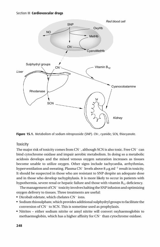
P1: PSB Printer: Yet To Come
9780521704632c15 CUFX213A/Peck 9780521618168 December 28, 2007 13:25
Section III Cardiovascular drugs
Red blood cell
Cyanocobalamine
Vitamin B12
Sulphydryl groups
Rhodanase
CN
SCN
Kidney
Liver
OxyHb
MetHb
CyanoMetHb
NO
SNP
CN
Figure 15.1. Metabolism of sodium nitroprusside (SNP). CN–, cyanide; SCN, thiocyanate.
ToxicityThe major risk of toxicity comes from CN−, although SCN is also toxic. Free CN− canbind cytochrome oxidase and impair aerobic metabolism. In doing so a metabolicacidosis develops and the mixed venous oxygen saturation increases as tissuesbecome unable to utilize oxygen. Other signs include tachycardia, arrhythmias,hyperventilation and sweating. Plasma CN− levels above 8 µg.ml−1 result in toxicity.It should be suspected in those who are resistant to SNP despite an adequate doseand in those who develop tachyphylaxis. It is more likely to occur in patients withhypothermia, severe renal or hepatic failure and those with vitamin B12 deficiency.
The management of CN− toxicity involves halting the SNP infusion and optimizingoxygen delivery to tissues. Three treatments are useful:� Dicobalt edetate, which chelates CN− ions.� Sodium thiosulphate, which provides additional sulphydryl groups to facilitate the
conversion of CN− to SCN. This is sometime used as prophylaxis.� Nitrites – either sodium nitrite or amyl nitrite will convert oxyhaemoglobin tomethaemoglobin, which has a higher affinity for CN− than cytochrome oxidase.
248
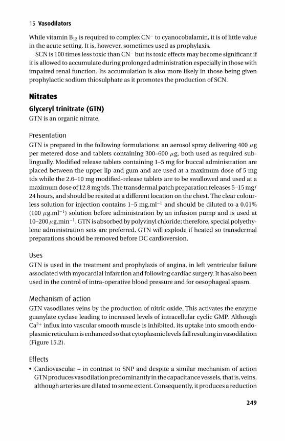
P1: PSB Printer: Yet To Come
9780521704632c15 CUFX213A/Peck 9780521618168 December 28, 2007 13:25
15 Vasodilators
While vitamin B12 is required to complex CN− to cyanocobalamin, it is of little valuein the acute setting. It is, however, sometimes used as prophylaxis.
SCN is 100 times less toxic than CN− but its toxic effects may become significant ifit is allowed to accumulate during prolonged administration especially in those withimpaired renal function. Its accumulation is also more likely in those being givenprophylactic sodium thiosulphate as it promotes the production of SCN.
Nitrates
Glyceryl trinitrate (GTN)GTN is an organic nitrate.
PresentationGTN is prepared in the following formulations: an aerosol spray delivering 400 µgper metered dose and tablets containing 300–600 µg, both used as required sub-lingually. Modified release tablets containing 1–5 mg for buccal administration areplaced between the upper lip and gum and are used at a maximum dose of 5 mgtds while the 2.6–10 mg modified-release tablets are to be swallowed and used at amaximum dose of 12.8 mg tds. The transdermal patch preparation releases 5–15 mg/24 hours, and should be resited at a different location on the chest. The clear colour-less solution for injection contains 1–5 mg.ml−1 and should be diluted to a 0.01%(100 µg.ml−1) solution before administration by an infusion pump and is used at10–200 µg.min−1. GTN is absorbed by polyvinyl chloride; therefore, special polyethy-lene administration sets are preferred. GTN will explode if heated so transdermalpreparations should be removed before DC cardioversion.
UsesGTN is used in the treatment and prophylaxis of angina, in left ventricular failureassociated with myocardial infarction and following cardiac surgery. It has also beenused in the control of intra-operative blood pressure and for oesophageal spasm.
Mechanism of actionGTN vasodilates veins by the production of nitric oxide. This activates the enzymeguanylate cyclase leading to increased levels of intracellular cyclic GMP. AlthoughCa2+ influx into vascular smooth muscle is inhibited, its uptake into smooth endo-plasmic reticulum is enhanced so that cytoplasmic levels fall resulting in vasodilation(Figure 15.2).
Effects� Cardiovascular – in contrast to SNP and despite a similar mechanism of actionGTN produces vasodilation predominantly in the capacitance vessels, that is, veins,although arteries are dilated to some extent. Consequently, it produces a reduction
249
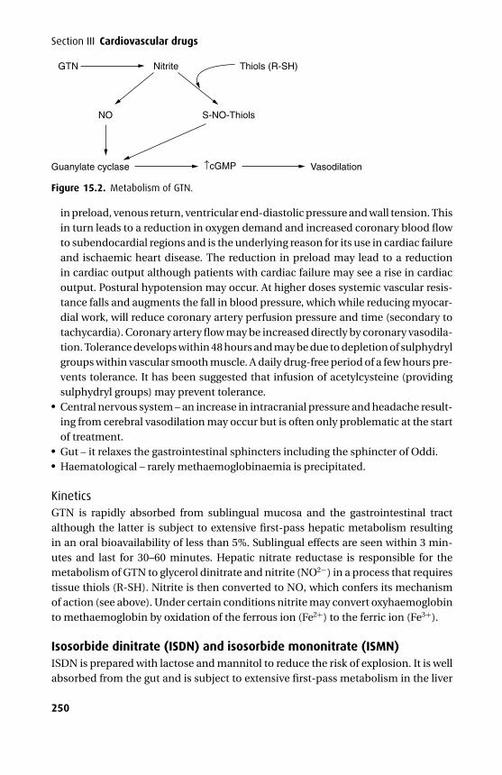
P1: PSB Printer: Yet To Come
9780521704632c15 CUFX213A/Peck 9780521618168 December 28, 2007 13:25
Section III Cardiovascular drugs
GTN Nitrite
NO
Thiols (R-SH)
S-NO-Thiols
Guanylate cyclase ↑cGMP Vasodilation
Figure 15.2. Metabolism of GTN.
in preload, venous return, ventricular end-diastolic pressure and wall tension. Thisin turn leads to a reduction in oxygen demand and increased coronary blood flowto subendocardial regions and is the underlying reason for its use in cardiac failureand ischaemic heart disease. The reduction in preload may lead to a reductionin cardiac output although patients with cardiac failure may see a rise in cardiacoutput. Postural hypotension may occur. At higher doses systemic vascular resis-tance falls and augments the fall in blood pressure, which while reducing myocar-dial work, will reduce coronary artery perfusion pressure and time (secondary totachycardia). Coronary artery flow may be increased directly by coronary vasodila-tion. Tolerance develops within 48 hours and may be due to depletion of sulphydrylgroups within vascular smooth muscle. A daily drug-free period of a few hours pre-vents tolerance. It has been suggested that infusion of acetylcysteine (providingsulphydryl groups) may prevent tolerance.� Central nervous system – an increase in intracranial pressure and headache result-ing from cerebral vasodilation may occur but is often only problematic at the startof treatment.� Gut – it relaxes the gastrointestinal sphincters including the sphincter of Oddi.� Haematological – rarely methaemoglobinaemia is precipitated.
KineticsGTN is rapidly absorbed from sublingual mucosa and the gastrointestinal tractalthough the latter is subject to extensive first-pass hepatic metabolism resultingin an oral bioavailability of less than 5%. Sublingual effects are seen within 3 min-utes and last for 30–60 minutes. Hepatic nitrate reductase is responsible for themetabolism of GTN to glycerol dinitrate and nitrite (NO2−) in a process that requirestissue thiols (R-SH). Nitrite is then converted to NO, which confers its mechanismof action (see above). Under certain conditions nitrite may convert oxyhaemoglobinto methaemoglobin by oxidation of the ferrous ion (Fe2+) to the ferric ion (Fe3+).
Isosorbide dinitrate (ISDN) and isosorbide mononitrate (ISMN)ISDN is prepared with lactose and mannitol to reduce the risk of explosion. It is wellabsorbed from the gut and is subject to extensive first-pass metabolism in the liver
250
P1: PSB Printer: Yet To Come
9780521704632c15 CUFX213A/Peck 9780521618168 December 28, 2007 13:25
15 Vasodilators
to isosorbide 2-mononitrate and isosorbide 5-mononitrate (ISMN), both of whichprobably confer the majority of the activity of ISDN. ISMN has a much longer half-life (4.5 hours) and is used in its own right. It is not subject to hepatic first-passmetabolism and has an oral bioavailability of 100%. Both are used in the prophylaxisof angina.
Potassium-channel activators
NicorandilNicorandil (nicotinamidoethyl nitrate) is a potassium-channel activator with anitrate moiety and as such differs from other potassium-channel activators. It hasbeen used in Japan since 1984 and was launched in the UK in 1994.
Presentation and usesNicorandil is available as tablets and the usual dose is 10–30 mg bd. It is used for thetreatment and prophylaxis of angina and in the treatment of congestive heart failureand hypertension. It has been used experimentally via the intravenous route.
Mechanism of actionATP-sensitive K+ channels are closed during the normal cardiac cycle but are open(activated) during periods of ischaemia when intracellular levels of ATP fall. In theopen state K+ passes down its concentration gradient out of the cell resulting inhyperpolarization, which closes Ca2+ channels resulting in less Ca2+ for myocardialcontraction.
Nicorandil activates the ATP-sensitive K+ channels within the heart and arterioles.In addition, nicorandil relaxes venous capacitance smooth muscle by stimulatingguanylate cyclase via its nitrate moiety, leading to increased intracellular cGMP.
Effects� Cardiovascular – nicorandil causes venodilation and arterial vasodilation result-ing in a reduced pre- and afterload. The blood pressure falls. Left ventricular end-diastolic pressure falls and there is an improved normal and collateral coronaryartery blood flow, partly induced by coronary artery vasodilation without a ‘steal’phenomenon. An increase in cardiac output is seen in patients with ischaemicheart disease and cardiac failure. It is effective at suppressing torsades de pointesassociated with a prolonged QT interval. High concentrations in vitro result in ashortened action potential by accelerated repolarization. It also reduces the sizeof experimentally induced ischaemia, the mechanism of which is uncertain, sothat an alternative, as yet undefined, cardioprotective mechanism has been pos-tulated. Unlike nitrate therapy it is not associated with tolerance during prolongedadministration. Contractility and atrioventricular conduction is not affected.� Central nervous system – headaches, these usually clear with continued therapy.
251
P1: PSB Printer: Yet To Come
9780521704632c15 CUFX213A/Peck 9780521618168 December 28, 2007 13:25
Section III Cardiovascular drugs� Metabolic – unlike other antihypertensives it has no effect on lipid profile or glucosecontrol.� Haematological – nicorandil inhibits in vitro ADP-induced platelet aggregation (ina similar manner to nitrates), which is associated with an increase of intraplateletcGMP.� Miscellaneous – giant oral apthous ulcers have been reported with its use.
KineticsNicorandil is well absorbed from the gut with insignificant first-pass metabolism.The main metabolic route is denitration with 20% excreted as metabolites in theurine. The elimination half-life is 1 hour, although its actions last up to 12 hours, butneither is increased in the presence of renal impairment. It is not plasma protein-bound to any significant extent.
Calcium channel antagonistsDespite their disparate chemical structures, the Ca2+ channel antagonists are alleffective in specifically blocking the entry of Ca2+ through L-type channels while leav-ing T-, N- and P-type Ca2+ channels unaffected. The L-type channel is widespreadin the cardiovascular system and is responsible for the plateau phase (slow inwardcurrent) of the cardiac action potential. It triggers the internal release of Ca2+ andis regulated by cAMP-dependent protein kinase. The T-type channel is structurallysimilar to the L-type channel and is present mainly in cardiac cells that lack a T-tubule system, that is, SA node and certain types of vascular smooth muscle. Theyare not present in ventricular myocardium. N-type channels are only found in nervecells.
Calcium channel antagonists have variable affinity for L-type channels inmyocardium, nodal and vascular smooth muscle resulting in variable effects. Theyare all useful in the treatment of essential hypertension, although some are alsoparticularly useful in the treatment of angina or arrhythmias.
VerapamilVerapamil is a racemic mixture and a synthetic derivative of papaverine.
Presentation and usesVerapamil is available as 20–240 mg tablets, some prepared as modified release and asan oral suspension containing 40 mg/5 ml. The colourless intravenous preparationcontains 2.5 mg.ml–1. It is used for certain supraventricular arrhythmias (not inatrial fibrillation complicating WPW syndrome) and angina. It has been used totreat hypertension although its negative inotropic properties limit its usefulness inthis setting.
252
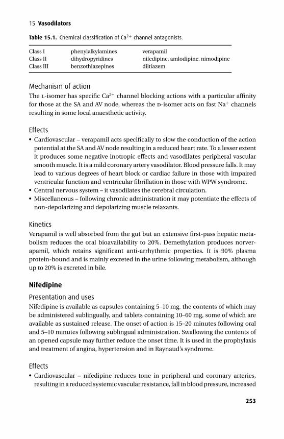
P1: PSB Printer: Yet To Come
9780521704632c15 CUFX213A/Peck 9780521618168 December 28, 2007 13:25
15 Vasodilators
Table 15.1. Chemical classification of Ca2+ channel antagonists.
Class I phenylalkylamines verapamilClass II dihydropyridines nifedipine, amlodipine, nimodipineClass III benzothiazepines diltiazem
Mechanism of actionThe l-isomer has specific Ca2+ channel blocking actions with a particular affinityfor those at the SA and AV node, whereas the d-isomer acts on fast Na+ channelsresulting in some local anaesthetic activity.
Effects� Cardiovascular – verapamil acts specifically to slow the conduction of the actionpotential at the SA and AV node resulting in a reduced heart rate. To a lesser extentit produces some negative inotropic effects and vasodilates peripheral vascularsmooth muscle. It is a mild coronary artery vasodilator. Blood pressure falls. It maylead to various degrees of heart block or cardiac failure in those with impairedventricular function and ventricular fibrillation in those with WPW syndrome.� Central nervous system – it vasodilates the cerebral circulation.� Miscellaneous – following chronic administration it may potentiate the effects ofnon-depolarizing and depolarizing muscle relaxants.
KineticsVerapamil is well absorbed from the gut but an extensive first-pass hepatic meta-bolism reduces the oral bioavailability to 20%. Demethylation produces norver-apamil, which retains significant anti-arrhythmic properties. It is 90% plasmaprotein-bound and is mainly excreted in the urine following metabolism, althoughup to 20% is excreted in bile.
Nifedipine
Presentation and usesNifedipine is available as capsules containing 5–10 mg, the contents of which maybe administered sublingually, and tablets containing 10–60 mg, some of which areavailable as sustained release. The onset of action is 15–20 minutes following oraland 5–10 minutes following sublingual administration. Swallowing the contents ofan opened capsule may further reduce the onset time. It is used in the prophylaxisand treatment of angina, hypertension and in Raynaud’s syndrome.
Effects� Cardiovascular – nifedipine reduces tone in peripheral and coronary arteries,resulting in a reduced systemic vascular resistance, fall in blood pressure, increased
253
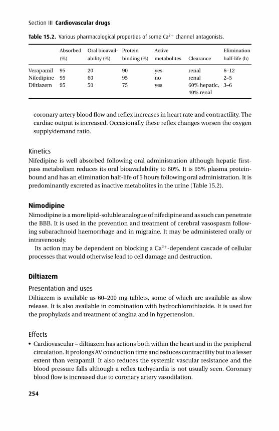
P1: PSB Printer: Yet To Come
9780521704632c15 CUFX213A/Peck 9780521618168 December 28, 2007 13:25
Section III Cardiovascular drugs
Table 15.2. Various pharmacological properties of some Ca2+ channel antagonists.
Absorbed
(%)
Oral bioavail-
ability (%)
Protein
binding (%)
Active
metabolites Clearance
Elimination
half-life (h)
Verapamil 95 20 90 yes renal 6–12Nifedipine 95 60 95 no renal 2–5Diltiazem 95 50 75 yes 60% hepatic,
40% renal3–6
coronary artery blood flow and reflex increases in heart rate and contractility. Thecardiac output is increased. Occasionally these reflex changes worsen the oxygensupply/demand ratio.
KineticsNifedipine is well absorbed following oral administration although hepatic first-pass metabolism reduces its oral bioavailability to 60%. It is 95% plasma protein-bound and has an elimination half-life of 5 hours following oral administration. It ispredominantly excreted as inactive metabolites in the urine (Table 15.2).
NimodipineNimodipine is a more lipid-soluble analogue of nifedipine and as such can penetratethe BBB. It is used in the prevention and treatment of cerebral vasospasm follow-ing subarachnoid haemorrhage and in migraine. It may be administered orally orintravenously.
Its action may be dependent on blocking a Ca2+-dependent cascade of cellularprocesses that would otherwise lead to cell damage and destruction.
Diltiazem
Presentation and usesDiltiazem is available as 60–200 mg tablets, some of which are available as slowrelease. It is also available in combination with hydrochlorothiazide. It is used forthe prophylaxis and treatment of angina and in hypertension.
Effects� Cardiovascular – diltiazem has actions both within the heart and in the peripheralcirculation. It prolongs AV conduction time and reduces contractility but to a lesserextent than verapamil. It also reduces the systemic vascular resistance and theblood pressure falls although a reflex tachycardia is not usually seen. Coronaryblood flow is increased due to coronary artery vasodilation.
254
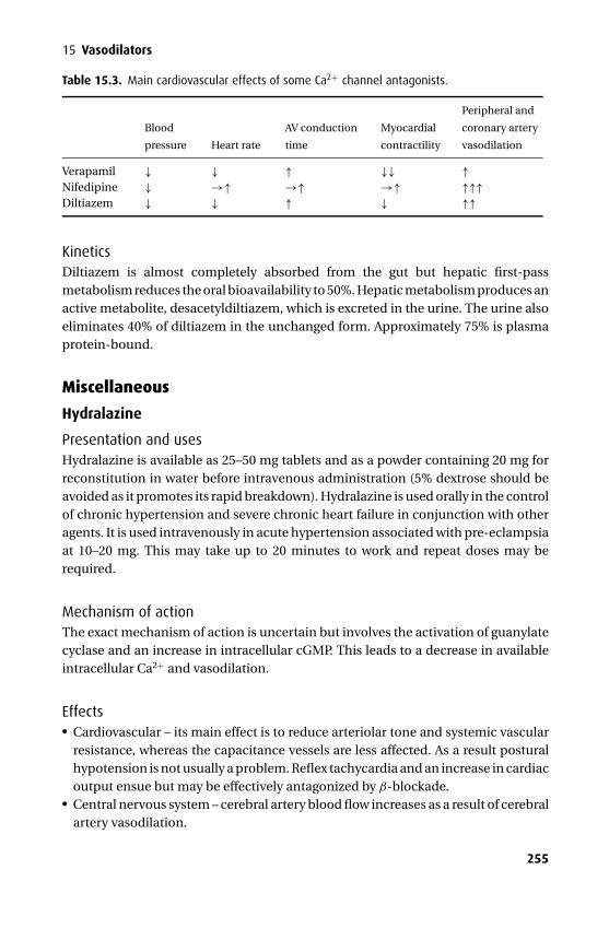
P1: PSB Printer: Yet To Come
9780521704632c15 CUFX213A/Peck 9780521618168 December 28, 2007 13:25
15 Vasodilators
Table 15.3. Main cardiovascular effects of some Ca2+ channel antagonists.
Blood
pressure Heart rate
AV conduction
time
Myocardial
contractility
Peripheral and
coronary artery
vasodilation
Verapamil ↓ ↓ ↑ ↓↓ ↑Nifedipine ↓ →↑ →↑ →↑ ↑↑↑Diltiazem ↓ ↓ ↑ ↓ ↑↑
KineticsDiltiazem is almost completely absorbed from the gut but hepatic first-passmetabolism reduces the oral bioavailability to 50%. Hepatic metabolism produces anactive metabolite, desacetyldiltiazem, which is excreted in the urine. The urine alsoeliminates 40% of diltiazem in the unchanged form. Approximately 75% is plasmaprotein-bound.
Miscellaneous
Hydralazine
Presentation and usesHydralazine is available as 25–50 mg tablets and as a powder containing 20 mg forreconstitution in water before intravenous administration (5% dextrose should beavoided as it promotes its rapid breakdown). Hydralazine is used orally in the controlof chronic hypertension and severe chronic heart failure in conjunction with otheragents. It is used intravenously in acute hypertension associated with pre-eclampsiaat 10–20 mg. This may take up to 20 minutes to work and repeat doses may berequired.
Mechanism of actionThe exact mechanism of action is uncertain but involves the activation of guanylatecyclase and an increase in intracellular cGMP. This leads to a decrease in availableintracellular Ca2+ and vasodilation.
Effects� Cardiovascular – its main effect is to reduce arteriolar tone and systemic vascularresistance, whereas the capacitance vessels are less affected. As a result posturalhypotension is not usually a problem. Reflex tachycardia and an increase in cardiacoutput ensue but may be effectively antagonized by β-blockade.� Central nervous system – cerebral artery blood flow increases as a result of cerebralartery vasodilation.
255
P1: PSB Printer: Yet To Come
9780521704632c15 CUFX213A/Peck 9780521618168 December 28, 2007 13:25
Section III Cardiovascular drugs� Renal – despite an increase in renal blood flow, fluid retention, oedema, and areduction in urine output is often seen. This may be overcome by concurrentadministration of a diuretic.� Gut – nausea and vomiting are common.� Miscellaneous – peripheral neuropathy and blood dyscrasias. A lupus erythemato-sus type syndrome is occasionally seen after long-term use and may be morecommon in slow acetylators and women. It may require long-term corticosteroidtherapy.
KineticsHydralazine is well absorbed from the gut but is subject to a variable first-passmetabolism resulting in an oral bioavailability of 25–55% depending on the acetyla-tor status of the individual. The plasma half-life is normally 2–3 hours but this may beshortened to 45 minutes in rapid acetylators. It is 90% protein-bound in the plasma.Up to 85% is excreted in the urine as acetylated and hydroxylated metabolites, someof which are conjugated with glucuronic acid. It crosses the placenta and may causea foetal tachycardia.
Minoxidil
Presentation and usesMinoxidil is prepared as 2.5–10 mg tablets, a 2% solution for intravenous use and a5% lotion. It is used for severe hypertension and alopecia areata.
Its mechanism of action and principal effects are essentially the same as thoseof hydralazine. The mechanism by which it stimulates the hair follicle is poorlyunderstood. It may also precipitate hypertrichosis of the face and arms and breasttenderness. It does not cause a lupus erythematosus-type reaction.
KineticsMinoxidil is well absorbed from the gut and has an oral bioavailability of 90%. It is notprotein-bound in the plasma. Its plasma half-life is only 3 hours but its hypotensiveeffects may last for as long as 3 days. It undergoes hepatic glucuronide conjugationand is subsequently excreted in the urine.
DiazoxideThis vasodilator is chemically related to the thiazide diuretics.
Presentation and usesDiazoxide is available as 50 mg tablets and as a solution for intravenous injectioncontaining 15 mg.ml−1. It is used intravenously to treat hypertensive emergenciesassociated with renal disease at 1–3 mg.kg−1 up to a maximum dose of 150 mg,
256
P1: PSB Printer: Yet To Come
9780521704632c15 CUFX213A/Peck 9780521618168 December 28, 2007 13:25
15 Vasodilators
which may be repeated after 15 minutes. It has also been used to treat intractablehypoglycaemia and malignant islet-cell tumours.
Mechanism of actionIts hypotensive effects are mediated through altered levels of cAMP in arterioles,producing vasodilation. It may also be due to a reduced Ca2+ influx. Its biochemi-cal effects are due to inhibition of insulin secretion and increased release of cate-cholamines.
Effects� Cardiovascular – diazoxide produces arteriolar vasodilation with little effect oncapacitance vessels. As a result the blood pressure falls and there is an increase inheart rate and cardiac output. Postural hypotension is not a problem.� Metabolic – it increases the levels of glucose, catecholamines, renin and aldos-terone. It also causes fluid retention (despite its chemical relation to the thiazides),which may require treatment with a loop diuretic.� Miscellaneous – the following effects may occur: nausea, especially at thestart of treatment, extra-pyramidal effects including occulogyric crisis, thrombo-cytopenia and hyperuricaemia.
KineticsDiazoxide is well absorbed from the gut with an oral bioavailability of 80%, and isextensively protein-bound in the plasma (about 90%). The hyperglycaemic effectslast about 8 hours while the hypotensive effects last about 5 hours. The plasma half-life, however, may last up to 36 hours. It is partly metabolized in the liver, beingexcreted in the urine as unchanged drug and as inactive metabolites.
257
P1: PSB Printer: Yet To Come
9780521704632c16 CUFX213A/Peck 9780521618168 December 28, 2007 13:34
16Antihypertensives
� Drugs affecting the renin–angiotensin–aldosterone system� Adrenergic neurone blockade� Centrally acting� Ganglion blockade� Diuretics� Adrenoceptor antagonists� Ca2+ channel antagonists
Renin–angiotensin–aldosterone system
PhysiologyThe juxtaglomerular apparatus within the kidney consists of three distinct celltypes:� Juxtaglomerular cells form part of the afferent arteriole as it enters the glomerulus
and are supplied by sympathetic nerves. They contain prorenin, which is convertedto the acid protease renin before systemic release.� The macula densa is a region of cells at the start of the distal convolutedtubule, which lie adjacent to the juxtaglomerular cells of the same nephron.� Agranular lacis cells, which lie between the afferent and efferent arterioles adjacentto the glomerulus.
Under the following conditions the juxtaglomerular apparatus will cause the releaseof renin into the circulation:� Reduced renal perfusion� Reduced Na+ at the macula densa� Stimulation of the renal sympathetic fibres via β1-adrenoceptorsRenin (half-life 80 minutes) splits the decapeptide angiotensin I from the cir-culating plasma protein angiotensinogen, which is synthesized in the liver andis present in the α2-globulin fraction of plasma proteins. Angiotensin-convertingenzyme (ACE) converts angiotensin I to the active octapeptide angiotensin II, andalso inactivates bradykinin. Angiotensin II is broken down in the kidney and liverto inactive metabolites and angiotensin III, which retains some activity (Figure16.1).
258
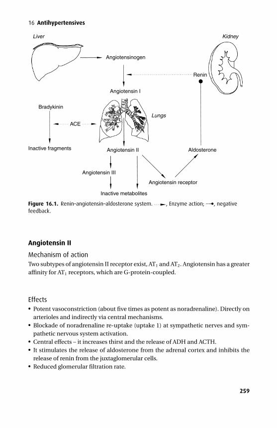
P1: PSB Printer: Yet To Come
9780521704632c16 CUFX213A/Peck 9780521618168 December 28, 2007 13:34
16 Antihypertensives
Liver
Lungs
Kidney
Angiotensinogen
Angiotensin I
Renin
Angiotensin II
Angiotensin III
Inactive metabolites
Angiotensin receptor
Aldosterone
Bradykinin
ACE
Inactive fragments
Figure 16.1. Renin–angiotensin–aldosterone system. , Enzyme action; —–•, negativefeedback.
Angiotensin II
Mechanism of actionTwo subtypes of angiotensin II receptor exist, AT1 and AT2. Angiotensin has a greateraffinity for AT1 receptors, which are G-protein-coupled.
Effects� Potent vasoconstriction (about five times as potent as noradrenaline). Directly onarterioles and indirectly via central mechanisms.� Blockade of noradrenaline re-uptake (uptake 1) at sympathetic nerves and sym-pathetic nervous system activation.� Central effects – it increases thirst and the release of ADH and ACTH.� It stimulates the release of aldosterone from the adrenal cortex and inhibits therelease of renin from the juxtaglomerular cells.� Reduced glomerular filtration rate.
259

P1: PSB Printer: Yet To Come
9780521704632c16 CUFX213A/Peck 9780521618168 December 28, 2007 13:34
Section III Cardiovascular drugs
ACE inhibitors and angiotensin II receptor antagonists are used widely in the treat-ment of hypertension. β-Blockers (cf. Chapter 13) reduce sympathetically mediatedrelease of renin, which contributes to their antihypertensive effects.
Angiotensin-converting enzyme (ACE) inhibitorsACE inhibitors are used in all grades of heart failure and in patients with myocardialinfarction with left ventricular dysfunction where it improves the prognosis. Theyare used in hypertension especially insulin-dependent diabetics with nephropathy.However hypertension is relatively resistant to ACE inhibition in the black poulationwhere concurrent diuretic thereapy may be required. While most drugs should becontinued throughout the peri-opereative period, ACE inhibitors and angiotensin IIreceptor antagonists should be omitted due to the increased frequency of peri-operative hypotension.
From a kinetic point of view ACE inhibitors may be divided into three groups:Group 1. Captopril – an active drug that is metabolized to active metabolites.Group 2. Enalopril, ramipril – prodrugs, which only become active following
hepatic metabolism to the diacid moiety.Group 3. Lisinopril – an active drug that is not metabolized and is excreted
unchanged in the urine.In other respects the effects of ACE inhibitors are similar and are discussed undercaptopril.
Captopril
PresentationCaptopril is available as 12.5–50 mg tablets that may also be combined withhydrochlorothiazide. The initial dose is 12.5 mg although 6.25 mg may be prudentfor those with heart failure.
Mechanism of actionCaptopril is a competitive ACE inhibitor and therefore prevents the formation ofangiotensin II and its effects. Afterload is reduced to a greater degree than preload.
Effects� Cardiovascular – captopril reduces the systemic vascular resistance significantly,resulting in a fall in blood pressure. The fall in afterload may increase the cardiacoutput particularly in those with heart failure. Heart rate is usually unaffected butmay increase. Baroreceptor reflexes are also unaffected. Transient hypotensionmay occur at the start of treatment, which should therefore be initiated in thehospital for patients with anything more than mild heart failure.� Renal – the normal function of angiotensin II to maintain efferent arteriolar pres-sure (by vasoconstriction) at the glomerulus in the presence of poor renal perfusion
260
P1: PSB Printer: Yet To Come
9780521704632c16 CUFX213A/Peck 9780521618168 December 28, 2007 13:34
16 Antihypertensives
is forfeit. Therefore renal perfusion pressure falls and renal failure may follow. Asa result bilateral renal artery stenosis or unilateral renal artery stenosis to a singlefunctioning kidney is considered a contraindication. Where normal renal perfu-sion is preserved, renal vasodilatation may occur leading to a naturesis.� Metabolic – reduced aldosterone release impairs the negative feedback to reninproduction so that renin levels become elevated. It may also lead to hyperkalaemiaand raised urea and creatinine, especially in those with even mildly impaired renalfunction.� Interactions – captopril reduces aldosterone release, which may result in hyper-kalaemia, so it should not be used with potassium-sparing diuretics. It has beenassociated with unexplained hypoglycaemia in Type I and II diabetes. These effectsusually decrease with continued treatment. Non-steroidal anti-inflammatorydrugs reduce captopril’s antihypertensive effects and may precipitate renal fail-ure.� Cough – a persistent dry cough may be the result of increased levels of bradykinin,which are normally broken down by ACE. Non-steroidal anti-inflammatory drugsmay alleviate this cough but at the expense of reduced antihypertensive effects.� Miscellaneous – rare but serious effects may complicate its use and include angio-oedema (0.2%, more common in black patients), agranulocytosis and thrombocy-topenia. Less serious side effects are more common and include loss of taste, rash,pruritus, fever and apthous ulceration. These are more common with higher dosesand in patients with impaired renal function.
KineticsCaptopril is well absorbed from the gut and has an oral bioavailability of 65%. It is25% plasma protein bound. Approximately 50% is oxidized in the liver to the dimerand mixed sulphides all of which are eventually excreted in the urine. It has anelimination half-life of 4 hours, although this will be increased in the presence ofrenal impairment.
Enalopril
KineticsEnalopril is a prodrug, which is hydrolyzed in the liver and kidney to the active com-pound enaloprilat. It may be given orally (as enalopril) or intravenously (as enalo-prilat). Its elimination half-life is 4–8 hours but increases to 11 hours in prolongedthereapy. It has a duration of action of approximately 20 hours.
Angiotensin II receptor antagonists (ARAs)
LosartanLosartan is a substituted imidazole compound.
261
P1: PSB Printer: Yet To Come
9780521704632c16 CUFX213A/Peck 9780521618168 December 28, 2007 13:34
Section III Cardiovascular drugs
Presentation and usesLosartan is available as 25–50 mg tablets and in combination with hydrochloroth-iazide. It is used in the treatment of hypertension where dry cough proves an unac-ceptable side effect of ACE inhibitor therapy.
Mechanism of actionLosartan is a specific angiotensin II receptor (type AT1) antagonist at all sites withinthe body. It blocks the negative feedback of angiotensin II on renin secretion, whichtherefore increases, leading to increased angiotensin II. This has little impact due tocomprehensive AT1 receptor blockade.
EffectsThese are broadly similar to those of the ACE inhibitors.� Metabolic – as it does not block the actions of ACE, bradykinin may be broken
down in the usual manner (by ACE). As a result, bradykinin levels are not raisedand the dry cough seen with ACE inhibitors does not complicate its use.
KineticsLosartan is well absorbed from the gut but undergoes significant first-passmetabolism to an active carboxylic acid metabolite (which acts in a non-competitivemanner), and several other inactive compounds. It has an oral bioavailability of30% and is 99% plasma protein bound. Its elimination half-life is 2 hours while theelimination half-life of the active metabolite is 7 hours. Less than 10% is excretedunchanged in an active form in the urine. The inactive metabolites are excreted inbile and urine.
ContraindicationsLike ACE inhibitors, Losartan is contraindicated in bilateral renal artery stenosis andpregnancy.
Comparing ARAs to ACE inhibitorsARA use may become more widespread for the following reasons.1. Blockade of the AT1-receptor is the most specific way of preventing the adverse
effects of angiotensin II seen in heart failure and hypertension, especially asangiotensin II may be synthesized by alternative non-ACE pathways.
2. The AT2-receptor is not blocked, which may possess cardioprotective properties.3. There is a much lower incidence of cough and angioedema and therefore
improved compliance.
262
P1: PSB Printer: Yet To Come
9780521704632c16 CUFX213A/Peck 9780521618168 December 28, 2007 13:34
16 Antihypertensives
Adrenergic neurone blockadeThis group of drugs interferes with the release of noradrenaline from adrenergicneurones.
PhysiologyNoradrenaline is synthesized from tyrosine (see Chapter 12) within the adrener-gic nerve terminal. It is held in storage vesicles and subsequently released into theneuronal cleft to act on adrenoceptors. Its actions are terminated by:� Uptake 1 – noradrenaline is taken back into the nerve terminal by a high-affinity
transport system. This provides the main route for terminating its effects. Withinthe neurone it is recycled and returned to storage vesicles. However, while in thecytoplasm some may be de-aminated by MAO.� Uptake 2 – noradrenaline diffuses away from the nerve terminal into the circulationwhere it is taken up by extraneuronal tissues including the liver and metabolizedby COMT.
Guanethidine
Presentation and usesGuanethidine is available as tablets and as a colourless solution for injection con-taining 10 mg.ml−1. It has been used as an antihypertensive agent but is currentlyused only for the control of sympathetically mediated chronic pain. The initial anti-hypertensive dose is 10 mg.day−1, which is increased to a maintenance dose of30–50 mg.day-1. A dose of 20 mg is used when performing an intravenous regionalblock for chronic pain. Repeated blocks are usually required.
Mechanism of actionGuanethidine gains access to the adrenergic neurone by utilizing the uptake 1transport mechanism. Following intravenous administration there is some initialhypotension by direct vasodilation of arterioles. Subsequently, it displaces nora-drenaline from its binding sites, which may cause transient hypertension. Finally,guanethidine reduces the blood pressure by preventing the release of what littlenoradrenaline is left in the nerve terminal. Oral administration does not producethe same triphasic response, as its onset of action is much slower. It does not alterthe secretion of catecholamines by the adrenal medulla.
Effects� Cardiovascular – hypotension is its main action. Postural hypotension is commonas it blocks any compensatory rise in sympathetic tone. Fluid retention leading tooedema may occur.� Gut – diarrhoea is common.� Miscellaneous – failure to ejaculate.
263
P1: PSB Printer: Yet To Come
9780521704632c16 CUFX213A/Peck 9780521618168 December 28, 2007 13:34
Section III Cardiovascular drugs� Drug interactions – drugs that block uptake 1 (tricyclic antidepressants, cocaine)prevent guanethidine from entering the nerve terminal and disrupting nora-drenaline storage. They therefore antagonize guanethidine.
Up-regulation of adrenoceptors follows the long-term use of guanethidine so thatthese patients are very sensitive to direct-acting sympathomimetic amines.
KineticsFollowing oral administration, guanethidine is variably and incompletely absorbed.Hepatic first-pass metabolism results in an oral bioavailability of 50%. It is not boundby plasma proteins and does not cross the BBB. It has an elimination half-life ofseveral days. Elimination is by hepatic metabolism and excretion of unchanged drugand its metabolites in the urine.
ReserpineReserpine is no longer available in the UK. It is a naturally occurring alkaloid that waswidely used to treat hypertension that failed to respond to β-blockers or diuretics.
Mechanism of actionReserpine acts centrally and peripherally by preventing storage vesicles incorporat-ing noradrenaline from neuronal cytoplasm leading to rapid de-amination by mito-chondrial MAO and noradrenaline depleted neurones. Serotonin is also depleted.
Effects� Cardiovascular – hypotension is mainly a result of a reduced cardiac output andsystemic vascular resistance. Postural hypotension is rarely a problem, but nasalcongestion is less well tolerated.� Central nervous system – depression, lethargy, and nightmares are caused by reser-pine’s ability to cross the BBB. Extrapyramidal effects may also be seen. Generalanaesthetic requirements are decreased.� Gut – diarrhoea and increased gastric acid secretions may lead to epigastric pain.� Miscellaneous – sexual dysfunction, hyperprolactinaemia, gynaecomastia andgalactorrhoea are seen rarely.� Drug interactions – patients taking reserpine will exhibit increased sensitivity todirect-acting sympathomimetic amines (adrenoceptor up-regulation) but reducedsensitivity to indirect-acting sympathomimetic amines (depleted noradrenalinestores).
KineticsFollowing absorption from the gut, reserpine is metabolized slowly in the liver. Somemetabolic products are excreted in the urine while most appear to be excreted
264
P1: PSB Printer: Yet To Come
9780521704632c16 CUFX213A/Peck 9780521618168 December 28, 2007 13:34
16 Antihypertensives
unchanged in the bile. It has a half-life of many days. It crosses the placenta andBBB and also reaches breast milk.
MetirosineMetirosine is a competitive inhibitor of tyrosine hydroxylase and can therefore pre-vent the synthesis of catecholamines. It should only be used to manage hypertensionassociated with phaeochromocytoma.
It may cause severe diarrhoea, sedation, extrapyramidal effects and hypersensi-tivity reactions.
Centrally acting
Methyldopa
Presentation and usesMethyldopa is available as film-coated tablets containing 125–500 mg and as anoral suspension containing 50 mg.ml–1. The rarely used intravenous preparation iscolourless and contains 50 mg.ml–1 and uses sodium metabisulphite as the preser-vative. It is used at 250 mg tds increasing to a maximum of 3 g.day–1 to controlhypertension, especially that associated with pregnancy.
Mechanism of actionMethyldopa readily crosses the BBB where it is decarboxylated to α-methyl-noradrenaline, which is a potent α2-agonist. It retains limited α1-agonist proper-ties (α2:α1 ratio 10:1). Stimulation of presynaptic α2-receptors in the nucleus tractussolitarii forms a negative feedback loop for further noradrenaline release so thatα-methyl-noradrenaline reduces centrally mediated sympathetic tone, leading to areduction in blood pressure.
Effects� Cardiovascular – its main effect is a reduction in systemic vascular resistance lead-ing to a fall in blood pressure. Postural hypotension is occasionally a problem.Cardiac output is unchanged despite a relative bradycardia. Rebound hyperten-sion may occur if treatment is stopped suddenly but this is less common than withclonidine.� Central nervous system – sedation is common, while dizziness, depression, night-mares and nausea are less common. The MAC of volatile anaesthetics is reduced.� Haematological – a positive direct Coombs’ test is seen in 10–20% of patients takingmethyldopa. Thrombocytopenia and leucopenia occur rarely.� Allergic – it may precipitate an auto-immune haemolytic anaemia. Eosinophiliaassociated with fever sometimes occurs within the first few weeks of therapy. Ahypersensitivity reaction may cause myocarditis.
265
P1: PSB Printer: Yet To Come
9780521704632c16 CUFX213A/Peck 9780521618168 December 28, 2007 13:34
Section III Cardiovascular drugs� Renal – urine may become darker in colour when exposed to air, due to the break-down of methyldopa or its metabolites.� Hepatic – liver function may deteriorate during long-term treatment and fatalhepatic necrosis has been reported.� Miscellaneous – it fluoresces at the same wavelengths as catecholamines so thatassays of urinary catecholamines may be falsely high. Assays of VMA are notaffected. It may cause constipation and gynaecomastia (due to suppressed pro-lactin release).
KineticsMethyldopa is erratically absorbed from the gut and has a very variable oral bioavail-ability and a slow onset of action. It is subject to hepatic first-pass metabolism, beingconverted to the O-sulphate. Less than 20% is bound to plasma proteins. Approxi-mately 50% is excreted unchanged in the urine.
ClonidineClonidine is an α-agonist with an affinity for α2-receptors 200 times that for α1-receptors. Some studies identify it as a partial agonist.
Presentation and usesClonidine is available as 25–300 µg tablets and as a colourless solution for injectioncontaining 150 µg.ml−1. A transdermal patch is available but this takes 48 hours toachieve therapeutic levels. It is used in the treatment of hypertension, acute andchronic pain, the suppression of symptoms of opioid withdrawal and to augmentsedation during ventilation of the critically ill patient.
Mechanism of actionThe useful effects of clonidine rest on its ability to stimulateα2-receptors in the lateralreticular nucleus resulting in reduced central sympathetic outflow, and in the spinalcord where they augment endogenous opiate release and modulate the descendingnoradrenergic pathways involved in spinal nociceptive processing. MAC appears tobe reduced by stimulation of central postsynaptic α2-receptors.
Transmembrane signalling of α2-receptors is coupled to Gi, leading to reducedintracellular cAMP. K+ channels are also activated.
Effects� Cardiovascular – following intravenous administration the blood pressure mayrise due to peripheral α1 stimulation, but this is followed by a more prolongedfall in blood pressure. Cardiac output is well maintained despite a bradycardia.The PR interval is lengthened, atrioventricular nodal conduction depressed andthe baroreceptor reflexes are sensitized by clonidine resulting in a lower heartrate for a given increase in blood pressure. Its effects on the coronary circulation
266
P1: PSB Printer: Yet To Come
9780521704632c16 CUFX213A/Peck 9780521618168 December 28, 2007 13:34
16 Antihypertensives
are complicated as any direct vasoconstriction may be offset by a reduction insympathetic tone and by the release of local nitric oxide. It stabilizes the cardio-vascular responses to peri-operative stimuli. Rebound hypertension is seen morecommonly when a dose of more than 1.2 g.day−1 is stopped abruptly. This is dueto peripheral vasoconstriction and increased plasma catecholamines and maybe exacerbated by β-blockade (leaving vasoconstriction unopposed). Increasingdoses have a ceiling effect and are limited by increasing α1 stimulation.� Central nervous system – it produces sedation and a reduction of up to 50% inthe MAC of volatile agents. It is anxiolytic at low doses but becomes anxiogenic athigher doses.� Analgesia – clonidine has been used via the subarachnoid and epidural routes.It provides prolonged analgesia with no respiratory depression and appears toact synergistically with concurrently administered opioids. It does not producemotor or sensory blockade. Clonidine also appears to reduce post-operative opioidrequirement when administered intravenously.� Renal system – a number of mechanisms including inhibition of release of ADHhave been implicated as the cause of diuresis during its use.� Respiratory – in contrast to opioids it does not produce significant respiratorydepression.� Endocrine – the stress response to surgical stimulus is inhibited. Insulin release isinhibited although this rarely increases blood-sugar levels.� Haematological – despite the presence of α2-adrenoceptors on platelets, therapeu-tic doses of clonidine do not promote platelet aggregation and its sympatholyticeffects block adrenaline-induced platelet aggregation.
KineticsClonidine is rapidly and almost completely absorbed after oral administration,achieving an oral bioavailability of nearly 100%. It is 20% plasma protein bound andhas a volume of distribution of about 2 l.kg−1. The elimination half-life of clonidineis between 9 and 18 hours, with approximately 50% of the drug being metabolizedin the liver to inactive metabolites, while the rest is excreted unchanged in the urine.The dose should be reduced in the presence of renal impairment.
DexmedetomidineMedetomidine is an α2-agonist that has been used widely in veterinary practice forits sedation and analgesic properties.
Presentation and usesMedetomidine is a racemic mixture but only the d-stereoisomer is active, so it hasbeen developed as dexmedetomidine.
267
P1: PSB Printer: Yet To Come
9780521704632c16 CUFX213A/Peck 9780521618168 December 28, 2007 13:34
Section III Cardiovascular drugs
Mechanism of actionThis is similar to that of clonidine although it is more potent and has a higher affinityfor the α2-receptor (α2:α1 ratio 1600:1). It is a full agonist.
EffectsThese are broadly similar to those of clonidine.
KineticsOral absorption is unpredictable but does avoid the initial hypertension that par-enteral administration produces. It has an elimination half-life of 2 hours.
AtipamezoleAtipamezole is a selective α2-antagonist that crosses the BBB to reverse the seda-tion and analgesia associated with clonidine and dexmedetomidine. At 2 hours itselimination half-life is similar to dexmedetomidine with obvious clinical benefit.
Ganglion blockadeThis group of drugs competitively antagonizes nicotinic receptors at both parasym-pathetic and sympathetic ganglia and also at the adrenal cortex. Ganglion blockers donot inhibit nicotinic receptors at the neuromuscular junction although some musclerelaxants (tubocurare) may demonstrate some ganglion-blocking properties.
TrimetaphanTrimetaphan is a quaternary ammonium compound.
Presentation and usesTrimetaphan is available as a clear pale-yellow solution for intravenous injectioncontaining 50 mg.ml−1. It was used previously via the oral route to treat essentialhypertension but drugs with better side-effect profiles have superseded it. Its solecurrent use is to induce hypotensive anaesthesia at 1–4 mg.min−1.
Mechanism of actionTrimetaphan is a competitive antagonist at all nicotinic ganglionic receptors includ-ing those at the adrenal cortex and has a direct vasodilator effect on peripheral vessels.Histamine is also released but may not be significant in producing hypotension.
Effects� Cardiovascular – the onset of hypotension is rapid when used by intravenous infu-sion. Pre- and afterload falls, and there may be a compensatory increase in heartrate to maintain cardiac output.
268
P1: PSB Printer: Yet To Come
9780521704632c16 CUFX213A/Peck 9780521618168 December 28, 2007 13:34
16 Antihypertensives� Central nervous system – cerebral blood flow is not reduced as long as the preloadis maintained with intravenous fluid and the mean blood pressure remains aboveapproximately 50 mmHg. The cerebral metabolic rate is unchanged and it doesnot cross the BBB to any extent.� Respiratory – histamine release may induce bronchospasm.� Parasympathetic – owing to parasympathetic ganglion blockade the followingeffects are seen: constipation, urinary retention, dry mouth, mydriasis, increasedintra-occular pressure and variable tachycardia. These may complicate recoveryfrom anaesthesia.� Miscellaneous – it inhibits plasma cholinesterase and prolongs the effects of depo-larizing and non-depolarizing muscle relaxants, although this is variable.
KineticsData are limited but it is mainly excreted unchanged in the urine. There may be someplasma hydrolysis. It crosses the placenta and has been associated with meconiumileus in neonates.
Hexamethonium (C6)This quaternary ammonium compound has similar effects to trimetaphan. It is nolonger used in the UK.
DiureticsSee Chapter 21.
Adrenoceptor antagonistsSee Chapter 13.
Ca2+ channel antagonistsSee Chapter 15 and 16.
269
P1: PSB Printer: Yet To Come
9780521704632c17 CUFX213A/Peck 9780521618168 December 28, 2007 13:44
SECTION IV Other important drugs
17Central nervous system
� Hypnotics and anxiolytics� Antidepressants� Anticonvulsants
Hypnotics and anxiolytics
Physiologyγ -Aminobutyric acid (GABA) is the main inhibitory neurotransmitter within the CNSand acts via two different receptor subtypes, GABAA and GABAB:� GABAA – this receptor is a ligand-gated Cl− ion channel. It consists of five
subunits (2α, β, δ and γ – each having a number of variants) arranged toform a central ion channel. GABA binds to and activates GABAA receptors andincreases the opening frequency of its Cl− channel, augmenting Cl−
conductance and thereby hyperpolarizing the neuronal membrane. Cl− ionconductance is potentiated by the binding of BDZs to the α subunit ofthe activated receptor complex. GABAA receptors are essentially (but notexclusively) postsynaptic and are widely distributed throughout the CNS.� GABAB – this receptor is metabotropic (i.e. acts via a G-protein and second messen-gers), and when stimulated it increases K+ conductance, thereby hyperpolarizingthe neuronal membrane. GABAB receptors are located both presynaptically onnerve terminals and postsynaptically in many regions of the brain, as well as in thedorsal horn of the spinal cord. Baclofen acts only via GABAB receptors to reducespasticity.
BDZs modulate the effects of GABA at GABAA receptors. The specific α-subunit typedetermines the BDZ pharmacology – anxiolytic or sedative. Two BDZ receptor sub-types have been identified: BZ1, found in the spinal cord and cerebellum – respon-sible for anxiolysis; and BZ2, found in the spinal cord, hippocampus and cerebralcortex – responsible for sedative and anticonvulsant activity.
270
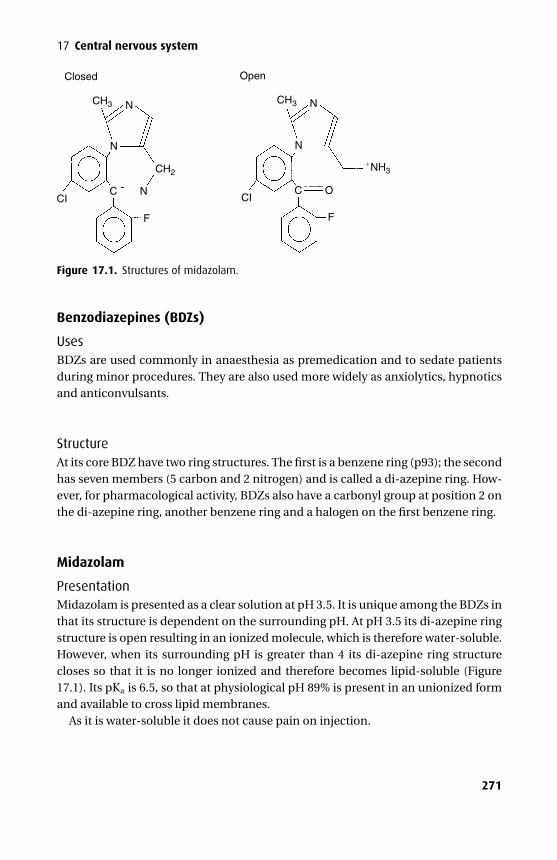
P1: PSB Printer: Yet To Come
9780521704632c17 CUFX213A/Peck 9780521618168 December 28, 2007 13:44
17 Central nervous system
Closed
CH3
CH2 NH3
CH3
N
N
N
N
C O
F
CCI CI
F
N
Open
Figure 17.1. Structures of midazolam.
Benzodiazepines (BDZs)
UsesBDZs are used commonly in anaesthesia as premedication and to sedate patientsduring minor procedures. They are also used more widely as anxiolytics, hypnoticsand anticonvulsants.
StructureAt its core BDZ have two ring structures. The first is a benzene ring (p93); the secondhas seven members (5 carbon and 2 nitrogen) and is called a di-azepine ring. How-ever, for pharmacological activity, BDZs also have a carbonyl group at position 2 onthe di-azepine ring, another benzene ring and a halogen on the first benzene ring.
Midazolam
PresentationMidazolam is presented as a clear solution at pH 3.5. It is unique among the BDZs inthat its structure is dependent on the surrounding pH. At pH 3.5 its di-azepine ringstructure is open resulting in an ionized molecule, which is therefore water-soluble.However, when its surrounding pH is greater than 4 its di-azepine ring structurecloses so that it is no longer ionized and therefore becomes lipid-soluble (Figure17.1). Its pKa is 6.5, so that at physiological pH 89% is present in an unionized formand available to cross lipid membranes.
As it is water-soluble it does not cause pain on injection.
271
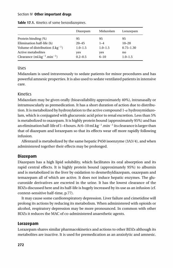
P1: PSB Printer: Yet To Come
9780521704632c17 CUFX213A/Peck 9780521618168 December 28, 2007 13:44
Section IV Other important drugs
Table 17.1. Kinetics of some benzodiazepines.
Diazepam Midazolam Lorazepam
Protein binding (%) 95 95 95Elimination half-life (h) 20–45 1–4 10–20Volume of distribution (l.kg−1) 1.0–1.5 1.0–1.5 0.75–1.30Active metabolites yes yes noClearance (ml.kg−1.min−1) 0.2–0.5 6–10 1.0–1.5
UsesMidazolam is used intravenously to sedate patients for minor procedures and haspowerful amnesic properties. It is also used to sedate ventilated patients in intensivecare.
KineticsMidazolam may be given orally (bioavailability approximately 40%), intranasally orintramuscularly as premedication. It has a short duration of action due to distribu-tion. It is metabolized by hydroxylation to the active compound 1-α hydroxymidazo-lam, which is conjugated with glucuronic acid prior to renal excretion. Less than 5%is metabolized to oxazepam. It is highly protein bound (approximately 95%) and hasan elimination half-life of 1–4 hours. At 6–10 ml.kg−1.min−1 its clearance is larger thanthat of diazepam and lorazepam so that its effects wear off more rapidly followinginfusion.
Alfentanil is metabolized by the same hepatic P450 isoenzyme (3A3/4), and whenadministered together their effects may be prolonged.
DiazepamDiazepam has a high lipid solubility, which facilitates its oral absorption and itsrapid central effects. It is highly protein bound (approximately 95%) to albuminand is metabolized in the liver by oxidation to desmethyldiazepam, oxazepam andtemazepam all of which are active. It does not induce hepatic enzymes. The glu-curonide derivatives are excreted in the urine. It has the lowest clearance of theBDZs discussed here and its half-life is hugely increased by its use as an infusion (cf.context-sensitive half-time, p.77).
It may cause some cardiorespiratory depression. Liver failure and cimetidine willprolong its actions by reducing its metabolism. When administered with opioids oralcohol, respiratory depression may be more pronounced. In common with otherBDZs it reduces the MAC of co-administered anaesthetic agents.
LorazepamLorazepam shares similar pharmacokinetics and actions to other BDZs although itsmetabolites are inactive. It is used for premedication as an anxiolytic and amnesic.
272
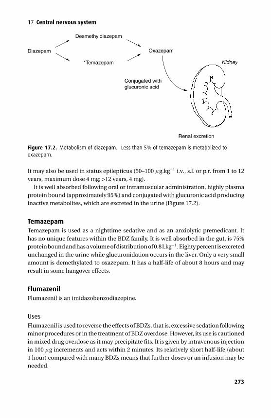
P1: PSB Printer: Yet To Come
9780521704632c17 CUFX213A/Peck 9780521618168 December 28, 2007 13:44
17 Central nervous system
Diazepam
*Temazepam
Conjugated withglucuronic acid
Renal excretion
Kidney
Oxazepam
Desmethyldiazepam
Figure 17.2. Metabolism of diazepam. ∗Less than 5% of temazepam is metabolized tooxazepam.
It may also be used in status epilepticus (50–100 µg.kg−1 i.v., s.l. or p.r. from 1 to 12years, maximum dose 4 mg; >12 years, 4 mg).
It is well absorbed following oral or intramuscular administration, highly plasmaprotein bound (approximately 95%) and conjugated with glucuronic acid producinginactive metabolites, which are excreted in the urine (Figure 17.2).
TemazepamTemazepam is used as a nighttime sedative and as an anxiolytic premedicant. Ithas no unique features within the BDZ family. It is well absorbed in the gut, is 75%protein bound and has a volume of distribution of 0.8 l.kg−1. Eighty percent is excretedunchanged in the urine while glucuronidation occurs in the liver. Only a very smallamount is demethylated to oxazepam. It has a half-life of about 8 hours and mayresult in some hangover effects.
FlumazenilFlumazenil is an imidazobenzodiazepine.
UsesFlumazenil is used to reverse the effects of BDZs, that is, excessive sedation followingminor procedures or in the treatment of BDZ overdose. However, its use is cautionedin mixed drug overdose as it may precipitate fits. It is given by intravenous injectionin 100 µg increments and acts within 2 minutes. Its relatively short half-life (about1 hour) compared with many BDZs means that further doses or an infusion may beneeded.
273
P1: PSB Printer: Yet To Come
9780521704632c17 CUFX213A/Peck 9780521618168 December 28, 2007 13:44
Section IV Other important drugs
Mechanism of actionFlumazenil is a competitive BDZ antagonist. However, it has some agonist activityas well and its ability to precipitate seizures in certain patients may be a result ofinverse agonist activity.
EffectsThese include nausea and vomiting. It may also precipitate anxiety, agitation andseizures especially in epileptic patients.
KineticsFlumazenil is 50% plasma protein bound and undergoes significant hepaticmetabolism to inactive compounds that are excreted in the urine.
Non-benzodiazepine hypnotics (the Z drugs)Zopiclone, zaleplon and zolpidem are hypnotics that act via the BDZ receptor butcannot be classified as benzodiazepines structurally. They were developed in orderto try to overcome some of the side effects of the BDZs, namely dependence andnext day sedation.
Zopiclone has the longest elimination half-life of approximately 5 hours whilezaleplon and zolpidem have elimination half-lives of 1 and 2 hours, respectively.
They have not had a major impact in the area of short-term hypnotics as they carryall the same side effects as the BDZs. Only where there has been a specific intoleranceto a specific BDZ is switching to a Z drug recommended. Where general intoleranceis a problem the patient is just as likely to expereince intolerance to the Z drugs.
AntidepressantsFour groups of drugs are used to treat depression:� Tricyclics� Selective serotonin re-uptake inhibitors� Monoamine oxidase inhibitors� Atypical agents
Tricyclics – TCAs (amitriptyline, nortriptyline, imipramine, dothiepin)As its name suggests, this group of drugs was originally based on a tricyclic ringstructure, although now many second-generation drugs contain different numbersof rings.
UsesTCAs are used to treat depressive illness, nocturnal enuresis and as an adjunct in thetreatment of chronic pain.
274
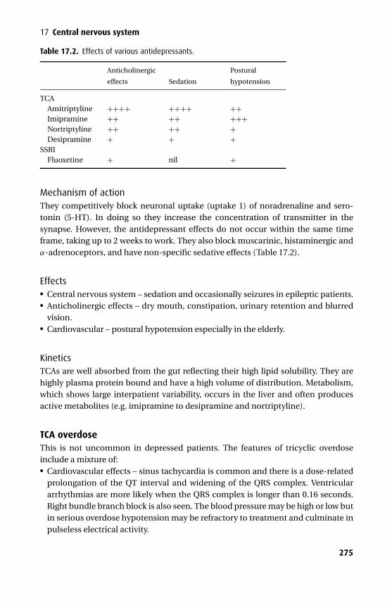
P1: PSB Printer: Yet To Come
9780521704632c17 CUFX213A/Peck 9780521618168 December 28, 2007 13:44
17 Central nervous system
Table 17.2. Effects of various antidepressants.
Anticholinergic
effects Sedation
Postural
hypotension
TCAAmitriptyline ++++ ++++ ++Imipramine ++ ++ +++Nortriptyline ++ ++ +Desipramine + + +
SSRIFluoxetine + nil +
Mechanism of actionThey competitively block neuronal uptake (uptake 1) of noradrenaline and sero-tonin (5-HT). In doing so they increase the concentration of transmitter in thesynapse. However, the antidepressant effects do not occur within the same timeframe, taking up to 2 weeks to work. They also block muscarinic, histaminergic andα-adrenoceptors, and have non-specific sedative effects (Table 17.2).
Effects� Central nervous system – sedation and occasionally seizures in epileptic patients.� Anticholinergic effects – dry mouth, constipation, urinary retention and blurredvision.� Cardiovascular – postural hypotension especially in the elderly.
KineticsTCAs are well absorbed from the gut reflecting their high lipid solubility. They arehighly plasma protein bound and have a high volume of distribution. Metabolism,which shows large interpatient variability, occurs in the liver and often producesactive metabolites (e.g. imipramine to desipramine and nortriptyline).
TCA overdoseThis is not uncommon in depressed patients. The features of tricyclic overdoseinclude a mixture of:� Cardiovascular effects – sinus tachycardia is common and there is a dose-related
prolongation of the QT interval and widening of the QRS complex. Ventriculararrhythmias are more likely when the QRS complex is longer than 0.16 seconds.Right bundle branch block is also seen. The blood pressure may be high or low butin serious overdose hypotension may be refractory to treatment and culminate inpulseless electrical activity.
275

P1: PSB Printer: Yet To Come
9780521704632c17 CUFX213A/Peck 9780521618168 December 28, 2007 13:44
Section IV Other important drugs� Central effects – excitation, seizures (correlating with a QRS duration of more than0.1 seconds) and then depression. Mydriasis is a feature, as is hyperthermia.� Anticholinergic effects.
TreatmentThis includes gastric lavage followed by activated charcoal. Supportive care mayrequire supplementation with specific treatment. Seizures may be treated with ben-zodiazepines or phenytoin and ventricular arrhythmias with phenytoin or lidocaine.Inotropes should be avoided where possible as this may precipitate arrhythmias.Intravascular volume expansion is usually sufficient to correct hypotension. Theanticholinergic effects may be reversed by an anticholinesterase, but this is not rec-ommended as it may precipitate seizures, bradycardia and heart failure.
Selective serotonin re-uptake inhibitors – SSRIs (fluoxetine, paroxetine,sertraline, venlafaxine)As their name suggests, SSRIs selectively inhibit the neuronal re-uptake of 5-HT. Theyare no more effective than standard antidepressants but do not have their associatedside-effect profile. SSRIs are less sedative, have fewer anticholinergic effects andappear less cardiotoxic in overdose although they are associated with gastrointestinalside-effects (nausea and constipation).
Despite their side-effect profile, when combinations of serotonergic drugs areused the potentially fatal serotonergic syndrome may result, which is characterizedby hyper-reflexia, agitation, clonus and hyperthermia. The commonest combinationis an MAOI and SSRI – however, the phenylpiperidine opioids (particularly pethi-dine) have weak serotonin reuptake inhibitor properties and can also precipitate thesyndrome.
Fluoxetine is an effective antidepressant causing minimal sedation. It is a 50:50mix of two isomers that are equally active. It is well absorbed and metabolized in theliver by cytochrome P450 enzymes. In addition, there are non-saturable enzymesthat prevent an unchecked rise in levels. However, the dose should be reduced inrenal failure as accumulation may result. Side effects include nausea and vomiting,headache, insomnia, reduced libido and mania or hypomania in up to 1%.
Venlafaxine appears to block the re-uptake of both noradrenaline and 5-HT (andto a lesser extent dopamine) while having little effect on muscarinic, histaminergicor α-adrenoceptors.
Monoamine oxidase inhibitors – MAOIsThis group of drugs is administered orally for the treatment of resistant depression,obsessive compulsive disorders, chronic pain syndromes and migraine.
MAO is present as a variety of isoenzymes within presynaptic neurones and isresponsible for the deamination of amine neurotransmitters. They have been clas-sified as types A and B. Following their inhibition there is an increase in the level of
276
P1: PSB Printer: Yet To Come
9780521704632c17 CUFX213A/Peck 9780521618168 December 28, 2007 13:44
17 Central nervous system
amine neurotransmitters, which is thought to be the basis of their central activity.MAO-A preferentially deaminates 5-HT and catecholamines, while MAO-B prefer-entially deaminates tyramine and phenylethamine.
There are now two generations of MAOIs. The original generation inhibit MAOirreversibly and non-selectively (i.e. MAO-A and -B) while the new generation selec-tively and reversibly inhibit only MAO-A (RIMA). Neither group is used as first linetherapy because of the potential for serious side effects and hepatic toxicity.
Non-selective irreversible MAOIs (phenelzine,isocarboxazid, tranylcypromine)Phenelzine and isocarboxazid are hydrazines while tranylcypromine is a non-hydrazine compound. Tranylcypromine is potentially the most dangerous as it pos-sesses stimulant activity.
EffectsIn addition to controlling depression they also produce sedation, blurred vision,orthostatic hypotension and hypertensive crises following tyramine rich foods(cheese, pickled herring, chicken liver, Bovril and chocolate) and indirectly actingsympathomimetics. Hepatic enzymes are inhibited and the hydrazine compoundsmay cause hepatotoxicity. Interaction with pethidine may precipitate cerebral irri-tability, hyperpyrexia and cardiovascular instability. Interaction with fentanyl hasalso been reported.
Selective reversible MAOIs – RIMA (moclobemide)Moclobemide causes less potentiation of tyramine than the older generation MAOIsand in general patients do not need the same level of dietary restriction. How-ever, some patients are especially sensitive to tyramine and so all patients shouldbe advised to avoid tyramine-rich foods and indirect-acting sympathomimeticamines.
It is completely absorbed from the gut but undergoes significant first-passmetabolism resulting in an oral bioavailability of 60–80%. It is metabolized in theliver by cytochrome P450 and up to 2% of the Caucasian and 15% of the Asian pop-ulation have been shown to be slow metabolizers. The metabolites are excreted inthe urine.
Linezolid is a new antibiotic indicated for methacillin-resistant Staphlococcusaureus and vancomycin-resistant enetrococci. It is also a MAOI and as such has thetypical range of cautions and contraindications.
MAOIs and general anaesthesiaThose patients taking MAOIs and presenting for emergency surgery should not begiven pethidine or any indirectly acting sympathomimetic amines (e.g. ephedrine).If cardiovascular support is indicated, direct-acting agents should be used but
277
P1: PSB Printer: Yet To Come
9780521704632c17 CUFX213A/Peck 9780521618168 December 28, 2007 13:44
Section IV Other important drugs
with extreme caution as they may also precipitate exaggerated hypertension. Theelective case presents potential difficulties. If MAOI therapy is withdrawn for therequired 14–21 days before surgery, the patient may suffer a relapse of their depres-sion with potentially disastrous consequences. However, the newer agents may con-trol depression more effectively and reduce the chance of a serious peri-operativedrug interaction.
Indirectly acting sympathomimetic amines are heavily dependent on MAO fortheir metabolism and therefore may produce exaggerated hypertension and arrhyth-mias when administered with an MAOI. The directly acting sympathomimeticamines should also be used with caution although they are also metabolized byCOMT and therefore are not subject to the same degree of exaggerated response.
MAOIs should be stopped for 2 weeks before starting alternative antidepressanttherapy and 2 weeks should have elapsed from the end of tricyclic therapy to thestart of MAOI therapy.
Atypical agents
MianserinMianserin is a tetracyclic compound used in depressive illness, especially wheresedation is required. It does not block the neuronal re-uptake of transmitters incontrast to the TCAs. It does, however, block presynaptic α2-adrenoceptors, whichreduces their negative feedback, resulting in increased synaptic concentrations ofneurotransmitters.
It has very little ability to block muscarinic and peripheral α-adrenoceptors andas such causes less in the way of antimuscarinic effects or postural hypotension.
Its important side effects are agranulocytosis and aplastic anaemia, which aremore common in the elderly.
Lithium carbonateLithium is used in the treatment of bipolar depression, mania and recurrent affectivedisorders. It has a narrow therapeutic index and plasma levels should be maintainedat 0.5–1.5 mmol.l−1. In excitable cells, lithium imitates Na+ and decreases the releaseof neurotransmitters.
It may increase generalized muscle tone and lower the seizure threshold in epilep-tics. Many patients develop polyuria and polydipsia due to antidiuretic hormoneantagonism. It may also produce raised serum levels of Na+, Mg2+ and Ca2+. Lithiumprolongs neuromuscular blockade and may decrease anaesthetic requirements as itblocks brain stem release of noradrenaline and dopamine.
The thyroid gland may become enlarged and underactive and the patient mayexperience weight gain and tremor. Above the therapeutic level patients suffer withvomiting, abdominal pain, ataxia, convulsions, arrhythmias and death.
278
P1: PSB Printer: Yet To Come
9780521704632c17 CUFX213A/Peck 9780521618168 December 28, 2007 13:44
17 Central nervous system
AnticonvulsantsWhere possible a single agent should be used to treat epilepsy as it avoids the potentialfor drug interaction. In addition patients rarely improve with a second agent.
Phenytoin
UsesPhenytoin has been used widely for many years in the treatment of grand mal and par-tial seizures, trigeminal neuralgia and ventricular arrhythmias following TCA over-dose. It may be given orally or intravenously but the dose must be tailored to theindividual patient as wide interpatient variation exists (about 9% of the populationare slow hydroxylators) and blood assays are useful in this regard. It is incompatiblewith 5% dextrose, in which it becomes gelatinous. The normal therapeutic level is10–20 µg.ml−1.
Mechanism of actionThe action of phenytoin is probably dependent on its ability to bind to and stabilizeinactivated Na+ channels. This prevents the further generation of action potentialsthat are central to seizure activity. It may also reduce Ca2+ entry into neurones,blocking transmitter release and enhancing the actions of GABA.
Effects� Idiosyncratic – acne, coarsening of facial features, hirsutism, gum hyperplasia,folate-dependent megaloblastic anaemia, aplastic anaemia, various skin rashesand peripheral neuropathy.� Dose-related – ataxia, nystagmus, parasthesia, vertigo and slurred speech. Rapidundiluted intravenous administration is associated with hypotension and heartblock.� Teratogenicity – it causes craniofacial abnormalities, growth retardation, limb andcardiac defects and mental retardation.� Drug interactions – as phenytoin induces the hepatic mixed function oxidases itincreases the metabolism of warfarin, BDZs and the oral contraceptive pill. Itsmetabolism may be inhibited by metronidazole, chloramphenicol and isoniazidleading to toxic levels. Furthermore phenytoin’s metabolism may be induced bycarbamazepine or alcohol resulting in reduced plasma levels.
KineticsThe oral bioavailability is approximately 90% and it is highly plasma protein bound(90%). It undergoes saturable hepatic hydroxylation resulting in zero-order kineticsjust above the therapeutic range. It can induce its own metabolism and that of otherdrugs. Its major metabolite is excreted in the urine.
279
P1: PSB Printer: Yet To Come
9780521704632c17 CUFX213A/Peck 9780521618168 December 28, 2007 13:44
Section IV Other important drugs
CarbamazepineCarbamazepine is also used in the treatment of trigeminal neuralgia. Its mode ofaction is similar to that of phenytoin. It may be given orally or rectally.
Effects� Central nervous system – mild neurotoxic effects including headache, diplopia,ataxia, vomiting and drowsiness are common and often limit its use.� Metabolic – it may produce an antidiuretic effect leading to water retention.� Miscellaneous – drug-induced hepatitis, rashes in 5–10% and rarely agranulocyto-sis.� Teratogenicity – it causes facial abnormalities, intrauterine growth retardation,microcephaly and mental retardation. The incidence increases with dose. Overallincidence is about 1/300–2000 live births.� Drug interactions – as carbamazepine induces hepatic enzymes, it demonstratesmany of the interactions seen with phenytoin. Levels of concurrently administeredphenytoin may be elevated or reduced. Erythromycin can increase serum levels ofcarbamazepine.
KineticsCarbamazepine is well absorbed from the gut with a high oral bioavailability.It is approximately 75% plasma protein bound and undergoes extensive hepaticmetabolism to carbamazepine 10,11-epoxide, which retains about 30% of carba-mazepine’s anticonvulsant properties. It powerfully induces hepatic enzymes andinduces its own metabolism. Its excretion is almost entirely in the urine as unconju-gated metabolites.
Sodium valproate
UsesSodium valproate is used in the treatment of various forms of epilepsy includingabsence (petit mal) seizures and in the treatment of trigeminal neuralgia.
Mechanism of actionSodium valproate appears to act by stabilizing inactive Na+ channels and also bystimulating central GABA-ergic inhibitory pathways. It is generally well tolerated.
Effects� Abdominal – it may cause nausea and gastric irritation. Pancreatitis and potentiallyfatal hepatotoxicity are recognized following its use.� Haematological – thrombocytopenia and reduced platelet aggregation.� Miscellaneous – transient hair loss.� Teratogenicity – it causes neural tube defects.
280
P1: PSB Printer: Yet To Come
9780521704632c17 CUFX213A/Peck 9780521618168 December 28, 2007 13:44
17 Central nervous system
KineticsSodium valproate is well absorbed orally, highly protein bound (approximately 90%)and undergoes hepatic metabolism to products (some of which are active), whichare excreted in the urine.
Phenobarbitone is an effective anticonvulsant but its use is associated with signif-icant sedation, which limits its use. It is a long-acting barbiturate that induces hepaticenzymes and interacts with other agents (warfarin, oral contraceptives, other anti-convulsants).
BDZs (see above) are widely used in the emergency treatment of status epilepticusand act by enhancing the chloride-gating function of GABA.
Other agentsVigabatrin, gabapentin, lamotrigine and pregabalin are newer agents that are usedin the control of persistent partial seizures.
Vigabatrin irreversibly inhibits GABA-transaminase, the enzyme responsible forthe breakdown of GABA and therefore its duration of action is about 24 hours whileits elimination half-life is only 6 hours. It is excreted unchanged in the urine. Itinteracts with phenytoin reducing its concentration by about 25% by an unknownmechanism. It may cause sedation, fatigue, headache, agitation and depression.
Lamotrigine acts on the presynaptic neuronal membrane and stabilizes the inac-tive Na+ channel leading to a reduction in excitatory neurotransmitter release. Itundergoes hepatic metabolism to an inactive conjugate. Its rate of metabolism isincreased by other enzyme-inducing drugs (carbamazepine and phenytoin) butreduced by sodium valproate. It may precipitate headache, nausea, diplopia, ataxiaand tremor. Approximately 0.1% of adults develop Stevens–Johnson or Lyell’s syn-drome. The incidence is higher in children.
Gabapentin’s mode of action is uncertain but it may bind to Ca2+ channels withinthe brain. It is not plasma protein bound and has a terminal elimination half-life of5–7 hours. It is excreted unchanged in the urine and does not interfere with otheranticonvulsants as it does not affect hepatic enzyme systems. It is well tolerated.Gabapentin also has a role to play in the management of chronic pain.
Pregabalin is a potent ligand for the alpha-2-delta subunit of voltage-gated cal-cium channels in the central nervous system. Once bound there is a reduction incalcium influx leading to a reduction of excitatory neurotransmitter release. It is astructural analogue of gamma-aminobutyric acid (GABA – although it bears no phar-macological resemblance to GABA) and is prepared as the S-enantiomer. Its kineticsare predictable and its oral bioavailability is >90% and it readily crosses the BBB. Itsterminal elimination half-life is 6 hours. In a manner similar to gabapentin it is notmetabolized so that it does no interact with other anticonvulsants. It is excreted inthe urine unchanged.
281
P1: PSB Printer: Yet To Come
9780521704632c18 CUFX213A/Peck 9780521618168 December 28, 2007 13:51
18Antiemetics and related drugs
Nausea and vomiting has many causes including drugs, motion sickness, fear, preg-nancy, vestibular disease and migraine. In previous decades anaesthesia was almostsynonymous with vomiting, but with the advent of new anaesthetic agents and moreaggressive treatment the incidence of vomiting has decreased. However, even thelatest agents have failed to eradicate this troublesome symptom encountered in theperi-operative period.
PhysiologyThe vomiting centre (VC) coordinates vomiting. It has no discrete anatomical sitebut may be considered as a collection of effector neurones situated in the medulla.This collection projects to the vagus and phrenic nerves and also to the spinal motorneurones supplying the abdominal muscles, which when acting together bring aboutthe vomiting reflex.
The VC has important input from the chemoreceptor trigger zone (CTZ), which liesin the area postrema on the floor of the fourth ventricle but is functionally outside theblood–brain barrier. The CTZ is rich in dopamine (D2) receptors and also serotonin(5-HT) receptors. Acetylcholine (ACh) is important in neural transmission from thevestibular apparatus. Other input is summarized in Figure 18.1.
The treatment of nausea and vomiting is aimed at reducing the afferent sup-ply to the VC. While the administration of antiemetics forms a vital part of treat-ment, attention should also be given to minimizing the administration of opioids bythe use of non-steroidal anti-inflammatory drugs and avoiding unnecessary anti-cholinesterase administration. When propofol is used to maintain anaesthesia forminor surgery, where the use of opioids is limited, it may reduce the incidence ofpost-operative nausea and vomiting (PONV).
The following types of agents have been used:� Dopamine antagonists� Anticholinergics� Antihistamines� 5-HT3 antagonists� Miscellaneous
282
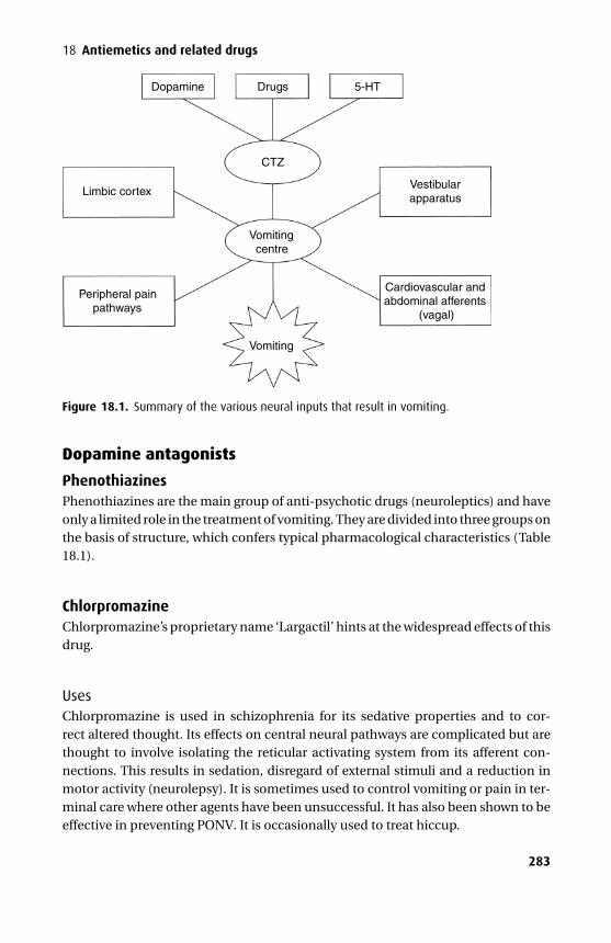
P1: PSB Printer: Yet To Come
9780521704632c18 CUFX213A/Peck 9780521618168 December 28, 2007 13:51
18 Antiemetics and related drugs
Vomitingcentre
CTZ
Vomiting
Dopamine
Limbic cortex
Peripheral painpathways
Drugs 5-HT
Vestibularapparatus
Cardiovascular andabdominal afferents
(vagal)
Figure 18.1. Summary of the various neural inputs that result in vomiting.
Dopamine antagonists
PhenothiazinesPhenothiazines are the main group of anti-psychotic drugs (neuroleptics) and haveonly a limited role in the treatment of vomiting. They are divided into three groups onthe basis of structure, which confers typical pharmacological characteristics (Table18.1).
ChlorpromazineChlorpromazine’s proprietary name ‘Largactil’ hints at the widespread effects of thisdrug.
UsesChlorpromazine is used in schizophrenia for its sedative properties and to cor-rect altered thought. Its effects on central neural pathways are complicated but arethought to involve isolating the reticular activating system from its afferent con-nections. This results in sedation, disregard of external stimuli and a reduction inmotor activity (neurolepsy). It is sometimes used to control vomiting or pain in ter-minal care where other agents have been unsuccessful. It has also been shown to beeffective in preventing PONV. It is occasionally used to treat hiccup.
283
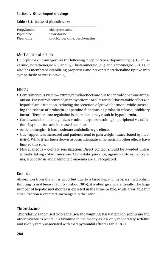
P1: PSB Printer: Yet To Come
9780521704632c18 CUFX213A/Peck 9780521618168 December 28, 2007 13:51
Section IV Other important drugs
Table 18.1. Groups of phenothiazines.
Propylamine chlorpromazinePiperidine thioridazinePiperazine prochlorperazine, perphenazine
Mechanism of actionChlorpromazine antagonizes the following receptor types: dopaminergic (D2), mus-carinic, noradrenergic (α1 and α2), histaminergic (H1) and serotinergic (5-HT). Italso has membrane-stabilizing properties and prevents noradrenaline uptake intosympathetic nerves (uptake 1).
Effects� Central nervous system – extrapyramidal effects are due to central dopamine antag-onism. The neuroleptic malignant syndrome occurs rarely. It has variable effects onhypothalamic function, reducing the secretion of growth hormone while increas-ing the release of prolactin (dopamine functions as prolactin release inhibitoryfactor). Temperature regulation is altered and may result in hypothermia.� Cardiovascular – it antagonizes α-adrenoceptors resulting in peripheral vasodila-tion, hypotension and increased heat loss.� Anticholinergic – it has moderate anticholinergic effects.� Gut – appetite is increased and patients tend to gain weight (exacerbated by inac-tivity). While it has been shown to be an adequate antiemetic, its other effects havelimited this role.� Miscellaneous – contact sensitization. Direct contact should be avoided unlessactually taking chlorpromazine. Cholestatic jaundice, agranulocytosis, leucope-nia, leucocytosis and haemolytic anaemia are all recognized.
KineticsAbsorption from the gut is good but due to a large hepatic first-pass metabolism(limiting its oral bioavailability to about 30%), it is often given parenterally. The largenumber of hepatic metabolites is excreted in the urine or bile, while a variable butsmall fraction is excreted unchanged in the urine.
ThioridazineThioridazine is not used to treat nausea and vomiting. It is used in schizophrenia andother psychoses where it is favoured in the elderly as it is only moderately sedativeand is only rarely associated with extrapyramidal effects (Table 18.2).
284
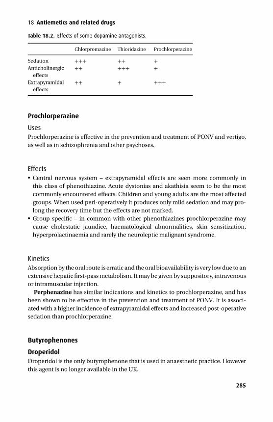
P1: PSB Printer: Yet To Come
9780521704632c18 CUFX213A/Peck 9780521618168 December 28, 2007 13:51
18 Antiemetics and related drugs
Table 18.2. Effects of some dopamine antagonists.
Chlorpromazine Thioridazine Prochlorperazine
Sedation +++ ++ +Anticholinergic
effects++ +++ +
Extrapyramidaleffects
++ + +++
Prochlorperazine
UsesProchlorperazine is effective in the prevention and treatment of PONV and vertigo,as well as in schizophrenia and other psychoses.
Effects� Central nervous system – extrapyramidal effects are seen more commonly inthis class of phenothiazine. Acute dystonias and akathisia seem to be the mostcommonly encountered effects. Children and young adults are the most affectedgroups. When used peri-operatively it produces only mild sedation and may pro-long the recovery time but the effects are not marked.� Group specific – in common with other phenothiazines prochlorperazine maycause cholestatic jaundice, haematological abnormalities, skin sensitization,hyperprolactinaemia and rarely the neuroleptic malignant syndrome.
KineticsAbsorption by the oral route is erratic and the oral bioavailability is very low due to anextensive hepatic first-pass metabolism. It may be given by suppository, intravenousor intramuscular injection.
Perphenazine has similar indications and kinetics to prochlorperazine, and hasbeen shown to be effective in the prevention and treatment of PONV. It is associ-ated with a higher incidence of extrapyramidal effects and increased post-operativesedation than prochlorperazine.
Butyrophenones
DroperidolDroperidol is the only butyrophenone that is used in anaesthetic practice. Howeverthis agent is no longer available in the UK.
285
P1: PSB Printer: Yet To Come
9780521704632c18 CUFX213A/Peck 9780521618168 December 28, 2007 13:51
Section IV Other important drugs
UsesDroperidol has been shown to be effective in the prevention and treatment of PONVat doses from 0.25 to 5 mg, although the incidence of side effects increases with dose.It is also used in neurolept analgesia and in the control of mania.
Mechanism of actionDroperidol antagonizes central dopamine (D2) receptors at the CTZ.
EffectsThese are similar to those seen with phenothiazines.� Central nervous system – sedation is more pronounced compared to the phenoth-
iazines. The true incidence of extrapyramidal effects is unknown but increases withhigher doses. They may develop more than 12 hours after administration and upto 25% of patients may experience anxiety up to 48 hours after administration. Insufficient dose it induces neurolepsis.� Metabolic – it may cause hyperprolactinaemia.� Cardiovascular – hypotension resulting from peripheral α-adrenoceptor blockademay occur.
KineticsDroperidol is usually given intravenously although it is absorbed readily after intra-muscular injection. It is highly plasma protein bound (approximately 90%) andextensively metabolized in the liver to products that are excreted in the urine, only1% as unchanged drug.
DomperidoneThis D2 antagonist is less likely to cause extrapyramidal effects as it does not cross theblood–brain barrier. Its use in children is limited to nausea and vomiting followingchemotherapy or radiotherapy. It also increases prolactin levels and may cause galac-torrhoea and gynaecomastia. The intravenous preparation was withdrawn followingserious arrhythmias during the administration of large doses. It is only available astablets or suppositories.
Benzamides
Metoclopramide
UsesMetoclopramide is used as an antiemetic and a prokinetic. Approximately half of theclinical studies have demonstrated placebo to be as effective as metoclopramide asan antiemetic. However, metoclopramide appears to be most effective when 20 mgis given at the end of anaesthesia rather than at induction.
286
P1: PSB Printer: Yet To Come
9780521704632c18 CUFX213A/Peck 9780521618168 December 28, 2007 13:51
18 Antiemetics and related drugs
Mechanism of actionMetoclopramide exerts its antiemetic actions primarily through dopamine (D2)receptor antagonism at the CTZ, although it does have prokinetic effects on thestomach (cf. Chapter 19). It also blocks 5-HT3 receptors, which may account forsome of its antiemetic properties.
Effects� Central nervous system – metoclopramide crosses the blood–brain barrier andmay precipitate extrapyramidal effects up to 72 hours after administration. Sucheffects are more common in young females (1 in 5000). Rarely it may precipitatethe neuroleptic malignant syndrome. Sedation is seen more commonly duringlong-term administration. Agitation is occasionally seen following intramuscularpremedication with 10–20 mg.� Cardiovascular – hypotension, tachy- and bradycardias have been reported fol-lowing rapid intravenous administration.
KineticsMetoclopramide is well absorbed from the gut although first-pass metabolism variessignificantly producing a wide range in oral bioavailability (30–90%). It may be givenintravenously. It is conjugated in the liver and excreted along with unchanged drugin the urine.
AnticholinergicsWhile so-called ‘anticholinergic’ agents are effective antagonists at muscarinic recep-tors, they have very little activity at nicotinic receptors and may therefore be thoughtof as essentially selective agents at normal doses.
The naturally occurring tertiary amines, atropine and hyoscine, are esters formedby the combination of tropic acid and an organic base (tropine or scopine) and areable to cross the blood–brain barrier. Their central effects include sedation, amnesia,anti-emesis and the central anticholinergic syndrome. Glycopyrrolate is a syntheticquaternary amine (therefore charged) with no central effects as it is unable to crossthe blood–brain barrier.
HyoscineHyoscine is a racemic mixture, but only l-hyoscine is active.
UsesHyoscine has traditionally been given with an intramuscular opioid as premedica-tion, and in this setting has been shown to reduce PONV. It has also been used as asedative and amnesic agent.
287
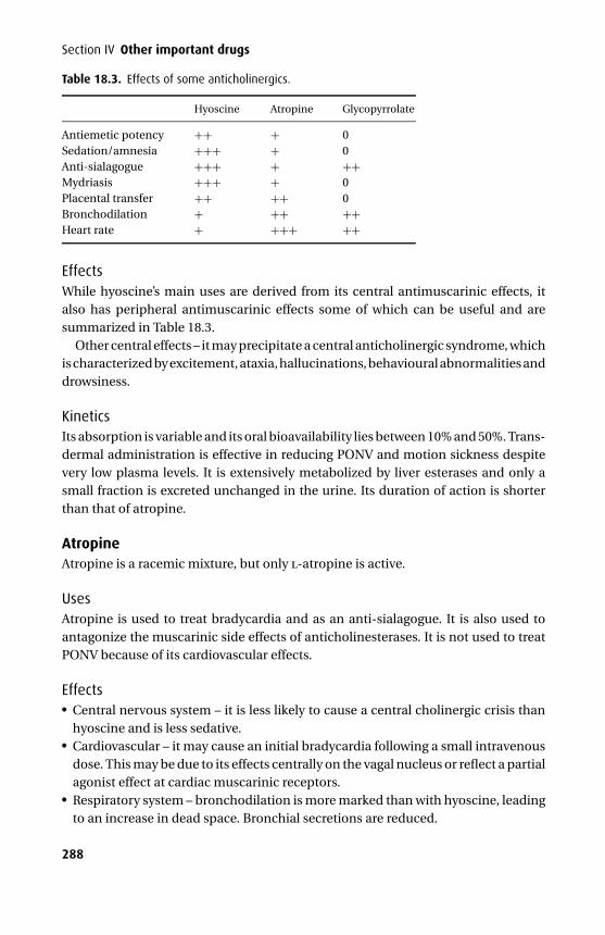
P1: PSB Printer: Yet To Come
9780521704632c18 CUFX213A/Peck 9780521618168 December 28, 2007 13:51
Section IV Other important drugs
Table 18.3. Effects of some anticholinergics.
Hyoscine Atropine Glycopyrrolate
Antiemetic potency ++ + 0Sedation/amnesia +++ + 0Anti-sialagogue +++ + ++Mydriasis +++ + 0Placental transfer ++ ++ 0Bronchodilation + ++ ++Heart rate + +++ ++
EffectsWhile hyoscine’s main uses are derived from its central antimuscarinic effects, italso has peripheral antimuscarinic effects some of which can be useful and aresummarized in Table 18.3.
Other central effects – it may precipitate a central anticholinergic syndrome, whichis characterized by excitement, ataxia, hallucinations, behavioural abnormalities anddrowsiness.
KineticsIts absorption is variable and its oral bioavailability lies between 10% and 50%. Trans-dermal administration is effective in reducing PONV and motion sickness despitevery low plasma levels. It is extensively metabolized by liver esterases and only asmall fraction is excreted unchanged in the urine. Its duration of action is shorterthan that of atropine.
AtropineAtropine is a racemic mixture, but only l-atropine is active.
UsesAtropine is used to treat bradycardia and as an anti-sialagogue. It is also used toantagonize the muscarinic side effects of anticholinesterases. It is not used to treatPONV because of its cardiovascular effects.
Effects� Central nervous system – it is less likely to cause a central cholinergic crisis thanhyoscine and is less sedative.� Cardiovascular – it may cause an initial bradycardia following a small intravenousdose. This may be due to its effects centrally on the vagal nucleus or reflect a partialagonist effect at cardiac muscarinic receptors.� Respiratory system – bronchodilation is more marked than with hyoscine, leadingto an increase in dead space. Bronchial secretions are reduced.
288
P1: PSB Printer: Yet To Come
9780521704632c18 CUFX213A/Peck 9780521618168 December 28, 2007 13:51
18 Antiemetics and related drugs� Gut – it is a less effective anti-sialagogue than hyoscine. The tone of the loweroesophageal sphincter is decreased and there is a small decrease in gastric acidsecretion.� Miscellaneous – sweating is inhibited and this may provoke a pyrexia in paedi-atric patients. When administered topically it may increase intra-occular pressure,which may be critical for patients with glaucoma.
KineticsIntestinal absorption is rapid but unpredictable. It is 50% plasma protein boundand extensively metabolized by liver esterases. It is excreted in the urine, only a tinyfraction unchanged.
GlycopyrrolateGylcopyrrolate is indicated for anti-sialagogue premedication, the treatment ofbradycardias and to protect against the unwanted effects of anticholinesterases.Its charged quaternary structure gives it a different set of characteristics comparedto the tertiary amines. Intestinal absorption is negligible and the oral bioavailabilityis consequently less than 5%. It does not cross the blood–brain barrier and so it isdevoid of central effects. It is minimally metabolized and 80% is excreted in the urineunchanged.
Antihistamines
CyclizineCyclizine is a piperazine derivative. The parenteral preparation is prepared withlactic acid at pH 3.2. Consequently intramuscular and intravenous injection may beparticularly painful.
UsesCyclizine is used as an antiemetic in motion sickness, radiotherapy, PONV and emesisinduced by opioids. It is also used to control the symptoms of Meniere’s disease.
Mechanism of actionCyclizine is a histamine (H1) antagonist, but also has anticholinergic properties thatmay contribute significantly to its antiemetic actions.
Effects� Gut – it increases lower oesophageal sphincter tone.� Anticholinergic – these are mild although it may cause an increase in heart ratefollowing intravenous injection.� Extrapyramidal effects and sedation do not complicate its use.
289
P1: PSB Printer: Yet To Come
9780521704632c18 CUFX213A/Peck 9780521618168 December 28, 2007 13:51
Section IV Other important drugs
KineticsCyclizine is well absorbed orally and has a high oral bioavailability (approximately75%). Surprisingly little is known regarding the rest of the kinetics of this drug.
PromethazinePromethazine has traditionally been used in combination with pethidine for intra-muscular premedication. However, it may also be used as an oral premed for children.It has significant anticholinergic properties and sedative effects. It is well absorbedfrom the gut but is subject to a significant first-pass hepatic metabolism so that itsoral bioavailability is 25%. It has a duration of action of 3–6 hours and its metabolitesare eliminated entirely in the urine.
5-HT3 antagonists
OndansetronOndansetron is a carbazole.
PresentationOndansetron is available as tablets (4–8 mg), a strawberry-flavoured lyophilisate(4–8 mg) to dissolve on the tongue, a suppository (16 mg) and as a clear solutioncontaining 2 mg.ml−1 for slow intravenous injection.
UsesOndansetron is indicated for the treatment of nausea and vomiting associated withchemo- or radiotherapy and in the peri-operative period. It is ineffective for vomitinginduced by motion sickness or dopamine agonists. It is licensed for children above2 years of age.
Mechanism of actionThe activation of 5-HT3 receptors peripherally and centrally appears to induce vom-iting. Chemo- and radiotherapy may cause the release of 5-HT from enterochro-maffin cells. Peripheral 5-HT3 receptors in the gut are then activated and stimulatevagal afferent neurones that connect to the VC, again via 5-HT3 receptors. Thusondansetron may antagonize 5-HT3 both peripherally and centrally.
EffectsOndansetron is well tolerated and its other effects are limited to headache, flushing,constipation and bradycardia following rapid intravenous administration.
KineticsOndansetron is well absorbed from the gut with an oral bioavailability of about60%. It is 75% protein bound and undergoes significant hepatic metabolism by
290
P1: PSB Printer: Yet To Come
9780521704632c18 CUFX213A/Peck 9780521618168 December 28, 2007 13:51
18 Antiemetics and related drugs
hydroxylation and subsequent glucuronide conjugation to inactive metabolites. Itshalf-life is 3 hours. The dose should be reduced in hepatic impairment.
While ondansetron clearly has a place in the treatment of nausea and vomitingit has not universally been shown to be superior to low-dose droperidol or a phe-nothiazine. This together with its higher cost suggests it should be used only whenconventional therapy is expected to or has failed.
Miscellaneous
SteroidsThe role of dexamethasone as an antiemetic has traditionally been confined tochemotherapy induced nausea and vomiting. However more recently it has beenused at a dose of 2.5–10 mg to prevent post-operative nausea and vomiting. Its modeof action in this area is uncertain.
AcupunctureSeveral studies have demonstrated the effectiveness of acupuncture in the preven-tion of PONV. The acupuncture point lies between the tendons of flexor carpi radialisand palmaris longus about 4 cm from the distal wrist skin crease. It should be per-formed on the awake patient and is free from side effects.
CanabinoidsNabilone acts at the VC and has been used as an antiemetic following chemotherapy.
BenzodiazepinesLorazepam is used as an antiemetic during chemotherapy. It has amnesic and seda-tive properties. Its mode of action as an antiemetic is uncertain but it may modifycentral connections to the VC and prevent the anticipatory nausea that is seen withrepeated doses of chemotherapy.
291
P1: PSB Printer: Yet To Come
9780521704632c19 CUFX213A/Peck 9780521618168 December 28, 2007 14:1
19Drugs acting on the gut
� Antacids� Drugs influencing gastric secretion� Drugs influencing gastric motility� Mucosal protectors� Prostaglandin analogues
AntacidsAntacids neutralize gastric acidity. They are used to relieve the symptoms of dyspep-sia and gastro-oesophageal reflux. They promote ulcer healing but less effectivelythan other therapies.
Aluminium and magnesium-containing antacidsNeither is absorbed from the gut significantly and due to their relatively lowwater solubility they are long-acting providing that they remain in the stomach.Aluminium-containing antacids have a slower action and produce constipation,while magnesium-containing antacids produce diarrhoea. Aluminium ions formcomplexes with some drugs (e.g. tetracycline) and reduce their absorption.
Sodium bicarbonate and sodium citrateThese antacids are water-soluble and their onset of action is faster than thealuminium- and magnesium-containing antacids. They are absorbed into the sys-temic circulation and may cause a metabolic alkalosis if taken in excess. Sodiumbicarbonate releases carbon dioxide as it reacts with gastric acid, resulting in belch-ing. Thirty milliliters of 0.3 m sodium citrate is often used with ranitidine to reducegastric acidity before caesarean section. It should be given less than 10 minutesbefore the start of surgery due to its limited duration of action.
Drugs influencing gastric secretion
PhysiologyGastrin and ACh stimulate parietal cells (via gastrin and muscarinic receptors) tosecrete H+ into the gastric lumen. ACh is released from parasympathetic postgan-glionic fibres while gastrin is released from G-cells in the antral mucosa. How-ever, the main stimulus for parietal cell acid secretion is via histamine receptor
292
P1: PSB Printer: Yet To Come
9780521704632c19 CUFX213A/Peck 9780521618168 December 28, 2007 14:1
19 Drugs acting on the gut
activation. Gastrin and ACh also stimulate the adjacent paracrine cells to produceand release histamine, which acts on the parietal cell, increasing cAMP and thereforeacid secretion.
H2 receptor antagonists
CimetidineCimetidine is the only H2 receptor antagonist with an imidazole structure.
UsesIt is used in peptic ulcer disease, reflux oesophagitis, Zollinger–Ellison syndrome andpre-operatively in those at risk of aspiration. It has not been shown to be of benefit inactive haematemesis, although it does have a prophylactic role in those with criticalillness.
Mechanism of actionCimetidine is a competitive and specific antagonist of H2 receptors at parietal cells.
Effects� Gut – the gastric pH is raised and the volume of secretions reduced, while there isno change in gastric emptying time or lower oesophageal sphincter tone.� Cardiovascular – bradycardia and hypotension follow rapid intravenous adminis-tration.� Central nervous system – confusion, hallucinations and seizures are usually onlyseen when impaired renal function leads to high plasma levels.� Respiratory system – low-grade aspiration of gastric content that has been strippedof its acidic, antibacterial environment will result in increased nosocomial pul-monary infections in critically ill ventilated patients.� Endocrine – gynaecomastia, impotence and a fall in sperm count is seen in mendue to its anti-androgenic effects.� Metabolic – it inhibits hepatic cytochrome P450 and will slow the metabolism ofthe following drugs: lidocaine, propranolol, diazepam, phenytoin, tricyclic antide-pressants, warfarin and aminophylline.
KineticsCimetidine is well absorbed from the small bowel (oral bioavailability approximately60%), poorly plasma protein bound (20%), partially metabolized (up to 60% if admin-istered orally) in the liver by cytochrome P450 and approximately 50% excretedunchanged in the urine.
RanitidineRanitidine is more potent than cimetidine.
293
P1: PSB Printer: Yet To Come
9780521704632c19 CUFX213A/Peck 9780521618168 December 28, 2007 14:1
Section IV Other important drugs
UsesRanitidine has similar uses to cimetidine. However, because it does not inhibit hep-atic cytochrome P450, it is often used in preference to cimetidine. It is used in com-bination with antibiotics to eradicate H. pylori. It is also used widely in labour withapparently no deleterious effects on the fetus or progress of labour.
Mechanism of actionSimilar to that of cimetidine.
Effects� Gut – similar to that of cimetidine.� Cardiovascular – it may produce cardiac arrhythmias during rapid intravenousadministration.� Metabolic – it should be avoided in porphyria, although reports detailing this inter-action are inconclusive. It has no anti-androgenic effects.� Miscellaneous – rarely it may cause thrombocytopenia, leucopenia, reversibleabnormalities of liver function and anaphylaxis.
KineticsRanitidine is well absorbed from the gut, poorly protein bound (15%) and partiallymetabolized in the liver. It undergoes a greater degree of first-pass metabolism thancimetidine (oral bioavailability approximately 50%), while 50% of an administereddose is excreted unchanged in the urine.
Nizatidine and famotidine are newer H2 antagonists with increased potency. Likeranitidine they do not inhibit hepatic cytochrome P450.
Proton pump inhibitors
Omeprazole
UsesOmeprazole is used for similar indications to that of ranitidine but also in caseswhere H2 blockade is insufficient. It may be given orally or intravenously.
Mechanism of actionA proton pump (K+/H+ ATPase) in the membrane of the parietal cell mediates thefinal common pathway of gastric acid secretion. Omeprazole reversibly blocks theproton pump and so achieves complete achlorhydria.
Effects� Gut – the acidity and volume of gastric secretions is reduced, while no change isseen in lower oesophageal sphincter tone or gastric emptying.
294
P1: PSB Printer: Yet To Come
9780521704632c19 CUFX213A/Peck 9780521618168 December 28, 2007 14:1
19 Drugs acting on the gut� Metabolic – inhibition of hepatic cytochrome P450. This is limited, and althoughclose monitoring is recommended with concurrent use of warfarin and phenytoin,their effects are rarely potentiated. The effects of diazepam may be increased via asimilar mechanism.� Miscellaneous – rashes and gastrointestinal upset are rare.
KineticsOmeprazole is degraded in gastric acid and so is prepared as a capsule with enteric-coated granules so that absorption occurs in the small intestine. It is a prodrug,becoming active within the parietal cell. It undergoes complete hepatic metabolismby cytochrome P450 to inactive metabolites, which are excreted in the urine (80%)and bile (20%).
Lansoprazole may be considered as an alternative to omeprazole.
AntimuscarinicsPirenzipine is a selective antimuscarinic that was used in the treatment of gas-tric ulcers. It is relatively selective on the gut and decreases acid secretion but lesseffectively than H2 blockers and the proton pump inhibitors. Its use has been dis-continued.
Drugs influencing gastric motility
MetoclopramideMetoclopramide is a dopamine antagonist with structural similarities to pro-cainamide although it has no local anaesthetic properties.
UsesMetoclopramide is used as a prokinetic and an antiemetic (see Chapter 18).
Mechanism of actionIts prokinetic actions are mediated by antagonism of peripheral dopaminergic (D2)receptors and selective stimulation of gastric muscarinic receptors (which can beblocked by atropine).
Effects� Central nervous system – extrapyramidal effects, the most common manifestationsof which are akinesia and occulogyric crisis, are only seen when metoclopramideis given in high doses, in renal impairment, and to the elderly and the young. Theycan be treated with the anticholinergic agent benztropine. Metoclopramide maycause some sedation and enhance the actions of antidepressants. The neurolepticmalignant syndrome may also be triggered. Its central effects on the chemoreceptortrigger zone are discussed in Chapter 18.
295
P1: PSB Printer: Yet To Come
9780521704632c19 CUFX213A/Peck 9780521618168 December 28, 2007 14:1
Section IV Other important drugs� Gut – its peripheral actions result in an increased lower oesophageal sphincter toneand relaxation of the pylorus. It has no effect on gastric secretion.� Cardiovascular – acute conduction abnormalities follow rapid intravenous admin-istration, and acute hypertension occurs in phaeochromocytoma.� Metabolic – it may precipitate hyperprolactinaemia and galactorrhoea, and shouldbe avoided in porphyria. It inhibits plasma cholinesterase activity in vitro and maytherefore prolong the effects of drugs metabolized by this enzyme.
KineticsMetoclopramide is well absorbed from the gut although first-pass metabolism variessignificantly producing a wide range in oral bioavailability (30–90%). It may be givenintravenously. It is conjugated in the liver and excreted along with unchanged drugin the urine.
DomperidoneThis dopamine antagonist is less likely to cause extrapyramidal effects as it does notcross the blood–brain barrier. Its use in children is limited to nausea and vomitingfollowing chemotherapy or radiotherapy. It also increases prolactin levels and maycause galactorrhoea and gynaecomastia. The intravenous preparation was with-drawn following serious arrhythmias during administration of large doses. It is onlyavailable as tablets or suppositories.
Mucosal protectors
SucralfateSucralfate exerts a generalized cytoprotective effect by forming a barrier over thegut lumen. It protects ulcerated regions specifically. It does not alter gastric pH,motility or lower oesophageal sphincter tone, although it has been reported to havebacteriostatic effects.
Its actions are due to its local effects and virtually none is absorbed from the gut.Consequently it has no effect on the central nervous or cardiorespiratory systems.
Effects� Gut – minor gastric disturbances. Enhanced aluminium absorption in patientswith renal dysfunction or on dialysis. It may reduce the absorption of certain drugs(ciprofloxacin, warfarin, phenytoin and H2 antagonists) by direct binding.
Prostaglandin analogues
MisoprostilMisoprostil is a synthetic analogue of prostaglandin E1.
296
P1: PSB Printer: Yet To Come
9780521704632c19 CUFX213A/Peck 9780521618168 December 28, 2007 14:1
19 Drugs acting on the gut
UsesIt is used for the prevention and treatment of non-steroidal anti-inflammatoryinduced ulcers.
Mechanism of actionIt inhibits gastric acid secretion and increases mucous secretion thereby protectingthe gastric mucosa.
Effects� Endocrine – it increases uterine tone and may precipitate miscarriage. Menorrha-gia and vaginal bleeding have been reported.� Gut – severe diarrhoea and other intestinal upset.� Cardiovascular – at normal doses it is unlikely to produce hypotension but itsuse is cautioned where hypotension could precipitate severe complications (i.e. incerebrovascular or cardiovascular disease).
KineticsMisoprostil is rapidly absorbed from the gut. Metabolism is by fatty acid-oxidizingsystems throughout the body and no alteration in dose is required in renal or hepaticimpairment.
297
P1: PSB Printer: Yet To Come
9780521704632c20 CUFX213A/Peck 9780521618168 December 28, 2007 14:10
20Intravenous fluids
Body fluid compartmentsTotal body water makes up approximately 60% of total body weight. Two-thirds ofbody water is intracellular, the remaining third is divided between the intravascu-lar (plasma, 20%) and interstitial (80%) compartment, that is, 3L intravascular, 12Linterstitial, and 25L intracellular.
IntracellularThe composition of the intracellular volume is maintained by a metabolically activemembrane. It has a low sodium concentration (10 mmol.l–1) and a high potassium(150 mmol.l–1) concentration.
InterstitialThe interstitial volume is that part of the extracellular volume that is not presentin the plasma – it is the fluid that bathes the cells. During illness or injury itsmembrane becomes leaky allowing immunological mediators access and the for-mation of oedema. It has an electrolyte composition that is similar to the plasmawith a high sodium concentration (140 mmol.l–1) and a low potassium concentra-tion (4 mmol.l–1). It has less protein than the plasma and therefore a lower oncoticpressure.
IntravascularThe intravascular compartment has a composition similar to that of the intersti-tial space. When the red cell volume is added, the total blood volume is derived.Clearly the main function of the red cell is to transport oxygen from the lungs tothe tissues. The plasma has a number of key functions, which include providingthe fluid volume necessary to suspend the red cells, clotting and immunologicalfunctions.
Fluid replacementIn order to make appropriate choices about fluid replacement it is essential that theprocesses of distribution between the three compartments are understood. Theseprinciples can then be used to guide the use of the different types of fluid replacementavailable in order to achieve specific aims.
298
P1: PSB Printer: Yet To Come
9780521704632c20 CUFX213A/Peck 9780521618168 December 28, 2007 14:10
20 Intravenous fluids
Processes of distributionThere are essentially three components to intravenous fluids: water, electrolytes(principally sodium) and large molecules (gelatins, starches, albumin). It is no sur-prise that each behaves differently because from a molecular point of view they arevery dissimilar.
Water has no charge but can be encouraged to be polar and will distribute rapidlyacross all compartments in the body, resulting in a minimal increase in plasmavolume. Sodium carries a charge and is distributed rapidly into the extracellularspace, whereas potassium is transported into cells. Therefore, solutions with a highsodium content are distributed across the extracellular space resulting in a greatereffect on plasma volume compared with 5% dextrose but still not a profound volumeincrease. By way of contrast, fluids with a significant component of large moleculesremain largely contained in the plasma and as a result the contribution to the plasmavolume is more significant.
However there are additional factors that govern fluid movement between thecompartments, and these are linked in the Starling equation. They are:� The shape and size of the molecules� Hydrostatic pressure gradients (i.e. the actual pressure in certain anatomical
spaces)� Oncotic pressure gradients (i.e. the pressure generated by the components withinthese anatomical spaces)� The time over which the fluid is given� The endothelial barrier
The Starling equationFluid movement = k[(Pc + πp) − (Pi + πp)]
where k = filtration constant for the capillary membranePc = capillary hydrostatic pressurePi = interstitial hydrostatic pressureπp = plasma oncotic pressureπ i = interstitial fluid oncotic pressure
So the administration of intravenous fluid depends on the specific aims in any givensituation. For example when a patient is starved pre-operatively for a long period, itmay become appropriate to administer maintenance fluid which consists of waterand electrolytes. However if the patient is given bowel prep then the amount of waterand electrolytes will need to be increased in order to maintain normality. Pyloricstenosis is a specific example in which careful fluid and electrolyte replacement arevital prior to surgery.
The picture may be complicated further as vascular permeability increases inburns, trauma, sepsis and surgery allowing immunological mediators to leave the
299
P1: PSB Printer: Yet To Come
9780521704632c20 CUFX213A/Peck 9780521618168 December 28, 2007 14:10
Section IV Other important drugs
intravascular space. Albumin follows resulting in a reduction of colloid oncotic pres-sure and the formation of oedema.
CrystalloidsMaintenance fluids only need to replace what is normally lost (i.e. ∼70 mmol ofsodium and ∼2 litres of water). Smaller amounts of potassium and other electrolytesare also required.
Where crystalloids are used for volume expansion, 0.9% saline remains a com-mon choice. However its chloride content is significantly higher than that found inplasma and this will promote a hyperchloraemic acidosis, which may also impairhaemostasis and urine ouput.
Ringer’s solutions were designed around the solutions used to bathe culturedcells. Hartmann added lactate, which allowed a reduced chloride concentration,and therefore prevented hyperchloraemic acidosis formation when used in vivo.However the addition of acetate instead of lactate may be advantageous in that it ismetabolized not only in the liver and kidney but by all tissues so that its bufferingcapacity is retained during times of hypovolaemic shock. Both lactate and acetateare metabolized to bicarbonate.
Colloids
GelatinsGelatins are large molecular weight proteins that are commonly suspended in saline-like solutions. They are used for plasma replacement and initially increase colloidosmotic pressure. However the effects are not long lasting and therefore gelatins arenot considered as serious volume expanders.
FormulationThe gelatins that were used initially had a very high molecular weight (100 000 Dal-tons), which provided a large colloid osmotic effect and subsequent plasma vol-ume expansion, however at low temperatures the solution became gel-like. As aresult lower molecular weight formulations were developed. These formulationsonly increase the plasma volume by the amount infused; they do not draw water infrom the extracellular space.
Gelatins in use today are manufactured from bovine gelatin, which is heated,allowing the protein to denature, and cooled, allowing new inter-chain bonds to form.These bonds are either succinylated or urea cross-linked depending on the chemicalconditons during cooling. The resultant product has a range of molecular weights,which is quoted in one of two ways: the weight average or the number average. Thenumber average best reflects the mean osmotically active particle weight of a colloidand is the total weight of all the molecules divided by the total number of all the
300
P1: PSB Printer: Yet To Come
9780521704632c20 CUFX213A/Peck 9780521618168 December 28, 2007 14:10
20 Intravenous fluids
molecules. The weight average is a more complicated calculation, usually larger andusually quoted by the manufacturer.
KineticsMore than 95% of infused gelatin is excreted unchanged in the urine. Unlike hydrox-yethyl starches, gelatins are not stored in the reticulo-endotheial system. Their vol-ume of dristribution varies greatly (minimum 120 ml.kg–1) depending on the natureof the capillaries that alter in states of sepsis.
Side effectsThe only signficant side effect is anaphylactic and anaphlactoid reactions, whichoccur in approximately 1 in 10 000 administrations.
The evidence on disturbances of coagulation is inconclusive.
Hydroxyethyl starchesHydroxyethyl starch (HES) is a derivative of amylopectin that is a highly branchedstarch compound. When anhydroglucose residues are substituted with hydroxyethylgroups two important changes occur: first, increased solubility and second, reducedmetabolism, which increases its half-life.
FormulationThere are many more preparations of HES than there are of gelatins. The typesof preparation are based around concentration, molecular weights, the degree ofsubstitution as described above and the C2:C6 ratio, which reflects the position ofthe substitution.
KineticsThe high molecular weight HES solutions provide the longest plasma-volume-expanding effect but are also associated with unwanted effects. The low molecu-lar weight HES solutions only have very limited duration of action, whereas themedium molecular weight HES solutions provide the best balance. However,high molecular weight HES solutions are now available in balanced electrolyteformulations, which are reported to reduce side effects.
Side effects� Coagulation – the effects on coagulation are variable and depend on the molec-ular weight and degree of substitution of the HES. High molecular weight HESand/or those with a high degree of substitution may cause a type I von Willebrand-like syndrome and post-operative bleeding. Balanced electrolye preperations mayeliminate these side effects.� Renal–HES may be involved in the generation of renal tubular swelling as aresult of reabsorption of macromolecules, which may lead to tubular obstruction,
301
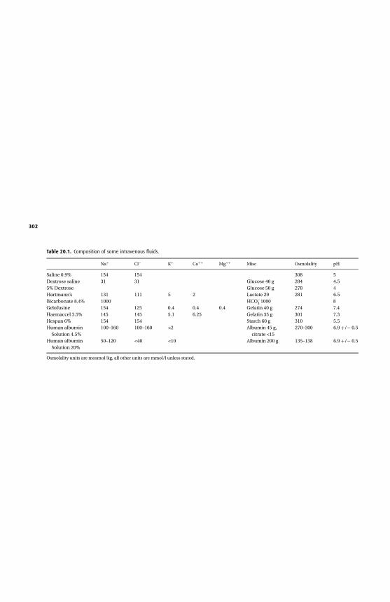
P1:P
SBP
rinter:YetTo
Co
me
9780521704632c20C
UF
X213A
/Peck9780521618168
Decem
ber
28,200714:10
Table 20.1. Composition of some intravenous fluids.
Na+ Cl− K+ Ca++ Mg++ Misc Osmolality pH
Saline 0.9% 154 154 308 5Dextrose saline 31 31 Glucose 40 g 284 4.55% Dextrose Glucose 50 g 278 4Hartmann’s 131 111 5 2 Lactate 29 281 6.5Bicarbonate 8.4% 1000 HCO−
3 1000 8Gelofusine 154 125 0.4 0.4 0.4 Gelatin 40 g 274 7.4Haemaccel 3.5% 145 145 5.1 6.25 Gelatin 35 g 301 7.3Hespan 6% 154 154 Starch 60 g 310 5.5Human albumin
Solution 4.5%100–160 100–160 <2 Albumin 45 g,
citrate <15270–300 6.9 +/− 0.5
Human albuminSolution 20%
50–120 <40 <10 Albumin 200 g 135–138 6.9 +/− 0.5
Osmolality units are mosmol/kg, all other units are mmol/l unless stated.
302
P1: PSB Printer: Yet To Come
9780521704632c20 CUFX213A/Peck 9780521618168 December 28, 2007 14:10
20 Intravenous fluids
ischaemia and ultimately renal failure. This hyperoncotic acute renal failure isparticularly likely in the severely dehydrated patient who receives large volumesof colloid.� Accumulation–the reticuloendothelial system takes up HES and stores it. Thedegree of substitution and the total dose administered are the determining factors.It has been reported that following administration of large doses of HES, patientsoccur an itch, which may develop weeks later.� Anaphylaxis–the incidence is in the order of 1 in 20 000.
Hydroxyethyl starch is clearly a very heterogenious group. Some of the older productshave been associated with significant problems, but there is hope that the newermedium molecular weight products in balanced preperations may provide fewerproblems.
AlbuminAlbumin accounts for ∼50% of the 5 available plasma proteins, the other 50% com-prising α1-, α2-, β globulin and γ globulin. It is made up of 585 amino acids, has amolecular weight of 69 000 Da and is folded into a series of α helices, which form 3domains each with a hydrophobic core and a polar outer exterior.
Hypoalbuminaemia in the critically ill has traditionally been corrected with infu-sions of albumin. However its use has become less widespread following work thatshowed no benefit over other colloids. Further research is required to clarify thesituation.
FunctionsAlbumin exerts a greater colloid osmotic effect than its molecular weight alone mightsuggest due to its highly negative charge.
Albumin binds and transports drugs and endogenous molecules in the plasma.Acidic drugs (warfarin, aspirin, furosemide and amiodarone) tend to bind to albuminwhile basic drugs bind toα1- acid glycoprotein. Reductions in albumin concentrationcan increase the effects of drugs such as warfarin by increasing the free fraction.Albumin carries bilirubin, fatty acids, calcium and magnesium. Despite a low bindingaffinity, a significant amount of steroid is carried by albumin due to its abundance.
KineticsAlbumin is synthesized in the liver and has a half-life of 20 days. Just under 50% isintravascular the remainder being present in the skin and muscle with lesser amountsin the gut, liver and subcutaneous tissue. In health albumin synthesis is balancedto match metabolism. Where metabolism or loss (renal or gastrointestinal) are sig-nificantly increased synthesis may not be able to keep up and hypoalbuminaemiaensues.
303
P1: PSB Printer: Yet To Come
9780521704632c20 CUFX213A/Peck 9780521618168 December 28, 2007 14:10
Section IV Other important drugs
BicarbonateBicarbonate serves various physiological functions including buffering and carbondioxide carriage.
Its main use is to correct hyperkalaemia and in the treatment of certain types ofmetabolic acidosis.
304
P1: PSB Printer: Yet To Come
9780521704632c21 CUFX213A/Peck 9780521618168 December 28, 2007 14:15
21Diuretics
The kidney is a complex organ maintaining fluid, electrolyte, and acid–base balance.It also serves an endocrine function by secreting renin and erythropoietin. Diureticsare drugs that act on the kidney to increase urine production and can be divided intothe following groups:� Thiazides� Loop diuretics� Potassium sparing� Aldosterone antagonists� Osmotic� Carbonic anhydrase inhibitors
Thiazides (bendroflumethazide, chlorothiazide, metolazone)Thiazides (which are chemically related to the sulphonamides) are widely used inthe treatment of mild heart failure and hypertension, alone or in combination withother drugs. They have also been used in diabetes insipidus where they may increasethe sensitivity of the collecting ducts to remaining ADH or cause Na+ depletion andtherefore reduced water absorption in the proximal tubule. In addition, they havebeen used in renal tubular acidosis. The main difference among the drugs is theirrate of absorption from the gut due to differences in lipid solubility and rate of onsetand duration of action due to differences in handling by the renal tubule.
Mechanism of actionThiazides are moderately potent and act mainly on the early segment of the distaltubule by inhibiting Na+ and Cl− reabsorption. This leads to increased Na+ and Cl−
excretion and therefore increased water excretion. The increased Na+ load reachingthe distal tubule stimulates an exchange with K+ and H+ so that thiazides tend toprecipitate a hypokalaemic, hypochloraemic alkalosis. Thiazides also reduce car-bonic anhydrase activity resulting in an increased bicarbonate excretion; however,this effect is small and of little clinical significance.
Metolazone has very powerful synergistic effects when combined with a loopdiuretic and is useful in cases of renal failure.
305
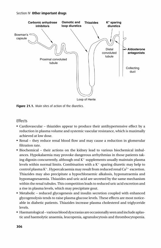
P1: PSB Printer: Yet To Come
9780521704632c21 CUFX213A/Peck 9780521618168 December 28, 2007 14:15
Section IV Other important drugs
Carbonic anhydraseinhibitors
Osmotic and loop diuretics
Thiazides
Collectingduct
Aldosteroneantagonists
Loop of Henle
Distalconvoluted
tubuleProximal convoluted
tubule
Bowman’scapsule
K+ sparingdiuretics
Figure 21.1. Main sites of action of the diuretics.
Effects� Cardiovascular – thiazides appear to produce their antihypertensive effect by areduction in plasma volume and systemic vascular resistance, which is maximallyachieved at low dose.� Renal – they reduce renal blood flow and may cause a reduction in glomerularfiltration rate.� Biochemical – their actions on the kidney lead to various biochemical imbal-ances. Hypokalaemia may provoke dangerous arrhythmias in those patients tak-ing digoxin concurrently, although oral K+ supplements usually maintain plasmalevels within normal limits. Combination with a K+ sparing diuretic may help tocontrol plasma K+. Hypercalcaemia may result from reduced renal Ca2+ excretion.Thiazides may also precipitate a hypochloraemic alkalosis, hyponatraemia andhypomagnesaemia. Thiazides and uric acid are secreted by the same mechanismwithin the renal tubules. This competition leads to reduced uric acid excretion anda rise in plasma levels, which may precipitate gout.� Metabolic – reduced glycogenesis and insulin secretion coupled with enhancedglycogenolysis tends to raise plasma glucose levels. These effects are most notice-able in diabetic patients. Thiazides increase plasma cholesterol and triglyceridelevels.� Haematological – various blood dyscrasias are occasionally seen and include aplas-tic and haemolytic anaemia, leucopenia, agranulocytosis and thrombocytopenia.
306
P1: PSB Printer: Yet To Come
9780521704632c21 CUFX213A/Peck 9780521618168 December 28, 2007 14:15
21 Diuretics� Miscellaneous – impotence, rash and photosensitivity occur rarely. Bendrofluazidemay precipitate pancreatitis.� Interactions – the hypokalaemia produced by thiazides may prolong the durationof action of non-depolarizing muscle relaxants. Non-steroidal anti-inflammatorydrugs antagonize the effects of thiazides.
KineticsBendroflumethazide is well absorbed from the gut in contrast with chlorothiazide,which is incompletely and variably absorbed. They both produce effects within 90minutes of oral administration although bendroflumethazide has twice the dura-tion of action at 18–24 hours. The lower lipid solubility of chlorothiazide results inelimination solely via the kidney – none being metabolized, unlike bendrofluazide,which is 70% metabolized in the liver to active metabolites, the rest being eliminatedunchanged in the urine.
Loop diuretics (furosemide, bumetanide)Loop diuretics (carboxylic acid derivatives) are used in severe heart failure to reduceperipheral and pulmonary oedema. They are also used in chronic and acute renalfailure.
Mechanism of actionLoop diuretics inhibit Na+ and Cl− reabsorption in the thick ascending limb of theloop of Henle and to a lesser extent in the early part of the distal tubule. This impairsthe action of the counter-current multiplier system and reduces the hypertonicityof the medulla, which reduces reabsorption of water in the collecting system. Thethick ascending part of the loop of Henle has a large capacity for NaCl reabsorption,so when ion reabsorption is blocked the effects are marked.
Effects� Cardiovascular – like thiazides they produce arteriolar vasodilatation whichreduces systemic vascular resistance. The preload is also reduced ahead of anychange that could be attributed to a diuresis.� Renal – in contrast with the thiazides an increase in renal blood flow is seen withblood being diverted from the juxtaglomerular region to the outer cortex.� Biochemical – the disturbances seen are similar to those induced by the thiazidesand include hyponatraemia, hypokalaemia, hypomagnesaemia, and a hypochlo-raemic alkalosis. In contrast with the thiazides the loop diuretics may precipitatehypocalcaemia due to increased Ca2+ excretion. Hyperuricaemia is sometimesseen and occasionally precipitates gout.� Metabolic – hyperglycaemia is less common than with thiazides. Loop diureticsincrease plasma cholesterol and triglyceride levels although these usually returnto normal during long-term treatment.
307
P1: PSB Printer: Yet To Come
9780521704632c21 CUFX213A/Peck 9780521618168 December 28, 2007 14:15
Section IV Other important drugs� Miscellaneous – deafness occasionally follows rapid administration of a large doseand is more common in patients with renal failure, and those on aminoglycosidetherapy. Bumetanide is less ototoxic than furosemide but may also cause myalgia.� Interactions – lithium levels may rise when given with loop diuretics.
KineticsBoth furosemide and bumetanide are well absorbed from the gut although their oralbioavailability is different (65% and 95% respectively). Both drugs are highly plasmaprotein bound (>95%) and excreted largely unchanged in the urine.
Potassium sparing (amiloride)Amiloride is a weak diuretic that is frequently used in combination with loop diureticsto prevent hypokalaemia.
Mechanism of actionAt the distal convoluted tubule it blocks Na+/K+ exchange, creating a diuresis, anddecreasing K+ excretion.
Effects� Metabolic – hyperkalaemia may sometimes follow its use. The hypokalaemic,hypochloraemic metabolic alkalosis seen with thiazide and loop diuretics is not afeature of K+ sparing diuretics.
KineticsAmiloride is poorly absorbed from the gut, minimally protein bound and not metab-olized.
Aldosterone antagonists (spironolactone)Spironolactone is only available as an oral preparation. Potassium canrenoate isavailable parenterally and is metabolized to canrenone, which is also a metaboliteof spironolactone.
Owing to its mode of action its effects take a few days to become established. It isused to treat ascites, nephrotic syndrome and primary hyperaldosteronism (Conn’ssyndrome).
Mechanism of actionSpironolactone is a competitive aldosterone antagonist. Normally aldosterone stim-ulates the reabsorption of Na+ in the distal tubule, which provides the driving forcefor the excretion of K+. When aldosterone is antagonized, K+ excretion is significantlyreduced while increased Na+ excretion produces a diuresis. The diuresis producedis limited as only 2% of Na+ reabsorption is under the control of aldosterone.
308
P1: PSB Printer: Yet To Come
9780521704632c21 CUFX213A/Peck 9780521618168 December 28, 2007 14:15
21 Diuretics
Effects� Biochemical – hyperkalaemia is most common in patients with renal impair-ment. Angiotensin-converting enzyme (ACE) inhibitors also predispose to hyper-kalaemia because they reduce aldosterone secretion. It may also produce hypona-traemia.� Hormonal – gynaecomastia in men and menstrual irregularity in women are seendue to spironolactone’s anti-androgenic effects. It is contraindicated in Addison’sdisease.
KineticsSpironolactone is incompletely absorbed from the gut and highly protein bound. Itis rapidly metabolized and excreted in the urine.
Osmotic (mannitol)The carbohydrate derivative mannitol is a polyhydric alcohol. Mannitol is used toreduce intracranial pressure (ICP) in the presence of cerebral oedema or duringneurosurgery. It is also used to preserve peri-operative renal function in the jaundicedpatient and those undergoing major vascular surgery. It is prepared in water as a10 or 20% solution. For the treatment of raised ICP%, the initial dose is 1 g.kg−1,which is then reduced to 0.1–0.2 g.kg−1. However, if the serum osmolality rises above320 mosm.kg−1, further doses of mannitol should not be given due to an increasedrisk of renal failure.
Mechanism of actionMannitol is freely filtered at the glomerulus but not re-absorbed in the tubules. Itincreases the osmolality of the filtrate so that by an osmotic effect the volume ofurine is increased. It is unable to pass through an intact blood–brain barrier and byvirtue of an increased plasma osmolality it draws extracellular brain water into theplasma. However, a head injury may give rise to a damaged blood–brain barrier. Inthis situation mannitol traverses the blood–brain barrier and will drag water with it,thereby increasing cerebral oedema.
Effects� Cardiovascular – initially circulating volume is increased producing an increasedpreload and cardiac output. In susceptible patients this may precipitate heart fail-ure.� Renal – it produces an osmotic diuresis and increased renal blood flow that mayresult in a reduced intravascular volume.� Central nervous system – it produces an acute reduction in ICP, which is not sus-tained during prolonged administration. It may also reduce the rate of formationof cerebrospinal fluid.� Allergic – these responses occur rarely.
309
P1: PSB Printer: Yet To Come
9780521704632c21 CUFX213A/Peck 9780521618168 December 28, 2007 14:15
Section IV Other important drugs
KineticsMannitol is minimally absorbed from the gut. Following intravenous administrationmannitol is distributed throughout the extracellular fluid space. It is freely filteredat the glomerulus and not reabsorbed, resulting in an elimination half-life of 100minutes. It is not metabolized to any significant extent.
Carbonic anhydrase inhibitors (acetazolamide)Acetazolamide is a weak diuretic that is more commonly used as prophylaxis againstmountain sickness. When used as eye drops it inhibits aqueous humour productionand is useful in the treatment of glaucoma.
Mechanism of actionAcetazolamide inhibits (non-competitively) carbonic anhydrase, which catalyzesthe formation of H+ and HCO−
3 from CO2 and H2O via H2CO3 in the proximal tubule.As a result less H+ ions are available for excretion resulting in a metabolic acidosis,counteracting the respiratory alkalosis associated with mountain sickness.
Effects� Biochemical − H+ excretion is inhibited and HCO3− is not re-absorbed leading
to an alkaline urine. Na+ and water excretion are slightly increased providing anincreased drive for K+ secretion. Therefore, it produces an alkaline urine in thepresence of a hyperchloraemic acidosis.
KineticsAcetazolamide has an oral bioavailability of more than 95%, is highly protein boundand is excreted unchanged in the urine.
310
P1: PSB Printer: Yet To Come
9780521704632c22 CUFX213A/Peck 9780521618168 December 29, 2007 14:51
22Antimicrobials
Antimicrobial agents are used to kill or suppress the growth of microorganisms.The term antibiotic specifically refers to a chemical substance that is produced bymicroorganisms and has the capacity to kill or inhibit the growth of another microor-ganism.
Antimicrobials are used widely in intensive care where the selection and dissem-ination of resistance is a constant concern. Methicillin Resistant StaphylococcusAureus (MRSA) and Vancomycin Resistant Enterococcus (VRE) have caused out-breaks in developed countries costing millions to control. Antimicrobial therapyshould be directed by microbiological evidence wherever possible, empirical treat-ment should be rationalised to appropriate narrow spectrum cover and treatmentshould be discontinued at the earliest opportunity. The choice of agent must beinfluenced by unit protocols that will reflect the local pathogen prevalence and theirknown pattern of sensitivity.
This chapter can, therefore, only generalize about each agent’s spectrum of activ-ity and will refrain from suggesting appropriate treatment regimes. The followingheadings are used to discuss the various antimicrobials:� Antibacterial drugs� Antifungal drugs� Antiviral drugs
Antibacterial drugs
β-Lactam antibacterial drugsβ-Lactam drugs can be subdivided into� Penicillins� Cephalosporins� Carbapenems� Monobactams
PenicillinsThis group of antimicrobial drugs are based around the penicillin nucleus, whichcontains a fused β-lactam/thiazolidine ring.
311
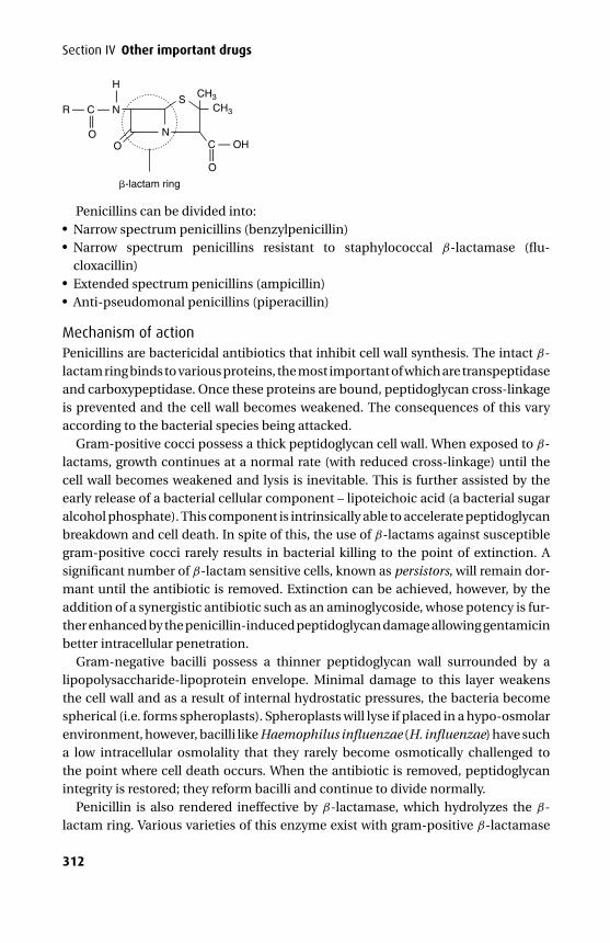
P1: PSB Printer: Yet To Come
9780521704632c22 CUFX213A/Peck 9780521618168 December 29, 2007 14:51
Section IV Other important drugs
R — C — N
C — OH
—
— —H
O
— —
O
N
CH3
CH3S
O
-lactam ring
Penicillins can be divided into:� Narrow spectrum penicillins (benzylpenicillin)� Narrow spectrum penicillins resistant to staphylococcal β-lactamase (flu-cloxacillin)� Extended spectrum penicillins (ampicillin)� Anti-pseudomonal penicillins (piperacillin)
Mechanism of actionPenicillins are bactericidal antibiotics that inhibit cell wall synthesis. The intact β-lactam ring binds to various proteins, the most important of which are transpeptidaseand carboxypeptidase. Once these proteins are bound, peptidoglycan cross-linkageis prevented and the cell wall becomes weakened. The consequences of this varyaccording to the bacterial species being attacked.
Gram-positive cocci possess a thick peptidoglycan cell wall. When exposed to β-lactams, growth continues at a normal rate (with reduced cross-linkage) until thecell wall becomes weakened and lysis is inevitable. This is further assisted by theearly release of a bacterial cellular component – lipoteichoic acid (a bacterial sugaralcohol phosphate). This component is intrinsically able to accelerate peptidoglycanbreakdown and cell death. In spite of this, the use of β-lactams against susceptiblegram-positive cocci rarely results in bacterial killing to the point of extinction. Asignificant number of β-lactam sensitive cells, known as persistors, will remain dor-mant until the antibiotic is removed. Extinction can be achieved, however, by theaddition of a synergistic antibiotic such as an aminoglycoside, whose potency is fur-ther enhanced by the penicillin-induced peptidoglycan damage allowing gentamicinbetter intracellular penetration.
Gram-negative bacilli possess a thinner peptidoglycan wall surrounded by alipopolysaccharide-lipoprotein envelope. Minimal damage to this layer weakensthe cell wall and as a result of internal hydrostatic pressures, the bacteria becomespherical (i.e. forms spheroplasts). Spheroplasts will lyse if placed in a hypo-osmolarenvironment, however, bacilli like Haemophilus influenzae (H. influenzae) have sucha low intracellular osmolality that they rarely become osmotically challenged tothe point where cell death occurs. When the antibiotic is removed, peptidoglycanintegrity is restored; they reform bacilli and continue to divide normally.
Penicillin is also rendered ineffective by β-lactamase, which hydrolyzes the β-lactam ring. Various varieties of this enzyme exist with gram-positive β-lactamase
312
P1: PSB Printer: Yet To Come
9780521704632c22 CUFX213A/Peck 9780521618168 December 29, 2007 14:51
22 Antimicrobials
being fundamentally different from the gram-negative types. Gram-negative β-lactamase is encoded on bacterial chromosomes and plasmids, which may bedisseminated; the basis of acquired resistance.
KineticsIntestinal absorption is variable and certain penicillins are only available as par-enteral preparations. Plasma half-lives are short (benzylpenicillin, 30 min; ampi-cillin, 2 hrs) and protein binding widely variable (ampicillin, 20%; flucloxacillin,93%). Tissue penetration is generally good although inflammation is necessary forpenicillins to pass into bone and through the blood-brain barrier. Penicillins areexcreted by the kidneys in the unchanged form (60–90%), mainly by renal tubularsecretion, but they are also excreted in bile (10%) and up to 20% is metabolized.
Probenecid blocks renal tubular secretion so that when given in combination withpenicillin the plasma concentration of the latter doubles. In addition probenecidinhibits active removal of penicillin from the CSF via the choroid plexus so that thereis increased risk of CNS toxicity.
Use during continuous veno-venous haemodiafiltration (CVVHDF)Penicillin dose adjustment is unnecessary for patients on CVVHDF with the excep-tion of benzylpenicillin – the dose of which should be reduced by 30%. Peni-cillins combined with clavulanic acid (AugmentinTM, TimentinTM) or tazobactam(TazocinTM) require no dose adjustment for CVVHDF. However, if an anuric patientremains without filtration for a prolonged period then dose frequency reductionshould be considered.
Side effects� Hypersensitivity – allergy to penicillins occurs in up to 10% of the popula-tion, anaphylaxis occurs in 0.01%. Cross-reactivity exists between penicillins,cephalosporins and carbapenems in up to 10% of allergic people. Patients witha history of immediate reaction (urticaria, anaphylaxis or interstitial nephritis)should therefore avoid any of these β-lactams.� Encephalopathy – benzylpenicillin is the most pro-convulsant β-lactam. Toxic CSFlevels can be reached by intrathecal injection (maximum dose of benzylpenicillinis 12 mg), by using large doses in meningitic patients with renal impairment andby concomitant probenecid use.� Gut – diarrhoea is common during oral therapy. Ampicillin is also associated witha low risk of pseudomembranous colitis (0.3–0.7%).� Miscellaneous – ampicillin produces a maculopapular rash in 10% of all patientsand 95% of patients with infectious mononucleosis. Bone marrow suppression hasalso been reported with penicillins.
313
P1: PSB Printer: Yet To Come
9780521704632c22 CUFX213A/Peck 9780521618168 December 29, 2007 14:51
Section IV Other important drugs
Specific penicillins
Narrow spectrum penicillinBenzylpenicillin (penicillin G) is inactivated by gastric acid and must be given par-enterally. It is active against a wide variety of gram-positive pathogens, gram-negativecocci, and occasional gram-negative bacilli. Streptococci and Neisseria are extremelysensitive, whilst some Proteus mirabilis (P. mirabilis), Salmonella and Enterococcusfaecalis (E. faecalis) are mildly so. H. influenzae, staphylococci, and Pseudomonas areresistant. Some anaerobic gram-positive cocci are sensitive but Bacteroides fragilis(B. fragilis) is resistant.
Narrow spectrum penicillins resistant to staphylococcal β-lactamaseMost staphylococci are resistant to benzylpenicillin as a result of β-lactamaseproduction. However, the semi-synthetic penicillin flucloxacillin is moderatelyresistant and provides effective therapy in this setting. It is well absorbed fromthe gut but should be given intravenously for serious infections. It is less activeagainst gram-positive cocci than benzylpenicillin but is effective againstβ-lactamasepositive staphylococci. Metabolism produces both active and inactive metabo-lites. It may cause cholestatic jaundice several weeks after the end of a course oftreatment.
Extended spectrum penicillinsAmpicillin is effective against the same range of organisms as benzylpenicillin(although slightly less active) as well as activity against some H. influenzae,Salmonella, Escherichia coli (Esch. coli), and E. faecalis. Amoxycillin has an iden-tical spectrum to ampicillin but has better bioavailability and is bactericidal to sus-ceptible gram-negative organisms at a lower concentration. Both are inactivated byβ-lactamase. Clavulanic acid irreversibly inhibits a large range of β-lactamases andwhen combined with amoxycillin as Co-Amoxiclav reduces the minimum inhibitoryconcentration (MIC) against H. influenzae, Branhamella catarrhalis (B. catarrhalis),Esch. coli, B. fragilis, Klebsiella and Staphylococcus aureus (Staph. aureus) eight tosixty-four-fold. Clavulanic acid is weakly antimicrobial in its own right and may alsobe responsible for the rare occurrence of cholestatic jaundice several weeks aftertreatment has been stopped.
Anti-pseudomonal penicillinsThe carboxypenicillin ticarcillin and the ureidopenicillins azlocillin andpiperacillin have a broad spectrum (albeit with a lower activity than benzylpeni-cillin) and are particularly indicated for use against Pseudomonas, Citrobacter andSerratia. They are β-lactamase sensitive so ticarcillin is presented with clavulanicacid and piperacillin is presented with tazobactam (a β-lactamase inhibitor with no
314
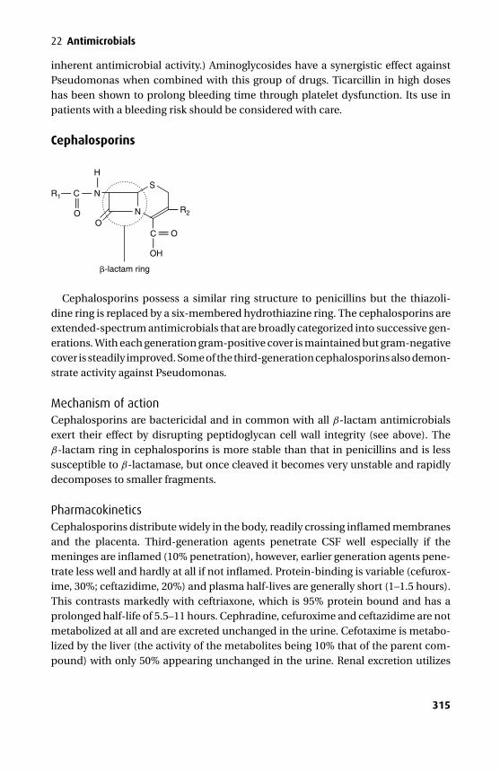
P1: PSB Printer: Yet To Come
9780521704632c22 CUFX213A/Peck 9780521618168 December 29, 2007 14:51
22 Antimicrobials
inherent antimicrobial activity.) Aminoglycosides have a synergistic effect againstPseudomonas when combined with this group of drugs. Ticarcillin in high doseshas been shown to prolong bleeding time through platelet dysfunction. Its use inpatients with a bleeding risk should be considered with care.
Cephalosporins
R1— C — N
C
—
— O
R2
—
— —
H
O
———
OH
NO
-lactam ring
S
Cephalosporins possess a similar ring structure to penicillins but the thiazoli-dine ring is replaced by a six-membered hydrothiazine ring. The cephalosporins areextended-spectrum antimicrobials that are broadly categorized into successive gen-erations. With each generation gram-positive cover is maintained but gram-negativecover is steadily improved. Some of the third-generation cephalosporins also demon-strate activity against Pseudomonas.
Mechanism of actionCephalosporins are bactericidal and in common with all β-lactam antimicrobialsexert their effect by disrupting peptidoglycan cell wall integrity (see above). Theβ-lactam ring in cephalosporins is more stable than that in penicillins and is lesssusceptible to β-lactamase, but once cleaved it becomes very unstable and rapidlydecomposes to smaller fragments.
PharmacokineticsCephalosporins distribute widely in the body, readily crossing inflamed membranesand the placenta. Third-generation agents penetrate CSF well especially if themeninges are inflamed (10% penetration), however, earlier generation agents pene-trate less well and hardly at all if not inflamed. Protein-binding is variable (cefurox-ime, 30%; ceftazidime, 20%) and plasma half-lives are generally short (1–1.5 hours).This contrasts markedly with ceftriaxone, which is 95% protein bound and has aprolonged half-life of 5.5–11 hours. Cephradine, cefuroxime and ceftazidime are notmetabolized at all and are excreted unchanged in the urine. Cefotaxime is metabo-lized by the liver (the activity of the metabolites being 10% that of the parent com-pound) with only 50% appearing unchanged in the urine. Renal excretion utilizes
315
P1: PSB Printer: Yet To Come
9780521704632c22 CUFX213A/Peck 9780521618168 December 29, 2007 14:51
Section IV Other important drugs
both filtration and tubular secretion and probenecid does increase peak concentra-tion and plasma half-life but to a lesser extent than penicillin.
Use during CVVHDFAlthough the same dose should be given, dose interval adjustment is required forpatients on CVVHDF. Cefuroxime should be given 8 hourly, cefotaxime 12 hourly,and ceftazidime 12–24 hourly. No interval adjustment is required for ceftriaxone.
Side effects� Hypersensitivity – cross-reactivity with other β-lactams (see above).� Haematological – a positive Coombs test has been reported with cefuroxime butwithout clinical consequences. Platelet abnormalities are much rarer than withticarcillin, and ceftazidime has been associated with transient eosinophilia.� Liver function – ceftazidime has been reported to cause abnormal liver function.
First-generationCefradine is active against β-lactamase producing staphylococcus, streptococ-cus and anaerobic gram-positive cocci. It has less activity against Neisseria whileH. influenzae is resistant. It is used commonly when surgical prophylaxis for gram-positive organisms is required (classically orthopaedic surgery). Cefradine’s lack ofgram-negative cover makes it unsuitable for GI surgery.
Second-generationThis generation is more resistant to β-lactamase and has greater activity againstH. influenzae and Neisseria gonorrhoea (N. gonorrhoea). Cefuroxime is activeagainst Salmonella, Proteus sp., Esch. coli, Klebsiella, Citrobacter and Enterobacter.E. faecalis, Acinetobacter, Serratia and Pseudomonas remain resistant. Cefuroximeis commonly used for surgical prophylaxis during bowel surgery but lacks sufficientanaerobic cover to be used as a sole agent.
Third-generationThese drugs have improved gram-negative activity but their activity against somegram-positive bacteria (i.e. Staph. aureus) is less than the second-generation agents.Cefotaxime has the same gram-negative spectrum as cefuroxime except Acineto-bacter, Serratia and some Pseudomonas are covered in addition. Ceftazidime ishighly active against Pseudomonas, including strains resistant to aminoglycosides,but much less active against staphylococcus. Ceftriaxone has a long duration ofaction due to its long half-life and is administered once daily. These agents all havea very broad spectrum of activity and may encourage superinfection with resistantbacteria or fungi. Injudicious use of third-generation agents has been implicated inthe encouragement of MRSA.
316
P1: PSB Printer: Yet To Come
9780521704632c22 CUFX213A/Peck 9780521618168 December 29, 2007 14:51
22 Antimicrobials
CarbapenemsCarbapenems have the broadest spectrum of any class of antimicrobial coveringgram-positive and gram-negative aerobic and anaerobic organisms. They do notcover MRSA or E. faecalis and if used in isolation tend to generate Pseudomonasresistance; however, they still prove useful when treating neutropenic fever or life-threatening multi-resistant gram-negative sepsis.
ImipenemThis bactericidal carbapenem has a very broad spectrum of activity including manyaerobic and anaerobic organisms especially Legionella, Streptococcus faecium (Strep.Faecium), Yersinia and all types of Pseudomonas (except Pseudomonas maltophilia(P. maltophilia). It is resistant to β-lactamase-producing organisms but is only mod-erately effective against Clostridia perfringens (C. perfringens). In spite of imipenem’sbroad spectrum some strains of Citrobacter, Enterobacter and Serratia are resistant(though not through the application of β-lactamase). Imipenem also inducesβ-lactamase expression in Pseudomonas which, does not alter susceptibility toimipenem but does impart substantial protection from other β-lactams.
KineticsImipenem is partially metabolized by renal dehydropeptidase-I and is presentedin combination with cilastatin (itself devoid of antimicrobial activity) to preventthis metabolism. As a result plasma and urine concentration are increased. It ismainly excreted unchanged in the urine and will accumulate in renal failure. Whenimipenem/cilastatin is used in patients on CVVHDF the dose must be halved andthe dose-interval doubled to 12 hourly.
Side effectsImipenem is generally well tolerated but may cause vomiting and diarrhoea (includ-ing pseudomembranous colitis), a positive Coombs’ test, allergic reactions and con-vulsions. Imipenem has similar convulsive potential to benzylpenicillin but onlypresents problems in people with pre-existing CNS disorders or cases of relativeoverdose (renal insufficiency or concurrent probenecid use).
MeropenemMeropenem is similar to imipenem but is not metabolized by renal dehy-dropeptidase-I and does not require combination with cilastatin. It is very resistantto β-lactamases although Pseudomonas has been reported to produce protectiveenzymes. Meropenem is less active than imipenem against gram-positive organ-isms but more active against gram-negative organisms that are moderately resistantto imipenem, however, the clinical significance of this is negligible.
317
P1: PSB Printer: Yet To Come
9780521704632c22 CUFX213A/Peck 9780521618168 December 29, 2007 14:51
Section IV Other important drugs
Monobactams
AztreonamAztreonam is a synthetic monobactam antimicrobial that is potently active againsta wide range of gram-negative bacteria only. It has no useful activity against gram-positive or anaerobic organisms. Both Enterobacter and Pseudomonas are sensitivealthough it is often combined with gentamicin when treating severe Pseudomonalinfections. It can be used in penicillin-allergic patients and has been used safely inpatients with known IgE antibodies to benzylpenicillin.
PharmacokineticsAztreonam is a parenteral preparation with a plasma half-life of 1–2 hours. It onlyhas moderate CSF penetration and sputum penetration is poor (0.05%). Serous fluidpenetration is better with good penetration into synovial fluid and bone. It undergoesminimal metabolism and is predominantly excreted unchanged in the urine. It canbe used in patients on CVVHDF but the dose must be decreased by 50–70% (samedose interval.)
Side effectsAztreonam is well tolerated but can cause an increase in bleeding time – INR andAPTT should be monitored during use. Reversible marrow suppression has beenreported as well as liver enzyme rises.
MacrolidesMacrolides have a range of activity similar to penicillin and may be used as alterna-tive therapy in penicillin allergy. They cover most gram-positive organisms, Neisseriaand Haemophilus, with activity against gram-positive and gram-negative anaerobes.However, they also have specific activity against Mycoplasma and Legionella forwhich they are indicated. Erythromycin represents the parent member of this groupbut more modern drugs are also now available. Clarithromycin causes less gastroin-testinal upset and gives better cover against streptococci, Listeria and Legionella.Azithromycin provides better gram-negative cover (Branhamella catarrhalis, Neis-seria and H. influenzae MIC being up to 8 times lower), improved bioavailability andlonger half-life.
Mechanism of actionMacrolides are bactericidal or bacteriostatic depending on plasma concentration.They halt bacterial protein synthesis by binding to the 50S ribosomal subunit afterformation of the initiation complex. Once bound the inhibition of translocationoccurs at the first step.
318
P1: PSB Printer: Yet To Come
9780521704632c22 CUFX213A/Peck 9780521618168 December 29, 2007 14:51
22 Antimicrobials
KineticsMacrolides can be administered orally or parenterally. Erythromycin does irritategastric mucosa and is also rapidly degraded in an acidic environment. CSF pen-etration is non-therapeutic even with inflamed meninges, but sputum and lungpenetration is good. Protein binding is highly variable in this group with 70–90% oferythromycin and 12% of azithromycin being bound. Macrolides tend to be metab-olized and excreted mainly via the liver; however, 40% of clarithromycin and 15% oferythromycin is excreted unchanged in the urine. Doses for both erythromycin andclarithromycin should be halved in patients on CVVHDF (protein binding of thesedrugs is saturable and they are not removed by dialysis) but dose adjustment forazithromycin is not required.
Erythromycin inhibits hepatic cytochrome P4503A, which is responsible for themetabolism of alfentanil and midazolam so that concurrent use leads to raised serumlevels of these drugs.
Side effects� Gut – nausea, vomiting and diarrhoea occur especially following intravenous injec-tion due to a prokinetic effect. Various hepatic dysfunctions have been reportedbut are usually reversible.� Cardiovascular – a prolonged QT interval is associated with erythromycin andclarithromycin and may precipitate ventricular tachycardia or torsades de pointesespecially when taken with terfenadine.� Interactions – the actions of theophylline, warfarin and digoxin may all be aug-mented when given with macrolides. Erythromycin should be avoided in por-phyria.
AminoglycosidesThe aminoglycoside group contains a large number of naturally occurring and syn-thetic drugs including gentamicin, netilmicin and tobramycin. They cover a widerange of gram-negative enterobacteria and have gram-positive cover that includesstaphylococci and to a limited extent streptococci. Aminoglycosides have no anaero-bic activity but are synergistic with β-lactams and vancomycin. They are large polarmolecules that need active transportation to gain entry into bacterial cells. Thistransportation is inhibited by divalent cations (calcium and magnesium), acidosisand low oxygen tensions. (Antimicrobial activity dramatically improves in urine ifpH is raised.) Streptomycin is active against Mycobacterium tuberculosis and is onlyused in this setting. Neomycin is too toxic for systemic administration and may beused orally for pre-operative bowel sterilization (not absorbed).
Mechanism of actionAminoglycosides are bactericidal antimicrobials that block protein synthesis bybinding to the bacterial 30S ribosomal RNA subunit. Bacterial ribosomal RNA
319
P1: PSB Printer: Yet To Come
9780521704632c22 CUFX213A/Peck 9780521618168 December 29, 2007 14:51
Section IV Other important drugs
subunits are smaller than mammalian subunits and this difference is utilized bya number of antimicrobials that interfere with protein synthesis. Once bound toribosomal RNA, aminoglycosides block tRNA attachment and mRNA is either nottranscribed or totally misread (depending on aminoglycoside concentration). Ente-rococcal resistance in the UK is low and likely to be caused by enzyme-mediateddeactivation. Altered cell permeability and altered ribosomal binding have also beenfound in resistant strains.
KineticsOwing to their low lipid solubility, aminoglycosides are not absorbed from the gutand must be given parenterally; aminoglycoside concentration being directly pro-portional to the rate of bacterial killing. They have low protein binding (20–30%),distribute predominantly in extracellular fluid and penetrate cells, CSF and spu-tum poorly. They are not metabolized and are excreted unchanged in the urine byfiltration. Owing to their narrow therapeutic index, blood assays should be checkedbefore and 1 hour after a dose to determine the most appropriate dose and theadequacy of its clearance.
Side effects� Ototoxicity (vestibular and rarely auditory dysfunction) occurs when a significantamount of drug accumulates in the inner ear perilymph and is usually perma-nent. Perilymph penetration is related to peak aminoglycoside concentration anddecreased removal due to high trough levels. This risk is increased in renal failureand with simultaneous use of furosemide.� Nephrotoxicity is seen in up to 37% of intensive care patients treated with amino-glycosides and tends to be reversible on discontinuing treatment. Accumulationof the drug in the renal cortex leads to acute tubular necrosis that manifests withinthe first week of treatment. Synergistic toxicity is seen with some cephalosporinsbut the most likely independent risk factor is patient age. Netilmicin is the leasttoxic in this respect.� Muscle weakness – aminoglycosides decreases the pre-junctional release ofacetylcholine; reduces post-junctional sensitivity to acetylcholine and as a resultincreases non-depolarizing muscle relaxant potency. While intravenous calciummay reverse this effect, their use is still cautioned in myasthenia gravis.
QuinolonesNalidixic acid is a synthetic 4-quinolone that is used for urinary tract infections.It provides no gram-positive or anaerobic cover and although it has a wide gram-negative spectrum, Pseudomonas is known to be resistant. Fluoroquinolones rep-resent a much more significant category of antimicrobials. When compared tothe 4-quinolones, this group has increased gram-positive cover and also includesLegionella, Mycoplasma, Rickettsia and Chlamydia. Pseudomonas aeruginosa
320

P1: PSB Printer: Yet To Come
9780521704632c22 CUFX213A/Peck 9780521618168 December 29, 2007 14:51
22 Antimicrobials
(P. aeruginosa) tends to be sensitive but resistance is acquired rapidly if used inisolation. Ciprofloxacin is the most commonly employed drug in this group and isactive against a wide range of gram-negative and some gram-positive bacteria (strep-tococcus and enterococcus have moderate sensitivity). Norfloxacin is only availableas an oral preparation for uncomplicated urinary tract infections and Levofloxacinhas increased pneumococcal cover that lends itself for use in lower respiratory tractinfections. Ofloxacin is more potent against both gram-negative and gram-positiveorganisms and has acquired less resistance. It is also indicated in lower respiratorytract infection as well as urinary sepsis.
Ciprofloxacin is also used as a first line treatment for anthrax infection and mustbe administered with rifampicin or clindamycin when infection is suspected. Peni-cillins and cephalosporins are not indicated if the potential for molecular biologicalmanipulation of the presenting strain exists. Pneumonic plague also has bioterrorismpotential and ciprofloxacin is similarly recommended as first-line treatment. Vac-cines are available for both these conditions.
Mechanism of actionThe quinolones are bactericidal antimicrobials that inhibit the α subunit of the DNA-gyrase enzyme. This enzyme is responsible for the negative supercoiling of bacterialDNA and when inactivated rapidly results in cell death.
KineticsQuinolones are well absorbed from the gut although this is reduced in the presence ofmagnesium, calcium/iron salts and sucralfate. They are widely distributed and haveexcellent CSF and tissue penetration. Protein binding lies between 15–40%. Theyundergo limited metabolism (active metabolites), and excretion is via urine andfaeces mainly in the unchanged form (60–80% of ofloxacin is excreted in the urine).Dose reduction is only required in severe renal impairment and ciprofloxacin doseadjustment is unnecessary when patients are on CVVHDF.
Side effects� Central nervous system – it should be used with caution in epileptic patients andit may cause restlessness, confusion and seizures.� Gut – nausea, vomiting and abdominal pain can all occur.� Haematological – haemolytic reactions can occur in patients with defects of glucose6-phosphate dehydrogenase.� Interactions – the half-life of concurrently administered theophylline is increased.It is recommended that where concurrent administration is necessary theophyllinelevels are measured.� Allergic reactions and transiently altered liver function have been reported.
321
P1: PSB Printer: Yet To Come
9780521704632c22 CUFX213A/Peck 9780521618168 December 29, 2007 14:51
Section IV Other important drugs
GlycopeptidesGlycopeptides are naturally occurring agents that are therapeutically importantbecause of their gram-positive activity. They are active against most gram-positivebacteria including Staph. aureus (especially methicillin resistant), coagulase-negative staphylococci and gram-positive anaerobes such as Clostridia. They havevery limited gram-negative action because the drugs are too large to penetrate theouter lipid layer of the bacteria. They are not used as first-line agents due to potentialtoxicity but are used for prophylaxis in patients at high risk of endocarditis who areallergic to penicillin. Vancomycin and teicoplanin are the only agents in this groupavailable in the United Kingdom. They have very similar spectrum and differ mainlyin pharmacokinetic and side-effect profiles.
Mechanism of actionThese agents are bactericidal antimicrobials that work by inhibiting glycopeptidesynthetase and thus preventing peptidoglycan formation in the bacterial cell wall.
VancomycinVancomycin is a complex glycopeptide containing vancosamine and several aminoacid moieties. Initial preparations were associated with a high incidence of nephro-toxicity and ototoxicity but the incidence of this has declined as the commer-cial preparations became more refined. Increased use of vancomycin has led toisolated cases of vancomycin-resistant Staph. aureus in the United Kingdom andvancomycin-resistant enterococcus has caused extensive outbreaks in other coun-tries.
KineticsVancomycin is not absorbed from healthy intestine but is used orally for antibi-otic associated pseudomembranous colitis due to Clostridium difficile (C. difficile).Following intravenous administration there is marked individual variability in drugkinetics with an elimination half-life between 3 and 13 hours. Plasma protein-bindingis quoted between 10% and 80% (the variation is probably due to methodological dif-ferences). It is well distributed to serous fluids but CSF penetration is very poor evenin the presence of meningitis. Bone penetration is similarly poor with undetectablebone concentrations in 50% of patients treated for osteomyelitis. Ninety percent iseliminated in the urine as unchanged drug.
Intravenous administration should be monitored with blood assays to ensure ade-quate clearance bearing in mind that peak levels are governed predominantly by doseand trough levels by dose and interval. Maintaining trough levels between 10 and 15mg.l−1 will ensure therapeutic efficacy whilst minimizing the chance of renal toxic-ity. If appropriate doses are administered within therapeutic trough levels, then peaklevels will not exceed safe limits in adults and these levels are therefore not routinelymeasured.
322
P1: PSB Printer: Yet To Come
9780521704632c22 CUFX213A/Peck 9780521618168 December 29, 2007 14:51
22 Antimicrobials
Side effects� Renal – nephrotoxicity is rare but is usually seen with concurrent administrationof aminoglycosides or with pre-existing renal impairment. It usually resolves onwithdrawal of vancomycin.� Ototoxicity – very rare and usually associated with pre-existing hearing loss,renal impairment or concomitant treatment with another ototoxic drug. It is veryuncommon if peak levels do not exceed 50 mg.l−1 and exceedingly rare if they donot exceed 30 mg.l−1.� Phlebitis – the intravenous preparation should be diluted when given peripherally.� Histamine release – if administered too rapidly histamine release may causehypotension, tachycardia and a widespread rash (known as the ‘red man syn-drome’). Administration should not exceed 10 mg.min−1.� Haematological – neutropenia and thrombocytopenia are rare and reversible.
TeicoplaninTeicoplanin is a similar antibiotic to vancomycin but with a longer duration of actionand 2–4 times the potency, allowing once daily administration. The preparation con-tains two components A1 (a phosphoglycolipid) and A2 (a complex of 5 glycopep-tides). Resistance to teicoplanin is more common with up to 25% of Staph. epider-midis showing intermediate sensitivity.
KineticsTeicoplanin is not absorbed when given orally but can be given by intramuscularinjection. It is 90% protein bound and has better distribution than vancomycin. Boneand CSF penetration are more reliable than with vancomycin but urine penetration isless. It is given twice daily for 48 hours to facilitate loading, then once daily thereafter(prolonged elimination half-life). In patients on CVVHDF the maintenance intervalshould be increased to once every 48 hours.
Side effectsTeicoplanin is better tolerated than vancomycin but side effects do include rash,eosinophilia, thrombocytopenia and fever. It has been used safely in patients knownto exhibit red man syndrome after vancomycin.
Lincosamides
ClindamycinParenteral clindamycin phosphate is a semi-synthetic derivative that is highly activeagainst gram-positive aerobes but demonstrates little activity against any gram-negative aerobic organisms. Anaerobic organisms are highly susceptible. It is notablyactive against a significant proportion of MRSA and covers Toxoplasma and Plasmod-ium falciparum (but not Plasmodium vivax).
323
P1: PSB Printer: Yet To Come
9780521704632c22 CUFX213A/Peck 9780521618168 December 29, 2007 14:51
Section IV Other important drugs
Mechanism of actionClindamycin is a bacteriostatic antimicrobial that inhibits bacterial protein synthesisby disrupting the function of the 50S ribosomal subunit. Resistance is inducible ingram-positive organisms that are normally sensitive and when present often impartsresistance to macrolides. Clindamycin has also been shown to inhibit the action ofgentamicin on Enterobacter and Staph. aureus.
KineticsClindamycin is widely distributed and highly protein bound (94%). It is hydrolyzedto four different forms (clindamycin base, clindamycose, demethyl and sulphoxidederivatives) with the sulphoxide derivative being the least active. CSF penetrationis poor and clindamycin is not recommended for CNS infections. Bone and jointpenetration, however, is very good and clindamycin is commonly employed to treatstaphylococcal osteomyelitis. Drug metabolism is not fully elucidated but less than10% of the drug is cleared in the urine. Although levels rise in renal failure, patientson CVVHDF should receive the same dose.
Side effectsClindamycin is generally well tolerated although up to 30% of patients may sufferwith diarrhoea. Pseudomembranous colitis (C. difficile) has been associated withclindamycin use but the incidence is less than 2%. Fever, rash and eosinophilia aremore common (10%) and transient drop in neutrophil and platelet counts have alsobeing described.
Rifamycins
RifampicinRifampicin is a semi-synthetic antimicrobial with potent activity against gram-positive bacteria. It demonstrates marked activity against staphylococci and strep-tococci but E. faecalis is more resistant. Legionella sp., Neisseria and H. Influenzaeare susceptible but other gram-negative organisms remain resistant. Most mycobac-teria (including M. tuberculosis) are sensitive to rifampicin; however M. avium tendsto be resistant.
Mechanism of actionRifampicin is bactericidal and binds to the β-subunit of DNA-dependant RNA poly-merase. This prevents bacterial DNA being transcribed into RNA and thus preventsprotein synthesis. Resistance is readily inducible with rifampicin and this antimi-crobial should not be used as a single agent. Combination therapy needs carefulconsideration as some antimicrobials (ciprofloxacin) are antagonized by rifampicin.
324
P1: PSB Printer: Yet To Come
9780521704632c22 CUFX213A/Peck 9780521618168 December 29, 2007 14:51
22 Antimicrobials
KineticsRifampicin is very lipid-soluble and correspondingly widely distributed. It penetratesCSF, abscesses and heart valves well and is 80% protein bound. It is metabolizedby liver microsomes and excreted into the bile. Active transport of the drug intobile can become saturated and remaining drug will be excreted unchanged inthe urine (where it imparts the characteristic red colour). Rifampicin also induceshepatic microsomes and other drug metabolism may be accelerated as a result(anticonvulsants, warfarin, steroids). Plasma levels are unaffected by renal failureand dose alteration for CVVHDF is unnecessary.
Side effectsRifampicin is well tolerated causing transient gastrointestinal upset and a red dis-colouration of body fluids. Liver function is rarely impaired (more common withintermittent use) but should be monitored throughout therapy. It should be usedwith caution if liver function is known to be impaired. Rarely haemolytic anaemiaand acute renal failure have also been reported.
Fusidanes
Fusidic acidFusidic acid is a naturally occurring bactericidal antibiotic that exhibits activityagainst gram-positive bacteria and some gram-negative organisms (bacilli are highlyresistant). It has appreciable activity against Staph. aureus, MRSA and Staph. epider-midis but streptococci and pneumococci are relatively resistant.
Mechanism of actionFusidic acid does not bind to bacterial ribosome but forms a complex with elon-gation factor and GTP. This complex is involved in protein translocation and willallow the incorporation of one amino acid residue before blocking further chainelongation. Consequently protein synthesis is inhibited and cell death follows. Clin-ical resistance remains uncommon especially if fusidic acid is administered withanti-staphylococcal penicillin. Mono-therapy is not recommended for this reason.
KineticsFusidic acid is well distributed and highly protein bound (95%). Although it pene-trates cerebral abscesses well, it is not active in the cerebral spinal fluid. Bone penetra-tion is good and fusidic acid is often used to treat staphylococcal bone and joint sep-sis. The drug is concentrated and excreted unchanged in the bile but little active drugappears in faeces. Similarly little drug activity is detected in urine and renal failurehas little effect on plasma levels. Dose alteration while on CVVHDF is unnecessary.
325
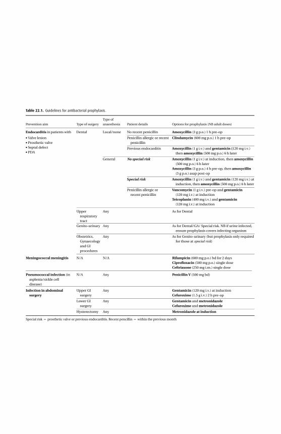
Table 22.1. Guidelines for antibacterial prophylaxis.
Type of
Prevention aim Type of surgery anaesthesia Patient details Options for prophylaxis (NB adult doses)
Endocarditis in patients with Dental Local/none No recent penicillin Amoxycillin (3 g p.o.) 1 h pre-op
• Valve lesion• Prosthetic valve• Septal defect• PDA
Penicillin allergic or recentpenicillin
Clindamycin (600 mg p.o.) 1 h pre-op
Previous endocarditis Amoxycillin (1 g i.v.) and gentamicin (120 mg i.v.)then amoxycillin (500 mg p.o.) 6 h later
General No special risk Amoxycillin (1 g i.v.) at induction, then amoxycillin(500 mg p.o.) 6 h later
Amoxycillin (3 g p.o.) 4 h pre-op, then amoxycillin(3 g p.o.) asap post-op
Special risk Amoxycillin (1 g i.v.) and gentamicin (120 mg i.v.) atinduction, then amoxycillin (500 mg p.o.) 6 h later
Penicillin allergic orrecent penicillin
Vancomycin (1 g i.v.) pre-op and gentamicin(120 mg i.v.) at induction
Teicoplanin (400 mg i.v.) and gentamicin(120 mg i.v.) at induction
Upperrespiratorytract
Any As for Dental
Genito-urinary Any As for Dental/GA/ Special risk. NB if urine infected,ensure prophylaxis covers infecting organism
Obstetrics,Gynaecologyand GIprocedures
Any As for Genito-urinary (but prophylaxis only requiredfor those at special risk)
Meningococcal meningitis N/A N/A Rifampicin (600 mg p.o.) bd for 2 daysCiprofloxacin (500 mg p.o.) single doseCeftriaxone (250 mg i.m.) single dose
Pneumococcal infection (inasplenia/sickle celldisease)
N/A Any Penicillin V (500 mg bd)
Infection in abdominalsurgery
Upper GIsurgery
Any Gentamicin (120 mg i.v.) at inductionCefuroxime (1.5 g i.v.) 2 h pre-op
Lower GIsurgery
Any Gentamicin and metronidazoleCefuroxime and metronidazole
Hysterectomy Any Metronidazole at induction
Special risk = prosthetic valve or previous endocarditis. Recent pencillin = within the previous month
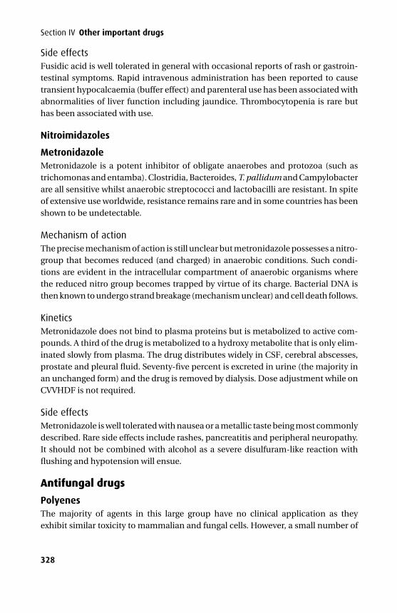
P1: PSB Printer: Yet To Come
9780521704632c22 CUFX213A/Peck 9780521618168 December 29, 2007 14:51
Section IV Other important drugs
Side effectsFusidic acid is well tolerated in general with occasional reports of rash or gastroin-testinal symptoms. Rapid intravenous administration has been reported to causetransient hypocalcaemia (buffer effect) and parenteral use has been associated withabnormalities of liver function including jaundice. Thrombocytopenia is rare buthas been associated with use.
Nitroimidazoles
MetronidazoleMetronidazole is a potent inhibitor of obligate anaerobes and protozoa (such astrichomonas and entamba). Clostridia, Bacteroides, T. pallidum and Campylobacterare all sensitive whilst anaerobic streptococci and lactobacilli are resistant. In spiteof extensive use worldwide, resistance remains rare and in some countries has beenshown to be undetectable.
Mechanism of actionThe precise mechanism of action is still unclear but metronidazole possesses a nitro-group that becomes reduced (and charged) in anaerobic conditions. Such condi-tions are evident in the intracellular compartment of anaerobic organisms wherethe reduced nitro group becomes trapped by virtue of its charge. Bacterial DNA isthen known to undergo strand breakage (mechanism unclear) and cell death follows.
KineticsMetronidazole does not bind to plasma proteins but is metabolized to active com-pounds. A third of the drug is metabolized to a hydroxy metabolite that is only elim-inated slowly from plasma. The drug distributes widely in CSF, cerebral abscesses,prostate and pleural fluid. Seventy-five percent is excreted in urine (the majority inan unchanged form) and the drug is removed by dialysis. Dose adjustment while onCVVHDF is not required.
Side effectsMetronidazole is well tolerated with nausea or a metallic taste being most commonlydescribed. Rare side effects include rashes, pancreatitis and peripheral neuropathy.It should not be combined with alcohol as a severe disulfuram-like reaction withflushing and hypotension will ensue.
Antifungal drugs
PolyenesThe majority of agents in this large group have no clinical application as theyexhibit similar toxicity to mammalian and fungal cells. However, a small number of
328
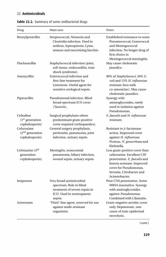
P1: PSB Printer: Yet To Come
9780521704632c22 CUFX213A/Peck 9780521618168 December 29, 2007 14:51
22 Antimicrobials
Table 22.2. Summary of some antibacterial drugs.
Drug Main uses Notes
Benzylpenicillin Streptococcal, Neisseria andClostridia infection. Used inanthrax, leptospirosis, Lyme,tetanus and necrotising fasciitis.
Established resistance to somePneumococcal, Gonococcaland Meningococcalinfection. No longer drug offirst choice inMeningococcal meningitis.
Flucloxacillin Staphylococcal infection (joint,soft tissue, endocarditis, toxicshock syndrome).
May cause cholestaticjaundice.
Amoxycillin Enterococcal infection andfirst-line treatment forListeriosis. Useful agent forsensitive urological sepsis.
90% of Staphylococci, 50% E.coli and 15% H. influenzaeresistant (less withco-amoxiclav). May causecholestatic jaundice.
Piperacillin Pseudomonal infection. Blindbroad spectrum ICU cover(Tazocin).
Synergy withaminoglycosides, rarelyused in isolation againstPseudomonas.
Cefradine(1st generationcephalosporin)
Surgical prophylaxis wherepredominant gram-positivecover required (orthopaedics).
E. faecalis and H. influenzaeresistant.
Cefuroxime(2nd generationcephalosporin)
General surgery prophylaxis,peritonitis, pneumonia, jointinfection, urinary sepsis.
Resistant to β-lactamaseaction. Improved coveragainst H. influenzae,Proteus, N. gonorrhoea andKlebsiella.
Cefotaxime (3rd
generationcephalosporin)
Meningitis, nosocomialpneumonia, biliary infection,wound sepsis, urinary sepsis.
Less gram-positive cover thancefuroxime. Excellent CSFpenetration. E. faecalis andlisteria resistant. Improvedcover for Pseudomonas,Serratia, Citrobacter andAcinetobacter.
Imipenem Very broad antimicrobialspectrum. Role in blindtreatment of severe sepsis inICU. Used in neutropaenicsepsis.
Poor CNS penetration. SomeMRSA insensitive. Synergywith aminoglycosidesagainst Pseudomonas.Combined with Cilastatin.
Aztreonam ‘Third’-line agent, reserved for useagainst multi-resistantorganisms.
Gram-negative aerobic coveronly. Hepatotoxic, rarecause of toxic epidermalnecrolysis.
(cont.)
329
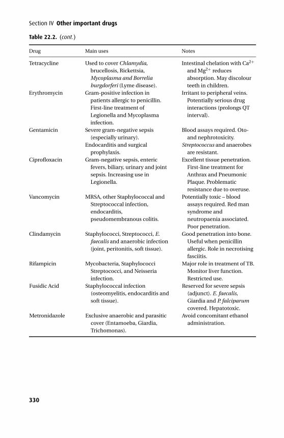
P1: PSB Printer: Yet To Come
9780521704632c22 CUFX213A/Peck 9780521618168 December 29, 2007 14:51
Section IV Other important drugs
Table 22.2. (cont.)
Drug Main uses Notes
Tetracycline Used to cover Chlamydia,brucellosis, Rickettsia,Mycoplasma and Borreliaburgdorferi (Lyme disease).
Intestinal chelation with Ca2+
and Mg2+ reducesabsorption. May discolourteeth in children.
Erythromycin Gram-positive infection inpatients allergic to penicillin.First-line treatment ofLegionella and Mycoplasmainfection.
Irritant to peripheral veins.Potentially serious druginteractions (prolongs QTinterval).
Gentamicin Severe gram-negative sepsis(especially urinary).
Endocarditis and surgicalprophylaxis.
Blood assays required. Oto-and nephrotoxicity.
Streptococcus and anaerobesare resistant.
Ciprofloxacin Gram-negative sepsis, entericfevers, biliary, urinary and jointsepsis. Increasing use inLegionella.
Excellent tissue penetration.First-line treatment forAnthrax and PneumonicPlaque. Problematicresistance due to overuse.
Vancomycin MRSA, other Staphylococcal andStreptococcal infection,endocarditis,pseudomembranous colitis.
Potentially toxic – bloodassays required. Red mansyndrome andneutropaenia associated.Poor penetration.
Clindamycin Staphylococci, Streptococci, E.faecalis and anaerobic infection(joint, peritonitis, soft tissue).
Good penetration into bone.Useful when penicillinallergic. Role in necrotisingfasciitis.
Rifampicin Mycobacteria, StaphylococciStreptococci, and Neisseriainfection.
Major role in treatment of TB.Monitor liver function.Restricted use.
Fusidic Acid Staphylococcal infection(osteomyelitis, endocarditis andsoft tissue).
Reserved for severe sepsis(adjunct). E. faecalis,Giardia and P. falciparumcovered. Hepatotoxic.
Metronidazole Exclusive anaerobic and parasiticcover (Entamoeba, Giardia,Trichomonas).
Avoid concomitant ethanoladministration.
330
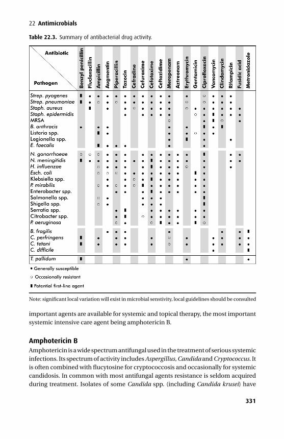
P1: PSB Printer: Yet To Come
9780521704632c22 CUFX213A/Peck 9780521618168 December 29, 2007 14:51
22 Antimicrobials
Table 22.3. Summary of antibacterial drug activity.
Note: significant local variation will exist in microbial senstivity, local guidelines should be consulted
important agents are available for systemic and topical therapy, the most importantsystemic intensive care agent being amphotericin B.
Amphotericin BAmphotericin is a wide spectrum antifungal used in the treatment of serious systemicinfections. Its spectrum of activity includes Aspergillus, Candida and Cryptococcus. Itis often combined with flucytosine for cryptococcosis and occasionally for systemiccandidosis. In common with most antifungal agents resistance is seldom acquiredduring treatment. Isolates of some Candida spp. (including Candida krusei) have
331
P1: PSB Printer: Yet To Come
9780521704632c22 CUFX213A/Peck 9780521618168 December 29, 2007 14:51
Section IV Other important drugs
been reported after prolonged treatment, especially from heart valve vegetations,and fungal endocarditis normally requires surgical intervention to cure.
Mechanism of actionAmphotericin reacts with ergosterol in the fungal cell membrane, creating poresthrough which cell contents are lost. It reacts less with cholesterol and is not as toxicto human cells. As the dose is increased, the pore size created increases and celldamage occurs at a faster rate.
KineticsAmphotericin is only administered parenterally and is highly plasma protein bound.Tissue penetration is poor and negligible levels are found in CSF or urine. It appearsto be metabolized by the liver and plasma levels do not rise in renal failure. However,due to its potential for nephrotoxicity, dose reduction is still considered in renallyimpaired patients. Liposomal preparations are available that allow higher doses tobe given with less renal toxicity – they are considerably more expensive.
Side effects� Renal – renal impairment is only reversible if treatment is stopped early. Renaltoxicity can be limited if the dose is gradually increased over the first 48 hours.� Miscellaneous – it may cause any of the following: gastrointestinal upsets, blooddyscrasias, febrile reactions, muscle pains, visual and hearing disturbances, con-vulsions, anaphylaxis and altered liver function tests. Some of these effects can beameliorated by reducing the rate of administration or by the co-administration ofsteroids or antihistamines.
AzolesThis category can be further subdivided into triazoles (fluconazole and itraconazole)and imidazoles (ketoconazole and miconazole). They are active against a wide rangeof fungi and yeasts including Candida spp., Coccidioides spp., Cryptococcus spp. andHistoplasma spp. Itraconazole and miconazole also cover Aspergillus spp. infection.Resistance is rarely problematic but isolates are known to demonstrate resistance tomore than one agent.
Mechanism of actionThese agents all work by disrupting ergosterol synthesis. The blockade of 14-αdemethylation during ergosterol manufacture leads to an alteration in membranefunction. Higher concentrations of azoles also damage ergosterol directly leading toloss of membrane integrity, cell leakage and death.
332
P1: PSB Printer: Yet To Come
9780521704632c22 CUFX213A/Peck 9780521618168 December 29, 2007 14:51
22 Antimicrobials
KineticsThese drugs are all absorbed well via the oral route with the exception of miconazole,which is available in a parenteral form (prepared in cremophor EL).
Fluconazole is minimally protein bound (12%) and penetrates CSF well. It is notmetabolized in man and excreted unchanged in the urine. Itraconazole, ketocona-zole and miconazole are all highly protein bound (99%) and do not penetrate CSF.They are not therapeutically active in urine but tissue levels are high. Ketoconazoleand miconazole are metabolized by the liver and inactive metabolites excreted inbile. Itraconazole is degraded before inactive metabolites are excreted in bile. Nodose adjustment for renal failure or CVVHDF is required with these antifungals.
Side effectsDrug interaction� Cisapride metabolism is inhibited and high plasma concentrations increase the
risk of prolonged QT syndrome and fatal arrhythmias.� Warfarin effect is enhanced.Ketoconazole� Gastrointestinal symptoms (20%), rash and pruritus� Hepatitis (rare)� Steroid synthesis inhibition (subclinical adrenocortical deficiency)Miconazole� Thrombophlebitis� Cardiac arrhythmias with rapid i.v. administration
Antiviral drugsViruses utilize the biochemical components of their host cell and it is, therefore,difficult to prevent viral replication without causing damage to the host cell.
AcyclovirAcyclovir is used to treat infection due to Herpes simplex (type I and II) and Varicellazoster.
Mechanism of actionAcyclovir inhibits nucleic acid synthesis. Cells infected with H. simplex or V. zostercontain a virally encoded thymidine kinase that converts acyclovir to acyclovirmonophosphate. This is then converted to an active triphosphate that inhibits viralDNA polymerase and acts as a chain terminator. The thymidine kinase present inuninfected cells has a low affinity for acyclovir and therefore only very small amountsof acyclovir triphosphate are produced. This variable affinity forms the basis of itsselectivity. Whilst useful against H. simplex and V. zoster it does not eradicate themand is only effective if given at the start of an infection.
333
P1: PSB Printer: Yet To Come
9780521704632c22 CUFX213A/Peck 9780521618168 December 29, 2007 14:51
Section IV Other important drugs
KineticsIntestinal absorption is erratic and the oral bioavailability is low at approximately25%. Acyclovir is widely distributed with 15% being protein bound. It is partiallymetabolized to inactive compounds but largely undergoes active tubular excretionunchanged into the urine, but this can be blocked by probenecid. Dose reductionin renal failure is required and a dose of 3.5 mg.kg−1.day−1 has been suggested forpatients on CVVHDF.
Side effects� Renal – rapid intravenous administration may precipitate renal impairment.� Thrombophlebitis – it is highly irritating to veins and may cause ulcers whenextravasated.� Central nervous system – tremors, confusion, seizures and coma during rapidintravenous administration are recognized.
ZidovudineZidovudine is used to treat the human immunodeficiency virus (HIV) in combinationwith other antiviral agents.
Mechanism of actionZidovudine is a nucleoside reverse transcriptase inhibitor (or nucleoside analogue)that is converted by various kinase enzymes to zidovudine triphosphate, the activecompound. This binds to HIV-reverse transcriptase and when it becomes incorpo-rated into proviral DNA chain termination occurs. Its affinity for HIV-reverse tran-scriptase is 100 times greater than for host DNA polymerase.
KineticsZidovudine is given orally or intravenously and distributes widely. It is transported bythe choroid plexus into the CSF where levels are 50% plasma levels. It is conjugatedin the liver and up to 80% is excreted in the urine as the glucuronide. Accumulationoccurs in renal failure and dose reduction is required. Patients on CVVHDF shouldreceive doses at the lower end of the dosage range.
Side effects� Haematological – anaemia and neutropenia.� Gut – gastrointestinal upset, anorexia, deranged liver function, steatorrhoea andlactic acidosis.� Miscellaneous – headache, myalgia, parasthesia and pigmentation of nail beds andoral mucosa.
334
P1: PSB Printer: Yet To Come
9780521704632c23 CUFX213A/Peck 9780521618168 December 29, 2007 14:58
23Drugs affecting coagulation
� Anti-platelet drugs� Heparins and protamine� Oral anticoagulants� Drugs affecting the fibrinolytic system
PhysiologyHaemostasis is complicated. Two models are currently used to explain what isthought to occur.
The classical modelThis has three elements: platelets, the coagulation cascade and fibrinolysis. Thefirst two are involved in preventing haemorrhage by thrombus formation, whilefibrinolysis is an essential limiting mechanism.
Thrombus formation is initially dependent on platelet adhesion, which istriggered by exposure to subendothelial connective tissue. The von Willebrandfactor, which is part of the main fraction of factor VIII, is essential in this process.Subsequent platelet aggregation and vasoconstriction is enhanced by the releaseof thromboxane A2 (TXA2) from platelets. Adjacent undamaged vascular endothe-lium produces prostacyclin (PGI2), which inhibits aggregation and helps to localizethe platelet plug to the damaged area. The localized primary platelet plug is thenenmeshed by fibrin converting it to a stable haemostatic plug.
The coagulation cascade is formed by an intrinsic and extrinsic pathway, whichconverge to activate factor X and the final common pathway (Figure 23.1). The intrin-sic pathway is triggered by the exposure of collagen, thereby activating factor XII,while the extrinsic pathway is triggered by leakage of tissue factors, activating factorVII.
Venous thrombus consists mainly of a fibrin web enmeshed with plateletsand red cells. Arterial thrombus relies more on platelets and less on the fibrinmesh.
A crucial part of this process is its limitation to the initial site of injury. Circulatinginhibitors, of which anti-thrombin III is the most potent, perform this function. In
335
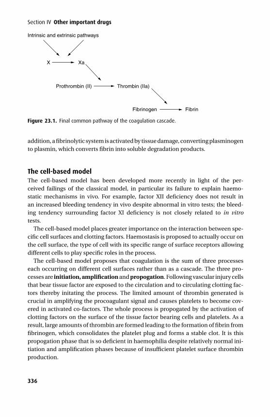
P1: PSB Printer: Yet To Come
9780521704632c23 CUFX213A/Peck 9780521618168 December 29, 2007 14:58
Section IV Other important drugs
Intrinsic and extrinsic pathways
X
Prothrombin (II) Thrombin (IIa)
Fibrinogen Fibrin
Xa
Figure 23.1. Final common pathway of the coagulation cascade.
addition, a fibrinolytic system is activated by tissue damage, converting plasminogento plasmin, which converts fibrin into soluble degradation products.
The cell-based modelThe cell-based model has been developed more recently in light of the per-ceived failings of the classical model, in particular its failure to explain haemo-static mechanisms in vivo. For example, factor XII deficiency does not result inan increased bleeding tendency in vivo despite abnormal in vitro tests; the bleed-ing tendency surrounding factor XI deficiency is not closely related to in vitrotests.
The cell-based model places greater importance on the interaction between spe-cific cell surfaces and clotting factors. Haemostasis is proposed to actually occur onthe cell surface, the type of cell with its specific range of surface receptors allowingdifferent cells to play specific roles in the process.
The cell-based model proposes that coagulation is the sum of three processeseach occurring on different cell surfaces rather than as a cascade. The three pro-cesses are initiation, amplification and propogation. Following vascular injury cellsthat bear tissue factor are exposed to the circulation and to circulating clotting fac-tors thereby initating the process. The limited amount of thrombin generated iscrucial in amplifying the procoagulant signal and causes platelets to become cov-ered in activated co-factors. The whole process is propogated by the activation ofclotting factors on the surface of the tissue factor bearing cells and platelets. As aresult, large amounts of thrombin are formed leading to the formation of fibrin fromfibrinogen, which consolidates the platelet plug and forms a stable clot. It is thispropogation phase that is so deficient in haemophilia despite relatively normal ini-tiation and amplification phases because of insufficient platelet surface thrombinproduction.
336
P1: PSB Printer: Yet To Come
9780521704632c23 CUFX213A/Peck 9780521618168 December 29, 2007 14:58
23 Drugs affecting coagulation
Anti-platelet drugs
Cyclo-oxygenase I inhibition
Aspirin
UsesAspirin reduces the risk of unstable angina progressing to acute myocardial infarction(MI) and reduces the mortality following acute MI. The risk of stroke is also reducedfor patients with transient ischaemic attacks.
Mechanism of actionAspirin acts by irreversible inhibition of cyclo-oxygenase (by acetylation) within theplatelet resulting in reduced production of TXA2. This may be achieved with only75 mg daily. Its effects and kinetics are discussed in Chapter 9.
Platelet phosphodiesterase inhibition
Dipyridamole
UsesDipyridamole has been used with limited success in conjunction with warfarin toprevent thrombus formation on prosthetic valves. There is some evidence that whencombined with aspirin, dipyridamol may further reduce the risk of stroke, comparedto aspirin alone.
Mechanism of actionDipyridamole inhibits platelet adhesion to damaged vessel walls (by inhibitingadenosine uptake), potentiates the affects of prostacyclin and at high doses inhibitsplatelet phosphodiesterase activity resulting in increased cAMP levels and lowerintraplatelet calcium levels. Compared to aspirin it inhibits platelet adhesion to ves-sel walls more than platelet aggregation.
It is a potent coronary artery vasodilator and may be used in conjunction withthallium–201 during myocardial imaging.
Inhibition of ADP binding
Ticlodipine and clopidogrelLike ticlopidine, clopidogrel is a thienopyridine derivative. Neutropenia may beseen with toclopidine (2.4%) and clopidogrel (0.02%). Thrombocytopenia may alsoaccompany their use.
337
P1: PSB Printer: Yet To Come
9780521704632c23 CUFX213A/Peck 9780521618168 December 29, 2007 14:58
Section IV Other important drugs
Uses� Clopidogrel – When used for patients with peripheral vascular disease it reducesthe risk of ischaemic stroke and myocardial infarction when compared to aspirin.It is also used following coronary artery stenting in combination with aspirin toprevent stent thrombosis. It is associated with less gastrointestinal bleeding thanaspirin but severe rash is more common.� Ticlodipine – Because of its side effects ticlodipine should only be used whenaspirin has failed. It may be used to prevent stroke and during coronary stenting.
Mechanism of actionClopidogrel and ticlodipine irreversibly prevent ADP from binding to its receptoron the platelet surface, thereby preventing the glycoprotein IIb/IIIa-receptor trans-forming into its active form.
KineticsClopidogrel’s effects are apparent 2 hours after an oral dose but its full effects areseen only after 3 to 7 days of treatment. Therapy should be stopped for a minimumof 7 days preoperatively to prevent excessive bleeding.
Glycoprotein IIb/IIIa-receptor anatagonistsAbciximab is a monoclonal antibody with a high affinity for the platelet glycoproteinIIb/IIIa receptor. It has a plasma half-life of 20 minuts but remains bound in thecirculation for up to 15 days with some residual activity.
Eptifibatide is a cyclic heptapeptide with a lower receptor affinity and a plasmahalf-life of 200 minutes. Fifty percent undergoes renal clearance.
Tirofiban has intermediate receptor affinity and undergoes renal (65%) and faecal(25%) clearance.
UsesThey are used around the time of acute coronary events. They are used intravenouslyfor a short duration during concurrent unfractionated heparin therapy with closecontrol of APTT and or ACT. They carry the rare but serious complication of throm-bocytopenia.
Mechanism of actionThey act by inhibiting the platelet glycoprotein IIb/IIIa-receptor and as such blockthe final common pathway of platelet aggregation. However, they do not blockplatelet adhesion, secretion of platelet products, inflammatory effects or thrombinactivation.
338
P1: PSB Printer: Yet To Come
9780521704632c23 CUFX213A/Peck 9780521618168 December 29, 2007 14:58
23 Drugs affecting coagulation
Miscellaneous
Dextran 40 and dextran 70These are polysaccharides that contain chains of glucose. They are produced by thefermentation of sucrose by the bacterium Leuconostoc mesenteroides.
UsesThey are used as prophylaxis against peri-operative venous thrombosis and asplasma volume expanders.
Mechanism of actionTheir anticoagulant activity appears to depend on reducing platelet adhesivenessand a specific inhibitory effect on the von Willebrand factor. They also reduce red cellaggregation and provide a protective coat over vascular endothelium and erythro-cytes. The dilution of clotting factors and volume expansion also improves micro-circulatory flow.
Other effectsThese include fluid overload, allergic reactions and anaphylaxis. Dextran 70 canimpair cross matching by rouleaux formation. Renal failure is a rare complication.
Epoprostenol (prostacyclin – PGI2)This is a naturally occurring prostaglandin.
UsesEpoprostenol is used to facilitate haemofiltration by continuous infusion into theextracorporeal circuit.
Mechanism of actionEpoprostenol causes inhibition of platelet adhesion and aggregation by stimulatingadenylate cyclase. The ensuing rise in cAMP reduces intracellular Ca2+, effecting thechange. It may also have a fibrinolytic effect. The recommended dose range is 2–12ng.kg−1.min−1.
Side effectsThese relate to its vasodilator properties (hypotension, tachycardia, facial flushingand headache).
339
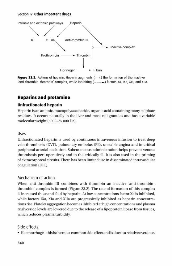
P1: PSB Printer: Yet To Come
9780521704632c23 CUFX213A/Peck 9780521618168 December 29, 2007 14:58
Section IV Other important drugs
Intrinsic and extrinsic pathways
X
Prothrombin
Fibrinogen Fibrin
Xa Anti-thrombin III
Heparin
Thrombin
Inactive complex
Figure 23.2. Actions of heparin. Heparin augments (−→) the formation of the inactive‘anti-thrombin–thrombin’ complex, while inhibiting ( ) factors Xa, IXa, XIa, and XIIa.
Heparins and protamine
Unfractionated heparinHeparin is an anionic, mucopolysaccharide, organic acid containing many sulphateresidues. It occurs naturally in the liver and mast cell granules and has a variablemolecular weight (5000–25 000 Da).
UsesUnfractionated heparin is used by continuous intravenous infusion to treat deepvein thrombosis (DVT), pulmonary embolus (PE), unstable angina and in criticalperipheral arterial occlusion. Subcutaneous administration helps prevent venousthrombosis peri-operatively and in the critically ill. It is also used in the primingof extracorporeal circuits. There has been limited use in disseminated intravascularcoagulation (DIC).
Mechanism of actionWhen anti-thrombin III combines with thrombin an inactive ‘anti-thrombin–thrombin’ complex is formed (Figure 23.2). The rate of formation of this complexis increased thousand-fold by heparin. At low concentrations factor Xa is inhibited,while factors IXa, XIa and XIIa are progressively inhibited as heparin concentra-tions rise. Platelet aggregation becomes inhibited at high concentrations and plasmatriglyceride levels are lowered due to the release of a lipoprotein lipase from tissues,which reduces plasma turbidity.
Side effects� Haemorrhage – this is the most common side effect and is due to a relative overdose.
340
P1: PSB Printer: Yet To Come
9780521704632c23 CUFX213A/Peck 9780521618168 December 29, 2007 14:58
23 Drugs affecting coagulation� Thrombocytopenia – A non-immune based thrombocytopenia (type I) occurswithin four days of anticoagulant doses of heparin. This rarely has clini-cal significance and the platelet count recovers without stopping heparin.
This contrasts with the more severe (type II) immune-mediated thrombocy-topenia, which occurs within 4 to 14 days of starting intravenous or subcutaneousheparin (fractionated and unfractionated). Heparin complexes with platelet fac-tor 4, which is bound by IgG causing platelet aggregation and thrombosis. Half ofthese patients develop serious thrombosis after the platelet count starts to fall andmortality is high, mainly from pulmonary embolus. In addition 20% get arterialthrombosis leading to stroke and limb ischaemia.� Cardiovascular – hypotension may follow rapid intravenous administration of alarge dose.� Miscellaneous – osteoporosis, due to complexing of mineral substance from bone,and alopecia have been reported. Heparin (including LMWH) can cause hyper-kalaemia, which may be due to inhibition of aldosterone secretion. Patients withdiabetes mellitus and chronic renal failure are particularly susceptible and the riskincreases with the duration of therapy.
KineticsAs the potency of commercial preparations varies, unfractionated heparin is pre-sented as units.ml−1 rather than mg.ml−1. It is ineffective orally and may only begiven subcutaneously or intravenously. It has a low lipid solubility and does notcross the BBB or placenta. Owing to its negative charge it is highly bound in plasmato anti-thrombin III, albumin, proteases and fibrinogen. It is metabolized by hep-atic heparinase and the products are excreted in the urine. During hypothermia (e.g.cardiopulmonary bypass) clearance is reduced. Its effects are reversed by protamine.
Low-molecular-weight heparins (LMWHs)
Enoxaparin, dalteparin, tinzaparinThese drugs are derived from the depolymerization of heparin by either chemical orenzymatic degradation. Their molecular weights vary from 2000 to 8000 Da.
UsesLMWHs have proven benefit over unfractionated heparin in reducing the incidenceof fatal PE after major orthopaedic surgery.
Advantages include:� Single daily dose, due to a longer half-life� Reduced affinity for von Willebrand factor� Less effect on platelets� Reduced risk of heparin induced thrombocytopenia� Reduced need for monitoring
341
P1: PSB Printer: Yet To Come
9780521704632c23 CUFX213A/Peck 9780521618168 December 29, 2007 14:58
Section IV Other important drugs
Mechanism of actionCompared with unfractionated heparin, LMWHs are more effective at inhibit-ing factor Xa and less effective at promoting the formation of the inactive ‘anti-thrombin–thrombin’ complex.
Protamine is not fully effective in reversing the effects of LMWH.
Monitoring heparin therapyThe activated partial thromboplastin time (APTT) measures the intrinsic systemfactors (VIII, IX, XI and XII) in addition to the factors common to both systems.Phospholipid, a surface activator (e.g. kaolin) and Ca2+ are added to citrated blood.The clotting time varies between laboratories but is about 28–35 seconds.
Monitoring LMWH is difficult as its main effects are inhibiting factor Xa, so theAPTT is not altered.
ProtamineProtamine is a basic protein prepared from fish sperm. Its positive charge enablesit to neutralize the anticoagulant effects of heparin by the formation of an inactivecomplex that is cleared by the reticulo-endothelial system. It is also used in theformulation of certain insulins.
It is given intravenously, 1 mg reversing 100 i.u. heparin.
Side effects� Cardiovascular – hypotension (due to histamine release), pulmonary hypertension(complement activation and thromboxane release), dyspnoea, bradycardia andflushing follow rapid intravenous administration.� Allergic reactions – patients having previously received insulin containing pro-tamine and those allergic to fish may be at increased risk of allergic reactions.� Anticoagulation – when given in excess it has some anticoagulant effects.
Oral anticoagulants
Warfarin
UsesWarfarin is a coumarin derivative and is used for the prophylaxis of systemicthrombo-embolism in patients with atrial fibrillation, rheumatic valve disease orprosthetic valves. It is also used in the prophylaxis and treatment of DVT and PE.
Mechanism of actionWarfarin inhibits the synthesis of the vitamin K-dependent clotting factors: II (pro-thrombin), VII, IX and X. The precursors of these clotting factors are produced in
342
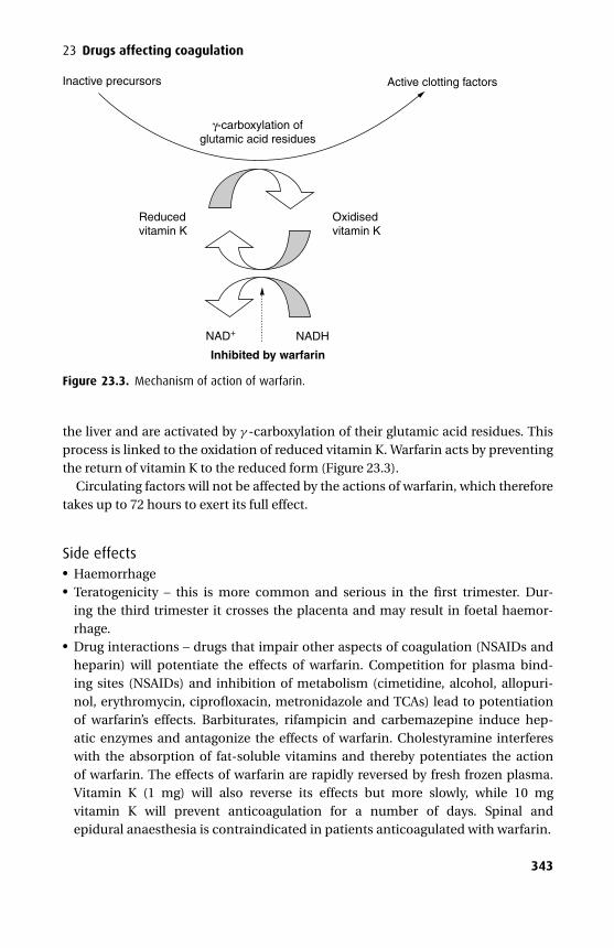
P1: PSB Printer: Yet To Come
9780521704632c23 CUFX213A/Peck 9780521618168 December 29, 2007 14:58
23 Drugs affecting coagulation
Inactive precursors Active clotting factors
γ-carboxylation ofglutamic acid residues
NAD+ NADH
Inhibited by warfarin
Reduced vitamin K
Oxidised vitamin K
Figure 23.3. Mechanism of action of warfarin.
the liver and are activated by γ -carboxylation of their glutamic acid residues. Thisprocess is linked to the oxidation of reduced vitamin K. Warfarin acts by preventingthe return of vitamin K to the reduced form (Figure 23.3).
Circulating factors will not be affected by the actions of warfarin, which thereforetakes up to 72 hours to exert its full effect.
Side effects� Haemorrhage� Teratogenicity – this is more common and serious in the first trimester. Dur-ing the third trimester it crosses the placenta and may result in foetal haemor-rhage.� Drug interactions – drugs that impair other aspects of coagulation (NSAIDs andheparin) will potentiate the effects of warfarin. Competition for plasma bind-ing sites (NSAIDs) and inhibition of metabolism (cimetidine, alcohol, allopuri-nol, erythromycin, ciprofloxacin, metronidazole and TCAs) lead to potentiationof warfarin’s effects. Barbiturates, rifampicin and carbemazepine induce hep-atic enzymes and antagonize the effects of warfarin. Cholestyramine interfereswith the absorption of fat-soluble vitamins and thereby potentiates the actionof warfarin. The effects of warfarin are rapidly reversed by fresh frozen plasma.Vitamin K (1 mg) will also reverse its effects but more slowly, while 10 mgvitamin K will prevent anticoagulation for a number of days. Spinal andepidural anaesthesia is contraindicated in patients anticoagulated with warfarin.
343
P1: PSB Printer: Yet To Come
9780521704632c23 CUFX213A/Peck 9780521618168 December 29, 2007 14:58
Section IV Other important drugs
KineticsWarfarin is completely absorbed from the gut and highly protein bound (>95%). Itundergoes complete hepatic metabolism by oxidation and reduction to productsthat are subsequently conjugated and excreted in the urine.
Monitoring warfarin therapyProthrombin time (PT) is a measure of the extrinsic system (VII) and factors com-mon to both systems. Tissue thromboplastin (a brain extract) and Ca2+ are added tocitrated plasma. Clotting normally takes place in 10–14 seconds. The internationalnormalized ratio (INR) is a ratio of the sample PT to a control international standardPT. Warfarin treatment should aim to increase the PT so that the INR is raised tobetween 2.0 and 4.5 depending on the clinical situation.
XimelagatranXimelagatran is an oral direct thrombin inhibitor. It is a prodrug, which is convertedto the active form – melagatran – in the liver. Melagatran is a potent competitiveinhibitor of α-thrombin, which is highly selective for thrombin rather than otherserine proteases.
It is taken twice a day and blood tests are not required to monitor its effects. How-ever, in contrast to warfarin there is no antidote to reverse its effects should thatbecome necessary. Overdose may be treated with prothrombin complex concen-trates, fresh frozen plasma or recombinant factor VIIa. Ximelagatran may find itsniche where warfarin is currently used and has proven efficacy in these areas (atrialfibrillation and venous thromboembolism). It requires no further precautions overLMWH when used alongside neuraxial blockade.
KineticsThe oral bioavailability of meligatran after oral ximelagatran (compared with sub-cutaneous melagatran) is about 20%. It has a volume of distribution of 2 l/kg, andan elimination half-life of 3–5 hours, and as it is not metabolized further it is almostexclusively excreted via the kidneys. It has no interactions with drugs metabolizedby the hepatic P450 system.
Drugs affecting the fibrinolytic system
Fibrinolytics
StreptokinaseStreptokinase is an enzyme produced by group C β-haemolytic streptococci.
344
P1: PSB Printer: Yet To Come
9780521704632c23 CUFX213A/Peck 9780521618168 December 29, 2007 14:58
23 Drugs affecting coagulation
UsesStreptokinase is used to dissolve clot in arterial (acute MI, occluded peripheral arter-ies and PE) and venous (DVT) vessels.
Mechanism of actionStreptokinase activates the fibrinolytic system by forming a complex with plasmino-gen, which then facilitates the conversion of further plasminogen to active plasmin.The clot can then be lysed.
Side effects� Haemorrhage – streptokinase is contraindicated in patients for whom a risk ofserious bleeding outweighs any potential benefit of therapy (i.e. active internalbleeding, recent stroke, severe hypertension).� Cardiovascular – it may precipitate reperfusion arrhythmias and hypotension dur-ing treatment of acute MI.� Allergic reactions may occur as it is very antigenic. Where high antibody titresare expected (5 days to 12 months from previous exposure to streptokinase), analternative should be sought.
KineticsStreptokinase is administered intravenously as a loading dose followed by an infu-sion. The loading dose is sufficient to neutralize any antibodies that are often presentfrom previous exposure to streptococcal infection. The streptokinase–antibody com-plex is cleared rapidly and the streptokinase–plasminogen complex is degraded to anumber of smaller fragments during its action.
Alteplase (rt-PA, tissue-type plasminogen activator) is a glycoprotein that becomesactivated only when bound to fibrin, inducing the conversion of plasminogen toplasmin. Because of its mode of activation, systemic fibrinolysis occurs to a lesserextent than with streptokinase.
Urokinase was originally extracted from human adult male urine but is now pre-pared from renal cell cultures. Recombinant DNA technology is currently used toproduce pro-urokinase. It is not antigenic and is used in the lysis of clots in arteri-ovenous shunts and in hyphaema (anterior chamber haemorrhage with refractorythrombi).
Antifibrinolytics
AprotininAprotinin is a polypeptide with 58 amino acids and has a molecular weight of 6512 Da.It is a naturally occurring proteolytic enzyme inhibitor acting on trypsin, plasmin andtissue kallikrein. It inhibits the fibrinolytic activity of the streptokinase–plasminogen
345
P1: PSB Printer: Yet To Come
9780521704632c23 CUFX213A/Peck 9780521618168 December 29, 2007 14:58
Section IV Other important drugs
complex. In addition it has been suggested that it preserves platelet function anddecreases activation of the clotting cascade.
It has been used for the treatment of haemorrhage due to hyperplasminaemia at adose of 500 000–1 000 000 units followed by 200 000 units per hour until the bleedingstops.
It has also been used in patients at high risk of bleeding during and after cardiopul-monary bypass. The dose here is 2 000 000 prior to sternotomy followed by 500 000units per hour. In addition, 2 000 000 units is added to the cardiopulmonary bypassmachine.
Side effects� Hypersensitivity reactions including anaphylaxis.� Coagulation – it prolongs the activated clotting time (ACT) of heparinized bloodso that heparin/protamine protocols will need alteration during treatment withaprotinin.
KineticsAprotinin is metabolized and eliminated by the kidney.
Tranexamic acid competitively inhibits the conversion of plasminogen to activeplasmin. It is used to control bleeding due to excessive fibrinolysis, which may belocal (prostatectomy or surgical procedures in haemophiliacs) or systemic (followingintravenous fibrinolytic therapy or DIC). It is excreted essentially (95%) unchangedin the urine.
346
P1: PSB Printer: Yet To Come
9780521704632c24 CUFX213A/Peck 9780521618168 December 29, 2007 15:7
24Drugs used in Diabetes
� Insulin� Sulphonylureas� Biguanides� Other antidiabetics
Insulin
PhysiologyHuman insulin is a polypeptide of 51 amino acids and is formed by the removal of aconnecting or ‘C’ peptide (34 amino acids) from pro-insulin. It has A and B chains,which are joined by two disulphide bridges. A third disulphide bridge connects tworegions of the A chain.
Glucose forms the most potent stimulus for insulin release. It enters the β-cells ofthe islets of Langerhans in the pancreas, resulting in an increase in ATP, which closesK+ channels. This causes depolarization and Ca2+ influx through voltage-sensitiveCa2+ channels, which triggers insulin release. By way of negative feedback the K+
channels are re-opened. In health there is a continuous basal insulin release, whichis supplemented by bursts when plasma glucose levels rise. Following its release itis carried in the portal circulation to the liver (its main target organ) where aboutone-half is extracted and broken down, as glucose is converted to glycogen.
Insulin binds to the α subunit of the insulin receptor, which consist of two α andtwo β subunits that span the cell membrane. Once bound, the whole complex isinternalized. The mechanism by which this complex produces its effects is unclearbut the tyrosine kinase activity of the β subunit appears important.
Insulin affects carbohydrate, fat and protein metabolism. It promotes hepatic (andextrahepatic) uptake of glucose and subsequently facilitates the actions of enzymesrequired to convert glucose into glycogen. Glycogenolysis is inhibited. Fat depositionis also increased by the promotion of hepatic fatty acid synthesis and subsequentstorage as triglycerides. Lipolysis is inhibited. The storage of amino acids as proteins(anabolism) is promoted and catabolism inhibited.
PreparationsInsulin used to be extracted from porcine or beef pancreas but now is almost entirelyproduced by recombinant DNA technology using E. coli. Preparations are classified
347
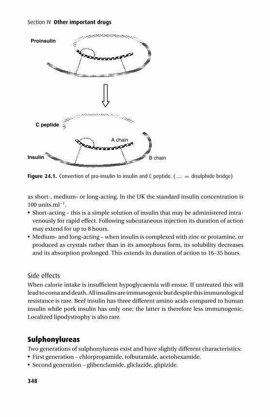
P1: PSB Printer: Yet To Come
9780521704632c24 CUFX213A/Peck 9780521618168 December 29, 2007 15:7
Section IV Other important drugs
Insulin
A chain
B chain
C peptide
Proinsulin
Figure 24.1. Convertion of pro-insulin to insulin and C peptide. (..... = disulphide bridge)
as short-, medium- or long-acting. In the UK the standard insulin concentration is100 units.ml−1.� Short-acting – this is a simple solution of insulin that may be administered intra-
venously for rapid effect. Following subcutaneous injection its duration of actionmay extend for up to 8 hours.� Medium- and long-acting – when insulin is complexed with zinc or protamine, orproduced as crystals rather than in its amorphous form, its solubility decreasesand its absorption prolonged. This extends its duration of action to 16–35 hours.
Side effectsWhen calorie intake is insufficient hypoglycaemia will ensue. If untreated this willlead to coma and death. All insulins are immunogenic but despite this immunologicalresistance is rare. Beef insulin has three different amino acids compared to humaninsulin while pork insulin has only one; the latter is therefore less immunogenic.Localized lipodystrophy is also rare.
SulphonylureasTwo generations of sulphonylureas exist and have slightly different characteristics:� First generation – chlorpropamide, tolbutamide, acetohexamide.� Second generation – glibenclamide, gliclazide, glipizide.
348
P1: PSB Printer: Yet To Come
9780521704632c24 CUFX213A/Peck 9780521618168 December 29, 2007 15:7
24 Drugs used in Diabetes
UsesSulphonylureas are used to control type II or non-insulin dependent diabetes(NIDDM), but should not replace dietary control.
Mechanism of actionIn general these drugs work at the pancreas by displacing insulin from β-cells inthe islets of Langerhans. For this reason they are ineffective in insulin-dependentdiabetics who have no functioning β-cells. They may also induce β-cell hyperplasia,while reducing both glucagon secretion and hepatic insulinase activity. During long-term administration they also reduce peripheral resistance to insulin.
KineticsSulphonylureas are all well absorbed from the gut and have oral bioavailabilitiesgreater than 80%. Albumin binds these drugs extensively in the plasma. Chlor-propamide undergoes hepatic metabolism but to a much lesser extent than theothers. A significant fraction is excreted unchanged in the urine and in the presenceof renal failure its half-life is prolonged. The other drugs in this class are extensivelymetabolized in the liver to inactive metabolites (with the exception of glibenclamidewhose active metabolites are probably of little clinical significance), which are sub-sequently excreted in the urine. Cimetidine inhibits their metabolism and, therefore,potentiates their actions. Drugs that tend to increase blood sugar (thiazides, corti-costeroids, phenothiazines) antagonize the effects of the sulphonylureas and makediabetic control harder.
The second-generation sulphonylureas bind to albumin by non-ionic forces incontrast with tolbutamide and chlorpropamide that bind by ionic forces. Thus,anionic drugs such as phenylbutazone, warfarin and salicylate do not displacesecond-generation drugs to such an extent and may be safer during concurrentdrug therapy.
Other effectsSide effects are generally mild but sometimes include gastrointestinal disturbancesand rashes.� Chlorpropamide – its long half-life and partial reliance on renal elimination results
in a greater chance of hypoglycaemia, especially in the elderly. It may also causefacial flushing and vomiting following alcohol and rarely may enhance antidiuretichormone secretion resulting in hyponatraemia. Photosensitivity may also occur.� Tolbutamide – this drug has been associated with cholestatic jaundice, derangedliver function and blood dyscrasias.
349
P1: PSB Printer: Yet To Come
9780521704632c24 CUFX213A/Peck 9780521618168 December 29, 2007 15:7
Section IV Other important drugs
Biguanides
MetforminMetformin is the only currently available biguanide in the UK.
Mechanism of actionThis is thought to include delayed uptake of glucose from the gut, increased periph-eral insulin sensitivity (increasing peripheral glucose utilization) and inhibition ofhepatic and renal gluconeogenesis.
Other effectsGastrointestinal disturbances including diarrhoea sometimes occur but are gener-ally mild. More significantly is its ability to precipitate severe lactic acidosis especiallyif taken by alcohol abusers or in the presence of renal impairment. It also lowersplasma cholesterol, triglycerides and low-density lipoproteins.
KineticsMetformin is slowly absorbed from the gut with a bioavailability of 60%. It is notbound by plasma proteins and is excreted unchanged in the urine. Renal impairmentsignificantly prolongs its actions.
Other antidiabetics
AcarboseAcarbose is a competitive inhibitor of intestinal α-glucosidase with a specific actionon sucrase. Polysaccharides are therefore not metabolized into their absorbablemonosaccharide form. This delay in intestinal glucose absorption reduces the post-prandial hyperglycaemia normally seen in diabetics.
It is not absorbed significantly and has few other systemic effects. Owing to anincreased carbohydrate load reaching the bacterial flora in the large bowel patientsmay experience borborygmi, flatulence and diarrhoea. Hepatic transaminases maybe transiently raised.
Thiazolidinediones
Rosiglitazone and pioglitazoneThiazolidinediones are oral antidiabetic agents with a high oral bioavailability. Thereeffects may take weeks to be fully established due to their mechanism of action.Troglitazone was withdrawn due to hepatitis and consequently patients taking thi-azolidinediones require liver function monitoring.
350
P1: PSB Printer: Yet To Come
9780521704632c24 CUFX213A/Peck 9780521618168 December 29, 2007 15:7
24 Drugs used in Diabetes
Mechanism of actionThiazolidinediones activate peroxisome proliferator-activated receptor γ (PPARγ –situated within the cell nucleus) and thereby regulate a number of genes involved inglucose and lipid metabolism. As a result they act to improve insulin sensitivity.
Other effectsThey may cause fluid retention and should not be given to those with significantheart failure.
KineticsBoth drugs are extensively metabolized by hepatic cytochrome P450. Rosiglitazonemetabolites are essentially inactive and mainly excreted in the urine. Some of thepioglitazone metabolites are active and are excreted in faeces and urine. Neitherdrug appears to have interactions with other drugs utilising the cytochrome P450system.
Meglitinides
Repaglinide and metiglinideMeglitinides are oral antidiabetic agents that may be of most use in treating patientswith erratic eating habits as they are relatively short acting. They are taken just beforemeals.
Mechanism of actionMeglitinides block the potassium channels in beta cells in the pancreas, closing theATP-dependent potassium channels and opening the cells’ calcium channels. Theresulting calcium influx causes the cells to secrete insulin.
KineticsRepaglinide has an oral bioavailability of 50%, is 98% plasma protein bound and ismetabolized in the liver by CYP3A4 and therefore is potentially susceptible to druginteractions. Its elimination half-life is 1 hour.
Peri-operative care of the diabetic patientA regional technique is often more appropriate as the patient will be able to eat andreturn to their normal drug regime more rapidly.
If a general anaesthetic is indicated the aim is to minimize the metabolic derange-ment by providing a balance of fluid, electrolytes, glucose and insulin. There is nosingle best way to achieve this and the approach chosen will reflect the type of dia-betes, medication and proposed operation.
351
P1: PSB Printer: Yet To Come
9780521704632c24 CUFX213A/Peck 9780521618168 December 29, 2007 15:7
Section IV Other important drugs
Diabetes Type IIMedication Long-acting tabletsOperation MinorAction Transfer to short-acting tablets 1 week before surgery. Omit tablets on
morning of operation. First on list. Treat as non-diabetic if blood sugar<7 mmol.l−1. Restart tablets with first meal.
Diabetes Type IIMedication Long-acting tabletsOperation MajorAction Treat as Type I diabetes. Once eating convert to soluble insulin before meals
and revert to tablets when total insulin requirement is <20 i.u.day−1.Diabetes Type IMedication InsulinOperation Major or minorAction Alberti regime or insulin sliding scale
Alberti regimeInfusion of 500 ml 10% dextrose with 10 i.u. actrapid and 10 mmol KCl starting at 100ml.h−1. Blood sugar and potassium are measured every 2 hours and if adjustmentsare required a new infusion is prepared.
Insulin sliding scaleAn infusion of 10% dextrose at 100 ml.h−1 is given alongside an infusion of solubleinsulin. The starting hourly insulin rate is calculated by dividing the total daily unitsby 24. Blood sugar is measured hourly and the insulin rate is adjusted accordingly.
352
P1: PSB Printer: Yet To Come
9780521704632c25 CUFX213A/Peck 9780521618168 December 29, 2007 15:15
25Corticosteroids and otherhormone preparations
� Glucocorticoids� Drugs used in thyroid disease� Oral contraceptives� Hormone replacement therapy
GlucocorticoidsThe adrenal cortex releases two classes of steroidal hormones into the circulation:� Glucocorticoids (from the zona fasciculata and zona reticularis)� Mineralocorticoids (from the zona glomerulosa)The main endogenous glucocorticoid in humans is hydrocortisone. Synthetic gluco-corticoids include prednisolone, methylprednisolone, betamethasone, dexametha-sone and triamcinolone. They have metabolic, anti-inflammatory and immuno-suppressive effects.
Metabolic effectsGlucocorticoids facilitate gluconeogenesis (i.e. synthesis of glucose from a non-carbohydrate source, e.g. protein). Glycogen deposition and glucose release fromthe liver are stimulated but the peripheral uptake of glucose is inhibited. Proteincatabolism is stimulated and synthesis inhibited.
When exogenous glucocorticoid is given in high doses or for prolonged periods, thealtered metabolism causes unwanted effects. Increased protein catabolism leads tomuscle weakness and wasting. The skin becomes thin leading to striae, and gastricmucosa becomes susceptible to ulceration. Dietary protein will not reverse thesechanges. Altered carbohydrate metabolism leads to hyperglycaemia and glycosuria.Diabetes may be provoked. Fat is redistributed from the extremities to the trunk,neck and face. Bone catabolism leads to osteoporosis.
Anti-inflammatory effectsGlucocorticoids reduce the production of tissue transudate and cell oedema in acuteinflammation. Circulating polymorphs and macrophages are prevented from reach-ing inflamed tissue. The production of inflammatory mediators (prostaglandins,leukotrienes and platelet-activating factor) is suppressed by the stimulation oflipocortin, which inhibits phospholipase A2. Normally phospholipase A2 would
353
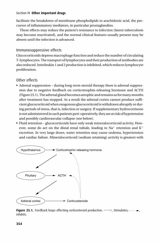
P1: PSB Printer: Yet To Come
9780521704632c25 CUFX213A/Peck 9780521618168 December 29, 2007 15:15
Section IV Other important drugs
facilitate the breakdown of membrane phospholipids to arachidonic acid, the pre-cursor of inflammatory mediators, in particular prostaglandins.
These effects may reduce the patient’s resistance to infection (latent tuberculosismay become reactivated), and the normal clinical features usually present may beabsent until the infection is advanced.
Immunosuppressive effectsGlucocorticoids depress macrophage function and reduce the number of circulatingT-lymphocytes. The transport of lymphocytes and their production of antibodies arealso reduced. Interleukin 1 and 2 production is inhibited, which reduces lymphocyteproliferation.
Other effects� Adrenal suppression – during long-term steroid therapy there is adrenal suppres-sion due to negative feedback on corticotrophin-releasing hormone and ACTH(Figure 25.1). The adrenal gland becomes atrophic and remains so for many monthsafter treatment has stopped. As a result the adrenal cortex cannot produce suffi-cient glucocorticoid when exogenous glucocorticoid is withdrawn abruptly or dur-ing periods of stress, that is, infection or surgery. If supplementary hydrocortisoneis not administered in such patients peri-operatively, they are at risk of hypotensionand possibly cardiovascular collapse (see below).� Fluid retention – glucocorticoids have only weak mineralocorticoid activity. How-ever, some do act on the distal renal tubule, leading to Na+ retention and K+
excretion. In very large doses, water retention may cause oedema, hypertensionand cardiac failure. Mineralocorticoid (sodium retaining) activity is greatest with
Hypothalamus Corticotrophin releasing hormone
ACTH
Corticosteroids
Pituitary
Adrenal cortex
Figure 25.1. Feedback loops affecting corticosteroid production. ——→, Stimulates; ,inhibits.
354
P1: PSB Printer: Yet To Come
9780521704632c25 CUFX213A/Peck 9780521618168 December 29, 2007 15:15
25 Corticosteroids and other hormone preparations
Table 25.1. Anti-inflammatory potential of various steroids.
Relative anti- Equivalent anti-
Drug inflammatory potency inflammatory dose (mg)
Hydrocortisone 1 100Prednisolone 4 25Methylprednisolone 5 20Triamcinolone 5 20Dexamethasone 25 4
hydrocortisone and cortisone, lesser with prednisolone and least with methylpred-nisolone and dexamethasone.� Vascular reactivity – glucocorticoids have a ‘permissive action’ on vascular smoothmuscle, allowing them to respond efficiently to circulating catecholamines. There-fore, glucocorticoid deficiency leads to an ineffective response by vascular smoothmuscle to circulating catecholamines. In sepsis there may be an inappropri-ately reduced production of cortisol so that higher levels of inotropic support arerequired. This may be improved by the administration of intravenous hydrocorti-sone.Other ‘permissive actions’ of glucocorticoids include the calorigenic effects of
glucagon and catecholamines, and the lipolytic and bronchodilator effects of cate-cholamines.
Peri-operative steroid supplementationThe magnitude of the surgical stimuls and the pre-operative dose of steroid needassessment in order to decide the most appropriate peri-operative supplementationregimen. Patients taking <10 mg prednisolone (or equivalent) pre-operatively have anormal hypothalamic-pituitary axis (HPA) and do not require peri-operative supple-mentation. Those taking >10 mg prednisolone require peri-operative supplementa-tion in addition to their normal steroid. For minor surgery 25 mg hydrocortisone atinduction is sufficient, for moderate surgery a further 100 mg hydrocortisone shouldbe added over the first 24 hours post-operatively, which should be prolonged for upto 72 hours for major surgery. The HPA should be considered to be suppressed forup to 3 months and maybe longer after stopping steroids.
Drugs used in thyroid disease
Thyroid replacement
Thyroxine (T4)The thyroid gland synthesizes triiodothyronine (T3) and thyroxine (T4) by combin-ing iodine with tyrosine residues present in thyroglobulin. The release of T3 and T4 is
355
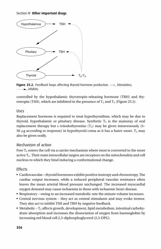
P1: PSB Printer: Yet To Come
9780521704632c25 CUFX213A/Peck 9780521618168 December 29, 2007 15:15
Section IV Other important drugs
Hypothalamus TSH
TSH
T3/T4
Pituitary
Thyroid
Figure 25.2. Feedback loops affecting thyroid hormone production. −→, Stimulates;, inhibits.
controlled by the hypothalamic thyrotropin-releasing hormone (TRH) and thy-rotropin (TSH), which are inhibited in the presence of T3 and T4 (Figure 25.2).
UsesReplacement hormone is required to treat hypothyroidism, which may be due tothyroid, hypothalamic or pituitary disease. Synthetic T4 is the mainstay of oralreplacement therapy but l-triiodothyronine (T3) may be given intravenously (5–50 µg according to response) in hypothyroid coma as it has a faster onset. T3 mayalso be given orally.
Mechanism of actionFree T4 enters the cell via a carrier mechanism where most is converted to the moreactive T3. Their main intracellular targets are receptors on the mitochondria and cellnucleus to which they bind inducing a conformational change.
Effects� Cardiovascular – thyroid hormones exhibit positive inotropy and chronotropy. Thecardiac output increases, while a reduced peripheral vascular resistance oftenleaves the mean arterial blood pressure unchanged. The increased myocardialoxygen demand may cause ischaemia in those with ischaemic heart disease.� Respiratory – owing to an increased metabolic rate the minute volume increases.� Central nervous system – they act as central stimulants and may evoke tremor.They also act to inhibit TSH and TRH by negative feedback.� Metabolic – T3 affects growth, development, lipid metabolism, intestinal carbohy-drate absorption and increases the dissociation of oxygen from haemoglobin byincreasing red blood cell 2,3-diphosphoglycerol (2,3-DPG).
356
P1: PSB Printer: Yet To Come
9780521704632c25 CUFX213A/Peck 9780521618168 December 29, 2007 15:15
25 Corticosteroids and other hormone preparations
KineticsWhen administered enterally both T3 and T4 are well absorbed from the gut. Oncein the plasma they are more than 99% bound to albumin and thyroxine-bindingglobulin. Some T4 is converted within the liver and kidneys to T3 or to the inactivereverse T3. Metabolism occurs in the liver and the conjugated products are excretedin bile. Up to 40% may be excreted unchanged in the urine.
Drugs used in the treatment of hyperthyroidism
CarbimazoleCarbimazole is a prodrug, being rapidly converted to methimazole in vivo.
Mechanism of actionCarbimazole prevents the synthesis of new T3 and T4 by inhibiting the oxidationof iodide to iodine and by inhibiting thyroid peroxidase, which is responsible foriodotryrosine synthesis and the coupling of iodotyrosines. Stored T3 and T4 is unaf-fected and so treatment with carbimazole requires weeks before the patient becomeseuthyroid.
Side effectsThese include rashes, arthralgia and pruritus. Agranulocytosis occurs rarely andmay be heralded by a sore throat. It crosses the placenta and may cause foetalhypothyroidism; however, it is not contraindicated during breast feeding as onlytiny amounts enter the breast milk.
KineticsCarbimazole is well absorbed from the gut and completely converted to methimazoleduring its passage through the liver. It increases the vascularity of and is metabolizedwithin the thyroid gland. It is minimally plasma protein-bound.
PropylthiouracilThis oral preparation blocks the iodination of tyrosine and partially blocks theperipheral conversion of T4 to T3. There is no parenteral formulation and it is reservedfor those patients intolerant of carbimazole. It has a slightly higher incidence ofagranulocytosis.
PropranololPropranolol is a β-blocker with negative inotropic and chronotropic activity. It willhelp control the sympathetic effects of a thyrotoxic crisis. However, it also blocks theperipheral conversion of T4 to T3 and blocks hypersensitivity to the actions of cate-cholamines. It is relatively contraindicated for patients with heart failure or reversibleairflow limitation. Guanethidine may be considered as an alternative in these cases.
357
P1: PSB Printer: Yet To Come
9780521704632c25 CUFX213A/Peck 9780521618168 December 29, 2007 15:15
Section IV Other important drugs
IodidesIodide has been given as potassium iodide orally, sodium iodide intravenously andiodide containing radiographic contrast dyes. It is concentrated in the follicular cellswhere peroxidases oxidize it to iodine, which is then combined with tyrosine resi-dues.
Iodide is required for normal thyroid function and when levels are too low or toohigh, thyroid function becomes abnormal. Large doses cause inhibition of iodidebinding and hence reduced hormone production. This effect is greatest when iodidetransport is increased, that is, in thyrotoxicosis. Iodide also reduces the effect of TSHon the thyroid gland and inhibits proteolysis of thyroglobulin. Thyroid vascularityis decreased, which is useful pre-operatively, but iodide is not used for prolongedperiods as its actions tend to diminish with time.
Lithium may be considered in those with iodide sensitivity.
Oral contraceptivesThe oral contraceptive pill (OCP) takes two forms: a combined oestrogen/progesterone preparation and a progesterone-only preparation. The former is takenfor 21 consecutive days followed by a 7 day break, while the latter is taken continu-ously.
Mechanism of actionThe combined preparation inhibits ovulation by suppressing gonadotrophin-releasing hormone and inhibiting gonadotrophin secretion. The progesterone-onlypill prevents ovulation in only 25% of women and appears to work by producing anunfavourable endometrium for implantation.
Side effects� Major – these include cholestatic jaundice and an increased incidence of thrombo-embolic disease. The following are considered a contraindication for the combinedOCP: a history of thrombo-embolism, obesity, smoking, hypertension or diabetesmellitus.� Minor – these include breakthrough bleeding, weight gain, breast tenderness,headache and nausea. Hirsutism and depression may occur following the com-bined OCP. Side effects are lower in the progesterone-only pill.
Hormone replacement therapyHormone replacement therapy (HRT; oestrogen or oestrogen/progesterone combi-nations) is used to relieve menopausal symptoms, reduce osteoporosis and reducethe risk of cardiovascular disease.
358
P1: PSB Printer: Yet To Come
9780521704632c25 CUFX213A/Peck 9780521618168 December 29, 2007 15:15
25 Corticosteroids and other hormone preparations
Side effects� Endometrial cancer – oestrogen therapy alone increases the risk of endometrialcancer and is therefore reserved for those who have had a hysterectomy. Womenwith a uterus require combined oestrogen/progesterone therapy.� Breast cancer – oncerns have been raised regarding HRT and breast cancerand there may be a slightly higher risk of breast cancer with combined oestro-gen/progesterone therapy compared to oestrogen alone.� Deep vein thrombosis – HRT may increase the risk of deep vein thrombosisalthough the absolute risk is low. The mechanism is unclear, but HRT may pro-voke changes in relation to the haemostatic system, increasing age of patients andunmasking of thrombotic traits.
Peri-operative HRTThere is no evidence to support stopping HRT in the peri-operative period providedthe usual thromboprophylaxis protocols are followed.
359
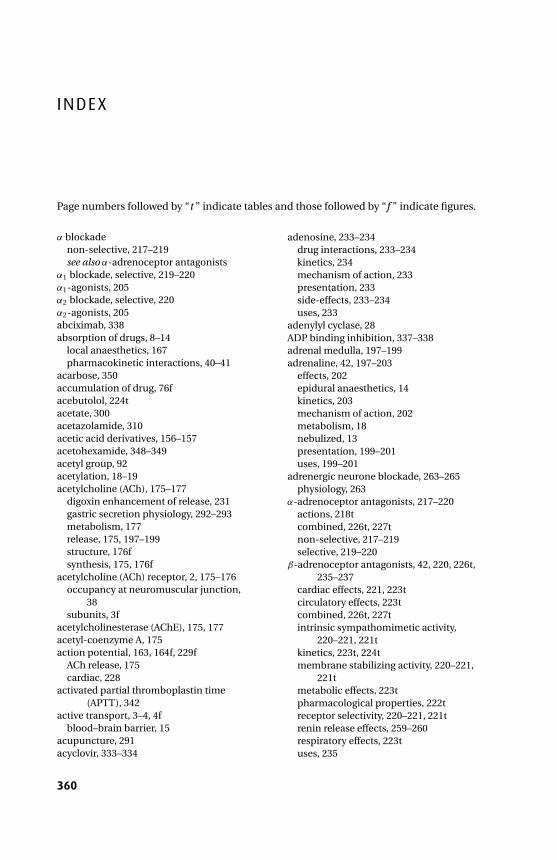
P1: PSB Printer: Yet To Come
9780521704632ind CUFX213A/Peck 9780521618168 December 29, 2007 17:19
INDEX
Page numbers followed by “t ” indicate tables and those followed by “f ” indicate figures.
α blockadenon-selective, 217–219see also α-adrenoceptor antagonists
α1 blockade, selective, 219–220α1-agonists, 205α2 blockade, selective, 220α2-agonists, 205abciximab, 338absorption of drugs, 8–14
local anaesthetics, 167pharmacokinetic interactions, 40–41
acarbose, 350accumulation of drug, 76facebutolol, 224tacetate, 300acetazolamide, 310acetic acid derivatives, 156–157acetohexamide, 348–349acetyl group, 92acetylation, 18–19acetylcholine (ACh), 175–177
digoxin enhancement of release, 231gastric secretion physiology, 292–293metabolism, 177release, 175, 197–199structure, 176fsynthesis, 175, 176f
acetylcholine (ACh) receptor, 2, 175–176occupancy at neuromuscular junction,
38subunits, 3f
acetylcholinesterase (AChE), 175, 177acetyl-coenzyme A, 175action potential, 163, 164f, 229f
ACh release, 175cardiac, 228
activated partial thromboplastin time(APTT), 342
active transport, 3–4, 4fblood–brain barrier, 15
acupuncture, 291acyclovir, 333–334
adenosine, 233–234drug interactions, 233–234kinetics, 234mechanism of action, 233presentation, 233side-effects, 233–234uses, 233
adenylyl cyclase, 28ADP binding inhibition, 337–338adrenal medulla, 197–199adrenaline, 42, 197–203
effects, 202epidural anaesthetics, 14kinetics, 203mechanism of action, 202metabolism, 18nebulized, 13presentation, 199–201uses, 199–201
adrenergic neurone blockade, 263–265physiology, 263
α-adrenoceptor antagonists, 217–220actions, 218tcombined, 226t, 227tnon-selective, 217–219selective, 219–220
β-adrenoceptor antagonists, 42, 220, 226t,235–237
cardiac effects, 221, 223tcirculatory effects, 223tcombined, 226t, 227tintrinsic sympathomimetic activity,
220–221, 221tkinetics, 223t, 224tmembrane stabilizing activity, 220–221,
221tmetabolic effects, 223tpharmacological properties, 222treceptor selectivity, 220–221, 221trenin release effects, 259–260respiratory effects, 223tuses, 235
360
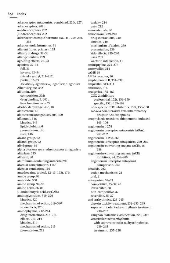
P1: PSB Printer: Yet To Come
9780521704632ind CUFX213A/Peck 9780521618168 December 29, 2007 17:19
361 Index
adrenoceptor antagonists, combined, 226t, 227tadrenoceptors, 201tα–adrenoceptors, 202β–adrenoceptors, 202adrenocorticotropic hormone (ACTH), 259–260,
354adrenosteroid hormones, 31afferent fibres, primary, 135affinity of drugs, 32–33after-potentials, 229age, drug effects, 22–23agonists, 32–33
full, 33inverse, 32–34mixed α and β, 211–212partial, 32–33see also α1-agonists; α2-agonists; β-agonists
Alberti regime, 352albumin, 303t
composition, 302tdrug binding, 7, 303tliver function tests, 22
alcohol dehydrogenase, 18aldosterone, 45aldosterone antagonists, 308–309alfentanil, 146
kinetics, 146lipid solubility, 6presentation, 146uses, 146
alkane group, 92alkanol group, 92alkyl group, 92alpha blockers see α-adrenoceptor antagonistsalteplase, 345althesin, 90aluminium-containing antacids, 292alveolar concentration, 116falveolar ventilation, 116amethocaine, topical, 12–13, 173t, 174tamide group, 92amiloride, 308amine group, 92–93amino acids, 86–88γ -aminobutyric acid see GABAaminoglycosides, 319–320
kinetics, 320mechanism of action, 319–320side-effects, 320
aminophylline, 212–214drug interactions, 213–214effects, 213–214kinetics, 214mechanism of action, 213presentation, 212
toxicity, 214uses, 212
aminosteroids, 90amiodarone, 239–240
drug interactions, 240kinetics, 240mechanism of action, 239presentation, 239side-effects, 239–240uses, 239warfarin interaction, 41
amitriptyline, 274–276amoxycillin, 314cAMP, 28AMPA receptor, 26amphoterocin B, 331–332ampicillin, 313–314amrinone, 216analgesics, 135–162
COX-2 inhibitorspreferential, 152t, 158–159specific, 152t, 159–162
non-specific COX inhibitors, 152t, 153–158see also non-steroidal anti-inflammatory
drugs (NSAIDs); opioidsanaphylactic reactions, thiopentone-induced,
105–106angiotensin I, 258angiotensin I receptor antagonists (ARAs),
261–262angiotensin II, 258–260angiotensin II receptor antagonists, 259–260angiotensin-converting enzyme (ACE), 18,
258angiotensin-converting enzyme (ACE)
inhibitors, 24, 259–260angiotensin I receptor antagonist
comparison, 262antacids, 292
action mechanisms, 24oral, 8
antagonists, 32–33competitive, 35–37, 42irreversible, 38non-competitive, 37reversible, 35–37
anti-arrhythmics, 228–245digoxin toxicity treatment, 232–233, 245supraventricular tachyarrhythmia treatment,
230–237Vaughan–Williams classification, 229, 231tventricular tachyarrhythmia
with supraventricular tachyarrhythmias,239–245
treatment, 237–238
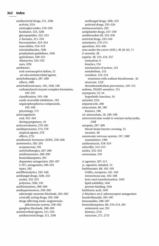
P1: PSB Printer: Yet To Come
9780521704632ind CUFX213A/Peck 9780521618168 December 29, 2007 17:19
362 Index
antibacterial drugs, 311, 328tactivity, 331taminoglycosides, 319–320fusidanes, 325, 328tglycopeptides, 322–323β-lactams, 311–318lincosamides, 323–324macrolides, 318–319nitroimidazoles, 328tprophylaxis guidelines, 326tquinolones, 320–321rifamycins, 324–325uses, 329t
antibioticsoral contraceptive failure, 21see also antimicrobial agents
anticholinergics, 287–289effects, 288t
anticholinesterases, 192, 194f, 196carbamylated enzyme complex formation,
193–195classification, 193–196easily reversible inhibition, 193organophosphorus compounds,
195–196physiology, 175
anticoagulantsoral, 342–344during pregnancy, 16
anticonvulsants, 279–281antidepressants, 274–278
atypical agents, 278effects, 275t
antidiuretic hormone (ADH), 259–260antiemetics, 282–291
acupuncture, 291anticholinergics, 287–289antihistamines, 289–290benzodiazepines, 291dopamine antagonists, 283–2875-HT3 antagonists, 290–291types, 282
antifibrinolytics, 345–346antifungal drugs, 328t, 333
azoles, 332–333polyenes, 328t, 332
antihistamines, 289–290antihypertensives, 258–269
adrenergic neurone blockade, 263–265centrally acting drugs, 265–268drugs affecting renin–angiotensin–
aldosterone system, 258–262ganglion blockade, 268–269
antimicrobial agents, 311–334antibacterial drugs, 311, 328t
antifungal drugs, 328t, 333antiviral drugs, 333–334
antimuscarinics, 295antiplatelet drugs, 337–339antithrombin III, 335–336antiviral drugs, 333–334anxiolytics, 270–274aprotinin, 345–346area under the curve (AUC), 9f, 62–63, 71β-arrestin, 28aspirin, 38, 153–154, 337
ionization, 6kinetics, 154mechanism of action, 153metabolism, 153overdose, 153–154
treatment with sodium bicarbonate, 42structure, 152fthromboembolism prevention, 149–151
asthma, NSAID-sensitive, 151asymptote, 52–54
hyperbolic function, 54atenolol, 224tatipamezole, 268atracurium, 90, 189
kinetics, 189cis-atracurium, 18, 189–190atrioventricular nodal re-entrant tachycardia,
230fatropine, 287–289
blood–brain barrier crossing, 15racemic, 48
autonomic nervous system, 197, 198ftransmitters, 199t
azithromycin, 318–319azlocillin, 314–315azoles, 332–333aztreonam, 318
β-agonists, 207–211β2-agonists, inhaled, 13barbiturates, 88, 102–103
GABAA receptors, 101–102intravenous use, 103–108keto-enol transformation, 105flipid solubility, 104tprotein binding, 104t
barbituric acid, 103fβ-blockers see β-adrenoceptor antagonistsbendrofluazide, 305–307benzamides, 286–287benzodiazepines, 89, 270–274, 281
antiemetic use, 291kinetics, 272tstructure, 271, 271f
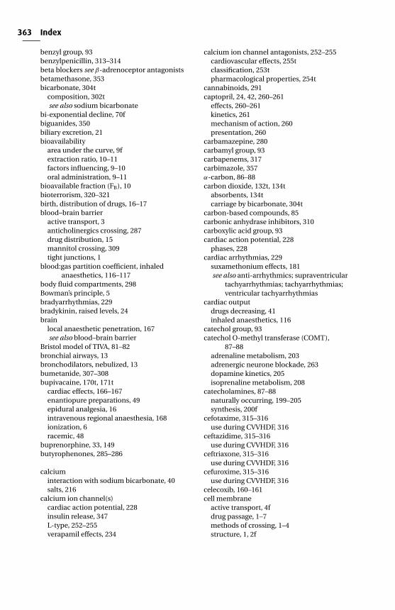
P1: PSB Printer: Yet To Come
9780521704632ind CUFX213A/Peck 9780521618168 December 29, 2007 17:19
363 Index
benzyl group, 93benzylpenicillin, 313–314beta blockers see β-adrenoceptor antagonistsbetamethasone, 353bicarbonate, 304t
composition, 302tsee also sodium bicarbonate
bi-exponential decline, 70fbiguanides, 350biliary excretion, 21bioavailability
area under the curve, 9fextraction ratio, 10–11factors influencing, 9–10oral administration, 9–11
bioavailable fraction (FB), 10bioterrorism, 320–321birth, distribution of drugs, 16–17blood–brain barrier
active transport, 3anticholinergics crossing, 287drug distribution, 15mannitol crossing, 309tight junctions, 1
blood:gas partition coefficient, inhaledanaesthetics, 116–117
body fluid compartments, 298Bowman’s principle, 5bradyarrhythmias, 229bradykinin, raised levels, 24brain
local anaesthetic penetration, 167see also blood–brain barrier
Bristol model of TIVA, 81–82bronchial airways, 13bronchodilators, nebulized, 13bumetanide, 307–308bupivacaine, 170t, 171t
cardiac effects, 166–167enantiopure preparations, 49epidural analgesia, 16intravenous regional anaesthesia, 168ionization, 6racemic, 48
buprenorphine, 33, 149butyrophenones, 285–286
calciuminteraction with sodium bicarbonate, 40salts, 216
calcium ion channel(s)cardiac action potential, 228insulin release, 347L-type, 252–255verapamil effects, 234
calcium ion channel antagonists, 252–255cardiovascular effects, 255tclassification, 253tpharmacological properties, 254t
cannabinoids, 291captopril, 24, 42, 260–261
effects, 260–261kinetics, 261mechanism of action, 260presentation, 260
carbamazepine, 280carbamyl group, 93carbapenems, 317carbimazole, 357α-carbon, 86–88carbon dioxide, 132t, 134t
absorbents, 134tcarriage by bicarbonate, 304t
carbon-based compounds, 85carbonic anhydrase inhibitors, 310carboxylic acid group, 93cardiac action potential, 228
phases, 228cardiac arrhythmias, 229
suxamethonium effects, 181see also anti-arrhythmics; supraventricular
tachyarrhythmias; tachyarrhythmias;ventricular tachyarrhythmias
cardiac outputdrugs decreasing, 41inhaled anaesthetics, 116
catechol group, 93catechol O-methyl transferase (COMT),
87–88adrenaline metabolism, 203adrenergic neurone blockade, 263dopamine kinetics, 205isoprenaline metabolism, 208
catecholamines, 87–88naturally occurring, 199–205synthesis, 200f
cefotaxime, 315–316use during CVVHDF, 316
ceftazidime, 315–316use during CVVHDF, 316
ceftriaxone, 315–316use during CVVHDF, 316
cefuroxime, 315–316use during CVVHDF, 316
celecoxib, 160–161cell membrane
active transport, 4fdrug passage, 1–7methods of crossing, 1–4structure, 1, 2f
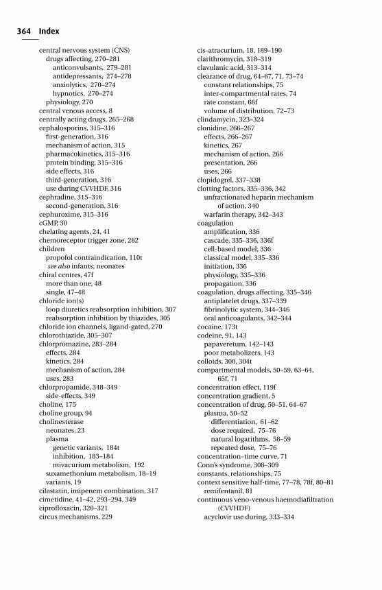
P1: PSB Printer: Yet To Come
9780521704632ind CUFX213A/Peck 9780521618168 December 29, 2007 17:19
364 Index
central nervous system (CNS)drugs affecting, 270–281
anticonvulsants, 279–281antidepressants, 274–278anxiolytics, 270–274hypnotics, 270–274
physiology, 270central venous access, 8centrally acting drugs, 265–268cephalosporins, 315–316
first-generation, 316mechanism of action, 315pharmacokinetics, 315–316protein binding, 315–316side effects, 316third-generation, 316use during CVVHDF, 316
cephradine, 315–316second-generation, 316
cephuroxime, 315–316cGMP, 30chelating agents, 24, 41chemoreceptor trigger zone, 282children
propofol contraindication, 110tsee also infants; neonates
chiral centres, 47fmore than one, 48single, 47–48
chloride ion(s)loop diuretics reabsorption inhibition, 307reabsorption inhibition by thiazides, 305
chloride ion channels, ligand-gated, 270chlorothiazide, 305–307chlorpromazine, 283–284
effects, 284kinetics, 284mechanism of action, 284uses, 283
chlorpropamide, 348–349side-effects, 349
choline, 175choline group, 94cholinesterase
neonates, 23plasma
genetic variants, 184tinhibition, 183–184mivacurium metabolism, 192
suxamethonium metabolism, 18–19variants, 19
cilastatin, imipenem combination, 317cimetidine, 41–42, 293–294, 349ciprofloxacin, 320–321circus mechanisms, 229
cis-atracurium, 18, 189–190clarithromycin, 318–319clavulanic acid, 313–314clearance of drug, 64–67, 71, 73–74
constant relationships, 75inter-compartmental rates, 74rate constant, 66fvolume of distribution, 72–73
clindamycin, 323–324clonidine, 266–267
effects, 266–267kinetics, 267mechanism of action, 266presentation, 266uses, 266
clopidogrel, 337–338clotting factors, 335–336, 342
unfractionated heparin mechanismof action, 340
warfarin therapy, 342–343coagulation
amplification, 336cascade, 335–336, 336fcell-based model, 336classical model, 335–336initiation, 336physiology, 335–336propagation, 336
coagulation, drugs affecting, 335–346antiplatelet drugs, 337–339fibrinolytic system, 344–346oral anticoagulants, 342–344
cocaine, 173tcodeine, 91, 143
papaveretum, 142–143poor metabolizers, 143
colloids, 300, 304tcompartmental models, 50–59, 63–64,
65f, 71concentration effect, 119fconcentration gradient, 5concentration of drug, 50–51, 64–67
plasma, 50–52differentiation, 61–62dose required, 75–76natural logarithms, 58–59repeated dose, 75–76
concentration–time curve, 71Conn’s syndrome, 308–309constants, relationships, 75context sensitive half-time, 77–78, 78f, 80–81
remifentanil, 81continuous veno-venous haemodiafiltration
(CVVHDF)acyclovir use during, 333–334
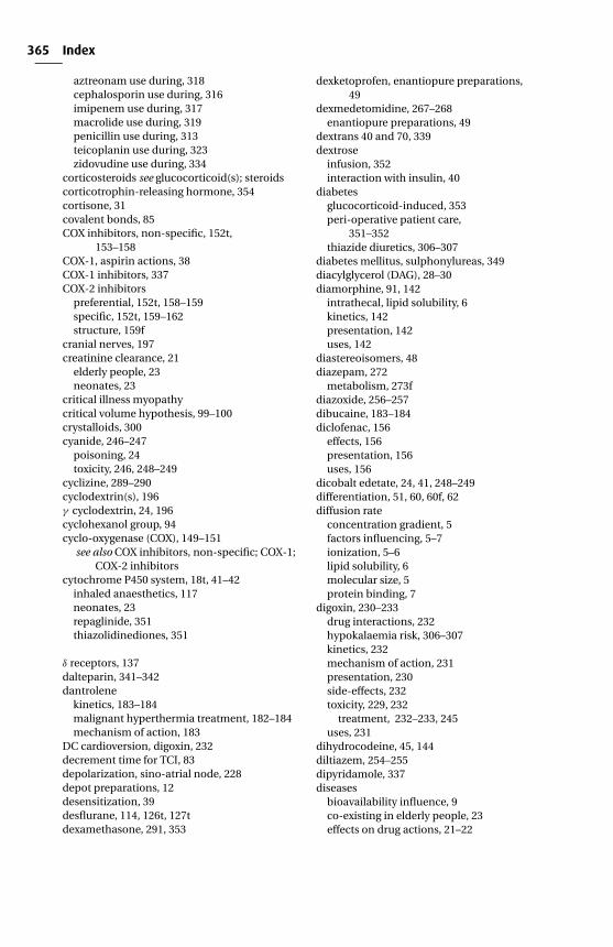
P1: PSB Printer: Yet To Come
9780521704632ind CUFX213A/Peck 9780521618168 December 29, 2007 17:19
365 Index
aztreonam use during, 318cephalosporin use during, 316imipenem use during, 317macrolide use during, 319penicillin use during, 313teicoplanin use during, 323zidovudine use during, 334
corticosteroids see glucocorticoid(s); steroidscorticotrophin-releasing hormone, 354cortisone, 31covalent bonds, 85COX inhibitors, non-specific, 152t,
153–158COX-1, aspirin actions, 38COX-1 inhibitors, 337COX-2 inhibitors
preferential, 152t, 158–159specific, 152t, 159–162structure, 159f
cranial nerves, 197creatinine clearance, 21
elderly people, 23neonates, 23
critical illness myopathycritical volume hypothesis, 99–100crystalloids, 300cyanide, 246–247
poisoning, 24toxicity, 246, 248–249
cyclizine, 289–290cyclodextrin(s), 196γ cyclodextrin, 24, 196cyclohexanol group, 94cyclo-oxygenase (COX), 149–151
see also COX inhibitors, non-specific; COX-1;COX-2 inhibitors
cytochrome P450 system, 18t, 41–42inhaled anaesthetics, 117neonates, 23repaglinide, 351thiazolidinediones, 351
δ receptors, 137dalteparin, 341–342dantrolene
kinetics, 183–184malignant hyperthermia treatment, 182–184mechanism of action, 183
DC cardioversion, digoxin, 232decrement time for TCI, 83depolarization, sino-atrial node, 228depot preparations, 12desensitization, 39desflurane, 114, 126t, 127tdexamethasone, 291, 353
dexketoprofen, enantiopure preparations,49
dexmedetomidine, 267–268enantiopure preparations, 49
dextrans 40 and 70, 339dextrose
infusion, 352interaction with insulin, 40
diabetesglucocorticoid-induced, 353peri-operative patient care,
351–352thiazide diuretics, 306–307
diabetes mellitus, sulphonylureas, 349diacylglycerol (DAG), 28–30diamorphine, 91, 142
intrathecal, lipid solubility, 6kinetics, 142presentation, 142uses, 142
diastereoisomers, 48diazepam, 272
metabolism, 273fdiazoxide, 256–257dibucaine, 183–184diclofenac, 156
effects, 156presentation, 156uses, 156
dicobalt edetate, 24, 41, 248–249differentiation, 51, 60, 60f, 62diffusion rate
concentration gradient, 5factors influencing, 5–7ionization, 5–6lipid solubility, 6molecular size, 5protein binding, 7
digoxin, 230–233drug interactions, 232hypokalaemia risk, 306–307kinetics, 232mechanism of action, 231presentation, 230side-effects, 232toxicity, 229, 232
treatment, 232–233, 245uses, 231
dihydrocodeine, 45, 144diltiazem, 254–255dipyridamole, 337diseases
bioavailability influence, 9co-existing in elderly people, 23effects on drug actions, 21–22
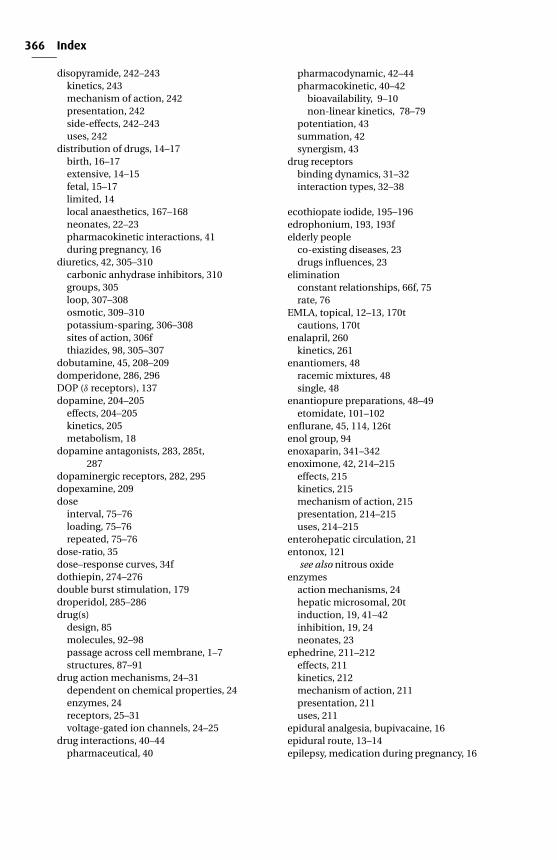
P1: PSB Printer: Yet To Come
9780521704632ind CUFX213A/Peck 9780521618168 December 29, 2007 17:19
366 Index
disopyramide, 242–243kinetics, 243mechanism of action, 242presentation, 242side-effects, 242–243uses, 242
distribution of drugs, 14–17birth, 16–17extensive, 14–15fetal, 15–17limited, 14local anaesthetics, 167–168neonates, 22–23pharmacokinetic interactions, 41during pregnancy, 16
diuretics, 42, 305–310carbonic anhydrase inhibitors, 310groups, 305loop, 307–308osmotic, 309–310potassium-sparing, 306–308sites of action, 306fthiazides, 98, 305–307
dobutamine, 45, 208–209domperidone, 286, 296DOP (δ receptors), 137dopamine, 204–205
effects, 204–205kinetics, 205metabolism, 18
dopamine antagonists, 283, 285t,287
dopaminergic receptors, 282, 295dopexamine, 209dose
interval, 75–76loading, 75–76repeated, 75–76
dose-ratio, 35dose–response curves, 34fdothiepin, 274–276double burst stimulation, 179droperidol, 285–286drug(s)
design, 85molecules, 92–98passage across cell membrane, 1–7structures, 87–91
drug action mechanisms, 24–31dependent on chemical properties, 24enzymes, 24receptors, 25–31voltage-gated ion channels, 24–25
drug interactions, 40–44pharmaceutical, 40
pharmacodynamic, 42–44pharmacokinetic, 40–42
bioavailability, 9–10non-linear kinetics, 78–79
potentiation, 43summation, 42synergism, 43
drug receptorsbinding dynamics, 31–32interaction types, 32–38
ecothiopate iodide, 195–196edrophonium, 193, 193felderly people
co-existing diseases, 23drugs influences, 23
eliminationconstant relationships, 66f, 75rate, 76
EMLA, topical, 12–13, 170tcautions, 170t
enalapril, 260kinetics, 261
enantiomers, 48racemic mixtures, 48single, 48
enantiopure preparations, 48–49etomidate, 101–102
enflurane, 45, 114, 126tenol group, 94enoxaparin, 341–342enoximone, 42, 214–215
effects, 215kinetics, 215mechanism of action, 215presentation, 214–215uses, 214–215
enterohepatic circulation, 21entonox, 121
see also nitrous oxideenzymes
action mechanisms, 24hepatic microsomal, 20tinduction, 19, 41–42inhibition, 19, 24neonates, 23
ephedrine, 211–212effects, 211kinetics, 212mechanism of action, 211presentation, 211uses, 211
epidural analgesia, bupivacaine, 16epidural route, 13–14epilepsy, medication during pregnancy, 16
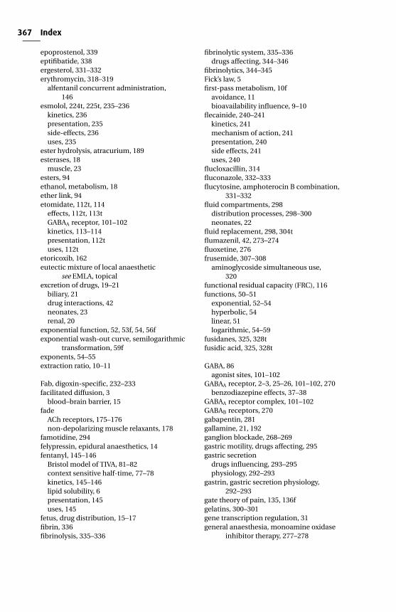
P1: PSB Printer: Yet To Come
9780521704632ind CUFX213A/Peck 9780521618168 December 29, 2007 17:19
367 Index
epoprostenol, 339eptifibatide, 338ergesterol, 331–332erythromycin, 318–319
alfentanil concurrent administration,146
esmolol, 224t, 225t, 235–236kinetics, 236presentation, 235side-effects, 236uses, 235
ester hydrolysis, atracurium, 189esterases, 18
muscle, 23esters, 94ethanol, metabolism, 18ether link, 94etomidate, 112t, 114
effects, 112t, 113tGABAA receptor, 101–102kinetics, 113–114presentation, 112tuses, 112t
etoricoxib, 162eutectic mixture of local anaesthetic
see EMLA, topicalexcretion of drugs, 19–21
biliary, 21drug interactions, 42neonates, 23renal, 20
exponential function, 52, 53f, 54, 56fexponential wash-out curve, semilogarithmic
transformation, 59fexponents, 54–55extraction ratio, 10–11
Fab, digoxin-specific, 232–233facilitated diffusion, 3
blood–brain barrier, 15fade
ACh receptors, 175–176non-depolarizing muscle relaxants, 178
famotidine, 294felypressin, epidural anaesthetics, 14fentanyl, 145–146
Bristol model of TIVA, 81–82context sensitive half-time, 77–78kinetics, 145–146lipid solubility, 6presentation, 145uses, 145
fetus, drug distribution, 15–17fibrin, 336fibrinolysis, 335–336
fibrinolytic system, 335–336drugs affecting, 344–346
fibrinolytics, 344–345Fick’s law, 5first-pass metabolism, 10f
avoidance, 11bioavailability influence, 9–10
flecainide, 240–241kinetics, 241mechanism of action, 241presentation, 240side effects, 241uses, 240
flucloxacillin, 314fluconazole, 332–333flucytosine, amphoterocin B combination,
331–332fluid compartments, 298
distribution processes, 298–300neonates, 22
fluid replacement, 298, 304tflumazenil, 42, 273–274fluoxetine, 276frusemide, 307–308
aminoglycoside simultaneous use,320
functional residual capacity (FRC), 116functions, 50–51
exponential, 52–54hyperbolic, 54linear, 51logarithmic, 54–59
fusidanes, 325, 328tfusidic acid, 325, 328t
GABA, 86agonist sites, 101–102
GABAA receptor, 2–3, 25–26, 101–102, 270benzodiazepine effects, 37–38
GABAA receptor complex, 101–102GABAB receptors, 270gabapentin, 281gallamine, 21, 192ganglion blockade, 268–269gastric motility, drugs affecting, 295gastric secretion
drugs influencing, 293–295physiology, 292–293
gastrin, gastric secretion physiology,292–293
gate theory of pain, 135, 136fgelatins, 300–301gene transcription regulation, 31general anaesthesia, monoamine oxidase
inhibitor therapy, 277–278
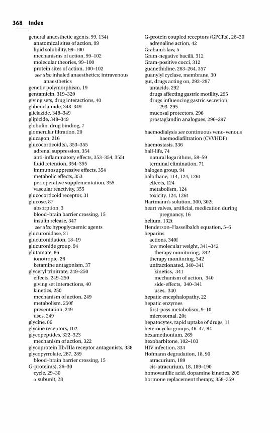
P1: PSB Printer: Yet To Come
9780521704632ind CUFX213A/Peck 9780521618168 December 29, 2007 17:19
368 Index
general anaesthetic agents, 99, 134tanatomical sites of action, 99lipid solubility, 99–100mechanisms of action, 99–102molecular theories, 99–100protein sites of action, 100–102see also inhaled anaesthetics; intravenous
anaestheticsgenetic polymorphism, 19gentamicin, 319–320giving sets, drug interactions, 40glibenclamide, 348–349gliclazide, 348–349glipizide, 348–349globulin, drug binding, 7glomerular filtration, 20glucagon, 216glucocorticoid(s), 353–355
adrenal suppression, 354anti-inflammatory effects, 353–354, 355tfluid retention, 354–355immunosuppressive effects, 354metabolic effects, 353perioperative supplementation, 355vascular reactivity, 355
glucocorticoid receptor, 31glucose, 87
absorption, 3blood–brain barrier crossing, 15insulin release, 347see also hypoglycaemic agents
glucuronidase, 21glucuronidation, 18–19glucuronide group, 94glutamate, 86
ionotropic, 26ketamine antagonism, 37
glyceryl trinitrate, 249–250effects, 249–250giving set interactions, 40kinetics, 250mechanism of action, 249metabolism, 250fpresentation, 249uses, 249
glycine, 86glycine receptors, 102glycopeptides, 322–323
mechanism of action, 322glycoprotein IIb/IIIa receptor antagonists, 338glycopyrrolate, 287, 289
blood–brain barrier crossing, 15G-protein(s), 26–30
cycle, 29–30α subunit, 28
G-protein coupled receptors (GPCRs), 26–30adrenaline action, 42
Graham’s law, 5Gram-negative bacilli, 312Gram-positive cocci, 312guanethidine, 263–264, 357guanylyl cyclase, membrane, 30gut, drugs acting on, 292–297
antacids, 292drugs affecting gastric motility, 295drugs influencing gastric secretion,
293–295mucosal protectors, 296prostaglandin analogues, 296–297
haemodialysis see continuous veno-venoushaemodiafiltration (CVVHDF)
haemostasis, 336half-life, 74
natural logarithms, 58–59terminal elimination, 71
halogen group, 94halothane, 114, 124, 126t
effects, 124metabolism, 124toxicity, 124, 126t
Hartmann’s solution, 300, 302theart valves, artificial, medication during
pregnancy, 16helium, 132tHenderson–Hasselbalch equation, 5–6heparins
actions, 340flow molecular weight, 341–342
therapy monitoring, 342therapy monitoring, 342unfractionated, 340–341
kinetics, 341mechanism of action, 340side-effects, 340–341uses, 340
hepatic encephalopathy, 22hepatic enzymes
first-pass metabolism, 9–10microsomal, 20t
hepatocytes, rapid uptake of drugs, 11heterocyclic groups, 46–47, 94hexamethonium, 269hexobarbitone, 102–103HIV infection, 334Hofmann degradation, 18, 90
atracurium, 189cis-atracurium, 18, 189–190
homovanillic acid, dopamine kinetics, 205hormone replacement therapy, 358–359
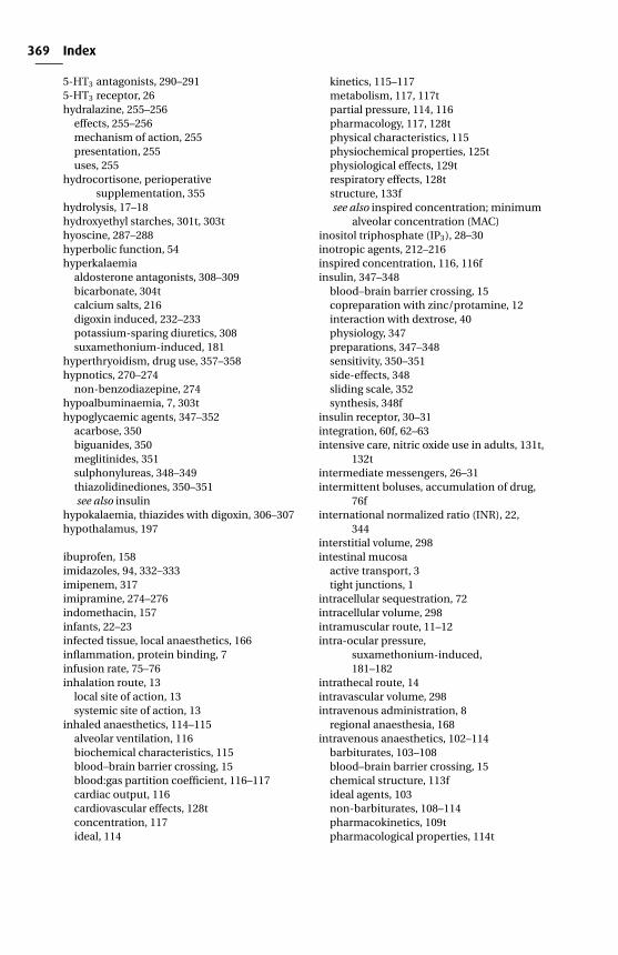
P1: PSB Printer: Yet To Come
9780521704632ind CUFX213A/Peck 9780521618168 December 29, 2007 17:19
369 Index
5-HT3 antagonists, 290–2915-HT3 receptor, 26hydralazine, 255–256
effects, 255–256mechanism of action, 255presentation, 255uses, 255
hydrocortisone, perioperativesupplementation, 355
hydrolysis, 17–18hydroxyethyl starches, 301t, 303thyoscine, 287–288hyperbolic function, 54hyperkalaemia
aldosterone antagonists, 308–309bicarbonate, 304tcalcium salts, 216digoxin induced, 232–233potassium-sparing diuretics, 308suxamethonium-induced, 181
hyperthryoidism, drug use, 357–358hypnotics, 270–274
non-benzodiazepine, 274hypoalbuminaemia, 7, 303thypoglycaemic agents, 347–352
acarbose, 350biguanides, 350meglitinides, 351sulphonylureas, 348–349thiazolidinediones, 350–351see also insulin
hypokalaemia, thiazides with digoxin, 306–307hypothalamus, 197
ibuprofen, 158imidazoles, 94, 332–333imipenem, 317imipramine, 274–276indomethacin, 157infants, 22–23infected tissue, local anaesthetics, 166inflammation, protein binding, 7infusion rate, 75–76inhalation route, 13
local site of action, 13systemic site of action, 13
inhaled anaesthetics, 114–115alveolar ventilation, 116biochemical characteristics, 115blood–brain barrier crossing, 15blood:gas partition coefficient, 116–117cardiac output, 116cardiovascular effects, 128tconcentration, 117ideal, 114
kinetics, 115–117metabolism, 117, 117tpartial pressure, 114, 116pharmacology, 117, 128tphysical characteristics, 115physiochemical properties, 125tphysiological effects, 129trespiratory effects, 128tstructure, 133fsee also inspired concentration; minimum
alveolar concentration (MAC)inositol triphosphate (IP3), 28–30inotropic agents, 212–216inspired concentration, 116, 116finsulin, 347–348
blood–brain barrier crossing, 15copreparation with zinc/protamine, 12interaction with dextrose, 40physiology, 347preparations, 347–348sensitivity, 350–351side-effects, 348sliding scale, 352synthesis, 348f
insulin receptor, 30–31integration, 60f, 62–63intensive care, nitric oxide use in adults, 131t,
132tintermediate messengers, 26–31intermittent boluses, accumulation of drug,
76finternational normalized ratio (INR), 22,
344interstitial volume, 298intestinal mucosa
active transport, 3tight junctions, 1
intracellular sequestration, 72intracellular volume, 298intramuscular route, 11–12intra-ocular pressure,
suxamethonium-induced,181–182
intrathecal route, 14intravascular volume, 298intravenous administration, 8
regional anaesthesia, 168intravenous anaesthetics, 102–114
barbiturates, 103–108blood–brain barrier crossing, 15chemical structure, 113fideal agents, 103non-barbiturates, 108–114pharmacokinetics, 109tpharmacological properties, 114t
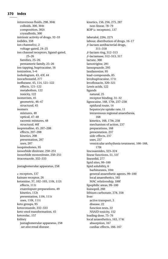
P1: PSB Printer: Yet To Come
9780521704632ind CUFX213A/Peck 9780521618168 December 29, 2007 17:19
370 Index
intravenous fluids, 298, 304tcolloids, 300, 304tcomposition, 302tcrystalloids, 300
intrinsic activity of drugs, 32–33iodides, 358ion channel(s), 2
voltage-gated, 24–25ion channel receptors, ligand-gated,
25–26families, 25–26pentameric family, 25–26
ion tapping, bupivacaine, 16ionization, 5–6isobologram, 43, 43f, 44isocarboxazid, 277isoflurane, 45, 114, 121–122
effects, 121–122metabolism, 122toxicity, 122
isomerism, 45geometric, 46–47structural, 45
isomersmixtures, 48optical, 47–48racemic mixtures, 48structural, 46f
isoprenaline, 45, 207–208effects, 207–208kinetics, 208presentation, 207uses, 207
isoquinolones, 95isosorbide dinitrate, 250–251isosorbide mononitrate, 250–251itraconazole, 332–333
juxtaglomerular apparatus, 258
κ receptors, 137kainate receptor, 26ketamine, 37, 102–103, 110t, 112t
effects, 111tenantiopure preparations, 49kinetics, 112tpresentation, 110t, 111tuses, 110t, 111t
keto groups, 95ketoconazole, 332–333keto-enol transformation, 45ketorolac, 157kidney
juxtaglomerular apparatus, 258see also renal disease
kinetics, 156, 256, 275, 287non-linear, 78–79
KOP (κ receptors), 137
labetalol, 226t, 227tlabour, distribution of drugs, 16–17β-lactam antibacterial drugs,
311–318β-lactam ring, 312–313β-lactamase, 312–313, 317lactate, 300lamotrigine, 281lansoprazole, 295laudanosine, 95lead-compounds, 85levobupivacaine, 171tlevofloxacin, 320–321Lewis acids, 122ligands
natural, 25receptor binding, 31–32
lignocaine, 168, 170t, 237–238epidural route, 14hepatocyte uptake rate, 11intravenous regional anaesthesia,
168kinetics, 168, 170t, 238mechanism of action, 237preparations, 168presentation, 237side-effects, 237uses, 237ventricular arrhythmia treatment, 166–168,
170tlincosamides, 323–324linear functions, 51, 51flinezolid, 277lipid sites, 99–100lipid solubility, 6
barbiturates, 104tgeneral anaesthetic agents, 99–100local anaesthetics, 165MAC relationship, 100f
lipophilic areas, 99–100lisinopril, 260lithium carbonate, 278, 358liver
active transport, 3disease, 22function tests, 22NSAID toxicity, 152
loading dose, 75–76local anaesthetics, 163, 174t
absorption, 167cardiac effects, 166–167
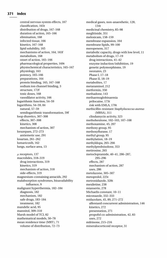
P1: PSB Printer: Yet To Come
9780521704632ind CUFX213A/Peck 9780521618168 December 29, 2007 17:19
371 Index
central nervous system effects, 167classification, 165tdistribution of drugs, 167–168duration of action, 165–166elimination, 168infected tissue, 166kinetics, 167–168lipid solubility, 165mechanisms of action, 164, 165fmetabolism, 168onset of action, 165–166pharmacological properties, 169tphysiochemical characteristics, 165–167physiology, 163potency, 165–166preparations, 164protein binding, 165, 167–168sodium ion channel binding, 3structure, 172ftoxic doses, 168vasodilator activity, 166
logarithmic function, 54–59logarithms, 54–59, 84
natural, 57–59semilogarithmic transformation, 59f
loop diuretics, 307–308effects, 307–308kinetics, 308mechanism of action, 307
lorazepam, 272–273antiemetic use, 291
losartan, 261–262lumaricoxib, 162lungs, surface area, 13
µ receptors, 137macrolides, 318–319
drug interactions, 319kinetics, 319mechanism of action, 318side-effects, 319
magnesium-containing antacids, 292malabsorption syndromes, bioavailability
influence, 9malignant hyperthermia, 182–184
diagnosis, 182mechanism, 182safe drugs, 183–184treatment, 182
mandelic acid, 95mannitol, 309–310Marsh model of TCI, 82mathematical models, 50–78mean residence time (MRT), 71
volume of distribution, 72–73
medical gases, non-anaesthetic, 128,134t
medicinal chemistry, 85–98meglitinide, 351meloxicam, 158–159membrane expansion, 164membrane lipids, 99–100meropenem, 317metabolic capacity, drugs with low level, 11metabolism of drugs, 17–19
drug interactions, 41–42enzyme induction/inhibition, 19genetic polymorphisms, 19neonates, 23Phase I, 17–18Phase II, 18–19
metabolites, 17metaraminol, 212metformin, 350methadone, 143methaemoglobinaemia
prilocaine, 173trisk with EMLA, 170t
methicillin-resistant Staphylococcus aureus(MRSA), 311
clindamycin activity, 323methohexitone, 102–103, 107–108methoxamine, 45, 207methoxy group, 95methoxyflurane, 17methyl group, 95methylation, 18–19methyldopa, 265–266methylprednisolone, 353metirosine, 265metoclopramide, 40–41, 286–287,
295–296effects, 287mechanism of action, 287uses, 286
metolazone, 305–307metoprolol, 225tmetronidazole, 328tmexiletine, 238mianserin, 278Michaelis constant, 10–11miconazole, 332–333midazolam, 45, 89, 271–272
alfentanil concurrent administration, 146kinetics, 272presentation, 271propofol co-administration, 42, 83uses, 272
milrinone, 215–216mineralocorticoid receptor, 31
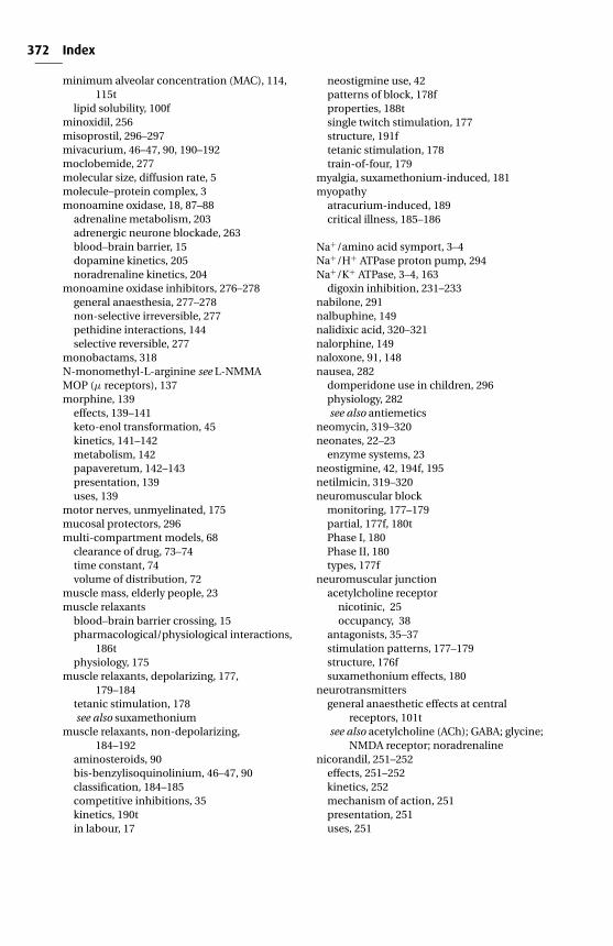
P1: PSB Printer: Yet To Come
9780521704632ind CUFX213A/Peck 9780521618168 December 29, 2007 17:19
372 Index
minimum alveolar concentration (MAC), 114,115t
lipid solubility, 100fminoxidil, 256misoprostil, 296–297mivacurium, 46–47, 90, 190–192moclobemide, 277molecular size, diffusion rate, 5molecule–protein complex, 3monoamine oxidase, 18, 87–88
adrenaline metabolism, 203adrenergic neurone blockade, 263blood–brain barrier, 15dopamine kinetics, 205noradrenaline kinetics, 204
monoamine oxidase inhibitors, 276–278general anaesthesia, 277–278non-selective irreversible, 277pethidine interactions, 144selective reversible, 277
monobactams, 318N-monomethyl-L-arginine see L-NMMAMOP (µ receptors), 137morphine, 139
effects, 139–141keto-enol transformation, 45kinetics, 141–142metabolism, 142papaveretum, 142–143presentation, 139uses, 139
motor nerves, unmyelinated, 175mucosal protectors, 296multi-compartment models, 68
clearance of drug, 73–74time constant, 74volume of distribution, 72
muscle mass, elderly people, 23muscle relaxants
blood–brain barrier crossing, 15pharmacological/physiological interactions,
186tphysiology, 175
muscle relaxants, depolarizing, 177,179–184
tetanic stimulation, 178see also suxamethonium
muscle relaxants, non-depolarizing,184–192
aminosteroids, 90bis-benzylisoquinolinium, 46–47, 90classification, 184–185competitive inhibitions, 35kinetics, 190tin labour, 17
neostigmine use, 42patterns of block, 178fproperties, 188tsingle twitch stimulation, 177structure, 191ftetanic stimulation, 178train-of-four, 179
myalgia, suxamethonium-induced, 181myopathy
atracurium-induced, 189critical illness, 185–186
Na+/amino acid symport, 3–4Na+/H+ ATPase proton pump, 294Na+/K+ ATPase, 3–4, 163
digoxin inhibition, 231–233nabilone, 291nalbuphine, 149nalidixic acid, 320–321nalorphine, 149naloxone, 91, 148nausea, 282
domperidone use in children, 296physiology, 282see also antiemetics
neomycin, 319–320neonates, 22–23
enzyme systems, 23neostigmine, 42, 194f, 195netilmicin, 319–320neuromuscular block
monitoring, 177–179partial, 177f, 180tPhase I, 180Phase II, 180types, 177f
neuromuscular junctionacetylcholine receptor
nicotinic, 25occupancy, 38
antagonists, 35–37stimulation patterns, 177–179structure, 176fsuxamethonium effects, 180
neurotransmittersgeneral anaesthetic effects at central
receptors, 101tsee also acetylcholine (ACh); GABA; glycine;
NMDA receptor; noradrenalinenicorandil, 251–252
effects, 251–252kinetics, 252mechanism of action, 251presentation, 251uses, 251

P1: PSB Printer: Yet To Come
9780521704632ind CUFX213A/Peck 9780521618168 December 29, 2007 17:19
373 Index
nicotinic acetylcholine receptor, 25nifedipine, 253–254nimodipine, 254nitrates, 249–251
organic, 131ttolerance, 39
nitric oxide (NO), 130t, 132tadult intensive care use guidelines,
131t, 132teffects, 131tguanylyl cyclase stimulation, 30inhaled, 13synthesis, 130t, 131t
nitric oxide synthase (NOS)constitutive, 130t, 131tinducible, 130t, 131t
nitrites, 248–249nitroimidazoles, 328tnitrous oxide (N2O), 120f
concentration effect, 118–120diffusion hypoxia, 120effects, 118manufacture, 117–118second gas effect, 120storage, 118toxicity, 120–121
nizatidine, 294NMDA receptor, 2–3, 26, 102
ketamine antagonism with glutamate,37
L-NMMA, 131tnon-compartment models, 71
volume of distribution, 72–73non-linear kinetics, 79fnon-steroidal anti-inflammatory drugs
(NSAIDs), 135–136, 149–152asthma, 151classification, 152tclinical data, 157tdrug interactions, 152gastric irritation, 151–152hepatotoxicity, 152kinetics, 157tmechanisms of action, 149–151platelets, 151–152renal function, 151–152
NOP receptor, 137–139noradrenaline, 203–204
effects, 203kinetics, 204mechanism of action, 203metabolism, 18presentation, 203release, 197–199uses, 203
norfloxacin, 320–321nortriptyline, 274–276nucleic acids, 86nucleosides, 86nucleotides, 86
oestrogen therapy, 358–359ofloxacin, 320–321omeprazole, 294–295ondansetron, 290–291one-compartment model, 64–68
clearance of drug, 73time constant, 74volume of distribution, 72
opioids, 91, 135–149minimizing of administration, 282partial agonists, 139t, 149pharmacological properties, 138treceptors, 136, 136t, 137t, 139structure, 140f
oral administration, 8–11bioavailability, 9–11
oral contraceptives, 358failure with antibiotics, 21
organic chemistry, 85organophosphorus compounds, 195–196, 196fosmotic diuretics, 309–310oxicam group, 95, 158oxidation, 17–18oxygen, 128, 130t
effects, 130tmanufacture, 128measurement, 130tstorage, 128toxicity, 130tuses, 130t
pain, 135modification, 135–136physiology, 135–136
pancuronium, 187–188volume of distribution, 72
papaveretum, 142–143papaverine, 90, 96, 142–143para-aminophenols, 154–156paracetamol, 154–156
kinetics, 155overdose, 155–156presentation, 154–155structure, 152ftoxicity, 155uses, 154–155
parasympathetic nervous system, 197parecoxib, 161
kinetics, 161
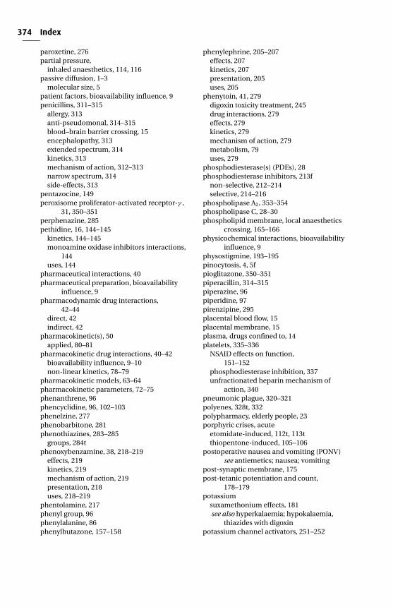
P1: PSB Printer: Yet To Come
9780521704632ind CUFX213A/Peck 9780521618168 December 29, 2007 17:19
374 Index
paroxetine, 276partial pressure,
inhaled anaesthetics, 114, 116passive diffusion, 1–3
molecular size, 5patient factors, bioavailability influence, 9penicillins, 311–315
allergy, 313anti-pseudomonal, 314–315blood–brain barrier crossing, 15encephalopathy, 313extended spectrum, 314kinetics, 313mechanism of action, 312–313narrow spectrum, 314side-effects, 313
pentazocine, 149peroxisome proliferator-activated receptor-γ ,
31, 350–351perphenazine, 285pethidine, 16, 144–145
kinetics, 144–145monoamine oxidase inhibitors interactions,
144uses, 144
pharmaceutical interactions, 40pharmaceutical preparation, bioavailability
influence, 9pharmacodynamic drug interactions,
42–44direct, 42indirect, 42
pharmacokinetic(s), 50applied, 80–81
pharmacokinetic drug interactions, 40–42bioavailability influence, 9–10non-linear kinetics, 78–79
pharmacokinetic models, 63–64pharmacokinetic parameters, 72–75phenanthrene, 96phencyclidine, 96, 102–103phenelzine, 277phenobarbitone, 281phenothiazines, 283–285
groups, 284tphenoxybenzamine, 38, 218–219
effects, 219kinetics, 219mechanism of action, 219presentation, 218uses, 218–219
phentolamine, 217phenyl group, 96phenylalanine, 86phenylbutazone, 157–158
phenylephrine, 205–207effects, 207kinetics, 207presentation, 205uses, 205
phenytoin, 41, 279digoxin toxicity treatment, 245drug interactions, 279effects, 279kinetics, 279mechanism of action, 279metabolism, 79uses, 279
phosphodiesterase(s) (PDEs), 28phosphodiesterase inhibitors, 213f
non-selective, 212–214selective, 214–216
phospholipase A2, 353–354phospholipase C, 28–30phospholipid membrane, local anaesthetics
crossing, 165–166physicochemical interactions, bioavailability
influence, 9physostigmine, 193–195pinocytosis, 4, 5fpioglitazone, 350–351piperacillin, 314–315piperazine, 96piperidine, 97pirenzipine, 295placental blood flow, 15placental membrane, 15plasma, drugs confined to, 14platelets, 335–336
NSAID effects on function,151–152
phosphodiesterase inhibition, 337unfractionated heparin mechanism of
action, 340pneumonic plague, 320–321polyenes, 328t, 332polypharmacy, elderly people, 23porphyric crises, acute
etomidate-induced, 112t, 113tthiopentone-induced, 105–106
postoperative nausea and vomiting (PONV)see antiemetics; nausea; vomiting
post-synaptic membrane, 175post-tetanic potentiation and count,
178–179potassium
suxamethonium effects, 181see also hyperkalaemia; hypokalaemia,
thiazides with digoxinpotassium channel activators, 251–252
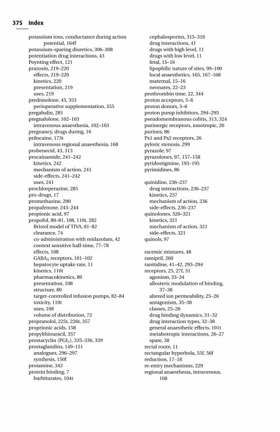
P1: PSB Printer: Yet To Come
9780521704632ind CUFX213A/Peck 9780521618168 December 29, 2007 17:19
375 Index
potassium ions, conductance during actionpotential, 164f
potassium-sparing diuretics, 306–308potentiation drug interactions, 43Poynting effect, 121prazosin, 219–220
effects, 219–220kinetics, 220presentation, 219uses, 219
prednisolone, 45, 353perioperative supplementation, 355
pregabalin, 281pregnalolone, 102–103
intravenous anaesthesia, 102–103pregnancy, drugs during, 16prilocaine, 173t
intravenous regional anaesthesia, 168probenecid, 43, 313procainamide, 241–242
kinetics, 242mechanism of action, 241side-effects, 241–242uses, 241
prochlorperazine, 285pro-drugs, 17promethazine, 290propafenone, 243–244propionic acid, 97propofol, 80–81, 108, 110t, 282
Bristol model of TIVA, 81–82clearance, 74co-administration with midazolam, 42context sensitive half-time, 77–78effects, 108GABAA receptors, 101–102hepatocyte uptake rate, 11kinetics, 110tpharmacokinetics, 80presentation, 108structure, 80target-controlled infusion pumps, 82–84toxicity, 110tuses, 108volume of distribution, 72
propranolol, 225t, 226t, 357proprionic acids, 158propylthiouracil, 357prostacyclin (PGI2), 335–336, 339prostaglandins, 149–151
analogues, 296–297synthesis, 150f
protamine, 342protein binding, 7
barbiturates, 104t
cephalosporins, 315–316drug interactions, 41drugs with high level, 11drugs with low level, 11fetal, 15–16lipophilic nature of sites, 99–100local anaesthetics, 165, 167–168maternal, 15–16neonates, 22–23
prothrombin time, 22, 344proton acceptors, 5–6proton donors, 5–6proton pump inhibitors, 294–295pseudomembranous colitis, 313, 324purinergic receptors, ionotropic, 26purines, 86Px1 and Px2 receptors, 26pyloric stenosis, 299pyrazole, 97pyrazolones, 97, 157–158pyridostigmine, 193–195pyrimidines, 86
quinidine, 236–237drug interactions, 236–237kinetics, 237mechanism of action, 236side-effects, 236–237
quinolones, 320–321kinetics, 321mechanism of action, 321side-effects, 321
quinols, 97
racemic mixtures, 48ramipril, 260ranitidine, 41–42, 293–294receptors, 25, 27f, 31
agonism, 33–34allosteric modulation of binding,
37–38altered ion permeability, 25–26antagonism, 35–38classes, 25–26drug binding dynamics, 31–32drug interaction types, 32–38general anaesthetic effects, 101tmetabotropic interactions, 26–27spare, 38
rectal route, 11rectangular hyperbola, 55f, 56freduction, 17–18re-entry mechanisms, 229regional anaesthesia, intravenous,
168
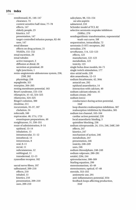
P1: PSB Printer: Yet To Come
9780521704632ind CUFX213A/Peck 9780521618168 December 29, 2007 17:19
376 Index
remifentanil, 81, 146–147clearance, 74context sensitive half-time, 77–78effects, 147elderly people, 23kinetics, 147presentation, 147target-controlled infusion pumps, 82–84uses, 147
renal diseaseeffects on drug actions, 21NSAIDs, 151–152
renal excretion, 20renal tubules
active transport, 3diffusion at distal, 20secretion at proximal, 20tight junctions, 1
renin–angiotensin–aldosterone system, 258,259f, 262
physiology, 258repaglinide, 351reserpine, 264–265resting membrane potential, 163Reye’s syndrome, 153–154rifampicin, 41–42, 324–325rifamycins, 324–325Ringer’s solution, 300ritodrine, 211rocuronium, 35–37, 187
chelation, 24rofecoxib, 160ropivacaine, 48, 171t, 172t
enantiopure preparations, 49rosiglitazone, 31, 350–351route of administration, 8–14
epidural, 13–14inhalation, 13intramuscular, 11–12intrathecal, 14intravenous, 8oral, 8–11rectal, 11subcutaneous, 12sublingual, 11transdermal, 12–13
ryanodine receptor, 182
sacral nerve fibres, 197salbutamol, 209–210
effects, 210inhaled, 13kinetics, 210presentation, 209–210uses, 209–210
salicylates, 98, 153–154see also aspirin
salmeterol, 210Schnider model of TCI, 82selective serotonin reuptake inhibitors
(SSRIs), 276semilogarithmic transformation, exponential
wash-out curve, 59fsequestration, intracellular, 72serotonin (5-HT) receptors, 282sertraline, 276sevoflurane, 114, 122–123
effects, 123manufacture, 122–123metabolism, 123toxicity, 123
single bolus dose models, 64–71single twitch stimulation, 177sino-atrial node, 228skin anaesthesia, 12–13sodium bicarbonate, 42, 304t
antacid use, 292epidural anaesthetics, 14interaction with calcium, 40
sodium calcium edetate, 41sodium citrate, 292sodium ion(s)
conductance during action potential,164f
loop diuretics reabsorption inhibition, 307reabsorption inhibition by thiazides, 305
sodium ion channel, 163–164cardiac action potential, 228local anaesthetic binding, 3quinidine blocking, 236
sodium nitroprusside, 24, 131t, 246, 248f, 249effects, 247kinetics, 247mechanism of action, 246metabolism, 247presentation, 246toxicity, 248–249uses, 246
sodium thiosulphate, 248–249sodium valproate, 280–281sotalol, 226t, 244spironolactone, 308–309Starling equation, 299stereoisomerism, 45–49stereoisomers, optical, 47–48steroids, 353–355
antiemetic use, 291anti-inflammatory potential, 355tfeedback loops affecting production,
354f
P1: PSB Printer: Yet To Come
9780521704632ind CUFX213A/Peck 9780521618168 December 29, 2007 17:19
377 Index
inhaled, 13intravenous anaesthesia, 102–103nucleus, 90perioperative supplementation, 355
stilboestrol, 16straight lines with same gradient, 61fstreptokinase, 344–345streptomycin, 319–320structure-activity relationships (SAR), 85subcutaneous route, 12sublingual route, 11sucralfate, 296sugars, 87sulphation, 18–19sulphonylureas, 348–349
kinetics, 349mechanism of action, 349side-effects, 349uses, 349
summation drug interactions, 42supraventricular tachyarrhythmias, 229–237
with ventricular tachyarrhythmias, 239–245verapamil side-effects, 234–235
suxamethonium, 17, 179–184cardiac output, 41effects, 181–184genetic polymorphisms, 19kinetics, 180malignant hyperthermia, 182–184mechanism of action, 180metabolism, 18–19, 181fpresentation, 180uses, 180
suxamethonium apnoea, 183–184sympathetic nervous system, 197, 198f, 199sympathomimetics, 197–199, 216
direct-/indirect acting, 199naturally occurring catecholamines,
199–205phosphodiesterase inhibitors
non-selective, 212–214selective, 214–216
physiology, 197–199structure, 199, 206fsynthetic agents, 205–212
synergisms, 43syringes, drug interactions, 40
tachyarrhythmias, 229atrioventricular nodal re-entrant tachycardia,
230ftachyphylaxis, 38–39target-controlled infusion pumps (TCI), 76,
82–84decrement time, 83
pumps, 83safety, 83–84
tautomerism, 45tazobactam, 313teicoplanin, 322–323temazepam, 273tenoxicam, 158teratogens, 16terbutaline, 211terminal elimination half-life, 71, 77–78tetanic stimulation, 178therapeutic index, 75thiazides, 98, 305–307
effects, 306–307kinetics, 307mechanism of action, 305
thiazolidinediones, 350–351thiocyanate (SCN), 246–249thiopentonal, 16–17thiopentone, 102–107
effects, 105–106intra-arterial injection, 107keto-enol transformation, 45kinetics, 106plasma, 106fpresentation, 104uses, 104
thioridazine, 284three compartment models, 70–71thrombin, 336thromboxane(s), 149–151, 153thromboxane A2 (TXA2), 335–336thrombus formation, 335–336thyroid disease, 355–358thyroid hormones
feedback loops affecting production, 356freplacement, 355–357see also triiodothyronine (T3)
thyroxine (T4), 216, 355–357effects, 356kinetics, 357mechanism of action, 356uses, 356
ticarcillin, 314–315ticlopidine, 337–338tight junctions, 1time constant, 67, 67f, 68, 74
natural logarithms, 58–59tinzaparin, 341–342tirofiban, 338tissue binding of drugs, 72tobramycin, 319–320tolbutamide, 348–349
side-effects, 349tolerance, 39
P1: PSB Printer: Yet To Come
9780521704632ind CUFX213A/Peck 9780521618168 December 29, 2007 17:19
378 Index
total intravenous anaesthesia (TIVA), 81–82safety, 83–84
train-of-four, 177f, 179tramadol, 147–148
effects, 148interactions, 148kinetics, 148mechanism of action, 148presentation, 148
tranexamic acid, 346transdermal route, 12–13tranylcypromine, 277triamcinolone, 353triazoles, 332–333tricyclic antidepressants, 274–276
effects, 275mechanism of action, 275overdose, 275–276
phenytoin treatment, 279uses, 274
triiodothyronine (T3), 216, 355–357kinetics, 357
trimetaphan, 268–269tubocurarine, 90, 188–189two-compartment models, 68–70tyrosine, 87–88tyrosine kinase, membrane, 30–31
urokinase, 345
vacuum insulated evaporator (VIE), 128valdecoxib, 161
kinetics, 161vancomycin, 322–323
oral, 8vancomycin-resistant enterococci (VRE), 311,
322vanillylmandelic acid
adrenaline metabolism, 203noradrenaline kinetics, 204
vasodilators, 246–257activity and local anaesthetics, 166calcium channel antagonists, 252–255potassium channel activators, 251–252sodium nitroprusside, 246–249see also nitrates
vecuronium, 35–37, 41–42, 90, 185–186venlafaxine, 276ventricular tachyarrhythmias, 229, 237–238
with supraventricular tachyarrhythmias,239–245
verapamil, 234–235, 252–253kinetics, 235mechanism of action, 234presentation, 234side-effects, 234–235uses, 234
vigabatrin, 281vitamin B12, 248–249vitamin K, warfarin therapy, 342–343volatile anaesthetic agents, 13
racemic mixtures, 48volume of distribution, 72–73
constant relationships, 75plasma concentration, 75–76total, 72–73
vomiting, 282domperidone use in children, 296neural inputs, 283fphysiology, 282see also antiemetics
vomiting centre, 282
warfarin, 342–344actions, 343fdrug interactions, 343
amiodarone, 41kinetics, 344mechanism of action, 342–343side-effects, 343therapy monitoring, 344uses, 342
wash-out curve, 52–54asymptote, 52–54semilogarithmic transformation, 59f
Wolff–Parkinson–White syndrome, 234–235xanthenes, 98xenon, 114, 127t, 128t
effects, 127t, 128telimination, 128tmanufacture, 127t
ximelagatran, 344kinetics, 344
xylidine, 98
yohimbine, 220
zaleplon, 274zero-order kinetics, 78–79, 79fzidovudine, 334zolpidem, 274zopiclone, 274