
PNG_REDD+ Scheme for April Salumei Sustainable Forest- Biatus Bito
The Journal of Neuroscience, April 1995, 15(4): 2995-3012
Pharmacological Dissection of Multiple Types of Ca*+ Channel Currents in Rat Cerebellar Granule Neurons
Andrew RandalP and Richard W. Tsien
Department of Molecular and Cellular Physiology, The Beckman Center, Stanford University Medical Center, Stanford, California 94305
The diversity of Ca2+ channel types in rat cerebellar granule neurons was investigated with whole-cell recordings (5 mu external Ba*+). Contributions of five different high-voltage- activated Ca2+ channel current components were distin- guished pharmacologically. Nimodipine-sensitive L-type current and w-CTx-GVIA-sensitive N-type current contrib- uted 15 and 20% of the total current, respectively. The bulk of the remaining current (46%) was inhibited by w-Aga-IVA. The current blocked by this toxin was further subdivided into two components, P-type and Q-type, on the basis of differences in their inactivation kinetics and sensitivity to w-Aga-IVA. P-Type current was noninactivating during 0.1 set depolarizations, half-blocked at about l-3 nu w-Aga- IVA, and contributed -11% of the total current; Q-type cur- rent was prominently inactivating, half-blocked at -90 nu w-Aga-IVA, and comprised 35% of the total current. Both P- and Q-type currents were potently inhibited by the Con- us magus toxin w-CTx-MVIIC. A current component resis- tant to all of the aforementioned blockers (R-type) dis- played more rapid inactivation than the other components and constituted 19% of the total current. The Q-type cur- rent, the largest of the current components in the granule neurons, resembles currents that can be generated in Xen- opus oocytes by expression of cloned 01,~ subunits.
[Key words: calcium channel, calcium current, cerebel- lum, granule neuron, m-conotoxin-GVIA, w-Aga-WA]
The diversity of Ca2+ channels in vertebrate neurons is of fun- damental interest because voltage-gated Ca2+ entry through these channels controls a wide variety of physiological func- tions, encompassing neurotransmitter release, membrane excit- ability, neurite outgrowth, bioenergetics, and gene expression (Reuter, 1983; Hille, 1992). The existence of more than one type of voltage-gated calcium channel in vertebrate neurons has been appreciated for some time (Carbone and Lux, 1984; Armstrong
Received Aug. 17, 1994; revised Oct. 31, 1994; accepted Nov. 4, 1994.
We thank Beverly Wendland, Felix Schweizer, Karl Deisseroth, and Haru- hiko Bito for preparation of cerebellar granule cells, George Miljanich (Neu- rex) for providing gifts of w-CTx-MVIIC and o-CTx-GVIA, A. J. G. Baxter (Fisons PLC) for providing FPL 64176, and Michael Adams (UC Riverside) for a gift of w-Aga-IVA and helpful discussions. We are grateful to Ilya Bez- prozvanny, Karl Deisseroth, William Sather, David Wheeler, and Ji-Fang Zhang for comments on the MS. This work was supported by NIH Grant NS24067 (R.W.T.) and an NIMH-Silvio Conte Center for Neuroscience Research. A.R. is a Lucille I? Markey Visiting Fellow.
Correspondence should be addressed to Richard W. Tsien at the above ad- dress.
“Present address: Neurobiology Division, The MRC Laboratory of Molecular Biology, Hills Road, Cambridge, CB2 2QH, UK.
Copyright 0 1995 Society for Neuroscience 0270-6474/95/152995-18$05.00/O
and Matteson, 1985; Fedulova et al., 1985; Nowycky et al., 1985). Biophysical and pharmacological analysis has led to the description of several classes of Ca2+ channels, usually referred to as L-, N-, T-, and P-type (Nowycky et al., 1985; Fox et al., 1987a,b; Bean 1989; Llinas et al., 1989; Hess, 1990). These channel types differ considerably in their responsiveness to neu- romodulators (Tsien et al., 1988; Bean, 1989) their distribution among various types of neurons (Miller and Fox, 1990; Tsien et al., 1991; Mintz et al., 1992b; Snutch and Reiner, 1992), and their localization in different regions within individual neurons (Westenbroek et al., 1992). The variety of Ca*+ channel types allows many possibilities for multiplicity of function (Hille, 1992).
The full extent of CaZ+ channel diversity remains incomplete- ly understood. On one hand, molecular cloning uncovers an ever-increasing number of Ca*+ channel subunits with a wide distribution in the CNS (Perez-Reyes et al., 1990; Tsien et al., 1991; Snutch and Reiner, 1992; Miller, 1993). On the other hand, pharmacological evidence points to the possible existence of ad- ditional types of Ca2+ channels outside of the T, N, L, and P classification (Regan et al., 1991; Artelejo et al., 1992; Mintz et al., 199213, Swartz et al., 1993; Forti et al., 1994). While both lines of investigation suggest additional complexity, the match- up between the cloned subunits and CaZ+ channels in neurons is incomplete. Some Ca2+ channel 01, subunits appear in good cor- respondence with established channel types. For example neu- ronal L-type channel currents are generated by class C and D CK, subunits (Mikami et al., 1989; Williams et al., 1992a) and N-type Ca2+ channels arise from the class B (Y, (Williams et al., 1992b). Other CX,S, however, are more difficult to identify as known calcium channel types in nerve cells. A leading example is the class A (Y, subunit (also known as brain-I or BI), the first o(, subunit to be isolated from nervous tissue (Mori et al., 1991; Starr et al., 1991). Transcripts for oIA abound in the mammalian brain, particularly in cerebellar Purkinje and granule cells (Mori et al., 1991, Fujita et al., 1993). This has led to the presumption that this subunit is equivalent to the P-type channel, the domi- nant Ca2+ conductance in the Purkinje neurons (Snutch and Rei- ner, 1992; but see Tsien et al., 1991). However, the expression of oIA in oocytes (Sather et al., 1993; Stea et al., 1994) or in a mammalian cell line (Niidome et al., 1994) produces a pheno- type different from the P-type current, suggesting that (Y,, can also generate a novel non-P-type Ca2+ channel.
We have undertaken a study of the classes of Ca*+ channel in rat cerebellar granule cells, the most abundant neuron in the mammalian CNS (Ramon y Cajal, 1904). Granule cells give rise to parallel fiber synapses onto the dendrites of Purkinje neurons,

2996 Randall and Tsien - Five Components of Ca2+ Channel Current in Granule Neurons
synapses whose strength undergoes modification during long- term depression and other forms of synaptic plasticity (see Ito, 1989, for review). Granule cells are electrically compact (Silver et al.; 1992, Randall and Tsien 1993), express a range of differ- ent cx, subunits, and survive well in tissue culture. All of these traits proved favorable for the analysis of Ca2+ channels in our experiments. Using a series of potent channel inhibitors, we have defined five readily distinguishable components of Ca*+ channel current, including L-, N-, and P-type Ca*+ channels with con- ventional properties, as well as additional components, Q-type and R-type. Together, Q-type and R-type currents constitute half of the peak inward Ca2+ channel current. The Q-type current is the largest component in the granule cells and displays electro- physiological and pharmacological properties that match up closely with the qA subunit expressed in oocytes.
Some of the results have been presented in abstract form (Randall et al., 1993a).
Materials and Methods
Cell preparation and culture. Cerebellar neurons were prepared using minor modifications of a procedure for preparation of hippocampal cul- tures (Malgaroli and Tsien, 1992). Cerebella were removed from the brains of decapitated 2-5-d-old rat pups of either sex. Following re- moval of the dura, the cerebellum was cut into small (-1 mm3) pieces and digested with trypsin (type XI, Sigma, 10 mg/ml) for 4 min at 24°C in a Ca2+- and Mg2+ -containing, HEPES-buffered salt solution supple- mented with 0.5 mg/ml DNAase (type IV Sigma). This treatment was followed by trituration with fire-polished Pasteur pipettes in a high Mg*+, 0 Ca2+, bicarbonate-buffered saline. Following three centrifuga- tion/resuspension steps, the cells were plated on square No. 1 coverslips (VWR), previously coated with Matrigel (Collaborative Research Inc.). The cells were maintained in culture under 5% CO, at 37°C in minimal essential medium (5.3 mM K+, GIBCO), supplemented with glucose 5 gm/liter, transferrin 100 mg/liter, insulin 25 mg/liter, glutamine 300 mg/ liter, cytosine arabinoside 2-5 FM, and 10% fetal calf serum (Hyclone). Cultures were fed after 2 d by replacement of 50-70% of the culture medium and sometimes were fed a second time after 5-6 d in vitro.
Electrophysiology. Prior to recording, dishes containing a single cov- erslip were removed from the incubator, the culture medium being re- placed with Tyrode solution containing (mu) NaCl 119, KC1 5, CaCl, 2, MgCl, 1, glucose 30, HEPES-NaOH 25 (pH 7.3), 313 mOsm. The coverslip was broken into numerous pieces and a single shard placed in the recording chamber, while the remaining shards were kept in Ty- rode at room temperature until used or discarded. In control experiments we were unable to detect any change in the size or properties of the Ba*+ currents from cells that were maintained in Tyrode at room tem- perature for < 10 min or more than 5 hr. Standard patch clamp record- ings (Hamill et al., 1981) (Axopatch 1D amplifier, Axon Instruments) were made with fire-polished patch-clamp pipettes, fabricated from 1.2 mm diameter, thick-walled borosilicate glass (Clark Electromedical) with a horizontal electrode puller (Sutter Instruments). The pipettes were filled with CsMeSO, 108, M&l, 4.5, EGTA 9, ATP 4, GTP 0.3, HEPES 24 (pH 7.3 with CsOH), 292 mOsm, and showed resistances of 2-8 Mfl (tvoicallv 5 Ma). For recordings of Ba*+ currents. the external solution coniained TEA-Cl 160, BaCl, 5, HEPES-CsOH 10 (pH 7.3), 313 mOsm. Gigaseals (> 5 GR) were formed by gentle mouth suction, following which fast capacitance transients were neutralized. The whole-cell configuration was achieved using a combination of fur- ther suction and application of 200 psec, 2 V DC pulses. Series resis- tances were < 20 FM (typically 8-10 Ma) and were typically not compensated. In control experiments, the recorded currents were essen- tially the same with and without series resistance compensation, pre- sumably because the cell capacitance and total current magnitudes were relatively small.
For the analysis of toxin actions on voltage-activated Ba*+ currents, we adopted a standard protocol of applying a 105 msec duration step to a test potential of 0 mV every 10 set, the standard holding potential being -80 mV. Each test pulse was preceded by a +lO mV pulse of identical length that was used to monitor series resistance, capacitance, and input (leak) resistance, and to allow subsequent leak subtraction. In
the solutions used to acquire Ba*+ currents, the leak resistance varied between 6 and 20 Gfl
Data storage and analysis. Occasionally, current and voltage signals were recorded on digital tape and digitized following appropriate filter- ing off line. Usually, however, data were directly filtered (1.5 to 8 kHz, S-pole Bessel filter, Frequency Devices) and digitized (3.33 to 25 kHz) within the PCLAMP acquisition environment (Axon Instruments). Leak subtraction and analysis were performed by custom-written programs (AXOBASIC), used in conjunction with a spreadsheet program (QUATTRO
PRO, Borland). All data are presented as mean ? 1 SEM. Rundown correction. Corrections for rundown of non-L-, non-N-type
Ba2+ current were based on direct observations of time-dependent changes in the amplitude of this current, which decreased from nor- malized values of 1.0 to 0.907 over a 5 min period under our whole- cell recording conditions (see Fig. 5A), with no detectable change in waveform. A straight line provided a very close fit to the rundown data (while of no theoretical significance), and was used to correct data points at various times to what would have been expected if rundown had not occurred (see Fig. 5B). Thus, with rundown correction. the magnitude of a drug-sensitive current at 5 min would be estimated as [(2,,,,/0.907) - Z,,.,,,,]. This kind of correction was extended to estimates of the waveform of drug-sensitive currents, making the assumption that various components of the non-L-, non-N-type current undergo run- down at the same rate. This was supported empirically by the finding that the time course of the non-L-, non-N-type current did not change with rundown. Stated in another way, the experimentally determined rundown correction factor was the same at all points during a depolar- izing test pulse.
Drug application. Drug-containing solutions were delivered by grav- ity feed from a linear array of glass capillaries, positioned near the recorded cell. In addition, the recording chamber was continuously per- fused at -1 ml/min, with a peristaltic pump connected to a small an- techamber. Bath solution was removed by continuous suction. The ef- ficiency of the cell superfusion was verified by comparison of the blockade of Ba*+ current produced by 1 FM Cd*+, applied either via the glass capillaries, or by slow bath perfusion. All drugs were stored as 100-5000X stock solutions at -20°C. Cytochrome c (0.1 mg/ml) was included in all recording solutions to block nonspecific peptide binding sites; in control experiments, this agent was shown to have no effect on Ba*+ currents (n = 6).
Toxin preincubation experiments. The preincubation experiments (see Fig. 7) were carried out by incubating granule cells for the prescribed period in Tyrode solution at 24°C in the presence of cytochrome c and the relevant toxin. Sham-preincubated cells were subjected to the same procedure in the absence of toxin, but in the presence of cytochrome c. For any particular experiment, the sham- and toxin-preincubated cells were obtained from the same cell culture, and all recordings were made on the same day. To avoid any possibility of reversal of the effects of the toxin preincubation, the preincubating concentration of toxin was included in the recording solutions at all times.
In a similar fashion to toxin preincubation experiments, tests of ef- fects of elevation of intracellular cyclic AMP (CAMP) were performed on two groups of cells. For the treated group, preincubation time was > 30 min in Tyrode solution supplemented with 1 mM 8-Br-CAMP and 10 PM forskolin, and the recording solution contained 0.1 mu 8-Br- CAMP and 2 FM forskolin. The control group were preincubated in Tyrode solution without additions.
Fine chemicals. Nimodipine was provided by Dr. A. Scriabine (Miles Pharmaceuticals, West Haven, CT),-FPL 64176 by Dr. A. J. G. Baxter (Fisons PLC, Loughborough, UK), o-CTx-MVIIC by George Miljanich (Neurex Corp, Menlo Park, CA). Purified w-Aga-IVA was a generous gift from Dr. M. E. Adams (UC Riverside). Synthetic o-Aga-IVA was obtained from Peotides International (Louisville. KY). w-CTx-GVIA was obtained from Peninsula Laboratories (Belmont, CA) or as a gift from Neurex Corporation. 8-Br-CAMP was obtained from Boehringer USA and forskolin from Research Biochemicals Inc. (Natick, MA).
Results
Whole-cell recordings were made from more than 1000 cultured rat cerebellar granule cells maintained for l-l 1 d in vitro (DIV). The cell bodies of granule cells were identified by their round or oval shape and their small size (4-6 pm) and low capacitance (5.26 5 0.08 pE mean ? SEM). Cells with this morphology comprise -90% of the neurons in our cerebellar cultures.

The Journal of Neuroscience, April 1995, 15(4) 2997
A V,=-40 mV
C 25
V,=-20 mV c
0.8 - c8
0.4 -
0.2 -
0.0 -I
-100 -80 -60 -40 -20
_ o/O-0--o t 4 C;PF) 2
0 1 2 3 4 5 6 7 8 9 10 Days in vitro
Properties of global Ca2+ channel current
In recordings from cells bathed in Tyrode solution with 2 mM Ca2+ (data not shown), a depolarizing step from -80 mV to 0 mV evoked a rapidly activating and inactivating TTX-sensitive Na+ current (102.7 ? 9.9 pA/pF), followed by a more prolonged Cd2+-sensitive Ca*+ current (7.0 + 2.2 pA/pF). Upon replace- ment of Tyrode solution with a sodium-free extracellular solu- tion containing 5 mu Ba2+ and 1 pM TTX, the Na+ current was blocked, leaving an inward BaZ+ current with the characteristics illustrated in Figure 1. The Ba2+ current was entirely high-volt- age activated (HVA), significant inward current not being ob- served until the depolarizing step reached -40 mV (Fig. lA,B). The onset of the inward current became considerably faster as the depolarization was increased. Maximal inward current was observed at a test potential (V,) of 0 mV. The Z/V relationship (Fig. lB, data pooled from six cells) shows a decrease in peak current with further depolarization. As expected for HVA chan- nels, 20 FM Cdl+ reversibly blocked -95% of the Ba*+ current. Evidence for low-voltage activated (LVA) Ba2+ current such as that carried by T-type channels (Carbone and Lux, 1984; Now-
Figure 1. Properties of global BaZ+ currents in cerebellar granule cells. A, Activation of Ba*+ currents with in- creasingly large depolarizing steps (V, = 40 mV to 0 mV, V,, = -80 mV). B, Peak Ba2+ current plotted against test potential (V, = -80 mV). Peak current values were normalized by maximal peak current (I,,,, almost always at V, = 0 mV) for each cell. Data pooled from six cells. C, Dependence of Ba*+ current on holding potential. Holding potential (V,) varied from - 100 mV to -20 mV in 10 mV increments, and was maintained for 10 set before the test pulse (V, = 0 mV). Note that cur- rents are larger and exhibit increasinalv prominent decav at more negat& holding potentials. D, Steady-state in- activation data oooled from four cells. Peak currents dere normalized by the largest current observed in the holding potential range -100 to 20 mV. Star- dard errors smaller than the symbols are not shown. Smooth curve is a Boltzmann function, h, = [ 1 + exp (( V - V,,&}]-‘, with V,,, = -52.4 mV, k = 13.8 mV. E, Development of Ba*+ current amplitude with increasing pe- riods in vitro. Mean amplitude of peak Ba*+ current recorded in the absence of any Ca2+ channel inhibitors (open sym- bols). Number of cells used for Dooled data varied from 6 (8 DIV) to-35 (6 DIV). Also shown are mean values of total cell capacitance @filled symbols). Numbers of cells varied from 8 (4 DIV) to 132 (6 DIV). Some of the ca- pacitance determinations were carried out in the presence of Ca*+ channel in- hibitors.
ycky et al., 1985; Bean, 1989; Hess, 1990) was found in < 1% of the cells studied.
The inactivation properties of the Ba2+ current were complex, as previously reported in mouse cerebellar granule neurons (Sle- singer and Lansman, 1991a,b). The waveform of the Ba*+ cur- rent displayed considerably less decay during the test pulse as the holding potential (V,,) was varied from - 100 mV towards 0 mV (Fig. 1C). The steady-state voltage dependence of inacti- vation (Fig. 10) showed some deviation from a simple Boltz- mann relationship (smooth curve). These findings might be ex- plained if the total Ba2+ current were comprised of multiple components (but see also Slesinger and Lansman, 1991b).
The electrophysiological properties of the cells undergo de- velopmental changes with the number of days in vitro. The in- ward Ba*+ current evoked by a step from -80 mV to 0 mV increased rapidly over the first 5 DIV while the total cell capac- itance remained largely unchanged (Fig. 1E). A similar increase in the size of the sodium current was also found (data not shown). In the experiments described below we have focused on cells > 5 DIV.

2998 Randall and Tsien * Five Components of Ca2+ Channel Current in Granule Neurons
A Nimodipine 10 FM -500 - a’
I
o-CTx-GVIA 1 FM I
-400 - Cd*+ 10 PM
I I 3 -300 -
4
4 -200 -
-100 -
0 , I I I I I
0 100 200 300 400 500
Time (s)
Figure 2. Effects of L- and N-type channel inhibitors on granule cell Ba*+ current. A, Plot of peak BaZ+current versus time for an experiment in which 10 pM nimodipine, 1 PM w-CTx- GVIA, and 10 PM Cd2+ were sequen- tially applied to a 5 DIV granule neu- ron. B, Examples of Ba*+currents (averages of three successive sweeps) taken from the same experiment. Sweets taken at times indicated bv a- d in A. C, Subtractive determinafions of the nimodipine-sensitive (L-type) and w-CTx-GVIA-sensitive (N-type) current waveforms.
200 pA
--I
60 pA
--J 50 ms 50 ms
Pharmacologically dejined components of the total Baz+ current
In order to determine the calcium current complement of these cells, we carried out a pharmacological dissection of the global Ba2+ current with agents known to block L- and N-type channels (Fig. 2). We first applied the dihydropyridine (DHP) compound nimodipine (10 pM) a selective blocker of the L-type calcium channels (Bean, 1989; Hess, 1990; Regan et al., 1991; McCarthy and TanPiengco, 1992). This compound promptly decreased the Ba2+ current by 10.1 2 1 .O pAlpF or about 18%. This blockade could be rapidly reversed upon removal of the drug (data not shown). Next, in the continued presence of nimodipine, N-type channels were inhibited by means of o-CTx-GVIA, a cone snail toxin with considerable selectivity for N-type channels in other neurons (Aosaki and Kasai, 1987; Plummer et al., 1989; Regan et al., 1991; Boland et al., 1994). Application of I FM o-CTx- GVIA rapidly and irreversibly blocked an additional fraction of Ba2+ current, typically 20-25%. The remainder of the current (average, 60.8 + 3.2%) was reversibly inhibited by application of 10 FM Cd2+, a generally effective blocker of HVA Ca*+ chan- nels.
Tests of the effectiveness of channel blockade
Since more than half of the total Ba*+ current remains unblocked in the combined presence of nimodipine and o-CTx-GVIA, it appears that channels other than L- or N-type make a substantial contribution to the global Ba*+ current in cerebellar granule neu- rons, as has been demonstrated in other neurons (Regan et al., 1991). However, the validity of this conclusion depends on the completeness of the pharmacological blockade by the various inhibitors. This was tested by experiments like those illustrated in Figure 3. The effectiveness of the blockade of L-type channels by 10 PM nimodipine was assessed by means of FPL 64176, a
powerful agonist on L-type channels (McKechnie et al., 1989), that acts at a locus independent of the DHP binding site (Zheng et al., 1991). As expected from its known actions in other sys- tems (Zheng et al., 1991; Sather et al., 1993), FPL 64176 strong- ly affected Ca2+ channel currents in the granule neurons (Fig. 3A), greatly increasing the total current during the depolarization and prolonging the tail current upon membrane repolarization (trace b, right). The stimulatory actions of FPL 64176 were com- pletely abolished by 10 p,M nimodipine (Fig. 3A, bottom) (n = 6). This indicates that nimodipine has completely blocked the L-type current.
The effectiveness of 1 pM o-CTx-GVIA in achieving a com- plete blockade of N-type channels was tested by comparing ef- fects of this compound applied successively at 1 and 3 pM (Fig. 3B). The Ba2+ current in 3 pM w-CTx-GVIA (c) showed no greater degree of block than that in 1 PM toxin (b) relative to the control record in the absence of toxin (a). As illustrated in Figure 3B (right), this was a consistent finding (n = 6 cells). In comparison to the average of currents sensitive to 1 FM w-CTx- GVIA (a, b), the average of the additional toxin-sensitive cur- rent, blocked at the higher dose (b-c) was essentially zero. Thus, 1 pM o-CTx-GVIA appears sufficient to yield a saturating block of current carried by N-type channels. This point was confirmed in another series of experiments that demonstrated that the rate of block but not its magnitude was increased as the w-CTx- GVIA concentration was increased from 0.3 to 1 to 3 pM (data not shown). We conclude that block of L- and N-type Ca2+ chan- nels is virtually complete under the experimental conditions of Figure 2. This brings to the fore the question of what type(s) of channel(s) carry the remaining current.
Non-L-, non-N-type current can be divided into o-Aga-IVA- sensitive and -insensitive components The large current (46.8 ? 1.61 pA/pF) that remains in the pres- ence of nimodipine and o-CTx-GVIA (Fig. 28) is high-voltage

The Journal of Neuroscience, April 1995, 75(4) 2999
O-
+Nimodipine
0 50 100 150 200
Time (s)
B o-CTx-GVIA
1 FM 3vM ,
-200 1 N a-b
0 100 200 300 Time (s)
Figure 3. L- and N-type channels are completely blocked by nimodipine and o-CTx-GVIA. A, Application of FPL 64176, a potent stimulator of L-type
1. r--- channels, as a test of the effectiveness of 10 FM nimodipine in blocking L-type channels. FPL 64176 enhance- ment of BaZ+ current amplitude (upper
L 100 pA graph) is completely prevented in the presence of 10 p,M nimodipine (lower
30 Ills ,5yuph). Right, representative traces (averages of three sweeps), taken at the noints indicated on the grauhs. FPL
b-c -w
l- 30 pA
25 ms
64176 greatly prolongs ta?l &rent in the absence of nimodipine but has no effect in its presence. B, Effects of suc- cessive applications of 1 pM and 3 pM
o-CTx-GVIA. Nimodipine (10 PM)
present throughout. Right, average cur- rent blocked by 1 FM w-CTx-GVIA (a-b), compared with the current fur- ther blocked by 3 pM o-CTx-GVIA (b-c). Data pooled from six cells. Note that scatter in a and b largely reflects cell-to-cell variations in magnitude of o-Aga-IVA-sensitive current (see Fig. 10 for indication of consistency in the degree of inactivation).
activated, completely and reversibly blocked by Cd2+, and de- cays by 34.8 ? 0.8% during a 105 msec depolarization (data from 202 typical cells). These properties are reminiscent of the current supported by class A 01, subunits expressed in Xenopus oocytes (Mori et al., 1991; Sather et al., 1993). While the olA current is not reduced by DHPs or w-CTx-GVIA (Mori et al., 1991), it can be blocked by the funnel web spider toxin w-Aga- IVA and the cone snail toxin w-CTx-MVIIC (Sather et al., 1993). w-Aga-IVA is a well-known and very potent blocker (Kd - 1 nM ) of P-type channels in cerebellar Purkinje cells (Mintz et al., 1992a,b; Mintz and Bean, 1993; but cf. Sutton et al., 1994). Figure 4A illustrates the action of 1 pM w-Aga-IVA, ap- plied in the presence of nimodipine and w-CTx-GVIA. The spi- der toxin produced a promptly developing decrease in the non- L-type, non-N-type current. When analyzed by subtraction, as demonstrated in Figure 4C, the waveform of the current blocked by this dose of w-Aga-IVA (b-c) was invariably decaying in nature.
The current remaining in the presence of micromolar doses of nimodipine, w-CTx-GVIA and o-Aga-IVA (Fig. 4B, trace c) has been designated as R-type (Ellinor et al., 1993; Zhang et al., 1993). This terminology carries no presumption that R-type cur- rent is generated by a single class of channels; this remains to be determined. As described previously, this residual component is also resistant to the cone snail toxin w-CTx-MVIIC (see also p. 21). R-type current (c) decays more rapidly and more com- pletely than the current blocked by w-Aga-IVA (b-c). Further consideration of the characteristics of R-type current will be taken up below.
Concentration-dependent kinetics of o-Aga-IVA action
The dose and time dependence of w-Aga-IVA block were char- acterized in an extensive series of experiments (Fig. 5). Nimo- dipine (10 FM) and w-CTx-GVIA (1 FM) were present through- out the recordings. Measurements of the peak current amplitude from individual cells were normalized to their value before ex- posure to w-Aga-IVA, time aligned, and averaged. Figure 5A
compares the time-dependent decrease in Ba*+ current amplitude in cells exposed to 1.5 nM o-Aga-IVA and in a control group subjected to a sham solution change but no agatoxin. The toxin block is small but significantly greater than rundown in the con- trol group. Figure 5B shows the block at 1.5 nM toxin after correction for rundown as estimated from the control group (see Materials and Methods for details). Raising the toxin concentra- tion over the range between 1.5 nM and 3 PM greatly increased the rapidity and extent of block (Fig. 5B). At 3 pM o-Aga-IVA, block reached a steady state within 30 sec. A similar dose de- pendence of the rate of blockade was observed when individual cells were subjected to two successive applications of w-Aga- IVA, at concentrations of 15 nM and then 1 p,M (n = 6, data not shown). The dose dependence of the rate and completeness of o-Aga-IVA-induced block was not detectably different when shorter test pulses were applied at a higher frequency (0.4 or 2 Hz, e.g., Fig. 8A). Furthermore, the inhibitory effect of 300 nM w-Aga-IVA we observed in the absence of any test pulses was indistinguishable from that seen with frequent test pulses to monitor the time course of the onset of block (n = 5, data not shown).
Figure 5C shows the maximal degree of block plotted against toxin concentration (filled symbols). Also included are data for the actions of 100 nM purified w-Aga-IVA (open diamond) and for the toxin preincubation experiments described later in the paper (cross, open circle). A saturating degree of block is achieved at the highest agatoxin concentrations (1, 2, or 3 pM
w-Aga-IVA), with an unblocked fraction of 0.234, which con- sists of R-type current (Fig. 4B). Allowing for this residuum, the dose dependence of the agatoxin block could be fitted rea- sonably well with a one-to-one binding relationship (dashed curve) with an IC,, of 28 nM. This value is considerably higher than the -1 nM IC,, of P-type current in Purkinje neurons, but somewhat lower than the - 100 nM IC,, reported for currents generated by (Y,~ subunit expression in Xenopus oocytes (Sather et al., 1993).

3000 Randall and Tsien * Five Components of Ca2+ Channel Current in Granule Neurons
A w-CTx-GVIA (1 FM) and Nimodipine (10 pM)
-300 - r 1
a I
o-Aga IVA (1 pM) I
-200 -
a 4
I
-100 -
0 1 I I
B
0 150 300 Time (s)
C b-c
1 I’JOpA 50 PA
50 ms
Figure 4. Effect of o-Aga-IVA on non-L-, non-N-type current. A, Time course of an experiment in which 1 FM o-Aga-IVA was applied, following inhibition of L- and N-type current with 10 PM nimodipine and 1 pM o-CTx-GVIA. B, Averages of three traces obtained at the times shown (u-c) in A. C, Subtractive determination of the waveform of the w-Aga-IVA-sensitive current from the sweeps shown in B.
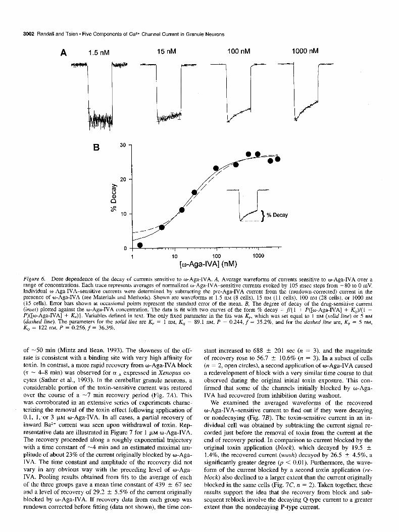
Concentration dependence of the waveform of the w-Aga-IVA- sensitive current
The characteristics of the o-Aga-IVA-sensitive current in cere- bellar granule neurons were examined in greater detail in order to see if components with properties more reminiscent of the o-Aga-IVA-sensitive currents previously described in Purkinje neurons and oocytes could be identified. These previously de- scribed currents differ markedly in their time course of inacti- vation during a depolarizing step. P-Type current in Purkinje neurons shows virtually no decay during depolarizations lasting up to several seconds (Regan, 1991; Llimis et al., 1992; Mintz et al., 1992a,b; Usowicz et al., 1993). In contrast, expression of the (Y,~ subunit in Xenopus oocytes can generate a current that inactivates substantially during the first 0.1 set of a test depo- larization (Sather et al., 1993).
Indeed, we found that a small fraction of the total o-Aga- IVA-sensitive current in the granule neurons exhibited the non- decaying waveform of P-type current. As illustrated in Figure 6A, the waveform of the current blocked by w-Aga-IVA de- pended critically on the dose of toxin. This is shown for data sets corrected for rundown, but it was also true if the rundown correction were omitted. As illustrated in the left-most record, the toxin-sensitive current derived by subtracting currents before and after a 5 min exposure to 1.5 nM o-Aga-IVA was essentially nondecaying. The degree of decay markedly increased, however, as the toxin concentration was elevated into the micromolar range. This trend is documented quantitatively in Figure 6B, which shows pooled results for the degree of decay during the
105 msec depolarization. The fractional decay shows a graded increase over the range between 1.5 nM and 3 p,M, reaching a saturating level of about 30%. These results are not consistent with all-or-nothing block of either a single component of current, or of two components with different waveforms but equal toxin sensitivity. In either case, the fractional decay should be the same at all toxin doses. The observations are also inconsistent with what would be expected if w-Aga-IVA were to act as an open channel blocker (e.g., like D600 on L-type Ca2+ channels). If this were the case, the degree of block would deepen during the test pulse, and the waveform of the toxin-sensitive current would show a time-dependent increase rather than a sustained or decaying time course as actually found.
Subdivision of the w-Aga-IVA-sensitive current into two components The data in Figure 6B was compared to a theoretical relationship based on the premise that the total w-Aga-IVA-sensitive current is comprised of two components: one that is nondecaying and highly toxin sensitive (possibly like P-type current in cerebellar Purkinje cells), and another component, henceforth designated as “Q-type,” that is decaying and less susceptible to toxin blockade (perhaps like currents that can be generated by 01,~ subunits in oocytes). Let us assume that the nondecaying P-type current contributes a fraction P, while the remainder (1 - P) of the total o-Aga-IVA-sensitive current is Q-type, and that o-Aga-IVA blocks each of the components in a one-to-one man- ner (Mintz and Bean, 1993), without changing their individual time course of decay (Mintz et al., 1992a,b; Sather et al., 1993). Then the percentage of decay of the overall o-Aga-IVA differ- ence current will show the following dose dependence:
% decay = f/[ 1 + P(T + K,)l( 1 - P)(T + Kp)] (1)
where f is the fractional decay of the Q-type current during the 105 msec test pulse, T is the toxin concentration, Kp is the dis- sociation constant of the P-type current, and KQ is the dissoci- ation constant of the Q-type current (see figure legend for de- tails). ‘The solid curve in Figure 6B is Equation 1 with f = 35.2%, P = 0.244, Kp = 1 nM, and KQ = 89.1 nM. The theo- retical curve provides a good fit. Thus, the data appears consis- tent with expectations if the w-Aga-IVA-sensitive current were approximately one-fourth P-type and three-fourths Q-type.
The predicted toxin dependence of the percentage decay is strongly influenced by f, P, and K,. It is consistent with a range of values for K,, ranging between about 1 and 3 nM. The the- oretical relationship for Kp = 5 nM (dashed curve, Fig. 6B) pre- dicts somewhat more decay than that observed at the lowest toxin concentrations.
The predictions of the two-component hypothesis can be com- pared with the dose-response relationship in Figure 5C. The solid curve, generated with the same set of parameter values as for the solid curve in Figure 6B, provides a good fit to the pooled data for the o-Aga-IVA-sensitive current. The data points for block by 1.5 and 5 nM w-Aga-IVA lie somewhat above the curve as would be expected if toxin block at these low concentrations had not achieved steady state at 5 min. Thus, the set of data in Figure 5C is consistent with the two component hypothesis (Fig. 6), although it is not sufficient by itself to exclude a one-com- ponent scheme.
Recovery from w-Aga-IVA-induced block Following removal of w-Aga-IVA, block of P-type current in cerebellar Purkinje cells is slowly reversed, with a time constant

The Journal of Neuroscience, April 1995, I!?(4) 3001
0.8 i
100
Time (s)
1.0 -
0.8
0.6
o.oI -100 0 100 200 300
Time (s)
1.5 nM
5
15
30
100
300
1000
3000
1 10 100 1000 [o-Aga-WA] (nM)
Figure 5. Dose dependence of the kinetics and magnitude of w-Aga-IVA-induced block. A, Effect of application of 1.5 nM o-Aga-IVA (filled triangles, n = 8 cells), compared with rundown determined in the absence of o-Aga-IVA (open circles, n = 8). Nimodipine (10 pM) and 1 pM
o-CTx-GVIA were present throughout, and solution changes were made at t = 0 (switch to o-Aga-WA-containing solution or sham solution change). Data from individual cells were normalized to the period -40 to 0 set and averaged. Error bars are not shown when smaller than symbol. The solid line represents the best straight line fit to the open symbols and provides an empirical measure of rundown. B, Dose-dependent blockade of non-L-, non-N-type Ca*+ channel currents by o-Aga-IVA. The graph illustrates the time course of the actions of synthetic o-Aga-IVA in the dose range 1.5 nM (least block, slowest rate) to 3000 nM (most block, fastest rate). The curves represent data (acquired at 0.1 Hz) from between 8 and 29 cells. Data from each cell was normalized to the pre-Aga-IVA current level (t = -40 to 0 set) before averaging. Pooled data at each o-Aga-IVA concentration terminates at the conclusion of the w-Aga-IVA application and has been corrected for the amount rundown demonstrated by the control cells shown in A (see Materials and Methods for details). Data for 100 nM o-Aga-IVA was recorded from a particular batch of neurons several months after the results for all other doses had been obtained and is somewhat out of line with other data sets. This was due to a paucity in R-type current (current resistant to nimodipine, o-CTx-GVIA, and o-Aga-IVA), as verified by direct application of 600 nM toxin to the same batch of cells (data not shown). In a similar vein, small variations in the density and degree of decay of non-L-, non-N-type current were observed in comparisons between cell preparations separated by periods of several months. C, Dose-response relationship for o-Aga-IVA block. Abscissa plots current remaining after exposure to o-Aga-IVA. Synthetic o-Aga-IVA unless otherwise stated. Filled circles represent isochronal data (t = 5 min) for applications of w-Aga-IVA in the range between 1.5 and 15 nM, and steady-state data for o-Aga-IVA applications in the range between 30 and 3000 nM. The cross shows the result of a preincubation experiment with 5 nM w-Aga-IVA (Fig. 9A). The open circle depicts the result of a preincubation experiment with 100 nM o-Aga-IVA (Fig. 9&C). The open diamond is the average amount of block seen at steady state with 100 nM purified o-Aga-IVA (n = 8). The data represented by the jlled symbols are fit with two curves. The dashed line is a one-to-one binding curve with a Kd of 28 nM and a saturating level of block at 0.234. The residual toxin-insensitive component represents R-type current (Fig. 4A, trace c). The solid line represents the sum of two one-to-one binding curves, combining to give a saturating level of block at 0.235. The Kd and amplitude of the two binding sites are 1.0 nM and 19.5%, and 89.1 nM and 57.0%, respectively.
3002 Randall and Tsien - Five Components of Ca2+ Channel Current in Granule Neurons
A 1.5 nM 15nM 100 nM 1000 nM
l/J > % Decay
I 0 , , , , , , , I ' sl'llll
1 10 100 1000
[o-Aga-WA] (nM)
Figure 6. Dose dependence of the decay of currents sensitive to w-Aga-IVA. A, Average waveforms of currents sensitive to o-Aga-IVA over a range of concentrations. Each trace represents averages of normalized o-Aga-IVA-sensitive currents evoked by 105 msec steps from -80 to 0 mV. Individual o-Aga-IVA-sensitive currents were determined by subtracting the pre-Aga-IVA current from the (rundown-corrected) current in the presence of w-Aga-IVA (see Materials and Methods). Shown are waveforms at 1.5 nM (8 cells), 15 nM (11 cells), 100 nM (28 cells), or 1000 nM (15 cells). Error bars shown at occasional points represent the standard error of the mean. B, The degree of decay of the drug-sensitive current (inset) plotted against the o-Aga-IVA concentration. The data is fit with two curves of the form % decay = fl( 1 + P([w-Aga-IVA] + &J/(1 - P)([w-Aga-IVA] + &)). Variables defined in text. The only fixed parameter in the fits was Kp, which was set equal to 1 nM (solid line) or 5 nM (dashed line). The parameters for the solid line are K,, = 1 nM, K, = 89.1 nM, P = 0.244, f = 35.2%, and for the dashed line are, Kp = 5 nM, K0 = 122 ntvr, P = 0.256, f = 36.3%.
of -50 min (Mintz and Bean, 1993). The slowness of the off- rate is consistent with a binding site with very high affinity for toxin. In contrast, a more rapid recovery from w-Aga-IVA block (7 - 4-8 min) was observed for olA expressed in Xenopus oo- cytes (Sather et al., 1993). In the cerebellar granule neurons, a considerable portion of the toxin-sensitive current was restored over the course of a -7 min recovery period (Fig. 7A). This was corroborated in an extensive series of experiments charac- terizing the removal of the toxin effect following application of 0.1, 1;or 3 p,M u-Aga-IVA. In all cases, a partial recovery of inward BaZ+ current was seen upon withdrawal of toxin. Rep- resentative data are illustrated in Figure 7 for 1 pM w-Aga-IVA. The recovery proceeded along a roughly exponential trajectory with a time constant of -4 min and an estimated maximal am- plitude of about 23% of the current originally blocked by o-Aga- IVA. The time constant and amplitude of the recovery did not vary in any obvious way with the preceding level of w-Aga- IVA. Pooling results obtained from fits to the average of each of the three groups gave a mean time constant of 439 ? 67 set and a level of recovery of 29.2 ? 5.5% of the current originally blocked by o-Aga-IVA. If recovery data from each group was rundown corrected before fitting (data not shown), the time con-
stant increased to 688 + 201 set (IZ = 3), and the magnitude of recovery rose to 56.7 ? 10.6% (n = 3). In a subset of cells (n = 2, open circles), a second application of w-Aga-IVA caused a redevelopment of block with a very similar time course to that observed during the original initial toxin exposure. This con- firmed that some of the channels initially blocked by o-Aga- IVA had recovered from inhibition during washout.
We examined the averaged waveforms of the recovered w-Aga-IVA-sensitive current to find out if they were decaying or nondecaying (Fig. 7B). The toxin-sensitive current in an in- dividual cell was obtained by subtracting the current signal re- corded just before the removal of toxin from the current at the end of recovery period. In comparison to current blocked by the original toxin application (block), which decayed by 19.5 + 1.4%, the recovered current (wash) decayed by 26.5 + 4.5%, a significantly greater degree 0, < 0.01). Furthermore, the wave- form of the current blocked by a second toxin application (re- block) also declined to a larger extent than the current originally blocked in the same cells (Fig. 7C, n = 2). Taken together, these results support the idea that the recovery from block and sub- sequent reblock involve the decaying Q-type current to a greater extent than the nondecaying P-type current.

The Journal of Neuroscience, April 1995, 7~74) 3003
w-Aga-IVA (1 pM) A I
w-Aga-IVA (1 pM)
a 1.0
d a 1.00 II
J
Time (s)
Wash (d-c) Block (a-b)
Reblock (e-t) Block
60 ms
Figure 7. Characterization of the reversal of o-Aga-IVA-induced block. A, Reversal of 1 FM o-Aga-IVA-induced block following removal of w-Aga-IVA (10 FM nimodipine and 1 p,~ w-CTx-GVIA present throughout). Filled symbols represent pooled data from seven cells. The data from individual cells were resynchronized 20 set prior to toxin removal (gaps between groups of data points are due to slight differences in the time of toxin exposure among the individual cells). Data has not been corrected for rundown to avoid any possible overemphasis of recovery. open symbols represent data from two cells in which toxin was reapplied following washout. The data from these cells was renormalized (right ordinate) and shown on a scale to match up with the average Ba2+ current in the larger population of cells at the time immediately before the second application of o-Aga-IVA. B, A comparison of the waveform of the current blocked by the initial o-Aga-IVA application (u-b) with that recovered by prolonged toxin washout (d-c). Averaged waveforms from the cells represented by jilled symbols in A. C, A comparison of the waveforms of the currents blocked by initial toxin exposure and by toxin reapplication (e-f). Data for the two cells depicted with open symbols in A.
Recovery from w-Aga-IVA-induced block is hastened by strong depolarizing pulses
The application of a series of strong depolarizing pulses speeds recovery from w-Aga-IVA block, both for P-type current in cer- ebellar Purkinje cells (Mintz et al., 1992a,b) and currents sup- ported by (Y,~ expressed in Xenopus oocytes (Sather et al., 1993). This was also found for the o-Aga-IVA-sensitive currents de- scribed here. Pooled results from 53 experiments showed a res- titution of 58.1 ? 2.8% (range 21 to 105%) of the current orig- inally blocked by o-Aga-IVA (100-3000 nM), following a combination of toxin washout and strong depolarizing pulses. The percentage recovery of current showed little dependence on the previous dose of toxin or on the magnitude of the toxin- sensitive or -insensitive current (data not shown). In the absence of w-Aga-IVA, application of strong pulses did not appreciably alter the nimodipine- and o-CTx-GVIA-insensitive current (n = 5, data not shown).
Toxin block could be largely reversed even in the maintained presence of o-Aga-IVA by application of a series of very strong depolarizing pulses (Fig. 8A). The time course of the subsequent redevelopment of the inhibitory effect of toxin agrees very well with original onset of block. Figure 8B illustrates the recovery from block after toxin has been removed. The effects of separate groups of depolarizing pulses are cumulative and greatly accel- erate the recovery relative to that observed later in the same cell in the absence of strong depolarizing pulses. Even after recovery reaches a near steady state following removal of toxin, a delayed application of strong depolarizations promotes further reversal of toxin blockade (Fig. 8C). The waveform of the component that is restored by strong depolarizations (knockoff) is clearly
decaying, like the current recovered during the previous washoff period (washoff). Thus, Q-type current seems to contribute to both of these difference currents.
Preincubation experiments with w-Aga-IVA
The results up to this point suggest that the bulk of the current blocked by o-Aga-IVA in rat cerebellar granule neurons is sig- nificantly different in waveform and o-Aga-IVA sensitivity from the P-type current as originally defined in rat cerebellar Purkinje neurons (Mintz et al., 1992a,b; Mintz and Bean, 1993). We have called this Q-type current. However, a smaller fraction (-25%) of the overall w-Aga-IVA-blocked current appears to be non- decaying in waveform and quite sensitive to w-Aga-IVA, like the P-type current of Purkinje neurons. It is reasonable to ask if the apparent difference between the magnitudes of Q-type and P-type currents is genuine, and not simply due to incomplete equilibration at low toxin concentrations over the span of a typ- ical experiment. Accordingly, we performed additional experi- ments to test for o-Aga-IVA block during preincubation periods lasting > 1 hr prior to the initiation of whole-cell recording.
Figure 9A compares the pooled behavior of two groups of granule neurons, one subjected to sham incubation (cytochrome c but no w-Aga-IVA, the other incubated with 5 nM o-Aga-IVA, a concentration well above the IC,, for blockade of P-type cur- rent in Purkinje neurons (-1 nM). Recording was carried out in standard solutions containing 10 pM nimodipine and 1 pM
w-CTx-GVIA, with o-Aga-IVA absent or present just as in the respective preincubation solutions. There was no significant dif- ference in the current magnitudes among the control and toxin- preincubated groups (p > 0.1). Thus, no evidence was obtained

3004 Randall and Tsien l Five Components of Ca2+ Channel Current in Granule Neurons
A
-200
g -# -100
0
B -800
-600
g I -400
-200
0
0
I
o-Aga-WA (1 uM) /
+I50 mV lllll1llllllllllllllllllllllll
I I
50 Time (s) 100 150
wAga-IVA (100 nM) o-Aga-IVA (100 nM) L I -
~ 0 500 Time (s) 1000 1500
-400 w-Aga-IVA (1 PM) l- D Wash off
-1 P -300 +Qm I I $
-# -200
I 0 000&j 0 0
o%Kto +100 mv Knock off
-100
0 200 400 600 800 60 ms
Time (s)
Figure 8. Relief of w-Aga-IVA block by large depolarizations. A, Temporary relief of w-Aga-IVA block of Ca2+ channel current induced by a train of strong depolarizations (vertical lines), applied in the main- tained presence of the toxin. Ba*+ current was assayed every 500 msec with 11.2 msec test pulses to 0 mV, and relief of toxin block was achieved by application at 1 Hz of 30 pulses of 50 msec duration to + 150 mV. B, Use of strong depolarizing pulses to greatly accelerate the recovery from block following o-Aga-IVA removal. Voltage-dependent relief of toxin block was followed by a reapplication of o-Aga-IVA and a prolonged washout. In this experiment, test pulses were of the stan- dard frequency (0.1 Hz), amplitude (to 0 mV), and duration (105 msec) described in Materials and Methods. Knockoff was achieved using five cycles of a standard protocol (arrows). Each m-row represents a train of 20 pulses (+lOO mV for 50 msec) at 1 Hz); recovery was assessed with a solitary test pulse, applied 10 set following each train. C, Relief of toxin block with strong depolarizing pulses following partial recov- ery from toxin block due to toxin washout. Standard test pulse protocol (0.1 Hz, 105 msec duration, 0 mV) was used to monitor partial recov- ery; strong pulse protocol identical to that in B. D, Averages of nor- malized waveforms of difference currents in experiments like that il- lustrated in C. The upper truce (‘wash off’) represents the average of the currents recovered during wash off of l-3 FM o-Aga-IVA (b-u). The lower truce (‘knock off’) represents the waveform of the current subsequently recovered by the application of strong depolarizing pulses (c-b) to a subset of these cells (n = 10).
for any major contribution of a current component with the na- nomolar sensitivity of previously defined P-type channels. Pre- sumably, the variability in current density from one cell to the next was too large to allow detection of a small degree of block like that seen with acute application of 5 nM toxin to individual cells (Fig. 5A).
trol group of mock preincubated cells, and 28.4 ? 1.8 pA/pF in the neurons that had been preincubated for 0.5-2.2 hr with 100 nM w-Aga-IVA, a significant difference (p < 0.004) (Fig. 9B). Both groups of cells were subsequently exposed to 3 pM
w-Aga-IVA to determine how much w-Aga-IVA-sensitive cur- rent had been left unblocked by the prior preincubation. The acute reduction in current averaged 52.1 pA/pF in the control group and 16.4 pA/pF in the toxin-preincubated cells (p < 0.01; Fig. 9B) and proceeded with a similar time course in both cases (Fig. 9C). This confirms that the preincubation with toxin was effective in strongly reducing the o-Aga-IVA-sensitive current, while leaving a substantial fraction unblocked (16.4/52.1 or -31%). Following the acute application of 3 FM w-Aga-IVA, the residual currents are not detectably different (p > 0.35), as expected with only R-type current left unblocked.
Additional experiments were undertaken to test the effects of Relative contributions of the Ca2+ channel components
preincubation with 100 nM o-Aga-IVA (Fig. 9B-D). The non- Figure 10 shows the contributions of the various Ca*+ channel L-, non-N-type current averaged 66.5 2 11.2 pA/pF in the con- components to the total whole-cell CaZ+ channel current, based
A Non-L, non-N current B Non-L, non-N Acutely Residual current Aga-IVA-sew. current
60
Sham Aga IVA 0
PK?l”C 5 nM
c ^̂ -0” 1
Sham Aga-IVA Sham Aga-IVA Sham Aga-IVA preinc 100 nM preinc 100 nM preinc 100 nM
3 PM Aga-IVA I
-70 -
-60 -
-50 - % ? s -40 - J
D
Sham
pwnc 0
Aga-IVA
100 nM
-30 -
-20 - 60 ms
-10 -
01 -80 -40 0 40 80 120
Time (s)
Figure 9. Preincubation experiments as a test for steady-state block by w-Aga-IVA. A, Comparison of the magnitudes of non-L-type, non- N-type current in cells preincubated for l-5 hr in Tyrode containing cytochrome c and 5 nM o-Aga-IVA or in sham-preincubated cells (Ty- rode plus cytochrome c). The symbols indicate data from individual cells contributing to the group mean and standard error as illustrated. B, Effect of a 0.5 to 2.2 hr preincubation with 100 nM w-Aga-IVA on the magnitude of subsequently recorded BaZ+ currents. Pooled results for amplitudes of non-L-, non-N-type current (left), current blocked by acute application of 3 FM o-Aga-IVA (center), and current spared by acute toxin application (right). C, Time course of the block of non-L-, non-N-type current induced by acute application of 3 PM w-Aga-IVA. Cells were either sham preincubated (squares) or preincubated and maintained in 100 nM w-Aga-IVA (circles). D, Averages of normalized waveforms of currents blocked by 3 FM w-Aga-IVA in the two groups shown in C.

10 FM Nimodipine 1 FM o-CTx-GVIA
l-3 pM o-Aga-IVA Insensitive
B
35
30
25
k 20
3 p 15
10
5
0
10 pA/pF l-
60 ms
35%+11%
T
Q
Nimodipine w-CTx-GVIA w-Aga-WA Insensitive
Figure IO. Comparison of pharmacologically dissected current com- ponents. A, Pharmacologically defined current components from indi- vidual cells were normalized to peak current, averaged, then scaled by their mean peak current density. Error bars are shown for individual time points. L-Type currents dissected with 10 FM nimodipine (n = 23); N-type currents determined with 1 PM w-CTx-GVIA (n = 25); P-type + Q-type current (n = 64) obtained with l-3 FM o-Aga-IVA following blockade of N- and L-type currents. R-Type current (n = 26) is estimated as the current that remains unblocked in the combined presence of 10 FM nimodipine, 1 PM o-CTx-GVIA, 3 PM o-Aga-IVA. B, Pooled results for mean current density of the four current compo- nents shown in A. The number above each bar denotes the percentage contribution that the current makes to the global BaZ+ current. The bar denoting o-Aga-IVA sensitivity is divided into fractions corresponding to the amount of P-type (shaded) and Q-type (unshaded) current as determined by the analysis illustrated in Figure 6. The asterisk in A marks the time course and estimated magnitude of the corresponding P-type current (waveform determined by application of 1.5 nM w-Aga- IVA in Figure 6A, smoothed by Hanning filter).
on pooled data from a large number of cerebellar granule neu- rons. Individual components of Ba2+ current were defined by the pharmacological procedures described in the preceding sections. The mean time course of each component during a 105 msec depolarization was determined by normalizing the individual drug-sensitive current components (illustrated for individual ex- amples in Figs. 2, 4) to an amplitude of unity before averaging
The Journal of Neuroscience, April 1995, 15(4) 3005
across the population of cells. The relative magnitudes of the components (B) were obtained by dividing the peak drug-sen- sitive current amplitude by cell capacitance before averaging across different cells. The average time course was scaled by the mean current density (B) to produce each of the current waveforms in (A).
As defined by 10 FM nimodipine, L-type current averaged 10.1 t 1.0 pA/pF (n = 27) and showed a nondecaying time course as expected from recordings in other systems. The o-CTx-GVIA-sensitive N-type current displayed a mean ampli- tude of 13.8 + 1.2 pA/pF (n = 50). The partially decaying waveform (decrease of 30.3 f 3.1% from peak in 105 msec) is typical of N-type currents in other neurons (Kongsamut et al., 1989; Plummer et al., 1989; Regan et al., 1991). The o-Aga- IVA-sensitive current averaged 32.5 + 3.0 pA/pE or 46% of the total current. This was further subdivided into noninactivat- ing P-type current and inactivating Q-type current in a -1:3 ratio, as determined by the analysis in Figure 6. The contribution of P-type current is indicated in Figure 10 by the asterisk (A) and the shaded bar (B). According to this analysis, P-type cur- rent was 8 pA/pF, or 11% of the total current, while the Q-type current was 24 pA/pF in amplitude or 35% of total current. The R-type current, the remaining component, accounts for 13.1 f 1.0 pA/pF or about 19% of the total Ba*+ current.
Because P-type current appears to be essentially nondecaying, its relative contribution to the total current will increase pro- gressively during a sustained depolarization. Currents recorded in the presence of nimodipine and o-CTx-GVIA decayed to 16.9 & 1.6% of their peak value at the end of a 2.1 set pulse to 0 mV. This residual current is comparable in magnitude to the P-type current, which comprises about 17% of the total non-L-, non-N-type current.
Pharmacological analysis with w-CTx-MVIIC
The cone snail toxin w-CTx-MVIIC provides another pharma- cological tool for separation of Caz+ channel currents. It has been shown to block nondecaying P-type channel current in cer- ebellar Purkinje cells (Hillyard et al., 1992), and decaying cur- rents supported by class A (Y, subunits expressed in Xenopus oocytes (Sather et al., 1993). N-Type current is also susceptible to block by w-CTx-MVIIC (Swartz et al., 1993; Grantham et al., 1994; J.-E Zhang, P T. Ellinor, and R. W. Tsien, unpublished observations), although this is of no consequence if o-CTx- GVIA is already present. Figure 11A illustrates the actions of w-CTx-MVIIC on non-L-, non-N-type Ba2+ current in a typical cerebellar granule cell. Almost 70% of the Ca*+ channel current remained in the presence of 10 pM nimodipine and 1 pM
w-CTx-GVIA. Application of o-CTx-MVIIC (5 FM) caused a further slow reduction of this current (7 = 55 set), leaving a residual current amounting to less than one-fifth of the original current in the absence of inhibitors. On average, o-CTx-MVIIC blocked 71 + 3% (n = 13) of the nimodipine and o-CTx- GVIA-insensitive Ba*+ current. The waveform of the o-CTx- MVIIC-sensitive current (Fig. 11 B) was very similar to that of the current sensitive to high doses of o-Aga-IVA (that is, P-type + Q-type). Likewise, the waveform of the nimodipine and w-CTx-GVIA-insensitive current remaining in the presence of w-CTx-MVIIC was very similar to that remaining after l-3 FM
o-Aga-IVA. Furthermore, pooled results for the magnitudes of the o-CTx-MVIIC-sensitive and -insensitive components (Fig. 11 C) were in excellent agreement with the w-Aga-IVA-sensitive and -insensitive components in Figure 10. Thus, both pharma-

3006 Randall and Tsien * Five Components of Ca2+ Channel Current in Granule Neurons
Nimodipine (10)
w-CTx-GVIA (1) I
w-CTx-MVIIC (5)
0 100 200 300 400 500 600
Time (9
o-CTx-MVIIC-sensitive co-CTx-MVIIC-insensitive 30 1 -1
G”’ 10 pAlpF
I 60 ms
10
0
MVIIC Insensitive
Figure II. Block of non-L-, non-N-type current by w-CTx-MVIIC. A, Effect of 5 pM o-CTx-MVIIC after prior block of L- and N-type currents by nimodipine and o-CTx-GVIA. The current spared by o-CTx-MVIIC was reversibly inhibited by application of 100 pM Ni*+. B, Averages of normalized waveforms of currents blocked (n = 13) and spared (n = 22), by application of 5 p.~ w-CTx-MVIIC. Current waveforms were scaled to mean peak current density (see C). All results obtained in the continuous presence of nimodipine and w-CTx-GVIA. C, Mean peak current density of w-CTx-MVIIC-sensitive (P + Q-type) and w-CTx-MVIIC-insensitive (R-type) currents.
cological procedures are equivalent to the extent of isolating the R-type current.
Long applications of low doses of w-CTX-MVIIC block the Q-type current
The c1,, subunit expressed in Xenopus oocytes is rather sensitive to w-CTx-MVIIC (IC,, < 150 nM), although the block is rela- tively slow (t,,, -40 min at 150 nM) (Sather et al., 1993). This raises the question of whether w-CTx-MVIIC-mediated block of Q-type current is comparably potent. Slowly developing effects of toxin would be difficult to examine in granule cells because Ca*+ channel activity runs down during continuous whole-cell recording (e.g., Fig. SA). We circumvented this problem by preincubating neurons with a lower concentration of toxin before commencement of whole-cell recording. The pharmacology of granule cells preincubated with 0.5 p,M w-CTx-MVIIC for 24 hr was compared with the behavior of cells subjected to a sham preincubation. Figure 12 illustrates the impact of the preincu- bation on the total Ba*+ current and the component blocked by acutely applied 5 pM o-CTx-MVIIC. Representative records from individual cells (A) and pooled data from many cells (B) are shown. Preincubation with 0.5 FM w-CTx-MVIIC lowers the total Ba*+ current to slightly more than half its control value. The block of Ba*+ current by acute application of 5 PM o-CTx- MVIIC, prominent in the sham-incubated cell (left traces), is almost entirely occluded by the toxin preincubation (right trac- es). This is a very consistent finding, as indicated by the pooled data. In six cells, the acutely o-CTx-MVIIC-sensitive current
A Sham Preincubated with
preincubated 0.5 FM o-CTx-MVIIC -
Acute 5 pM
w-CTx-MVIIC
Control
B
! w-CTx-MVIIC-
/ 40 ms
50
40
30
2 20
10
0
Total isa Acutely w-CTx-MVIIC
sensitive
I
- Sham MVIIC Sham MVIIC preinc 0.5 VM preinc 0.5 pM
Figure 12. Block of P- and Q-type currents by preincubation with low doses of o-CTx-MVIIC. A, Current traces from exemplar cells showing effect of 2-4 hr preincubation with 500 nM o-CTx-MVIIC on Ba*+ current in subsequent whole-cell recordings and its response to acute application of 5 PM o-CTx-MVIIC. While o-CTx-MVIIC produced a typical inhibition (30 pA/pF) in a sham preincubated cell (left), it was almost ineffective at reducing Ba*+ current following preincubation with 500 nM w-CTx-MVIIC (n’ght). B, Pooled data comparing eight sham preincubated cells and six cells preincubated with 500 nM w-CTx- MVIIC. Lej?, The magnitude of global Ba2+ current following sham preincubation (open bar) or o-CTx-MVIIC preincubation (shaded bar). Right, The mean reduction of inward current produced by acute appli- cation of 5 PM o-CTx-MVIIC in the same cells. Note that nimodipine and o-CTx-GVIA were not present.
was 29.3 ? 3.8 pA/pF in sham-treated cells, and only 3.0 & 0.5 pA/pF in the cells preincubated with toxin. Thus, preincu- bation with 0.5 FM w-CTx-MVIIC effectively occludes the ef- fect of the IO-fold higher toxin concentration. Indeed, the wave- form of the extra current blocked by the higher dose of o-CTx-MVIIC was sustained rather than decaying, consistent with the idea that it arose from blockade of P-type channels that were spared by the toxin preincubation (Hillyard et al., 1992).
Comparison of fast gating characteristics of various current types There are appreciable differences between P-, Q-, and R-type currents with respect to their rates of inactivation during a main- tained depolarization, while Q- and N-type channels are similar. This leads to the question of whether the various channel types also differ with regard to activation and deactivation kinetics. We approached this question by characterizing the rapid gating properties of toxin-sensitive components, evoked by brief de- polarizing test pulses and studied on a fast time scale. Depicted

a-CTx-GVIA m-Aga-WA -sensitive -sensitive
(N) P+Q)
Cd*+ -sensitive (P+Q+R)
Figure 13. Activation and deactivation kinetics of drug-sensitive cur- rents. Examples of drug-sensitive current components recorded at high time resolution. Representative records illustrating 1 FM o-CTx-GVIA- sensitive current recorded in the continuous presence of nimodipine (left); current sensitive to 2 pM o-Aga-IVA, recorded in the presence of nimodipine and w-CTx-GVIA (center); current sensitive to 50 FM
Cd2+, recorded in the presence of nimodipine and o-CTx-GVIA (right). Each trace shows the difference between an average of six to eight predrug sweeps and an average of six to eight sweeps at steady-state block in an individual experiment. The current amplitudes at 0 mV ranged from 120 to 240 pA but are shown normalized to allow com- parison of kinetics. Data points were acquired every 40 set, but for clarity, only every other point (open circles) is shown. The smooth curves are single exponential fits of the rising phase and tail current with time constants as indicated. Fits to the rising phase were made to a set of data points of greater magnitude than that indicated by the UYYOW. All data points in the tail current were used to fit the exponential decay corresponding to current deactivation.
in Figure 13 are examples of (A) the o-CTx-GVIA-sensitive current (N-type), (B) the w-Aga-IVA-sensitive current (P-type + Q-type), and (C) the nimodipine- and o-CTx-GVIA-insen- sitive, Cdz+-sensitive current (P-, Q-, + R-type). The mean val- ues obtained from a number of different cells for the exponential time constants for activation at 0 mV were 0.93 * 0.09 msec (N), 1.15 2 0.05 msec (PSQ), and 1.22 ? 0.06 msec (P+Q+R). The time constants of deactivation at -80 mV, ob- tained by fitting the tail current decay to a single exponential, were 259 + 40 (*.sec (N), 229 ? 11 psec (P+Q), and 240 +- 21 psec (PSQSR). In no case was there an additional slow exponential component of significance, consistent with the ab- sence of T-type current, which is known to deactivate much more slowly than HVA currents (Armstrong and Matteson, 1985). The values for onset and decay time constants are very similar to those reported for L-type, N-type, and non-L-, non- N-type currents in a variety of central neurons lacking T-type currents (Regan et al., 1992; Brown et al., 1993). Taken together, these results suggest that Q-type and R-type channels are similar to N-type channels with regard to activation-deactivation kinet- ics. This is not surprising, in light of the overall structural ho- mology among non-L-type channels in regions of the (Y, subunit known to underlie voltage-dependent gating (Tsien et al., 1991), but it discourages the use of tail current analysis as a means of component dissection.
Tests of effects of elevation of intracellular cyclic AMP
Additional experiments were undertaken to determine if the rel- ative magnitudes of Q-type and P-type currents were subject to modulatory control. In the extreme case, one might imagine some sort of reciprocal interconversion between these compo-
The Journal of Neuroscience, April 1995, 75(4) 3007
P
b
a
a Total current . 80
i
Non-L, non-N-type 6. current
Ll
Fractional decay
.
! :
ti
i ! .
. :ontrol t CAMP
Figure 14. Maneuvers that raise intracellular CAMP levels fail to alter inactivation kinetics. A, Examples of currents from control (left) or for- skolin and 8-bromo-CAMP-treated cells (right) (see Materials and Methods). In each case, the larger current (a,~) is the global Ca*+ chan- nel current during a 10.5 msec test depolarization to 0 mV. The smaller currents (b,d) were recorded following application of 10 PM nimodipine and 1 U,M o-CTx-GVIA. B. Pooled data from control cells (filled cir- cles, n’ = 14) and forskolin and 8-bromo-CAMP-treated c&s (jilled squares, n = 15). Global current density (left), density of nimodipine and o-CTx-GVIA-insensitive current (center), and the degree of decay of the nimodipine and w-CTx-GVIA-insensitive current (n’ght). The symbols represent data from individual neurons yielding the values for mean and standard error indicated for each group.
nents. This question was prompted by consideration of the ex- periments of Fournier et al. (1993). They described Ba*+ currents in Xenopus oocytes injected with mRNA prepared from rat cer- ebellum that strongly resemble the w-Aga-IVA-sensitive current we observed in granule cells. Their current was promptly acti- vated at strong depolarizations and displayed -40% decay dur- ing a 400 msec pulse to + 10 mV. It was not enhanced by Bay K 8644 or reduced by w-CTx-GVIA, but it was strongly inhib- ited by crude venom from Agenolopsis aperta, the source of w-Aga-IVA and FIX. The resemblance to currents in granule neurons is not surprising, given the great abundance of these cells in the cerebellum. Fournier et al. (1993) found that eleva- tion of cyclic AMP increased the expressed currents by nearly twofold in amplitude, and caused a marked slowing of their inactivation (possible changes in the sensitivity to A. aperta ven- om were not reported). These observations led us to ask if sim- ilar changes occurred in the granule neurons and if they might represent a conversion of decaying Q-type current to nondecay- ing P-type current. Accordingly, we compared the behavior of groups of granule cells subjected to sham treatment or exposed to 8-bromo-cyclic AMP plus forskolin (Fig. 14). I f anything, under conditions of elevated cyclic AMP the global current was marginally smaller 0, > 0.03), and slightly less current remained after exposure to nimodipine and w-CTx-GVIA 0, > 0.02). Most importantly, the percentage decay during the 105 msec test depolarization was not different in control and under conditions that raise cyclic AMP (35% vs. 36%, p > 0.6). Thus, we failed to see any hint of an interconversion between Q-type and P-type channels under the conditions of these experiments.

3008 Randall and Tsien - Five Components of Ca 2+ Channel Current in Granule Neurons
Discussion
Five distinguishable components of Ca2+ channel current This article presents evidence that Ca2+ channel currents in cer- ebellar granule neurons are comprised of at least five compo- nents, referred to as L-, N-, P-, Q-, and R-type currents. The individual components were distinguished on the basis of their pharmacological properties and, to some extent, their voltage- dependent gating. Kinetics were studied with Ba2+ as the charge carrier to avoid secondary complications due to Ca2+-induced current changes. Each of the five currents may be categorized as high voltage-activated (HVA); there was little or no low- voltage activated current such as that carried by T-type Ca2+ channels (Fig. 1). This array of currents is more diverse than previously reported at the whole-cell level for cerebellar granule neurons or any other mammalian neuron described thus far. However, the diversity is not totally unexpected, given the mul- tiplicity of Ca2+ channel 01, subunits. Indeed, as we discuss be- low, the individual components correspond well with Ca2+ chan- nel currents derived from the expression of various (Y, subunits.
Together, L- and N-type currents accounted for about one- third of the total Caz+ channel current (Figs. 2, 4). Their contri- butions were assessed by use of nimodipine (10 PM) and w-CTx- GVIA (l-3 FM), respectively. Control experiments indicated that the blockade was complete in both cases, as expected from results in other systems (Fig. 3). Thus, the remaining two-thirds of the total current must be attributed to other types of Ca2+ channels. After elimination of L- and N-type currents, applica- tion of micromolar concentrations of o-Aga-IVA blocked almost half of the overall Ca*+ channel current (Fig. 4). The o-Aga- IVA-sensitive current was of particular interest because it dif- fered significantly from classical P-type current. As originally defined in cerebellar Purkinje cells (Llinas et al., 1989; Regan, 1991; Llinas et al., 1992), P-type current is virtually noninacti- vating with 1 set depolarizations (Usowicz et al., 1992) and half-blocked at - 1 nM w-Aga-IVA (Mintz et al., 1992a,b; Mintz and Bean, 1993). In contrast, in granule neurons, the current inhibited by w-Aga-IVA was prominently inactivating (Figs. 4, 6) and was 50% blocked at a dose around 30 nM. These con- clusions were based on extensive analysis of the time and dose dependence of o-Aga-IVA block over a wide range of toxin concentrations (Fig. 5); 100 nM w-Aga-IVA failed to block com- pletely, even with toxin exposures lasting for > 1 hr (Fig. 9). At the very least, this indicates the diversity of currents that can be targeted by w-Aga-IVA in various neurons.
P- and Q-type currents: two components of w-Aga-IVA- sensitive current
One of the main points of this study was the subdivision of the o-Aga-IVA-sensitive current in granule neurons into two dis- tinct components. The key observation was that the waveform of the w-Aga-IVA difference current was strongly dose depen- dent (Fig. 6). This finding runs contrary to expectations for a single class of channels whose contribution is simply scaled down by toxin block, as previously found for o-Aga-IVA-sen- sitive currents (Mintz et al., 1992a,b; Mintz and Bean, 1993; Sather et al., 1993; Niidome et al., 1994). More detailed analysis revealed that a fraction of the total w-Aga-IVA-sensitive current is a component with classical P-type properties, while the bulk of it arises from a novel current that we have called Q-type. The distinction between P-type and Q-type was based on differences in the waveforms of these components and their sensitivities to
w-Aga-IVA (Fig. 6). The P-type component showed no decay during 0.1 set depolarizing pulses and was potently blocked by w-Aga-IVA, with an IC,, in the low nanomolar range (Fig. 6). It recovered very slowly from w-Aga-IVA block, as might be expected for a high affinity interaction (Fig. 7). In contrast, the Q-type current showed -35% inactivation during 0.1 set de- polarizations, and was less sensitive to w-Aga-IVA, with an IC,, of -90 nM (Fig. 6). Q-Type current displayed a relatively rapid, albeit partial, recovery upon removal of the o-Aga-IVA, with a time constant of -5 min (Fig. 7). In certain other respects, the P-type and Q-type currents were quite similar. For example, in both cases o-Aga-IVA block could be reversed by application of very strong depolarizations (Fig. 8). Both currents were com- pletely blocked by o-CTx-MVIIC at concentrations no more than 0.5 FM (Figs. 11, 12).
As described in this study, the P-type current in granule neu- rons exhibits properties very similar to those of the P-type cur- rent as originally defined in cerebellar Purkinje cells (Llinas et al., 1989, 1992; Mintz et al., 1992a,b; Usowicz et al., 1992). Likewise, the Q-type current in granule neurons strongly resem- bles currents that can be generated by expression of the (Y,* subunit in Xenopus oocytes (Sather et al., 1993; Stea et al., 1994) or in stably transfected baby hamster kidney cells (Niidome et al., 1994).
These results have relevance to the functional contribution of oIA subunits in neurons. Given the close resemblance between currents generated by oIA in oocytes and Q-type current in cer- ebellar granule neurons, it seems likely that oIA underlies the Q-type current. It is also possible that oIA can generate P-type channels as well (Sather et al., 1993; Stea et al., 1994). Indeed, differences in gating kinetics between P- and Q-type might arise from alternative splicing or variability in the identity of the B subunit (Stea et al., 1994). In combination, Q-type and P-type currents are considerably larger than other types of Caz+ currents in granule neurons. Since these neurons greatly outnumber all other neurons in the cerebellum (Ramon y Cajal, 1904), it is to be expected that oIA transcripts should be so abundant in the cerebellum (Snutch et al., 1989).
The finding that Q-type and P-type channels can coexist in a single cell largely excludes the possibility that these distinct channel phenotypes arise solely from general differences in membrane or cellular environment. It will be interesting to learn more about the regulatory mechanisms that give the P-type cur- rent such prominence in cerebellar Purkinje cells while allowing Q-type current to outweigh the P-type current by a 3:l ratio in cerebellar granule neurons. So far, we have found that the bal- ance between these currents is not significantly affected by acutely raising intracellular cyclic AMP (Fig. 13).
R-type current
R-Type current contributed 15-20% of the total current in the granule neurons. It was isolated as the current remaining in the presence of nimodipine, o-CTx-GVIA, and either or both w-Aga-IVA and o-CTx-MVIIC (Figs. 4, 10, 11; Ellinor et al., 1993; Zhang et al., 1993). The robustness of the pharmacolog- ical distinction between Q-type current and R-type current is supported by various findings. First, prolonged preincubation with 0.5 PM w-CTx-MVIIC occludes the effects of an acute application of 5 PM toxin (Fig. 12A), consistent with a saturating effect of the toxin, and presumably, complete inhibition of Q-type current. Second, close agreement exists between R-type currents determined with w-Aga-IVA (Fig. 4) or o-CTx-MVIIC

The Journal of Neuroscience, April 1995, 1~74) 3009
Table 1. Pharmacological actions of the agents used in this study on various Ca2+ channel currents
L N (%k,) (%I) ; + a,,)
Q R (%) (%E)
Nimodipine
o-CTx-GVIA
o-Aga-IVA
blocks 12
++ 2,3,4,11
ts 17,18
o-CTx-MVIIC t) blocks 19 19,20,21
t)
XV
blocks 3,4,11,12
e-3 17,18
H
56
e-3
13
blocks
ICSO - 1 nM
8,17
blocks 19
w
738
H 8,14
blocks
IGO - 90 nM
73
blocks 8
H
8,9,10
w 1516
H 8,9,10
tf 8,9,10,16
tf, no effect. Numbers indicate representative, not exhaustive, references to studies: 1, McCarthy and TanPiengco, 1992; 2, Regan et al., 1991; 3, Aosaki and Kasai, 1987; 4, Plummer et al., 1988; 5, Mintz et al., 1992a; 6, Usowicz et al., 1992; 7, Sather et al., 1993; 8, present study; 9, Ellinor et al., 1993; 10, Zhang et al., 1993; 11, Jones and Marks, 1989; 12, Williams et al., 1992b; 13, Mintz et al., 1992b, 14, Mori et al., 1991, 15, Soong et al., 1993; 16, Williams et al., 1994; 17. Mintz and Bean. 1993: 18. Brown et al.. 1994: 19. Hillyard et al., 1992; 20, Grantham et al., 1994; 21, Swartz et al., 1993.
(Fig. 11). Third, the magnitudes of nimodipine-, w-CTx-GVIA- and o-CTx-MVIIC-insensitive currents were not significantly different in the absence or presence of w-Aga-IVA (100 nM), further indicating that w-Aga-IVA fails to block R-type current. Fourth, the properties of the pharmacologically dissected R-type current were quite distinct from L-, N-, P-, or Q-type channels with regard to rapidity of inactivation and sensitivity to block by Ni*+ (Figs. 10, 11; Zhang et al., 1993). Currents with R-type properties have also been found in AtT20 cells and both cultured and acutely dissociated hippocampal neurons (A. Randall, un- published observations). In all of these systems, studies would greatly benefit from the availability of a selective blocker for R-type current. Since the R-type current has been isolated thus far by exclusion of other currents, one must be particularly alert to the possibility that it may be generated by more than one type of channel.
The R-type current shares a number of characteristics with the current generated by (Y,~ subunits expressed in Xenopus oocytes or HEK293 cells (Soong et al., 1993; Williams et al., 1994), which also lacks responsiveness to DHPs and w-CTx-GVIA, while displaying sensitivity to both Ni*+ and Cd*+. There is a particularly striking resemblance with cxIE in HEK293 cells (Wil- liams et al., 1994) with respect to voltage dependence, speed of activation and inactivation, and the relative sizes of currents car- ried by Ba2+ and Ca*+. In light of the overall similarities, and the presence of (Y,~ transcripts in rat cerebellar granule neurons (Soong et al., 1993), it seems reasonable to attribute the R-type current to (Y,,.
Assessment of the validity of the pharmacological analysis
Table 1 provides an overview of the known pharmacological effects of nimodipine, w-CTx-GVIA, o-Aga-IVA, and w-CTx- MVIIC. Based on work in other neurons and in expression sys- tems for Ca2+ channel IX, subunits, as well as our own experi- ments, these agents can be used to block some type or types of channel completely, while fully sparing others. Insofar as pos- sible, we performed control experiments to check that this was the case (e.g., Fig. 3). As a further precaution, we arranged the order of application of inhibitors so as to apply the most thor- oughly characterized and possibly most selective agents first.
Nimodipine was used to pharmacologically dissect the L-type current with the expectation that it would exert a negligible ef- fect on N-, P-, Q-, and R-type channels. At 10 FM, nimodipine appeared to block L-type channels completely (- 15% of the global CaZ+ channel current); this was verified by use of the L-type stimulator FPL 64176 (Fig. 3A). Previous work has dem- onstrated the effectiveness of nimodipine in blocking neuronal L-type channels (Regan et al., 1991; McCarthy and TanPiengco, 1992), and at 5 10 FM nimodipine or nifedipine, the absence of block of N-type channels (Aosaki and Kasai, 1987; Plummer et al., 1988; Regan et al., 1989; Brown et al., 1994), P-type chan- nels (Mintz et al., 1992a,b; Usowicz et al., 1992), or channels derived from (Y,* (Mori et al., 1991; Sather et al., 1993) or IX,, (Ellinor et al., 1993; Soong et al., 1993; Zhang et al., 1993). The possibility of nonselective nimodipine effects on P- or Q-type channels was not supported by preincubation experi- ments. These showed that prior blockade of P- and Q-type chan- nels wjth o-CTx-MVIIC failed to decrease the current subse- quently inhibited by nimodipine (data not shown). The ineffectiveness of nimodipine in blocking channels other than L-type is further supported by the time course of the nimodipine- sensitive current, which shows slower activation than the other currents (Fig. lo), as expected from comparisons between the behavior of aIc and other subunits in oocytes (e.g., Sather et al., 1993). Furthermore, the nimodipine-sensitive current contained no hint of the decaying waveforms of N-, Q-, and R-type chan- nels. While we feel satisfied that nimodipine was effective in targeting L-type current, we cannot exclude the possibility that the L-type current may be further subdivided by more detailed analysis (see Forti and Pietrobon, 1993; Bossu et al., 1994).
w-CTx-GVIA was used to block the N-type current (-20% of the total Ca2+ channel current). o-CTx-GVIA produced max- imal block of N-type current at l-3 ~J,M (Fig. 3B), and we have no reason to think that it affected the other components. This toxin acts as an essentially irreversible inhibitor of N-type chan- nels in other neurons (e.g., Aosaki and Kasai, 1987; Fox et al., 1987; Bean, 1989; Jones and Marks, 1989; Kongsamut et al., 1989; Plummer et al., 1989), and CK,, subunits expressed in hu- man embryonic kidney cells (Williams et al., 1992b; Grantham et al., 1994). Previous studies in oocytes indicate that this toxin

3010 Randall and Tsien * Five Components of Ca*.) Channel Current in Granule Neurons
lacks effectiveness in blocking P-type channels in Purkinje neu- rons (Mintz et al., 1992b) and currents derived from (Y, subunits other than (Y,~, including IX,* (Mori et al., 1991; Stea et al., 1994) and oIE (Soong et al., 1993; Williams et al., 1994).
In general, w-Aga-IVA and o-CTx-MVIIC were applied after L-and N-type currents had already been blocked. Our experi- ments support the idea that both of these agents exert effects on more than one type of channel (Table 1). w-Aga-IVA block of Q-type as well as P-type current must be considered, particularly at o-Aga-IVA concentrations 2 20 nM (Figs. 5, 6; Sather et al., 1993). o-CTx-MVIIC is actually a relatively nonselective agent, capable of blocking N-type channels in central neurons (Swartz et al., 1993) ollB channels expressed in HEK cells (Grantham et al., 1994), or Xenopus oocytes (Zhang, Ellinor, and Tsien, un- published observations), P-type channels (Hillyard et al., 1992), as well as Q-type channels (Zhang et al., 1993; this paper). While o-CTx-MVIIC cannot be used as a selective inhibitor of Q-type channels, it can be used in combination with nimodipine for isolation of R-type current (Fig. 11).
Comparisons with other studies in cerebellar granule neurons
Single channel recordings in rat cerebellar granule cells have revealed three novel classes of Ca2+ channels (Gl , G2, G3) that are different in pharmacological or biophysical properties from T-, L-, N-, or P-type channels (Forti et al., 1994). This provides an independent indication of the degree of Ca*+ channel diver- sity in these neurons. It seems likely that at least some of the single channel activity is related to Q-type and R-type currents, but further work will be needed to establish their exact relation- ship (see also Bossu et al., 1994).
Q- and R-type currents have not been reported in earlier whole-cell recordings from granule cells. While early studies found little or no Ca2+ channel current (Hirano et al, 1986; Hockberger et al., 1987; Cull-Candy et al., 1989), more recent work points to a dominance of L-type currents (Huston et al., 1990; De Waard et al., 1991; Marchetti et al., 1991; Slesinger and Lansman, 199la,b; Haws et al., 1993). Current sensitive to o-CTx-GVIA has also been reported (De Waard et al., 1991; Haws et al., 1993; Sutton et al., 1994). There are various expla- nations for why Q- and R-type currents may not have been de- tected previously. The recent availability of potent toxins has certainly been helpful in bringing their contributions into focus. Furthermore, the expression of Q-type current in granule cells may vary with culture conditions. The extracellular K+ concen- tration may be an important factor. In previous electrophysio- logical studies, culture media containing elevated K+ (usually 25 ITIM, Gallo et al., 1987) were used to enhance survival of granule cells, while in our experiments, the granule neurons were maintained in media containing 5 mM extracellular K+. While this did decrease long-term neuronal survival (Randall et al., 1993b), it also greatly enhanced the degree of synaptic con- nectivity, in line with previous comparisons between granule cells grown in high and low K+ with respect to levels of en- zymes, neurotransmitter receptors, neurotransmitter release and, most importantly, depolarization-induced Ca*+ entry (Moran and Pate1 1989; Peng et al., 1991; Pearson et al., 1992; Zhoa and Peng, 1993). It is also noteworthy that sympathetic neurons cul- tured in elevated K+ have been found deficient in their expres- sion of non-L-type channels (Murrell and Tolkovsky, 1993). An intriguing implication of these results is that the expression of Q-type (and possibly N-type) channels might go hand in hand with the formation of functional synapses.
Possible function of Q-type current
The Q-type current was the largest single component in the gran- ule neurons, raising questions about its possible functional role. R. D. Randall et al. (1994) have studied transmission between granule neurons and large Purkinje-like neurons in cerebellar cultures, and have found that a combination of nickel, nifedipine and w-CTx-GVIA failed to abolish evoked EPSCs. At 100 nM, u-Aga-IVA largely blocked transmission. This would be consis- tent with participation of P-type channels, or with involvement of Q-type channels, given the steep power-law dependence of neurosecretion on Ca*+ entry (Wheeler et al., 1994; Wu and Saggau, 1994). As additional information, we have found that w-CTx-MVIIC greatly depresses action potential-dependent syn- aptic transmission in granule cell cultures (Randall et al., 1993). Further studies with lower doses of o-Aga-IVA would be help- ful.
Acutely dissociated hippocampal neurons from area CA3 dis- play a substantial amount of nimodipine-, o-CTx-GVIA-, and o-Aga-IVA-resistant current with the waveform and o-CTx- MVIIC sensitivity expected for Q-type current (Swartz et al., 1993; A. Randall and R. W. Tsien, unpublished observations). Furthermore, glutamatergic synaptic transmission between CA3 and CA1 neurons is dominated by a Ca2+ entry pathway whose pharmacology matches that of the Q-type current (Wheeler et al.,l994). The Q-type current of hippocampal neurons and cer- ebellar granule cells is modulated by activation of certain neu- rotransmitter receptors, including GABA, and metabotropic glu- tamate receptors (Randall et al., 1993~). Thus, modulation of Q-type channels may contribute to the overall regulation of syn- aptic strength produced by activation of presynaptic signaling systems (Wheeler et al., 1994).
The Q-type current also appears to be present in non-neuronal excitable cells. AtT20 cells, a cell line derived from mouse an- terior pituitary, display a current component with a time course and pharmacological profile similar to Q-type current in granule neurons (Randall and Tsien, 1994). In all of these systems, new- found knowledge of the diversity of Ca*+ channel subtypes leads immediately to questions about their function and regulation. It will be interesting to find out to what extent the various subtypes may be differentially modulated (see Wu and Saggau, 1994).
References
Aosaki T, Kasai H (1987) Characterization of two kinds of high-volt- age-activated Ca-channel currents in chick sensory neurons: differ- ential sensitivity to dihydropyridines and o-conotoxin GVIA. Pflue- gers Arch 4143150-156.
Armstrong CM, Matteson DR (1985) Two distinct populations of cal- cium channels in a clonal line of oituitarv cells. Science 227:65-67.
Artalejo CR, Perlman RL, Fox Ad (1992j Omega-conotoxin GVIA blocks a CaZ+ current in bovine chromaffin cells that is not of the “classic” N type. Neuron 8:85-95.
Bean BP (1989) Classes of calcium channels in vertebrate cells. Annu Rev Physiol 5 1:367-384.
Boland LM, Morrill JA, Bean BP (1994) w-Conotoxin block of N-type calcium channels in frog and rat sympathetic neurons. J Neurosci, in press.
Bossu JL, DeWaard M, Fagni L, Tanzi E Feltz A (1994) Characteristics of calcium channels responsible for voltage-activated calcium entry in rat cerebellar granule cells. Eur J Neurosci 6:335-344.
Brown AM, Schwindt PC, Grill WE (1993) Voltage dependence and activation kinetics of pharmacologically defined components of the high threshold calcium current in rat neocortical neurones. J Neuro- physiol 70:1530-1543.
Brown AM, Sayer RJ, Schwindt PC, Grill WE (1994) P-Type channels in rat neocortical neurones. J Physiol (Lond) 475:197-205.
The Journal of Neuroscience, April 1995, 15(4) 3011
Carbone E, Lux HD (1984) A low voltage-activated, fully inactivating Ca channel in vertebrate sensory neurones. Nature 310:501-502.
Cull-Candy SG, Marshall CG, Ogden D (1989) Voltage-activated membrane currents in rat cerebellar granule neurones. J Physiol (Lond) 414:179-199.
De Waard M, Feltz A, Bossu JL (1991) Properties of a high-threshold voltage-activated calcium current in rat cerebellar granule cells. Eur J Neurosci 3:771-777.
Ellinor PT, Zhang J-E Randall AD, Zhou M, Schwarz TL, Tsien RW, Horne WA (1993) Functional expression of a rapidly inactivating neuronal calcium channel. Nature 363:455458.
Fedulova SA, Kostyuk PG, Veselovsky NS (1985) Two types of cal- cium channels in the somatic membrane of new-born rat dorsal root ganglion neurones. J Physiol (Lond) 359:431-446.
Forti L, Pietrobon D (1993) Functional diversity of L-type calcium channels in rat cerebellar neurons. Neuron 10:437450.
Forti L, Tottene A, Moretti A, Pietrobon D (1994) Three novel types of voltage-dependent calcium channels in rat cerebellar neurons. J Neurosci, in press.
Fournier F, Bourinet E, Nargeot J, Charnet P (1993) Cyclic AMP- dependent regulation of P-type calcium channels expressed in Xen- opus oocytes. Pfluegers Arch 423:173-180.
Fox AP, Nowycky MC, Tsien RW (1987a) Kinetic and pharmacolog- ical properties distinguishing three types of calcium currents in chick sensory neurones. J Physiol (Lond) 394:149-172.
Fox AP, Nowycky MC, Tsien RW (1987b) Single channel recordings of three types of calcium channels in chick sensory neurons. J Physiol (Lond) 394: 173-200.
Fujita Y, Mynlieff M, Dirksen RT, Kim M-S, Niidome T, Nakai J, Fried- rich T, Iwabe N, Miyata T, Furuichi T, Furutama D, Mikoshiba K, Mori Y, Beam KG (1993) Primary structure and functional expres- sion of the o-conotoxin-sensitive N-type channel from rabbit brain. Neuron 10:585-598.
Gallo V, Kingsbury A, Balazs R, Jorgensen, OS (1987) The role of depolarization in the survival and differentiation of cerebellar granule cells in culture. J Neurosci 7:2203-2213.
Grantham CJ, Bowman D, Bath CP, Bell DC, Bleakman D (1994) w-Conotoxin MVIIC reversibly inhibits a human N-type calcium channel and calcium influx into chick synaptosomes. Neuropharma- cology 33:255-258.
Hamill OP, Marty A, Neher E, Sakmann B, Sigworth FJ (1981) Im- proved patch-clamp techniques for high-resolution current recording from cells and cell- free membrane patches. Pfluegers Arch 391:85- 100.
Haws CM, Slessinger PA, Lansman JB (1993) Dihydropyridine- and o-conotoxin-sensitive Ca2+ currents in cerebellar neurons: persistent block of L-type channels by a pertussis toxin-sensitive G-protein. J Neurosci 13:1148-l 156.
Hess P (1990) Calcium channels in vertebrate cells. Annu Rev Neu- rosci 13:1337-1356.
Hille B (1992) Ionic channels of excitable membranes, 2nd ed. Sun- derland, MA: Sinauer.
Hillyard DR, Monje VD, Mintz IM, Bean BP, Nadasdi L, Ramachan- dran J, Miljanich G, Azimi-Zoonooz A, McIntosh JM, Cruz LJ, Im- perial JS, Olivera BM (1992) A new Conus peptide ligand for mam- malian presynaptic Ca*+ channels. Neuron 9:69-77.
Hirano T, Kubo Y, Wu MM (1986) Cerebellar granule cells in culture: monosynaptic connections with Purkinje cells and ionic currents. Proc Nat1 Acad Sci 83:49574961.
Hockberger PE, Tseng HY, Connor JA (1987) Immunocytochemical and electrophysiological differentiation of rat cerebellar granule cells in explant cultures. J Neurosci 7:1370-1383.
Huston E, Scott RH, Dolphin AC (1990) A comparison of the effect of calcium channel ligands and GABA, agonists and antagonists on transmitter release and somatic calcium channel currents in cultured neurones. Neuroscience 38:721-729.
Ito M (1989) Long-term depression. Annu Rev Neurosci 12:85-102. Jones SW, Marks TN (1989) Calcium currents in bullfrog sympathetic
neurons. I. Activation kinetics and pharmacology. J Gen Physiol 94: 151-167.
Kongsamut S, Lipscombe D, Tsien RW (1989) The N-type Ca channel in frog sympathetic neurons and its role in a-adrenergic modulation of transmitter release. Ann NY Acad Sci 560:312-333.
Llinas R, Sugimori M, Lin J-W, Cherksey B (1989) Blocking and iso- lation of a calcium channel from neurons in mammals and cephalo-
pods utilizing a toxin fraction (FTX) from funnel-web spider poison. Proc Nat1 Acad Sci USA 86:1689-1693.
Llinas R, Sugimori M, Hillman DE, Cherksey B (1992) Distribution and functional significance of the P- type, voltage-dependent Ca2+ channels in the mammalian central nervous system. Trends Neurosci 15:351-355.
Malgaroli A, Tsien RW (1992) Glutamate-induced long-term potentia- tion of the frequency of miniature synaptic currents in cultured hip- pocampal neurons. Nature 357:134-357.
Marchetti C, Carignani C, Robe110 M (1991) Voltage-dependent cal- cium currents in dissociated granule cells from rat cerebellum. Neu- roscience 43:121-133.
McCarthy RT, TanPiengco PE (1992) Multiple types of high-threshold calcium channels in rabbit sensory neurons: high-affinity block of neuronal L-type by nimodipine. J Neurosci 12:2225-2234.
McKechnie K, Killingback PG, Naya I, O’Conner SE, Smith GW, Wat- tam DG, Wells E, Whitehead YM, Williams GE (1989) Calcium channel activator properties in a novel non-dihydropyridine, FPL 64176. Br J Pharmacol98:673P.
Mikami A, Imoto K, Tanabe T, Niidome T, Mori Y, Takashima H, Na- rumiya S, Numa S (1989) Primary structure and functional expres- sion of the cardiac dihydropyridine-sensitive calcium channel. Nature 340:230-233.
Miller RJ (1993) A tale of two toxins. Curr Biol 4:481482. Mintz IM, Bean BP (1993) Block of calcium channels in rat neurons
by synthetic o-Aga-IVA. Neuropharmacology 32:1161-l 169. Miller RJ, Fox AP (1990) In: Intracellular calcium regulation (Bronner
E ed), pp 97-138. New York: Wiley-Liss. Mintz IM, Venema VJ, Swiderek K, Lee T Bean BP, Adams ME
(1992a) P-Type calcium channels blocked by the spider toxin o-Aga- IVA. Nature 355:827-829.
Mintz IM, Adams ME, Bean BP (1992b) P-Type calcium channels in rat central and peripheral neurons. Neuron 9:85-95.
Moran J, Pate1 AJ (1989) Effect of potassium depolarization on phos- phate-activated glutaminase activity in primary cultures of cerebellar granule neurons and astroglial cells during development. Dev Brain Res 46:97-105.
Mori Y, Friedrich T, Kim M-S, Mikami A, Nakai J, Ruth P, Bosse E, Hofmann E Flockerzi V, Furuichi T, Mikoshiba K, Imoto K, Tanabe T, Numa S (1991) Primary structure and functional expression from complementary DNA of a brain calcium channel. Nature 350:398- 402.
Murrell RD, Tolkovsky AM (1993) Role of voltage-gated Ca2+ chan- nels and intracellular Ca*+ m rat sympathetic neuron survival and function promoted by high K+ and cyclic AMP in the presence and absence of NGE Eur J Neurosci 5:1261-1272. -
Niidome T. Teramoto T. Murata Y. Tanaka I. Seto T. Sawada K. Mori Y, Katayama K (1994) Stable expression’of the neuronal BI (Class A) calcium channel in baby hamster kidney cells. Biochem Biophys Res Commun 203:1821-1827.
Nowycky MC, Fox AP, Tsien RW (1985) Three types of neuronal calcium channel with different calcium agonist sensitivity. Nature 316:440-443.
Pearson H, Graham ME, Burgoyne RD (1992) N-Methyl-D- aspartate responses in rat cerebellar granule cells are modified by chronic de- polarisation in culture. Neurosci Lett 137:198-202.
Peng LA, Juurlink BH, Hertz L (1991) Differences in transmitter re- lease, morphology, and ischemia-induced cell injury between cere- bellar granule cell cultures developing in the presence of and absence of a depolarizing potassium concentration. Dev Brain Res 63: 1-12.
Perez-Reyes E, Wei -X, Castellano A, Birnbaumer L (1990) Molecular diversitv of L-tvoe calcium channels. J Biol Chem 265:20430-20436.
Plummer ‘MR, Logothetis DE, Hess P (1989) Elementary properties and pharmacological sensitivities of calcium channels in mammalian peripheral neurons. Neuron 2:1453-1463.
Raabe W, Randall RD (1994) A non T-, N-, L- or P-type channel mediates release of transmitter from cerebellar granule cells in tissue culture. In: Molecular neurobiology, Proceedings of the second NIMH conference (Zalcman S, Scheller R, Tsien R, eds), p 248. Rockville, MD: NIH Publications.
Ramon y Cajal S (1904) La textura de1 sistema nervioso de1 hombre y 10s vertebrados. Madrid: Moya.
Randall AD, Tsien RW (1993) Kinetic analysis of EPSCs and IPSCs recorded from cultured rat cerebellar granule cells. J Physiol (Land) 473:154P
3012 Randall and Tsien - Five Components of Ca*+ Channel Current in Granule Neurons
Randall AD, Tsien RW (1994) Pharmacological dissection of multiple classes of calcium channels in the mouse AtT20 cell line. Br J Phar- macol 111: 194P.
Randall AD, Wendland B, Schweizer E Miljanich G, Adams ME, Tsien RW (1993a) Five pharmacologically distinct high voltage-activated Ca*+ channels in cerebellar granule cells. Sot Neurosci Abstr 19: 1478.
Randall AD, Deisseroth K, Tsien RW (1993b) Excitatory and inhibi- tory synaptic connectivity between cultured rat cerebellar granule cells. J Physiol (Lond) 4733153P.
Randall AD, Wheeler DB, Tsien RW (1993~) Modulation of Q-type Ca*+ channels and Q-type channel-mediated synaptic transmission by metabotropic and other G-protein linked receptors. Funct Neurol [Suppl] 8:44.
Regan LJ (1991) Voltage-dependent calcium currents in Purkinje cells from rat cerebellar vermis. J Neurosci 11:2259-2269.
Regan LJ, Sah DWY, Bean BP (1991) Ca2+ channels in rat central and peripheral neurons: high-threshold current resistant to dihydropyri- dine blockers and o-conotoxin. Neuron 6:269-280.
Reuter H (1983) Calcium channel modulation by neurotransmitters, enzymes and drugs. Nature 301:569-574.
Sather WA, Tanabe T, Zhang J-E Mori Y, Adams ME, Tsien RW (1993) Distinctive biophysical and pharmacological properties of class A (BI) calcium channel (Y, subunits. Neuron 11:291-303.
Silver RA, Traynelis SF, Cull-Candy SG (1992) Rapid-time-course miniature and evoked excitatory currents at cerebellar synapses in situ. Nature 355:163-166.
Slesinger PA, Lansman JB (1991a) Inactivation of calcium currents in granule cells cultured from the mouse cerebellum. J Physiol (Lond) 435:101-121.
Slesinger PA, Lansman JB (199lb) Inactivating and non-inactivating dihydropyridine-sensitive Caz+ channels in mouse cerebellar granule cells. J Physiol (Lond) 439:301-323.
Snutch TP, Reiner PB (1992) Ca*+ channels: diversity of form and function. Curr Opin Neurobiol 2:247-253.
Snutch TP, Leonard JP, Gilbert MM, Lester HA (1990) Rat brain ex- presses a heterogeneous family of calcium channels. Proc Nat1 Acad Sci USA 87:3391-3395.
Soong TW, Stea A, Hodson CD, Dubel SJ, Vincent SR, Snutch TP (1993) Structure and functional expression of a member of the low voltage-activated calcium channel family. Science 260: 1133-l 136.
Starr TVP, Prystay W, Snutch TP (1991) Primary structure of a calcium channel that is highly expressed in the rat cerebellum. Proc Nat1 Acad Sci USA 88:5621-5625.
Stea A, Tomlinson WJ, Soong TW, Bourinet E, Dubel SJ, Vincent SR, Snutch TP (1994) The localization and functional properties of a rat
brain qA calcium channel reflect similarities to neuronal Q- and P-tvne channels. Proc Nat1 Acad Sci USA, in press.
Sutton-KG, Pearson HA, Scott RH, Dolphin AC- (1994) Characterisa- tion of Ca2+ channel currents in cultured rat cerebellar granule neu- rons. J Physiol (Lond), in press.
Swartz KJ, Mintz IM, Boland LM, Bean BP (1993) Block of calcium channels in central and peripheral rat neurons by o-CTx-MVIIC. Sot Neurosci Abstr 19: 1478.
Tsien RW, Lipscombe D, Madison DV, Bley KR, Fox AP (1988) Mul- tiple types of neuronal calcium channels and their selective modu- lation. Trends Neurosci 11:43 1438.
Tsien RW, Ellinor PT, Horne WA (1991) Molecular diversity of volt- age-dependent Ca*+ channels. Trends Pharmacol Sci 12:349-354.
Usowicz M, Sugimori M, Cherksey B, Llinas R (1992) P-Type calcium channels in the somata and dendrites of adult cerebellar Purkinje cells. Neuron 9: 1185-l 199.
Westenbroek RE, Hell JW, Warner C, Dubel SJ, Snutch TC, Catterall WA (1992) Biochemical properties and subcellular distribution of an N-type calcium channel alpha 1 subunit. Neuron 9: 1099-l 115.
Wheeler DB. Randall A. Tsien RW (1994) Roles of N-tvoe and 0-tvoe Caz+ channels in supporting hipp&amial synaptic tra%missioc. sci- ence 264:107-111.
Williams ME, Feldman DH, McCue AR Brenner R, VeliGelebi G, Ellis SB, Harpold MM (1992a) Structure and functional expression of c1,, (Y*, and p subunits of a novel human neuronal calcium channel sub- type. Neuron 8:71-84.
Williams ME, Brust PF, Feldman DH, Saraswathi & Simerson S, Mar- oufi A, McCue AR VeliGelebi G, Ellis SB, Harpold MM (1992b) Structure and functional expression of an o-conotoxin-sensitive hu- man N-type calcium channel. Science 257:389-395.
Williams ME, Marubio LM, Deal CR, Hans M, Brust PF, Philipson LH, Miller, RJ, Johnson EC, Harpold MM, Ellis SB (1994) Structure and functional characterization of neuronal 01,~ calcium channel subtypes. .I __ J Biol Chem, in press.
Wu LG. Saggau P (1994) Adenosine inhibits evoked svnaptic trans- mission p&arily by reducing presynaptic calcium influx &area CA1 of hippocampus. Neuron 12:1139-l 148.
Zhang J-F, Randall AD, Ellinor PT, Horne WA, Sather WA, Tanabe T, Schwarz TL, Tsien RW (1993) Distinctive pharmacology and ki- netics of cloned neuronal Ca*+ channels and their possible counter- parts in mammalian CNS neurons. Neuropharmacology 32: 1075- 1088.
Zhao Z, Peng L (1993) Glutamate effects on calcium homeostasis in cerebellar granule cells in primary cultures grown under depolarizing and non-depolarizing conditions. Synapse 13:315-321.
Zheng W, Rampe D, Triggle DJ (1991) Pharmacological, radioligand binding, and electrophysiological characteristics of FPL 64176, a
*+ novel nondihydropyridine Ca channel activator, in cardiac and vas- cular preparations. Mol Pharmacol 40:734-741.


![IJCNN 2019 Preliminary Programme - MemberClicks10:10AM Learning with Coherence Patterns in Multivariate Time-series Data via Dynamic Mode Decomposition [#19278] Takehito Bito, Masashi](https://static.fdocuments.in/doc/165x107/5ecac5820fcb482252281003/ijcnn-2019-preliminary-programme-memberclicks-1010am-learning-with-coherence.jpg)


