
Pharmacokinetics and Efficacy of the Spleen Tyrosine Kinase Inhibitor R406 … · 2017-08-28 ·...
13
RESEARCH PAPER Pharmacokinetics and Efficacy of the Spleen Tyrosine Kinase Inhibitor R406 after Ocular Delivery for Retinoblastoma Eleanor M. Pritchard & Elizabeth Stewart & Fangyi Zhu & Cori Bradley & Lyra Griffiths & Lei Yang & Praveen Kumar Suryadevara & Jiakun Zhang & Burgess B. Freeman III & R. Kiplin Guy & Michael A. Dyer Received: 29 January 2014 /Accepted: 28 April 2014 /Published online: 7 June 2014 # The Author(s) 2014. This article is published with open access at Springerlink.com ABSTRACT Purpose Retinoblastoma is a childhood cancer of the retina. Clinical trials have shown that local delivery of broad spectrum chemotherapeutic agents is efficacious. Recent studies character- izing the genomic and epigenomic landscape of retinoblastoma identified spleen tyrosine kinase (SYK) as a promising candidate for targeted therapy. The purpose of this study was to conduct preclinical testing of the SYK antagonist R406 to evaluate it as a candidate for retinoblastoma treatment. Methods The efficacy of the SYK antagonist R406 delivered locally in a human orthotopic xenograft mouse model of retino- blastoma was tested. Intraocular exposure of R406 was deter- mined for various routes and formulations. Results There was no evidence of efficacy for subconjunctival. R406. Maximal vitreal concentration was 10-fold lower than the minimal concentration required to kill retinoblastoma cells in vitro. Dosage of R406 subconjunctivally from emulsion or sus- pension formulations, direct intravitreal injection of the soluble prodrug of R406 (R788), and repeated topical administration of R406 all increased vitreal exposure, but failed to reach the exposure required for retinoblastoma cell death in culture. Conclusion Taken together, these data suggest that R406 is not a viable clinical candidate for the treatment of retinoblastoma. This study highlights the importance of pharmacokinetic testing of molecular targeted retinoblastoma therapeutics. KEY WORDS Ocular drug delivery . R406 . Retinoblastoma . Spleen tyrosine kinase ABBREVIATIONS ACN Acetonitrile ALP Alkaline phosphatase AUC Area under the curve BLOQ Below limit of quantitation BBB Blood brain barrier BRB Blood retinal barrier CV Coefficient of variation DMSO Dimethyl sulfoxide EdU 5-Ethynyl-2’-deoxyuridine HPβCD 2-Hydroxylpropyl-β-cyclodextrin LLOQ Lower limit of quantitation NCA Non-compartmental analysis PK Pharmacokinetics Electronic supplementary material The online version of this article (doi:10.1007/s11095-014-1399-y) contains supplementary material, which is available to authorized users. E. M. Pritchard : F. Zhu : L. Yang : P . K. Suryadevara : R. K. Guy (*) Department of Chemical Biology and Therapeutics St. Jude Children ’s Research Hospital, Memphis, Tennessee, USA e-mail: [email protected] E. M. Pritchard : E. Stewart : C. Bradley : L. Griffiths : J. Zhang : M. A. Dyer Department of Developmental Neurobiology, St. Jude Children ’s Research Hospital Memphis, Tennessee, USA B. B. Freeman III Department of Preclinical Pharmacokinetics St. Jude Children ’s Research Hospital, Memphis, Tennessee, USA M. A. Dyer Department of Ophthalmology, University of Tennessee Health Sciences Center, Memphis, Tennessee, USA M. A. Dyer Howard Hughes Medical Institute, Chevy Chase, Maryland, USA M. A. Dyer (*) Department of Developmental Neurobiology St Jude Children ’s Research Hospital 262 Danny Thomas Place, Memphis, Tennessee 38105, USA e-mail: [email protected] Pharm Res (2014) 31:3060–3072 DOI 10.1007/s11095-014-1399-y
Transcript of Pharmacokinetics and Efficacy of the Spleen Tyrosine Kinase Inhibitor R406 … · 2017-08-28 ·...
RESEARCH PAPER
Pharmacokinetics and Efficacy of the Spleen Tyrosine KinaseInhibitor R406 after Ocular Delivery for Retinoblastoma
Eleanor M. Pritchard & Elizabeth Stewart & Fangyi Zhu & Cori Bradley & Lyra Griffiths & Lei Yang & Praveen Kumar Suryadevara &
Jiakun Zhang & Burgess B. Freeman III & R. Kiplin Guy & Michael A. Dyer
Received: 29 January 2014 /Accepted: 28 April 2014 /Published online: 7 June 2014# The Author(s) 2014. This article is published with open access at Springerlink.com
ABSTRACTPurpose Retinoblastoma is a childhood cancer of the retina.Clinical trials have shown that local delivery of broad spectrumchemotherapeutic agents is efficacious. Recent studies character-izing the genomic and epigenomic landscape of retinoblastomaidentified spleen tyrosine kinase (SYK) as a promising candidate fortargeted therapy. The purpose of this study was to conductpreclinical testing of the SYK antagonist R406 to evaluate it as acandidate for retinoblastoma treatment.Methods The efficacy of the SYK antagonist R406 deliveredlocally in a human orthotopic xenograft mouse model of retino-blastoma was tested. Intraocular exposure of R406 was deter-mined for various routes and formulations.Results There was no evidence of efficacy for subconjunctival.R406. Maximal vitreal concentration was 10-fold lower than theminimal concentration required to kill retinoblastoma cells in vitro.Dosage of R406 subconjunctivally from emulsion or sus-pension formulations, direct intravitreal injection of thesoluble prodrug of R406 (R788), and repeated topicaladministration of R406 all increased vitreal exposure, butfailed to reach the exposure required for retinoblastoma cell deathin culture.
Conclusion Taken together, these data suggest that R406 is not aviable clinical candidate for the treatment of retinoblastoma. Thisstudy highlights the importance of pharmacokinetic testing ofmolecular targeted retinoblastoma therapeutics.
KEY WORDS Ocular drug delivery . R406 . Retinoblastoma .Spleen tyrosine kinase
ABBREVIATIONSACN AcetonitrileALP Alkaline phosphataseAUC Area under the curveBLOQ Below limit of quantitationBBB Blood brain barrierBRB Blood retinal barrierCV Coefficient of variationDMSO Dimethyl sulfoxideEdU 5-Ethynyl-2’-deoxyuridineHPβCD 2-Hydroxylpropyl-β-cyclodextrinLLOQ Lower limit of quantitationNCA Non-compartmental analysisPK Pharmacokinetics
Electronic supplementary material The online version of this article(doi:10.1007/s11095-014-1399-y) contains supplementary material, which isavailable to authorized users.
E. M. Pritchard : F. Zhu : L. Yang : P. K. Suryadevara : R. K. Guy (*)Department of Chemical Biology and TherapeuticsSt. Jude Children’s Research Hospital, Memphis, Tennessee, USAe-mail: [email protected]
E. M. Pritchard : E. Stewart : C. Bradley : L. Griffiths : J. Zhang :M. A. DyerDepartment of Developmental Neurobiology, St. Jude Children’sResearch Hospital Memphis, Tennessee, USA
B. B. Freeman IIIDepartment of Preclinical PharmacokineticsSt. Jude Children’s Research Hospital, Memphis, Tennessee, USA
M. A. DyerDepartment of Ophthalmology, University of Tennessee Health SciencesCenter, Memphis, Tennessee, USA
M. A. DyerHoward Hughes Medical Institute, Chevy Chase, Maryland, USA
M. A. Dyer (*)Department of Developmental NeurobiologySt Jude Children’s Research Hospital262 Danny Thomas Place, Memphis, Tennessee 38105, USAe-mail: [email protected]
Pharm Res (2014) 31:3060–3072DOI 10.1007/s11095-014-1399-y

PBS Phosphate buffered salineSYK Spleen tyrosine kinase
INTRODUCTION
Retinoblastoma is the most common primary intraocular ma-lignancy of childhood and the third most common pediatriccancer in infants. Each year, approximately 300 cases of retino-blastoma are diagnosed in the United States and 5,000 cases arediagnosed worldwide (1). While mortality is low with aggressivemultimodal therapy, partial or full loss of vision occurs in ap-proximately 50% of patients with advanced bilateral retinoblas-toma (2). In addition, there are significant late effects of therapyincluding facial malformations and increased incidence of sec-ondary malignancies (3–5). Locally delivered targeted therapiesmay maintain high cure rates while improving ocular salvageand vision preservation and reducing late effects of therapy.
Recently, characterization of the genetic and epigeneticlandscapes of retinoblastoma revealed increases in expressionof the proto-oncogene spleen tyrosine kinase (SYK) (6).Though not expressed in normal human retinas, SYK wasfound to be upregulated in 100% (82/82) of theretinoblastoma samples and SYK was shown to be requiredfor retinoblastoma cell survival (6). The preclinical candidateSYK inhibitor BAY-61-3606 was efficacious in blocking pro-liferation of human orthotopic retinoblastoma xenograftsin vivo (6). In this study, we focused on the SYK inhibitorR406. The orally available prodrug of R406, fostamatinib(R788), has advanced into late-phase clinical trials for oraltherapy of autoimmune disorders (7, 8). Fostamatinib is aprodrug that is processed to the active form (R406) in theintestine and previous studies have shown that R406 caninduce retinoblastoma cell death in culture (6).
We evaluated in vivo efficacy of R406 in an orthotopicxenograft mouse model of retinoblastoma using the approachthat was previously successful for nutlin-3a and BAY-61-3606(6, 11): 1) developing a solution formulation suitable for localdelivery using FDA-approved ophthalmic additives; 2) thenperforming preclinical efficacy and pharmacokinetics studiesof subconjunctival R406 in combination with systemictopotecan (R406oc/TPTsys), following a clinically relevantdose and schedule. We found no evidence of efficacy forR406 delivered using this route and formulation, so we per-formed in vitro assays to estimate the minimal exposure re-quired for caspase-induced retinoblastoma cell death andin vivo pharmacokinetics studies to determine the intraocularexposure following a subconjunctival dose of R406 in solution.Comparing in vitro pharmacodynamic response with the in vivopharmacokinetic profile, we showed that vitreal exposure ofR406 following subconjunctival delivery of R788 was approx-imately 100-fold lower than the minimal exposure required tokill retinoblastoma cells in culture (>1 μM for at least 12 h).
Next, we performed PK studies of alternate formulations ofR406 to determine if intraocular exposure followingsubconjunctival dosing could be increased. Formulation ofR406 in higher concentration emulsions or suspensions in-creased vitreal exposure compared with solution formulationbut still did not achieve target exposure, so we expanded ourinvestigation to include alternative routes and prodrugs.
Intravitreal injections of aqueous soluble drugs have beenused for retinoblastoma treatment. To determine if R788 (themore water soluble prodrug form of R406) was a candidatefor local delivery we examined conversion of R788 to R406 inextracted murine vitreous. After demonstrating that R788 isconverted to R406 by phosphatases in the vitreous, we per-formed pharmacokinetic studies of locally delivered R788.Intravitreal and subconjunctival delivery of R788 failed toachieve target doses of R406. Topical delivery of a lipophilicR406 palmitate salt in eye drops achieved the highest intra-ocular levels of R406 (~1-2 μM), which were sustainable viarepeated topical dosing, but exposure was still below ourdesignated therapeutic target.
We examined all feasible delivery routes and formulationsfor the SYK inhibitor R406 and found that none achieved anintraocular exposure needed to induce retinoblastoma celldeath. These data combined with the lack of evidence ofefficacy in a preclinical mouse model suggest that R406 isnot a viable clinical candidate for retinoblastoma. These dataon R788/R406 highlight the importance of careful drugformulation and route selection along with comprehensivepharmacokinetics in preclinical models before moving newtherapies into clinical trials.
MATERIALS AND METHODS
Chemicals and Materials
The internal standard OSI-906 (>99% purity, batch lotS109103) was purchased from Selleck Chemicals (Houston,TX). R406 phenylsulfonate salt and R788 were purchasedfrom Selleck Chemicals (Houston, TX, USA). LC-MSChromasolv grade acetonitrile (ACN) was purchased fromFisher Scientific (Loughborough, UK). LC-MS Chromasolvgrade formic acid was obtained from Sigma-Aldrich (St.Louis, MO). Milli-Q water as an ultrapure laboratory gradewater was used in aqueous mobile phase. Blank murine plas-ma was obtained from Hilltop Lab Animals, Inc. (Scottdale,PA, USA). Blank murine vitreous was harvested from a mixedpopulation of mice and stored at −80°C until use. All otherreagents were of analytical grade or higher. Preparation ofR406 free base and palmitate salt are described in theSupplementary Information. For chemical structure of R406free base, R406 phenylsulfonate salt, R406 palmitate salt andR788, see Fig. S1.
PK and Efficacy of R406 after Ocular Delivery 3061
Formulations
For oral delivery of R788, the target dose of 25 mg/kg R788was administered by oral gavage as a 4 mM solution in 0.01 Ncitrate buffer, pH 6 (9). For subconjunctival administration,R788 was prepared at a concentration of 4 mM in 2% PEG-800 and filtered through a 0.22 μm filter prior to use (seeTable S1). R788 was also administered subconjunctivally as asuspension at a concentration of 25 mM in 10% 2-hydroxylpropyl-β-cyclodextrin (HPβCD, Sigma-Aldrich).For subconjunctival administration, R406 phenylsulfonatesalt was administered at a concentration of 800–900 μM asa cosolvent solution in a formulation of 5% Cremaphor-eL,0.5% ethanol, 0.2% Tween 80 and 94.3% PBS, filteredthrough a 0.22 μm filter prior to use (see Table S2). R406phenylsulfonate salt was also administered subconjunctivallyat a dose of 31.4 mM solution in DMSO. R406 free base wasadministered subconjunctivally at a concentration of 6.5 mMas a 25% castor oil (Sigma-Aldrich) emulsion. This emulsionwas prepared by mixing castor oil: ethanol (1:1) solution ofR406 (approx. 35 mM), in vacuo evaporation of the ethanol,and centrifugation to remove any excess R406, then emulsi-fying the R406 oil phase in an aqueous phase comprised ofCremaphor-eL, Tween 80 and PBS (final composition of theemulsion: 25% castor oil, 5%Cremaphor-eL, 0.2%Tween-80,69.8% PBS) by probe sonication. For intravitreal injection,R788 solution was prepared by filtering a 0.12 mM (120 μM)solution of R788 in saline through a 0.22 μm filter. R788solution was diluted to a concentration 10-fold lower than thecompound’s maximum solubility in saline (approx. 1.2-1.4 mM) before administration to mice to better approximatethemaximum concentration achievable in a human intravitrealinjection. For topical delivery, 25–30 mM suspensions wereprepared in 5% HPβCD. R406 or R788 concentration in allformulations was determined by UPLC prior to study (in caseswhere formulation was filtered before administration, concen-tration was determined post-filtering) Table I.
Efficacy Testing of R406 in a Human OrthotopicXenograft Mouse Model of Retinoblastoma
Preclinical testing was carried out in human orthotopic xeno-graft mice using the methods previously described (10, 11). SJ-39 retinoblastoma cells were injected into the vitreous of 30immunocompromised mice (J:NU, The Jackson Laboratory(Bar Harbor, ME, USA)): 10 were untreated and 20 receivedsubconjunctival injections of R406 in solution formulation(R406oc) and systemic topotecan (TPTsyst) combination ther-apy administered over a 5-day course every third week asfollows: 10 μL R406 per eye at a concentration of 0.8 -0.9 mmol (8–9 nanomoles/eye) on day 1, and TPTsyst
(0.7 mg/kg per dose, i.p.) on days 1 to 5 (see Fig. 1a). Micewere scheduled to receive six courses of therapy (18 weeks
total). Before each course of therapy, mice were examined andintraocular pressure (IOP) was read as previously de-scribed (10, 11). If intraocular pressure was elevatedabove normal (IOP>20) or eye rupture was observed,mice were taken off study and time to event was re-corded. At the completion of the study event-free sur-vival (EFS) was analyzed for the R406oc/TPTsyst treatedand untreated groups.
Bioconversion of R788 to R406 in Mouse Vitreous
Conversion of R788 to R406 was examined in freshly har-vested murine vitreous, 2 U/mL bovine alkaline phosphatasein PBS (positive control), or plain PBS (no enzyme, negativecontrol) at 37°C using a modified version of previously de-scribed phosphate prodrug conversion protocols (9, 12).Alkaline phosphatase (ALP) from bovine intestinal mucosawas obtained from Invitrogen and was reconstituted anddiluted according to the manufacturer’s instructions.Vitreous was harvested from a mixed population of mice,pooled, and kept on ice for 1 - 2 h. The incubation mediawere pre-warmed to 37°C before the reaction was initiated byaddition of 1 mM of R788 in saline (6 μL of R788 in saline into234 μL of incubation media in a 1.5 mL Eppendorf tube).R788 was diluted into the incubation media at a 1:40 ratio(volume:volume) to mimic the dilution encountered duringintravitreal injection in humans (typically 100 μL injectionvolume into 4–5 mL of human vitreous volume (13, 14).Initial estimated concentration of R788 was 25 μM. At thedesired time points (0, 5, 10, 20, 30, 45, 60, and 120 min) analiquot of 25 μl was collected from each incubation vial andtransferred to a 1.5 mL Eppendorf tube, containing25 μl of acetonitrile to terminate the reaction. 3 sam-ples were tested per time point. Samples were stored at−80°C until quantification.
Animals
All procedures were approved by the St. Jude InstitutionalAnimal Care and Use Committee and conducted in accor-dance with the National Institutes of Health guidelines for thecare and use of laboratory animals (15). The animal facility isaccredited by the American Association for Accreditation ofLaboratory Animal Care. For oral gavage and subconjunctivalstudies, adult female C57BL/6 and B6D2F1/J mice werepurchased from The Jackson Laboratory (Bar Harbor, ME,USA). Heterozygous female nudes approximately 11 weeks inage were used for intravitreal injection studies. For topicalstudies, adult female C57BL/6 mice were purchased fromCharles River (Wilmington, MA, USA). Mice werehoused in a temperature-controlled room on a normal 12-hlight/dark cycle, with free access to water and standard labo-ratory food.
3062 Pritchard et al.

PK Study Drug Administration and Sample Collection
For the systemic delivery study R788 was administered as asingle bolus dose (25mg/kg, 10mL/kg volume) by oral gavage.For the subconjunctival administration studyR788 orR406 (10µL of formulation) was injected into the subconjunctival spaceof an anesthetized mouse using a Hamilton microliter syringe(Hamilton company, Reno NV). For the intravitreal adminis-tration study, R788 (5 μL of 0.15 mM in saline) was adminis-tered to each eye via direct injection. To perform the intravit-real injection, mice were anesthetized, a small incision betweenthe sclera and the cornea was created with a 13G needle, andthe R788 was injected into the vitreous using a Hamiltonmicroliter syringe. For the topical administration study, R406(2 μL of 25–30 mM suspension) was applied as a drop to thesurface of each eye of an anesthetized mouse. All routes, dosesand formulations tested are summarized in Table I.
At serial time points (0.5, 1, 2, 4, and 8 h post-injection, n=3 for each time point), blood was collected by terminal cardiacpuncture after isoflurane anesthesia. Whole-blood sampleswere centrifuged immediately at 10,000×g for 5 min at 4°Cto separate plasma. Once the cardiac puncture and bloodcollection was completed, the animal was perfused with10 mL of saline and euthanized by cervical dislocation. Eyeswere removed, rinsed in PBS, gently patted dry, then rinsedand dried again. Eyes were dissected and vitreous was collect-ed. Plasma and vitreous samples were put on dry ice immedi-ately after collection and stored at −80°C until analysis. Forquantitative analysis methods and assay validation methodsand results, see Supplementary Information.
PK Data Analysis
Plasma and vitreous concentration time data were analyzedusing non-compartmental analysis in WinNonlin 6.2
(Pharsight, Mountain View, CA), providing PK parametersincluding Cmax (maximum observed concentration), Tmax (timeto reach maximum observed concentration), AUC (area underthe concentration time curve) andMRT (mean residence time).AUC and AUMC (area under the concentration time firstmoment curve) values were estimated with the linear trapezoi-dal method, with parameters calculated using mean concen-tration values at each time point. To obtain the mean concen-trations at time points with data below the lower limit ofquantitation (BLOQ), BLOQ observations were replaced witha value of ½ the lower limit of quantitation (LLOQ) if≥2/3rdsof the observed concentrations were above the LLOQ; other-wise, the mean concentration was treated as missing.
RESULTS
Preclinical Testing of R406 in Mouse Modelsof Retinoblastoma
Previous studies have shown that R406 and BAY61-3606 caninduce caspase-mediated cell death of retinoblastoma cellsin vitro (6). In vivo administration of BAY61-3606 showed efficacyin human orthotopic xenografts models (6) but R406 was nottested in vivo in that previous study. Since R406 is the SYKinhibitor farthest along in clinical development (Phase 3), andtherefore the most likely compound to be tested clinicallyagainst retinoblastoma, we decided to focus upon its evaluation.In order to test the efficacy of R406 in vivo, we developed asolution formulation using FDA approved adjuvants for oph-thalmic applications (11). The maximum concentration ofR406 in solution was approximately 900 μM and R406 waschemically stable in formulation for 48 h at room temperature(Table S3 and Fig. S2). Next, we performed in vivo efficacystudies using the solution formulation of R406 in combination
Table 1 Formulation, administration route and dosing summary for all in vivo pharmacokinetic studies
Compound Administration Route Formulation Dose Total Dose PerEye (nmol)
R788 (Fostamatinib disodium) Oral Solution in citric acid buffer 25 mg/kg N/A
Subconjunctival Co-solvent Solution: 2% PEG in PBS 10 μL of 4 mM per eye 40
Subconjunctival Suspension: 10% cyclodextrin in PBS 10 μL of 25 mM per eye 250
Intravitreal Saline 5 μL of 120 μM per eye 0.6
R406 phenyl-sulfonate salt Subconjunctival Co-solvent Solution: 5% Cremaphor-eL,0.5% ethanol, 0.2% Tween 80 in PBS
10 μL of 800–900 μM per eye 8−9
Subconjunctival DMSO 10 μL of 31.4 mM per eye 314
R406 free base Subconjunctival Emulsion: 25–30% castor oil, 5%Cremaphor-eL, 0.2% Tween-80 in PBS
10 μL of 6.5 mM per eye 65
Topical Suspension: 5% HPβCD in PBS 2 μL of 25 mM 50
R406 palmitate salt Topical Suspension: 5% HPβCD in PBS 2 μL of 25 mM 50
Topical Suspension: 5% HPβCD in PBS 2 μL of 40 mM, administered hourly1, 2, 3 or 4 times
80, 160, 240or 320
PK and Efficacy of R406 after Ocular Delivery 3063

with systemic topotecan (standard for our preclinical evaluationof new retinoblastoma agents (11) following a clinically relevantdose and schedule as was previously done for nutlin-3a andBAY-61-3606 (6, 11). SJ-39 retinoblastoma cells were injectedinto the vitreous of 30 immunocompromised mice and animalswere randomized into two treatment groups: 20 receivedsubconjunctival injections of R406 in solution formulation(R406oc) and systemic topotecan (TPTsyst) combination therapyadministered over a 5-day course every third week (see Fig. 1a)and 10 were untreated. Animals were scheduled to receive 6courses of treatment (18 weeks total), but no animal completedthe entire 6 courses. All animals were taken off study by 63 daysdue to elevated intraocular pressure (IOP>20) or eye rupture.Event free survival (EFS) was analyzed for the two groups(Fig. 1b). Log-rank test showed that there was a statisticallysignificant difference in the distribution of time to event be-tween the two groups (p=0.005), and the treated group had alonger EFS than the untreated group. The proportion of eventfree survivals at 21 days were 50% and 90% for the untreatedand treated group, respectively; at 42 days the EFS proportionswere 0% and 30% for the untreated and treated group, re-spectively. Median event free survivals were 32.5 days and
42 days for untreated and treated group, respectively. This isvery similar to our historical studies with TPTsyst alone.Though there is a slight improvement in outcome forthe R406oc/TPTsyst treated group, no animals survivedbeyond 63 day, suggesting R406 delivered with thisroute and formulation does not substantially delay diseaseprogression.
Pharmacokinetics of Subconjunctival R406 Ocular Solutionin Mice
To determine if the lack of evidence of efficacy ofsubconjunctival R406 in a solution formulation was due tosuboptimal ocular exposure, we performed a pharmacokineticexperiment. Briefly, 10 μl of R406 solution formulation wasdelivered to each eye of 15 mice and the plasma and vitreouswere harvested at different time points (0.5, 1, 2, 4, and 8 h).The concentration of R406 was measured at each time pointin each tissue and the concentration of drug was plotted as afunction of time (Fig. 2). The 4 h and 8 h data are not shown,as the majority of R406 concentrations in these samples wereBLOQ. These data show that systemic exposure (plasma) was
Fig. 1 In a human orthoptopicxenograft preclinical mouse modelof retinoblastoma subconjunctivalR406 does not improve provideefficacy. (a) Chemotherapyschedule for 1 course of therapy.Animals were scheduled tocomplete 6 courses of therapy(18 weeks). (b) Kaplan Meier plot ofevent free survival (EFS) oforthoptopic xenograft mice overtime (event defined here aselevated intraocular pressure(IOP>20) or eye rupture). Onegroup received subconjunctivalR406 in a solution formulation(10 μL per eye of approx.800–900 μMR406 phenylsulfonatesalt per dose) in combinationwith systemic topotecan (TPT)(0.7 mg/kg per dose, i.p.) (n=20)and another group was untreated(n=10). No animals in either groupcompleted the entire 6 courses oftreatment; all animals were taken offstudy by 63 day.
3064 Pritchard et al.
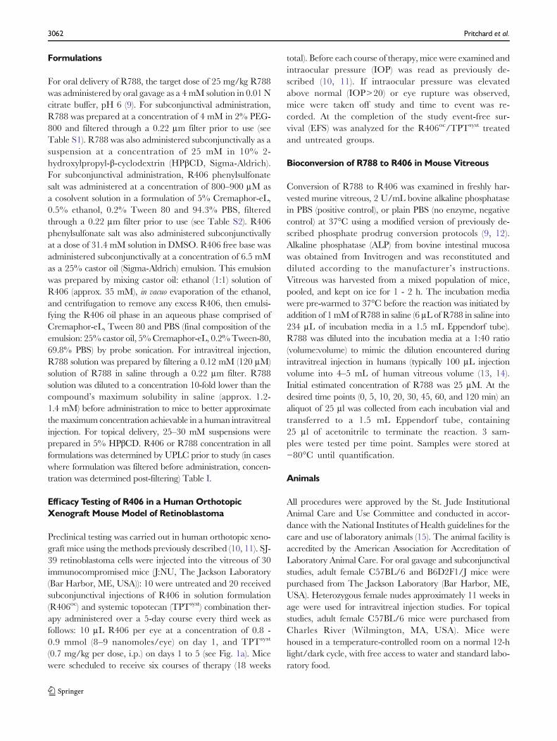
low following subconjunctival delivery of R406 as expected(Fig. 2). However, exposure of R406 in the vitreous failed toreach pharmacologically significant thresholds (Fig. 2).Importantly, the maximal concentration achieved (0.05 μM)was well below the concentration required to kill the mostsensitive retinoblastoma cell line (RB355 in culture, Fig. S3).RB355 cells in culture were treated with DMSO, 1, 2.5, or5 μM of R406 for varied exposure durations (0, 6, 12, 24, 48and 72 h). For the cells in the 2.5 and 5 μM treatment groups,the proportion of activated caspase 3+ cells measured byimmunostaining increased with increasing exposure duration.Incorporation of 5-ethynyl-2′-deoxyuridine (EdU) was used todetect DNA synthesis. The proportion of cells that incorpo-rated EdU in the 5 μM treatment groups decreased withincreasing exposure time (Fig. 3e). In contrast, cells treatedwith 1 μM R406 did not show any increase in caspase 3+activation compared to untreated cells or cells treated withDMSO for any exposure time tested up to 48 h (Fig. 3f to j).Viability of RB355 cells in culture is reduced to 50% of theviability of DMSO treated cells when exposed to 2.5 μMR406 for an exposure duration between 24 and 48 h or5 μM R406 for an exposure duration between 12 and 24 h.There is no reduction in viability compared to DMSO treatedcontrols for RB355 cells in culture treated with 1 μMR406 forup to 24 h exposure, and at longer exposure durations (48 –72 h), viability only drops by approximately 20% (Fig. S3).Comparing the in vivo pharmacokinetics (Fig. 2) and in vitropharmacodynamics data (Fig. 3, Fig. S3) suggests that the lackof evidence of efficacy observed in the preclinical study
(Fig. 1b) was due to insufficient intraocular exposure ofR406 from the solution formulation.
Pharmacokinetics of R406 Emulsion, Suspension, and Drops
In order to increase the concentration of R406 delivered tothe eye from subconjunctival injection, we developed anemulsion formulation using castor oil and other FDA ap-proved adjuvants for ophthalmic emulsions (11). Using thisapproach, we achieved a concentration of 6.5 mM of R406 inemulsion and the emulsion was stable for 48 h at roomtemperature (Fig. S10, Tables S12 and S13). This provideda formulation with 7-fold increased R406 concentration com-pared to the solution formulation. We performed a pharma-cokinetic experiment after delivery of 10 μl of R406 emulsionas described above for the R406 solution (Fig. 4a). As acontrol, we used a high concentration (31.4 mM) of R406 inDMSO. While it is not clinically feasible to deliver R406 inDMSO and DMSO might disrupt biological membranescausing an overestimate of vitreous exposure, it provides auseful benchmark for peak concentration of R406 achievablefrom subconjunctival delivery (Fig. 4b). The R406 emulsionand R406 in DMSO both led to a significant increase inmaximal vitreal concentration and overall exposure but nei-ther reached the targeted efficacious exposure (Fig. 4a and b,Table II). Subconjunctival R406 suspension was not evaluat-ed, as the DMSO control indicated the maximum intraocularconcentration that could be reached for R406 from this routewas approximately 1 μM.
Previous work has shown that ocular absorption of a drugvia the topical route (drops applied to the cornea) can beenhanced by increasing its lipophilicity (16). To evaluate thetopical route for R406 delivery, we tested the intraocularconcentration after a single topical dose of R406 free base oran R406 palmitate salt. R406 levels in the eye were 0.1±0.06 μM at 0.5 h and 0.1±0.1 μM at 1 h post dosing withR406 free base drops, and 0.3±0.3 μM at 0.5 h and 0.3±0.2 μM 1 h post dosing with R406 palmitate salt drops. TheR406 palmitate salt achieved higher intraocular concentra-tions than the R406 free base, but the difference betweenaverage intraocular concentration at both time points wasnot statistically significant (t-test, p-value for 0.5 h=0.3, p-value for 1 h=0.2). No detectable drug was present in theplasma samples (data not shown). The advantage of oculardrops is that repeat daily dosing can be readily achieved. Todetermine if repeated dosing of the R406 palmitate salt dropscould increase intraocular exposure, we dosed mice up to fourtimes with a 40 mM R406 palmitate 5% cyclodextrin suspen-sion and measured the vitreal concentration of R406 at 0.5, 1,and 2 h after the last of 1, 2, 3, or 4 hourly doses (Fig. 5).Whileexhibiting high variability (coefficient of variation (CV)>40%at nearly all observations), mean intraocular concentration-time profiles during the period of time 0.5 to 2 h post-dosing
Fig. 2 Pharmacokinetic behavior of R406 administered as in the efficacystudy. Concentration of R406 measured at 0.5, 1.0, 2.0, and 4.0 h in theplasma (dashed line, empty circles) and vitreous (solid line, filled circles)following subconjunctival delivery of R406 salt in cosolvent solution (10 μLper eye of 880 μM R406 phenylsulfonate salt).
PK and Efficacy of R406 after Ocular Delivery 3065

were similar for repeated doses, with some increase in themean value at the 1 h post dosing time point for 3 or 4doses compared with 1 or 2 doses. A vitreous concen-tration of ~1-2 μM is sustained for 1.5 h after eachapplication of eye drops, demonstrating that R406 con-centrations above 1 μM could be sustained using the topicalroute. However the maximal concentration achieved is stillbelow the targeted concentration.
Intravitreal Injection of R788
Previously, dexamethasone phosphate prodrugs have beensuccessfully administered as subconjunctival injections,
resulting in high concentrations of dexamethasone in thevitreous (17, 18). This suggests phosphatases are present inocular tissues, but phosphatase activity in extracted vitreoushas not been characterized. To determine if R788 can beconverted to R406 in the mouse eye, we incubated freshmurine vitreous with R788 and measured the accumulationof R406 over time (Fig. 6a). As a negative control, we usedPBS; as a positive control, we used PBS with alkaline phos-phatase (ALP) (2 U/mL concentration). R788 was convertedto R406 (100% conversion within 2 h) in extracted mousevitreous suggesting that the prodrug can be effectively deliv-ered directly to the eye. High variability in concentration andapparent concentration decrease at later time points in the
Fig. 3 Cellular pharmacodynamic study defining target time concentration curve RB355 cells in culture are exposed to DMSO, 1, 2.5 and 5 μM R406 for 0, 6,12, 24, 48, or 72 h. (a to e) EdU staining of RB355 cells treated with 0 (DMSO), 1, 2.5, or 5 μM R406 for 0, 6, 12, 24, 48, or 72 h. (f to j) Caspase staining ofRB355 cells treated with 0 (DMSO), 1, 2.5 or 5 μM R406 for 0, 6, 12, 24, 48, or 72 h.
Fig. 4 Pharmacokinetics of R406 following subconjunctival administration. Concentration of R406 measured at 0.5, 1.0, 2.0, and 4.0 h in the plasma (dashedline, empty circles) and vitreous (solid line, filled circles) following (a) subconjunctival delivery of R406 free base in a 25–30% castor oil emulsion (10 μL per eye of6.5 mM R406 free base). (b) subconjunctival delivery of R406 in DMSO (10 μL per eye of 31.4 mM R406 phenylsulfonate salt). Neither formulation achieves thetarget drug concentration in the eye.
3066 Pritchard et al.

TableII
Summaryofvitreousandplasmapharmacokineticparametersafter
asingle
administration(systemicdelivery/oralrouteor
localdelive
ry/subconjunctivalinjection)ofR4
06or
R788
tomice
R788
(FostamatinibDiso
dium
)R4
06PhenylsulfonateSalt
R406
Free
Base
Route
Oral
Local
Local
Local
Local
Local
Form
ulation
BufferedSolution
Co-solve
ntSolution
Suspensio
nCo-solve
ntSolution
DMSO
CastorO
ilEm
ulsio
n
Concentration(m
M)
44
250.88
31.4
6.5
Dose(nmolpere
ye,exceptw
here
noted)
25mg/kg
40250
8.8
314
65
PlasmaPK
Parameters:
Cmax(nM)
2,009.7
574.2
2,906.6
79370.2
118.6
T max(h)
0.5
0.5
0.5
0.5
0.5
0.5
AUClast(nM∙h)
1,500
306
1,800
42.9
298
119
Dosenorm
alizedAU
C(nM∙h/nmol)
1.9
3.8
3.6
2.4
0.5
0.9
T last(h)
41
41
42
Vitre
ousPK
Parameters:
Cmax(nM)
164±
68969±
39.8
267±
76.4
41.3±18
892±
350
169±
35.9
T max(h)
0.5
0.5
0.5
0.5
0.5
0.5
AUClast(nM∙h)
125
724
456
33.7
1,960
430
Dosenorm
alizedAU
C(nM∙h/nmol)
0.2
9.1
0.9
1.9
3.1
3.3
T last(h)
24
42
44
MRT
0-4h(h)
0.772
0.861
1.69
0.782
2.02
2.19
Ratio
Vitre
ousAU
Clast/PlasmaAU
Clast
0.08
NA
0.3
NA
6.6
NA
PK and Efficacy of R406 after Ocular Delivery 3067

ALP positive control samples (Fig. 6a) may be related toR406’s solubility limitations.
There are now several examples of direct injection ofchemotherapy or biological agents into the vitreous for thetreatment of retinoblastoma in patients (19–21). Direct intra-ocular administration of R788 provides maximal R406 con-centration of 1 μM at 0.5 h post dosing (Fig. 6b, the earliesttime point measured). Systemic exposure is lower following
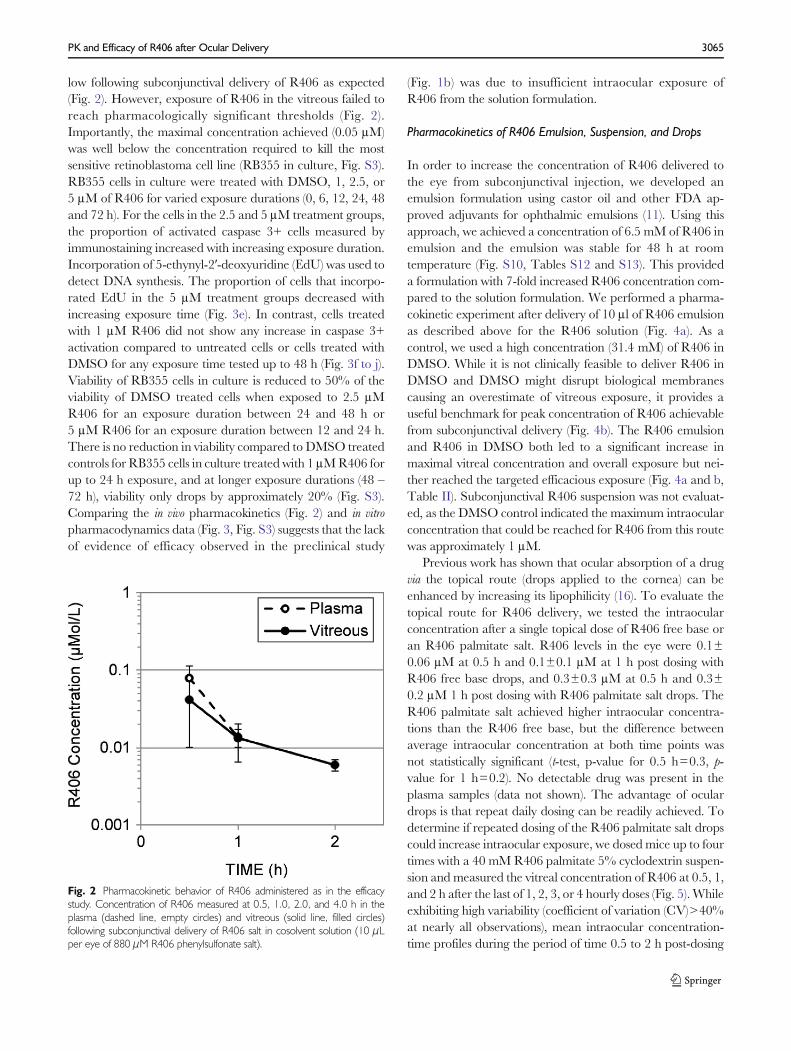
intravitreal injection, but the vitreous concentration-timecurve is similar to that following subconjunctival injection ofR788 in solution formulation (Fig. 7b). Oral delivery of R788and subconjunctival injection of R788 in suspension show noimprovement in overall exposure compared to intravitrealinjection of R788 (Fig. 7a and c).
DISCUSSION
Local Delivery of Molecular Targeted Therapyfor Retinoblastoma
Due to the unique physiology of the eye, numerous routes ofdelivery are available. Systemic routes can be used for oculartherapeutics, but are typically limited by the Blood RetinalBarrier (BRB), which drives up doses needed to reach effica-cious concentrations, thus causing increased systemic toxicity(22–24). Therefore local delivery, which can drastically reducesystemic exposure while simultaneously increasing ocular ex-posure, is an attractive option.
Local ocular delivery routes generally fall into four catego-ries: topical (transcorneal), periocular (transcleral), intravitreal(direct injection), and intra-arterial infusion (21, 25, 26).Though multiple delivery routes are available for retinoblas-toma therapeutics, all routes are not amenable to all com-pounds. Some chemotherapy drugs like topotecan effectivelycross the BRB, with equivalent intraocular PK profiles fromeither systemic or local topotecan delivery (10, 27). In contrast,carboplatin (28–30) and nutlin-3a (11, 31) cannot be admin-istered systemically due to insufficient penetration across theBRB and in these cases subconjunctival injection has been
Fig. 5 Pharmacokinetics of R406 following topical administration. IntraocularR406 concentration measured at 0.5, 1, and 2 h post dosing with one, two,three, or four topical administrations of R406 palmitate salt in suspension (2μLper eye of 40 mM R406 palmitate salt in 5% HPβCD per dose). Vitreouscollected 0.5, 1, and 2 h after the latest dose. N=3-5. Error bars representstandard deviations (where error bars are not shown they fall intobackground).
Fig. 6 Pharmacokinetics of R406 following intravitreal administration of R788. (a) Bioconversion of R788 to R406 in freshly harvested, pooled murine vitreous(filled circles), PBS alone (empty circles), and PBS with 2U/mL alkaline phosphate enzyme (ALP) (triangles). N=3 samples per incubation media. Error barsrepresent standard deviations (where error bars are not shown they fall into background). (b) Concentration of R406 measured at 0.5, 1.0, 2.0, and 4.0 h in thevitreous (blue, squares) following intravitreal injection of R788 in saline (5 μL per eye of 120 μMR788). R406 levels BLOQ in all plasma samples. Administration ofmaximal dose of R788 fails to provide target concentrations of R406 for sufficient duration.
3068 Pritchard et al.
significantly more effective. This contrast highlights the im-portance of utilizing PK to direct delivery route selection forocular therapeutics.
We examined all feasible routes of delivery to the eye includ-ing oral administration of R788 (the conventional route ofadministration in clinical RA trials), subconjunctival deliveryof R406, and topical delivery of R406 and a more lipophilicR406-palmitate salt. Bioconversion of R788 to R406 in pooledextractedmurine vitreous supported testing local administrationof the phosphate prodrug, so subconjunctival administration ofR788 and intravitreal injection of R788 was also evaluated.
To achieve efficacy as a retinoblastoma therapeutic, R406must reach therapeutically effective intraocular exposure. Inpreclinical mouse models of retinoblastoma, subconjunctivalR406 did not result in evidence of efficacy, and our studies
suggest this can be attributed to insufficient intraocular expo-sure. Though local administration routes improved intraocu-lar exposure relative to systemic delivery (Fig. 6), none of thesubconjunctival or intravitreal formulations tested provided avitreous Cmax above 1 μM, which our in vitro studies suggestwould be required to drive efficacy even at prolonged expo-sure times (i.e., up to 72 h) (Fig. 3). Only topical delivery of theR406 palmitate salt in cyclodextrin drops provided a vitreousCmax above 1 μM, which can be sustained via repeated dosing.The topical route achieved higher intraocular concentrationsof R406 than any other route tested and has the advantages ofnon-invasiveness and ease of administration of repeated doses.No chemotherapy drugs are currently administered topically,but these results highlight the need for further exploration ofthis route for retinoblastoma therapeutics.
Fig. 7 Pharmacokinetics of R406 following administration of R788 by several routes. Concentration of R406 measured at 0.5, 1.0, 2.0, and 4.0 h in plasma(dashed line, empty circles) and vitreous (solid line, filled circles) following (a) oral delivery of R788 (25 mg/kg R788 in citric acid buffer) (b) subconjunctival deliveryof R788 cosolvent solution (10 μL per eye of 4 mM R788). (c) subconjunctival delivery of R788 suspension (10 μL per eye of 25 mM R788). No route achievesthe targeted drug exposure.
PK and Efficacy of R406 after Ocular Delivery 3069

R406 Ocular Pharmacokinetics
Our results suggest that the primary drivers of ocular PK forR406 are (1) aqueous solubility (2) lipophilicity and (3) dissolu-tion behavior. In PBS, the maximum solubility of R788 is1.4 mM, the maximum solubility of the R406 phenylsulfonatesalt is 0.4 μMandR406 free base is insoluble (below the limit ofquantitation). Lipophilicity of the compounds tested follows theorder (least lipophilic to most): R788<R406 phenylsulfonatesalt<R406 free base, while aqueous solubility follows the order(lowest solubility to highest): R406 free base<R406phenylsulfonate salt<R788. At similar total doses, the morewater soluble prodrug R788 in cosolvent solution (40 nmoltotal dose per eye) achieves a maximum vitreous concentration6-fold higher than the water insoluble R406 free base in emul-sion (65 nmol total dose per eye) (Figs. 6b and 3a, Table II),which suggests aqueous solubility may control maximal equi-librium ocular drug concentrations. R406 administered at ahigh dose in DMSO achieves a comparable vitreous Cmax toR788 cosolvent formulation (Figs. 3b and 6b, Table II), sug-gesting that the upper limit of compound solubility in vitreous isdriven by solubility of the converted R406. A 6.25-fold increasein dose of locally administered R788 does not increase vitreousCmax or vitreous exposure, but dramatically increases systemicexposure (Table II). This suggests rapid clearance occurs oncethe maximum vitreous concentration is reached, and thatincreasing the amount of drug administered to thesubconjunctival space will not increase vitreous Cmax beyondthis limit. When comparable total doses of R788 and R406 saltare administered locally (250 and 314 nmol total per eye,respectively), the R406 salt increases vitreous exposure anddecreases plasma exposure, achieving a ratio of vitreous AUCto plasma AUC 22-fold higher than the more water-solubleR788 in suspension (Table II). This suggests that when localdose is sufficient, R406 phenylsulfonate salt exhibits greaterretention at the injection site due to its limited water solubility,while themore water soluble prodrug diffuses more readily fromthe injection site into systemic circulation. R406 phenylsulfonatesalt and R788 have very similar molecular weights (628.6 and624.4, respectively), suggesting that their differences in lipophi-licity, solubility and dissolution are driving PK.
Ocular mean residence times (from shortest to longest) wereas follows: R788 systemic/oral≈R406 salt local solution<R788local solution (all less than 1 h)<R788 local suspension<R406salt local DMSO<R406 base local emulsion (Table II). Theemulsion and the R406 salt in DMSO exhibit the longest meanresidence times (MRT≥2 h). The R406 emulsion may form adrug depot in the subconjunctival space, analogous to the use ofintramuscular injections of oil carriers and suspensions in de-livery of long acting injectable (LAI) antipsychotics where slowdiffusion of the drug ester out of the oil phase and into the bloodstream prolongs the maintenance of therapeutic drugconcentrations (32). When added to buffer, R406 salt in
DMSO precipitates out of solution (see Supplement). Thesolution of R406 salt in DMSO may therefore be functionallycomparable to the long-acting injectable formulation ofolanzapine, which is injected into the gluteal muscle as asuspension of micron sized crystals of olanzapine and pamoicacid in aqueous solution, providing sustained release (2–4weeks) as the salt gradually dissolves and diffuses into systemiccirculation (33). If R406 salt precipitates out of DMSO in thesubconjunctival space, depot formation will lead to re-dissolution of the R406 precipitate sustaining release into theeye and limiting rapid clearance from the injection site. Thisinterpretation is consistent with the rapid clearance and highsystemic exposure observed when a relatively high concentra-tion of R788 in suspension is administered locally.
In the case of subconjunctival delivery of R406, increasingcompound lipophilicity improves the ratio of vitreous AUC toplasma AUC, but an increase in drug lipophilicity may notimprove ocular PK overall if there is a trade-off in aqueoussolubility: the most lipophilic of the compounds tested (R406free base), reaches relatively low intraocular concentrationscompared with other local formulations, while the least lipo-philic (R788) has the shortest MRT. Compare these results tothe previous success with local delivery of nutlin-3a (AUCratio of vitreous to plasma for subconjunctival administra-tion=28.6), which has a water solubility of approximately40 μM (100-fold higher than R406 phenylsulfonate salt) anda higher logP than any of the compounds tested in this study.Our current understanding of subconjunctival administrationsuggests that successful delivery depends on optimizing athree-way balance between aqueous solubility, lipophilicity,and dissolution because these properties impact absorptionacross barriers, tissue partition, maximum concentrationachievable in vitreous, retention at the injection site, and ratioof compound diffusing out systemically relative to the amountabsorbed into the eye. Because inverse relationships may existamongst solubility, lipophilicity, and dissolution, it is unlikelythat any one of those properties can be optimized individually;improvements in one property will need to be weighed againstpotential trade-offs in another.
Targeting SYK/BCL2 in Retinoblastoma
Despite the failing of R406 as a retinoblastoma clinical can-didate, SYK remains a promising target as there are manyother small molecule SYK inhibitors with diverse physio-chemical properties to evaluate as retinoblastoma therapeutics(34–38). Previous studies of SYK inhibition have implicated anumber of downstream signalingmolecules, including the Bcl-2 family of proteins as mediators of the SYK survival signal(35, 39). This suggests that some of the small molecule Bcl-2inhibitors currently being developed for other cancer, such asobatoclax and TW-37 (40) may also prove to be effectivetherapeutic agents in retinoblastoma.
3070 Pritchard et al.

Conclusion
The SYK inhibitor R406, currently in clinical developmentfor rheumatoid arthritis, can induce caspase-mediated celldeath of retinoblastoma cells in culture. Subconjunctivaladministration of R406 failed to provide any evidence ofimprovement in tumor response in preclinical models ofretinoblastoma. We found that vitreal exposure followingsubconjunctival delivery of the R406 solution formulationwas below the exposure required to induce caspase-mediated cell death of retinoblastoma cells in culture. Wedeveloped emulsion and suspension formulations of R406,which increased the vitreal exposure of R406, but still failedto reach the target exposure. The R788 prodrug was effi-ciently converted to R406 in extracted vitreous and in vivo,but following direct intravitreal injection of R788, R406exposure was still insufficient. Topical delivery of R406-palmitate achieved intraocular concentrations above 1 μM,which was sustainable via repeated dosing. Though localdelivery via the subconjunctival, topical and intravitrealroutes improved vitreous exposure compared with oral de-livery, vitreous exposure from all routes and formulationstested was not within the range required to kill retinoblas-toma cells in culture. Therefore, the preclinical modelsstrongly suggest that R406 is not a viable clinical candidatefor retinoblastoma. Future efforts may focus on combiningR406 with other agents, testing other SYK inhibitors withdifferent physiochemical properties that may improve intra-ocular pharmacokinetics, or targeting other proteins in thepathway.
ACKNOWLEDGMENTS AND DISCLOSURES
The authors thank Justin Thurman and Alex Su for helpcollecting tissue samples. The authors thank JustinaMcEvoy, Claudia Benavente and Daniel Hiler for providingmurine vitreous. The authors thankWilliamWu and JianrongWu in the SJCRH Department of Biostatistics. The authorsthank William Caufield for preliminary bioanalytical assaydevelopment. We thank the American Lebanese SyrianAssociated Charities (ALSAC) and St Jude Children’sResearch Hospital for funding. Eleanor Pritchard was fundedby an SJCRH Academic Programs special fellowship. Thiswork was also supported by a grant from the National CancerInstitute [R01CA168875-01] and by the Howard HughesMedical Institute.
Open Access This article is distributed under the terms ofthe Creative Commons Attribution License which permitsany use, distribution, and reproduction in any medium, pro-vided the original author(s) and the source are credited.
REFERENCES
1. Federico S, Brennan R, Dyer MA. Childhood cancer a developmen-tal biology: a crucial partnership. Curr Top Dev Biol. 2011;94:1–13.
2. Shields CL, Shields JA. Diagnosis and management of retinoblasto-ma. Cancer Control. 2004;11(5):317–27.
3. Eng C, Li FP, Abramson DH, Ellsworth RM, Wong RL, GoldmanMB, et al. Mortality from second tumors among long-term survivorsof retinoblastoma. J Natl Cancer Inst. 1993;85(14):1121–8.
4. Nahum MP, Gdal-On M, Kuten A, Herzl G, Horovitz Y, ArushMWB. Long-term follow-up of children with retinoblastoma.Pediatric Hematol Oncol. 2001;18(3):173–9.
5. Hijiya N, Ness KK, Ribeiro RC, Hudson MM. Acute leukemia as asecondary malignancy in children and adolescents: current findingsand issues. Cancer. 2009;115(1):23–35.
6. Zhang J, Benavente CA,McEvoy J, Flores-Otero J, Ding L, Chen X,et al. A novel retinoblastoma therapy from genomic and epigeneticanalyses. Nature. 2012;481(7381):329–34.
7. Braselmann S, Taylor V, Zhao H, Wang S, Sylvain C, Baluom M,et al. R406, an orally available spleen tyrosine kinase inhibitor blocksFc receptor signaling and reduces immune complex-mediated in-flammation. J Pharmacol Exp Ther. 2006;319(3):998–1008.
8. Weinblatt ME, Kavanaugh A, GenoveseMC,Musser TK, GrossbardEB, Magilavy DB. An oral spleen tyrosine kinase (Syk) inhibitor forrheumatoid arthritis. N Engl J Med. 2010;363(14):1303–12.
9. Sweeny DJ, Li W, Clough J, Bhamidipati S, Singh R, Park G, et al.Metabolism of fostamatinib, the oral methylene phosphate prodrugof the spleen tyrosine kinase inhibitor R406 in humans: contributionof hepatic and gut bacterial processes to the overall biotransforma-tion. Drug Metab Dispos. 2010;38(7):1166–76.
10. Nemeth KM, Federico S, Carcaboso AM, Shen Y, Schaiquevich P,Zhang J, et al. Subconjunctival carboplatin and systemic topotecantreatment in preclinical models of retinoblastoma. Cancer. 2011;117(2):421–34.
11. Brennan RC, Federico S, Bradley C, Zhang J, Flores-Otero J,WilsonM, et al. Targeting the p53 pathway in retinoblastoma withsubconjunctival nutlin-3a. Cancer Res. 2011;71(12):4205–13.
12. Yuan H, Li N, Lai Y. Evaluation of in vitro models for screeningalkaline phosphatase-mediated bioconversion of phosphate esterprodrugs. Drug Metab Dispos. 2009;37(7):1443–7.
13. Cardillo JA, Melo Jr LA, Costa RA, Skaf M, Belfort Jr R, Souza-Filho AA, et al. Comparison of intravitreal versus posterior sub–Tenon’scapsule injection of triamcinolone acetonide for diffuse dia-betic macular edema. Ophthalmology. 2005;112(9):1557–63.
14. Smith JR, Rosenbaum JT, Wilson DJ, Doolittle ND, Siegal T,Neuwelt EA, et al. Role of intravitreal methotrexate in the manage-ment of primary central nervous system lymphoma with ocularinvolvement. Ophthalmology. 2002;109(9):1709–16.
15. Institute of Laboratory Animal Resources. Commission on LifeSciences, National Research Council. Guide for the Care and Useof Laboratory Animals. Washington: National Academy Press; 1996.
16. Järvinen T, Järvinen K. Prodrugs for improved ocular drug delivery.Adv Drug Deliv Rev. 1996;19(2):203–24.
17. Weijtens O, Feron EJ, Schoemaker RC, Choen AF, Lentjes EGWM,Romijn FPHTM, et al. High concentration of dexamethasone in aque-ous and vitreous after subconjunctival injection. Am J Ophthalmol.1999;128(2):192–7.
18. Weijtens O, Schoemaker RC, Lentjes EGWM, Romijn FPHTM,Cohen AF, van Meurs JC. Dexamethasone concentration in thesubretinal fluid after a subconjunctival injection, a peribulbar injection,or an oral dose. Ophthalmology. 2000;107(10):1932–8.
19. Munier FL, Gaillard MC, Balmer A, Soliman S, Podilsky G, MoulinAP, et al. Intravitreal chemotherapy for vitreous disease in retinoblas-toma revisited: from prohibition to conditional indications. Br JOphthalmol. 2012;96(8):1078–83.
PK and Efficacy of R406 after Ocular Delivery 3071

20. Ghassemi F, Shields CL. Intravitreal melphalan for refractory orrecurrent vitreous seeing from retinoblastoma. Arch Ophthalmol.2012;130(10):1268–71.
21. Shields CL, Fulco EM, Arias JD, Alarcon C, Pellegrini M, Rishi P,et al. Retinoblastoma frontiers with intravenous, intra-arterial,periocular, and intravitreal chemotherapy. Eye. 2013;27(2):253–64.
22. Janoria KG, Gunda S, Boddu SH, Mitra AK. Novel approaches toretinal drug delivery. Expert Opin Drug Deliv. 2007;4(4):371–88.
23. Campbell M, Ozaki E, Humphries P. Systemic delivery of therapeu-tics to neuronal tissues: a barrier modulation approach. Expert OpinDrug Deliv. 2010;7(7):859–69.
24. Zhang K, Zhang L, Weinreb RN. Ophthalmic drug discovery: noveltargets and mechanisms for retinal diseases and glaucoma. Nat RevDrug Discov. 2012;11(7):541–59.
25. Short BG. Safety evaluation of ocular drug delivery formulations:techniques and practical considerations. Toxicol Pathol. 2008;36(1):49–62.
26. Geroski DH, Edelhauser HF. Drug delivery for posterior segmenteye disease. Invest Ophthalmol Vis Sci. 2000;41(5):961–4.
27. Carcaboso AM, Bramuglia GF, Chantada GL, Fandiño AC,Chiappetta DA, de Davila MTG, et al. Topotecan vitreous levelsafter periocular or intravenous delivery in rabbits: an alternative forretinoblastoma chemotherapy. Invest Ophthalmol Vis Sci.2007;48(8):3761–7.
28. Murray TG, Cicciarelli N, O’Brien JM, Hernández E, Mueller RL,Smith BJ, et al. Subconjunctival carboplatin therapy and cryotherapyin the treatment of transgenic murine retinoblastoma. ArchOphthalmol. 1997;115(10):1286–90.
29. Abramson DH, Frank CM, Dunkel IJ. A phase I/II study ofsubconjunctival carboplatin for intraocular retinoblastoma.Ophthalmology. 1999;106(10):1947–50.
30. Hayden BC, Jockovich M-E, Murray TG, Voigt M, Milne P,Kralinger M, et al. Pharmacokinetics of systemic versus focalcarboplatin chemotherapy in the rabbit eye: possible implication inthe treatment of retinoblastoma. Invest Ophthalmol Vis Sci.2004;45(10):3644–9.
31. Zhang F, Tagen M, Throm S, Mallari J, Miller L, Guy RK, et al.Whole-body physiologically based pharmacokinetic model for nutlin-3a in mice after intravenous and oral administration. Drug MetabDispos. 2011;39(1):15–21.
32. Taylor D. Psychopharmacology and adverse effects of antipsychoticlong-acting injections: a review. Br J Psychiatry Suppl. 2009;195:S13–9.
33. Di Lorenzo R, Brogli A. Profile of olanzapine long-acting injectionfor the maintenance treatment of adult patients with schizophrenia.Neuropsychiatr Dis Treat. 2010;6:573–81.
34. Singh R, Masuda ES, Payan DG. Discovery and development ofspleen tyrosine kinase (SYK) inhibitors. J Med Chem. 2012;55(8):3614–43.
35. D’Cruz OJ, Uckun FM. Targeting spleen tyrosine kinase(SYK) for treatment of human disease. J Pharm Drug DelivRes 201;1:2.
36. Ruzza P, Biondi B, Calderan A. Therapeutic prospect ofSyk inhibitors. Expert Opin Ther Patents. 2009;19(10):1361–76.
37. Spurgeon SE, Coffey G, Fletcher LB, Burke R, Tyner JW,Druker BJ, et al. The selective SYK inhibitor P505-15(PRT062607) inhibits B cell signaling and function in vitroand in vivo and augments the activity of fludarabine inchronic lymphocytic leukemia. J Pharmacol Exp Ther.2013;344(2):378–87.
38. Reilly MP, Sinha U, André P, Taylor SM, Pak Y, DeGuzman FR,et al. PRT-060318, a novel Syk inhibitor, prevents heparin inducedthrombocytopenia and thrombosis in a transgenic mouse model.Blood. 2011;117(7):2241–6.
39. Gobessi S, Laurenti L, Longo PG, Carsetti L, Berno V, Sica S, et al.Inhibition of constitutive and BCR-induced Syk activationdownregulates Mcl-1 and induces apoptosis in chronic lymphocyticleukemia B cells. Leukemia. 2009;23(4):686–97.
40. Juin P, Geneste O, Gautier F, Depil SJ, Campone M. Decoding andunlocking the BCL-2 dependency of cancer cells. Nat Rev Cancer.2013;13(7):455–65.
3072 Pritchard et al.


![HMPL-523, a Novel SYK Inhibitor, Showed Anti-tumor ...€¦ · [1] Pharmacology & Therapeutics 144 (2014) 338–348 Spleen Tyrosine Kinase (SYK) plays a pivotal role in the regulation](https://static.fdocuments.in/doc/165x107/604cbdbedbcfc6166042bf65/hmpl-523-a-novel-syk-inhibitor-showed-anti-tumor-1-pharmacology-therapeutics.jpg)