Pharmacokinetic, Physicochemical and Medicinal Properties ...
12
Journal of Pharmacy and Pharmacology 7 (2019) 165-176 doi: 10.17265/2328-2150/2019.04.003 Pharmacokinetic, Physicochemical and Medicinal Properties of N-glycoside Anti-cancer Agent More Potent than 2-Deoxy-D-Glucose in Lung Cancer Cells Fidelis Toloyi Ndombera 1,2 , Geoffrey K. K. Maiyoh 3,4 and Vivian C. Tuei 5 1. Covance Laboratories Inc. BioPharm CMC solutions, Greenfield, 46140, Indiana, USA 2. Chemistry Department, Wayne State University, Detroit, 48201, Michigan, USA 3. Department of Medical Biochemistry, School of Medicine, Moi University, 30100, Eldoret, Kenya 4. Johannesburg Institute for Advanced Study, An initiative of the University of Johannesburg and Nanyang Technological University, Singapore,1 Tolip Street, Westdene 2092, P.O Box 524, Auckland Park 2006, Johannesburg, South Africa 5. Department of Chemistry and Biochemistry, School of Science, University of Eldoret, 30100, Eldoret, Kenya Abstract: Acetylated N-xyloside of 1-naphthylamine (K8A) has been shown to be more potent than 2-deoxy-D-glucose in lung cancer cells and has therapeutic potential for further drug development. In this paper we evaluate and report cytotoxicity, pharmacokinetic, physicochemical and medicinal properties of this D-Xylose derivative (K8A) as a lead anticancer agent with greater therapeutic potential than 2-deoxy-D-glucose (2-DG). 2-DG has been in clinical trials for treatment of solid tumors and other types of cancer. We demonstrate using virtual tools that K8A has better “drug-likeness” than 2-DG and does not violate any Lipinski, Ghose, Veber, Egan or Muegge rules. On the other hand, 2-DG violates Ghose and Muegge rules. A “BOILEDegg evaluation”, predicts that K8A has higher gastrointestinal absorption (HIA) than 2-DG and is not effluxed by P-glycoprotein (P-gp). Additionally, K8A does not penetrate the blood brain barrier (BBB) and is not a substrate of most Cytochrome P450 (CYP) enzymes. Importantly, K8A did not show false positive alert from PAINS screening enabling us to narrow down and rule out false targets. Importantly, K8A is more potent than 2-DG in H1299 and A549 lung cancer cells. Key words: Anticancer agent, N-glycoside, 2-deoxy-D-glucose, pharmacokinetics, lung cancer. 1. Introduction 2-deoxy-D-glucose (2-DG) induces cellular stress [1-4] and cell death [5]. 2-DG inhibits glycolysis [6], disrupts N-glycosylation [7] and alters the pentose phosphate pathway (PPP) to cause oxidative [8] and endoplasmic reticulum (ER) stress [4]. Upon 2-DG phosphorylation, 2-DG-6-phosphate noncompetitively inhibits hexokinase, and competitively inhibits phosphoglucose isomerase (PGI) [6]. 2-DG has been in clinical trial for potential use as an anticancer agent [9, 10]. The use of 2-DG is however limited by high-dose Corresponding author: Fidelis Ndombera, Ph.D., Research Associate II, Covance Labs. Inc, research fields: molecular biology & drug development. Email: [email protected]. systemic toxicity [11] because the drug discovery process ignored toxicity and in silico pharmacodynamic and pharmacokinetic indicators. For this reason, high concentrations are required to out-compete intracellular glucose concentrations in the human body. In most humans, glucose concentration varies from about 80 mg/dl to 110 mg/dl. Additionally, 2-DG promotes multiple prosurvival pathways [12] and can activate migration and invasion of glioblastomas [13], thus necessitating the need for drug discovery of alternative carbohydrate-based inducers of cellular stress [14]. One such molecule is acetylated N-xyloside of 1-naphthylamine (K8A) and has been shown to be more potent than 2-deoxy-D-glucose in lung cancer cells. K8A has therapeutic potential for D DAVID PUBLISHING
Transcript of Pharmacokinetic, Physicochemical and Medicinal Properties ...

Journal of Pharmacy and Pharmacology 7 (2019) 165-176
doi: 10.17265/2328-2150/2019.04.003
Pharmacokinetic, Physicochemical and Medicinal
Properties of N-glycoside Anti-cancer Agent More
Potent than 2-Deoxy-D-Glucose in Lung Cancer Cells
Fidelis Toloyi Ndombera1,2
, Geoffrey K. K. Maiyoh3,4
and Vivian C. Tuei5
1. Covance Laboratories Inc. BioPharm CMC solutions, Greenfield, 46140, Indiana, USA
2. Chemistry Department, Wayne State University, Detroit, 48201, Michigan, USA
3. Department of Medical Biochemistry, School of Medicine, Moi University, 30100, Eldoret, Kenya
4. Johannesburg Institute for Advanced Study, An initiative of the University of Johannesburg and Nanyang Technological University,
Singapore,1 Tolip Street, Westdene 2092, P.O Box 524, Auckland Park 2006, Johannesburg, South Africa
5. Department of Chemistry and Biochemistry, School of Science, University of Eldoret, 30100, Eldoret, Kenya
Abstract: Acetylated N-xyloside of 1-naphthylamine (K8A) has been shown to be more potent than 2-deoxy-D-glucose in lung cancer
cells and has therapeutic potential for further drug development. In this paper we evaluate and report cytotoxicity, pharmacokinetic,
physicochemical and medicinal properties of this D-Xylose derivative (K8A) as a lead anticancer agent with greater therapeutic
potential than 2-deoxy-D-glucose (2-DG). 2-DG has been in clinical trials for treatment of solid tumors and other types of cancer. We
demonstrate using virtual tools that K8A has better “drug-likeness” than 2-DG and does not violate any Lipinski, Ghose, Veber, Egan
or Muegge rules. On the other hand, 2-DG violates Ghose and Muegge rules. A “BOILEDegg evaluation”, predicts that K8A has higher
gastrointestinal absorption (HIA) than 2-DG and is not effluxed by P-glycoprotein (P-gp). Additionally, K8A does not penetrate the
blood brain barrier (BBB) and is not a substrate of most Cytochrome P450 (CYP) enzymes. Importantly, K8A did not show false
positive alert from PAINS screening enabling us to narrow down and rule out false targets. Importantly, K8A is more potent than 2-DG
in H1299 and A549 lung cancer cells.
Key words: Anticancer agent, N-glycoside, 2-deoxy-D-glucose, pharmacokinetics, lung cancer.
1. Introduction
2-deoxy-D-glucose (2-DG) induces cellular stress
[1-4] and cell death [5]. 2-DG inhibits glycolysis [6],
disrupts N-glycosylation [7] and alters the pentose
phosphate pathway (PPP) to cause oxidative [8] and
endoplasmic reticulum (ER) stress [4]. Upon 2-DG
phosphorylation, 2-DG-6-phosphate noncompetitively
inhibits hexokinase, and competitively inhibits
phosphoglucose isomerase (PGI) [6]. 2-DG has been in
clinical trial for potential use as an anticancer agent [9,
10]. The use of 2-DG is however limited by high-dose
Corresponding author: Fidelis Ndombera, Ph.D., Research
Associate II, Covance Labs. Inc, research fields: molecular
biology & drug development. Email: [email protected].
systemic toxicity [11] because the drug discovery
process ignored toxicity and in silico
pharmacodynamic and pharmacokinetic indicators. For
this reason, high concentrations are required to
out-compete intracellular glucose concentrations in the
human body. In most humans, glucose concentration
varies from about 80 mg/dl to 110 mg/dl. Additionally,
2-DG promotes multiple prosurvival pathways [12]
and can activate migration and invasion of
glioblastomas [13], thus necessitating the need for drug
discovery of alternative carbohydrate-based inducers
of cellular stress [14]. One such molecule is acetylated
N-xyloside of 1-naphthylamine (K8A) and has been
shown to be more potent than 2-deoxy-D-glucose in
lung cancer cells. K8A has therapeutic potential for
D DAVID PUBLISHING
Pharmacokinetic, Physicochemical and Medicinal Properties of N-Glycoside Anti-cancer Agent More Potent than 2-Deoxy-D-Glucose in Lung Cancer Cells
166
further drug development.
Modern drug development involves evaluation of
efficiency of potent molecules and their ability to reach
targets in bioactive form. This involves cellular, animal
and human clinical trials that are often costly and laden
with safety risks. Currently, computer aided
assessment of absorption, distribution, metabolism and
excretion of drugs (ADME) has gained traction to
mitigate these concerns [15]. Importantly, computer
models provide predictive and reliable data fast and
thus compliment experimental approaches. These
computer models also predict pharmacokinetic,
physicochemical and medicinal properties of small
molecules and assist in optimization of lead
compounds to patient drugs.
Free web tools are now available that give access to
these predictive models and include molinspiration
(http://www.molinspiration.com), Silicos-it can be
traced online at
(http://silicos-it.be.s3-website-eu-west-1.amazonaws.c
om/software/software.html), omicX found at
(https://omictools.com/qed-tool), molsoft,
(http://www.molsoft.com/mprop/) and swissADME
(http://www.swissadme.ch) [16], among others.
Molinspiration is a website that allows one to predict
the bioactivity of a structure against several different
common drug targets. Molinspiration offers broad
range of cheminformatics software tools supporting
molecule manipulation and processing. Silicos-it was
developed to share information on open source
cheminformatics with downloadable tools that aid in
drug design and development. SwissADME is a more
recent and comprehensive site run by the Swiss
institute of bioinformatics (SIB) which provides
bioinformatics services and resources for scientists
worldwide. SIB has over 65 bioinformatics research
groups and 800 scientists from the major Swiss schools
of higher education and research institutes.
SwissADME enables assessment of ADME
parameters of drug candidates and small molecules and
provides information that allows early risk assessment
in the drug development process. Notably,
swissADME provides a platform to assess Lipinski’s
rule of five [17] for drug-likeness of oral
bioavailability. Drug-likeness is a complex balance of
molecular properties and structural features which
determine whether an unknown molecule is like the
known drugs. These molecular properties include
hydrophobicity, electronic distribution, and hydrogen
bonding characteristics, molecule size and flexibility.
SwissADME includes “BOILEDegg evaluation” [18]
that predict gastrointestinal absorption (HIA) and
efflux/retention by P-glycoprotein (P-gp).
“BOILEDegg” is a graphical evaluation of HIA as a
function of the position of the small molecule in the
WLOGP-versus-TPSA plot. Additionally, blood brain
barrier (BBB) penetration and Cytochrome P450 (CYP)
enzyme substrate-inhibition prediction can be made.
Importantly, false positive results commonly seen in
biochemical assays of small molecules are predicted
with a fair degree of certainty [19].
2. Materials and Methods
SIB website http://www.swissadme.ch was
accessed in a web browser that displays the
submission page of SwissADME. Previously
synthesized and characterized hit molecules K8 and
K8A [20] with positive control (2-deoxyD-glucose)
and other controls were input for estimation of ADME,
physicochemistry, drug-likeness, pharmacokinetics
and medicinal chemistry properties. The input zone
comprises a molecular sketcher based on ChemAxon’s
Marvin JS (http://www.chemaxon.com) that allowed
the user to draw and edit 2D chemical structures. The
structures are transferred as a list to the right-hand
side of the submission page, which is the actual input
for computation. The list is made to contain one input
molecule per line, defined by simplified
molecular-input line-entry system (SMILES) and
results are presented for each molecule in tables,
graphically and as an excel spreadsheet.
We then synthesized and evaluated K8 and K8A
Pharmacokinetic, Physicochemical and Medicinal Properties of N-Glycoside Anti-cancer Agent More Potent than 2-Deoxy-D-Glucose in Lung Cancer Cells
167
with D-xylose and naphthylamine as described
previously [20]. Briefly, equimolar D-xylose and
appropriate naphthylamine was refluxed in ethanol for
8 hours. This was followed by acetylation in pyridine
with acetic anhydride in an ice bath for 3 hours and in
the cold room (4 0C) for another 13 hours. The reactant
mixture was then quenched by pouring into ice/water
mixture, extracted, washed with aqueous sodium
bicarbonate, water and brine, and purified by column
chromatography (1:4 ethyl acetate/hexane solvent
conditions). Bioactive K8A was characterized by 1H
NMR and 13
C NMR (supporting information).
We evaluated acetylated N-Naphthyl-β-D-Xylose
(K8A) vs 2-DG in two lung cancer cell lines (Table 7).
Cell viability was measured by using the
3-(4,5-dimethylthiazol-2-yl)-diphenyl tetrazolium
bromide assay (MTT assay) after a 48-hour incubation
period.
3. Results and Discussion
We evaluated our lead compound K8A, with
controls that included 1-naphthylamine
(K8A-aglycone), D-Xylose (K8A-glycone), acetylated
D-Xylose and the positive control 2-deoxy-D-glucose.
The structural features of these small molecules were
entered in the SwissADME website
(http://swissadme.ch) using the ChemAxon’s Marvin
JS structure drawing tool. Structural features of a
pharmacophore influence the behavior of a molecule in
humans, including bioavailability, transport properties,
affinity to proteins, reactivity, toxicity and metabolic
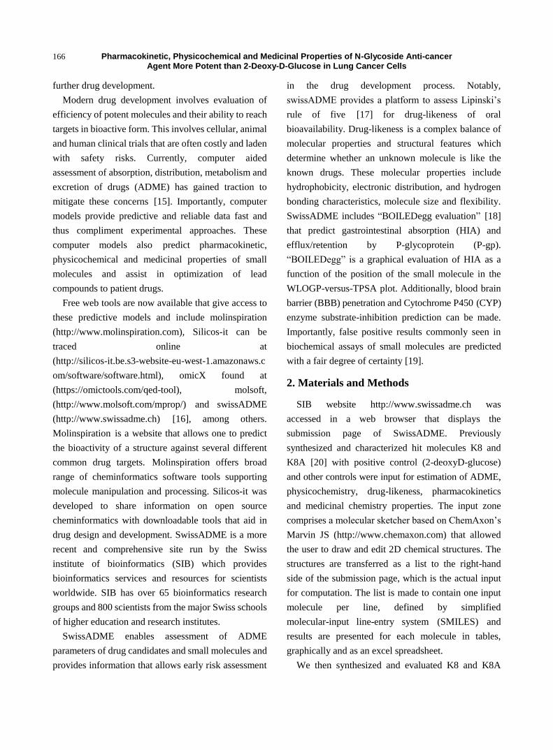
stability. Unique to swissADME is the bioavailability
radar [16] that provides a graphical snapshot of the
drug-likeness parameters of an orally available
bioactive drug. The drug-likeness graph is presented as
a hexagon (Fig. 1) with each of the vertices
representing a parameter that define a bioavailable
drug. The pink area within the hexagon represents the
optimal range for each property (lipophilicity: XLOGP3
between -0.7 and +5.0, size: MW between 150 and 500
g/mol, polarity: TPSA between 20 and 130 Å2
,
solubility: log S not higher than 6, saturation: fraction
of carbons in the sp3 hybridization not less than 0.25,
and flexibility: no more than 9 rotatable bonds).
Fig. 1 The bioavailability radar of the small molecules evaluated using swissADME web tool. 2-deoxy-D-glucose (Molecule 1),
K8 (Molecule 2), K8A (Molecule 3), 1-naphthylamine (Molecule 4), D-xylose (Molecule 5) and acetylated D-xylose (Molecule 6).
(Lipophilicity (LIPO): XLOGP3 between -0.7 and +5.0, Molecular weight (SIZE): MW between 150 and 500 g/mol, Polarity
(POLAR) TPSA between 20 and 130 Å2, Solubility (INSOLU): log S not higher than 6, Saturation (INSATU): fraction of
carbons in the sp3 hybridization not less than 0.25, and Flexibility (FLEX): no more than 9 rotatable bonds.

Pharmacokinetic, Physicochemical and Medicinal Properties of N-Glycoside Anti-cancer Agent More Potent than 2-Deoxy-D-Glucose in Lung Cancer Cells
168
3.1 Drug-Likeness
2-deoxy-D-glucose (Molecule 1), K8 (Molecule 2)
and K8A (Molecule 3) drug-likeness properties are
represented by the red distorted hexagon within the
pink shade (Fig. 1). Notably, 2-DG, K8 and K8A
drug-likeness fall within parameters of a bioavailable
drug. 1-naphthylamine (Molecule 4), D-xylose
(Molecule 5) and acetylated D-xylose (Molecule 6)
were negative controls with no bioactivity in cancer
cells. Molecule 4 has high unsaturation indicated by an
off-shoot of one of the saturation (INSATU) vertex
(Fig. 1).
SwissADME also has computational filters that
include Ghose [21], Egan [22], Veber [23], and
Muegee [24] developed by leading pharmaceutical
companies and cheminfomaticians to evaluate the
drug-likeness of small molecules. The Ghose filter
quantitatively characterizes small molecules based on
computed physicochemical property profiles that
include log P, molar refractivity (MR), molecular
weight (MW), and number of atoms. Additionally, the
Ghose [21, 25] filter include a qualitative
characterization based on the presence of functional
groups and important substructures. The qualifying
range of calculated log P (ClogP) is between -0.4 and
5.6. For MW, the qualifying range is between 160 and
480. For MR, the qualifying range is between 40 and
130 and for the total number of atoms, the qualifying
range is between 20 and 70 atoms in a small molecule.
Our hit compound K8A met the Ghose qualifying
criteria but not molecule 4 with a MW around 143,
molecule 1 (2-DG) with a MR less than 40, and
Molecule 5 with a MR of around 30 (Table 1).
Egan (pharmacia) filter [22] provides a prediction of
drug absorption based on physical processes involved
in membrane permeability of a small molecule. The
descriptors in the Egan model are polar surface area
(PSA) and AlogP98v with exclusion of redundant
descriptors such as MW. PSA is a reference point for
AlogP98 [25], since the latter descriptor is a ratio of
lipophilicity to hydrophilicity which contains no
information on the absolute measure of either factor.
Importantly, the Egan computational model for human
passive intestinal absorption (HIA) of small molecule
accounts for active transport and efflux mechanisms
and is therefore robust in predicting absorption of
drugs. Exclusion of redundant descriptors by this
model allowed K8A and all the small molecules to
obey the Egan rules (Table 2).
Veber (GSK filter) [23] model characterizes
molecules as drug-like if they have 10 or fewer
rotatable bonds and a PSA equal to or less than 140 Å2
with 12 or fewer H-bond donors and acceptors. K8A
and all the small molecules evaluated met Veber
criteria (Tables 1 and 2). Molecules with these
properties have a high probability of good oral
bioavailability in a rat model. Reduced PSA correlates
better with increased permeation rate than lipophilicity
does (ClogP). Conversely, increased rotatable bond count
Table 1 Physicochemical properties of the small molecules (MW; molecular weight, MR; molar refractivity, TPSA; total
polar surface area).
Pharmacokinetic, Physicochemical and Medicinal Properties of N-Glycoside Anti-cancer Agent More Potent than 2-Deoxy-D-Glucose in Lung Cancer Cells
169
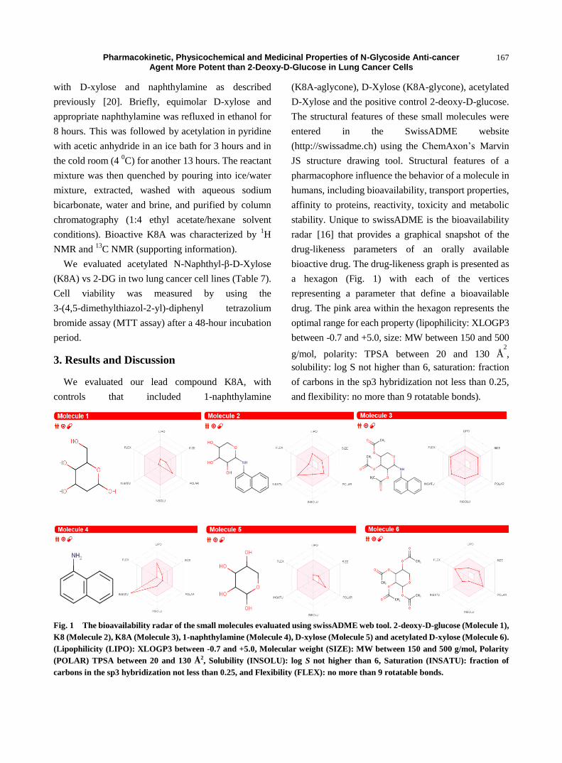
Table 2 Drug-likeness evaluation of small molecules using swissADME shows the hit compounds K8A and K8 do not violate
any of the drug-likeness criteria.
has a negative effect on the permeation rate.
Consequently, a threshold permeation rate is a
prerequisite of oral bioavailability (F). Of note is the
Abbot bioavailability score [26] that predicts the
probability of a compound to have F > 10% based on
the predominant charge at biological pH in a rat model.
Abbot bioavailability model distinguishes compounds
that are poorly permeable from those that are
permeable in Caco-2 cells.
Muegge (Bayer filter) [24] model is a
database-independent pharmacophore point filter that
discriminates between drug-like and nondrug-like
chemical matter. It is based on the observation that
non-drugs are often less functionalized. Four
functional motifs are defined to be important in
drug-like molecules and include ketone, hydroxyl,
sulfonyl and amine groups. Therefore, a minimum
count of well-defined pharmacophore points is
required to pass the filter. The occurrence of these
functional motifs guarantees hydrogen-bonding
capabilities that are essential for specific drug
interactions with its targets. These functional groups
can be combined to what Muegge model [24] refers to
as pharmacophore points. The pharmacophore points
include amine, amide, alcohol, ketone, sulfone,
sulfonamide, carboxylic acid, carbamate, guanidine,
amidine, urea, and ester functional groups. These
pharmacophore points in small molecules potentially
provide key interactions with the target protein.
Bioactive K8 and K8A have an amine linker with
hydroxyl groups on 2-DG. The hydroxyl groups in
K8A are made available by esterases when the small
molecule enters cells and likely provides these
important hydrogen bonds.
3.2 PAINS, Brenk and Leadlikeness Screening
Complimentary to models that predict the
drug-likeness of small molecules are models that
exclude those that are likely to show false positives in
biological assays. PAINS [27] is a screening computer
model that identifies compounds that appear as hits
(promiscuous compounds) in many biochemical high
throughput screens. Notably, such compounds have
been reported to be active in many different assays and
are often reported in the literature as potential starting
points for further exploration, whereas they may not be
active. SwissADME evaluation did not post any
PAINS alert of any of the molecules (Table 3).
In another selection model, Brenk [28] considered
compounds that are smaller and less hydrophobic and
not those defined by “Lipinski’s rule of 5” to widen
opportunities for lead optimization. This was after
exclusion of compounds with potentially mutagenic,
reactive and unfavorable groups such as nitro groups,
sulfates, phosphates, 2-halopyridines and thiols. Brenk
model restricts the ClogP/ClogD between zero and four,
the number of hydrogen-bond donors and acceptors to
fewer than 4 and 7, respectively, and the number of
heavy atoms to be between 10 and 27 [28]. Additionally,
only compounds with limited complexity defined as fewer
Pharmacokinetic, Physicochemical and Medicinal Properties of N-Glycoside Anti-cancer Agent More Potent than 2-Deoxy-D-Glucose in Lung Cancer Cells
170
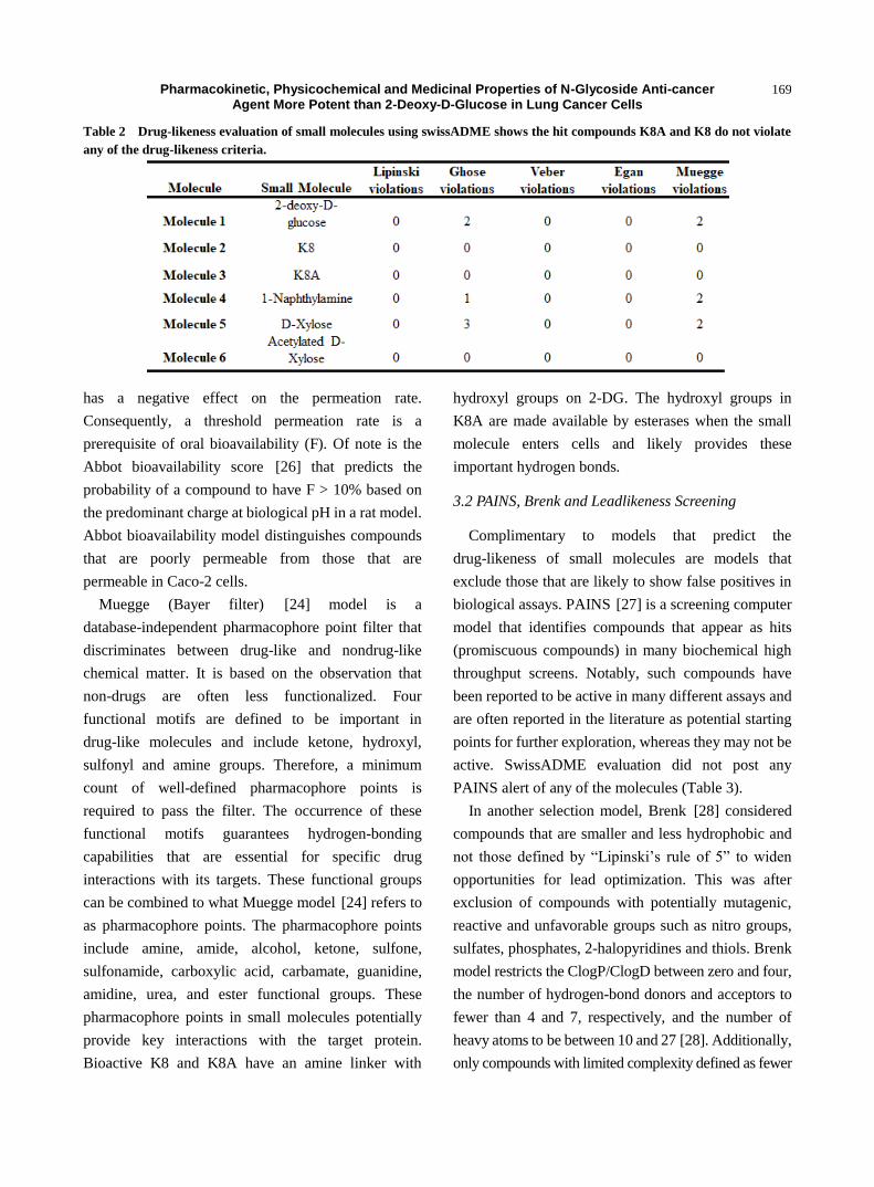
Table 3 Medicinal chemistry evaluation of the small molecules.
than eight rotatable bonds, fewer than five ring systems,
and no ring systems with more than two fused rings are
considered medicinal [28]. K8A with 3 ester groups
flouted 1 Brenk rule, luckily however, these ester
groups are cleaved off in-vivo to generate non-ester
containing K8 (Table 3). Molecule 4 with an aniline
moiety also flouted 1 Brenk rule (Table 3).
Conversely, Teague [29] and others propose that
there is a great deal of precedent to suggest that
libraries consisting of molecules with MW in the range
100 ± 350 and ClogP in the range 1 ± 3.0 are greatly
superior to those considered drug-like compounds and
are therefore lead-like [29]. With these stringent
lead-like criteria K8A with a MW of 401 and 7 rotors
failed two Leadlikeness criteria (Table 3). Of note,
bioactive K8 passed all Leadlikeness criteria but all
other controls failed (Table 3). Leadlikeness tests are
intended to provide leads with high affinity in
high-throughput screens that allow for the discovery
and exploitation of additional interactions in the
lead-optimization phase.
3.3 P-glycoprotein and CYP Enzyme Activity
Prediction
SwissADME also enables the estimation for a
chemical to be a substrate of p-glycoprotein (P-gp) or
inhibitor of the cytochrome p450 isoenzymes (CYP
isoenzymes). P-gp is extensively distributed and
expressed in the intestinal epithelium where it pumps
xenobiotics such as drugs back into the intestinal lumen
and in the capillary endothelial cells composing the
blood-brain barrier where it pumps them back into the
capillaries. CYP isoenzymes are responsible for the
biotransformation of drugs [30]. Drug metabolism via
CYP isoenzymes is an important determinant of drug
interactions that can lead to drug toxicities and reduced
pharmacological effect. The models return “Yes” or
“No” if the molecule under investigation has higher
probability to be substrate or non-substrate of P-gp or
inhibitor or non-inhibitor of a given CYP. All the
small molecules returned “No” for P-gp substrate and
“No” for most CYP isoenzymes. K8A returned “Yes”
for CYP3A4 inhibition (Table 4). While many drugs
are deactivated by CYP3A4, there are also some drugs
which are activated by the enzyme. Now, a biological
experiment will be required to determine if K8A is
activated or deactivated by CYP3A4.
3.4 HIA and BBB Prediction
Pertinent to P-gp and CYP enzyme kinetics is human
gastrointestinal absorption (HIA) and blood-brain
barrier penetration (BBB). SwissADME “BOILEDegg”
(Fig. 2) allows for evaluation of HIA as a function of
the position of the small molecules in the
WLOGP-versus-TPSA referential. The white region of
the “BOILEDegg” is for high probability of passive
absorption by the gastrointestinal tract, and the yellow
region (yolk) is for high probability of brain
penetration. Yolk and white areas are not mutually
exclusive. In addition, the points are colored in blue if
predicted as actively effluxed by P-gp (PGP+) and in
red if predicted as non-substrate of P-gp (PGP−). K8
(Molecule 2) and K8A (Molecule 3) are predicted as
absorbed by gastrointestines (white region) but are not
brain penetrant (yolk) (Fig. 2). The anti-cancer agent
2-deoxyD-glucose (Molecule 1) and the control molecule

Pharmacokinetic, Physicochemical and Medicinal Properties of N-Glycoside Anti-cancer Agent More Potent than 2-Deoxy-D-Glucose in Lung Cancer Cells
171
Table 4 Pharmacokinetic evaluation of the small molecules (GI: gastro-intestinal absorption, BBB: blood brain barrier,
CYP: Cytochromes, P-gp: P-glycoprotein).
Fig. 2 The BOILED-Egg allows for evaluation of passive gastrointestinal absorption (HIA), brain penetration (BBB) and
P-glycoprotein activity in presence of the molecule (P-gp).
D-xylose (Molecule 5) are predicted as not-absorbed
by the intestines and the brain. 1-naphthylamine
(Molecule 4) was predicted to be brain-penetrant. All
the molecules evaluated are PGP negative and are not
subject to active efflux (red dot).
HIA and BBB are dependent on water solubility and
lipophilicity of the drug. Two topological methods to
predict water solubility are included in SwissADME.
The first one is an implementation of the ESOL [31]
model and the second one is adapted from Ali et al.
[32]. SwissADME third predictor for solubility was
developed by SILICOS-IT. All predicted values are
the decimal logarithm of the molar solubility in water
(log S). Water solubility [31] of the small molecules
ranged from highly soluble (Molecule 1) to very
soluble (Molecule 6) using the ESOL [31] and other
criteria [32] (Table 5). Conversely, lipophilicity
evaluated as Consensus Log P indicated K8A
(molecule 3) to be the most lipophilic (Consensus
Log P = 2.38) whereas 2-DG was the least lipophilic

Pharmacokinetic, Physicochemical and Medicinal Properties of N-Glycoside Anti-cancer Agent More Potent than 2-Deoxy-D-Glucose in Lung Cancer Cells
172
Table 5 Water solubility evaluation of the small molecules.
Table 6 Lipophilicity evaluation of the small molecules.
Table 7 K8A Cytotoxicity in lung cancer cells.
(Consensus Log P = -1.67). Consensus Log P is the
average value of all Log P evaluated with various
lipophilicity criteria (Table 6).
3.5 Cytotoxicity of K8A
Cell viability studies revealed that K8A is more
potent than 2-DG, in the two cancer cell lines tested.
K8A cytotoxicity ranged between 0.03 mM and about
0.05 mM, whereas 2-DG cytotoxicity ranged between
1 mM and 4 mM in the same cell lines. K8 cytotoxicity
(IC50; 0.076 ± 0.19 mM) was about 10-fold higher than
2-DG (IC50; 0.79 ± 0.32 mM) in H1299 cells. On
acetylation (K8A), cytotoxicity improved about
20-fold higher (IC50; 0.045 ± 0.003 mM) than 2-DG
cytotoxicity in H1299 cell line, underscoring
importance of increased lipophilicity with acetylation.
Cytotoxicity of D-xylose and the acetylated form was
very low ranging between 3 and 6 mM.
4. Conclusions
Small molecules that block the altered metabolism
in cancer are emerging as potential anti-cancer agents.
Carbohydrate derivatives such as 2-deoxy-D-glucose
and K8A can be used for cellular energetics of which
interruption can lead to cellular stress. Indeed K8A, a
derivative of D-xylose was more potent than
clinically-tested 2-deoxy-D-glucose. We used the
swissADME virtual tools to further evaluate the hit
compound K8A and demonstrated that K8A has better
“drug-likeness” than 2-DG. A “BOILEDegg
evaluation”, predicts that K8A has higher
gastrointestinal absorption (HIA) than 2-DG and is not
effluxed by P-glycoprotein (P-gp). Additionally, K8A is
lipophilic but does not penetrate the blood brain barrier

Pharmacokinetic, Physicochemical and Medicinal Properties of N-Glycoside Anti-cancer Agent More Potent than 2-Deoxy-D-Glucose in Lung Cancer Cells
173
(BBB) and is not a substrate of most CYP enzymes. Of
note is the moderate synthetic accessibility of K8A that
provides medicinal chemists with opportunities for
synthesis of numerous analogues. Importantly, K8A
did not show false positive alert enabling us to rule out
false targets with confidence of pursuing potential
biologically relevant targets. Ultimately, biological
evaluation is required to validate the pharmacokinetics
and pharmacodynamics of any potential patient drug.
Acknowledgements
Synthesis, NMR characterization and cytotoxicity
evaluation of K8A was done at the Department of
Chemistry, Wayne State University with the help of
Prof. Young-Hoon Ahn.
Supporting Information: NMR Data 1H NMR of K8A
1H NMR (400 MHz, CD3OD) δ 7.90-7.82 (m, 1H),
7.80-7.70 (m, 1H), 7.46-7.38 (m, 2H), 7.35-7.26 (m,
2H), 6.89-6.83 (m, 1H), 5.44 (t, J = 9.5 Hz, 1H), 5.25 (t,
J = 9.2 Hz, 1H), 5.05 (ddd, J = 10.6, 9.5, 5.6 Hz, 1H),
5.00 (d, J = 8.9 Hz, 1H), 4.07 (dd, J = 11.3, 5.6 Hz, 1H),
3.63 (t, J = 10.9 Hz, 1H), 2.05 (s, 3H), 2.03 (s, 3H),
1.99 (s, 3H).
13
C NMR of K8A (101 MHz, CD3OD) 13
C NMR (101 MHz, CD3OD) δ 171.05, 170.28, 170.19, 140.84, 134.36, 127.93, 125.80, 125.33, 124.57, 120.51,
119.29, 107.52, 84.62, 72.92, 71.46, 69.47, 62.89, 19.32, 19.24, 19.16. [α]DRT
-45.9 (c, 0.5, methanol). HRMS
calculated mass 424.1372, found mass 424.1372.

Pharmacokinetic, Physicochemical and Medicinal Properties of N-Glycoside Anti-cancer Agent More Potent than 2-Deoxy-D-Glucose in Lung Cancer Cells
174
13
C NMR of K8A
References
[1] Zhang, S. Q., Yung, K. K., Chung, S. K., and Chung, S. S.,
2018. “Aldo-Keto Reductases Mediated Cytotoxicity of
2-Deoxyglucose: A Novel Anticancer Mechanism.”
Cancer Science.
[2] Le Pogam, P., Doue, M., Le Page, Y., Habauzit, D.,
Zhadobov, M., Sauleau, R., Le Drean, Y., and Rondeau, D.
2018. “Untargeted Metabolomics Reveal Lipid Alterations
upon 2-Deoxyglucose Treatment in Human HaCaT
Keratinocytes.” Journal of Proteome Research 17 (3):
1146-57.
[3] Xu, Y., Wang, Q., Zhang, L., and Zheng, M. 2018.
“2-Deoxy-D-Glucose Enhances TRAIL-Induced
Apoptosis in Human Gastric Cancer Cells through
Downregulating JNK-Mediated Cytoprotective
Autophagy.” Cancer Chemotherapy and Pharmacology
81 (3): 555-64.
[4] Xi, H., Kurtoglu, M., Liu, H., Wangpaichitr, M., You, M.,
Liu, X., Savaraj, N., and Lampidis, T. J. 2011.
“2-Deoxy-D-Glucose Activates Autophagy via
Endoplasmic Reticulum Stress Rather than ATP
Depletion.” Cancer Chemotherapy and Pharmacology 67
(4): 899-910.
[5] Aft, R. L., Zhang, F. W., and Gius, D. 2002. “Evaluation
of 2-Deoxy-D-Glucose as a Chemotherapeutic Agent:
Mechanism of Cell Death.” British Journal of Cancer 87:
805.
[6] Wick, A. N., Drury, D. R., Nakada, H. I., and Wolfe, J. B.
1957. “Localization of the Primary Metabolic Block
Produced by 2-Deoxyglucose.” The Journal of Biological
Chemistry 224 (2): 963-9.
[7] Kurtoglu, M., Gao, N., Shang, J., Maher, J. C., Lehrman,
M. A., Wangpaichitr, M., Savaraj, N., Lane, A. N., and
Lampidis, T. J. 2007. “Under Normoxia,
2-Deoxy-D-Glucose Elicits Cell Death in Select Tumor
Types Not by Inhibition of Glycolysis but by Interfering
with N-Linked Glycosylation.” Molecular Cancer
Therapeutics 6 (11): 3049-58.
[8] Shutt, D. C., O'Dorisio, M. S., Aykin-Burns, N., and Spitz,

Pharmacokinetic, Physicochemical and Medicinal Properties of N-Glycoside Anti-cancer Agent More Potent than 2-Deoxy-D-Glucose in Lung Cancer Cells
175
D. R. 2010. “2-Deoxy-D-Glucose Induces Oxidative
Stress and Cell Killing in Human Neuroblastoma Cells.”
Cancer Biology & Therapy 9 (11): 853-61.
[9] Raez, L. E., Rosenblatt, J., Schlesselman, J., Langmuir, V.,
Tidmarsh, G., Rocha-Lima, C., Papadopoulos, K.,
O’Connor, J., Baldie, P., and Lampidis, T. 2005.
“Combining Glycolytic Inhibitors with Chemotherapy:
Phase I Trial of 2-Deoxyglucose and Docetaxel in Patients
with Solid Tumors.” Journal of Clinical Oncology 23 (16):
3190.
[10] Dwarakanath, B., Singh, D., Banerji, A., Sarin, R.,
Venkataramana, N., Jalali, R., et al. 2009. “Clinical
Studies for Improving Radiotherapy with
2-Deoxy-D-Glucose: Present Status and Future Prospects.”
Journal of Cancer Research and Therapeutics 5 (9): 21-6.
[11] Vijayaraghavan, R., Kumar, D., N Dube, S., Singh, R., S
Pandey, K., Bag, B., Kaushik, M., Krishnamurthy, S.,
Dwarakanath, B., and Ravindranath, T. 2006. “Acute
Toxicity and Cardio-Respiratory Effects of
2-Deoxy-D-Glucose: A Promising Radio Sensitiser.” 19:
96-103.
[12] Zhong, D., Xiong, L., Liu, T., Liu, X., Liu, X., Chen, J., et
al. 2009. “The Glycolytic Inhibitor 2-Deoxyglucose
Activates Multiple Prosurvival Pathways through
IGF1R.” The Journal of Biological Chemistry 284 (35):
23225-33.
[13] Shukla, A., Gupta, P., Singh, R., and Mishra, D. P. 2018.
“Glycolytic Inhibitor 2-Deoxy-D-Glucose Activates
Migration and Invasion in Glioblastoma Cells through
Modulation of the miR-7-5p/TFF3 Signaling Pathway.”
Biochem Biophys Res Commun 2018.
[14] Ndombera Fidelis, T. 2017. “Anti-cancer Agents and
Reactive Oxygen Species Modulators That Target Cancer
Cell Metabolism.” Pure and Applied Chemistry 89: 1333.
[15] Sliwoski, G., Kothiwale, S., Meiler, J., and Lowe, E. W.,
Jr. 2014. “Computational Methods in Drug Discovery.”
Pharmacological Reviews 66 (1): 334-95.
[16] Daina, A., Michielin, O., and Zoete, V. 2017.
“SwissADME: A Free Web Tool to Evaluate
Pharmacokinetics, Drug-Likeness and Medicinal
Chemistry Friendliness of Small Molecules.” Scientific
Reports 7: 42717.
[17] Lipinski, C. A., Lombardo, F., Dominy, B. W., and
Feeney, P. J. 1997. “Experimental and Computational
Approaches to Estimate Solubility and Permeability in
Drug Discovery and Development Settings 1PII of
Original Article: S0169-409X(96)00423-1.” Advanced
Drug Delivery Reviews 46 (1): 3-26.
[18] Daina, A., and Zoete, V. 2016. “A BOILED-Egg to
Predict Gastrointestinal Absorption and Brain Penetration
of Small Molecules.” Chem. Med. 11 (11): 1117-21.
[19] Matlock, M. K., Hughes, T. B., Dahlin, J. L., and
Swamidass, S. J. 2018. “Modeling Small-Molecule
Reactivity Identifies Promiscuous Bioactive Compounds.”
J Chem Inf Model 58 (8): 1483-500.
[20] Ndombera, F. T., VanHecke, G. C., Nagi, S., and Ahn,
Y.-H. 2016. “Carbohydrate-Based Inducers of Cellular
Stress for Targeting Cancer Cells.” Bioorganic &
Medicinal Chemistry Letters 26 (5): 1452-6.
[21] Ghose, A. K., Viswanadhan, V. N., and Wendoloski, J. J.
1999. “A Knowledge-Based Approach in Designing
Combinatorial or Medicinal Chemistry Libraries for Drug
Discovery. 1. A Qualitative and Quantitative
Characterization of Known Drug Databases.” Journal of
Combinatorial Chemistry 1 (1): 55-68.
[22] Egan, W. J., Merz, K. M., Jr., and Baldwin, J. J. 2000.
“Prediction of Drug Absorption Using Multivariate
Statistics.” Journal of Medicinal Chemistry 43 (21):
3867-77.
[23] Veber, D. F., Johnson, S. R., Cheng, H. Y., Smith, B. R.,
Ward, K. W., and Kopple, K. D. 2002. “Molecular
Properties That Influence the Oral Bioavailability of Drug
Candidates.” Journal of Medicinal Chemistry 45 (12):
2615-23.
[24] Muegge, I., Heald, S. L., and Brittelli, D. 2001. “Simple
Selection Criteria for Drug-Like Chemical Matter.”
Journal of Medicinal Chemistry 44 (12): 1841-6.
[25] Ghose, A. K., Viswanadhan, V. N., and Wendoloski, J. J.
1998. “Prediction of Hydrophobic (Lipophilic) Properties
of Small Organic Molecules Using Fragmental Methods:
An Analysis of ALOGP and CLOGP Methods.” The
Journal of Physical Chemistry A 102 (21): 3762-72.
[26] Martin, Y. C. 2005. “A Bioavailability Score.” Journal of
Medicinal Chemistry 48 (9): 3164-70.
[27] Baell, J. B., and Holloway, G. A. 2001. “New
Substructure Filters for Removal of Pan Assay
Interference Compounds (PAINS) from Screening
Libraries and for Their Exclusion in Bioassays.” Journal
of Medicinal Chemistry 53 (7): 2719-40.
[28] Brenk, R., Schipani, A., James, D., Krasowski, A., Gilbert,
I. H., Frearson, J., and Wyatt, P. G. 2008. “Lessons Learnt
from Assembling Screening Libraries for Drug Discovery
for Neglected Diseases.” Chem. Med. Chem. 3 (3):
435-44.
[29] Teague, S. J., Davis, A. M., Leeson, P. D., and Oprea, T.
1999. “The Design of Leadlike Combinatorial Libraries.”
Angewandte Chemie International Edition 38 (24):
3743-8.
[30] Ogu, C. C., and Maxa, J. L. 2000. “Drug Interactions due
to Cytochrome P450.” Proceedings (Baylor University.
Medical Center) 13 (4): 421-3.
[31] Delaney, J. S. 2004. “ESOL: Estimating Aqueous
Solubility Directly from Molecular Structure.” Journal of
Chemical Information and Computer Sciences 44 (3):
Pharmacokinetic, Physicochemical and Medicinal Properties of N-Glycoside Anti-cancer Agent More Potent than 2-Deoxy-D-Glucose in Lung Cancer Cells
176
1000-5.
[32] Fagerberg, J. H., Karlsson, E., Ulander, J., Hanisch, G.,
and Bergström, C. A. S. 2015. “Computational Prediction
of Drug Solubility in Fasted Simulated and Aspirated
Human Intestinal Fluid.” Pharmaceutical Research 32 (2):
578-89.