
PHARMACEUTICAL CHEMISTRY FACULTY OF PHARMACEUTICAL … · i pharmaceutical chemistry ukwueze,...
187
i PHARMACEUTICAL CHEMISTRY UKWUEZE, STANLEY EJIKE (PG/PhD/05/40421) PHYTOCHEMICAL AND BIOACTIVITY-GUIDED EVALUATION OF THE ANTIBACTERIAL CONSTITUENTS OF PSIDIUM GUAJAVA (LINN.) AND LORANTHUS MICRANTHUS (LINN.) LEAVES Digitally Signed by: Content manager’s Name DN : CN = Webmaster’s name O = University of Nigeria, Nsukka OU = Innovation Centre Ugwoke Oluchi C. FACULTY OF PHARMACEUTICAL SCIENCES
Transcript of PHARMACEUTICAL CHEMISTRY FACULTY OF PHARMACEUTICAL … · i pharmaceutical chemistry ukwueze,...

i
PHARMACEUTICAL CHEMISTRY
UKWUEZE, STANLEY EJIKE
(PG/PhD/05/40421)
PHYTOCHEMICAL AND BIOACTIVITY-GUIDED
EVALUATION OF THE ANTIBACTERIAL CONSTITUENTS OF
PSIDIUM GUAJAVA (LINN.) AND LORANTHUS MICRANTHUS
(LINN.) LEAVES
Digitally Signed by: Content manager’s Name
DN : CN = Webmaster’s name
O = University of Nigeria, Nsukka
OU = Innovation Centre
Ugwoke Oluchi C.
FACULTY OF PHARMACEUTICAL SCIENCES

ii
PHYTOCHEMICAL AND BIOACTIVITY-GUIDED EVALUATION OF
THE ANTIBACTERIAL CONSTITUENTS OF PSIDIUM GUAJAVA
(LINN.) AND LORANTHUS MICRANTHUS (LINN.) LEAVES
UKWUEZE, STANLEY EJIKE
(PG/PhD/05/40421)
DEPARTMENT OF PHARMACEUTICAL CHEMISTRY,
FACULTY OF PHARMACEUTICAL SCIENCES,
UNIVERSITY OF NIGERIA, NSUKKA.
SUPERVISOR: PROF. (MRS.) P. O. OSADEBE
MARCH, 2014.

iii
PHYTOCHEMICAL AND BIOACTIVITY-GUIDED EVALUATION OF
THE ANTIBACTERIAL CONSTITUENTS OF PSIDIUM GUAJAVA
(LINN.) AND LORANTHUS MICRANTHUS (LINN.) LEAVES
UKWUEZE, STANLEY EJIKE
(PG/PhD/05/40421)
A THESIS SUBMITTED TO
THE DEPARTMENT OF PHARMACEUTICAL AND MEDICINAL
CHEMISTRY, FACULTY OF PHARMACEUTICAL SCIENCES,
UNIVERSITY OF NIGERIA, NSUKKA.
IN FULFILMENT OF THE REQUIREMENTS FOR THE AWARD OF
DOCTOR OF PHILOSOPHY (PhD) DEGREE IN PHARMACEUTICAL
& MEDICINAL CHEMISTRY.

iv
CERTIFICATION
Ukwueze, Stanley Ejike, a postgraduate student with registration number:
PG/PhD/05/40421 has satisfactorily completed the requirement for the award
of the degree of Doctor of Philosophy (PhD) of the Department of
Pharmaceutical and Medicinal Chemistry, Faculty of Pharmaceutical Sciences,
University of Nigeria, Nsukka.
The research embodied in this thesis is original and has not been submitted in
part or in full for the award of any other diploma or degree of this or any other
University.
_____________________________ ______________________
Prof. (Mrs.) P. O. Osadebe Prof. C. J. Mbah
(Supervisor) (Head of Department)
_____________________________
(External Examiner)

v
DEDICATION
To my beloved wife (Odimnobi), Nchedo, and my lovely roots: Osinachi,
Munachiso, Oluoma and Uchechi who have remained the priceless jewels and
adorable ornaments of my life.

vi
ACKNOWLEDGEMENT
To God be the Glory, great thing He has done! He has shown me His
faithfulness once again by making it possible for me to attain the zenith of this
tortuous and seemingly endless race. I will forever consecrate my life to Him!
My immeasurable gratitude goes to my supervisor, Prof. Mrs. P. O. Osadebe,
who God has used to guide and illuminate my academic path since my
undergraduate years. She and her inspirational husband, Rev. Prof. N. N.
Osadebe, have been more of motivators and mentors not just to me, but also to
my entire family in diverse realms. I will always remain indebted.
My appreciation also goes to my ever loving parents, Ichie and Mrs. S. K. C.
Ukwueze, who God has kept alive to witness their life-long dream of having
one of their children attain this academic summit. May God continue to
preserve their life for us!
My special acknowledgement goes to my brother, friend, colleague and
schoolmate, Dr. Okoye, F. B. C. who has always been of great support in times
of need. His role in carrying out the spectral analysis in Germany and the
eventual structure elucidations cannot be quantified. My appreciation also goes
to Dr. Peters at the Institute of Organic Chemistry, Heinrich-Heine-University
for the NMR measurements. I also acknowledge the contributions of the
Institute of Pharmaceutical Biology and Biotechnology, University of Heirich-
Heine-Düsseldorf for the HPLC/ESI-MS recordings.

vii
I would like at this juncture to appreciate the professional and elderly roles
played by some of my senior colleagues who kept prodding me on even when I
appeared distracted. Worthy of mention here include Prof. C. O. Onyeji (my
VC), Prof. Cyril Usifo (my amiable Prof), Assoc. Prof. Eseyin (my Oga), Prof.
Orisakwe, Prof. S. I. Ofoefule, Prof. Vincent Okore and Prof. O. K. Udeala (my
able Dean). The list is quite endless!
It will be so unfair if I shall end this eulogy without the appreciation of all the
laboratory staff that played one role or the other towards the accomplishment of
this study. Names like Chief Hassan and Mrs. Caleb come tops here. Also, my
colleagues in the Department of Pharmaceutical and Medicinal Chemistry,
University of Port Harcourt deserve special recognition.
Finally, my greatest felicitation goes to my beloved wife, Nchedo, and my
vibrant children: Osinachi, Munachiso, Oluomachukwu and Uchechi. I will
always remember the angelic voice of my sweet one (Odimnaheart) constantly
reminding me of her joy in seeing this work completed. Thank God it finally
came to a glorious end! My siblings equally deserve special recognition: Sis.
Vicky, Rev. Leo, Eby, Ify, Iyke, Chika and Eyinwa.
To anyone I might have forgotten to mention, please accept my apology and lets
all join hands to say three ‘Gbosas’ to one another for a job well done.
Merci! Danke!!
Ukwueze Stanley Ejike. February, 2014.

viii
ABSTRACT
Antibiotics have remained the mainstay of drug therapy of infectious diseases
worldwide. Their use is, however, limited by their numerous adverse effects and rapid
development of microbial resistance. Identification of natural products from plants that
may serve as valuable sources of antimicrobial agents for medicinal or agricultural uses
seems to be a viable alternative to the conventional antibiotics. To achieve this goal,
biological assays should be carried out in order to identify promising plant extracts,
guide the separation and isolation, and to evaluate "lead" compounds. Psidium guajava
Linn. and Loranthus micranthus Linn. have been employed traditionally in Nigeria and
other parts of the globe for the treatment of various human ailments such as wounds,
gastrointestinal tract disorders and other forms of infective and non-infective disorders.
The main objective of this study was to identify and isolate the antibacterial compounds
from the leaves of Psidium guajava and Loranthus micranthus L. The specific objectives
were to: (i) carry out phytochemical evaluation and isolation of antibacterial
constituents of the plants using standard methods, (ii) elucidate the structures of the
isolated secondary metabolites, and (iii) carry out antibacterial assay of the isolated
compounds.
Fresh leaves of Loranthus micranthus (Linn.) parasitic on the stem of Persea americana
were collected at Nsukka while those of Psidium guajava were collected from the bio-
resource area of the University of Port Harcourt in June 2010. The leaves were then
cleaned, air-dried for 14 days and milled to coarse powder. The powdered materials
(800 g each) were defatted with n-hexane (5 L) and extracted in a soxhlet extractor with
90.0 % methanol. The methanol extract was further fractionated to yield the
chloroform, ethyl acetate, acetone and methanol soluble fractions. Each of the fractions
was screened for antibacterial activity using Agar-well diffusion method. Phytochemical
tests were carried out using standard procedures. The fractions that had the best
antibacterial activity were subjected to column chromatographic separation and
monitored by analytical thin layer chromatography (TLC). The ethyl acetate fraction
(PsG-EF) from P. guajava that gave satisfactory bioassay result was subjected to further
Sephadex-LH 20 chromatographic fractionation and purification to afford ten fractions
(PsG-EF1 to PsG-EF10) which were pooled. Fractions PsG-EF4, PsG-EF5 and PsG-EF7
that had good antibacterial activity were subjected to semi-preparative reverse phase
high pressure liquid chromatography (HPLC) purification to isolate the phenolic
compounds; I-V. The structures of these compounds were elucidated by analytical and
spectral techniques which included: ultra violet (UV), proton nuclear magnetic
resonance (1H-NMR), carbon-13 nuclear magnetic resonance (13C-NMR), distortionless

ix
enhancement by polarization transfer (DEPT), proton-proton correlation spectroscopy
(1H-1HCOSY), heteronuclear multiple quantum correlation (HMQC), heteronuclear
multiple bond correlation (HMBC) and electron spray ionization-mass spectroscopy
(ESI-MS) analyses. The isolated compounds were screened against standard strains of
Staphylococus aureus (ATCC 25923) and Escherichia coli (ATCC 35219) using broth
dilution assay method, and the MIC values determined and compared with ceftriaxone.
All data obtained were analyzed by GraphPad Prism® 5 using differences in mean by
two-way ANOVA and further subjected to Bonferroni post-tests to compare replicate
means. The results were presented as mean ± SEM. Differences between means were
considered significant at P<0.05.
The results showed that the ethyl acetate fraction (PsG-EF) from P. guajava yielded one
known compound (IV) and four novel phenolic compounds (I, II, III & V). The isolated
compounds were elucidated as : 2,4-dihydroxy-6-O-βD-glucopyranosyl benzophenone
(I); 2,4-dihydroxy-3-methyl-6-O-βD-glucopyranosylbenzophenone (II); 2,4-dihydro xy-
3-methyl-6-O-βD-glucopyranosylbenzophenone (4→5", 6'→1") benzene-2",3",4",5"-
tetraol (III); quercertin-3-O-αL-arabinofuranoside (IV) and 2,4-dihydroxy-6-O-βD-
glucopyranosylbenzophenone (4→5", 6'→1") benzene-2",3",4",5"-tetraol (V).
Compounds I, II, III, and V are new natural products which have not been previously
reported in literature for this plant, guava, and the trivial names Guajaphenone A, B, C
and D were proposed, while Compound IV has been previously reported as Guaijaverin.
The various fractions of Psidium guajava L. exhibited significant (p < 0.05) antibacterial
activities while for Loranthus micranthus L., its various fractions showed significantly
lower values (p > 0.001) when compared with the control (ceftriaxone) suggestive of a
generally weak or negligible antibacterial action. All the isolated compounds from P.
guajava were also found to have moderate antibacterial activities against E. coli and S.
aureus in comparison with ceftriaxone whlie I and IV showed lower MICs than those of
the other isolates against the test organisms.
x
Table of Contents
Page
TITLE PAGE i
CERTIFICATION iii
DEDICATION iv
ACKNOWLEDGEMENT v
ABSTRACT vii
TABLE OF CONTENT ix
LIST OF FIGURES xiv
LIST OF TABLES xvii
CHAPTER ONE: GENERAL INTRODUCTION 1
1.0 PREAMBLE 1
1.1 INFECTIOUS DISEASES AND CONVENTIONAL ANTIBIOTIC THERAPY 2
1.2 LIMITATIONS OF CONVENTIONAL ANTIBACTERIAL
AGENTS 6
1.2.1 Bacterial Resistance 6
1.2.2 Adverse Reactions by Agents 7
1.3 HIGHER PLANTS AS ANTIMICROBIAL AGENTS 8
1.3.1 Medicinal Plants with Antibacterial Activity 9

xi
1.3.2 Plant Secondary Metabolites Associated with Antibacterial Effects 10
1.3.2.1 Flavones, Flavonoids and Flavonols 11
1.3.2.2 Alkaloids 12
1.3.2.3 Terpenoids and Essential Oils 13
1.3.2.4 Tannins 15
1.3.2.5 Miscellaneous Plant Constituents 17
1.4 LITERATURE REVIEWS OF PLANTS USED 21
1.4.1 Loranthus micranthus Linn 21
1.4.1.1 Taxonomy of L. Micranthus 21
1.4.1.2 Description of the Family, Genus and Species of L. micranthus 23
1.4.1.3 Ethnomedicinal Uses and Pharmacological Studies on
L. micranthus 24
1.4.2 Psidium guajava Linn 25
1.4.2.1 Taxonomy of Psidium guajava 25
1.4.2.2 Morphology of Psidium guajava 26
1.4.2.3 Ethnomedicinal Uses and Pharmacological Studies of
Psidium guajava 28
1.4.2.4 The Phytochemistry of Psidium guajava 32
1.5 BIOASSAY-GUIDED CHARACTERIZATION OF ANTI-BACTERIAL CONSTITUENTS FROM HIGHER PLANTS 37
1.5.1 Principles of Bioassay 38

xii
1.5.2 Screening Methods for Antibacterial Agents from Higher Plants 38
1.5.2.1 Test Organisms and Culture Media 38
1.5.2.2 Antibacterial Testing 40
1.6 STRUCTURE ELUCIDATION OF BIOACTIVE PLANT METABOLITES 42
1.6.1 Preliminary Analysis 42
1.6.2 Application of Modern Analytical Techniques 43
1.7 STATEMENT OF PROBLEM 44
1.8 JUSTIFICATION OF THE STUDY 44
1.9 AIMS AND SCOPE OF THE WORK 45
CHAPTER TWO: MATERIALS AND METHODS 46
2.1 MATERIALS 46
2.1.1 Plant Materials 46
2.1.2 Microorganisms Used 46
2.1.3 Solvents and Reagents 47
2.1.4 Materials and General Instruments 48
2.2 METHODS 50
2.2.1 Extraction and Fractionation of Plant Materials 50

xiii
2.2.2 Isolation and Purification of the Active Constituents from PsG-EF 54
2.2.3 HPLC Analysis of Active Constituents from PsG-EF 55
2.2.4 Electron Spray Ionization Mass Spectrometry (HPLC/ESI-MS)
of Isolates 55
2.2.5 Nuclear Magnetic Resonance (NMR) Spectroscopy of Isolates 56
2.2.6 Preliminary Screening of Extracts and Fractions
for Antimicrobial Activity 56
2.2.7 Phytochemical Screening of Plant Extracts and Fractions 57
2.2.8 Antibacterial Screening of Isolates 57
2.2.9 Statistical Analyses 58
CHAPTER THREE: RESULTS 59
3.1 Extraction and Solvent Fractionation 59
3.2 Preliminary Phytochemical and Antibacterial Screening Results 61
3.3 Isolation of Bioactive Constituents 73
3.4 Structure Elucidation of Isolated Compounds 74
3.4.1 PsG-EF4A (Compound I) 74
3.4.2 PsG-EF4B (Compound II) 76
3.4.3 PsG-EF7A (Compound III) 78
3.4.4 PsG-EF7D (Compound IV) 80

xiv
3.4.5 PsG-EF7E (Compound V) 81
3.5 Antibacterial Profile of Bioactive Constituents 83
CHAPTER FOUR: DISCUSSION AND CONCLUSION 84
4.1 Discussion 84
4.2 Conclusion 98
REFERENCES 100
APPENDICES 113

xv
LIST OF FIGURES
page
Fig. 1.0: Flowers and Leaves of Loranthus micranthus 22
Fig. 2.0: Loranthus micranthus parasitic on a host tree 23
Fig. 3.0 Psidium guajava Tree 27
Fig. 4.0 Leaves and Flowers of Psidium guajava 27
Fig. 5.0 Fruit of Psidium guajava 28
Fig. 6: Schematic Diagram of the Extraction/Fractionation 52
Procedure for L. micranthus Leaves.
Fig. 7: Schematic Diagram of the Extraction/Fractionation 53
Procedure for P. guajava Leaves.
Fig. 8: Graph of Mean IZD (mm) ± SEM of the extracts/fractions of
L. micranthus leaves against bacteria 68
Fig. 9a: Structure of Compound I 74
Fig. 9b: Numbering of Carbon Skeleton of Compound I 75
Fig. 10a: Structure of Compound II 76
Fig. 10b: Numbering of Carbon Skeleton of Compound II 77
Fig. 11a: Structure of Compound III 78
Fig. 11b: Numbering of Carbon Skeleton of Compound III 79
Fig. 12: Structure of Compound IV 80
Fig. 13: Structure of Compound V 81

xvi
Appendix 1: ESI-MS /UV Spectra of Compound I 113
Appendix 2: H-NMR Spectrum (500MHz; MeOD) of Compound I 114
Appendix 3: H-NMR Spectrum (600MHz; MeOD) of Compound I 115
Appendix 4: H-NMR Spectrum (600MHz; DMSO-d6) of Compound I 116
Appendix 5: 2-D COSY of Compound I 117
Appendix 6: C-13 NMR Spectrum of Compound I 118
Appendix 7: HMQC Spectrum of Compound I 119
Appendix 8: HMBC Spectrum of Compound I 120
Appendix 9: ESI-MS /UV Spectra Compound II 121
Appendix 10: H-NMR Spectrum (500MHz; MeOD) of Compound II 122
Appendix 11: H-NMR Spectrum (600MHz; DMSO) of Compound II 123
Appendix 12: 2-D COSY of Compound II 124
Appendix 13: C-13 NMR Spectrum of Compound II 125
Appendix 14: ESI-MS /UV Spectra Compound III 126
Appendix 15: H-NMR Spectrum (500MHz; MeOD) of Compound III 127
Appendix 16: H-NMR Spectrum (600MHz; DMSO) of Compound III 128
Appendix 17: 2-D COSY of Compound III 129
Appendix 18: C-13 NMR Spectrum of Compound III 130
Appendix 19: HMQC Spectrum of Compound III 131
Appendix 20: HMBC Spectrum of Compound III 132
Appendix 21: UV Spectrum of Compound IV 133
Appendix 22: ESI-MS Spectra of Compound IV 134

xvii
Appendix 23: H-NMR Spectrum (500MHz; MeOD) of Compound IV 135
Appendix 24: 2-D COSY of Compound IV 136
Appendix 25: H-NMR Spectrum (500MHz; MeOD) of Compound V 137
Appendix 26: 2-D COSY of Compound V 138
Appendix 27: 2-D COSY [aromatic region] of Compound V 139

xviii
LIST OF TABLES Page
Table 1: Some plant species with potential antimicrobial activities... 9
Table 2: Yield from extracts/fractions of the leaves of L. micranthus. 59
Table 3: Yield from extracts/fractions of the leaves of P. guajava …. 60
Table 4: Results of phytochemical tests on the leaf extract of
Loranthus micranthus parasitic on different host trees.... 61
Table 5: Results of the anti-microbial screening of extracts of
mistletoe from six different host plants... 62
Table 6: Results of phytochemical tests on the leaf extract of
Loranthus micranthus harvested at different seasons... 63
Table 7: Results of the anti-microbial screening of leaf extracts of
Loranthus harvested at different seasons... 64
Table 8: Result of MICs of leaf extracts of African mistletoe harvested
from P. americana against some fungi... 65
Table 9: Results of phytochemical tests on the solvent fractions of L.
micranthus leaves harvested from P.americana... 66
Table 10: Result of mean IZD (mm) ± SEM for L. micranthus
extracts/fraction… 67
Table 11: Results of phytochemical tests on the leaf extract of
P. guajava harvested at different seasons.... 69
Table 12: Results of the anti-microbial screening of leaf extracts of
P. guajava harvested at different seasons... 70
Table 13: Results of phytochemical tests on the solvent fractions
of Psidium guajava leaves... 71
Table 14: Table of Mean IZD (mm) +/- SEM for P. guajava
extracts/fractions... 72
Table 15: 1H and
13C-NMR data of Compound I... 75
Table 16: 1H and
13C-NMR data of Compound II... 77
Table 17: 1H and
13C-NMR data of Compound III... 79

xix
Table 18: 1H and
13C-NMR data of Compound V... 82
Table 19: Antibacterial profile of the isolated compounds against
Staphylococcus aureus and E. coli... 83
xx
CHAPTER ONE
GENERAL INTRODUCTION
1.0 PREAMBLE
Over the years, medicines and medicinal agents derived from plants have
made large contributions to human health and well-being. This is because they
are either used directly as phytomedicines for the treatment of various
ailments or they may become the base and the natural blueprint for the
development of new drugs (Cseke et al, 2006).
Herbal medicine also called phytotherapy or phytomedicine has been around
since the beginning of recorded history. It has also been described as the
therapeutic use of medicinal plants referred to as herbs (Thea et al, 2008).
Herbal medicine has become an integral part of standard health care, based on
a combination of time honored traditional usage and ongoing scientific
research. Surging interest in medicinal herbs has increased scientific scrutiny of
their therapeutic potential and safety. Some of the medicinal plants are
believed to enhance the natural resistance of the body to infections (Atal et al,
1986).
According to the World Health Organisation (WHO), herbal medicines could
also be referred to as phytopharmaceuticals sold as over the counter products
in modern dosage forms such as tablets, capsules, syrups or liquids for oral use
or dietary supplements containing herbal products, also called nutraceuticals

xxi
available in modern dosage forms, or even referred to as medicines consisting
of other crude, semi processed or processed medicines, which have a vital
place in primary health care and developing countries like Nigeria.
Traditional medicines are finished drug products intended for self-medication
or application that contain, as the active principles, herbal ingredients that
have received relatively little attention in world scientific literature, but for
which traditional or folkloric use is well documented in herbal references. It
may contain chemically defined or herbal based materials in addition to the
active principles (Canada, 1989).
The medicinal properties of plant have been investigated in the light of recent
scientific development throughout the world due to their potent
pharmaceutical activities and low toxicity. Today many countries still rely on
the medical values of herbs and use of medicinal plants for their therapeutic
practices (Thea et al, 2008). In this same vein, Nigeria which is having a vast
heritage of knowledge and expertise in herbal medicines is not an exception.
Finally, it has been variously established that the identification of these natural
products from plants that may serve as valuable sources of bioactive agents for
medicinal and agricultural uses largely depends on bioactivity-directed
isolation (Cseke et al, 2006).
1.1 INFECTIOUS DISEASES AND CONVENTIONAL ANTIBIOTIC THERAPY

xxii
Infectious diseases, also known as contagious diseases or transmissible
diseases, and include communicable diseases, comprise clinically evident
illness (i.e., characteristic medical signs and/or symptoms of disease) resulting
from the infection, presence and growth of pathogenic biological agents in an
individual host organism. In certain cases, infectious diseases may be
asymptomatic for much or their entire course. Infectious pathogens include
some viruses, bacteria, fungi, protozoa, multicellular parasites, and aberrant
proteins known as prions. These pathogens are the cause of disease epidemics,
in the sense that without the pathogen, no infectious epidemic occurs.
Transmission of pathogen can occur in various ways including physical contact,
contaminated food, body fluids, objects, airborne inhalation, or through vector
organisms (Ryan and Ray, 2004). Infectious diseases that are especially
infective are sometimes called contagious and can be easily transmitted by
contact with an ill person or their secretions. Infectious diseases with more
specialized routes of infection, such as vector transmission or sexual
transmission, are usually regarded as contagious but do not require medical
quarantine of victims.
The term infectivity describes the ability of an organism to enter, survive and
multiply in the host, while the infectiousness of a disease indicates the
comparative ease with which the disease is transmitted to other hosts; and as
such, an infection is not synonymous with an infectious disease, as some
infections do not cause illness in a host (Ryan and Ray, 2004).

xxiii
Among the almost infinite varieties of microorganisms, relatively few cause
disease in otherwise healthy individuals. Infectious disease results from the
interplay between those few pathogens and the defenses of the hosts they
infect. The appearance and severity of disease resulting from any pathogen
depends upon the ability of that pathogen to damage the host as well as the
ability of the host to resist the pathogen. Clinicians therefore classify infectious
microorganisms or microbes according to the status of host defenses - either
as primary pathogens or as opportunistic pathogens:
Primary pathogens cause disease as a result of their presence or activity within
the normal, healthy host, and their intrinsic virulence (the severity of the
disease they cause) is, in part, a necessary consequence of their need to
reproduce and spread. Many of the most common primary pathogens of
humans only infect humans; however many serious diseases are caused by
organisms acquired from the environment or which infect non-human hosts.
Organisms which cause an infectious disease in a host with depressed
resistance are classified as opportunistic pathogens. Opportunistic disease may
be caused by microbes that are ordinarily in contact with the host, such as
pathogenic bacteria or fungi in the gastrointestinal or the upper respiratory
tract, and they may also result from (otherwise innocuous) microbes acquired
from other hosts (as in Clostridium difficile colitis) or from the environment as
a result of traumatic introduction (as in surgical wound infections or compound
fractures). An opportunistic disease requires impairment of host defenses,
which may occur as a result of genetic defects (such as chronic granulomatous

xxiv
disease), exposure to antimicrobial drugs or immunosuppressive chemicals (as
might occur following poisoning or cancer chemotherapy), exposure to ionizing
radiation, or as a result of an infectious disease with immunosuppressive
activity (such as with measles, malaria or HIV disease). Primary pathogens may
also cause more severe disease in a host with depressed resistance than would
normally occur in an immunosufficient host.
Many human diseases are caused by pathogenic organisms resulting
sometimes in high mortality figures. Among these pathogens, bacteria account
for a reasonable percentage of causative organisms implicated in human
infectious diseases. Over the years, infections have been managed by the
conventional antibiotics. Antibiotics are microbial metabolites or synthetic
analogues inspired by them that, in small doses, inhibit the growth and survival
of microorganisms without serious toxicity to the host. They therefore exhibit
selective toxicity. In many cases, the clinical utility of natural antibiotics has
been through medicinal chemistry manipulations of the original structure
leading to broader antimicrobial spectrum, greater potency, lesser toxicity,
more convenient administration, and additional pharmacokinetic advantages.
Through customary usage, the many synthetic substances that are unrelated to
natural products but still inhibit or kill microorganisms are referred to as
antimicrobial agents instead (Martin, 1998).
Antibiotics are used to treat infections caused by organisms that are sensitive
to them, usually bacteria or fungi. They may alter the normal microbial content
xxv
of the body (e.g. in the intestine, lungs, bladder) by destroying one or more
groups of harmless or beneficial organisms, which may result in infections
(such as thrush in women) due to overgrowth of resistant organisms. These
side-effects are most likely to occur with broad-spectrum antibiotics (those
active against a wide variety of organisms). Resistance may also develop in the
microorganisms being treated; for example, through incorrect dosage or over-
prescription. Antibiotics should, therefore, not be used to treat minor
infections, which will clear up unaided. Some antibiotics may, in addition,
cause allergic reactions (Hendricks and Nemeth, 2010).
1.2 Limitations of Conventional Antibacterial Agents
1.2.1 Bacterial Resistance
Resistance is the failure of microorganisms to be killed or inhibited by
antimicrobial treatment. Resistance can either be intrinsic (exist before
exposure to drugs) or acquired (develop subsequent to exposure to a drug).
Resistance of bacteria to the toxic effects of antimicrobial agents and to
antibiotics develops fairly easily both in the laboratory and in the clinic and is
an ever-increasing public health hazard.
In clinical practice, resistance more commonly takes place by Resistance (R)
factor mechanisms. In more lurid examples, enzymes are elaborated that
attack the antibiotic and inactivate it. Mutations leading to resistance occur by
many mechanisms. They can result from point mutations, insertions, deletions,

xxvi
inversions, duplications and transpositions of segments of genes or by
acquisition of foreign DNA from plasmids, bacteriophages, and transposable
genetic elements.
These mechanisms can convert an antibiotic-sensitive cell to an antibiotic -
resistant cell. This can take place many times in a bacterium's already short
generation time.
Bacterial resistance generally is mediated through one of three mechanisms:
• Failure of the drug to penetrate into or stay in the cell
• Destruction of the drug by defensive enzymes, or
• Alterations in the cellular targets of the enzymes.
All these call for conservative but aggressive application of appropriate
antimicrobial chemotherapy. In many cases, however, a resistant
microorganism can still be controlled by achievable, though higher, doses than
are required to control sensitive populations. These higher doses must be
cautiously employed as they may predispose the patient to antibiotic adverse
reactions that can be life-threatening.
1.2.2 Adverse Reactions by Agents
Many patients placed on conventional antibiotics have reported various cases
of adverse drug reactions ranging from mild to severe/life-threatening
reactions including arrhythmias, hepatotoxicity, acute renal failure, and
antiretroviral therapy-induced lactic acidosis (Granowitz et al, 2008). Adverse
reactions associated with drug use include allergies, toxicities, and side effects.

xxvii
An allergy is a hypersensitivity reaction to a drug. Many allergies are IgE-
mediated and occur soon after drug administration. Examples of IgE-mediated
type 1 hypersensitivity reactions include early-onset urticaria, anaphylaxis,
bronchospasm, and angioedema. Non-IgE-mediated reactions include
hemolytic anemia, thrombocytopenia, acute interstitial nephritis, serum
sickness, vasculitis, erythema multiforme, Stevens-Johnson syndrome, and
toxic epidermal necrolysis. Toxicity, which is generally due to either excessive
dosing or impaired drug metabolism, is a consequence of administering a drug
in quantities exceeding those capable of being physiologically ‘‘managed’’ by
the host. Examples of toxicity caused by excessive dosing include penicillin-
related neurotoxicity (e.g. twitching, seizures) and the toxicities caused by
aminoglycosides. Side effects include adverse reactions that are neither
immunologically mediated nor related to toxic levels of the drug. An example is
the dyspepsia caused by erythromycin.
Various forms of frequently encountered toxicities/adverse reactions include
anaphylaxis, cardiotoxicity, nephrotoxicity, adverse heamatological and
dermatological reactions, neurotoxicity, hepatotoxicity, muscoskeletal
tocxicity, electrolyte and glucose abnormalities, fever, antibiotic-associated
diarrhea/colitis, etc (Granowitz et al, 2008).
1.3 HIGHER PLANTS AS ANTIMICROBIAL AGENTS
The emergence of pathogenic microbes with increased resistance to
established antibiotics provides a major incentive for the discovery of new

xxviii
antimicrobial agents. Antimicrobial screening of plant extracts and
phytochemicals then represents a starting point for antimicrobial drug
discovery.
Main-stream medicine is increasingly receptive to the use of antimicrobial and
other drugs derived from plants, as traditional antibiotics (products of
microorganisms or their synthesized derivative) become ineffective and as
new, particularly viral, diseases remain intractable to this type of drug. Another
driving factor for the renewed interest in plant antimicrobials in the past 20
years has been the rapid rate of (plant) species extinction (Lewis and Elvin-
Lewis, 1995). There is a feeling among natural-products chemists and
microbiologists alike that the multitude of potentially useful phytochemical
structures which could be synthesized chemically is at risk of being lost
irretrievably. There is a scientific discipline known as ethnobotany (or
ethnopharmacology), whose goal is to utilize the impressive array of
knowledge assembled by indigenous peoples about the plant and animal
products they have used to maintain health (Rojas et al, 1992). Lastly, the
ascendancy of the human immunodeficiency virus (HIV) has spurred intensive
investigation into the plant derivatives which may be effective, especially for
use in underdeveloped nations with little access to expensive Western
medicines.
1.3.1 Medicinal Plants with Antibacterial Activity
xxix
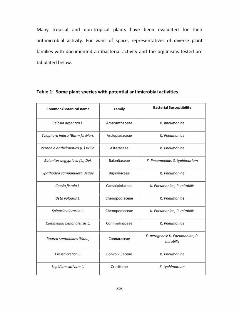
Many tropical and non-tropical plants have been evaluated for their
antimicrobial activity. For want of space, representatives of diverse plant
families with documented antibacterial activity and the organisms tested are
tabulated below.
Table 1: Some plant species with potential antimicrobial activities
Common/Botanical name Family Bacterial Susceptibility
Celosia argentea L. Amaranthaceae K. pneumoniae
Tylophora indica (Burm.f.) Merr. Asclepiadaceae K. Pneumoniae
Vernonia anthelmintica (L.) Willd. Asteraceae K. Pneumoniae
Balanites aegyptiaca (L.) Del. Balanitaceae K. Pneumoniae, S. typhimurium
Spathodea campanulata Beauv Bignonaceae K. Pneumoniae
Cassia fistula L. Caesalpiniaceae K. Pneumoniae, P. mirabilis
Beta vulgaris L. Chenopodiaceae K. Pneumoniae
Spinacia oleracea L. Chenopodiaceae K. Pneumoniae, P. mirabilis
Commelina benghalensis L. Commelinaceae K. Pneumoniae
Rourea santaloides (Vahl.) Connaraceae E. aerogenes; K. Pneumoniae; P.
mirabilis
Cressa cretica L. Convolvulaceae K. Pneumoniae
Lepidium sativum L. Cruciferae S. typhimurium

xxx
Momordica charantia L. Cucurbitaceae E. aerogenes; K. Pneumoniae; P.
mirabilis
Cyperus scarious R.Br. Cyperaceae E. aerogenes; K. Pneumoniae; P.
mirabilis
Ricinus communis L. Euphorbiaceae K. Pneumoniae, P. mirabilis
Arachis hypogaea L. Fabaceae E. aerogenes; K. Pneumoniae;
Vigna radiata L. Fabaceae K. Pneumoniae; P. mirabilis
Fumaria indica (Haussk.) Pugsley. Fumariaceae K. Pneumoniae; P. mirabilis
Ocimum kilimanjaricum L. Labiatae E. aerogenes; E. coli; K. Pneumoniae; P.
mirabilis; P. vulgaris.
Artocarpus hetrophyllus Lam. Moraceae E. aerogenes; P. mirabilis
Ficus elastica Roxb. Moraceae K. Pneumoniae; P. mirabilis
Piper longum L. Piperaceae E. aerogenes; K. Pneumoniae; P.
mirabilis; S. typhimurium .
Gardenia resinifera Roth. Rubiaceae K. Pneumoniae; P. mirabilis
Mesua ferra Linn. Guttiferae E. aerogenes; K. Pneumoniae; P.
mirabilis; P. vulgaris.
Alchornea cordifolia Euphorbiaceae K. Pneumoniae; E. coli; B. subtilis; S.
aureus
Chromolaena odorata Asteraceae Propionibacterium canes;
Mycobacterium spps.
(Parekh and Chanda, 2007; Okoye and Ebi, 2007)
1.3.2 Plant Secondary Metabolites Associated with Antimicrobial Effect

xxxi
Plants have been shown to possess an amazing potential to synthesize
aromatic substances, most of which are phenols or their oxygenated-
substituted derivatives. These substances have been reported to consist of
mostly secondary metabolites, of which at least 12,000 have been isolated, a
number estimated to still be less than 10% of the total (Schultes, 1978).
Plant secondary metabolites, in many cases, serve as plant defense
mechanisms against predation by microorganisms, insects, and herbivores.
Some, such as terpenoids give plants their odours; others (e.g. quinones and
tannins) are responsible for plant pigmentation. Many of these compounds are
also responsible for plant flavour. It is therefore not surprising that useful
antimicrobial phytochemicals have been derived from these plant secondary
metabolites.
1.3.2.1 Flavones, Flavonoids and Flavonols
Flavones are phenolic structures containing one carbonyl group (as opposed to
the two carbonyls in quinones). Addition of a 3-hydroxy group yields a flavonol
while flavonoids are also hydroxylated phenolic substances but occur as a C6-
C3 unit linked to an aromatic ring. These compounds have been known to be
synthesized by plants in response to microbial infection and have equally been
found in vitro to be effective antimicrobial substances against a wide array of
microorganisms (Dixon et al, 1983; Cowan, 1999). Their activity is probably due
to their ability to complex with extracellular and soluble proteins and to
complex with bacterial cell walls, often leading to the inactivation of the
xxxii
proteins, loss of function and cell lysis (Stern et al, 1996). More lipophilic
flavonoids may also disrupt microbial membrane (Tsuchiya et al, 1996).
Example of flavonoid compounds with known antimicrobial activity includes
the catechins. These are the most reduced form of the C-3 unit in flavonoid
compounds that have been extensively researched due to their occurrence in
oolong green teas. Teas have been reported to exert antimicrobial activity and
also contain a mixture of catechin compounds which inhibited in vitro activity
of Vibrio cholerae 01, Streptococcus mutans, Shigella, and other bacteria and
microorganisms (Toda et al, 1989; Batista et al, 1994; Borris, 1996; Sakanaka et
al, 1989; Sakanaka et al, 1992; Vijaya et al, 1995).
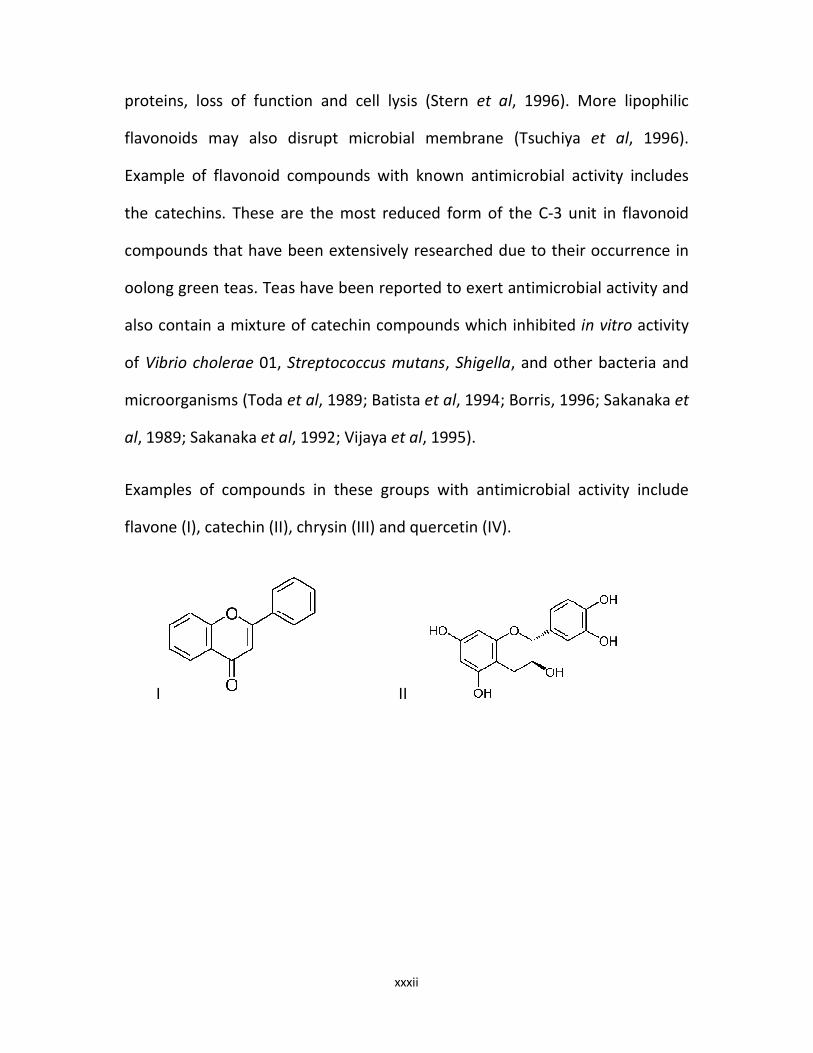
Examples of compounds in these groups with antimicrobial activity include
flavone (I), catechin (II), chrysin (III) and quercetin (IV).
I II

xxxiii
III IV
1.3.2.2 Alkaloids
These are basically heterocyclic nitrogenous compounds. Many of these
compounds found in higher plants have shown promising antibacterial activity.
For instance, diterpenoid alkaloids, commonly isolated from the plants of the
Ranunculaceae, or buttercup family, are commonly found to have
antimicrobial properties (Omulokoli et al, 1997). Some of the highly aromatic
planar quaternary alkaloids such as berberine (V) have their mechanism of
action attributable to their ability to intercalate with DNA (Phillipson and
O'Neill, 1987). Other alkaloids with antimicrobial actions are Harmane (VI),
Piperine (VII), etc.
V VI

xxxiv
VII
1.3.2.3 Terpenoids and Essential Oils
Essential oils are secondary metabolites that are highly enriched in compounds
based on an isoprene structure. The observed fragrance in most plants is
contained in their essential oil fraction. They consist mainly of compounds
belonging to the chemical group called terpenes. When the compounds
contain additional elements, usually oxygen, they are called terpenoids.
Terpenoids, even though synthesized from acetate units, differ from fatty acids
by their extensive branching and cyclization.
Many plant terpenoids have been found to be active against bacteria, fungi,
viruses and protozoa (Amaral et al, 1998; Habtemariam et al, 1993; Himejima
et al, 1992; Mendoza et al, 1997; Kubo et al, 1993; Hasegawa et al, 1994;
Ghoshal et al, 1996; Rao et al, 1993; Sun et al, 1996; Tassou et al, 1995; 2000).
Although the mechanism of their antibacterial action is not yet fully
understood, terpenes are speculated to act by disrupting cell membranes due
to their lipophilic nature. In fact, it has been reported as far back as 1977 that
of all the essential oil derivatives being examined, 30% were inhibitory to
xxxv
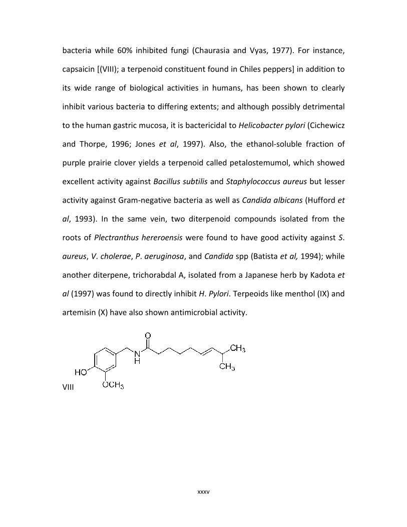
bacteria while 60% inhibited fungi (Chaurasia and Vyas, 1977). For instance,
capsaicin [(VIII); a terpenoid constituent found in Chiles peppers] in addition to
its wide range of biological activities in humans, has been shown to clearly
inhibit various bacteria to differing extents; and although possibly detrimental
to the human gastric mucosa, it is bactericidal to Helicobacter pylori (Cichewicz
and Thorpe, 1996; Jones et al, 1997). Also, the ethanol-soluble fraction of
purple prairie clover yields a terpenoid called petalostemumol, which showed
excellent activity against Bacillus subtilis and Staphylococcus aureus but lesser
activity against Gram-negative bacteria as well as Candida albicans (Hufford et
al, 1993). In the same vein, two diterpenoid compounds isolated from the
roots of Plectranthus hereroensis were found to have good activity against S.
aureus, V. cholerae, P. aeruginosa, and Candida spp (Batista et al, 1994); while
another diterpene, trichorabdal A, isolated from a Japanese herb by Kadota et
al (1997) was found to directly inhibit H. Pylori. Terpeoids like menthol (IX) and
artemisin (X) have also shown antimicrobial activity.
VIII

xxxvi
IX X
1.3.2.4 Tannins
The term 'tannin' is used to describe a group of polymeric phenolic substances
capable of tanning leather or precipitating gelatin from solution. The property
is known as astringency. Tannins which are found in almost every plant part
are divided into hydrolyzable and condensed tannins. Hydrolyzable tannins are
based on gallic acid, usually as multiple esters with D-glucose; while the more
numerous condensed tannins (often called proanthocyanidins) are derived
from flavonoid monomers (Cowan, 1999). Generally, tannins may be formed
by condensation of flavan derivatives which have been transported to woody
tissues of plant, or alternatively, by polymerization of quinone units (Geissman,
1963).
Tannins have been shown to act at the molecular levels by complexing with
microbial proteins through so-called non-specific forces such as hydrogen
bonding and hydrophobic effects, as well as covalent bond formation (Haslam,
1996; Stern et al, 1996). Thus, their mode of antimicrobial action may be
related to their ability to inactivate microbial adhesins, enzymes, cell envelope

xxxvii
transport proteins, etc. They may also complex with polysaccharide (Ya et al,
1988). Tannins have also shown to act via direct inactivation of microorganisms
(eg. low tannin concentrations modify the morphology of germ tubes of
Crinipellis perniciosa (Brownlee et al, 1990).
According to various documented studies reviewed by Scalbert (1991), tannins
were found to be toxic to filamentous fungi, yeasts and bacteria. Condensed
tannins have been described to bind cell walls of ruminal bacteria, preventing
growth and protease activity (Jones et al, 1994). Though still speculative,
tannins are considered to be wholly or partially responsible for the antibiotic
activity of various aqueous and solvent extracts of many tropical and
temperate plants scattered across the globe (Taylor et al, 1996). Pentagalloyl
glucose (XI; hydrolysable tannin) and procyanidine (XII; condensed tannin)
have shown remarkable antimicrobial activities (Cowan, 1999).
XI XII
1.3.2.5 Miscellaneous Plant Constituents

xxxviii
Other major groups of antimicrobial compounds from plants include the simple
phenols and phenolic acids, quinones, coumarins, lectins and polypeptides;
and mixtures of all these groups. There abound many documented plant
derivatives belonging to these chemical groups that have proven antimicrobial
activity.
The common herbs terragon and thyme both contain caffeic acid (XIII; a
phenylpropane-derived phenolic compound), which is effective against viruses,
bacteria and fungi (Wild, 1994; Brantner et al, 1996). Catechol (XIV) and
pyrogallol both are hydroxylated phenols, shown to be toxic to
microorganisms; while eugenol (XV; a phenolic compound possessing a C3 side
chain at lower level of oxidation but also classified as an essential oil) is
considered bacteriostatic against both fungi and bacteria (Duke, 1985). Gallic
acid (XVI) has also proven to be toxic to some microorganisms.
XIII XIV
XV XVI
xxxix
Quinones are aromatic rings with two ketone substitutions which are
ubiquitous in nature and are characteristically highly reactive. In addition to
providing a source of stable free radicals, they are known to complex
irreversibly with nucleophilic amino acids in proteins, often leading to
inactivation of the protein and loss of action (Stern et al, 1996). For these
reasons, the potential range of quinone antimicrobial effects is great. Kazmi et
al (1994) described an anthraquinone from Cassia italica, a Pakistani tree,
which was bacteriostatic for Bacillus anthracis, Corynebacterium
pseudodiphthericum and Pseudomonas aeruginosa but bactericidal for
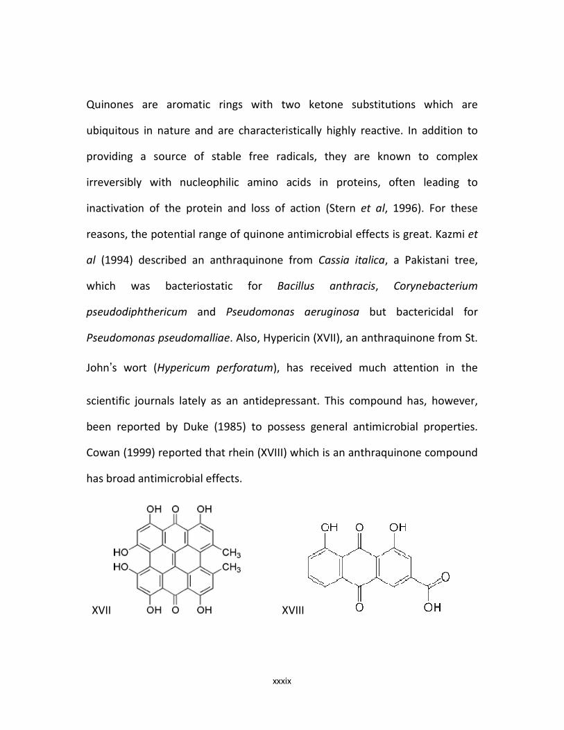
Pseudomonas pseudomalliae. Also, Hypericin (XVII), an anthraquinone from St.
John’s wort (Hypericum perforatum), has received much attention in the
scientific journals lately as an antidepressant. This compound has, however,
been reported by Duke (1985) to possess general antimicrobial properties.
Cowan (1999) reported that rhein (XVIII) which is an anthraquinone compound
has broad antimicrobial effects.
XVII XVIII

xl
Coumarins (XIX) are phenolic substances made up of fused benzene and α-
pyrone rings. They are responsible for the characteristic odour of hay. As a
group, coumarins have been found to stimulate macrophages which could
have an indirect negative effect on infections (Casley-Smith, 1997).
Hydroxycinamic acids, related to coumarins, seem to be inhibitory to Gram-
positive bacteria (Fernandez et al, 1996). Also, phytoalexins, which are
hydroxylated derivatives of coumarins, are produced in carrots in response to
fungal infection and can be presumed to have antifungal activity (Hoult and
Paya, 1996). General antimicrobial activity was equally documented in
coumarin compounds found in woodruff (Galium odoratum) extracts
(Thompson, 1978). Although data about specific antibiotic properties of
coumarins are scarce, many reports give reasons to believe that some utility
may reside in these phytochemicals (Cowan, 1999; Hamburger and
Hostettmann, 1991).
XIX

xli
Peptides which are inhibitory to microorganisms were first reported by Balls et
al (1942). They are often positively charged and contain disulfide bonds. Their
mechanism of action may be the formation of ion channels in the microbial
membrane, or by competitive inhibition of adhesion of microbial proteins to
host polysaccharide receptors (Zhang and Lewis, 1997; Sharon and Ofek, 1986).
Inhibition of bacteria and fungi by these macromolecules (e.g. peptides from
the herbaceous Amaranthus) has been documented (De Bolle et al, 1996).
Also, thionins, which are peptides commonly found in barley and wheat,
consisting of 47 amino acid residues are toxic to yeasts and both Gram-
negative and Gram-positive bacteria (Fernandes de Caleya et al, 1972).
Fabatin, a recently identified 47-residue peptide from fava beans, appears to
be structurally related to γ-thionins from grains and inhibits E. coli, P.
aeruginosa and Enterococcus hirae but not Candida or Saccharomyces (Zhang
and Lewis, 1997).
The antimicrobial activity of several extracts from plants has been linked to
compounds belonging to more than one chemical group. For instance, the
chewing sticks which are widely used in many African countries as an oral
hygiene aid come from different species of plants, and within one stick, the
chemically active component may be heterogeneous (Akpata and Akinrimisi,
1977). Crude extracts of one species used for this purpose, Serindeia
werneckei, inhibited the periodontal pathogens Porphyromonas gingivalis and
Bacteroides melaninogenicus in vitro (Rotimi et al, 1988). Also, the active
component of one of the Nigerian chewing sticks (Fagara zanthoxyloides) was

xlii
found to consist of various alkaloids (Odebiyi and Sofowora, 1979). Pawpaw
(Carica papaya) yields a milky sap, often called latex, which is a complex
mixture of chemicals (Cowan, 1999). Chief among them is papain, a well-
known proteolytic enzyme. It also contains carpaine (an alkaloid) and
terpenoids (Thomson, 1978). All these compounds in papaya have been shown
to contribute to the antimicrobial properties of its latex which was found to be
bacteriostatic to B. subtilis, Enterobacter cloacae, E. coli, Salmonella typhi,
Staphylococcus aureus and Proteus vulgaris (Osato et al, 1993).
1.4 LITERATURE REVIEWS OF PLANTS USED
1.4.1 Loranthus micranthus Linn
1.4.1.1 Taxonomy of L. Micranthus
The botanical profile of Loranthus micranthus is as summarized below:
Kingdom: Plantae
Phylum: Angiosperm
Sub-Phylum: Dicotyledons
Order: Santalales
Family: Loranthaceae
Sub-Family: Lorantheae
Genus: Loranthus

xliii
Species: micranthus
The mistletoe plant is an evergreen obligate parasite with over 700 species
which depends on its hosts for minerals and water only, as it can
photosynthesize its carbohydrate by means of its green leaves (Gill, 1973;
Griggs, 1991).
The most common species include: European mistletoe (Viscum album L.);
American mistletoe (Phoradendron flavescens); Australian/Argentine mistletoe
(Ligaria cuneifolia R et. T); African mistletoe, e.t.c.
Figure 1.0: Flowers and Leaves of Loranthus micranthus
xliv
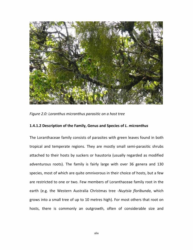
Figure 2.0: Loranthus micranthus parasitic on a host tree
1.4.1.2 Description of the Family, Genus and Species of L. micranthus
The Loranthaceae family consists of parasites with green leaves found in both
tropical and temperate regions. They are mostly small semi-parasitic shrubs
attached to their hosts by suckers or haustoria (usually regarded as modified
adventurous roots). The family is fairly large with over 36 genera and 130
species, most of which are quite omnivorous in their choice of hosts, but a few
are restricted to one or two. Few members of Loranthaceae family root in the
earth (e.g. the Western Australia Christmas tree -Nuytsia floribunda, which
grows into a small tree of up to 10 metres high). For most others that root on
hosts, there is commonly an outgrowth, often of considerable size and

xlv
complicated in shape, where the parasite root joins the host. The roots of the
parasites often branch within the tissue of the host (as in Viscum).
The genus, Loranthus, consists of several species scattered in many parts of
Africa. It belongs to the sub-family, Lorantheae, which is characterized by the
presence of stem without secretory canals and has extraxylary phloem. Their
flowers have below the petals an outgrowth from the axis in form of small ring
or fringe called calyculus. After some weeks of the seeds germinating on
branches of its host, the Loranthus plant produces proper flowers which are
generally bright red and conspicuous, although one species produces yellow
flowers with red tips. The flowers are soon followed by small, fleshy,
drupaceous fruits which are much sought after by birds. The red Loranthus is a
common sight in many parts of West Africa, particularly in cocoa and cola
plantations, where whole branches are often covered with this medicinal herb.
The African mistletoe species, micranthus, is found mainly in the Southeastern
part of Nigeria. It grows on a large number of hosts including kola nut (Kola
acuminata), avocado (Persea americana), dogoyaro/neem (Azadirachta
indica), oil bean (Pentaclethra macrophylla), ogbono (Irvigia gabonensis),
lemons/citrus, etc. It produces sympodial, often dichasical, stem and the leaves
are usually evergreen and leathery. The cymose inflorescences are in spikes,
with the flowers on the internodes as well as on the nodes. Thus, they have
clusters of narrowly tubular flowers that are bright-red which appear as
clusters of coloured ‘matches’.

xlvi
1.4.1.3 Ethnomedicinal Uses and Pharmacological Studies on L. micranthus
Several ethnomedicinal usages have been attributed to Loranthus micranthus.
These include: blood pressure control (antihypertensive activity), anti-diabetic
activity, anticancer, antimicrobial and in many other metabolic diseases which
qualified mistletoe as an “all-purpose herb” (Kafaru, 1993; Obatomi et al, 1994;
Oliver-Bever, 1986; Dalziel, 1955). Different research works have been carried
out on the several species of the plant to demonstrate and support the
existence of many of the ethno medicinal claims (Obatomi et al, 1996;
Osadebe and Ukwueze, 2004; Osadebe and Akabogu, 2006; Osadebe et al,
2004, 2010, 2012; Ukwueze and Osadebe, 2012; Agbo et al, 2013; Omeje et al,
2012). Previous works on Loranthus micranthus have equally shown that some
of its medicinal activities vary with the particular host tree from which it is
harvested (Osadebe and Ukwueze, 2004; Osadebe et al, 2004). Other factors
that have been shown to affect the phytochemical composition and
pharmacological activities of mistletoe plant include species, harvesting
season, etc. (Obatomi et al, 1994; Wagner et al, 1996; Osadebe et al, 2008).
1.4.2 Psidium Guajava Linn
1.4.2.1 Taxonomy of P. guajava
The botanical profile of Psidium guajava is as summarized below:

xlvii
Kingdom: Plantae
Phylum: Angiosperms
Sub-Phylum: Eudicots
(unranked): Rosids
Order: Myrtales
Family: Myrtaceae
Subfamily: Myrtoideae
Tribe: Myrteae
Genus: Psidium
Species: guajava
Binomial name: Psidium guajava L.
Psidium guajava L, a fruit-bearing tree commonly known as guava, of the
family Myrtaceae, is a native of tropical America. The French call it goyave or
goyavier; the Dutch, guyaba or goeajaaba; the Surinamese, guave or goejaba;
and the Portuguese, goiaba or goaibeira. Hawaiians call it guava or kuawa. In
Guam, it is abas. In Malaya, it is generally known either as guava or jambu batu
(Morton, 1987).
1.4.2.2 Morphology of P. guajava

xlviii
Cultivated varieties grow about 10 m in height and produce fruits within 4
years. Wild trees grow up to 20 m high and are well branched. The guava tree
can be easily identified by its distinctive thin, smooth, copper-colored bark that
flakes off, showing a greenish layer beneath. The trees might have spread
widely throughout the tropics because they thrive in a variety of soils,
propagate easily and bear fruits quickly. The fruits are enjoyed by humans,
birds and monkeys, which disperse guava seeds and cause spontaneous dumps
of guava saplings to grow throughout the rainforest (Wealth of India, 2003).
Figure 3.0: Psidium guajava Tree

xlix
Figure 4.0: Leaves and Flowers of Psidium guajava

l
Figure 5.0: Fruit of Psidium guajava
1.4.2.3 Ethnomedicinal Uses and Pharmacological Studies of P. guajava
Psidium guajava is a medicinal plant used in tropical and subtropical countries
to treat many health disorders. In the indigenous system of medicine, different
parts of the plant are used for the treatment of various human ailments such as
wounds, ulcers, bowels and cholera (Begum et al., 2002a). Investigations have
indicated that its bark, fruit and leaves possess antibacterial, hypoglycaemic,
anti-inflammatory, analgesic, antipyretic, spasmolytic and CNS depressant
activities ( Begum et al., 2002b). It has indeed been variously reported that
Psidium guajava leaf extract has a wide spectrum of biological activities such as
anticough, antibacterial, haemostasis (Jaiarj et al., 1999; 2000), antidiarrhoeal
and narcotic properties (Lozoya et al.,1990), and antioxidant properties (Qian
and Nihorimbere, 2004). According to Lutterodt and Maleque (1998) and
Meckes et al., 1996, the leaf extract is used to treat diarrhoea, abdominal pain,
convulsions, epilepsy, cholera, insomnia and has hypnotic effect.
The long history of guava use has led modern-day researchers to intensify their
study on guava extracts. Its traditional use against diarrhea, gastroenteritis and
other digestive complaints has been validated in numerous clinical studies. In a
study including 17 Thai medicinal plants on anti-proliferative effects on human

li
mouth epidermal carcinoma and murine leukemia cells using MIT assay, guava
leaf showed anti-proliferative activity, which was 4.37 times more than
vincristine (Manosroi et al. , 2006).
Bark and leaf extracts were shown to have in vitro toxic action against
numerous bacteria. Gallocatechin isolated from the methanol extract of guava
leaf showed antimutagenic activity against E. coli (Matsuo et al., 1994). Water
and chloroform extracts of guava were effective in activating the mutagenicity
of Salmonella typhimurium (Grover and Bala, 1993). The antimicrobial activities
of P. guajava and leaf extracts, determined by disk diffusion method (zone of
inhibition), were compared to tea tree oil (TTO), doxycycline and clindamycin
antibiotics. It was shown that P. guajava leaf extracts might be beneficial in
treating acne especially those that have anti-inflammatory activities (Qadan et
al., 2005). The active flavonoid compound-quercetin-3-O-alpha-l-
arabinopyranoside (guaijaverin) - extracted from guava leaves has high
potential antiplaque activity by inhibiting the growth of Streptococcus mutans
(Limsong et al., 2004). Guava leaf extract also inhibited the growth of
Streptococcus aureus in a study carried out by disc diffusion method
(Abdelrahim et al., 2002). In several other studies, guava showed significant
antibacterial activity against common diarrhea-causing bacteria such as
Staphylococcus, Shigella, Salmonella, Bacillus, E. coli, Clostridium and
Pseudomonas. Indeed, the aqueous, alcohol and chloroform extracts have
been found to be effective against Aeromonas hydrophila, Shigella spp. and
Vibrio spp., Staphylococcus aureus, Sarcinta lutea and Microbacterium phlei
(Jaiarj et al., 1999). In a more recent study, the aqueous and ethanol:water
extracts of P. guajava leaves, roots and stem bark were found to be active
against the Gram-positive bacteria Staphylococcus aureus and Bacillus subtilis,

lii
but virtually inactive against the Gram-negative bacteria Escherichia coli and
Pseudomonas aeruginosa (Sanches et al.,2005).
A double-blind clinical study of the effects of a Phytodrug (QG-5) developed
from guava leaf showed a decrease in duration of abdominal pain, which was
attributed to antispasmodic effect of quercetin present in leaf extract (Xavier
et al., 2002). Guava leaf extracts and fruit juices have also been clinically
studied for infantile diarrhea. In a clinical study with 62 infants with infantile
rotaviral enteritis, the recovery rate was 3 days (87.1%) in those treated with
guava, and diarrhea ceased in a shorter period than controls. It was concluded
in the study that guava has 'good curative effect on infantile rotaviral enteritis'
(Wei et al., 2000). Lectin chemicals in guava were shown to bind to E. coli (a
common diarrhea-causing organism), preventing its adhesion to the intestinal
wall and thus preventing infection and resulting diarrhea (Rodriguez et al.,
2001). Guava leaf extract has also shown to have tranquilizing effect on
intestinal smooth muscle, inhibit chemical processes found in diarrhea and aid
in the re-absorption of water in intestines. In another research, an alcoholic
leaf extract was reported to have a morphine-like effect, by inhibiting the
gastrointestinal release of chemicals in acute diarrheal disease. This morphine-
like effect was thought to be related to a chemical, quercetin. The effective use
of guava in diarrhea, dysentery and gastroenteritis can also be related to
guava's documented antibacterial properties (Tona et al., 2000). In a study
carried out with leaf extract of the plant, inhibition of gastrointestinal release

liii
of acetylcholine by quercetin present in extract was suggested as a possible
mode of action in the treatment of acute diarrheal disease (Lutterodt, 1992).
Guava fruit and leaf showed antioxidant and free radical scavenging capacity
(Hui-Yin and Gow-Chin, 2007). A study of aqueous extract of P. guajava in
acute experimental liver injury induced by carbon tetrachloride, paracetamol
and thioacetamide, showed its hepatoprotective activity. The effects observed
were compared with a known hepatoprotective agent, silymarin. Histological
examination of the liver tissues supported hepatoprotection (Roy et al., 2006).
During various episodes of screening of medicinal plants, extract from P.
guajava leaves was found to exhibit significantly inhibitory effect on the
protein tyrosine phosphatase1B (PTP1B). Significant blood glucose lowering
effects of the extract were observed after intraperitoneal injection of the
extract at a dose of 10mg/kg in both 1-and 3-month-old Lepr(db)/Lepr(db)
mice (Oh et al. , 2005). In a study undertaken to investigate the hypoglycemic
and hypotensive effects of P. guajava leaf aqueous extract in rats, it showed
hypoglycemic activity. The hypoglycemic effect of plant extract was examined
in normal and diabetic rats, using streptozotocin (STZ)-induced diabetes
mellitus model (Ojewole, 2005). Also, i.p. treatment with 1g/kg guava juice
produced a marked hypoglycemic action in normal and alloxan-treated
diabetic mice (Cheng and Yang, 1983). In two randomized human studies, the
consumption of guava fruit for 12 weeks was shown to reduce blood pressure
by an average 8%, decrease total cholesterol level by 9%, decrease
triglycerides by almost 8% and increase HDL cholesterol by 8%; while a

liv
randomized, single-blind, controlled trial conducted to examine the effects of
guava fruit intake on blood pressure and blood lipids in patients with essential
hypertension showed the possibility that an increased consumption of guava
fruit can cause a substantial reduction in blood pressure and blood lipids
without decreasing HDL-cholesterol level (Singh et al., 1992, 1993).
Leaf extract of guava had shown ionotropic effect on guinea pig atrium
(Conde-Garcia et al., 2003). Some studies reported that the leaf extract and its
derivative identified as quercetin has effect on the intracellular calcium levels
in gastrointestinal smooth muscle (Lozoya et al., 1990), in cardiac muscle cell
(Apisariyakul et al., 1999) and in neuromuscular junction (Chaichana and
Apisariyakul, 1996). In other animal studies, guava leaf extracts have shown
central nervous system (CNS) depressant activity (Shaheen, 2000). Guava leaf
extract showed anticough activity by reducing the frequency of cough induced
by capsaicin aerosol (Jaiarj et al., 1999).
1.4.2.4 The Phytochemistry of Psidium guajava
Guava has been found to be rich in tannins, phenols, triterpenes, flavonoids,
essential oils, saponins, carotenoids, lectins, vitamins, fibre and fatty acids.
According to Olajide et al (1999), the leaves of P. guajava contain an essential
oil rich in cineol, tannins, triterpenes and flavonoids. Various reports of
phytochemical screening of Psidium guajava leaf showed tannins in aqueous
extract; and anthocyans, alkaloids, flavonoids, tannins and steroids/terpenoids in
ethanolic extract.

lv
More than twenty identified compounds from Psidium guajava leaf have been
reported (Seshadri and Vasishta, 1965; Osman et al., 1974; Lutterodt and
Maleque, 1988). The major components are: β-selinene (XX), β-caryophyllene
(XXI), caryophyllene oxide (XXII), squalene (XXIII), selin-11-en-4α-ol
(XXIV), guaijavarin (XXV), isoquercetin (XXVI), hyperin (XXVII), quercitrin
(XXVIII) and quercetin-3-O-gentobioside; morin-3-O-α-L-lyxopyranoside
(XXIX), morin-3-O-α-L-arabopyranoside (XXX); β-sitosterol (XXXI), uvaol
(XXXII), oleanolic acid (XXXIII), ursolic acid (XXXIV) and one new
pentacyclic triterpenoid: guajanoic acid (Lozoya et al., 1994; Meckes et al.,
1996; Arima and Danno, 2002; Begum et al., 2004).
XX XXI
XXII XXIII

lvi
XXIV XXV
XXVI XXVII
XXVIII XXIX
XXX XXXI

lvii
XXXII XXXIII
XXXIV
Guava fruit is higher in vitamin C than citrus fruits (80 mg of vitamin C in 100g
of fruit) and contains appreciable amounts of Vitamin A as well and is also a
good source of pectin (Sunttornusk, 2002).
The bark of guava tree contains considerable amounts of tannins (11-27%), and
hence is used for tanning and dyeing purposes. Leucocyanidin (XXXV), luectic
acid, ellagic acid (XXXVI) and amritoside (XXXVII) have been isolated from the
stem bark.
Other compounds that have been isolated from guava plant include avicularin
(XXXVIII; 3-L-4-4-arabinofuranoside), α-pinene (XXXIX), β-pinene (XL), limonene
(XLI), terpenyl acetate (XLII), isopropyl alcohol (XLIII), longicyclene (XLIV), β-

lviii
bisabolene, β-copanene, farnesene, humulene, cardinene, curcumene, mallic
acids, ursolic, crategolic, guayavolic acids, cineol, etc (Shruthi et al., 2013).
XXXV XXXVI
XXXVII XXXVIII
XXXIX XL XLI

lix
XLII XLIII XLIV
1.5 BIOASSAYBIOASSAYBIOASSAYBIOASSAY----GUIDED CHARACTERIZATION OF ANTIGUIDED CHARACTERIZATION OF ANTIGUIDED CHARACTERIZATION OF ANTIGUIDED CHARACTERIZATION OF ANTIBACTERIALBACTERIALBACTERIALBACTERIAL CONSTITUENTS CONSTITUENTS CONSTITUENTS CONSTITUENTS
FROM HIGHER PLANTSFROM HIGHER PLANTSFROM HIGHER PLANTSFROM HIGHER PLANTS
The driving force behind much phytochemical research is the discovery of new
biological active compounds for medicinal or agricultural uses. Biological assays
then must be carried out in order to identify promising plant extracts, to guide
the separation and isolation, and to evaluate lead compounds. Identification of
natural products from plants that may serve as valuable sources of bioactive
agents for medicinal and agricultural uses largely depends on bioactivity-
directed isolation (Cseke et al, 2006).
The choices of bioassays depend a great deal on the amounts of materials to
be tested and the time and effort necessary to carry out the assays. Obviously,
an in vivo assay using the organism afflicted (humans or animals) would
provide the most meaningful results. However, exploratory screening using
whole animals is impractical (or unethical), and various in vitro screening
methods have been developed to provide guided separation and identification
of lead compounds. The latter have the advantage in that they can be

lx
automated with robotics and miniaturized, leading to rapid throughput
screening of large numbers of samples. In addition, the in vitro bioassays may
provide activity information that is precluded by poor bioavailability using a
whole-animal in vivo assay. For instance, natural products that inhibit the
growth of tumor cells or bacteria in an in vitro assay may identify promising
molecular structures that would benefit from semi-synthetic modifications
(Cseke et al, 2006).
1.5.1 Principles of Bioassay
Bioassay (or biological assay) is the estimation of the activity or potency of a
drug or other substance (e.g. plant extract) by comparing its effects on a test
organism with that of a standard preparation. It is a type of scientific
experiment conducted to measure the effects of a substance on a living
organism and is essential in the development of new drugs and other scientific
monitoring. Bioassays may be qualitative or quantitative.
1.5.2 Screening Methods for Antibacterial Agents from Higher Plants
The discovery of promising plant extracts and the subsequent activity-guided
isolation of constituents put specific requirements on the bioassays to be used
for that purpose. They have to be simple, rapid, reproducible and inexpensive
in order to be compatible with the large number of assays to be performed.
lxi
Antimicrobial activity of plants can be detected by observing the growth
response of various microorganisms to those plant tissues or extracts which
are placed in contact with them. Many methods for detecting such activity are
available, but since they are not equally sensitive or even based upon the same
principle, the results obtained will also be profoundly influenced not only by
the method selected, but also by the microorganisms used to carry out the test
(Vanden-Berghe and Vlietinck, 1991). In general, biological assays or evaluation
can be carried out much more efficiently on water-soluble, pure crystalline
substances than on mixtures like plant extracts.
1.5.2.1 Test Organisms and Culture Media
The purpose of any antimicrobial investigation will obviously determine to a
great extent the choice of test organisms to be used. For an investigation of a
general character, the test organisms selected should be as diverse as possible
and preferably representative of all important groups of pathogenic bacteria
according to their physical and chemical composition and resistance pattern.
Most screening studies on plant extracts, however, have been carried out on
one or two bacteria, including strains of Staphylococcus aureus and Escherichia
coli, although such findings may not adequately predict an interesting broad-
spectrum activity or a selective but pronounced activity against some of the
problem-pathogenic bacteria in chemotherapy such as resistant S. aureus, P.
aeruginosa, Proteus vulgaris, Klebsiella pneumoniae, Neisseria gonorrhoeae,
Candida albicans and others.

lxii
Most bacteria (and yeasts) can be cultivated on standard Mueller-Hinton agar
or diagnostic sensitivity test agar (DST) and American type culture collection
(ATCC) or similar standard microorganisms are available. Only few bacteria
(e.g. Neisseria gonorrhoeae and Campylobacter fetus) require special growth
factors which should be included in the standard medium.
In general, standard microorganisms should be preferably used as test bacteria
during screening for new antimicrobially-active plant components for ease of
reproducibility of results by other researchers. If the interest, however, is in
finding new products which are selectively active against problem
microorganisms causing certain diseases, e.g. resistant P. aeruginosa, it is
clearly appropriate to employ the corresponding isolated pathogenic
microorganisms (Rwangabo et al., 1988).
1.5.2.2 Antibacterial Testing
The currently available antimicrobial screening methods fall into three broad
groups, including diffusion, dilution and bioautographic methods (Rios et al,
1988). These testing methods will only give an idea of the presence or absence
of substances with antimicrobial activity in the plant extracts, as the potency of
the active ingredients can only be determined on pure compounds using
standardized methodologies. The results obtained using any of the methods,

lxiii
however, are influenced by such factors such as extraction method, inoculum
volume, culture medium composition, pH and incubation temperature.
In the diffusion technique, a reservoir (e.g. filter paper disc, porcelain/stainless
steel cylinder or hole punched in the media) containing the plant extract to be
tested is brought into contact with an inoculated medium (e.g. agar) and, after
incubation, the diameter of the clear zone around the reservoir (inhibition
zone diameter) is measured. In order to lower the detection limit using this
method, the inoculated system is kept at low a temperature during several
hours before incubation, which favors diffusion over microbial growth and thus
increases the inhibition diameter. In most studies, the inhibition zones
obtained are compared with those obtained for antibiotics so as to establish
the sensitivity of the test organism to the extract. Advantages of the diffusion
methods are the small size of the sample used in the screening and the
possibility of testing up to five or six compounds per plate against a single
microorganism.
For the dilution methods, samples being tested are mixed with a suitable
medium, which has previously been inoculated with the test organism. After
incubation, growth of the microorganism may be determined by direct visual
or turbidimetric comparison of the test culture with a control culture which did
not receive an addition of the sample being tested, or by plating out both test
and control cultures (Kavanagh, 1963). Usually a series of dilutions of the
original sample in the culture medium is made and then inoculated with the
test organism. After inoculation, the endpoint of the test (MIC-value) is taken

lxiv
as the highest dilution which will just prevent perceptible growth of the test
organism (Vanden-Berghe and Vlietinck, 1991). In comparison, several
different test microorganisms may be tested simultaneously on the same
dilution as against diffusion methods in which several substances or dilutions
of one substance may be tested simultaneously against one test
microorganism. The agar dilution method is thus very quick, time saving and
also very useful to guide the isolation of antimicrobially active components
from plant extracts (Bakana et al, 1987; Rwangabo et al, 1988).
Bioautographic methods are employed to localize antibacterial activity on a
chromatoGram. The procedures are based on the agar diffusion technique,
whereby the antimicrobial agent is transferred from the thin layer or paper
chromatoGram to an inoculated agar plate through a diffusion process. Zones
of inhibition are then visualized by appropriate vital stains. Although very
suitable for testing highly active antibiotics (MIC-values < 10ug/ml),
bioautographic methods might not be very promising for testing plant extracts,
which often contain much less potent antimicrobial agents than the currently
available antibiotics (Vanden-Berghe and Vlietinck, 1991).
1.6 STRUCTURE ELUCIDATION OF BIOACTIVE PLANT METABOLITES
Chemical compounds, usually derived from plants and other natural sources,
have been used by humans for thousands of years to alleviate pain, diarrhea,
infection, and various other maladies. Until recently, these ''remedies" were

lxv
primarily crude preparations of plant material of unknown constitution. The
revolution in the synthetic organic chemistry during the nineteenth century
produced a concerted effort towards identification of the structures of the
active constituents of these naturally derived medicinals and synthesis of what
were hoped to be more efficacious agents.
By determining the molecular structures of the active components of these
complex mixtures, it is hoped that a better understanding of how these
components work can be elucidated (Knittel and Zavod, 2008).
1.6.1 Preliminary Analysis
Bioassay-directed fractionation is the process of isolating pure active
constituents from some type of biomass (eg. plants, microbes, marine
invertebrates, etc.) using a decision tree that is dictated solely by bioactivity
(Kinghorn, 2008). A variety of chromatographic separation techniques are
available for these purposes, including those based on adsorption on sorbents,
such as silica gel, alumina, Sephadex, and more specialized solid phases, and
methods involving partition chromatography inclusive of counter-current
chromatography. Recent improvements have been made in column
technology, automation of high-performance liquid chromatography (HPLC; a
technique often used for final compound purification) and compatibility with
HTS methodology (Butler, 2004).
1.6.2 Application of Modern Analytical Techniques

lxvi
Routine structure elucidation is performed using combinations of
spectroscopic procedures, with particular emphasis on one and two-
dimensional 1H- and
13C-nuclear magnetic resonance (NMR) spectroscopy and
mass spectroscopy (MS). 2D NMR spectra, generally, provide more information
about a molecule than 1D NMR spectra and are especially useful in
determining the structure of a molecule, particular for molecules that are too
complicated to work with using 1D NMR. Thus, for the structural elucidation of
a compound, a simple 1D NMR (H1 and C
13) may not be sufficient. Advanced
techniques like correlation spectroscopic methods, when analyzed properly,
provide the perfect way to determine the structure of a given formula or an
entirely unknown compound. The combination of some or all of these 1D and
multi-dimensional NMR spectra are all very useful in identification and
characterization of the structure, and orientation of bonds in a molecule
(Becker, 2000).
Considerable progress has been made in the development of cryogenic and
microcoil NMR probe technology for the determination of structures in sub-
milligram amounts of natural products (Koehn and Carter, 2005). In addition,
the automated processing of spectroscopic data for the structure elucidation
of natural products is a practical proposition (Steinbeck, 2004).
Another significant advance is the use of "hyphenated" analytical techniques
for rapid determination of the structure of natural products without the need
for a separate isolation step, such as liquid chromatograph-nuclear magnetic
resonance (LC-NMR) and LC-NMR-MS (Koehn and Carter, 2005; Butler, 2004).
lxvii
1.7 STATEMENT OF PROBLEM
Antibiotics have remained the mainstay of clinical therapy of infectious
diseases worldwide. Many microorganisms, however, are becoming
increasingly resistant to most of these agents (Jawetz et al., 1989). Another
serious limitation of these chemotherapeutic agents is the numerous adverse
effects associated with their administration (Snavely and Hodges, 1984). These
limitations have, thus, made it imperative that extensive efforts must be made
to uncover new antimicrobial agents from alternative sources whose
structures and modes of action may very likely differ from those of microbial
sources (antibiotics). Compounds extracted from higher plants have the
potential of fulfilling this purpose and that has led to the increasing screening
studies on several plant extracts in search of new ‘leads’. Unfortunately, most
of these works are preliminary investigations of the pharmacological actions of
the crude plant extracts, rather than the isolation, identification or the
characterization of the active constituents of these extracts.
1.8 JUSTIFICATION OF THE STUDY
Several works have been carried out to verify and confirm the traditional
antibacterial uses of Psidium guajava and Loranthus micranthus growing in the
tropical rain forest region of Nigeria, but researches on the isolation,
characterization and detailed chemical investigations of their
pharmacologically active components have remained inexhaustive.

lxviii
There is, therefore, the need for comprehensive chemical and bio-molecular
studies on these active constituents, and hence the justifications for this
present research.
1.9 AIMS AND SCOPE OF THE WORK
Aims of the Study
• To carry out a detailed bioactivity-guided phytochemical evaluation of
the extracts/solvent fractions of the leaves of Psidium guajava and
Loranthus micranthus.
• To identify and isolate the antibacterial compounds from the leaves of
the plants.
• To characterize and elucidate the structure of the isolates.
• To compare the antibacterial potentials of these isolates with standard
antibiotics with a view to developing derivatives of the ‘lead’
compounds and optimize same for activity with respect to potency and
selectivity, especially against bacteria causing opportunistic AIDS
infections [ e.g. resistant strains of Pseudomonas aeruginosa, and
Staphylococcus aureus; including the multi-drug resistant (MDR) strains
of methicillin-resistant Staphylococcus aureus (MRSA), and
Mycobacterium avium complex (MAC)].

lxix
CHAPTER TWO
MATERIALS AND METHODS
2.1 MATERIALS
2.1.1 Plant Materials
The leaves of Loranthus micranthus L. were collected in mid-June at Nsukka,
South-Eastern Nigeria, from the stem of Persea americana and authenticated
by Mr. A. Ozioko, a taxonomist with the Bio-resources Development and
Conservation Project (BDCP), Nsukka, Nigeria. Voucher specimen was
deposited in the herbarium of the Faculty of Pharmaceutical Sciences,
University of Nigeria, Nsukka. The leaves of Psidium guajava L. were collected
at the same period from the bio-resource area of the University of Port
Harcourt in South-South region of Nigeria and authenticated by the Plant
Science Biology (PSB) Department of the University. The voucher specimen was
deposited in the herbarium of the Faculty of Pharmaceutical Sciences,
University of Port Harcourt.
The leaves were air-dried at room temperature to a constant weight,
pulverized and passed through a 1mm sieve. The powdered materials were
stored in air-tight containers and kept in a refrigerator.
2.1.2 Microorganisms Used
The studies were performed with standard cultures of Staphylococus aureus
(ATCC 25923), Pseudomonas aeruginosa (ATCC 27833) and Escherichia coli

lxx
(ATCC 35219) obtained from the Nigerian Institute of Medical Research
(NIMR),Yaba, Lagos, Nigeria. The clinical isolates of other microorganisms were
used. All the microorganisms were grown in nutrient broth (Biotec, Suffolk, UK)
at 37oC and maintained on nutrient agar (Biotec, Suffolk, UK) slants at 4
oC. The
standardized cultures of the organisms were used throughout the experiment.
2.1.3 Solvents and Reagents
N-Hexane (Sigma-Aldrich, South Africa), Chloroform (Sigma-Aldrich, South
Africa), Ethyl acetate (BDH, England), Methanol (Sigma-Aldrich, South
Africa), Acetone (BDH, England), DMSO (Sigma-Aldrich, USA), Acetic acid
(Hopkins and Williams, England). Cutter mill (Manesty, England), Electronic
Balance (Sartorius, Germany) and Weighing balance (Ohans, USA). Other
reagents used were freshly prepared in the laboratory according to official
specifications.
Solvents Used for Isolation and Characterization of Active Ingredients
Dichloromethane (DCM), ethyl acetate, hexane and methanol. These solvents
were purchased from the Institute of Chemistry, University of Duesseldorf,
Germany. They were distilled before use and special grades were used for
spectroscopic measurements.
Solvents for HPLC
Methanol (LiChroSolv HPLC; Merck) and nano-pure water (distilled and heavy
metals free water obtained by passing distilled water through nano- and
ionexchange filter cells; Barnstead, France).
Solvents for NMR

lxxi
Deuterated methanol (Uvasol, Merck) and DMSO-d6 were used for NMR
measurements.
2.1.4 Materials and General Instruments
Chromatography
Pre-coated TLC plates (Silica Gel 60 F254, layer thickness 0.2mm) Merck
Silica Gel 60, 40−63 μm mesh size Merck
Sephadex LH 20, 25−100 μm mesh size Merck
General instruments
Analytical balances MC-1 Sartorious
Half-micro and analytical balance MC-1 Sartorious
Glass ware Schott Duran
Drying Oven ET6130 Heraeus
Ultra sonicator RK 510H Bandelin
UV-Lamp (254 and 366 nm) Camag
Rotary evaporator Büchi Rotavapor R-200
Vacuum pump CVC 2000 Vacuubrand
Centrifuge Pico Heraeus

lxxii
Nitrogen generator UHPN 3001 Nitrox
Air generator ZA 20 WGA
Fraction collector Retriever II ISCO
Lyvac GT2 (Freeze dryer) Steris
Vacuum pump Trivag D10E (Freeze dryer) Leybol
Syringe Hamilton 1701 RSN
Magnetic stirrer Variomag
HP Behrotest PH 10-Set Multipoint
Semi-preparative HPLC
Pump: L-7100 Merck/Hitachi
Detector: UV-L7400 (Photodiode array detector) Merck/Hitachi
Printer: Chromato-Intergartor D-2000
Merck/Hitachi
Column: Eurospher 100-C18, [10 μm; 300 mm × 8 mm] Knauer
Pre-column: Eurospher 100-C18, [10 μm; 30 mm × 8 mm] Knauer
Analytical HPLC
Pump: P 580A LPG Dionex
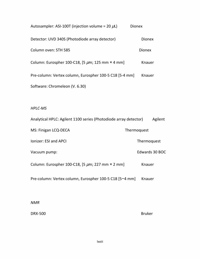
lxxiii
Autosampler: ASI-100T (injection volume = 20 μL) Dionex
Detector: UVD 340S (Photodiode array detector) Dionex
Column oven: STH 585 Dionex
Column: Eurospher 100-C18, [5 μm; 125 mm × 4 mm] Knauer
Pre-column: Vertex column, Eurospher 100-5 C18 [5-4 mm] Knauer
Software: Chromeleon (V. 6.30)
HPLC-MS
Analytical HPLC: Agilent 1100 series (Photodiode array detector) Agilent
MS: Finigan LCQ-DECA Thermoquest
Ionizer: ESI and APCI Thermoquest
Vacuum pump: Edwards 30 BOC
Column: Eurospher 100-C18, [5 μm; 227 mm × 2 mm] Knauer
Pre-column: Vertex column, Eurospher 100-5 C18 [5−4 mm] Knauer
NMR
DRX-500 Bruker

lxxiv
2.2 METHODS
2.2.1 Extraction and Fractionation of Plant of Materials
A portion of the powdered material (800 g) from L. micranthus plant was
defatted with n-hexane (4.5 L; to yield HFM) and the dried marc (540 g)
extracted in a soxhlet extractor with absolute methanol (3.5L). The dried
methanol soluble extract (MFM; 60.0 g) was reconstituted in 30 mL of
methanol, made up to 250 mL with distilled water, shaken for about 30
minutes and subjected to successive liquid-liquid extraction with chloroform (3
X 750 ml), ethyl acetate (3 X 750 ml) and acetone (3 X 750 ml) to yield the
chloroform (CFM; 11.4 g), ethyl acetate (EFM; 21.3 g) and acetone (AFM; 17.4
g) soluble fractions respectively. An aqueous portion (8.7 g) was left behind
after the fractionation process. The fractions were concentrated by
evaporating over water-baths set at 40oC. The concentrated/dried fractions
were stored in a refrigerator for further analysis.
The pulverized air-dried leaves (600 g) from Psidium guajava were defatted
with n-hexane (2.5L) and the dried marc (450 g) extracted with 5 L of 90 %
methanol for 4 days at room temperature (250C) and the extract concentrated
in vacuo with rotary evaporator. The dried methanol extract (35 g, 7.7 % w/w)
was reconstituted in 20 mL of methanol, made up to 200 mL with distilled
water, shaken for about 30 minutes and subjected to successive liquid-liquid
extraction with chloroform (3 x 750 ml), ethyl acetate (3 x 750 ml) and acetone
(3 x 750 ml) to yield PsG-CF (6.0 g; 1.3% w/w), PsG-EF (10.7g; 2.4% w/w) and

lxxv
PsG-AF (8.6 g; 1.9 % w/w) fractions respectively. An aqueous portion (6.8 g)
was left behind after the fractionation process.
Another portion (300 mg of the crude powder from L. micranthus) was
macerated in 90% methanol (3.5 L) for 48 hours and the filtrate concentrated
in vacuo to yield the crude methanol extract, CMFM.
Similarly, 100 mg of the crude powder from P. guajava was macerated in 90 %
methanol (1.5 L) for 48 hours and the filtrate concentrated in vacuo to yield
the crude methanol extract, PsG-CMF.
The schematic representaions for the extraction and fractionation procedures
for the plants are presented in Figures 6 & 7.

lxxvi
Dried Marc; 540g
Methanol; 5.5L
Hexane Fraction
(HFM); 44.2g
Chloroform; 3 X 750 ml
Methanol Fraction
(MFM); 60.0g
N-Hexane; 4.5L
Dried Marc
Acetone Fraction
(AFM); 17.4g
Ethyl acetate Fraction
(EFM); 21.3g
Chloroform Fraction
(CFM); 11.4g
Acetone; 3 X 750 ml
Ethyl acetate; 3 X 750 ml
Methanol;
3.5L
Crude Methanol
Fraction (CMFM);
31.4g 800g
PLANT MATERIAL
300g
Fig. 6: Schematic Diagram of the Extraction/Fractionation Procedure for L. micranthus Leaves.
Aqueous Residue
(8.7g)

lxxvii
Plant Material (100g)
Dried Marc; 450g
Methanol; 5L
Hexane Fraction (PsG-
HF); 32.6g
Chloroform; 3 X 750 ml
Methanol Fraction
(PsG-MF); 35g
N-Hexane; 2.5L
Dried Marc
Acetone Fraction
(PsG-AF); 8.9g
Ethyl acetate Fraction
(PsG-EF); 10.9g
Chloroform Fraction
(PsG-CF); 6.2g
Acetone; 3 X 750 ml
Ethyl acetate; 3 X 750 ml
Methanol;
1.5L
Crude Methanol
Fraction (PsG-
CMF); 14.4g 600g
Fig. 7: Schematic Diagram of the Extraction/Fractionation Procedure for P. guajava Leaves.
Aqueous Residue
(6.8g)

lxxviii
2.2.2 Isolation and Purification of the Active Constituents from PsG-EF
Thin layer and column chromatography
TLC was performed on pre-coated TLC plates with Silica gel 60 F254 (layer
thickness 0.2 mm, E. Merck, Darmstadt, Germany) with either CH2Cl2: MeOH
(9:1) for semi-polar compounds or n-Hexane: EtOAc (8:2) for non-polar
compounds as mobile phase. The compounds were detected by their UV
absorbtion at 254 and 366 nm or by spraying the TLC plates with anisaldehyde
reagent followed by heating at 1100C.
Column chromatography was carried out using Sephadex® LH-20. The ethyl
acetate fraction (PsG-EF) was separated on a sephadex LH-20 column (3 X 60
cm) eluted with Dichloromethane: MeOH (1:1) to afford 10 pooled fractions
PsG-EF1 to PsG-EF 10. Briefly, 200 g of Sephadex LH-20 was sonicated with 300
mL of DCM: MeOH (1:1) for about 30 min and the gel gradually transferred into
column (3 X 60 cm) with the tab open. Solvent was allowed to drain until the
gel was firmly formed. The sample to be separated was dispersed in 2 mL of
DCM: MeOH (1:1) sonicated for 10 min and then centrifuged. The supernatant
was transferred into the column and allowed to permeate the gel, when fresh
DCM: MeOH (1:1) was added and the whole set up connected to Fraction
collector (Retriever II ISCO, Germany) and adjusted to the flow rate of 0.2
mL/min.

lxxix
2.2.3 HPLC Analysis of Active Constituents from PsG-EF
Analytical high pressure liquid chromatography (HPLC)
The fractions (PsG-EF1 to PsG-EF 10) were subjected to analytical HPLC. The
solvent gradient used started with 10:90 (MeOH: nanopure water (adjusted to
pH 2 with phosphoric acid) increasing to 100 % MeOH in 45 minutes. The
compounds were detected by an UV-VIS Diode Array detector.
Semi-preparative high pressure liquid chromatography (HPLC)
Antibacterial bioactive fractions PsG-EF4 and PsG-EF7 were subjected to semi-
preparative HPLC purification to isolate the compounds PsG-EF4A, PsG-EF4B,
PsG-EF7A, PsG-EF7D and PsG-EF7E. Briefly, the separation column (125 x 21.4
mm, ID) was prefilled with Eurospher C-18 (Knauer, Berlin, Germany) or
Dynmax (250 x 21.4 mm, L.ID). The mobile phase used comprised a linear
gradient of nanopure water and methanol. 50 µL of approximately 40 mg/mL
solution of the substance was injected for each time. The flow rate was
stabilized at 5 mL/min, and the paper speed of the recorder was 5 mm/min.
The eluted peaks were collected respectively by manual work based on the
records of a UV-vis detector.
2.2.4 Electron Spray Ionization Mass Spectrometry (HPLC/ESI-MS) of Isolates

lxxx
HPLC/ESI-MS was carried out using a ThermoFinningan LCQ-Deca mass
spectrometer connected to an UV detector. The samples were each dissolved
in MeOH and injected to the HPLC/ESI-MS set up. A solution of the sample was
then sprayed at atmospheric pressure through a 2-5 kV potential. HPLC was
run on a Eurospher C-18 (6 x 2 mm, L. ID) reversed phase column. The mobile
phase was H2O containing 0.1% Formic acid (A), to which MeOH (B) or ACN (C)
was added by a linear gradient: initial, 0% of B; 45 min, 80% of B; 55 min, 100%
of B. The flow rate was at 400 μL/min and the absorbance detected at 254 nm.
ESI (electrospray ionization) was performed at a capillary temperature of 200
0C and drift voltage of 20eV. Since the molecular ion peak is the most abundant
ion in ESI spectra, it is also possible to perform MS/MS experiments.
Measurements were done at Institute of Pharmaceutical Biology and
Biotechnology, University of Heirich-Heine-Düsseldorf.
2.2.5 Nuclear magnetic resonance spectroscopy (NMR) of Isolates
The 1H NMR and
13C NMR spectra were recorded at 300
0K on DRX 500 or NMR
spectrometers. All 1D and 2D spectra were obtained using the standard Brüker
software. The sample was dissolved in a deuterated methanol (CD3OD), or
hexadeuterated dimethylsulfoxide (DMSO-d6) the choice of which is dependent
on the solubility of the sample. TMS was used as internal standard reference
signal. The observed chemical shifts (δ) were recorded in ppm and the coupling
constants (J) were recorded in Hz.

lxxxi
2.2.6 Preliminary Screening of Extracts and Fractions for Antimicrobial
Activity
Antibacterial Activity Testing
Each extract or fraction was screened for antibacterial activity using the
microorganisms provided. The inhibition zone diameters (IZDs) of the extracts
and reference antibiotics (Ceftriaxone - MAY & BAKER PLC, Nigeria) were
determined by agar-well diffusion method (Lovian, 1980). The standardized
broth culture (0.1ml) of the test microorganism was introduced into a sterile
petri-dish and 20ml of molten agar added. The content was mixed thoroughly
and allowed to solidify. Four 2-fold serial dilutions of each extract (including
the fractions/ sub-fractions) and reference antibiotics were obtained from
their stock solutions (20mg/ml in DMSO). Four quadrants were marked on
each petri-dish and a cup (6mm) was bored on each quadrant using a sterile
cork-borer. Two drops of each dilution were placed in each cup using Pasteur
pipettes, allowed to diffuse for about an hour and then incubated at 37oC. This
procedure was repeated for each of the microorganisms. The IZDs were
recorded after 24 hours of incubation.
2.2.7 Phytochemical Screening of Plant Extracts and Fractions
Phytochemical Tests
Phytochemical tests were carried out to detect the presence of steroids,
alkaloids, tannins, glycosides, reducing sugars, flavonoids and saponins. These
were carried out according to the procedures outlined by Harbourne (1998).
lxxxii
2.2.8 Antibacterial Screening of Isolates
Standard cultures of S. aureus and E. coli in Mueller-Hinton broth that had
been incubated for 24 hours were diluted 1000-fold with the same broth.
Aliquots of the dilution were mixed with 0.1 ml of the solutions of the isolated
compounds that have been dissolved or suspended in 10% aqueous dimethyl
sulfoxide (DMSO) in sterilized culture tubes. The mixtures were incubated for
24 hours at 37oC and the growth of each test bacterium determined by
turbidity. The minimum inhibitory concentration (MIC) of each compound
against a particular organism was expressed as the least concentration of that
compound which did not show turbidity. Ceftriaxone was used as the standard
antibacterial agent.
2.2.9 Statistical Analyses
Statistical Analysis
All the data obtained were analyzed by GraphPad Prism® (Model 5) using two-
way ANOVA and subjected to Bonferroni post-tests to compare replicate
means. The statistical results were presented as mean ± SEM. Differences
between means were considered significant at P<0.05.

lxxxiii
CHAPTER THREE
RESULTS
3.1 Extraction and Solvent Fractionation Result
The yield from the extraction and fractionation carried out on the leaves of
Loranthus micranthus using various solvents is presented in Table 2 below.
Table 2: Yield from Extracts/Fractions of the leaves of L. micranthus.
Extract/Fraction Weight of
Powder/Marc (g)
Yield (g) % Yield
HFM 800.0 44.2 5.5
MFM 540.0* 61.0 11.3
CFM 540.0* 11.4 2.1
EFM 540.0* 21.3 3.9

lxxxiv
AFM 540.0* 17.4 3.2
CMFM 300.0 31.4 10.5
Key: HFM = Hexane extract, MFM = Defatted Methanol extract, EFM= Ethyl acetate soluble
fraction, AFM = Acetone soluble fraction, CFM = Chloroform soluble fraction, CMFM
= Crude Methanolic Extract; * = Portion of marc obtained after defatting with
hexane.

lxxxv
The yield from the extraction and fractionation carried out on the leaves of
Psidium guajava using various solvents is presented in Table 3 below.
Table 3: Yield from Extracts/Fractions of the leaves of P. guajava.
Extract/Fraction Weight of
Powder/Marc
(g)
Yield (g) % Yield
PsG-HF 600.0 32.6 5.3
PsG-MF 450.0* 35.7 7.9
PSG-CF 450.0* 6.2 1.3
PsG-EF 450.0* 10.9 2.4
PsG-AF 450.0* 8.9 2.0
PsG-CMF 100.0 14.8 14.8
Key: PsG-HF = Hexane extract, PsG-MF = Methanol soluble fraction, PsG-EF=Ethyl acetate soluble
fraction, PsG-AF = Acetone soluble fraction, PsG-CF = Chloroform soluble fraction, PsG-CMF =
Crude Methanolic Extract; * = Portion of marc obtained after defatting with hexane.

lxxxvi
3.2 Preliminary Phytochemical and Antibacterial Screening Results
The results of the preliminary investigations carried out on the leaf
extracts/fractions of Loranthus micranthus and Psidium guajava are presented
below.
The result of the comparative study on the phytochemical and anti-microbial
properties of leaves of Loranthus micanthus harvested from different host
trees is shown in Tables 4 & 5.
Table 4: Results of Phytochemical Tests on the Leaf extract of Loranthus
micranthus parasitic on different host trees.
HOST TREE Steriods Alkaloids Glycosides Reducing sugars Flavonoids Saponins Tannins
K. acuminata + ++++ + + ++ + ++
P. americana + ++++ ++ + + + ++
B. nitida - + + + + + +
P.macrophylla - + + + + + +
I. gabonensis - ++ + + + + ++
A. indica + + ‽ + + - +
- = Absent; + = Present in small quantity; ++ = moderately present; +++ =
Present in large quantity; ‽ = not detected.

lxxxvii
Table 5: Results of the anti-microbial screening of extracts of mistletoe from
six different host plants.
Each value represents the mean ± SEM;*: P <.05, **: P < 0.01, ***: P < 0.001 significantly lower
when compared with control 1; blank spaces indicate no observable inhibition; IG = I. gabonensis;
PM = P. macrophylla; KA = K. acuminata; AI = A. indica; PA = P. americana; BN = B. nitida; AMX =
amoxicillin; KTZ = ketoconazole
Host tree Minimum Inhibitory Concentrations (MIC, mg/ml) ± SEM
S. aureus B. subtilis P. aeruginosa S. typhi A. niger C. albicans
IG 4.45±0.20* 4.19±0.30 6.08 ± 2.34 5.55± 0.55 - -
PM 5.76 ± 1.27 4.13 ± 0.59 7.97±1.81 - - -
KA 4.01 ± 0.17* 3.99 ± 0.30** 5.16± 2.03 6.48±.0.68 2.56± 0.14 7.98±1.86
AI 6.08 ± 0.23 4.53 ± 0.19 7.41±1.21 - - -
PA 3.76±0.25* 3.53±0.84** 4.53 ± 2.34*** 4.94± 0.2 4.63±1.0 6.05±0.32
BN 7.28±0.50 7.70±1.38 10.32±0.32 - - -
AMX 4.95±0.64 4.19±0.04 7.05±0.74 2.43± 0.49 - -
KTZ - - - - 0.44±0.07 2.26± 0.05

lxxxviii
The result of the study on the seasonal variations of the phytochemical and
anti-microbial constituents of L. miranthus is presented in Tables 6 & 7.
Table 6: Results of Phytochemical Tests on the Leaf extract of Loranthus
micranthus harvested at different seasons
MONTHS Tannins Flavonoids Alkaloids Reducing
sugars
Terpenoids Saponins Glycosides
Jan + + - + + + +
April ++ ++ ++ + + + +
July ++ +++ ++ + + + +
Nov + ++ - + + + +
Key: - = Absent; + = Present in small quantity; ++ = Moderately present; +++ = present in large
quantity.

lxxxix
Table 7: Results of the anti-microbial screening of leaf extracts of Loranthus
harvested at different seasons.
Months Inhibition Zone Diameter (IZD, mm) ± SEM
S. aureus B. subtilis P. aeruginosa S. kapemba E. coli
Jan. 14.7±0.0.67 13.3±0.33*** 16.0±0.58 10.7±0.67*** 18.3±0.88
April 16.7±1.67 17.0±0.58 17.3±0.67 20.7±0.67 16.0±0.58*
July 16.7±0.67 18.0±0.00 16.0± 0.58 19.7±.0.33 19.0± 0.58
Nov. 14.7±0.33 16.3±0.33* 16.0±1.0 18.7±0.33* 14.7±0.33**
Each value represents the mean ± SEM,*: P < 0.05, **: P < 0.01, ***: P < 0.001 significantly lower
when compared with values obtained at the other months.

xc
The result of the preliminary study to verify the folkloric utilization of the
leaves of the African mistletoe (L. micranthus) as antifungal agent is presented
in Table 8.
Table 8: Result of MIC of L. micranthus against some fungi
Each value represents the mean ± SEM; n = 3.
Extract MIC (mg/ml)
Candida Aspergillus
K. acuminata 7.73 ± 0.59 4.98 ± 2.02
P. americana 5.85 ± 0.26 4.07 ± 1.17
I. gabonensis 9.73 ± 2.31 8.14 ± 1.96
Ketoconazole (control) 1.09 ± 0.13 0.22 ± 0.11

xci
The results of the phytochemical and antimicrobial investigations on the
fractions from Loranthus micranthus are presented in Tables 9 & 10 below.
Table 9: Results of Phytochemical Tests on the solvent fractions of Loranthus
micranthus leaves harvested from Persea Americana
FRACTIONS Tannins Flavonoids Alkaloids Reducing
sugars
Terpenoids Saponins Glycosides
CMFM + ++ ++ + + + +
HFM + + - - + - -
MFM + + + + + + +
EFM + + - + - - +
AFM + ++ + + + + +
CFM + + - - + - -
Key: - = Absent; + = Present in small quantity; ++ = Moderately present; +++ = Present in large
quantity; CMFM = Crude Methanolic Extract, HFM = Hexane extract, MFM = Defatted
Methanol extract, EFM= Ethyl acetate soluble fraction, AFM = Acetone soluble fraction,
CFM = Chloroform soluble fraction.

xcii

xciii
Table 10: Result of Mean IZD (mm) +/- SEM for L. micranthus
Extracts/Fraction
FRACTIONS
{20mg/ml} S. aureus B. subtilis P. aeruginosa E. coli
CMFM 16.00 ± 0.58*** 18.00 ± 0.58 18.00 ± 0.58*** 10.00 ± 1.16***
HFM 1.98 ± 0.88*** 3.82 ± 1.20*** 2.10 ± 0.33*** 0.00 ± 0.00***
MFM 9.67 ± 1.20*** 13.00 ± 2.31*** 10.00 ± 2.08*** 5.67 ± 1.20***
EFM 4.67 ± 1.20*** 6.67 ± 1.20*** 6.67 ± 0.33*** 6.33 ± 0.88***
AFM 7.33 ± 1.20*** 9.67 ± 1.20*** 7.67 ± 0.88*** 5.00 ± 1.15***
CFM 2.67 ± 0.33*** 5.67 ± 0.67*** 3.00 ± 0.58*** 1.33 ± 0.33***
Control (10mg/ml)
26.00 ± 0.58 20.00 ± 0.58 33.67 ± 2.03 27.00 ± 1.15
CMFM = Crude Methanolic Extract, HFM = Hexane extract, MFM = Defatted Methanol
extract, EFM= Ethyl acetate soluble fraction, AFM = Acetone soluble fraction,
CFM = Chloroform soluble fraction, Control = Ceftriaxone; * P<0.05; ** P<0.01 and
*** P<0.001 significantly lower when compared with Control

xciv
Fig. 8.0: Graph of Mean IZD (mm) ± SEM of the extracts/fractions of L.
micranthus leaves against bacteria.

xcv
The results of the phytochemical and antimicrobial investigations on the leaves
of Psidium guajava harvested at differents seasons prevalent in Nigeria are
presented in Tables 11 & 12 below.
Table 11: Results of Phytochemical Tests on the Leaf extract of Psidium
guajava harvested at different seasons
MONTHS Tannins Flavonoids Alkaloids Reducing
sugars
Terpenoids Saponins Glycosides
Jan ++ - + + + - +
April ++ + - ++ + + +
July ++ ++ - ++ + + +
Nov ++ + + ++ + - +
- = Absent; + = Present in small quantity; ++ = Moderately present; +++ = Present in large quantity

xcvi
Table 12: Results of the anti-microbial screening of leaf extracts of Psidium
guajava harvested at different seasons
Months Inhibition Zone Diameter (IZD, mm) ± SEM
S. aureus B. subtilis P. aeruginosa S. kapemba E. coli
Jan. 31.0 ± 1.40 19.3 ± 0.47* 16.0 ± 1.40*** 26.0 ± 1.41 20.33 ± 0.40
April 31.0 ± 1.0 20.3 ± 0.44 20.0 ± 0.00 29.5 ± 0.45 22.3 ± 0.45
July 25.7 ± 0.24* 24.0 ± 1.41 18.7 ± 0.40* 15.3 ± 0.04*** 18.7 ± 0.59*
Nov. 19.3 ± 0.45*** 20.3 ± 0.04 24.7 ± 0.45 18.7 ± 0.04** 21.7 ± 0.47
Each value represents the mean ± SEM,*: P < 0.05, **: P < 0.01, ***: P < 0.001 significantly lower
when compared with values obtained at the other months

xcvii
The results of the phytochemical and antimicrobial investigations on the
fractions from Psidium guajava leaf are presented in Tables 13 & 14 below.
Table 13: Results of Phytochemical Tests on the solvent fractions of Psidium
guajava leaves
FRACTIONS
Tannins Flavonoids Alkaloids Reducing sugars
Terpenoids Saponins Glycosides
PSG-CMF ++ ++ + ++ + + ++
PSG-MF ++ ++ + + + + ++
PSG-EF ++ ++ - + - + ++
PSG-AF ++ + - + - + +
PSG-CF + - - + + + +
Key: PSG-CMF = Crude Methanolic Extract, PSG-MF=Methanol soluble fraction, PSG-EF=Ethyl
acetate soluble fraction, PSG-AF = Acetone soluble fraction, PSG-CF = Chloroform soluble fraction;-
= Absent; + = Present in small quantity; ++ = Moderately present; +++ = Present in large quantity.

xcviii
Table 14: Table of Mean IZD (mm) +/- SEM for P. guajava Extracts/Fractions
Fractions
(10mg/ml)
S. aureus B. subtilis P. aeruginosa E. coli
PSG-CMF 23.67 +/- 0.88 17.67 +/- 0.88 14.33 +/- 0.88*** 6.67 +/- 0.67***
PSG-MF 16.67 +/- 0.72*** 14.67 +/- 0.54*** 12.33 +/- 0.62*** 10.00 +/- 0.94***
PSG-EF 25.00 +/- 1.25 19.00 +/- 0.82 15.00 +/- 1.25*** 12.00 +/- 0.94***
PSG-AF 21.00 +/- 0.94** 17.33 +/- 0.27 14.33 +/- 0.72*** 9.67 +/- 0.54***
PSG-CF 9.67 +/- 0.54*** 7.00 +/- 0.94*** 5.67 +/- 0.72*** 1.67 +/- 0.27***
Control (10mg/ml)
26.00 +/- 0.58 20.00 +/- 0.58 33.67 +/- 2.03 27.00 +/- 1.15
Key: PSG-CMF = Crude Methanolic Extract, PSG-MF=Methanol soluble fraction, PSG-EF=Ethyl
acetate soluble fraction, PSG-AF = Acetone soluble fraction, PSG-CF = Chloroform soluble
fraction; Control = Ceftriaxone; * P<0.05; ** P<0.01 & *** P<0.001 significantly lower when
compared with Control.

xcix
3.3 Isolation of Bioactive Constituents
The ethyl acetate fraction (PsG-EF) from P. guajava that gave the best
antibacterial bioassay result was subjected to further Sephadex-LH 20
chromatographic fractionation and purifications to afford ten pooled fractions
(PsG-EF1 to PsG-EF10). Fractions PsG-EF4, PsG-EF5 and PsG-EF7 that had good
antibacterial potential were subjected to semi-preparative reverse phase HPLC
purification to isolate the phenolic compounds I - V.
The structures of these compounds were elucidated using a combination of
analytical and spectral techniques which included: UV, 1H-NMR,
13C-NMR,
DEPT, 1H
1HCOSY, HMQC, HMBC and ESI-MS analyses (see Appendices for
spectra details).
A summary of the spectral data and the elucidated structures of the isolates
are presented in pages 74- 82 while the detailed interpretation of analytical
data leading to the predicted structures is presented in chapter 4 under
“Discussion”.

c
3.4 Structure Elucidation of Isolated Compounds
3.4.1 PsG-EF4A (Compound I: Yellow semi-solid; 3.0mg)
1H-NMR (500 MHz, MeOD) δ = 7.70 (m, 2H), 7.52 (t, J=7.4, 1H), 7.41 (t, J=7.7, 2H), 6.21 (d, J=2.1, 1H),
6.07 (d, J=2.0, 1H), 4.81 (d, J=7.7, 1H), 3.85 (m, 1H), 3.66 (dd, J=5.7, 12.1, 1H), 3.22 (m, 1H), 2.83 (m,
1H).
1H-NMR (600 MHz, MeOD) δ = 7.70 (m, 2H), 7.52 (m, 1H), 7.42 (t, J=7.8, 2H), 6.22 (d, J=2.1, 1H), 6.07
(d, J=2.1, 1H), 4.81 (d, J=7.8, 3H), 3.85 (dd, J=2.2, 12.1, 1H), 3.66 (dd, J=5.6, 12.1, 1H), 3.22 (dd, J=6.6,
11.1, 1H), 2.84 (dd, J=7.8, 9.1, 1H).
EI.MS m/z: 392.8 [M+1], 391.3 [M-1], 231.1 [M+1 - 162]
UV λmax (MeOH) nm (Ɛ): 254; 296.
O O
HO OH
O
OH
OHHO
OH
[Molecular Formula: C19H20O9; Formula Mass = 392.4]

ci
Fig. 9a: Structure of Compound I

cii
Table 15: 1H and
13C-NMR data of compound I
Position δδδδH δδδδC HMBC
1 - 107.7
2 - 161.0
3 6.07 d (2.1) 96.6 1, 2, 4, 5
4 - 162.0
5 6.22 d (2.1) 94.2 1, 3, 4, 6
6 - 156.9
7 - 194.8
8 - 138.7
9/13 7.7 d 128.9 7, 11, 10, 12
10/12 7.42 t (7.8) 128.2 8, 9, 13
11 7.52 t 132.4 9, 13
1' 4.81 d (7.8) 100.6 6
2' 2.83 dd 73.1 1', 3'
3' 3.32 # 76.5 2', 4'
4' 3.22 t 69.4 6'
5' 3.34 # 77.1 4'
6' 3.66 dd Ha
3.86 dd Hb
60.6 5'
4'
NMR was measured at 500 MHz (1H) and 150 MHz (13C) (CD3OD).
# = overlapped with the broad solvent peak.

ciii
Fig. 9b: Numbering of Carbon Skeleton for Compound I [XXVII]

civ
3.4.2 PsG-EF4B (Compound II: Yellow semi-solid; 3.5mg)
1H NMR (500 MHz, MeOD) δ = 7.57 (m, 2H), 7.48 (t, J=7.4, 2H), 7.38 (dd, J=7.7, 15.3, 2H), 6.20 (s,
1H), 4.72 (d, J=7.7, 1H), 3.87 (dd, J=2.1, 12.0, 1H), 3.65 (dd, J=5.9, 12.0, 1H), 3.25 (d, J=9.1, 1H), 3.14
(t, J=9.4, 1H), 2.51 (dt, J=7.1, 14.3, 1H), 2.02 (s, 3H).
1H NMR (600 MHz, MeOD) δ = 7.58 (dd, J=3.4, 5.0, 2H), 7.49 (m, 1H), 7.39 (m, 3H), 6.20 (s, 1H), 4.73
(d, J=7.7, 1H), 3.87 (dd, J=2.2, 12.0, 1H), 3.66 (dd, J=6.0, 12.0, 1H), 3.28 (ddd, J=5.8, 10.9, 19.6, 2H),
3.16 (m, 2H), 2.52 (dt, J=8.2, 16.3, 1H), 2.02 (d, J=5.7, 3H).
EI.MS m/z: 406.8 [M+1], 405.3 [M-1], 245.2 [M+1 - 162]
UV λmax (MeOH) nm (Ɛ): 250; 302.
O O
HO OH
O
OH
OHHO
OH
[Molecular Formula: C20H22O9; Formula Mass- 406.38328]
Fig. 10a: Structure of Compound II

cv

cvi
Table 16: 1H and
13C-NMR data of compound II
Position δH δC HMBC
1 - 107.5
2 - 158.0
3 - 113.6
4 - 160.1
5 6.20 d (2.1) 94.2 1, 3, 4, 6
6 - 155.3
7 - 197.1
8 - 140.4
9/13 7.58 d (3.4; 5.0) 128.7 7, 11, 10, 12
10/12 7.39 m 127.6 8, 9, 13
11 7.49 m 131.5 9, 13
1' 4.73 d (7.7) 100.1 6
2' 2.52 dt 72.9 1', 3'
3' 3.28 # 73.4 2', 4'
4' 3.16 m 69.9 6'
5' 3.30 dd 77.0 4'
6'
3-CH3
3.66 dd Ha
3.87 dd Hb
2.02 s (5.7)
61.4
7.9
5'
4'
2, 3, 4

cvii
NMR was measured at 500 MHz (1H) and 150 MHz (
13C) (CD3OD).
# = overlapped with the broad solvent peak.
Fig. 10b: Numbering of Carbon Skeleton for Compound II

cviii
3.4.3 PsG-EF7A (Compound III: Brownish-yellow semi-solid; 2.5mg)
1H NMR (500 MHz, MeOD) δ = 7.50 (m, 2H), 7.41 (d, J=7.4, 1H), 7.27 (t, J=7.7, 2H), 7.10 (s, 1H), 6.15
(s, 1H), 4.76 (d, J=7.7, 1H), 4.55 (d, J=11.9, 1H), 4.34 (m, 1H), 3.50 (s, 2H), 2.50 (m, 1H), 2.02 (s, 3H).
1H NMR (600 MHz, MeOD) δ = 7.50 (d, J=7.3, 2H), 7.41 (q, J=7.4, 1H), 7.27 (t, J=7.7, 2H), 7.10 (s, 1H),
6.15 (s, 1H), 4.77 (d, J=7.7, 2H), 4.55 (dd, J=1.9, 11.9, 2H), 4.35 (dd, J=4.6, 12.0, 1H), 3.51 (m, 1H),
3.32 (dd, 1H), 3. 28 (dd, 1H), 2.51 (dd, J=9.1, 17.8, 1H), 2.02 (s, 3H).
EI.MS m/z: 545.0 [M+1], 543.3 [M-1]
UV λmax (MeOH) nm (Ɛ): 262; 362
[Molecular Formula: C26H24O13; Formula Mass- 544.46]
Fig. 11a: Structure of Compound III

cix

cx
Table 17: 1H and
13C-NMR data of compound III
Position δH δC HMBC
1 - 105.0
2 - 157.3
3 - 115.5
4 - 161.3
5 6.15 s 93.6 1, 3, 4, 6
6 - 156.4
7 - 177.7
8 - 138.5
9/13 7.50 d (7.3) 128.4 7, 11, 10, 12
10/12 7.27 t (7.7) 127.6 8, 9, 13
11 7.41 q (7.4) 134.2 9, 13
1' 4.77 d (7.7) 101.8 6
2' 2.51 dd 72.8 1', 3'
3' 3.28 dd 76.2 2', 4'
4' 3.32 dd 70.3 6'
5' 3.51 m 73.7 4'
6'
1''
2''
3''
4''
5''
4.35 Ha (dd)
4.55 Hb (dd)
-
-
-
-
-
69.0
148.4
145.6
120.7
145.2
165.8
5'
4'

cxi
6''
3-CH3
7.10 s
2.02 d (5.7)
108.6
8.0
4, 1'', 2'', 4'', 5''
2, 3, 4
NMR was measured at 600 MHz (1H) and 150 MHz (
13C) (CD3OD).
Fig. 11b: Numbering of Carbon Skeleton for Compound III

cxii
3.4.4 PsG-EF7D (Compound IV: Brownish-Yellow, needle-like solid; 4.5mg).
1H NMR (500 MHz, MeOD) δ = 7.52 (1H,s), 7.50 (1H,d,J=8.3), 6.9 (1H,d,J=8.4),
6.39 (1H,s), 6.20 (1H,s), 5.47 (1H,s), 4.85 (1H, d, J=7.3), 4.33 (1H, d, J=2.8), 3.90
(1H, d, J=3.1),3.87 (1H, m), 3.81 (1H, m), 3.50 (2H, t, J=3.6), 3.35 (1H, m).
EI.MS m/z: 435.0 [M+1], 433.1 [M-1], 303.1 [(M+1) - 132]
UV λmax (MeOH) nm (Ɛ): 257 (26, 595), 354 (23, 595).; m.p. 252-2630C.
PsG-EF7D was elucidated as the previously reported compound: Quercertin-3-
O-αL- arabinofuranoside with the chemical name: 2-(3,4-Dihydroxy phenyl)-
5,7-dihydroxy-4-oxo-4H-chromen-3-yl-α-L-arabinofuranoside. [Trivial name:
guaijaverin].

cxiii
Fig. 12: Structure of Compound IV

cxiv
3.4.5 PsG-EF7E (Compound V: Yellow semi-solid; 2.1mg)
1H NMR (500 MHz, MeOD) δ = 7.61 (d, J=8.4, 2H), 7.44 (s, 1H), 7.31 (t, J=7.8,
2H), 7.09 (s, 1H), 6.16 (s, 1H), 6.05 (s, 1H), 4.50 (s, 1H), 4.33 (s, 1H), 3.58 (s, 1H),
2.77 (s, 1H).
UV λλλλmax (MeOH) nm (ƐƐƐƐ): 265; 294.
O O
OH
O
OH
OHOH
O
O O
OH
OH
OH
[Molecular Formula: C25H22O13; Formula Mass- 530.43]

cxv
Fig. 13: Structure of Compound V

cxvi
Table 18: 1H and
13C-NMR data of compound V
Position δH δC HMBC
1 - 105.0
2 - 157.3
3 6.16 s 115.5
4 - 161.3
5 6.05 s 93.6 1, 3, 4, 6
6 - 156.4
7 - 177.7
8 - 138.5
9/13 7.61 d (8.4) 128.4 7, 11, 10, 12
10/12 7.31 t (7.8) 127.6 8, 9, 13
11 7.44 s 134.2 9, 13
1' 4.70 s 101.8 6
2' 2.77 s 72.8 1', 3'
3' 3.18 s 76.2 2', 4'
4' 3.42 s 70.3 6'
5' 3.58 s 73.7 4'
6'
1''
2''
3''
4''
5''
4.33 dd Ha
4.50 dd Hb
-
-
-
-
-
69.0
148.4
145.6
120.7
145.2
165.8
5'
4'

cxvii
6'' 7.09 s
108.6 4, 1'', 2'', 4'', 5''
NMR was measured at 500 MHz (1H) and 150 MHz (
13C) (CD3OD).

cxviii
3.5 Antibacterial Profile of Bioactive Constituents
Table 19: Antibacterial profile of the isolated compounds against
Staphylococcus aureus and E. coli.
Compound
MIC (µg/ml)
S. aureus E. coli
I 250 650
II 400 900
III 850 1950
IV 250 750
V 650 1200
Ceftriaxone 20 25

cxix
CHAPTER FOUR
DISCUSSION AND CONCLUSION
4.1 Discussion
The results of the preliminary investigations carried out on the leaf
extracts/fractions of Loranthus micranthus and Psidium gajava gave some
basic but revealing data that proved very vital for further assay on the plant
materials.
First, the comparative study of the phytochemical and anti-microbial
properties of leaves of Loranthus micanthus harvested from different host
trees showed significant variations in the parameters reviewed (Tables 4
& 5). The extracts from K. acuminata and P. americana exhibited appreciable
activities against some of the bacteria, but, showed only mild activity against
the fungi. The extract from P. americana showed significant activity (P < 0.05)
against some Gram-negative bacteria (P. aeruginosa) while maintaining
moderate activity against the Gram-positive bacteria. Also, alkaloids,
terpenoids and tannins were found to be relatively preponderant in the
extracts from P. americana and K. acuminata as against that from other hosts.
Thus, the study indicated that L. micranthus leaves to be used for the
treatment of non-specific infections should be preferentially sourced from
either P. americana or K. acuminata.
cxx
In the same vein, the study on the seasonal variations of the phytochemical
and anti-microbial constituents of the plants revealed some interesting
parameters. The investigation on leaves of L. micranthus (parasitic on P.
americana) harvested at different seasons of the year showed variations across
the seasons (Tables 6 & 7). Some phytoconstituents like alkaloids, terpenoids
and tannins were found to show significant variations across the seasons.
These constituents were found to be more abumdant in the extracts from the
leaves harvested in April and July. Higher antimicrobial activities were also
observed within these seasons. In effect, better antimicrobial activity was
exhibited by the extracts of the leaves harvested in April and July. This might
then suggest that Loranthus leaves intended for use in the treatment of non-
specific infections may be preferentially harvested either at the onset (April) or
the peak (July) of the rainy season. On the other hand, the seasonal variation
study data (Tables 11& 12) on the phytochemical and antimicrobial activities of
Psidium guajava were non-significant, even though the extracts harvested in
April (onset of rainy season) showed slightly better bioactivities than the other
seasons. This may be as a result of the relatively similar amount of the major
phytoconstituents like terpenoids, tannins, flavonoids, etc. found in the
extracts across the seasons.
The results (Table 8) of the study for determination of the folkloric utilization
of the leaves of the African mistletoe (L. micranthus) as an all-purpose
antimicrobial agent showed that it had negligible antifungal activity and thus
its paste should not be recommended for the treatment of wounds or any

cxxi
other topical infections where pathogenic fungal organisms might be
implicated.
Tables 9 & 10 on the investigation of the crude extract and various fractions of
L. mircranthus for antibacterial activity showed that although the plant had
some level of activity against the test organisms, when compared to the
control, all the extracts/fractions showed statistically lower values (up to
P<0.001) suggestive of a generally weak antibacterial action. Among the
extracts/fractions, there was a conspicuous and progressive loss of
antibacterial activity with the initial methanol extracts (CMFM and MFM)
having the highest activities while the other solvent fractions showed diverse
but significantly lower values (Figure 8.0). For the fractions, that of acetone
(AFM) exhibited the highest level of antibacterial activity. The activity trend is
therefore CMFM > MFM > AFM > EFM > CFM > HFM. An attempt at further
fractionation of the acetone soluble fraction (AFM) from Loranthus yielded
sub-fractions with no observable zones of inhibition. This suggested total loss
of antibacterial activity at this stage, and consequently, the bioactivity-guided
isolation process could not proceed any further. The phytochemical screening
results followed the same trend as above with CMFM and MFM showing the
presence of most of the constituents screened (Table 9). CFM and HFM only
showed traces of terpenoids and phenolic compounds. AFM with a slightly
higher activity than the other fractions tested positive for such constituents
like tannins, alkaloids, terpenoids and saponins, while the presence of tannins

cxxii
and flavonoids in EFM might have contributed to its improved antibacterial
activity over CFM and HFM.
The prevailing data for L. micranthus assay showed therefore that the
observed antibacterial activity in mistletoe might have arisen as a result of
interactions among the various plant constituents identified rather than that of
any one in isolation. As previously reported by different researchers, the
antimicrobial activity of several extracts from plants has been linked to
compounds belonging to more than one chemical group. For instance, the
chewing sticks which are widely used in many African countries as an oral
hygiene aid come from different species of plants, and within one stick, the
chemically active component may be heterogeneous (Akpata and Akinrimisi,
1977). Also, the active component of one of the Nigerian chewing sticks
(Fagara zanthoxyloides) was found to consist of various alkaloids (Odebiyi and
Sofowora, 1979). Pawpaw (Carica papaya) yields a milky sap, often called latex,
which is a complex mixture of chemicals (Cowan, 1999). All the compounds in
papaya have been shown to contribute to the antimicrobial properties of its
latex which was found to be bacteriostatic to B. subtilis, Enterobacter cloacae,
E. coli, Salmonella typhi, Staphylococcus aureus and Proteus vulgaris (Osato et
al, 1993). It follows therefore, using bioassay as a guide, that the utilization, in
isolation, of any of the constituents present in Loranthus for the management
of microbial infections might not only lead to treatment failure but could
equally result to the development of resistance among pathogenic organisms
being treated. We therefore recommended the use of the crude, aqueous or

cxxiii
alcoholic extracts of mistletoe leaves in the management of non-complicated
community acquired bacterial infections (Ukwueze et al, 2013; Ukwueze and
Osadebe, 2013).
Conversely, the results of the antimicrobial screening of the solvent fractions
and extracts from P. guajava yielded very positive significant data (Table 14).
Compared with control, all the test materials (except the chloroform-soluble
fraction, PsG-CF) showed antibacterial activities comparable to that of
ceftriaxone at similar concentrations. Most of the fractions from P. guajava
showed similar or slightly higher antibacterial activity than the crude methanol
extract (PsG-CMF) with the ethylacetate-soluble fraction (PsG-EF) exhibiting
the highest antibacterial activity against the test organisms and also showing a
statistically comparable activity with the control. Phytochemical analysis of the
P. guajava extracts/fractions showed that flavonoids and phenolic compounds
might be mainly responsible for the antimicrobial activities as the highly active
fractions had a preponderance of these phytoconstituents (Table 13). These
findings were in consonance with literature reports on the plant (Limsong et al.,
2004; Olajide et al 1999; Xavier et al., 2002; Arima and Danno, 2002; Begum
et al., 2004).
The ethyl acetate fraction (PsG-EF) from P. guajava that gave a satisfactory
bioassay result was subjected to further Sephadex-LH 20 chromatographic
fractionation and purifications to afford ten pooled fractions (PsG-EF1 to PsG-
EF10). Fractions PsG-EF4, PsG-EF5 and PsG-EF7 that had good antibacterial
yield were subjected to semi-preparative reverse phase HPLC purification to
isolate the phenolic compounds I - V. The structures of these compounds were
elucidated using a combination of analytical and spectral techniques which
included: UV, 1H-NMR,
13C-NMR, DEPT,
1H
1HCOSY, HMQC, HMBC and ESI-MS
analyses.
cxxiv
The phytochemical and spectral analysis of the isolated compounds indicated
that they were mainly phenolic glycosides having either hexose or pentose
sugars as the glycone portion. Ultraviolet (UV) spectral analysis of most
reported phenolic glycosides show that the nucleus of chromone (benzo-γ-
pyrone) constitutes the parent chromophore in flavonoids and other related
phenolic compounds like xanthones and anthocyanins. These chromones
which occur naturally in the hydroxylated or glucosylated forms have in
general two or three UV bands near 250, 300 and 350 nm. They represent the
principal bands but could be modified by substitutions as could be seen in the
UV spectral of these isolates. Also, the presence of prominent mass peaks of
fragments in the EI-MS spectra of the compounds indicative of loss of either
pentose (-132 m/z) or pyranose (-162 m/z) molecules confirm that they were
glycosides. Other spectral data of the compounds equally supported this
assertion (figures 9-13 and the appendices).
Compound I was isolated as yellowish viscous liquid with UV maxima at λmax
254.0 and 296.0 nm. The molecular formula of I was deduced as C19H20O9
based on the ESI-MS molecular ion peak at 392.8 [M+H]+. The
1H-NMR
spectrum of compound I showed aromatic signals of an AA’BB’X system at δH
7.7 (m, 2H), δH 7.42 (t, J=7.8 Hz) and δH 7.52 ((t, J=7.4 Hz) respectively. Each of
the AA’BB’ signals were found to be integrating for two protons assigned to H-
9/H-13 and H-10/H-12 respectively while the X signal at δH 7.52 integrated for
one proton assigned to H-11 (Table 15 & figure 9b). These five protons are
assigned to ring A of the aglycone nucleus as shown in the structure below
(XLIII).

cxxv
XLV
Another set of aromatic signals at δH 6.22 (d, J=2.1) and δH 6.07 (d, J=2.1)
integrating for one proton each were assigned to H-5 and H-3 respectively
attached to ring B of the phenolic nuclei (fig. 9b). The 2D-COSY spectrum and
the coupling constant (J = 2.1) of these protons indicated that they were meta-
coupled, while the HMBC showed no correlation between these protons and
the carbonyl carbon (δC 194.8; C-7). The various spectral correlations including
the fragmentation pattern of the ESI-MS spectrum correctly predicted the
structure of ring B of the aglycone backbone as shown (XLVI).
XLVI

cxxvi
The correlations equally showed that unlike in many traditional flavonoid
nuclei where we have fused structures, rings A & B were actually linked by a
carbonyl group. A careful correlation of the HMBC spectrum as presented in
Table 15 informed our proposal of a benzophenone skeleton as shown (XLVII).
XLVII
The 1H-NMR of compound I further showed signals from oxygenated C-Hs (δH
2.84-4.81). This strongly suggested the presence of a sugar moiety [Hobley et
al., 1996]. The signal at δH 4.81 is typical of proton attached to the anomeric
carbon (δC 100.6) assigned to H-1'. The signals at δH 3.85 (dd, J=2.2, 12.1), 3.66
(dd, J=5.6, 12.1), 3.34 (dd), 3.32 (dd), 3.22 (dd, J=6.6, 11.1), 2.84 (dd, J=7.8, 9.1)
were assigned to H-6'b, H-6'a, H-5', H-3', H-4' and H-2' respectively. The
spectral correlations and the molecular ion fragment peak at 231.1 m/z [M+1 (-
162)] indicated the presence of a pyranose sugar, which could be either
glucose or galactose [Qian et al., 2008; Al-Fadhli et al., 2006]. The HMBC
spectrum of the compound that showed no correlation between H-4'of the

cxxvii
sugar moiety and signal at δH 6.07 (H-5) of ring B confirmed this sugar to be d-
glucose [Li et al., 2013]. Galactose with its proton at C-4' projecting outwards
would have produced the reverse correlation. This is evident from the
structures of these sugars (XLVIII).
XLVIII
The attachment of the d-glucopyranosyl moiety (sugar unit) at the C-6 position
of the benzophenone was based on the HMBC correlations of H-5 signal to C-1,
C-3, C-4 and C-6 and that of H-1' to only C-6. Also, in the 1H-NMR spectrum, the
anomeric proton signal of glucose was observed as a doublet at δH 4.81 with
diaxial coupling constant J1’,2’ = 7.7 Hz, which confirmed a β configuration and
the pyranose form of sugar unit [Markham and Geiger, 1994; Olszewska and
Wolbis, 2002]. Compound I was thus elucidated as a benzophenone glycoside
with the name 2,4-dihydroxy-6-O-β-D-glucopyranosylbenzophenone for which
the trivial name guajaphenone A is proposed. To the best of our knowledge,
this compound is being isolated for the first time from Psidium guajava plant.

cxxviii
Our search also revealed that compound 1 is closely related to another
benzophenone glycoside, 2,6-dihydroxy-4-O-β-d-
glucopyranosylbenzophenone, which was isolated for the first time from the
leaves of Psidium guajava. (Fu et al., 2010).
Compound II was isolated as brownish-yellow viscous liquid with UV maxima at
λmax 250.0 and 302.0 nm and a molecular formula of C20H22O9 which was
deduced based on the ESI-MS molecular ion peak at 406.8 [M+H]+. This puts
the difference between the mass of I and II at exactly +14. This value is
equivalent to the mass of a methylene group (-CH2) and could imply the
replacement of a proton in compound I with a methyl (-CH3) group. A
comparison of the 1H and
13C NMR data of I and II showed that their spectra
have similar chemically equivalent signals in almost all the regions with just
few variations (Table 16). The 1H-NMR spectrum of compound II showed only
one signal at δH 6.20 (1H, s) assigned to H-5 of ring B as against two signals at
δH 6.22 (d, J=2.1, 1H) and 6.07 (d, J=2.1, 1H) assigned respectively to H-5 and H-
3 in compound I (figure 10b). This confirms the replacement of a proton in
compound II at the ring B region. The 1H-NMR spectrum of II also showed an
additional signal at the SP3
hybridized C-H region (δH 2.02: d, J=5.7) integrating
for three protons indicating the presence of a methyl group. 2-D COSY, DEPT,
HMQC and HMBC correlations confirmed the assignment of groups in ring B of
the compound as illustrated in this structure (XLIX):

cxxix
XLIX
A strong HMBC correlation of the methyl proton signals to C-3 supported a
methyl group substitution at this position. Further 1H-
1H COSY, DEPT, HMQC
and HMBC correlations confirmed II as a methyl substituted analogue of I and
was elucidated as 2,4-dihydroxy-3-methyl-6-O-β-D-glucopyranosylbenzophen-
one, for which we have proposed the trivial name guajaphenone B. This
compound is being reported for the first time in literature.
Compound III spectra are a little bit different from the previous ones, even
though there exists reasonable degree of symmetry among the spectra. It was
isolated as brownish viscous liquid with UV maxima at λmax 262.0 and 362.0 nm
and a molecular formula of C26H24O13 which was deduced based on the ESI-MS
molecular ion peak at 545.0 [M+H+]. The
1H-NMR spectrum of this compound
showed signals similar to that of Compound II in most of the analytical regions.
The proton signals at δH 7.50, 7.41 and 7.27 were basically identical as those
found at the same region in the previous compounds which corresponded to

cxxx
the proton arrangement already discussed for ring A of the glycosides.
Compared to compound II, however, there was an aromatic singlet proton
signal at δH 7.10 which neither 2D-COSY nor other heteronuclear correlations
were able to assign it as part of rings A or B of that compound. This suggested
the presence of another aromatic nucleus (ring C) in compound III. Its 13
C-NMR
spectrum confirmed this ring with signals between δC 115-125 ppm which were
clearly absent in the spectra of Compounds I & II (Appendices 6, 13 & 18).
HMBC spectrum of the compound showed that this ring C proton is remotely
linked to the signals at δC 108.6. Thus, HMBC and COSY correctly predicted the
structure of ring C of Compound III as in (L) below:
L
The EI-MS spectrum also showed a prominent fragment peak of about 153 m/z
which was closely linked to ring C fragment (Appendix 14). A combination of
all the spectral correlations thus elucidated Compound III as 2,4-dihydroxy-3-

cxxxi
methyl-6-O-βD-glucopyranosylbenzophenone (4→5", 6'→1") benzene-
2",3",4",5"-tetraol.
Compound IV spectra presented quite a different correlation from the
compounds so far discussed. The positive ionization from the EI-MS data was
435 [M+1] while the negative ionization gave 433 [M-1] implying that the
molar mass was 434. The major fragment of 435 was 303 [(M+1) -132]. This
signified a loss of 132 m/z corresponding to the loss of a pentose sugar. The
mass of the aglycone was thus 302 m/z. This with the UV absorption spectrum
confirmed the compound to be a glycoside. The 1H-NMR spectrum suggested
that there were two aromatic spin systems [one ABX (δ 7.52, 7.50 and 6.90:
two are otho-coupled while two are meta-coupled) and one AX (δ 6.39 and
6.20: the two are meta-coupled)]. The 2D-COSY therefore predicted the
structures (LI) of the AX (ring A) and ABX (ring C) aromatic systems to be:
LI
However, the EI-MS data and other spectral information indicated the
presence of another conjugated system (ring B) without proton signals. This

cxxxii
presented the possibility of a fused system, typical of most flavonoids as
indicated below (LII):
LII
The spin system from δH 5.45, 4.33, 3.90, 3.87 and 3.50 belonged to the sugar
(a pentose) protons. From the spectral correlations and literature reports, this
could either be rhamnose or arabinose. The absence of a C-methyl signal in the
1H-NMR spectrum rules out the presence of rahmnose whose occurrence could
equally have caused an upfield shift of H-2' and H-6' signals of the flavonoid
nucleus (Markham and Geiger, 1994). Comprehensive analysis of the NMR data
of IV showed that the compound is the previously reported quercertin-3-O-α-L-
arabinofuranoside with the trivial name, guaijaverin. The structure of the
compound was confirmed by comparison of its 1D and 2D NMR data with
those reported in the literature (Olszewska and Wolbis, 2002; Sanches et al.,
2005; Begum et al., 2002a).
Compound V was isolated as golden-brown viscous liquid with UV maxima at
λmax 265.0 and 294.0 nm and a molecular formula of C25H22O13 which was

cxxxiii
deduced based on the ESI-MS molecular ion peak at 531.4 [M+H+]. As observed
with compound II, the difference between the calculated positive ionization
peak for III and V is -14, a value that is equivalent to the mass of a methylene
group (-CH2) and which could imply the loss of a methyl (-CH3) group for a
proton in this instance. The 1H-NMR spectrum of the compound showed signal
for this additional proton at δH 6.05 (1H, s) assigned to H-3 of ring B as against
the single signal at the same aromatic region in compound III (Table 18). Other
analytical data were very similar to that of Compound III in most of the
regions. Compound V was thus elucidated as the demethylated analogue of
Compound III with the name 2,4-dihydroxy-6-O-βD-
glucopyranosylbenzophenone (4→5", 6'→1") benzene-2",3",4",5"-tetraol.
The antibacterial profiling of the isolated compounds showed that they all had
activities against S. aureus and E. coli but at different degrees. Compounds I &
IV showed activities closest to that of ceftriaxone against both organisms at
similar concentrations (Table 19). It appeared, however, that substitution (in
this case methylation) generally decreased antibacterial action in each case as
can be seen between I versus II and V versus III with the demethylated forms
showing lower MIC values against the bacteria. It was equally observed that
the compounds all had better antibacterial action against Staphylococcus
aureus than against Escherichia coli.

cxxxiv
4.2 Conclusion
Several medicinal plants have been used in traditional practice across Africa to
combat infectious disorders prevalent in that region. Currently, many of these
herbal remedies are available in Nigerian and other West African markets with
bogus claims of efficacy, hence the need for scientific validation of these claims
and establishment of the principle components and mechanisms behind their
activities.
Several works have been carried out in the past to verify the folkloric use of
the African mistletoe in the management of microbial infections. Previous
works by our research team on the crude plant powder and some of its
fractions have established low levels of antibacterial activity by L. micranthus,
but with negligible anti-fungal property. The optimal harvesting seasons as well
as the host trees of choice for mistletoe as an antimicrobial agent were equally
well documented. Further bioactivity-guided analysis on the antibacterial
potential of mistletoe, however, showed that the observed antibacterial
activity in mistletoe might have arisen as a result of interactions among the
various plant constituents identified rather than that of any one in isolation.
We therefore recommended the use of the crude, aqueous or alcoholic
extracts of mistletoe leaves in the management of non-complicated
community acquired bacterial infections.
The ethyl acetate fraction, as well as the Sephadex LH-20 fractions from P.
guajava exhibited significant (P < 0.05) antibacterial activity. The isolated

cxxxv
compounds (I-V) from the active fractions of P. guajava displayed moderate
antibacterial activities. The chemical names of the isolated compounds were
elucidated as : 2,4-dihydroxy-6-O-βD-glucopyranosylbenzophenone (I); 2,4-
dihydroxy-3-methyl-6-O-βD-glucopyranosylbenzophenone (II); 2,4-dihydroxy-
3-methyl-6-O-βD-glucopyranosylbenzophenone (4→5", 6'→1") benzene-
2",3",4",5"-tetraol (III); quercertin-3-O-α-L-arabinofuranoside (IV) and 2,4-
dihydroxy-6-O-β-D-glucopyranosylbenzophenone (4→5", 6'→1") benzene-
2",3",4",5"-tetraol (V). Compounds I, II, III and V are new natural products
which have not been previously reported in literature, and the trivial names
guajaphenone A, B, C and D were proposed, while Compound IV has been
previously reported. Compound I was found to be closely related to a new
benzophenone glycoside, 2,6-dihydroxy-4-O-β-d-
glucopyranosylbenzophenone, isolated for the first time from the leaves of
Psidium guajava (Fu et al., 2010). All the isolated compounds from P. guajava
were also found to have moderate antibacterial activities against E. coli and S.
aureus in comparison to ceftriaxone, with I and IV showing lower MICs than
that of the other isolates against the organisms.
The results of this study are therefore a major contribution to the field of
medicinal chemistry of drugs used in infectious disease states, considering the
observed limitations of the contemporary antibiotic agents. It is expected that
these isolated compounds could be optimized and developed into useful
therapeutic agents in management of infectious disease states and that further

cxxxvi
works should be carried out in order to maximize the full potentials of this
study.

cxxxvii
REFERENCES
Abdelrahim SI, Almagboul AZ, Omer ME, Elegami A. (2002). Antimicrobial
activity of Psidium guajava L. Fitoterapia, 73(7-8): 713-715.
Agbo MO, Lai D, Okoye FBC, Osadebe PO, Proksch P. (2013). Antioxidative
polyphenols from Nigerian mistletoe Loranthus micranthus (Linn.)
parasitizing on Hevea brasiliensis. Fitoterapia; 86:78–83
Akpata ES, Akinrimisi EO. (1977). Antibacterial activity of extracts from some
African chewing sticks. Oral Surg. Oral Med. Oral Pathol, 44:717-722.
Al-Fadhli A, Wahidulla S, D’Souza L. (2006). Glycolipids from the red alga
Chondria armata (Kütz.) Okamura. Glycobiology, 16 (10): 902-915.
Amaral JA, Ekins A, Richards SR, Knowles R. (1998). Effect of selected
monoterpenes on methane oxidation, denitrification, and aerobic
metabolism by bacteria in pure culture. Appl Environ Microbiol, 64:520-
525.
Apisariyakul A, Chaichana N, Takemura H. (1999). Dual effects of quercetin on
contraction in cardiac and skeletal muscle preparations. Res Commun
Molecul Pathol P, 105:129-138.
Arima H, Danno GI. (2002). Isolation of antimicrobial compounds from guava
(Psidium guajava L.) and their structural elucidation. Biosci Biotechnol
Biochem, 66:1727-1730.
Atal CK, Sharma ML, Kaul A, Khajuria A. (1986). Immunomodulating agents of
plant origin. I: Preliminary screening. J. Ethnopharmacol, 18: 133-141.
Bakana P, Claeys M, Totte J, Pieters LA, Van Hoof C, Tamba-Vemba L, van Den
Berghe DA., Vlie-Tink AJ. (1987). Structure and chemotherapeutical
activity of a polyisoprenylated benzophenone from the stem bark of
Garcinia huillensis. Journal of Ethnopharmacology 21, 75–84.

cxxxviii
Balls AK, Hale WS, Harris TH. (1942). A crystalline protein obtained from a
lipoprotein of wheat flour. Cereal Chem, 19:279-288.
Batista O, Duarte A, Nascimento J, Simones MF. (1994). Structure and
antimicrobial activity of diterpenes from the roots of Plectranthus
hereroensis. J Nat Prod 57:858-861.
Becker ED. (2000). High Resolution NMR-Theory and Chemical Applications.
Maryland: Academy Press; pp 251-277.
Begum S, Hassan SI, Ali SN, Siddiqui BS. (2004). Chemical constituents from the
leaves of Psidium guajava. Nat Prod. Res, 18(2): 135-140.
Begum S, Hassan SI, Siddiqui BS. (2002b). Two new triterpenoids from the
fresh leaves of Psidium guajava. Planta Med, 68, 1149-1152.
Begum S, Hassan SI, Siddiqui BS, Shaheen F, Ghayur AH. (2002a). Triterpenoids
from the leaves of Psidium guajava. Phytochemistry, 61, 399-403.
Borris RP. (1996). Natural products research: perspectives from a major
pharmaceutical company. J Ethnopharmacol, 51:29-38.
Brantner A, Males Z, Pepeljnjak S, Antolic A. (1996). Antimicrobial activity of
Paliurus spina-christi mill. J Ethnopharmacol, 52:119-122.
Brownlee HE, McEuen AR, Hedger J, Scott IM. (1990). Antifungal effects of
cocoa tannin on the witches’ broom pathogen Crinipellis perniciosa.
Physiol Mol Plant Pathol, 36:39-48.
Butler MS. (2004). The role of natural product chemistry in drug discovery. J
Nat Prod, 67:2141-2153.
Canada. (1989). Drugs Directorate guidelines. Ottawa: Health Protection
Branch, Health and Welfare Canada.
Casley-Smith JR. (1997). Coumarin in the treatment of lymphoedema and
other high-protein oedemas. In R. O’Kennedy and R. D. Thornes (eds.),
Coumarins: Biology, Applications and Mode of Action. John Wiley & Sons,
Inc., New York, p. 348.

cxxxix
Chaichana N, Apisariyakul A. (1996). Cholinergic blocking effect of quercetin.
Thai J Pharmacol, 18:1-15.
Chaurasia SC, Vyas KK. (1977). In vitro effect of some volatile oil against
Phytophthora parasitica var. piperina. J Res Indian Med Yoga Homeopath,
24-26.
Cheng JT, Yang RS. (1983). Hypoglycemic effect of guava juice in mice and
human subjects. Am. J. Clin Med, 11(1-4): 74-76.
Cichewicz RH, Thorpe PA. (1996). The antimicrobial properties of chile peppers
(Capsicum species) and their uses in Mayan medicine. J Ethnopharmacol,
52:61-70.
Conde-Garcia EA, Nascimento VT, Santiago-Santos AB. (2003). Inotropic effects
of extracts of Psidium guajava L (guava) leaves on the guinea pig atrium.
Braz J Med Biol Res, 36: 661-668.
Cowan, MM. (1999). Plant products as antimicrobial agents. Clinical
Microbiology Reviews, 12 (4): 564-582
Cseke LJ, Kirakosyan A, Kaufman PB, Warba S, Duke JA, Brielmann HL. (2006).
Natural Products from Plants (2nd. ed.), CRC/Taylor & Francis, Boca
Raton, FL.
Dalziel JM. The Useful Plant of West Tropical Africa. (1955). Crown Agents for
Overseas Governments and Administration: London, p. 297.
De Bolle MF, Osborn RW, Goderis IJ, Noe L, Acland D, Hart CA, Torrekens S,
Van Leuven F, Broekart NF. (1996). Antimicrobial properties from
Mirablis jalapa and Amaranthus caudalus: expression, processing,
localization and biological activity in transgenic tobacco. Plant Mol Biol,
31:993-1008.
Dixon RA, Dey PM, Lamb CJ. (1983). Phytoalexins: enzymology and molecular
biology. Adv Enzymol, 55:1-69.
Duke JA. (1985). Handbook of medicinal herbs. CRC Press, Inc.: Boca Raton, Fla.

cxl
Fernandes de Caleya R., Gonzalez-Pascual B. Garcia-Olmedo F, Carbonero, P.
(1972). Susceptibility of phytopathogenic bacteria to wheat purothionins
in vitro. Appl Microbiol, 23: 998-1000.
Fernandez MA, Garcia MD, Saenz MT. (1996). Antibacterial activity of the
phenolic acids fraction of Scrophularia frutescens and Scrophularia
sambucifolia. J Ethnopharmacol, 53:11-14.
Fu HZ, Luo YM, Li CJ, Yang JZ, Zhang DM. (2010). Psidials A-C, three unusual
meroterpenoids from the leaves of Psidium guajava L . Org Lett, 12: 656-
659.
Geissman TA. (1963). Flavonoid compounds, tannins, lignins and related
compounds. In M. Florkin and E. H. Stotz (eds.), Pyrrole pigments,
isoprenoid compounds and phenolic plant constituents, vol. 9. Elsevier:
New York, p. 265.
Ghoshal S, Krishna Prasad BN, Lakshmi V. (1996). Antiamoebic activity of Piper
longum fruits against Entamoeba histolytica in vitro and in vivo. J
Ethnopharmacol, 50:167-170.
Gill FB. (1973). The Machiavellian Fronter’s, 38 (2), pp. 2-5.
Granowitz EV, Brown RB. (2008). Antibiotic adverse reactions and drug
interactions. Crit Care Clin, 24:421-42.
Griggs P. (1991). Mistletoe, Myth, magic and medicine. The Biochemist, 13:3-4.
Grover IS, Bala S. (1993). Study on anti-mutagenic effects of guava in S.
typhimurium. Mutation Research/Genetic Toxicology, 300(1): 1-3.
Habtemariam S, Gray AI, Waterman PG. (1993). A new antibacterial
sesquiterpene from Premna oligotricha. J Nat Prod, 56:140-143.
Hamburger H, Hostettmann K. (1991). The link between phytochemistry and
medicine. Phytochemistry, 30:3864-3874.
Harborne, JB. (1998). Phytochemical methods: a guide to modern techniques of
plant analysis, 3rd
ed. Chapman and Hall, London.

cxli
Hasegawa H, Matsumiya S, Uchiyama M, Kurokawa T, Inouye Y, Kasai R,
Ishibashi S, Yamasaki K. (1994). Inhibitory effect of some triterpenoid
saponins on glucose transport in tumor cells and its application to in vitro
cytotoxic and antiviral activities. Planta Med, 6:240-243.
Haslam E. (1996). Natural polyphenols (vegetable tannins) as drugs: possible
modes of action. J Nat Prod, 59:205-215.
Hendricks K, Nemeth L. (Eds.). (2010). Handmark concise medical dictionary
(8th ed.). Oxford: Market House Books Limited.
Himejima M, Hobson KR., Otsuka T, Wood DL, Kubo I. (1992). Antimicrobial
terpenes from oleoresin of ponderosa pine tree Pinus ponderosa: a
defense mechanism against microbial invasion. J Chem Ecol, 18:1809-
1818.
Hobley P, Howarth O, Ibbett RN. (1996). 1H and
13C-NMR shifts for
aldopyranose and aldofuranose monosaccharides: Conformational
analysis and solvent dependence. Magn. Reson. Chem., 34:755–760.
Hoult JRS, Paya M. (1996). Pharmacological and biochemical actions of simple
coumarins: natural products with therapeutic potential. Gen Pharmacol,
27:713-722.
Hufford CD, Jia Y, Croom EM Jr., Muhammed I, Okunade AL, Clark AM, Rogers
RD. (1993). Antimicrobial compounds from Petalostemum purpureum. J
Nat Prod, 56:1878-1889.
Hui-Yin Chen, Gow-Chin Yen. (2007). Antioxidant activity and free radical-
scavenging capacity of extracts from guava (Psidium guajava L.) leaves,
Food Chemistry, 101(2): 686-694.
Jaiarj P, Khoohaswan P, Wongkrajang Y, Peungvicha P, Suriyawong P, Saraya
MLS, Ruangsomboo O. (1999). Anticough and antimicrobial activities of
Psidium guajava Linn leaf extract. J Ethnopharmacol, 67: 203-212.

cxlii
Jaiarj P, Wongkrajang Y, Thongpraditchote S, Peungvicha P, Bunyapraphatsara
N, Opartkiattikul N. (2000). Guava leaf extract and topical haemostasis.
Phytother Res, 14: 388-391.
Jawetz E, Melnick JL, Adelberg EA. (1989). Medical Microbiology; 8th
ed. USA:
Prentice-Hall Int’l Inc., pp. 143-172.
Jones GA, McAllister TA, Muir AD, Cheng KJ. (1994). Effects of sainfoin
(Onobrychis viciifolia scop.) condensed tannins on growth and proteolysis
by four strains of ruminal bacteria. Appl Environ Microbiol, 60:1374-1378.
Jones NL, Shabib S, Sherman PM. (1997). Capsaicin as an inhibitor of the
growth of the gastric pathogen Helicobacter pylori. FEMS Microbiol Lett,
146:223-227.
Kadota S, Basnet P, Ishii E, Tamura T, Namba T. (1997). Antibacterial activity of
trichorabdal from Rabdosia trichocarpa against Helicobacter pylori. Zentbl
Bakteriol, 286: 63-67.
Kafaru E. (1993). Herbal Remedies, The Guardian, Thursday June 3, p 24.
Kavanagh F. (1963). Analytical Microbiology, Vol. 1; California: Academic Press.
Kazmi MH, Malik A, Hameed S, Akhtar N, Noor Ali S. (1994). An anthraquinone
derivative from Cassia italica. Phytochemistry, 36:761-763.
Kinghorn AD. (2008). Drug Discovery from Natural Products, In, Lemke TL,
Williams DA (eds.) FOYE's Principles of Medicinal Chemistry (6th Ed.) India:
Wolters Kluwer (India ) Pvt. Ltd, Pp 12-25.
Knittel JJ, Zavod RM. (2008). Drug design and relationship of functional groups
to pharmacologic activity. In: Lemke TL, Williams DA (Eds.), Foye's
Principles of Medicinal Chemistry, (6th ed.). Philadelphia, PA: Lipincott
Williams & Wilkins, pp. 26-53.
Koehn FE, Carter GT. (2005). The evolving role of natural products in drug
discovery. Nat Rev Drug Discov, 4:206-220.

cxliii
Kubo I, Muroi H, Himejima M. (1993). Combination effects of antifungal
nagilactones against Candida albicans and two other fungi with
phenylpropanoids. J Nat Prod, 56:220-226.
Li X, Huang G, Zhao G, Chen W, Li J, Sun L. (2013). Two new monoterpenes
from Tithonia diversifolia and their anti-hyperglycemic activity. Rec. Nat.
Prod., 7: 351-354.
Lewis WH, Elvin-Lewis MP. (1995). Medicinal plants as sources of new
therapeutics. Ann Mol Bot Gard, 82:16-24.
Limsong J, Benjavong kulchai E, Kuvataanasuchati J. (2004). Inhibitory effects
of some herbal extracts on adherence of S. mutans. J Ethnopharmcol,
92(2-3): 281-289.
Lovian I. (1980). Antibiotics in Laboratory Medicine. London: Williams and
Williams, Baltimore. 7-22 p.
Lozoya X, Becerril G, Martinez ML. (1990). Model of intraluminal perfusion of
the guinea pig ileum in vitro in the study of the antidiarrheal properties of
the guava (Psidium guajava). Arch Invest Med, 21(2): 155-162.
Lozoya X, Meckes M, Abou-Zaid M, Tortoriello J, Nozzolillo C, Amason JT.
(1994). Quercetin glycosides in Psidium guajava L. leaves and
determination of spasmolytic principle. Arch Med Res; 25:11-15.
Lutterodt GD. (1992). Inhibition of Microlax-induced experimental diarrhea
with narcotic-like extracts of Psidium guajava leaf in rats. J
Ethnopharmacol, 37(2): 151-157.
Lutterodt GD, Maleque A. (1988). Effects on mice locomotor activity of a
narcotic-like principle from Psidium guajava leaves. J Ethnopharmacol.
24(2-3): 219-231.
Manosroi J, Dhumtanom P, Manosroi A. (2006). Anti-proliferative activity of
essential oil extracted from Thai medicinal plants on KB and P38 cell lines.
Cancer lett, 235(18): 114-120.

cxliv
Martin AR. (1988) Antibacterial Antibiotics, In: Delgado JN, Remers WA (eds.),
Wilson and Gisvold's Textbook of Organic Medicinal and Pharmaceutical
Chemistry (10th Ed.). USA: Lippincott-Raven Publishers, pp 253-325.
Markham KR, Geiger N. (1994) 1H nuclear magnetic resonance spectroscopy of
flavonoids and their glycosides in hexadeuterodimethylsulfoxide, In:
Harborne JB (ed.), The Flavonoids Advances in Research since 1986. New
York: Chapman and Hall; pp 441-473.
Matsuo N, Hanamure, Koyoko SY, Nakamura, Tomita I. (1994) Identification of
(+) gallocatechin as a bio-antimutagenic compound in Psidium guajava
leaves, Phytochemistry, 36(4): 1027-1029.
Meckes M, Calzada F, Tortoriello J, González JL, Martinez M. (1996).
Terpenoids isolated from Psidium guajava hexane extract with depressant
activity on central nervous system. Phytother Res, 10: 600-603.
Mendoza L, Wilkens M, Urzua A. (1997). Antimicrobial study of the resinous
exudates and of diterpenoids and flavonoids isolated from some Chilean
Pseudognaphalium (Asteraceae). J Ethnopharmacol, 58:85-88.
Mitscher LA, Lemke TL, Gentry EJ. (2008). Antibiotics and Antimicrobial
Agents, In: Lemke TL, Williams DA (eds.), FOYE's Principles of Medicinal
Chemistry (6th Ed.) India: Wolters Kluwer (India ) Pvt. Ltd, Pp 1028-1083.
Morton J. (1987). Guava. In: Fruits of warm climates, Julia F. Morton, Miami FL,
356-363
Obatomi DK, Aina VO, Temple VJ. (1996). Effect of African Mistletoe on blood
pressure in spontaneously hypertensive rats. Intl J pharmacog, 34 (2):
124-127.
Obatomi DK, Bikomo EO, Temple VJ. (1994). Anti-diabetic properties of African
Mistletoe in streptozotocin-induced diabetic rats. J Ethnopharmacol,
43:13-17.
Odebiyi OO, Sofowora EA. (1979). Antimicrobial alkaloids from a Nigerian
chewing sticks (Fagara zanthoxyloides). Planta Med, 36:204-207.
cxlv
Oh WK, Lee CH, Lee MS, Bae EY, Sohn CB, Oh H, Kim BY, Ahn JS. (2005).
Antidiabetic effects of extracts from Psidium guajava. J Ethnopharmacol,
96(3): 411-415.
Ojewole JA. (2005). Hypoglycemic and hypotensive effects of Psidium guajava
L, (Myrtaceae) leaf aqueous extracts. Methods Find Exp Clin Pharmacol,
27(10): 689-695.
Okoye FBC, Ebi GC. (2007). Preliminary antimicrobial and phytochemical
investigation of the plant extracts and column fractions of Alchornea
floribunda leaves. J Pharm Allied Sci, 4(1): 395-402.
Olajide OA, Awe SO, Makinde JM. (1999). Pharmacological studies on the
leaves of Psidtum guajava. Fitoterapia, 70: 25-31.
Oliver-Bever B. Medicinal Plants of Tropical West Africa. (1986). Cambridge
University: London, p. 375.
Olszewska M, Wolbis M. (2002). Further flavonoids from the flowers of Prunus
spinosa L, Acta Polon Pharm-Drug Res, 59 (2): 133-137.
Omeje EO, Osadebe PO, Esimone CO, Nworu CS, Kawamura A, Proksch P.
(2012). Three hydroxylatedlupeol-based triterpenoid esters isolated from
the Eastern Nigeria mistletoe parasitic on Kola acuminata. Natural
Product Research; 26 (19):1775-1781.
Omulokoli E, Khan B, Chhabra SC. (1997). Antiplasmodial activity of four
Kenyan medicinal plants. J Ethnopharmacol, 56:133-137.
Osadebe PO, Abba CC, Agbo MO. (2012). Antimotility effects of extracts and
fractions of Eastern Nigeria mistletoe (Loranthus micranthus Linn). Asian
Pacific Journal of Tropical Medicine; 5(7):556-560
Osadebe PO, Akabogu IC. (2006). Antimicrobial activity of Loranthus
micranthus harvested from Kolanut tree. Fitoterapia, 77: 54-56.
Osadebe PO, Dieke CA, Okoye, FBC. (2008). A study of the seasonal variation in
the antimicrobial constituents of the leaves of the leaves of Loranthus

cxlvi
micranthus sourced from Persea americana. Research Journal of
Medicinal Plant, 2 (1): 48-52.
Osadebe PO, Okide GB, Akabogu IC. (2004). Study on anti-diabetic activities of
crude methanolic extracts of Loranthus micranthus (Linn.) sourced from
five different host trees. J Ethnopharmacol, 95: 133-138.
Osadebe PO, Omeje EO, Uzor PF, David EK, Obiorah DC. (2010). Seasonal
variation for the antidiabetic activity of Loranthus micranthus methanol
extract. Asian Pacific Journal of Tropical Medicine, 3(3):196-199.
Osadebe PO, Ukwueze SE. (2004). A comparative study of the Phytochemical
and Antimicrobial properties of the Eastern Nigeria species of African
Mistletoe (Loranthus micranthus) sourced from different host trees.
Journal of Biological Research and Biotechnology, 2(1): 18-23.
Osato JA, Santiago LA, Remo GM, Cuadra MS, Mori A. (1993). Antimicrobial
and antioxidant activities of unripe papaya. Life Sci, 53: 1383-1389.
Osman AM, Younes ME, Sheta AE. (1974). Triterpenoids of the leaves of
Psidium guajava . Phytochemistry, 13: 2015-2016.
Phillipson JD, O’Neill MJ. (1987). New leads to the treatment of protozoal
infections based on natural product molecules. Acta Pharm. Nord, 1:131-
144.
Parekh J, Chanda, S. (2007). In vitro screening of antibacterial activity of
aqueous and alcoholic extracts of various Indian plant species against
selected pathogens from Enterobacteriaceae. Afr J Micro Res, 1(6):92-99.
Qadan F, Thewaini AJ, Ali DA, Afifi R, Elkhawad A, Matalka KZ. (2005). The
antimicrobial activities of Psidium guajava and Juglans regia leaf extracts
to acne-developing organisms. Am J Chin Med, 33(2): 197-204.
Qian H, Nihorimbere V. (2004). Antioxidant power of phytochemicals from
Psidium guajava leaf. J Zhejiang Univ Sci; 5(6): 676-683.
Qian WL, Khan Z, Watson DG, Fearnley J. (2008). Analysis of sugars in bee
pollen and propolis by ligand exchange chromatography in combination

cxlvii
with pulsed amperometric detection and mass spectrometry. Journal of
Food Composition and Analysis, 21: 78–83.
Rao, KV, Sreeramulu K, Gunasekar D, Ramesh D. (1993). Two new
sesquiterpene lactones from Ceiba pentandra. J Nat Prod, 56: 2041-2045.
Ríos JL, Recio MC, Villar A. (1988). Screening methods for natural products with
antimicrobial activity: a review of the literature. Journal of
ethnopharmacology, 23: 127-149.
Rodriguez RC, Cruz PH, Rios HG. (2001). Lectins in fruits having gastrointestinal
activity: their participation in hemagglunating property of Escherichia coli
O157. Arch.Med.Res, 32(4): 251-257.
Rojas A, Hernandez L, Pereda-Miranda R, Mata R. (1992). Screening for
antimicrobial activity of crude drug extracts and pure natural products
from Mexican medicinal plants. J Ethnopharmacol. 35:275-283.
Rotimi VO, Laughon BE, Bartlett JG, Mosadami HA. (1988). Activities of
Nigerian chewing stick extracts against Bacteroides gingivalis and
Bacteroides melaninogenicus. Antimicrob Agents Chemother, 32: 598-
600.
Roy CK, Kamath JV, Asad M. (2006). Hepatoprotective activity of Psidium
guajava L leaf extract, Indian J Exp Biol, 44(4): 305-311.
Rwangabo PC, Claeys M, Pieters L, Corthout J. (1988). Umuhengerin, new
antimicrobially active flavanoid from Lantana trifolia. J Nat Prod, 51: 966-
968.
Ryan KJ, Ray CG (eds.) (2004). Sherris Medical Microbiology (4th ed.). McGraw
Hill. ISBN 0-8385-8529-9.
Sakanaka S, Kim M, Taniguchi M, Yamamoto T. (1989). Antibacterial substances
in Japanese green tea extract against Streptococcus mutans, a cariogenic
bacterium. Agric Biol Chem, 53: 2307-2311.

cxlviii
Sakanaka S, Shimura N, Aizawa M, Kim M, Yamamoto T. (1992). Preventive
effect of green tea polyphenols against dental caries in conventional rats.
Biosci Biotechnol Biochem, 56: 592-594.
Sanches NR, Cortez DAG, Schiavini MS, Nakamura CV, Filho BPD. (2005). An
evaluation of antibacterial activities of Psidium guajava (L.). Brazilian Arch
Biol Tech, 48 (3): 429-436.
Scalbert A. (1991). Antimicrobial properties of tannins. Phytochemistry, 30:
3875-3883.
Schultes RE. (1978). The kingdom of plants, In: Thomson WAR (ed.), Medicines
from the Earth. McGraw-Hill Book Co.: New York, N.Y, p. 208.
Seshadri TR, Vasishta K. (1965). Polyphenols of the leaves of Psidium guajava:
quercetin, guaijiaverin, leucocyanidin and amritoside. Phytochemistry, 4:
989-992.
Shaheen HM. (2000). Effect of Psidium guajava leaves on some aspects of
central nervous system in mice. Phytother Res, 14(2): 107-111.
Sharon N, Ofek I. (1986). Mannose specific bacterial surface lectins, In:
Mirelman D (ed.), Microbial lectins and agglutinins. John Wiley & Sons,
Inc: New York, N.Y, p. 55-82.
Shruthi SD, Roshan A, Timilsina SS, Sunita S. (2013). A review on the medicinal
plant Psidium guajava Linn. (Myrtaceae). Journal of Drug Delivery &
Therapeutics, 3(2): 162-168.
Singh RB, Rastogi SS, Singh NK, Ghosh S, Gupta S, Niaz MA. (1993). Can guava
fruit intake decrease blood pressure and blood lipids? J Hum Hypertens,
7(1): 33-38.
Singh RB, Rastogi SS, Singh NK, Ghosh S, Niaz MA. (1992). Effects of guava
intake on serum total and high-density lipoprotein cholesterol levels and
on systemic blood pressure. Am J Cardiol, 70(15): 1287-1291.
Snavely SR, Hodges GR. (1984). The neurotoxicity of antibacterial agents. Arch
Intern Med, 101: 92-104.

cxlix
Steinbeck C. (2004). Recent developments in automated structure elucidation
of natural products. Nat Prod Rep, 21:512-518.
Stern JL, Hagerman AE, Steinberg PD, Mason PK. (1996). Phlorotannin-protein
interactions. J Chem Ecol, 22:1887-1899.
Sun, HD, Qiu SX, Lin LZ, Wang ZY, Lin ZW, Pengsuparp T, Pezzuto JM, Fong
HH, Cordell GA, Farnsworth NR. (1996). Nigranoic acid, a triterpenoid
from Schisandra sphaerandra that inhibits HIV-1 reverse transcriptase. J
Nat Prod, 59: 525-527.
Sunttornusk L. (2002). Quantitation of vitamin C content in herbal juice using
direct titration. J Pharm Biomed Anal, 28(5): 849-55.
Tassou C, Drosinos EH, Nychas GJE. (1995). Effects of essential oils from mint
(Mentha piperita) on Salmonella enteritidis and Listeria monocytogenes in
model food systems at 4°C and 10°C. Journal of Applied Bacteriology, 78:
593–600.
Tassou C, Koutsoumanis K, Nychas GJE. (2000). Inhibition of Salmonella
enteritidis and Staphylococcus aureus in nutrient broth by mint essential
oil. Food Research International, 33: 273–280.
Taylor RSL, Edel F, Manandhar NP, Towers GHN. (1996). Antimicrobial activities
of southern Nepalese medicinal plants. J Ethnopharmacol, 50: 97-102.
The wealth of India (Vol X). (2003). National Institute of science
communication and Information Resources, New Delhi, 411-413.
Thea LC, Kelvin MW, Chris M. (2008). Herbal Medicine, 212 (4) Retrieved from
http:// www.kcc.cc.il.us/full/url.
Thomson WAR (ed.). (1978). Medicines from the Earth. McGraw-Hill Book Co.:
Maidenhead, United Kingdom.
Toda M, Okubo S, Ohnishi R, Shimamura T. (1989). Antibacterial and
bactericidal activities of Japanese green tea. Jpn J Bacteriol, 45: 561-566.

cl
Tona L, Kambu K, Ngimbi N, Mesia K, Penge O, Lusakibanza M, Cimanga K, De
Bruyne T, Apers S, Totte J, Pieters L, Vlietinck AJ. (2000). Antiamoebic and
spasmolytic activities of extracts from some antidiarrhoeal traditional
preparations used in Kinshasa, Congo. Phytomedicine, 7(1): 31-38.
Tsuchiya H, Sato M, Miyazaki T, Fujiwara S, Tanigaki S, Ohyama M, Tanaka T,
Iinuma M. (1996). Comparative study on the antibacterial activity of
phytochemical flavanones against methicillin-resistant Staphylococcus
aureus. J Ethnopharmacol, 50:27-34.
Ukwueze SE, Osadebe PO. (2012). Determination of anti-fungal properties of
the African mistletoe species: Loranthus micranthus L. International
Journal of Pharma and Bio Sciences, 3(1): 454-458.
Ukwueze SE, Osadebe PO. (2013). Bioassay-guided fractionation for the
isolation of the antibacterial compounds from Loranthus species of the
mistletoe plant, In: Gupta VK. (ed.); Utilization and Management of
Medicinal Plants Vol. 2; New Delhi: Daya Publishing House, pp 421-430.
Ukwueze SE, Osadebe PO, Ezenobi NO. (2013). Bioassay-guided evaluation of
the antibacterial activity of Loranthus species of the African mistletoe.
Intl J Pharm Biomed Research, 4(2): 79-82.
Vanden-Berghe DA, Vlietinck AJ. (1991). Screening for antibacterial and
antiviral agents, In: Hostettmann K. (ed.); Methods in Plant Biochemistry,
Vol. 6, Assays for Bioactivity; London: Academic Press, pp 47-69.
Vijaya K, Ananthan S, Nalini R. (1995). Antibacterial effect of the aflavin,
polyphenon 60 (Camellia sinensis) and Euphorbia hirta on Shigella spp-a
cell culture study. J Ethnopharmacol, 49: 115-118.
Wagner ML, Teresa F, Elida A, Rafeal AG. (1996). Micromolecular and
macromolecular comparison of Argentina mistletoe (Ligaria Cuneifolia).
Acta Farmacentica Babaerense, 15 (2): 99 – 105.
Wei L, Li Z, Chen B. (2000). Clinical study on treatment of infantile rotaviral
enteritis with Psidium guajava L. Zhongguo Zhong Xi Yi Jie He Za Zhi,
20(12): 893-895.
cli
Wild R. (ed.) (1994). The complete Book of Natural and Medicinal Cures. Rodale
Press, Inc., Emmaus, Pa.
Xavier Lozoya, Hortesia Reyes, Soto-Gonzalez Y, Doubova SV. (2002). Intestinal
anti-spasmodic effect of a phytodrug of Psidium guajava folia in the
treatment of acute diarrheic disease. J Ethnopharmacol, 83(1-2): 19-24.
Ya, C, Gaffney SH, Lilley TH, Haslam E. (1988). Carbohydrate-polyphenol
complexation, In: R. W. Hemingway and J. J. Karchesy (eds.), Chemistry
and Significance of Condensed Tannins. Plenum Press, New York, N.Y., p.
553.
Zhang Y, Lewis K. (1997). Fabatins: new antimicrobial plant peptides. FEMS
Microbiol. Lett., 149:59–64

clii
APPENDICES

cliii
Appendix 1: ESI-MS /UV Spectra of Compound I

cliv
Appendix 2: H-NMR Spectrum (500MHz; MeOD) of Compound I
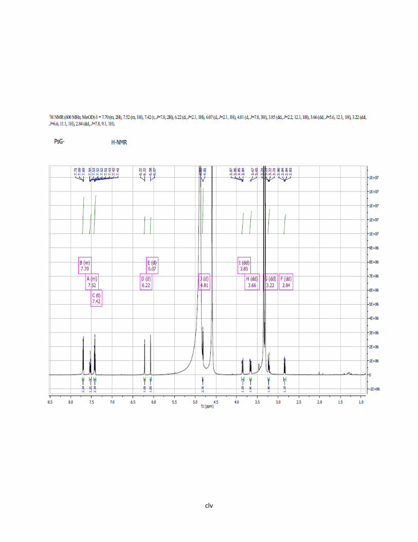
clv

clvi
Appendix 3: H-NMR Spectrum (600MHz; MeOD) of Compound I

clvii
Appendix 4: H-NMR Spectrum (600MHz; DMSO-d6) of Compound I

clviii

clix
Appendix 5: 2-D COSY of Compound I
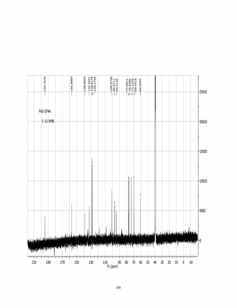
clx

clxi
Appendix 6: C-13 NMR Spectrum of Compound I

clxii
Appendix 7: HMQC Spectrum of Compound I

clxiii

clxiv
Appendix 8: HMBC Spectrum of Compound I

clxv

clxvi
Appendix 9: ESI-MS /UV Spectra Compound II

clxvii
Appendix 10: H-NMR Spectrum (500MHz;MeOD) of Compound II

clxviii

clxix
Appendix 11: H-NMR Spectrum (600MHz; DMSO) of Compound II

clxx
Appendix 12: 2-D COSY of Compound II

clxxi
Appendix 13: C-13 NMR Spectrum of Compound II
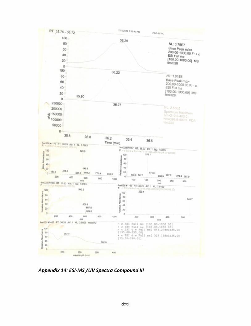
clxxii
Appendix 14: ESI-MS /UV Spectra Compound III

clxxiii
Appendix 15: H-NMR Spectrum (500MHz; MeOD) of Compound III

clxxiv
Appendix 16: H-NMR Spectrum (600MHz; DMSO) of Compound III

clxxv
Appendix 17: 2-D COSY of Compound III

clxxvi
Appendix 18: C-13 NMR Spectrum of Compound III

clxxvii
Appendix 19: HMQC Spectrum of Compound III

clxxviii
Appendix 20: HMBC Spectrum of Compound III
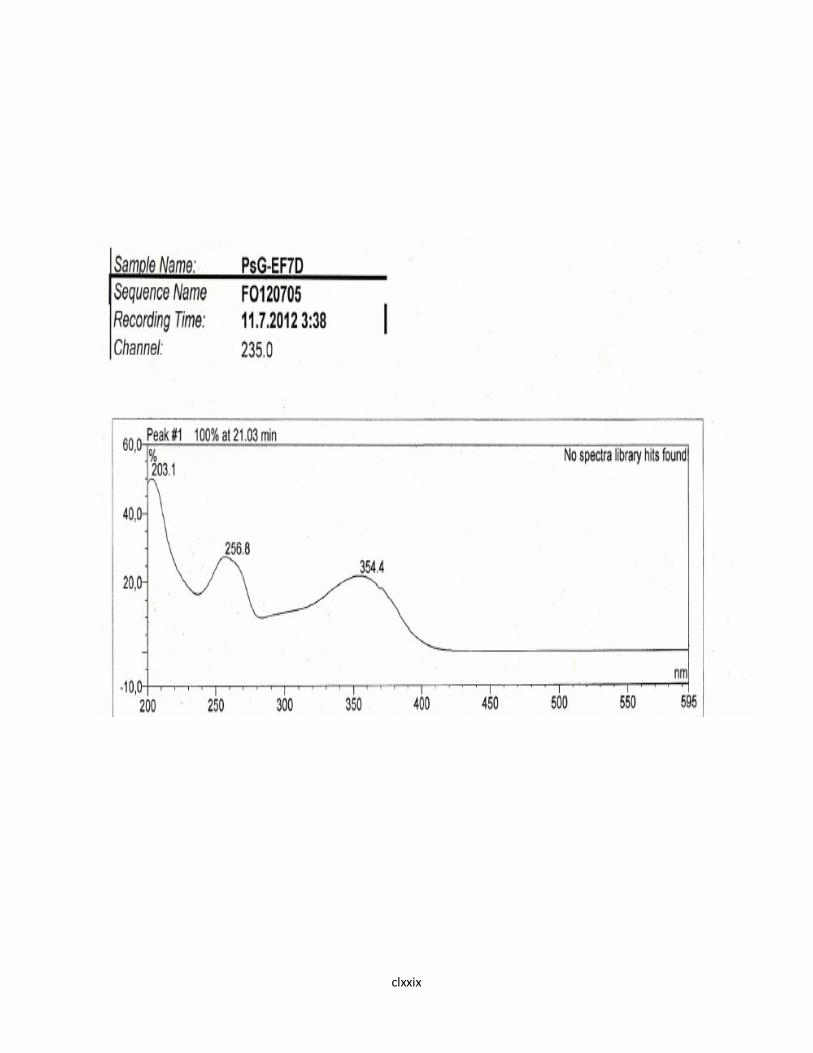
clxxix

clxxx
Appendix 21: UV Spectrum of Compound IV

clxxxi
Appendix 22: ESI-MS Spectra of Compound IV

clxxxii
Appendix 23: H-NMR Spectrum (500MHz; MeOD) of Compound IV

clxxxiii
Appendix 24: 2-D COSY of Compound IV

clxxxiv
Appendix 25: H-NMR Spectrum (500MHz; MeOD) of Compound V

clxxxv
Appendix 26: 2-D COSY of Compound V

clxxxvi

clxxxvii
Appendix 27: 2-D COSY [aromatic region] of Compound V


