
Pharmaceutical Applications of Carbon Nanotube-Mediated ...ijpsnonline.com/Issues/1685_full.pdf ·...
12
Review Article Pharmaceutical Applications of Carbon Nanotube-Mediated Drug Delivery Systems S. Pradeep Kumar 1 *, D. Prathibha 1 , N.L. Gowri Shankar 2 , R. Parthibarajan 3 , L. Mastyagiri 3 and M. Shankar 3 1 Department of Pharmacology, 2 Department of Pharmacognosy and 3 Department of Pharmaceutics, Swami Vivekananda Institute of Pharmaceutical Sciences, Vangapally Village, Yadagirigutta Mandal, Nalgonda District, Andhra Pradesh, India Received; December 18, 2011; accepted April 30, 2012 ABSTRACT Carbon nanotubes, which are elongated fullerenes, resemble graphite sheets wrapped into cylinders with a high length-to- width ratio (few nm in diameter and up to 1 mm in length). Carbon nanotubes are molecular-scale tubes of graphitic carbon with outstanding properties. Carbon nanotubes have drawn great interest and attraction in the field of novel drug delivery system. Nanomedicines can target, diagnose, monitor and treat cancerous cell also. The small nanoscale dimension and astonishing properties make them a distinctive carrier with a wide range of promising applications. These cylindrical carbon molecules have novel properties that make them potentially useful in many applications in nanotechnology. The various nano-size carrier systems are available for biotechnological applications including the drug delivery. Carbon nanotubes are typically used for bioactive delivery due to their some unique outstanding properties. Carbon nanotubes drug delivery system opens up new potential and possibilities over nanoparticles, dendrimers, liposomes etc. for biomedical applications and new drug delivery. In last few years, Carbon nanotubes (CNTs) have shown unexpected advantages in the field of cancer treatment and drug delivery systems. Present review article discuss in brief about the methods of synthesis, with purification as well as sorting techniques for giving different grades to different types of CNTs and biomedical applications. These show very good adsorption properties which helps in the detection of various chemicals, toxic agents etc. Research done using CNTs for cancer treatment is also discussed in brief. KEYWORDS: Chemical vapour deposition (CVD); magnetic purification; genetic engineering; biomedical applications. Introduction Carbon nanotubes were 'discovered' in 1991 by Sumio Iijima of NEC and are effectively long, thin cylinders of graphite. Nanotechnology is widely seen as a key technology of the 21 st century with a potential wide impact on many key sectors of the industry. Nanotechnology is likely to revolutionize our lives, in general, and health scenario, in particular. It is an emerging discipline that encompasses an increasingly sophisticated ability to manipulate matter at the nanoscale (0.1 nm to 1000 nm), resulting in new materials, products, and devices that demonstrate new and unusual behavior. It is one of the most important research and development areas in modern science. Apart from this, nanomaterials, nanoparticles, and nanocomposites used for biomedical purposes constitute a burgeoning new field called nanomedicine, which implies the medical application of nanotechnology and related research leading to the designing, testing, and optimizing of the pharmaceutical formulations. Nanotechnology is an applicable aspect of a broader area of nanoscience, which is one of the upcoming and highly challenging as well as a rewarding key research area in the modern scientific set-up. It is the science of small particles having unique properties which change upon altering the size of the particles. As nanotechnology is a very broad term, there are many disparate but sometimes overlapping subfields that could fall under its umbrella. It is a highly multidisciplinary field, drawing from fields such as applied physics, materials science, colloidal science, device physics and supramolecular chemistry. Nanotechnology, a contemporary discipline, has emerged in the field of cell biology in the form of nanosized particles such as liposomes to dendrimers and quantum dots to carbon-based nanoparticles including, fullerenes and carbon nanotubes (CNTs), also known as bucky tubes. Graphite is made up of layers of carbon atoms arranged in a hexagonal lattice (see Figure 1). Among the various nanomaterials being currently developed CNTs are often distinguished due to their great properties and the potential benefits they can deliver in many industrial applications (from materials engineering and electronics to medical devices and drug delivery systems). Nanoparticles are solid colloidal particles ranging in size from 10 nm to 1000 nm (1 micron). They consist of macromolecular materials in which the active principle is dissolved, entrapped or encapsulated and/or to which the active principle is International Journal of Pharmaceutical Sciences and Nanotechnology Volume 5 • Issue 2• July – September 2012 MS ID: IJPSN-12-18-11-KUMAR 1685
Transcript of Pharmaceutical Applications of Carbon Nanotube-Mediated ...ijpsnonline.com/Issues/1685_full.pdf ·...
K.S.C. Chaitanya et al: Development of Promethazine Hydrochloride Mucoadhesive Patches for Buccal Delivery: …1685
Review Article
Pharmaceutical Applications of Carbon Nanotube-Mediated Drug Delivery Systems
S. Pradeep Kumar1*, D. Prathibha1, N.L. Gowri Shankar2, R. Parthibarajan3, L. Mastyagiri3 and M. Shankar3
1Department of Pharmacology, 2Department of Pharmacognosy and 3Department of Pharmaceutics, Swami Vivekananda Institute of Pharmaceutical Sciences, Vangapally Village, Yadagirigutta Mandal, Nalgonda District, Andhra Pradesh, India
Received; December 18, 2011; accepted April 30, 2012 ABSTRACT Carbon nanotubes, which are elongated fullerenes, resemble graphite sheets wrapped into cylinders with a high length-to-width ratio (few nm in diameter and up to 1 mm in length). Carbon nanotubes are molecular-scale tubes of graphitic carbon with outstanding properties. Carbon nanotubes have drawn great interest and attraction in the field of novel drug delivery system. Nanomedicines can target, diagnose, monitor and treat cancerous cell also. The small nanoscale dimension and astonishing properties make them a distinctive carrier with a wide range of promising applications. These cylindrical carbon molecules have novel properties that make them potentially useful in many applications in nanotechnology. The various nano-size carrier systems are available for biotechnological applications including the drug delivery.
Carbon nanotubes are typically used for bioactive delivery due to their some unique outstanding properties. Carbon nanotubes drug delivery system opens up new potential and possibilities over nanoparticles, dendrimers, liposomes etc. for biomedical applications and new drug delivery. In last few years, Carbon nanotubes (CNTs) have shown unexpected advantages in the field of cancer treatment and drug delivery systems. Present review article discuss in brief about the methods of synthesis, with purification as well as sorting techniques for giving different grades to different types of CNTs and biomedical applications. These show very good adsorption properties which helps in the detection of various chemicals, toxic agents etc. Research done using CNTs for cancer treatment is also discussed in brief.
KEYWORDS: Chemical vapour deposition (CVD); magnetic purification; genetic engineering; biomedical applications.
Introduction Carbon nanotubes were 'discovered' in 1991 by Sumio Iijima of NEC and are effectively long, thin cylinders of graphite. Nanotechnology is widely seen as a key technology of the 21st century with a potential wide impact on many key sectors of the industry. Nanotechnology is likely to revolutionize our lives, in general, and health scenario, in particular. It is an emerging discipline that encompasses an increasingly sophisticated ability to manipulate matter at the nanoscale (0.1 nm to 1000 nm), resulting in new materials, products, and devices that demonstrate new and unusual behavior. It is one of the most important research and development areas in modern science. Apart from this, nanomaterials, nanoparticles, and nanocomposites used for biomedical purposes constitute a burgeoning new field called nanomedicine, which implies the medical application of nanotechnology and related research leading to the designing, testing, and optimizing of the pharmaceutical formulations. Nanotechnology is an applicable aspect of a broader area of nanoscience, which is one of the upcoming and highly challenging as well as a rewarding key research area in the modern scientific set-up. It is the science of small
particles having unique properties which change upon altering the size of the particles. As nanotechnology is a very broad term, there are many disparate but sometimes overlapping subfields that could fall under its umbrella. It is a highly multidisciplinary field, drawing from fields such as applied physics, materials science, colloidal science, device physics and supramolecular chemistry.
Nanotechnology, a contemporary discipline, has emerged in the field of cell biology in the form of nanosized particles such as liposomes to dendrimers and quantum dots to carbon-based nanoparticles including, fullerenes and carbon nanotubes (CNTs), also known as bucky tubes. Graphite is made up of layers of carbon atoms arranged in a hexagonal lattice (see Figure 1). Among the various nanomaterials being currently developed CNTs are often distinguished due to their great properties and the potential benefits they can deliver in many industrial applications (from materials engineering and electronics to medical devices and drug delivery systems). Nanoparticles are solid colloidal particles ranging in size from 10 nm to 1000 nm (1 micron). They consist of macromolecular materials in which the active principle is dissolved, entrapped or encapsulated and/or to which the active principle is
International Journal of Pharmaceutical Sciences and Nanotechnology
Volume 5 • Issue 2• July – September 2012MS ID: IJPSN-12-18-11-KUMAR
1685

1686 Int J Pharm Sci Nanotech Vol 5; Issue 2 • July−September 2012
adsorbed or attached and can be used for therapeutic disease management, silicon chip, bio-medical applications etc. Carbon nanotubes are hollow cylinders of carbon atoms. Their appear as rolled tubes of graphite such that their walls are hexagonal carbon rings and are often formed in large bundles (Figure 1).
Fig. 1. Layer structure of graphite.
Carbon nanotubes have become strongest candidates mainly in the field of biomedical engineering, biotechnology, defence research and pharmaceutical nanotechnology after their discovery in 1991. These are an important new class of technological materials that have numerous novel and useful properties. They have received very much attention as a new class of nanomaterials. These are the long hollow seamless cylinders (single walled as well as multiwalled carbon nanotubes) of graphene .The diameter of these tubes range from 1-100 nm. These tubes are normally capped with the half a full fullerence molecules at both the ends (Iijima et al., 1991, Strong et al., 2003).
In general, there are two types of CNTs: single-walled carbon nanotubes (SWNTs) and multi-walled carbon nanotubes (MWNTs). As their names imply, SWNTs consist of a single, cylindrical graphene layer, whereas MWNTs consist of multiple graphene layers telescoped about one another.
Structure Carbon nanotubes are allotropes composed entirely of
carbon in the form of a hallow sphere, ellipsoid, or tube, consisting of carbon atoms bonded to each other via sp2 bonds (C‐C distance of 1.4 Å) which are stronger than sp and sp3 bonds rendering the CNT’s excellent mechanical strength as well as high electrical and thermal conductivity. CNTs belong to the fullerene family of carbon allotropes, particularly those which have high
aspect ratio. These are hollow cylinders consisting of a hexagonal arrangement of sp2‐hybridized carbon atoms formed by rolling single or multiple layers of graphene sheets into seamless cylinders (Dresselhaus et al., 2004). These cylindrical structures have two forms: single‐walled carbon nanotubes (SWNTs) and multi-walled carbon nanotubes (MWNTs). SWNTs are composed of a single cylindrical graphene layer capped at both ends in a hemispherical arrangement of carbon networks. The inclusion of pentagonal and heptagonal C‐C structures during the growth process enables the closure of the cylinder. MWNTs comprise from several up to tens of concentric cylinders of graphitic shells, each one forming a SWNT. Schematic illustrations of the structures of carbon nanotubes are shown in Figure 2.
Fig. 2. Schematic illustrations of the structures of (A) armchair, (B) zigzag, and (C) chiral SWNTs. (D) Tunneling electron microscope image (72) showing the helical structure of a 1.3-nm-diameter chiral SWNT. (E) Transmission electron microscope (TEM) image of a MWNT containing a concentrically nestedarray of nine SWNTs. (F) TEM micrograph (18) showing the lateral packing of 1.4-nm-diameter SWNTs in a bundle. (G) Scanning electron microscope (SEM) image of an array of MWNTs grown as a nanotube forest.
MWNTs generally have a larger outer diameter (2.5-100 nm) than SWNTs (0.6-2.4 nm) and consist of a varying number of concentric SWNT layers. The interlayer separation between the SWNT layers of MWNT is about 0.34 nm. SWNTs have a better defined diameter, whereas MWNTs are more likely to have structural defects. Owing to its structure, MWNTS are a less stable nanostructure (Joselevich E et al., 2004). Both possess a high tensile strength, are ultra‐light weight, and have excellent chemical and thermal stability (Smart et al., 2006). CNTs have the ability to buckle and collapse reversibly due to high stiffness and resilience. The high C‐C bond stiffness of the hexagonal network produces an

Kumar et al: Pharmaceutical Applications of Carbon Nanotube-Mediated Drug Delivery Systems 1687
axial Young's modulus of approximately 1 TPa and a tensile strength of 150 GPa, making CNTs one of the stiffest materials known, yet with the capacity to deform elastically under compression (Trotter et al., 2005).
Classification of Carbon Nanotubes 1. Single-walled 2. Multi-walled 3. Double-walled 4. Torus 5. Fullerene 6. Nanobud 7. Functionalized CNTs
Types of CNTs There are several types of CNTs. They are as follows:
Single-walled carbon nanotube (SWNT) Single-walled nanotubes (SWNT) have a diameter of
about 1-10 nanometers, with a tube length that can be many thousands of times larger (Figure 3). The structure of a SWNT can be conceptualised by wrapping a one-atom-thick layer of graphite called graphene into a seamless cylinder.
Fig.3. 3D Representation of a Single Walled Carbon Nanotube (SWNT).
Double-wall Nanotubes (DWNT)
These materials combine similar morphology and other properties of SWNT, while significantly improving their resistance to chemicals. Double-wall nanotubes are ideal systems for studying the interwall interactions influencing the properties of nanotubes with two or more walls. This property is especially important when functionality is required to add new properties to the nanotube. Since DWNT are a synthetic blend of both SWNT and MWNT, they exhibit the electrical and thermal stability of the latter and the flexibility of the former (Alexander et al., 2009) (Figure 4).
Fig. 4. Double-wall Nanotubes.
Multi-walled carbon nanotube (MWNT)
Multi-walled nanotubes (MWNT) consist of multiple layers of graphite rolled in on themselves to form a tube shape (Figure 5). There are two models which can be used to describe the structures of multiwalled nanotubes. In the Russian Doll model, sheets of graphite are arranged in concentric cylinders, i.e. a single-walled nanotube (SWNT) within a larger single-walled nanotube. In the Parchment model, a single sheet of graphite is rolled in around itself, resembling a scroll of parchment or a rolled up newspaper. The MWNT's are much stiffer than the SWNT's, especially in compression.
Fig. 5. 3D Representation of a Multiple Walled Carbon Nanotube (MWNT).
Nanotorus
A nanotorus is a theoretically described carbon nanotube bent into a torus (donut shape). Nanotori have many unique properties, such as large magnetic moments, and thermal stability, which vary widely depending on the radius of the torus and radius of the tube.
Nanobud
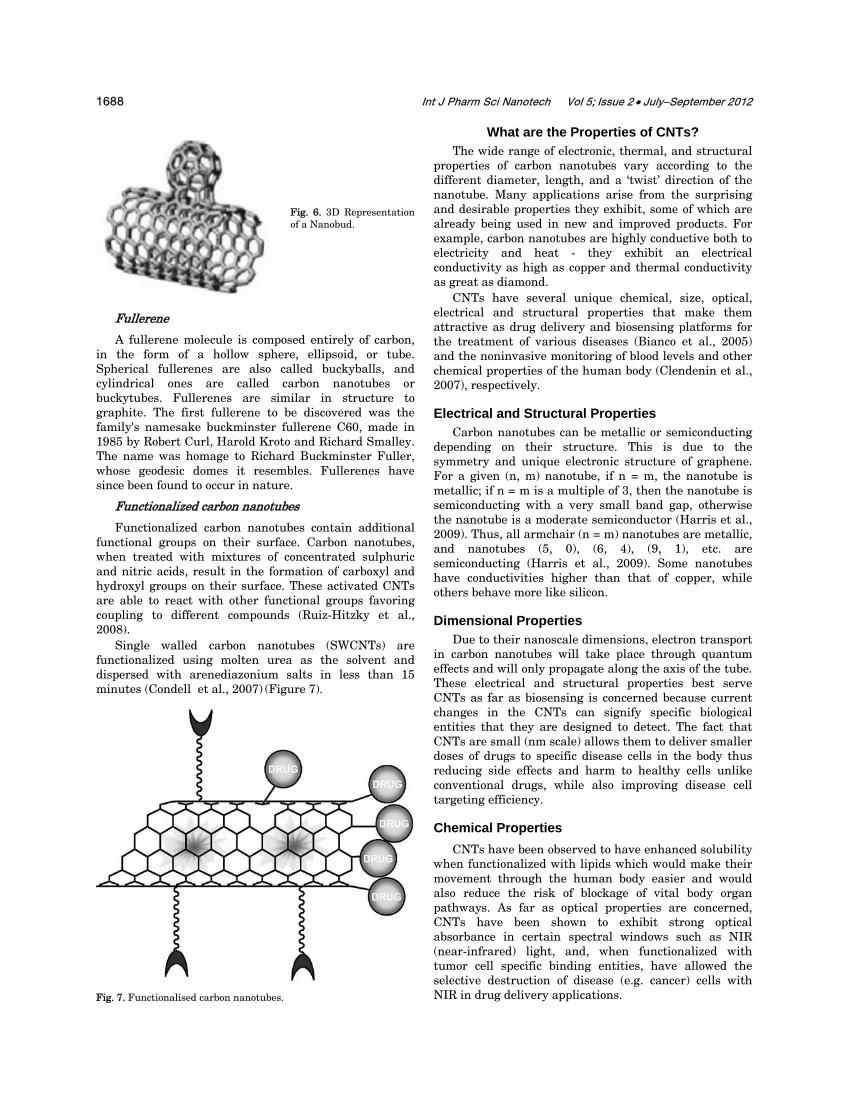
Carbon nanobuds are a newly discovered material combining two previously discovered allotropes of carbon: carbon nanotubes and fullerenes. In this new material, fullerene-like ‘buds’ are covalently bonded to the outer sidewalls of the underlying carbon nanotube (Figure 6). This hybrid material has useful properties of both fullerenes and carbon nanotubes.
1688 Int J Pharm Sci Nanotech Vol 5; Issue 2 • July−September 2012
Fig. 6. 3D Representation of a Nanobud.
Fullerene
A fullerene molecule is composed entirely of carbon, in the form of a hollow sphere, ellipsoid, or tube. Spherical fullerenes are also called buckyballs, and cylindrical ones are called carbon nanotubes or buckytubes. Fullerenes are similar in structure to graphite. The first fullerene to be discovered was the family's namesake buckminster fullerene C60, made in 1985 by Robert Curl, Harold Kroto and Richard Smalley. The name was homage to Richard Buckminster Fuller, whose geodesic domes it resembles. Fullerenes have since been found to occur in nature.
Functionalized carbon nanotubes
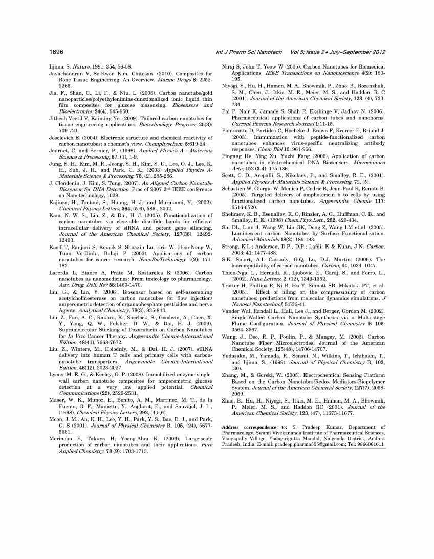
Functionalized carbon nanotubes contain additional functional groups on their surface. Carbon nanotubes, when treated with mixtures of concentrated sulphuric and nitric acids, result in the formation of carboxyl and hydroxyl groups on their surface. These activated CNTs are able to react with other functional groups favoring coupling to different compounds (Ruiz-Hitzky et al., 2008).
Single walled carbon nanotubes (SWCNTs) are functionalized using molten urea as the solvent and dispersed with arenediazonium salts in less than 15 minutes (Condell et al., 2007) (Figure 7).
Fig. 7. Functionalised carbon nanotubes.
What are the Properties of CNTs? The wide range of electronic, thermal, and structural
properties of carbon nanotubes vary according to the different diameter, length, and a ‘twist’ direction of the nanotube. Many applications arise from the surprising and desirable properties they exhibit, some of which are already being used in new and improved products. For example, carbon nanotubes are highly conductive both to electricity and heat - they exhibit an electrical conductivity as high as copper and thermal conductivity as great as diamond.
CNTs have several unique chemical, size, optical, electrical and structural properties that make them attractive as drug delivery and biosensing platforms for the treatment of various diseases (Bianco et al., 2005) and the noninvasive monitoring of blood levels and other chemical properties of the human body (Clendenin et al., 2007), respectively.
Electrical and Structural Properties Carbon nanotubes can be metallic or semiconducting
depending on their structure. This is due to the symmetry and unique electronic structure of graphene. For a given (n, m) nanotube, if n = m, the nanotube is metallic; if n = m is a multiple of 3, then the nanotube is semiconducting with a very small band gap, otherwise the nanotube is a moderate semiconductor (Harris et al., 2009). Thus, all armchair (n = m) nanotubes are metallic, and nanotubes (5, 0), (6, 4), (9, 1), etc. are semiconducting (Harris et al., 2009). Some nanotubes have conductivities higher than that of copper, while others behave more like silicon.
Dimensional Properties Due to their nanoscale dimensions, electron transport
in carbon nanotubes will take place through quantum effects and will only propagate along the axis of the tube. These electrical and structural properties best serve CNTs as far as biosensing is concerned because current changes in the CNTs can signify specific biological entities that they are designed to detect. The fact that CNTs are small (nm scale) allows them to deliver smaller doses of drugs to specific disease cells in the body thus reducing side effects and harm to healthy cells unlike conventional drugs, while also improving disease cell targeting efficiency.
Chemical Properties CNTs have been observed to have enhanced solubility
when functionalized with lipids which would make their movement through the human body easier and would also reduce the risk of blockage of vital body organ pathways. As far as optical properties are concerned, CNTs have been shown to exhibit strong optical absorbance in certain spectral windows such as NIR (near-infrared) light, and, when functionalized with tumor cell specific binding entities, have allowed the selective destruction of disease (e.g. cancer) cells with NIR in drug delivery applications.

Kumar et al: Pharmaceutical Applications of Carbon Nanotube-Mediated Drug Delivery Systems 1689
Fig. 8. Arc discharge apparatus for CNTs.
Methods of Production
A. ARC Discharge Method The arc discharge was the first available method for
the production of both SWNTs and MWNTs. This method creates nanotubes through arc-vaporisation of two carbon rods placed end-to-end, separated by approximately 1 mm, in an enclosure that is usually filled with inert gas (helium, argon) at low pressure (between 50 and 700 mbar). Recent investigations have shown that it is also possible to create nanotubes with the arc method in liquid nitrogen (Jung et al., 1992). A direct current of 50
to 100 A driven by approximately 20 V creates a high temperature discharge between the two electrodes. The discharge vaporises one of the carbon rods and forms a small rod shaped deposit on the other rod. Producing nanotubes in high yield depends on the uniformity of the plasma arc and the temperature of the deposit form on the carbon electrode (Ebbesen, T. W. et al., 1992). Depending on the exact technique, it is possible to selectively grow SWNTs or MWNTs, which is shown in Figure 8. Two distinct methods of synthesis can be performed with the arc discharge apparatus.
Differences between SWNTs and MWNTs.
SWNTs MWNTs 1. Single layer of graphene. 1. Multiple layers of graphene. 2. Catalyst is required for synthesis. 2. It can be produced without a catalyst. 3. Bulk synthesis is difficult as it requires proper
control over growth and atmospheric conditions. 3. Bulk synthesis is easy.
4. It can be easily twisted and is more pliable. 4. It cannot be easily twisted. 5. A chance of producing defects is more during
functionalazation. 5. A chance of producing defects is less, but once
occurred it is difficult to improve. 6. Purity is poor. 6. Purity is high. 7. Characterization and evaluation is easy. 7. Characterization and evaluation has very
complex structure. 8. Less accumulation in the body. 8. More accumulation in the body.
Synthesis of SWNTs
If SWNTs are preferable, the anode has to be doped with metal catalyst, such as Fe, Co, Ni, Y or Mo. A lot of elements and mixtures of elements have been tested by various authors (Journet et al., 1998) and it is noted that the results vary a lot, even though they use the same elements. This is not surprising as experimental conditions
differ. The quantity and quality of the nanotubes obtained depend on various parameters such as the metal concentration, inert gas pressure, the kind of gas, the current, and system geometry. Usually the diameter ranges from 1.2 to 1.4 nm. A few ways to improve the process of arc discharge are,
(a) Inert gas (b) Optical plasma control

1690 Int J Pharm Sci Nanotech Vol 5; Issue 2 • July−September 2012
(c) Catalyst (d) Improvement of oxidation resistance (e) Open air synthesis with welding arc torch
Synthesis of MWNTs
If both electrodes are graphite, the main product will be MWNTs. But next to MWNTs, a lot of side products are formed such as fullerenes, amorphous carbon, and some graphite sheets. Purifying the MWNTs means loss of structure and disorders of the walls. However scientists are developing ways to gain pure MWNTs in a large-scale process without purification. Typical sizes for MWNTs are an inner diameter of 1-3 nm and an outer diameter of approximately 10 nm. Because no catalyst is involved in this process, there is no need for a heavy acidic purification step. This means, the MWNT can be synthesised with a low amount of defects.
(a) Synthesis in liquid nitrogen (b) Magnetic field synthesis (c) Plasma rotating arc discharge
B. Laser Ablation Method Another method to grow SWNTs using laser ablation
was demonstrated in 1996 by Smalley’s group and has prompted a lot of interest. A pulsed (Yudasaka et al., 1999, Eklund et al., 2002) or continuous laser (Maser et al., 1998; Bolshakov et al., 2002) is used to vaporise a graphite target in an oven at 1200°C. The main difference between continuous and pulsed laser is that the pulsed laser demands a much higher light intensity (100 kW/cm2 compared with 12 kW/cm2). The oven is filled with helium or argon gas in order to keep the pressure at 500 Torr. A very hot vapour plume forms, expands and then cools rapidly. As the vaporised species cool, small carbon molecules and atoms quickly condense to form larger clusters, possibly including fullerenes. The catalysts also begin to condense, but more slowly at first, and attach to carbon clusters and prevent their closing into cage structures. Catalysts may even open cage structures when they attach to them. From these initial clusters, tubular molecules grow into single-wall carbon nanotubes until the catalyst particles become too large, or until conditions have cooled sufficiently that carbon no longer can diffuse through or over the surface of the catalyst particles. It is also possible that the particles become so coated with a carbon layer that they cannot absorb anymore and the nanotube stops growing. The SWNTs formed in this case are bundled together by van der Waals forces (Scott et al., 2001).
There are some striking, but not exact, similarities in the comparison of the spectral emission of excited species in laser ablation of a composite graphite target with that of laser-irradiated C60 vapour. This suggests that fullerenes are also produced by laser ablation of catalyst-filled graphite, as is the case when no catalysts are included in the target. However, subsequent laser pulses excite fullerenes to emit C2 that adsorbs on catalyst particles and feeds SWNT growth. However, there is insufficient evidence to conclude this with certainty
Laser ablation is almost similar to arc discharge, since the optimum background gas and catalyst mix is the same as in the arc discharge process. This might be due to the similar reaction conditions needed, and the reactions probably occur with the same mechanism.
Fig. 9. Laser ablation apparatus.
Large scale synthesis of SWNT
Because of the good quality of nanotubes produced by this method, scientists are trying to scale up laser ablation. However the results are not yet as good as for the arc-discharge method, but they are still promising. The two newest developments on large-scale synthesis of SWNTs are listed here:
1. Ultra fast Pulses from a free electron laser (FEL) method (Eklund et al., 2002),
2. Continuous wave laser-powder method. (Bolshakov et al., 2002).
Scaling up is possible, but the technique is rather expensive due to the laser and the large amount of power required.
C. Chemical Vapor Deposition Method (CVD)
Chemical vapour deposition CVD is known as irreversible deposition of a solid from a gas or a mixture of gases through a heterogeneous chemical reaction. This reaction takes place at the interface of gas-solid substrate, and, depending on the deposition conditions, the growth process can be controlled either by diffusion or by surface kinetics (Vander Wal et al., 2002, Endo et al., 1993).
It is carried out in two step process:- • Catalyst is deposited on substrate and then
nucleation of catalyst is carried via chemical etching or thermal annealing. Ammonia is used as an etchant. Metal catalysts used are Ni, Fe or Co.
• Carbon source is then placed in gas phase in a reaction chamber. Then, the carbon molecule is converted to an atomic level by using an energy source like plasma or heated coil. This carbon will diffuse towards a substrate, which is coated with a

Kumar et al: Pharmaceutical Applications of Carbon Nanotube-Mediated Drug Delivery Systems 1691
catalyst and nanotubes grow over this metal catalyst. The carbon source used is methane, carbon monoxide or acetylene. The temperature
used for synthesizing the nanotube is in the 650-9000 C range. The typical yield is 30%.
Short summary of the three most common techniques
Method Arc discharge method
Chemical vapour deposition
Laser ablation (vaporization)
Who
Ebbesen and Ajayan, NEC, Japan 1992
Endo, Shinshu University, Nagano, Japan (Endo et al., 1993)
Smalley, Rice,1995
How
Connect two graphite rods to a power supply, place them a few millimetres apart, and throw the switch. At 100 amps, carbon vaporises and forms a hot plasma.
Place the substrate in oven, heat to 600oC, and slowly add a carbon-bearing gas such as methane. As the gas decomposes it frees up carbon atoms, which recombine in the form of NTs.
Blast graphite with intense laser pulses; use the laser pulses rather than electricity to generate carbon gas from which the NTs form; try various conditions until one condition produces prodigious amounts of SWNTs.
Typical yield 30 to 90% 20 to 100% Up to 70%
SWNT
Short tubes with diameters of 0.6-1.4 nm
Long tubes with diameters ranging from 0.6-4 nm
Long bundles of tubes (5-20 microns), with individual diameter from 1-2 nm.
MWNT
Short tubes with inner diameter of 1-3 nm and outer diameter of approxi-mately 10 nm
Long tubes with diameter ranging from 10-240 nm
Not very much interest in this technique, as it is too expensive, but MWNT synthesis is possible.
Purification of CNTs A large problem with nanotube application is, next to
large-scale synthesis, the purification. The as-produced CNTs contain a lot of impurities. The main impurities in the soot are graphite (wrapped-up) sheets, amorphous
carbon, metal catalyst and the smaller fullerenes. These impurities will interfere with most of the desired properties of the CNTs. Basic steps are drawn in Figure 10.
Produced CNTs (With impurities, i.e. catalyst, graphite carbon)
Purification
Sorting
Length based Band based Diameter based
Purified CNTs Fig. 10. Basic steps involved in purification of CNTs.

1692 Int J Pharm Sci Nanotech Vol 5; Issue 2 • July−September 2012
Acid Treatment In general, the acid treatment will remove the metal
catalyst. First of all, the surface of the metal must be exposed by oxidation or sonication. The metal catalyst is then exposed to acid and solvated. The SWNTs remain in suspended form. When using a treatment in HNO3, the acid only has an effect on the metal catalyst. It has no effect on the SWNTs and other carbon particles (Borowiak-Palen, E. et al., 2005, Hou et al., 2001, Moon, J. M. et al., 2001). If a treatment in HCl is used, the acid has also a little effect on the SWNTs and other carbon particles. The mild acid treatment (Georgakilas et al., 2002) (4 M HCl reflux) is basically the same as the HNO3 reflux, but here the metal has to be totally exposed to the acid to solvate it.
Ultrasonication In this technique particles are separated due to
ultrasonic vibrations. Agglomerates of different nanoparticles will be forced to vibrate and will become more dispersed. The separation of the particles is highly dependable on the surfactant, solvent and reagent used. The solvent influences the stability of the dispersed tubes in the system. In poor solvents, the SWNTs are more stable if they are still attached to the metal. But in some solvents, such as alcohols, monodispersed particles are relatively stable (Thien-Nga et al., 2002). When an acid is used, the purity of the SWNTs depends on the exposure time. When the tubes are exposed to the acid for a short time, only the metal solvates, but for a longer exposure time, the tubes will also be chemically cut.
Magnetic Purification In this method ferromagnetic (catalytic) particles are
mechanically removed from their graphitic shells (Niyogi et al., 2001). The SWNTs suspension is mixed with inorganic nanoparticles (mainly ZrO2 or CaCO3) in an ultrasonic bath to remove the ferromagnetic particles. Then, the particles are trapped with permanent magnetic poles. After a subsequent chemical treatment, a high purity SWNT material will be obtained.
This process does not require large equipment and enables the production of laboratory-sized quantities of SWNTs containing no magnetic impurities (Figure 11).
Chromatography This technique is mainly used to separate small
quantities of SWNTs into fractions with small length and diameter distribution. The SWNTs are run over a column with a porous material, through which the SWNTs will flow. The columns used are GPC (Gel Permeation Chromatography) and HPLC-SEC (High Performance Liquid Chromatography - Size Exclusion Chromato-graphy) columns. The number of pores the SWNTs will flow through depends on their size. This means that, the smaller the molecule, the longer the pathway to the end of the column will be and that the larger molecules will come off first. The pore size will control what size
distribution can be separated. However, a problem is that the SWNTs have to be dispersed or solvated. This can be done by ultrasonication or functionalisation with soluble groups (Zhao et al., 2001).
Fig. 11. Schematic diagram of the apparatus for magnetic purification.
Applications of CNTs Various applications of CNTs are as follows:
1. Carrier for Drug delivery (a) Carbon nanohorns (CNHs) are the
spherical aggregates of CNTs with irregular horn like shape. Research studies have proved CNTs and CNHs as a potential carrier for drug delivery system.
(b) Functionalized carbon nanotubes are reported for targeting of Amphotericin B to Cells.
(c) Cisplatin incorporated oxidized SWNHs have showed slow release of Cisplatin in aqueous environment. The released Cisplatin had been effective in terminating the growth of human lung cancer cells, while the SWNHs alone did not show anticancer activity.
(d) The anticancer drug Polyphosphazene platinum, given with nanotubes, had enhanced permeability, distribution and retention in the brain due to controlled lipophilicity of nanotubes (Pai et al., 2001).
(e) The antibiotic Doxorubicin, given with nanotubes, is reported to have an enhanced intracellular penetration (Pantarotto et al., 2003).
(f) The gelatin CNT mixture (hydrogel) has been used as potential carrier system for biomedicals.

Kumar et al: Pharmaceutical Applications of Carbon Nanotube-Mediated Drug Delivery Systems 1693
(g) CNT-based carrier system can offer a successful oral alternative administration of Erythropoietin (EPO), which has not been possible so far because of the denaturation of EPO by the gastric environment conditions and enzymes.
(h) They can be used as lubricants or glidants in tablet manufacturing due to nanosize and sliding nature of graphite layers bound with vander Waals forces.
2. Genetic Engineering In genetic engineering, CNTs and CNHs are used
to manipulate genes and atoms in the development of bioimaging genomes, proteomics and tissue engineering. The unwound DNA (single-stranded) winds around SWNT by connecting its specific nucleotides and causes a change in its electrostatic property. This creates its potential application in diagnostics (polymerase chain reaction) and in therapeutics. Wrapping of carbon nanotubes by single-stranded DNA was found to be sequence-dependent, and hence can be used in DNA analysis. Nanotubes due to their unique cylindrical structure and properties are used as a carrier for genes (gene therapy) to treat cancer and genetic disorders. Their tubular nature has proved them to be a vector in gene therapy. Nanotubes complexed with DNA were found to release DNA before it was destroyed by cellular defense system, boosting transfection significantly. Nanostructures have showed antiviral effect in respiratory syncytical virus (RSV), a virus with severe bronchitis and asthma. The treatment is generally done by combining nanoparticles and gene slicing technologies. Here, RNA fragments capable of inhibiting a protein (which is needed for virus multiplication) is encapsulated within nanotubes and administered in the form of nasal sprays or drops. The promising results have been noted inhibiting further growth of virus (Kam et al., 2005). Nanotubes are reported for helical crystallization of proteins and growth of embryonic rat brain neurons. Streptavidin protein is successfully immobilized on CNT via 1-pyrene butanoic acid and succinimidyl ester (Pai et al., 2006). Nanotubes and nanohorns can adhere to various antigens on their surface, hence acting as a source of antigen in vaccines. Hence, by use of nanotubes, use of dead bacteria as a source for antigen, which is sometimes dangerous, can be avoided.
3. Biomedical Applications CNTs for protein delivery
Various low molecular weight proteins can adsorb spontaneously on the sidewalls of acidoxidized single-walled carbon nanotubes (Liu et al., 2009). The proteins are found to be readily transported
inside mammalian cells with nanotubes acting as the transporter via the endocytosis pathway. This research was reported by the Dai group. The results show that streptavidin (SA) and cytochrome c (Cyt-c) could easily be transported into the cytoplasm of cells by the CNTs and become physiologically active in the cell. Carbon nanotubes could become new class of protein transporters for various in vitro and in vivo delivery applications.
CNTs for chemical delivery
Recently, the Dai group reported that using supramolecular π–π stacking to load a cancer chemotherapy agent doxorubicin (DOX) onto branched polyethylene glycol (PEG) functionalized SWNTs for in vivo drug delivery applications. It has been found that the surface of PEGylated SWNTs could be efficiently loaded with DOX by supramolecular π–π stacking. These methods offer several advantages for cancer therapy, including enhanced therapeutic efficacy and a marked reduction in toxicity compared with free DOX.
CNTs for HIV/AIDS therapy
Recently, Liu et al. (2009) have shown the delivery of siRNA molecules conjugated to CNT to human T cells and primary cells. The results show that nanotubes are capable of siRNA delivery to afford efficient RNAi of CXCR4 and CD4 receptors on human T cells and peripheral blood mononuclear cells (PBMCs). The siRNA sequences used in these studies are able to silence the expression of the cell-surface receptors CD4 and coreceptors CXCR4 necessary for HIV entry and infection of T cells. This work demonstrates that siRNA linked through cleavable disulfide bonds to lipid molecules coating CNTs can be efficiently delivered, leading to knockdown (about 60%) of the CD4 and CXCR4 expression. Furthermore, the siRNA–S–S–lipid coated CNT conjugates greatly improve the silencing in T cells compared with Lipofectamine 2000 and other liposome based transfection agents. Even if preliminary at this stage, these results indicate the potential use of CNTs for the treatment of HIV.
4. Detection of other biomolecules NADH
The electrochemical oxidation of NADH at the electrode surface has received considerable interest due to the need to develop amperometric biosensors for substrates of NAD+ dependent dehydrogenases. Dihydronicotinamide adenine dinucleotide (NADH) and its oxidized form, nicotinamide adenine dinucleotide (NAD+), are the key central charge carriers in living cells. However, the oxidation of NADH at a conventional solid electrode surface is highly

1694 Int J Pharm Sci Nanotech Vol 5; Issue 2 • July−September 2012
irreversible with considerable overpotentials, which limits the selectivity of the determination in a real sample. CNTs have been devoted to decreasing the high overpotential for NADH oxidation on carbon paste electrodes and microelectrodes (Wang et al., 2003). By integrating the hydrophilic ion-conducting matrix of CHITn with electron mediator toluidine blue O and CNTs, the produced NADH sensor shows very low oxidation overpotential and good analytical performance (Zhang et al., 2005).
Glucose
The detection of glucose in blood is one of the most frequent checks for human health, since some diseases are related to the blood glucose concentration. However, the direct electron transfer for oxidation of FADH2 or reduction of FAD is hard to realize at conventional electrodes, because the FAD is deeply seated in a cavity and not easily accessible for conduction of electrons from the electrode surface. Thus, many CNTs based nanohybrids, such as MWCNT/AuNPs/ionic liquid (Jia F. et al., 2008), SWCNT/GOD/Nafion (Lyons et al., 2008), polyaniline (PANI) - coated Fe3O4 nanoparticle/MWCNT, and palladium/SWCNT, have been explored to immobilize GOD for glucose biosensing. More interestingly, Willner’s group demonstrated that aligned reconstituted GOD on the edge of SWCNT as conductive nanoneedles can be linked to an electrode surface for fast glucose response.
Organophosphate pesticides
The rapid detection of these toxic agents in the environment and public places has become increasingly important for homeland security and health protection. The flow injection amperometric biosensor for OPs has been developed by assembling AChE on CNTsmodified GCE. Under optimal conditions, the biosensor has been used to measure paraoxon as low as 0.4 pM with a 6 minutes inhibition time (Liu et al., 2006).
H2O2
H2O2 is a product of the enzymatic reactions between most oxidases and their substrates. This detection is very interesting for the development of biosensors for oxidase substrates. The earlier work on the electrocatalytic action of CNTs toward H2O2 was reported at an apparently decreased overvoltage using the CNTs/Nafion-coated electrode. With the introduction of MWCNT, the polyaniline-PB/MWCNT hybrid system showed the synergy between the PANI-PB and MWCNT, which amplified the sensitivity greatly.
5. Artificial implants Normally, the body shows rejection reaction for
implants with the post administration pain. But,
miniature-sized nanotubes and nanohorns get attached with other proteins and amino acids, avoiding rejection. Also, they can be used as implants in the form of artificial joints without host rejection reaction. Moreover, due to their high tensile strength, carbon nanotubes filled with calcium and arranged/grouped in the structure of bone can act as bone substitutes.
6. Preservative Carbon nanotubes and nanohorns are
antioxidants in nature. Hence, they are used to preserve drug formulations prone to oxidation. Their antioxidant property is used in anti aging cosmetics and with zincoxide as a dermatological sunscreen to prevent oxidation of important skin components.
7. Catalyst Nanohorns offer a large surface area and, hence,
the catalyst at molecular level can be incorporated into nanotubes in large amount and simultaneously can be released in required rate at a particular time. Therefore, a reduction in the frequency and amount of catalyst addition can be achieved by using CNTs and CNHs.
8. Diagnosis 1. Carbon nanotubes can also be employed as a
powerful carrier to pre-concentrate enzymes or electroactive molecules for electrochemical sensing of DNA hybridization as a novel indicator (Pingang et al., 2006).
2. Multiwalled carbon nanotubes functionalized with europium-doped Y2O3 nanophosphors give rise to species that are luminescent in the visible-light range analysed by Z-contrast imaging and such species have potential applications in cancer diagnosis and treatment.
3. Carbon nanotubes improve cancer diagnosis through better protein array detection limits.
4. In recent trends, biological functionalized carbon nanotubes have their potential applications for breast cancer diagnostics (Kasif et al., 2005).
5. Carbon nanotube-filled, nanocomposite-derived catheters exhibited outstanding properties when compared with neat polymer-derived catheters, and it is envisaged that these system will be widely utilized in various medical devices.
6. Nanotube-filled microcatheters were confirmed by measuring the systematic T-cells as well as a histopathological exam.

Kumar et al: Pharmaceutical Applications of Carbon Nanotube-Mediated Drug Delivery Systems 1695
9. Drug discovery The critical bottlenecks in drug discovery may be
overcome by using arrays of CNT sensors and current information technology solutions for identification of genes and genetic materials for drug discovery and development (Niraj et al., 2005).
10. Tissue engineering 1. Polyurethane foams with CNT coating have
the potential to be used as bioactive scaffolds in bone tissue engineering due to their high interconnected porosity, bioactivity and nanostructured surface topography.
2. Carbon nanotubes can also be incorporated into scaffolds providing structural reinforcement as well as imparting novel properties such as electrical conductivity into the scaffolds, which may aid in directing cell growth (Harrison et al., 2007).
3. In cardiovascular surgeries remarkable improvements in mechanical strength of implanted catheters is brought by carbon nanotubes and reduces thrombogenesis after surgery.
4. Though challenges still exist, the addition of CNT to improve the mechanical properties of CTS and ceramic (HAp) composite would surely support and stimulate the function of natural bone (Jayachandran et al., 2003).
Limitations of CNTs • Lack of solubility in most solvents compatible
with the biological milieu (aqueous-based). • The production of structurally and chemically
reproducible batches of CNTs with identical characteristics (Lacerda et al., 2006).
• Difficulty in maintaining a high quality and minimal impurities.
Conclusions
In this review, we have made an effort to provide the most contemporary overview possible of synthesis, properties, and potential biomedical applications of CNTs. Single and multiple-walled carbon nanotubes have already proven to serve as safer and more effective alternatives to previous drug delivery methods. They can pass through membranes, carrying therapeutic drugs, vaccines, and nucleic acids deep into the cell to targets previously unreachable. The properties of CNTs are still being researched. Functionalized carbon nanotubes have already proven to serve as safer and more effective alternatives to previous drug delivery methods and as diagnostic tool.
The exceptional physical, mechanical, and electronic properties of CNTs allow them to be used in sensors, probes, actuators, nanoelectronic devices, and drug
delivery systems within biomedical applications. With an increasing interest shown by the nanotechnology research community in this field, it is expected that plenty of applications of CNTs will be explored in future.
References Alexander A. Green, Mark C. Hersam (2009). Processing and
properties of highly enriched double wall carbon nanotubes. Natures Nanotechnology; 4: 64-70.
A. Bianco, K. Kostarelos, M. Prato, (2005). Applications of carbon nanotubes in drug delivery” Current Opinion in Biotechnology 9, 674.
Bandow, Shunji, Rao, A. M., Williams, K. A., Thess, A., Smalley, R. E., and Eklund, P. C., 1997, Journal of Physical Chemistry B, 101, (44).
Barroug A, Glimcher M. (2002). Hydroxyapatite crystals as a local delivery system for cisplatin: adsorption and release of cisplatin in vitro. J Orthop Res; 20: 274-280.
Bolshakov, A. P., Uglov, S. A., Saveliev, A. V., Konov, V. I., Gorbunov, A. A., Pompe, W., and Graff, A (2002). Diamond and Related Materials, 11, (3-6).
Borowiak-Palen, E., Pichler, T., Liu, X., Knupfer, M., Graff, A., Jost, O., Pompe, W., Kalenczuk, R. J., and Fink, J., ( 2002). Chemical Physics Letters, 363, (5-6), 567-572.
P.J.F. Harris (2009), Carbon nanotube science: Synthesis, Properties and Applications, (Cambridge University Press, Cambridge).
Chiang, I. W., Brinson, B. E., Huang, A. Y., Willis, P. A., Bronikowski, M. J., Margrave, J. L., Smalley, R. E., and Hauge, R. H., (2001). Journal of Physical Chemistry B, 105, (35), 8297-8301.
Chiang, I. W., Brinson, B. E., Smalley, R. E., Margrave, J. L., and Hauge, R. H., (2001), Journal of Physical Chemistry B, 105, (6), 1157-1161.
Condell D. Doyle., James M. Tour. (2009), Environmentally friendly functionalization of single walled carbon nanotubes in moltern urea. J Carbon; 47(14): 3215-3218.
Dresselhaus MS, Dresselhaus G, Charlier JC and Hernandez E (2004). Electronic, thermal and mechanical properties of carbon nanotubes. Philos Transact A Math Phys Eng Sci; 362: 2065‐98.
Ebbesen, T. W. and Ajayan, P. M., (1992). Nature, 358, (220-222). Eklund, P. C., Pradhan, B. K., Kim, U. J., Xiong, Q., Fischer, J. E.,
Friedman, A. D., Holloway, B. C.,Jordan, K., and Smith, M. W., (2002). Nano Letters, 2, (6).
Endo, Morinobu, Takeuchi, Kenji, Igarashi, Susumu, Kobori, Kiyoharu, Shiraishi, Minoru, and Kroto, Harold W (1993). Journal of Physics and Chemistry of Solids, 54, (12).
Ewelina Z, Monika Bil, Ryszkowska J, Showan N Nazhat, Johann Cho, Oana B, Judith AR, Aldo RB (2009). Polyurethane foams electrophoretically coated with carbon nanotubes for tissue engineering scaffolds. Biomedical Materials 4(1).
Farkas, E., Anderson, M. E., Chen, Z. H., and Rinzler, A. G., (2002). Chemical Physics Letters, 363, (1-2), 111-116.
Georgakilas, Vasilios, Voulgaris, Dimitrios, Vazquez, Ester, Prato, Maurizio, Guldi, Dirk M., Kukovecz, Akos, and Kuzmany, Hans, (2002). Journal of the American Chemical Society, 124, (48).
Guo, T., Nikolaev, P., Thess, A., Colbert, D. T., and Smalley, R. E., (1995). Chemical Physics Letters, 243, (1, 2).
Harrison B, Atala A. (2007). Carbon nanotube applications for tissue engineering. Biomaterials; 28(2): 344-353.
Hou, Peng Xiang, Liu, C., Tong, Y, Liu, M., and Cheng, H. M., (2001). Journal of Materials Research, 16, (9), 2526-2529
http://www.nanocyl.com/en/CNT-Expertise-Centre/Carbon-Nanotubes/Double-wall-Nanotubes-DWNT
http://www.nanowork.com/news/newsid=8296.php. http://www.pharmainfo.net Vol. 5 Issue 4, 2007. Methods of Carbon
Nanotube and Nanohorn Synthesis: A Review. Huang, Houjin, Shiraishi, Masashi, Yamada, Atsuo, Kajiura,
Hisashi, and Ata, Masafumi., 2001- JP10713, (0245812).
1696 Int J Pharm Sci Nanotech Vol 5; Issue 2 • July−September 2012
Iijima, S. Nature, 1991. 354, 56-58. Jayachandran V, Se-Kwon Kim, Chitosan. (2010). Composites for
Bone Tissue Engineering: An Overview. Marine Drugs 8: 2252-2266.
Jia, F., Shan, C., Li, F., & Niu, L. (2008). Carbon nanotube/gold nanoparticles/polyethylenimine-functionalized ionic liquid thin film composites for glucose biosensing. Biosensors and Bioelectronics, 24(4), 945-950.
Jithesh Veetil V, Kaiming Ye. (2009). Tailored carbon nanotubes for tissue engineering applications. Biotechnology Progress; 25(3): 709-721.
Joselevich E. (2004). Electronic structure and chemical reactivity of carbon nanotubes: a chemist's view. Chemphyschem; 5:619‐24.
Journet, C. and Bernier, P., (1998). Applied Physics A - Materials Science & Processing, 67, (1), 1-9.
Jung, S. H., Kim, M. R., Jeong, S. H., Kim, S. U., Lee, O. J., Lee, K. H., Suh, J. H., and Park, C. K., (2003) Applied Physics A-Materials Science & Processing, 76, (2), 285-286.
J. Clendenin, J. Kim, S. Tung, (2007). An Aligned Carbon Nanotube Biosensor for DNA Detection. Proc of 2007 2nd IEEE conference on Nanotechnology, 1028.
Kajiura, H., Tsutsui, S., Huang, H. J., and Murakami, Y., (2002). Chemical Physics Letters, 364, (5-6), 586-, 2002.
Kam, N. W. S., Liu, Z., & Dai, H. J. (2005). Functionalization of carbon nanotubes via cleavable disulfide bonds for efficient intracellular delivery of siRNA and potent gene silencing. Journal of the American Chemical Society, 127(36), 12492-12493.
Kasif T, Ranjani S, Kousik S, Shoaxin Lu, Eric W, Hisn-Neng W, Tuan Vo-Dinh., Balaji P (2005). Applications of carbon nanotubes for cancer research. NanoBioTechnology 1(2): 171-182.
Lacerda L, Bianco A, Prato M, Kostarelos K (2006). Carbon nanotubes as nanomedicines: From toxicology to pharmacology. Adv. Drug. Deli. Rev 58:1460-1470.
Liu, G., & Lin, Y. (2006). Biosensor based on self-assembling acetylcholinesterase on carbon nanotubes for flow injection/ amperometric detection of organophosphate pesticides and nerve Agents. Analytical Chemistry, 78(3), 835-843.
Liu, Z., Fan, A. C., Rakhra, K., Sherlock, S., Goodwin, A., Chen, X. Y., Yang, Q. W., Felsher, D. W., & Dai, H. J. (2009). Supramolecular Stacking of Doxorubicin on Carbon Nanotubes for In Vivo Cancer Therapy. Angewandte Chemie-International Edition, 48(41), 7668-7672.
Liu, Z., Winters, M., Holodniy, M., & Dai, H. J. (2007). siRNA delivery into human T cells and primary cells with carbon-nanotube transporters. Angewandte Chemie-International Edition, 46(12), 2023-2027.
Lyons, M. E. G., & Keeley, G. P. (2008). Immobilized enzyme-single-wall carbon nanotube composites for amperometric glucose detection at a very low applied potential. Chemical Communications (22), 2529-2531.
Maser, W. K., Munoz, E., Benito, A. M., Martinez, M. T., de la Fuente, G. F., Maniette, Y., Anglaret, E., and Sauvajol, J. L., (1998). Chemical Physics Letters, 292, (4,5,6).
Moon, J. M., An, K. H., Lee, Y. H., Park, Y. S., Bae, D. J., and Park, G. S (2001). Journal of Physical Chemistry B, 105, (24), 5677-5681.
Morinobu E, Takuya H, Yoong-Ahm K. (2006). Large-scale production of carbon nanotubes and their applications. Pure Applied Chemistry; 78 (9): 1703-1713.
Niraj S, John T, Yeow W (2005). Carbon Nanotubes for Biomedical Applications. IEEE Transactions on Nanobioscience 4(2): 180-195.
Niyogi, S., Hu, H., Hamon, M. A., Bhowmik, P., Zhao, B., Rozenzhak, S. M., Chen, J., Itkis, M. E., Meier, M. S., and Haddon, R. C (2001). Journal of the American Chemical Society, 123, (4), 733-734.
Pai P, Nair K, Jamade S, Shah R, Ekshinge V, Jadhav N. (2006). Pharmaceutical applications of carbon tubes and nanohorns. Current Pharma Research Journal 1:11-15.
Pantarotto D, Partidos C, Hoebeke J, Brown F, Kramer E, Briand J. (2003). Immunization with peptide-functionalized carbon nanotubes enhances virus-specific neutralizing antibody responses. Chem Biol 10: 961-966.
Pingang He, Ying Xu, Yuzhi Fang (2006), Application of carbon nanotubes in electrochemical DNA Biosensors. Microchimica Acta; 152 (3-4): 175-186.
Scott, C. D., Arepalli, S., Nikolaev, P., and Smalley, R. E., (2001). Applied Physics A: Materials Science & Processing, 72, (5).
Sebastien W, Giorgia W, Monica P, Cedric B, Jean-Paul K, Renato B. (2005). Targeted delivery of amphotericin b to cells by using functionalized carbon nanotubes. Angewandte Chemie 117: 6516-6520.
Shelimov, K. B., Esenaliev, R. O, Rinzler, A. G., Huffman, C. B., and Smalley, R. E., (1998) Chem.Phys.Lett., 282, 429-434.
Shi DL, Lian J, Wang W, Liu GK, Dong Z, Wang LM et.al. (2005). Luminescent carbon Nanotubes by Surface Functionalization. Advanced Materials 18(2): 189-193.
Strong, K.L.; Anderson, D.P., D.P.; Lafdi, K & Kuhn, J.N. Carbon, 2003; 41: 1477-488.
S.K. Smart, A.I. Cassady, G.Q. Lu, D.J. Martin: (2006). The biocompatibility of carbon nanotubes. Carbon, 44, 1034–1047.
Thien-Nga, L., Hernadi, K., Ljubovic, E., Garaj, S., and Forro, L., (2002), Nano Letters, 2, (12), 1349-1352.
Trotter H, Phillips R, Ni B, Hu Y, Sinnott SB, Mikulski PT, et al. (2005). Effect of filling on the compressibility of carbon nanotubes: predictions from molecular dynamics simulations. J Nanosci Nanotechnol; 5:536‐41.
Vander Wal, Randall L., Hall, Lee J., and Berger, Gordon M. (2002). Single-Walled Carbon Nanotube Synthesis via a Multi-stage Flame Configuration. Journal of Physical Chemistry B 106: 3564–3567.
Wang, J., Deo, R. P., Poulin, P., & Mangey, M. (2003). Carbon Nanotube Fiber Microelectrodes. Journal of the American Chemical Society, 125(48), 14706-14707.
Yudasaka, M., Yamada, R., Sensui, N., Wilkins, T., Ichihashi, T., and Iijima, S., (1999). Journal of Physical Chemistry B, 103, (30).
Zhang, M., & Gorski, W. (2005). Electrochemical Sensing Platform Based on the Carbon Nanotubes/Redox Mediators-Biopolymer System. Journal of the American Chemical Society, 127(7), 2058-2059.
Zhao, B., Hu, H., Niyogi, S., Itkis, M. E., Hamon, M. A., Bhowmik, P., Meier, M. S., and Haddon RC (2001). Journal of the American Chemical Society, 123, (47), 11673-11677.
Address correspondence to: S. Pradeep Kumar, Department of Pharmacology, Swami Vivekananda Institute of Pharmaceutical Sciences, Vangapally Village, Yadagirigutta Mandal, Nalgonda District, Andhra Pradesh, India. E-mail: [email protected]; Tel: 9866061611