Phantom validation of optical soft tissue navigation for - HAL
5
Phantom validation of optical soft tissue navigation for Brachytherapy Christoph Otte 1 , Gereon H¨ uttmann 2 , Gy¨ orgy Kovacs 3 , and Alexander Schlaefer 1,4 1 Institute for Robotics and Cognitive Systems, University of L¨ ubeck, L¨ ubeck 2 Institute for Biomedical Optics, University of L¨ ubeck, L¨ ubeck 3 Interdisciplinary Brachytherapy, University Clinics Schleswig Holstein, L¨ ubeck 4 Graduate School for Computing in Medicine and Life Sciences, University of L¨ ubeck, L¨ ubeck Abstract. In high dose rate brachytherapy, needles are inserted into soft tissue and subsequently radioactive sources are used to deliver a high dose inside the target region. While this approach can achieve a steep dose gradient and offers a focused, organ sparing treatment, it also requires a careful positioning of the needles with respect to the tissue. We have previously proposed to use an optical fiber embedded in the needle to detect soft tissue deformation. To validate the approach, we have developed an experimental setup to compare the actual needle motion with the motion estimated via the fiber. Our results show a good agreement between actual and estimated motion, indicating that optical deformation detection through the needle is possible. 1 Introduction In high dose rate (HDR) brachytherapy, needles are inserted into soft tissue and subsequently radioactive sources are used to deliver a high dose inside the target region. On the one hand, this approach can achieve a steep dose gradient and offers a focused, organ sparing treatment. On the other hand, it also requires a careful positioning of the needles with respect to the tissue, as the dose is highest in the direct proximity of the sources [1–3]. Different methods including gridlike templates and robotic needle drivers have been proposed for needle placement. One issue is the soft tissue deforma- tion resulting from the insertion force [4]. One approach is modeling the needle tissue interaction [5], or to study the resulting force [6]. Clearly, image guidance can also help identifying tissue motion. However, sophisticated modalities like magnetic resonance imaging (MRI) are typically not available in brachytherapy settings. Moreover, imaging can be subject to artifacts caused be the needles, e.g., phantom echoes in ultrasound images. We have previously proposed to embed an optical fiber into a brachytherapy needle to allow for optical coherence tomography (OCT) along the needle path [7]. Moreover, we have studied the feasibility of using Doppler data acquired through the needler to estimate the relative motion between needle tip and soft
Transcript of Phantom validation of optical soft tissue navigation for - HAL
Phantom validation of optical soft tissuenavigation for Brachytherapy
Christoph Otte1, Gereon Huttmann2, Gyorgy Kovacs3, and AlexanderSchlaefer1,4
1 Institute for Robotics and Cognitive Systems, University of Lubeck, Lubeck2 Institute for Biomedical Optics, University of Lubeck, Lubeck
3 Interdisciplinary Brachytherapy, University Clinics Schleswig Holstein, Lubeck4 Graduate School for Computing in Medicine and Life Sciences, University of
Lubeck, Lubeck
Abstract. In high dose rate brachytherapy, needles are inserted intosoft tissue and subsequently radioactive sources are used to deliver ahigh dose inside the target region. While this approach can achieve asteep dose gradient and offers a focused, organ sparing treatment, italso requires a careful positioning of the needles with respect to thetissue. We have previously proposed to use an optical fiber embedded inthe needle to detect soft tissue deformation. To validate the approach,we have developed an experimental setup to compare the actual needlemotion with the motion estimated via the fiber. Our results show a goodagreement between actual and estimated motion, indicating that opticaldeformation detection through the needle is possible.
1 Introduction
In high dose rate (HDR) brachytherapy, needles are inserted into soft tissue andsubsequently radioactive sources are used to deliver a high dose inside the targetregion. On the one hand, this approach can achieve a steep dose gradient andoffers a focused, organ sparing treatment. On the other hand, it also requires acareful positioning of the needles with respect to the tissue, as the dose is highestin the direct proximity of the sources [1–3].
Different methods including gridlike templates and robotic needle drivershave been proposed for needle placement. One issue is the soft tissue deforma-tion resulting from the insertion force [4]. One approach is modeling the needletissue interaction [5], or to study the resulting force [6]. Clearly, image guidancecan also help identifying tissue motion. However, sophisticated modalities likemagnetic resonance imaging (MRI) are typically not available in brachytherapysettings. Moreover, imaging can be subject to artifacts caused be the needles,e.g., phantom echoes in ultrasound images.
We have previously proposed to embed an optical fiber into a brachytherapyneedle to allow for optical coherence tomography (OCT) along the needle path[7]. Moreover, we have studied the feasibility of using Doppler data acquiredthrough the needler to estimate the relative motion between needle tip and soft
tissue [8]. However, the actual validation of the approach requires measuring thetissue deformation caused by the needle. We present a phantom setup to measurethe deformation and to compare the motion estimated from the Doppler datawith the measurements.
2 Material and Methods
Optical coherence tomography (OCT) is an interferometric approach that canpenetrate up to 3 mm into tissue, resulting in images with a depth resolution ofbetter than 10µm. While the images represent different scattering properties inthe probe, OCT it can also be used to obtain Doppler data [9]. As the phaseshift between subsequent A-scans is proportional to the relative velocity betweenfiber tip and scatter source, integrating over the velocities yields an estimate ofthe relative motion of the needle with respect to the tissue.
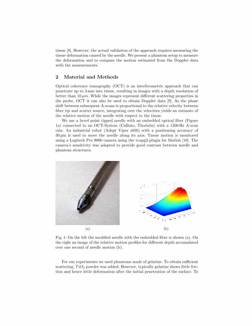
We use a bevel point tipped needle with an embedded optical fiber (Figure1a) connected to an OCT-System (Callisto, Thorlabs) with a 1200 Hz A-scanrate. An industrial robot (Adept Viper s850) with a positioning accuracy of30 µm is used to move the needle along its axis. Tissue motion is monitoredusing a Logitech Pro 9000 camera using the vcapg2-plugin for Matlab [10]. Thecamera’s sensitivity was adapted to provide good contrast between needle andphantom structures.
(a) (b)
Fig. 1: On the left the modified needle with the embedded fiber is shown (a). Onthe right an image of the relative motion profiles for different depth accumulatedover one second of needle motion (b).
For our experiments we used phantoms made of gelatine. To obtain sufficientscattering, TiO2 powder was added. However, typically gelatine shows little fric-tion and hence little deformation after the initial penetration of the surface. To
induce and detect deformations we added layers of colored gelatine with a differ-ent stiffness, such that the needle tip and the layers are well visible in the cameraimages. The robot was configured to continuously move the needle with a speedof approximately 0.05 mm/s while OCT and camera images were recorded with1200 Hz and 30 Hz, respectively. In a post-processing step, the camera imageswith maximum deformation of the gelatine layer were identified and the relatedtime stamp defined the end point of the motion trace. The deformation was thenestimated from the camera image by taking the needle as a reference, and therelative motion estimated by the OCT as well as the actual robot motion weredetermined from the recorded data.
As the OCT Doppler data can be considered as one-dimensional depth pro-files of the relative velocity vrel between tissue and needle tip, we need to decidein which depth to measure the motion. To this end we compute the accumulatedmotion for all depth and a windows of 1 s. Figure 1b shows the resulting motionprofiles, which generally show a maximum approximately 300 pixels or 1 mmfrom the needle tip.
3 Results
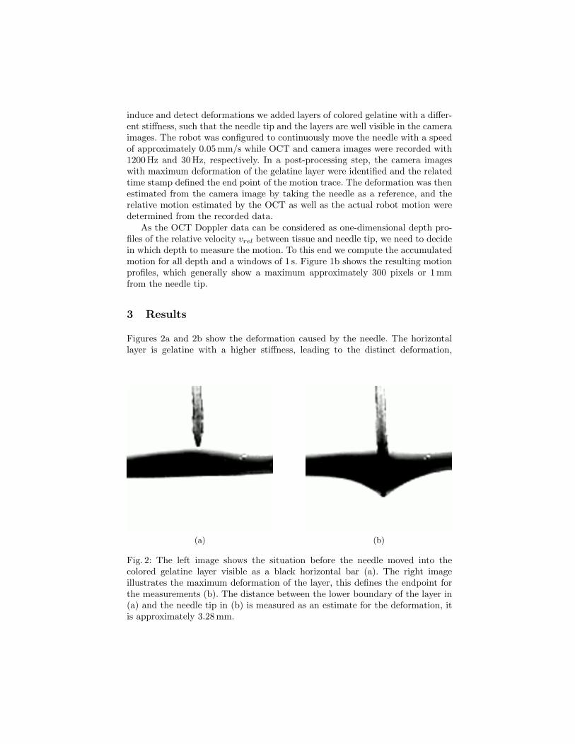
Figures 2a and 2b show the deformation caused by the needle. The horizontallayer is gelatine with a higher stiffness, leading to the distinct deformation,
(a) (b)
Fig. 2: The left image shows the situation before the needle moved into thecolored gelatine layer visible as a black horizontal bar (a). The right imageillustrates the maximum deformation of the layer, this defines the endpoint forthe measurements (b). The distance between the lower boundary of the layer in(a) and the needle tip in (b) is measured as an estimate for the deformation, itis approximately 3.28 mm.

Fig. 3: An overview of the OCT A-scans at different time steps / needle positions,from left to right.
(a) (b)
Fig. 4: The left plot shows the velocity of material in front of the needle over time/ needle position (a). The right plot shows the corresponding motion estimate(b).
which is approximately 3.28 mm in this case. In Figure 3 the OCT data recordedduring the needle motion is summarized. Note that each column in the imagecorresponds to an A-scan showing the real part of the signal, i.e., the left handside represents the situation before entering the layer and the last column on theleft corresponds to the A-scan in the situation shown in Figure 2b. A careful lookat the image reveals that there is a brighter region that gradually gets smalleron the left, indicating some compression.
The actual velocity of material in front of the needle and the resulting es-timate of the relative motion are presented in Figures 4a and 4b, respectively.Note that the total motion through the gelatine is estimated as approximately4.78 mm. The robot moved 8.16 mm while deformation of 3.28 mm and the esti-mated motion add to 8.06 mm
4 Discussion
Our experiments are preliminary in that they need to be repeated and the deter-mination of the deformation from the images should be automated. Moreover,the scattering and absorption in actual tissue may affect ability to detection ofmotion in sufficient depth, where the smaller SNR typically leads to an under-estimation of the Doppler signal [11].

However, the results still provide further indication that a precise estimationof the needle tip motion relative to soft tissue is possible using OCT Dopplerdata. Such information would be valuable when placing needles for brachyther-apy or biopsies, where proper placement of the needle with respect to soft tissueis important. The methods is not affected by artifacts from other needles and thehigh sampling rate allows for real-time control, e.g., of robotic needle drivers.
References
1. Damore, S.J., Syed, A., Puthawala, A.A., Sharma, A.: Needle displacement duringhdr brachytherapy in the treatment of prostate cancer. International Journal ofRadiation Oncology 46(5) (2000) 1205–1211
2. Nakamura, R., Ishiyama, H., Tanji, S., Satoh, T., Oikawa, H., Inatsu, W., Ehara, S.,K., H.: Effects of ellipsoid prostate deformation on dose delivery during permanentinterstitial brachytherapie. Brachytherapy 10(3) (2011) 208–213
3. Yoshida, K., Yamazaki, H., Nose, T., Shiomi, H., Yoshida, M., Mikami, M., Tak-enaka, T., Kotsuma, T., E.Tanaka, Kuriyama, K., Harada, Y., Tohda, A., Ya-sunaga, Y., Oka, T.: Needle applicator displacement during high-dose-rate inter-stitial brachytherapy for prostate cancer. Brachytherapy 9(1) (2010) 36–41
4. Lagerburg, V., Moerland, M.A., Lagendijk, J.J., Battermann, J.J.: Measurementof prostate rotation during insertion of needles for brachytherapy. Radiotherapyand Oncology 77(3) (2005) 318–23
5. Alterovitz, R., Goldberg, K.Y., Pouliot, J., Hsu, I.C.J.: Sensorless motion planningfor medical needle insertion in deformable tissues. IEEE Trans Inf Technol Biomed13(2) (Mar 2009) 217–225
6. Mahvash, M., Dupont, P.E.: Mechanics of dynamic needle insertion into a biologicalmaterial. IEEE TRANSACTIONS ON BIOMEDICAL ENGINEERING 57(4)(2010) 934 – 943
7. Schlaefer, A., Otte, C., Richter, L., Heinig, M., Bruder, R., Kovacz, G., Huttman,G.: Towards high resolution image guided needle navigation in prostate brachyther-apie. CARS 2010 2010 (2010) 24–25
8. Otte, C., Huttmann, G., Schlaefer, A.: Feasibiliy of optical detection of soft tissuedeformation during needle insertion. In III, D.R.H., Wong, K.H., eds.: MedicalImaging 2012: Image-Guided Procedures, Robotic Interventions, and Modeling.Volume 8316. (2012)
9. Wang, R.K., Ma, Z.H., Kirkpatrick, S.J.: Tissue doppler optical coherence elas-tography for real time strain rate and strain mapping of soft tissue. Appl. Phys.Lett 89(14) (2006) 144103–3
10. Kobayashi, K.: MATLAB Utilization Book. Shuwa System Co, Ltd. (2001)11. Szkulmowska, A., Szkulmowski, M., Kowalczyk, A., Wojtkowski, M.: Phase-
resolved doppler optical coherence tomography-limitations and improvements. OptLet 33(13) (2008) 1425–1427
![933 dji phantom-4 spec-sheet-rev[1] - PLASTICASE · 2019. 10. 23. · 933 DJI™ PHANTOM 4 For all DJI™ Phantom 4 models Phantom 4 Phantom 4 Pro Phantom 4 Pro + 2.0 Phantom 4 RTK](https://static.fdocuments.in/doc/165x107/60c827405a7e465133218fc4/933-dji-phantom-4-spec-sheet-rev1-plasticase-2019-10-23-933-djia-phantom.jpg)