Typing of Human Mycobacterium avium Isolates in Italy by IS1245

PFGE TYPING OF HUMAN CASE AND FOOD ISOLATES OF E. COLI O157:H7 IN
NEW ZEALAND
FINAL REPORT
Prepared as part of a New Zealand Food Safety Authority contract for scientific services
by
Angela Cornelius Brent Gilpin
Carolyn Nicol
November 2006
Institute of Environmental Science & Research Limited Christchurch Science Centre Location address: 27 Creyke Road, Ilam, Christchurch Postal address: P O Box 29 181, Christchurch, New Zealand Website: www.esr.cri.nz
A CROWN RESEARCH INSTITUTE

Client Report FW06101
PFGE TYPING OF HUMAN CASE AND FOOD ISOLATES OF E. COLI O157:H7 IN
NEW ZEALAND
FINAL REPORT
Stephen On Food Safety Programme Leader
Angela Cornelius Ellen Podivinsky Project Leader Peer Reviewer

DISCLAIMER This report or document ("the Report") is given by the Institute of Environmental Science and Research Limited ("ESR") solely for the benefit of the New Zealand Food Safety Authority (“NZFSA”), Public Health Services Providers and other Third Party Beneficiaries as defined in the Contract between ESR and the NZFSA, and is strictly subject to the conditions laid out in that Contract. Neither ESR nor any of its employees makes any warranty, express or implied, or assumes any legal liability or responsibility for use of the Report or its contents by any other person or organisation.
PFGE Typing of Human Case and Food November 2006 Isolates of E. coli O157:H7 in New Zealand

ACKNOWLEDGEMENTS The authors would like to thank Beth Robson (CSC), Philip Carter, Christopher Pope, Jenny Bennett, David Duncan and Moana Ngatai (KSC) for their contribution to the project. Thanks also to Robyn Clemens of AgResearch for supplying the North Island isolates from the Bobby Calf Longitudinal Study.
PFGE Typing of Human Case and Food November 2006 Isolates of E. coli O157:H7 in New Zealand

CONTENTS
SUMMARY ..............................................................................................................................1
1 BACKGROUND...........................................................................................................2
1.1 Current Best Practice for Interpreting PFGE Images ................................................2
2 RESULTS......................................................................................................................4 2.1 Analysis of US Isolates..............................................................................................4 2.2 XbaI Results (Stage 2) ...............................................................................................4 2.3 BlnI Results (Stage 3) ................................................................................................6 2.4 Combined XbaI:BlnI Patterns ....................................................................................7
3 CONCLUSIONS...........................................................................................................8
4 REFERENCES .............................................................................................................9
APPENDIX 1: PFGE THEORETICAL DIGESTS......................................................10
PFGE Typing of Human Case and Food November 2006 Isolates of E. coli O157:H7 in New Zealand

LIST OF FIGURES Figure 1: PFGE patterns of US E. coli O157:H7 isolates .....................................................2 Figure 2: Patterns of US isolates analysed in three laboratories ...........................................4 Figure 3: XbaI patterns for E. coli O157:H7 isolates ............................................................5 Figure 4: BlnI patterns for E. coli O157:H7 isolates .............................................................6 Figure 5: XbaI:BlnI patterns for isolates with XbaI or BlnI patterns indistinguishable for
the US Isolates .......................................................................................................7
PFGE Typing of Human Case and Food November 2006 Isolates of E. coli O157:H7 in New Zealand

SUMMARY From March to September 2006, 25 isolates were uploaded to the PulseNet USA E. coli O157:H7 pulsed field gel electrophoresis (PFGE) database with the XbaI:BlnI pattern EXHX01.0074:EXHA26.0569. Although this pattern is relatively common in the US database, this number of isolates suggests a potential common source outbreak. USDA-FSIS found E. coli O157:H7 isolates from two meat-processing plants with two similar XbaI:BlnI patterns (EXHX01.0074:EXHA26.0569 and EXHX01.1401:EXHA26.0569). One common link between these meat-processing plants is that both sourced some of their meat from New Zealand. As a consequence of the isolations in the US, in April 2006 the NZFSA and, independently, ESR (PulseNet Aotearoa) received an urgent request from the USDA-FSIS and US-CDC, requesting information on the prevalence of this pattern amongst New Zealand E. coli O157:H7 isolates. As the New Zealand database contained only limited data, responding to this request required the analysis by PFGE of over 200 additional isolates. Comparisons were made with the XbaI profiles of 203 human isolates and 229 meat isolates. Of these, 12 human isolates and three meat isolates had XbaI patterns that were indistinguishable from the US pattern EXHX01.0074. BlnI profiles were generated for these isolates and all differed from the USA pattern EXHA26.0569. No isolates were identified with the XbaI pattern EXHX01.1401. Consistent with US database, the XbaI pattern EXHX01.0074 appears relatively common in New Zealand isolates (5.9%). Furthermore it appears relatively stable having been isolated over at least five years, with no appreciable genetic changes. A further 10 human and 34 meat isolates, mostly with similar XbaI profiles, were genotyped using BlnI. Two of the meat isolates had BlnI patterns that were indistinguishable from the USA pattern EXHA26.0569. Their XbaI profiles however differed from the USA pattern. All of the New Zealand isolates were distinguishable from the USA patterns EXHX01.0074:EXHA26.0569, and EXHX01.1401:EXHA26.0569. There is no evidence to indicate that the E. coli O157:H7 isolates recovered from the US meat-processing plant came from New Zealand meat.
PFGE Typing of Human Case and Food November 2006 Isolates of E. coli O157:H7 in New Zealand
1

1 BACKGROUND In April 2006 the New Zealand Food Safety Authority (NZFSA) and, independently, ESR (PulseNet Aotearoa) received an urgent request from the USDA-FSIS and US-CDC, via PulseNet USA, for information on the occurrence in New Zealand of two pulsed field gel electrophoresis (PFGE) patterns for E. coli O157. These patterns, illustrated in Figure 1, were from two isolates from ground beef containing both US and New Zealand beef, and the patterns have also been implicated in a cluster of human illnesses in the United States of America. The patterns are relatively common in the US and one partial pattern (using one enzyme rather than two) is also common in New Zealand. However, the databases in New Zealand are not well populated with data, and as a consequence NZFSA was unable to provide a robust answer to the US request.
Figure 1: PFGE patterns of US E. coli O157:H7 isolates PFGE-XbaI
20.0
0
40.0
0
100.
00
150.
00
200.
00
250.
00
400.
00
500.
00
600.
00
800.
00
1500
PFGE-BlnI
20.0
0
40.0
0
100.
00
150.
00
200.
00
300.
00
400.
00
500.
00
600.
00
1000
2000 Key
USDA_OB060047
USDA_OB060052WA___9681
PFGE-XbaI-pattern
EXHX01.1401
EXHX01.0074EXHX01.1401
PFGE-BlnI-pattern
EXHA26.0569
EXHA26.0569EXHA26.0569
It was therefore considered imperative that NZ human and food (meat) isolates be PFGE-typed with urgency, using two appropriate enzymes, to answer the current request, and to facilitate a more rapid response to further requests. 1.1 Current Best Practice for Interpreting PFGE Images The objective of this study was to establish the prevalence in New Zealand of two E. coli O157:H7 PFGE patterns so that this information could be reported to PulseNet USA. An important step in this process was to ensure that PFGE results were interpreted in New Zealand, as they would have been by PulseNet USA. A brief review of the recent literature was undertaken to establish current best practice and identify what criteria PulseNet USA currently use to report PFGE patterns. In 1995 Tenover et al. published a paper suggesting guidelines for the interpretation of PFGE data. In the absence of alternative papers, the “Tenover criteria” have for the last decade been the defacto standard worldwide. The “Tenover criteria” suggested that profiles differing from each other by up to three bands should be considered closely related, and up to six bands possibly related. The basis of this was that a point mutation in a restriction site could result in loss of that site with two bands in one isolate merging to form a larger band – i.e. a “three band difference”. Using this logic, a three band difference could be the result of a single genetic event, and therefore isolates could be closely related. These guidelines were developed on the basis of comparison of nosocomial pathogens, particularly in situations of ongoing transmission. These guidelines also assume that all fragments are visible in a gel, that the plasmid content is stable, and that most mutations are
PFGE Typing of Human Case and Food November 2006 Isolates of E. coli O157:H7 in New Zealand
2

point mutations. For foodborne transmission of pathogens, and for E. coli O157 in particular, all three of these assumptions are flawed. Addressing these in order:
• PFGE usually only resolves a limited number of bands. Theoretical digestion of sequenced genomes of E. coli O157 strains should generate 41 fragments with XbaI and 31-33 fragments with BlnI. Some smaller bands run off the gel, while others are too similar in size and comigrate. As a consequence only half to three quarters of these bands can usually be distinguished by PFGE analysis (see Appendix 1).
• Plasmids are common in foodborne pathogens. If digested by a restriction enzyme they will migrate in a PFGE gel as a function of their size, just like chromosomal fragments. Often if they are multiple copy plasmids they are observed as intense bands. However if they are not in a linear conformation, their migration is unpredictable. Large undigested plasmids may not leave the wells of a gel, but may also be visible almost anywhere in the gel. Large megaplasmids are common in foodborne pathogens.
• Kudva et al. (2002) demonstrated that PFGE diversity in E. coli O157 is primarily attributed to insertions and deletions, not to point mutations as the “Tenover criteria” assume.
Under the “Tenover criteria”, single band differences should not be possible. They are however commonly observed, either because of the other bands involved not being resolved, or the presence of plasmids. Barrett et al. (2006) have recently re-evaluated the “Tenover criteria” in light of practical experiences of PulseNet USA. Their revised recommendations are:
• When any differences in PFGE patterns are observable, the patterns should be reported as different.
• Patterns that are indistinguishable should be reported as such, not as identical. Interpretation of patterns does however require adherence to the following steps: 1. The gel must be of sufficient quality to be properly interpreted. 2. The diversity of the organism under consideration must be considered. Even in an
organism with high diversity, some clonal populations may exist. 3. The outbreak setting should be considered. More variability is likely in ongoing
transmission than suspected point source. 4. The most important factor is how the laboratory data fit with the epidemiologic and
environmental information. PFGE results alone cannot establish an epidemiological connection between isolates.
Current best practice at PulseNet USA requires indistinguishable patterns using two enzymes for isolates to be considered indistinguishable by PFGE. The application of a second enzyme is well supported in the literature, pioneered perhaps by On et al. (1998). The validity of the second enzyme is of course subject to epidemiological investigation, and dependent on the nature of the outbreak. For common patterns a second enzyme is even more important.
PFGE Typing of Human Case and Food November 2006 Isolates of E. coli O157:H7 in New Zealand
3
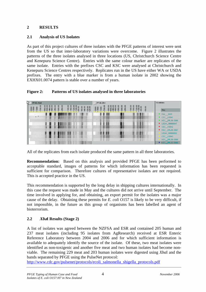
2 RESULTS 2.1 Analysis of US Isolates As part of this project cultures of three isolates with the PFGE patterns of interest were sent from the US so that inter-laboratory variations were overcome. Figure 2 illustrates the patterns of the three isolates analysed in three locations (US, Christchurch Science Centre and Kenepuru Science Centre). Entries with the same colour marker are replicates of the same isolate. Entries with the prefixes CSC and KSC were analysed at Christchurch and Kenepuru Science Centres respectively. Replicates run in the US have either WA or USDA prefixes. The entry with a blue marker is from a human isolate in 2002 showing the EXHX01.0074 pattern is stable over a number of years.
Figure 2: Patterns of US isolates analysed in three laboratories
All of the replicates from each isolate produced the same pattern in all three laboratories. Recommendation: Based on this analysis and provided PFGE has been performed to acceptable standard, images of patterns for which information has been requested is sufficient for comparison. Therefore cultures of representative isolates are not required. This is accepted practice in the US. This recommendation is supported by the long delay in shipping cultures internationally. In this case the request was made in May and the cultures did not arrive until September. The time involved in applying for, and obtaining, an export permit for the isolates was a major cause of the delay. Obtaining these permits for E. coli O157 is likely to be very difficult, if not impossible, in the future as this group of organisms has been labelled an agent of bioterrorism. 2.2 XbaI Results (Stage 2) A list of isolates was agreed between the NZFSA and ESR and contained 205 human and 237 meat isolates (including 95 isolates from AgResearch) received at ESR Enteric Reference Laboratory between 2004 and 2006 and for which sufficient information is available to adequately identify the source of the isolate. Of these, two meat isolates were identified as non-toxigenic and another five meat and two human isolates had become non-viable. The remaining 229 meat and 203 human isolates were digested using XbaI and the bands separated by PFGE using the PulseNet protocol: http://www.cdc.gov/pulsenet/protocols/ecoli_salmonella_shigella_protocols.pdf
PFGE Typing of Human Case and Food November 2006 Isolates of E. coli O157:H7 in New Zealand
4
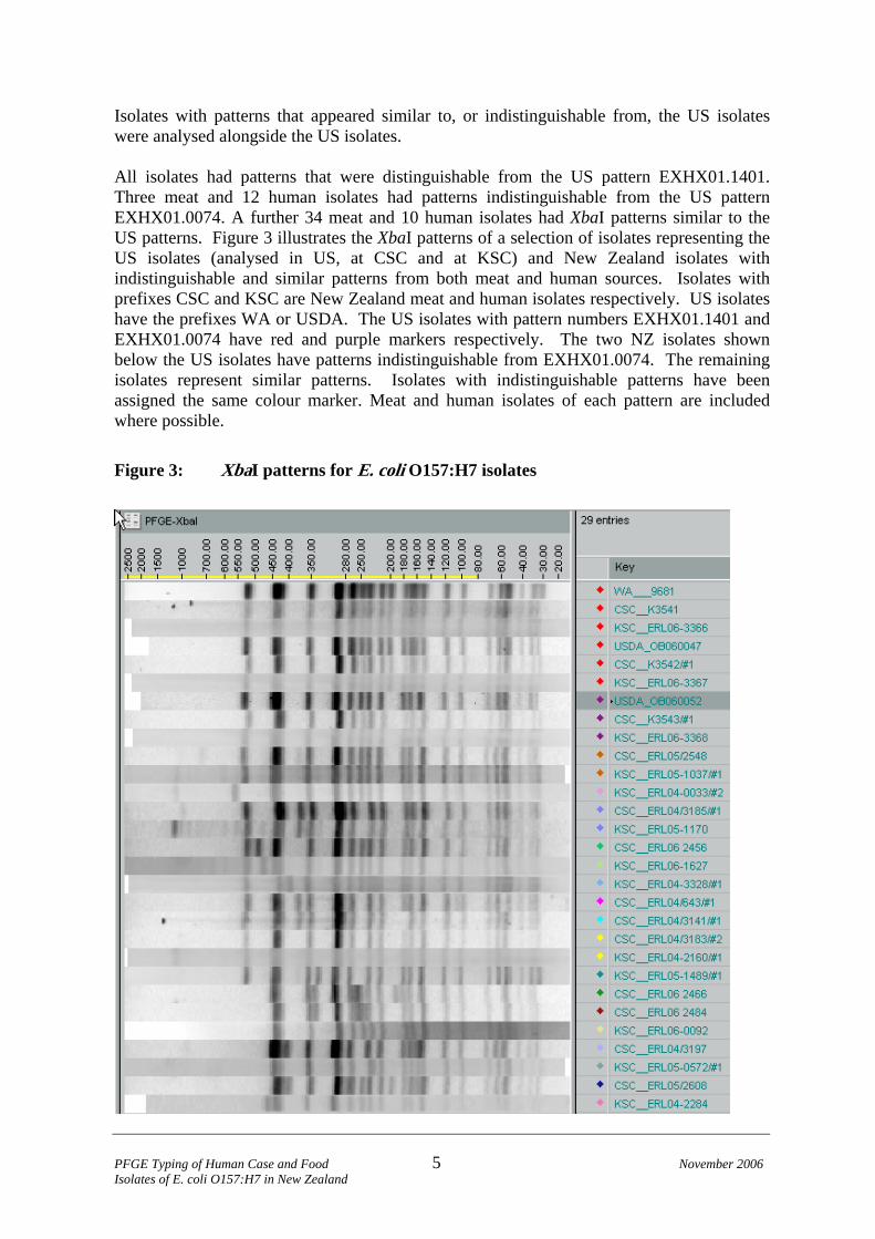
Isolates with patterns that appeared similar to, or indistinguishable from, the US isolates were analysed alongside the US isolates. All isolates had patterns that were distinguishable from the US pattern EXHX01.1401. Three meat and 12 human isolates had patterns indistinguishable from the US pattern EXHX01.0074. A further 34 meat and 10 human isolates had XbaI patterns similar to the US patterns. Figure 3 illustrates the XbaI patterns of a selection of isolates representing the US isolates (analysed in US, at CSC and at KSC) and New Zealand isolates with indistinguishable and similar patterns from both meat and human sources. Isolates with prefixes CSC and KSC are New Zealand meat and human isolates respectively. US isolates have the prefixes WA or USDA. The US isolates with pattern numbers EXHX01.1401 and EXHX01.0074 have red and purple markers respectively. The two NZ isolates shown below the US isolates have patterns indistinguishable from EXHX01.0074. The remaining isolates represent similar patterns. Isolates with indistinguishable patterns have been assigned the same colour marker. Meat and human isolates of each pattern are included where possible.
Figure 3: XbaI patterns for E. coli O157:H7 isolates
PFGE Typing of Human Case and Food November 2006 Isolates of E. coli O157:H7 in New Zealand
5

2.3 BlnI Results (Stage 3) All of the 37 meat and 22 human isolates that had XbaI patterns that were indistinguishable from, or similar to, the US patterns were digested using BlnI and separated by PFGE using the PulseNet Protocol: http://www.cdc.gov/pulsenet/protocols/ecoli_salmonella_shigella_protocols.pdf. The US isolates were analysed in each batch of isolates. Two meat isolates had patterns that were indistinguishable from the US pattern EXHA26.0569. Figure 4 illustrates the BlnI patterns of a selection of isolates representing the US isolates (analysed in US, at CSC and at KSC) and NZ isolates from both meat and human sources. The US isolates have fluorescent green markers and the two NZ meat isolates with patterns indistinguishable from these have red markers. Isolates with prefixes CSC and KSC are NZ meat and human isolates respectively, and US isolates have the prefixes WA or USDA. Isolates with indistinguishable patterns have been assigned the same colour marker and meat and human isolates of each pattern are included where possible.
Figure 4: BlnI patterns for E. coli O157:H7 isolates
PFGE Typing of Human Case and Food November 2006 Isolates of E. coli O157:H7 in New Zealand
6

2.4 Combined XbaI:BlnI Patterns All isolates had combined XbaI:BlnI patterns that were distinguishable from the US isolates. Figure 5 illustrates the XbaI and BlnI patterns for isolates that had XbaI or BlnI patterns that were indistinguishable from the US isolates. NZ isolates with XbaI patterns that were indistinguishable from US pattern EXHX01.0074 have a red marker. The US isolates analysed in the US, at CSC and at KSC have a fluorescent green marker. New Zealand isolates were BlnI patterns that were indistinguishable from US pattern EXHA26.0569 have a yellow marker.
Figure 5: XbaI:BlnI patterns for isolates with XbaI or BlnI patterns indistinguishable for the US Isolates
Extra band here
Missing a band here
PFGE Typing of Human Case and Food November 2006 Isolates of E. coli O157:H7 in New Zealand
7

3 CONCLUSIONS All of the New Zealand isolates were distinguishable from the USA patterns EXHX01.0074:EXHA26.0569, and EXHX01.1401:EXHA26.0569. There is no evidence to indicate that the E. coli O157:H7 isolates recovered from the US meat-processing plant came from New Zealand meat.
PFGE Typing of Human Case and Food November 2006 Isolates of E. coli O157:H7 in New Zealand
8

4 REFERENCES Barrett TJ, Gerner-Smidt P, Swaminathan B. (2006) Interpretation of pulsed-field gel electrophoresis patterns in foodborne disease investigations and surveillance. Foodborne Pathogens and Disease; 3(1): 20-31. Kudva IT, Evans PS, Perna NT, Barrett TJ, Ausubel FM, Blattner FR, Calderwood SB. (2002). Strains of Escherichia coli O157:H7 differ primarily by insertions or deletions, not single-nucleotide polymorphisms. Journal of Bacteriology; 184: 1873-1879. On SLW, Nielsen EM, Engberg J, Madsen M. (1998) Validity of SmaI-defined genotypes of Campylobacter jejuni examined by SalI, KpnI, and BamHI polymorphisms: evidence of identical clones infecting humans, poultry, and cattle. Epidemiology and Infection; 120: 231-237. Tenover FC, Arbeit RD, Goering RV, Mickelsen PA, Murray BE, Persing DH, Swaminathan B. (1995) Interpreting chromosomal DNA restriction patterns produced by pulsed-field gel electrophoresis: criteria for bacterial strain typing. Journal of Clinical Microbiology; 33(9): 2233-9.
PFGE Typing of Human Case and Food November 2006 Isolates of E. coli O157:H7 in New Zealand
9

APPENDIX 1: PFGE THEORETICAL DIGESTS Escherichia coli O157:H7 EDL933 - 41 fragments with XbaI, 33 with BlnI Escherichia coli O157:H7 VT2-Sakai, 41 fragments with XbaI, 31 with BlnI The schematic gels below illustrate that less fragments are likely to be visualised. Fragments below 20,000 bp will usually run off a gel.
EDL933 VT2-Sakai
PFGE Typing of Human Case and Food November 2006 Isolates of E. coli O157:H7 in New Zealand
10