Pex11mediates peroxisomal proliferation by promoting...
12
RESEARCH ARTICLE Pex11mediates peroxisomal proliferation by promoting deformation of the lipid membrane Yumi Yoshida 1 , Hajime Niwa 1 , Masanori Honsho 1 , Akinori Itoyama 2 and Yukio Fujiki 1,2,3, * ABSTRACT Pex11p family proteins are key players in peroxisomal fission, but their molecular mechanisms remains mostly unknown. In the present study, overexpression of Pex11pb caused substantial vesiculation of peroxisomes in mammalian cells. This vesicle formation was dependent on dynamin-like protein 1 (DLP1) and mitochondrial fission factor (Mff), as knockdown of these proteins diminished peroxisomal fission after Pex11pb overexpression. The fission-deficient peroxisomes exhibited an elongated morphology, and peroxisomal marker proteins, such as Pex14p or matrix proteins harboring peroxisomal targeting signal 1, were discernible in a segmented staining pattern, like beads on a string. Endogenous Pex11pb was also distributed a striped pattern, but which was not coincide with Pex14p and PTS1 matrix proteins. Altered morphology of the lipid membrane was observed when recombinant Pex11p proteins were introduced into proteo-liposomes. Constriction of proteo-liposomes was observed under confocal microscopy and electron microscopy, and the reconstituted Pex11pb protein localized to the membrane constriction site. Introducing point mutations into the N-terminal amphiphathic helix of Pex11pb strongly reduced peroxisomal fission, and decreased the oligomer formation. These results suggest that Pex11p contributes to the morphogenesis of the peroxisomal membrane, which is required for subsequent fission by DLP1. KEY WORDS: Peroxisomes, Pex11p, Liposomes, Membrane deformation, Amphiphathic helix INTRODUCTION Peroxisomes are single membrane-bound organelles that are involved in long-chain fatty acid oxidation, plasmalogen synthesis, and ROS elimination (van den Bosch et al., 1992; Wanders and Waterham, 2006). Peroxisomes are maintained by autonomous proliferation. Peroxisomal proliferation can occur through growth and division of pre-existing peroxisomes or de novo synthesis (Lazarow and Fujiki, 1985; Thoms and Erdmann, 2005; Hettema and Motley, 2009). Peroxisomal membrane structure is disrupted in fibroblasts from Zellweger syndrome patients, and this effect can be reversed by introduction of the responsible gene products (Honsho et al., 1998; Matsuzono et al., 1999; South and Gould, 1999; Ghaedi et al., 2000; Muntau et al., 2000; Shimozawa et al., 2000). Peroxisomes are well known to be maintained by growth and division (Lazarow and Fujiki, 1985). A multi-step reaction for this process has been proposed (Schrader et al., 1998; Koch et al., 2003; Li and Gould, 2003). First, peroxisomal matrix proteins and membrane proteins are imported to peroxisomes. Secondly, peroxisomes increase in size and develop an elongated morphology. Peroxisomal marker proteins were detectable in a segmented pattern on such elongated peroxisomes, like beads on string, with constriction of the membrane (Grabenbauer et al., 2000; Koch et al., 2004; Itoyama et al., 2012; Itoyama et al., 2013). The membrane is then cleaved at the restriction sites to form new peroxisomes. Several proteins involved in this final process have been identified. Dynamin-like protein 1 (DLP1) is a cytosolic large GTPase belonging to the dynamin superfamily (Praefcke and McMahon, 2004). DLP1 is thought to polymerize at the fission site, enabling the membrane to be cleaved by GTP hydrolysis (Yoon et al., 2001; Zhu et al., 2004). Fission 1 (Fis1) and mitochondrial fission factor (Mff) have been identified as the membrane receptor for DLP1 (Kobayashi et al., 2007; Gandre-Babbe and van der Bliek, 2008; Otera et al., 2010; Itoyama et al., 2013). Knockdown or mutation of DLP1, Fis1, and Mff results in fission-deficient, elongated peroxisomes (Koch et al., 2003; Koch et al., 2004; Koch et al., 2005; Tanaka et al., 2006; Kobayashi et al., 2007; Waterham et al., 2007; Gandre-Babbe and van der Bliek, 2008; Otera et al., 2010; Itoyama et al., 2013). These factors were shown to be essential in mitochondrial fission as well as peroxisomal proliferation (Koch et al., 2005; Tanaka et al., 2006; Waterham et al., 2007; Gandre-Babbe and van der Bliek, 2008; Otera et al., 2010). However, spherical peroxisomes require elongation steps prior to fission (Itoyama et al., 2012), which mitochondria do not. Pex11p family proteins are thought to function in the membrane elongation step (Schrader et al., 1998; Li and Gould, 2002). Pex11p proteins are peroxisomal membrane proteins consisting of three isoforms in mammalian cells, Pex11pa (Abe et al., 1998; Li et al., 2002b), Pex11pb (Abe and Fujiki, 1998; Schrader et al., 1998; Li et al., 2002a), and Pex11pc (Li et al., 2002b; Tanaka et al., 2003). Pex11pb is the most well-characterized isoform, due to its ubiquitous expression and involvement in peroxisomal fission. Overexpression of Pex11pb results in acceleration of peroxisomal fission, and Pex11pb knockout mice have a decreased number of peroxisomes (Schrader et al., 1998; Li et al., 2002a; Kobayashi et al., 2007). Pex11pb is a homo- or hetero-oligomeric protein through an N-terminal amphipathic helix (Kobayashi et al., 2007; Opalin ´ski et al., 2011; Bonekamp et al., 2013). In yeast, a synthetic peptide corresponding to this 1 Department of Biology, Faculty of Sciences, Kyushu University Graduate School, 6-10-1 Hakozaki, Higashi-ku, Fukuoka 812-8581, Japan. 2 Graduate School of Systems Life Sciences, Kyushu University Graduate School, 6-10-1 Hakozaki, Higashi-ku, Fukuoka 812-8581, Japan. 3 International Institute for Carbon-Neutral Energy Research (I 2 CNER), Kyushu University, 744 Motooka, Nishi-ku, Fukuoka 819-0395, Japan. *Author for correspondence ([email protected]) This is an Open Access article distributed under the terms of the Creative Commons Attribution License (http://creativecommons.org/licenses/by/3.0), which permits unrestricted use, distribution and reproduction in any medium provided that the original work is properly attributed. Received 30 October 2014; Accepted 6 March 2015 ß 2015. Published by The Company of Biologists Ltd | Biology Open (2015) 4, 710–721 doi:10.1242/bio.201410801 710 Biology Open by guest on June 5, 2018 http://bio.biologists.org/ Downloaded from
Transcript of Pex11mediates peroxisomal proliferation by promoting...
RESEARCH ARTICLE
Pex11mediates peroxisomal proliferation by promotingdeformation of the lipid membrane
Yumi Yoshida1, Hajime Niwa1, Masanori Honsho1, Akinori Itoyama2 and Yukio Fujiki1,2,3,*
ABSTRACT
Pex11p family proteins are key players in peroxisomal fission, but
their molecular mechanisms remains mostly unknown. In the
present study, overexpression of Pex11pb caused substantial
vesiculation of peroxisomes in mammalian cells. This vesicle
formation was dependent on dynamin-like protein 1 (DLP1) and
mitochondrial fission factor (Mff), as knockdown of these proteins
diminished peroxisomal fission after Pex11pb overexpression. The
fission-deficient peroxisomes exhibited an elongated morphology,
and peroxisomal marker proteins, such as Pex14p or matrix
proteins harboring peroxisomal targeting signal 1, were discernible
in a segmented staining pattern, like beads on a string. Endogenous
Pex11pb was also distributed a striped pattern, but which was not
coincide with Pex14p and PTS1 matrix proteins. Altered morphology
of the lipid membrane was observed when recombinant Pex11p
proteins were introduced into proteo-liposomes. Constriction of
proteo-liposomes was observed under confocal microscopy and
electron microscopy, and the reconstituted Pex11pb protein localized
to the membrane constriction site. Introducing point mutations into
the N-terminal amphiphathic helix of Pex11pb strongly reduced
peroxisomal fission, and decreased the oligomer formation. These
results suggest that Pex11p contributes to the morphogenesis of the
peroxisomal membrane, which is required for subsequent fission by
DLP1.
KEY WORDS: Peroxisomes, Pex11p, Liposomes, Membranedeformation, Amphiphathic helix
INTRODUCTIONPeroxisomes are single membrane-bound organelles that are
involved in long-chain fatty acid oxidation, plasmalogen
synthesis, and ROS elimination (van den Bosch et al., 1992;
Wanders and Waterham, 2006). Peroxisomes are maintained by
autonomous proliferation. Peroxisomal proliferation can occur
through growth and division of pre-existing peroxisomes or de
novo synthesis (Lazarow and Fujiki, 1985; Thoms and Erdmann,
2005; Hettema and Motley, 2009). Peroxisomal membrane
structure is disrupted in fibroblasts from Zellweger syndrome
patients, and this effect can be reversed by introduction of the
responsible gene products (Honsho et al., 1998; Matsuzono et al.,
1999; South and Gould, 1999; Ghaedi et al., 2000; Muntau et al.,
2000; Shimozawa et al., 2000).
Peroxisomes are well known to be maintained by growth and
division (Lazarow and Fujiki, 1985). A multi-step reaction for this
process has been proposed (Schrader et al., 1998; Koch et al., 2003;
Li and Gould, 2003). First, peroxisomal matrix proteins and
membrane proteins are imported to peroxisomes. Secondly,
peroxisomes increase in size and develop an elongated
morphology. Peroxisomal marker proteins were detectable in a
segmented pattern on such elongated peroxisomes, like beads on
string, with constriction of the membrane (Grabenbauer et al.,
2000; Koch et al., 2004; Itoyama et al., 2012; Itoyama et al., 2013).
The membrane is then cleaved at the restriction sites to form new
peroxisomes. Several proteins involved in this final process have
been identified. Dynamin-like protein 1 (DLP1) is a cytosolic large
GTPase belonging to the dynamin superfamily (Praefcke and
McMahon, 2004). DLP1 is thought to polymerize at the fission site,
enabling the membrane to be cleaved by GTP hydrolysis (Yoon
et al., 2001; Zhu et al., 2004). Fission 1 (Fis1) and mitochondrial
fission factor (Mff) have been identified as the membrane receptor
for DLP1 (Kobayashi et al., 2007; Gandre-Babbe and van der Bliek,
2008; Otera et al., 2010; Itoyama et al., 2013). Knockdown or
mutation of DLP1, Fis1, and Mff results in fission-deficient,
elongated peroxisomes (Koch et al., 2003; Koch et al., 2004; Koch
et al., 2005; Tanaka et al., 2006; Kobayashi et al., 2007; Waterham
et al., 2007; Gandre-Babbe and van der Bliek, 2008; Otera et al.,
2010; Itoyama et al., 2013). These factors were shown to be
essential in mitochondrial fission as well as peroxisomal
proliferation (Koch et al., 2005; Tanaka et al., 2006; Waterham
et al., 2007; Gandre-Babbe and van der Bliek, 2008; Otera et al.,
2010). However, spherical peroxisomes require elongation steps
prior to fission (Itoyama et al., 2012), which mitochondria do not.
Pex11p family proteins are thought to function in the membrane
elongation step (Schrader et al., 1998; Li and Gould, 2002).
Pex11p proteins are peroxisomal membrane proteins consisting
of three isoforms in mammalian cells, Pex11pa (Abe et al., 1998;
Li et al., 2002b), Pex11pb (Abe and Fujiki, 1998; Schrader et al.,
1998; Li et al., 2002a), and Pex11pc (Li et al., 2002b; Tanaka
et al., 2003). Pex11pb is the most well-characterized isoform, due
to its ubiquitous expression and involvement in peroxisomal
fission. Overexpression of Pex11pb results in acceleration of
peroxisomal fission, and Pex11pb knockout mice have a
decreased number of peroxisomes (Schrader et al., 1998; Li
et al., 2002a; Kobayashi et al., 2007). Pex11pb is a homo- or
hetero-oligomeric protein through an N-terminal amphipathic
helix (Kobayashi et al., 2007; Opalinski et al., 2011; Bonekamp
et al., 2013). In yeast, a synthetic peptide corresponding to this
1Department of Biology, Faculty of Sciences, Kyushu University Graduate School,6-10-1 Hakozaki, Higashi-ku, Fukuoka 812-8581, Japan. 2Graduate School ofSystems Life Sciences, Kyushu University Graduate School, 6-10-1 Hakozaki,Higashi-ku, Fukuoka 812-8581, Japan. 3International Institute for Carbon-NeutralEnergy Research (I2CNER), Kyushu University, 744 Motooka, Nishi-ku, Fukuoka819-0395, Japan.
*Author for correspondence ([email protected])
This is an Open Access article distributed under the terms of the Creative Commons AttributionLicense (http://creativecommons.org/licenses/by/3.0), which permits unrestricted use, distributionand reproduction in any medium provided that the original work is properly attributed.
Received 30 October 2014; Accepted 6 March 2015
� 2015. Published by The Company of Biologists Ltd | Biology Open (2015) 4, 710–721 doi:10.1242/bio.201410801
710
Bio
log
yO
pe
n
by guest on June 5, 2018http://bio.biologists.org/Downloaded from
helical stretch could directly bind to liposomes and disrupt theirstructure, resulting in tubule-like formations (Opalinski et al.,
2011). Pex11pb is also reported to interact with DLP1 throughbinding to Fis1 or Mff (Kobayashi et al., 2007; Koch and Brocard,2012; Itoyama et al., 2013). Introduction of EGFP-tagged or YFP-tagged Pex11pb promoted peroxisomal fission and resulted in the
formation of clusters of elongated peroxisomes (Delille et al.,2010; Koch et al., 2010). Although these data suggest thatPex11pb has a fundamental role in peroxisomal morphogenesis,
the characteristics of Pex11pb have not been fully elucidated,particularly with respect to its role in membrane deformation.
In the present work, we characterized the role of Pex11pb in
peroxisomal morphogenesis in mammalian cells. The localizationof endogenous Pex11pb was analyzed in fission-deficient cells,which revealed an alternating staining pattern of Pex11pb and
Pex14p/PTS1. The effect of Pex11pb on the morphology ofartificial lipid membranes was also tested. Pex11pb altered themorphology of reconstituted proteo-liposomes in a manner thatdepended on the N-terminal amphipathic helix. In this in vitro
assay, the recombinant Pex11pa and Pex11pc protein were alsoeffective in morphology of liposomes membrane because of theproperty of the amphipathic helix. Knockdown of PEX11bdecreased the number of peroxisomes. In this in vivo assay,Pex11pc had a similar effect on peroxisome formation asPex11pb, but Pex11pa had little effects. These results strongly
demonstrate that Pex11p functions in the morphogenesis ofperoxisomes, particularly at the elongation and constriction steps.
RESULTSPex11pb induces peroxisome membrane extension beforefissionPex11pb induces peroxisomal fission when overexpressed in
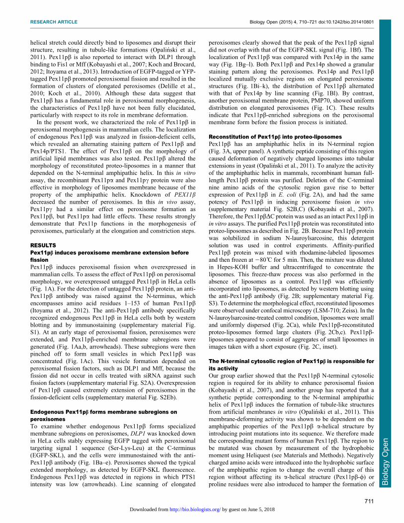
mammalian cells. To assess the effect of Pex11pb on peroxisomalmorphology, we overexpressed untagged Pex11pb in HeLa cells(Fig. 1A). For the detection of untagged Pex11pb protein, an anti-
Pex11pb antibody was raised against the N-terminus, whichencompasses amino acid residues 1–153 of human Pex11pb(Itoyama et al., 2012). The anti-Pex11pb antibody specifically
recognized endogenous Pex11pb in HeLa cells both by westernblotting and by immunostaining (supplementary material Fig.S1). At an early stage of peroxisomal fission, peroxisomes wereextended, and Pex11pb-enriched membrane subregions were
generated (Fig. 1Aa,b, arrowheads). These subregions were thenpinched off to form small vesicles in which Pex11pb wasconcentrated (Fig. 1Ac). This vesicle formation depended on
peroxisomal fission factors, such as DLP1 and Mff, because thefission did not occur in cells treated with siRNA against suchfission factors (supplementary material Fig. S2A). Overexpression
of Pex11pb caused extremely extension of peroxisomes in thefission-deficient cells (supplementary material Fig. S2Eb).
Endogenous Pex11pb forms membrane subregions onperoxisomesTo examine whether endogenous Pex11pb forms specializedmembrane subregions on peroxisomes, DLP1 was knocked down
in HeLa cells stably expressing EGFP tagged with peroxisomaltargeting signal 1 sequence (Ser-Lys-Leu) at the C-terminus(EGFP-SKL), and the cells were immunostained with the anti-
Pex11pb antibody (Fig. 1Ba–e). Peroxisomes showed the typicalextended morphology, as detected by EGFP-SKL fluorescence.Endogenous Pex11pb was detected in regions in which PTS1
intensity was low (arrowheads). Line scanning of elongated
peroxisomes clearly showed that the peak of the Pex11pb signaldid not overlap with that of the EGFP-SKL signal (Fig. 1Bf). The
localization of Pex11pb was compared with Pex14p in the sameway (Fig. 1Bg–l). Both Pex11pb and Pex14p showed a granularstaining pattern along the peroxisomes. Pex14p and Pex11pblocalized mutually exclusive regions on elongated peroxisome
structures (Fig. 1Bi–k), the distribution of Pex11pb alternatedwith that of Pex14p by line scanning (Fig. 1Bl). By contrast,another peroxisomal membrane protein, PMP70, showed uniform
distribution on elongated peroxisomes (Fig. 1C). These resultsindicate that Pex11pb-enriched subregions on the peroxisomalmembrane form before the fission process is initiated.
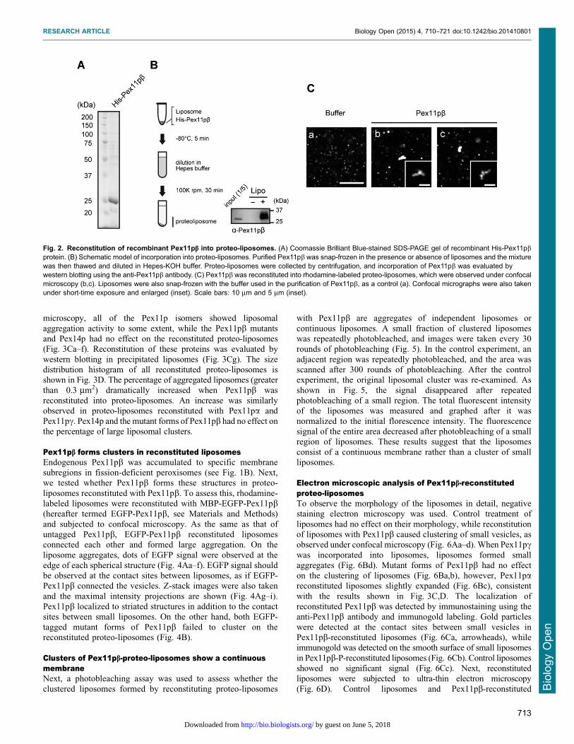
Reconstitution of Pex11pb into proteo-liposomesPex11pb has an amphiphathic helix in its N-terminal region
(Fig. 3A, upper panel). A synthetic peptide consisting of this regioncaused deformation of negatively charged liposomes into tubularextensions in yeast (Opalinski et al., 2011). To analyze the activityof the amphiphathic helix in mammals, recombinant human full-
length Pex11pb protein was purified. Deletion of the C-terminalnine amino acids of the cytosolic region gave rise to betterexpression of Pex11pb in E. coli (Fig. 2A), and had the same
potency of Pex11pb in inducing peroxisome fission in vivo
(supplementary material Fig. S2B,C) (Kobayashi et al., 2007).Therefore, the Pex11pbDC protein was used as an intact Pex11pb in
in vitro assays. The purified Pex11pb protein was reconstituted intoproteo-liposomes as described in Fig. 2B. Because Pex11pb proteinwas solubilized in sodium N-lauroylsarcosine, this detergent
solution was used in control experiments. Affinity-purifiedPex11pb protein was mixed with rhodamine-labeled liposomesand then frozen at 280 C for 5 min. Then, the mixture was dilutedin Hepes-KOH buffer and ultracentrifuged to concentrate the
liposomes. This freeze-thaw process was also performed in theabsence of liposomes as a control. Pex11pb was efficientlyincorporated into liposomes, as detected by western blotting using
the anti-Pex11pb antibody (Fig. 2B; supplementary material Fig.S3). To determine the morphological effect, reconstituted liposomeswere observed under confocal microscopy (LSM-710; Zeiss). In the
N-lauroylsarcosine-treated control condition, liposomes were smalland uniformly dispersed (Fig. 2Ca), while Pex11pb-reconstitutedproteo-liposomes formed large clusters (Fig. 2Cb,c). Pex11pb-liposomes appeared to consist of aggregates of small liposomes in
images taken with a short exposure (Fig. 2C, inset).
The N-terminal cytosolic region of Pex11pb is responsible forits activityOur group earlier showed that the Pex11pb N-terminal cytosolicregion is required for its ability to enhance peroxisomal fission
(Kobayashi et al., 2007), and another group has reported that asynthetic peptide corresponding to the N-terminal amphipathichelix of Pex11pb induces the formation of tubule-like structures
from artificial membranes in vitro (Opalinski et al., 2011). Thismembrane-deforming activity was shown to be dependent on theamphipathic properties of the Pex11pb a-helical structure byintroducing point mutations into its sequence. We therefore made
the corresponding mutant forms of human Pex11pb. The region tobe mutated was chosen by measurement of the hydrophobicmoment using Heliquest (see Materials and Methods). Negatively
charged amino acids were introduced into the hydrophobic surfaceof the amphipathic region to change the overall charge of thisregion without affecting its a-helical structure (Pex11pb-w) or
proline residues were also introduced to hamper the formation of
RESEARCH ARTICLE Biology Open (2015) 4, 710–721 doi:10.1242/bio.201410801
711
Bio
log
yO
pe
n
by guest on June 5, 2018http://bio.biologists.org/Downloaded from
the a-helix (Pex11pb-P) (Fig. 3A). Using multiple sequencealignment of Pex11p proteins from different species and thethree human isoforms, the amphipathic helices in human Pex11pa,Pex11pb, and Pex11pc were determined (supplementary material
Fig. S4A, underline). The average of the hydrophobic moment ofthis region was 0.342 in Pex11pa, 0.359 in Pex11pb, and 0.501 inPex11pc (supplementary material Fig. S4B, black line). To
confirm the effect, the point-mutated Myc-Pex11pb-w and Myc-Pex11pb-P were overexpressed in HeLa cells, and the cells weresubjected to immunostaining using antibodies to Myc and Pex14p
(Fig. 3B). While the wild-type Myc-Pex11pb enhancedperoxisomal fission as shown in Fig. 1A, overexpression of themutated forms did not change the number of peroxisomes in HeLa
cells (supplementary material Fig. S2C). The effect of Pex11paand Pex11pc on peroxisomal fission was also examined. Both
Pex11p isomers had weak effect on the fission. The fissionproperties of the series of Pex11p proteins were also tested in cellsin which DLP1 had been knocked down. The wild-type Pex11pband Pex11pc were incorporated into peroxisomes, forming Myc-
Pex11p-enriched regions and enhancing peroxisomal elongationoverall, while the Pex11pb mutants and Pex11pa did not result inadditional elongation of peroxisomes (supplementary material Fig.
S2E). Then, as the wild-type Pex11pb (Fig. 2C), the mutants,Pex11pa, and Pex11pc proteins were purified and reconstitutedinto proteo-liposomes to determine the effect of these proteins on
the liposomal morphology. As a control experiment, a typicalperoxisomal membrane protein, Pex14p, was also examined.Pex14p was heavily precipitated in the absence of liposomes so
that the reconstitution was carried out by the floating method asshown in supplementary material Fig. S3. Under confocal
Fig. 1. Pex11pb forms Pex11pb-enriched membranesubregions on the peroxisomal membrane. (A) HeLacells were transfected with a plasmid encoding humanPex11pb for 6–24 h and immunostained with antibodiesagainst Pex11pb (green) and Pex14p (red). Panels arearranged according to the stage of peroxisomalproliferation. Arrowheads indicate regions enriched inPex11pb compared with Pex14p. Scale bars: 10 mm and5 mm (enlarged). (B) HeLa cells stably expressing EGFP-SKL (a–e) and control HeLa cells (g–k) were treated witha control siRNA (a,g) or DLP1 siRNA (b–e,h–k) for 72 h.Cells were subjected to immunostaining using the anti-Pex11pb antibody (a–e) or double-stained with the anti-Pex11pb antibody and an anti-Pex14p antibody (g–k).Magnified view of DLP1 siRNA-treated cells (c–e,i–k),and pixel intensity by line scanning along the elongatedperoxisome (arrow) were plotted (f,l). Clear peaks ofPex11pb were highlighted by arrowheads. Scale bars:10 mm and 5 mm (enlarged). (C) HeLa cells were treatedwith a control siRNA (a) or DLP1 siRNA (b) for 72 h, andstained the DLP1 siRNA-treated cells with antibodies toPex14p and PMP70 in magnified views (c–e). Linescanning of elongated peroxisomes was shown in (f).Scale bars: 10 mm and 5 mm (magnified view).
RESEARCH ARTICLE Biology Open (2015) 4, 710–721 doi:10.1242/bio.201410801
712
Bio
log
yO
pe
n
by guest on June 5, 2018http://bio.biologists.org/Downloaded from
microscopy, all of the Pex11p isomers showed liposomalaggregation activity to some extent, while the Pex11pb mutantsand Pex14p had no effect on the reconstituted proteo-liposomes(Fig. 3Ca–f). Reconstitution of these proteins was evaluated by
western blotting in precipitated liposomes (Fig. 3Cg). The sizedistribution histogram of all reconstituted proteo-liposomes isshown in Fig. 3D. The percentage of aggregated liposomes (greater
than 0.3 mm2) dramatically increased when Pex11pb wasreconstituted into proteo-liposomes. An increase was similarlyobserved in proteo-liposomes reconstituted with Pex11pa and
Pex11pc. Pex14p and the mutant forms of Pex11pb had no effect onthe percentage of large liposomal clusters.
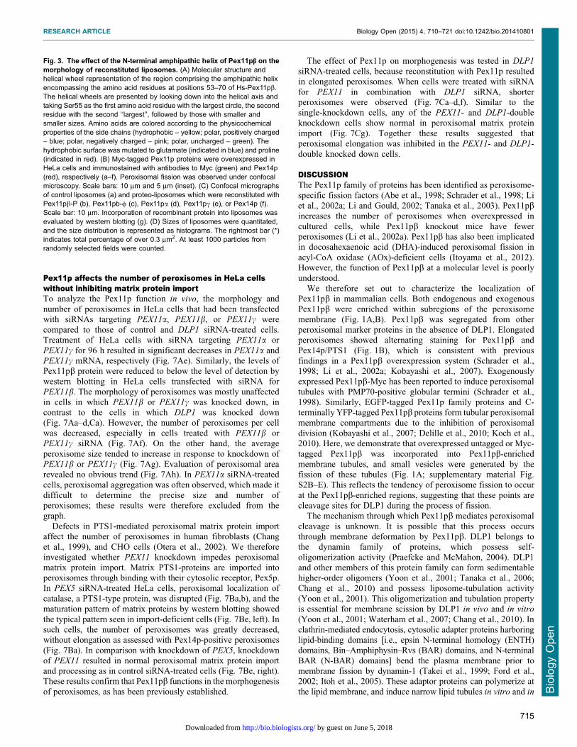
Pex11pb forms clusters in reconstituted liposomesEndogenous Pex11pb was accumulated to specific membranesubregions in fission-deficient peroxisomes (see Fig. 1B). Next,we tested whether Pex11pb forms these structures in proteo-
liposomes reconstituted with Pex11pb. To assess this, rhodamine-labeled liposomes were reconstituted with MBP-EGFP-Pex11pb(hereafter termed EGFP-Pex11pb, see Materials and Methods)
and subjected to confocal microscopy. As the same as that ofuntagged Pex11pb, EGFP-Pex11pb reconstituted liposomesconnected each other and formed large aggregation. On the
liposome aggregates, dots of EGFP signal were observed at theedge of each spherical structure (Fig. 4Aa–f). EGFP signal shouldbe observed at the contact sites between liposomes, as if EGFP-Pex11pb connected the vesicles. Z-stack images were also taken
and the maximal intensity projections are shown (Fig. 4Ag–i).Pex11pb localized to striated structures in addition to the contactsites between small liposomes. On the other hand, both EGFP-
tagged mutant forms of Pex11pb failed to cluster on thereconstituted proteo-liposomes (Fig. 4B).
Clusters of Pex11pb-proteo-liposomes show a continuousmembraneNext, a photobleaching assay was used to assess whether the
clustered liposomes formed by reconstituting proteo-liposomes
with Pex11pb are aggregates of independent liposomes orcontinuous liposomes. A small fraction of clustered liposomeswas repeatedly photobleached, and images were taken every 30rounds of photobleaching (Fig. 5). In the control experiment, an
adjacent region was repeatedly photobleached, and the area wasscanned after 300 rounds of photobleaching. After the controlexperiment, the original liposomal cluster was re-examined. As
shown in Fig. 5, the signal disappeared after repeatedphotobleaching of a small region. The total fluorescent intensityof the liposomes was measured and graphed after it was
normalized to the initial florescence intensity. The fluorescencesignal of the entire area decreased after photobleaching of a smallregion of liposomes. These results suggest that the liposomes
consist of a continuous membrane rather than a cluster of smallliposomes.
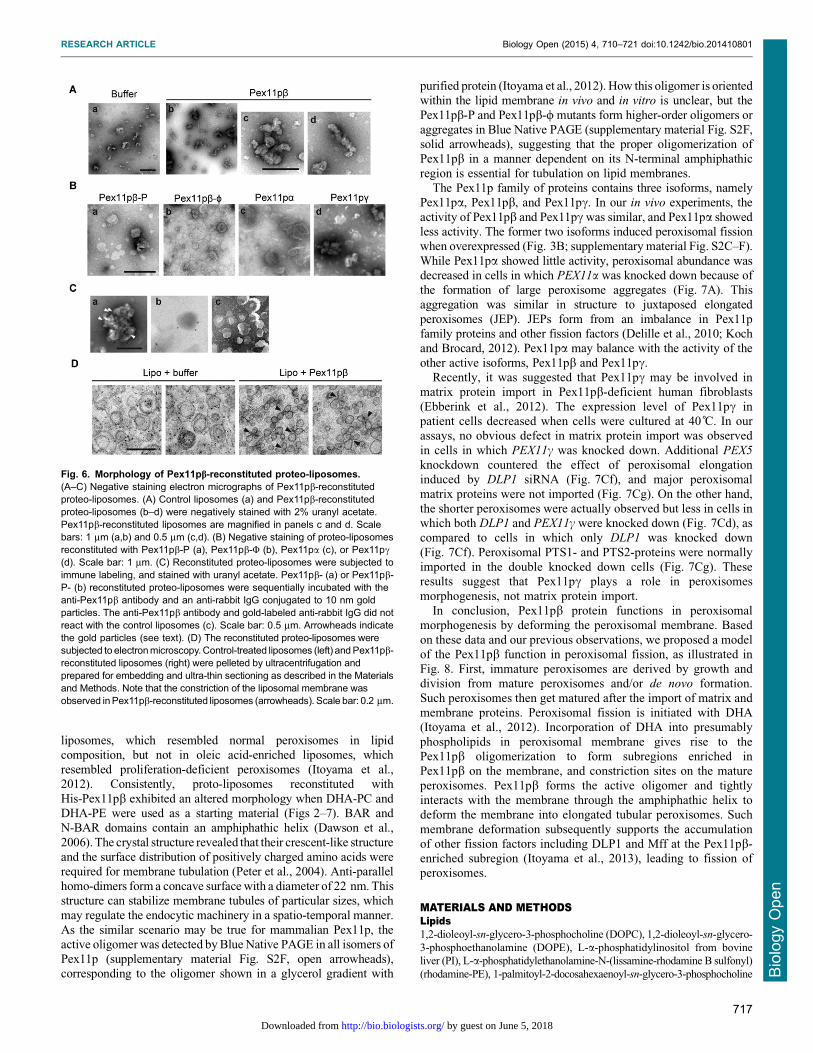
Electron microscopic analysis of Pex11pb-reconstitutedproteo-liposomesTo observe the morphology of the liposomes in detail, negativestaining electron microscopy was used. Control treatment of
liposomes had no effect on their morphology, while reconstitutionof liposomes with Pex11pb caused clustering of small vesicles, asobserved under confocal microscopy (Fig. 6Aa–d). When Pex11pcwas incorporated into liposomes, liposomes formed smallaggregates (Fig. 6Bd). Mutant forms of Pex11pb had no effecton the clustering of liposomes (Fig. 6Ba,b), however, Pex11pareconstituted liposomes slightly expanded (Fig. 6Bc), consistent
with the results shown in Fig. 3C,D. The localization ofreconstituted Pex11pb was detected by immunostaining using theanti-Pex11pb antibody and immunogold labeling. Gold particles
were detected at the contact sites between small vesicles inPex11pb-reconstituted liposomes (Fig. 6Ca, arrowheads), whileimmunogold was detected on the smooth surface of small liposomes
in Pex11pb-P-reconstituted liposomes (Fig. 6Cb). Control liposomesshowed no significant signal (Fig. 6Cc). Next, reconstitutedliposomes were subjected to ultra-thin electron microscopy
(Fig. 6D). Control liposomes and Pex11pb-reconstituted
Fig. 2. Reconstitution of recombinant Pex11pb into proteo-liposomes. (A) Coomassie Brilliant Blue-stained SDS-PAGE gel of recombinant His-Pex11pbprotein. (B) Schematic model of incorporation into proteo-liposomes. Purified Pex11pb was snap-frozen in the presence or absence of liposomes and the mixturewas then thawed and diluted in Hepes-KOH buffer. Proteo-liposomes were collected by centrifugation, and incorporation of Pex11pb was evaluated bywestern blotting using the anti-Pex11pb antibody. (C) Pex11pb was reconstituted into rhodamine-labeled proteo-liposomes, which were observed under confocalmicroscopy (b,c). Liposomes were also snap-frozen with the buffer used in the purification of Pex11pb, as a control (a). Confocal micrographs were also takenunder short-time exposure and enlarged (inset). Scale bars: 10 mm and 5 mm (inset).
RESEARCH ARTICLE Biology Open (2015) 4, 710–721 doi:10.1242/bio.201410801
713
Bio
log
yO
pe
n
by guest on June 5, 2018http://bio.biologists.org/Downloaded from
liposomes were concentrated by ultracentrifugation, then fixedwith glutaraldehyde and embedded in Epon. In control samples,small liposomes were dispersed separately. On the contrary, in the
Pex11pb-reconstituted samples, small clusters of connectedliposomes were observed. Liposomes were associated with eachother, and appeared to be constricted at the contact sites.
Fig. 3. See next page for legend.
RESEARCH ARTICLE Biology Open (2015) 4, 710–721 doi:10.1242/bio.201410801
714
Bio
log
yO
pe
n
by guest on June 5, 2018http://bio.biologists.org/Downloaded from
Pex11p affects the number of peroxisomes in HeLa cellswithout inhibiting matrix protein importTo analyze the Pex11p function in vivo, the morphology and
number of peroxisomes in HeLa cells that had been transfectedwith siRNAs targeting PEX11a, PEX11b, or PEX11c werecompared to those of control and DLP1 siRNA-treated cells.Treatment of HeLa cells with siRNA targeting PEX11a or
PEX11c for 96 h resulted in significant decreases in PEX11a andPEX11c mRNA, respectively (Fig. 7Ae). Similarly, the levels ofPex11pb protein were reduced to below the level of detection by
western blotting in HeLa cells transfected with siRNA forPEX11b. The morphology of peroxisomes was mostly unaffectedin cells in which PEX11b or PEX11c was knocked down, in
contrast to the cells in which DLP1 was knocked down(Fig. 7Aa–d,Ca). However, the number of peroxisomes per cellwas decreased, especially in cells treated with PEX11b orPEX11c siRNA (Fig. 7Af). On the other hand, the average
peroxisome size tended to increase in response to knockdown ofPEX11b or PEX11c (Fig. 7Ag). Evaluation of peroxisomal arearevealed no obvious trend (Fig. 7Ah). In PEX11a siRNA-treated
cells, peroxisomal aggregation was often observed, which made itdifficult to determine the precise size and number ofperoxisomes; these results were therefore excluded from the
graph.Defects in PTS1-mediated peroxisomal matrix protein import
affect the number of peroxisomes in human fibroblasts (Chang
et al., 1999), and CHO cells (Otera et al., 2002). We thereforeinvestigated whether PEX11 knockdown impedes peroxisomalmatrix protein import. Matrix PTS1-proteins are imported intoperoxisomes through binding with their cytosolic receptor, Pex5p.
In PEX5 siRNA-treated HeLa cells, peroxisomal localization ofcatalase, a PTS1-type protein, was disrupted (Fig. 7Ba,b), and thematuration pattern of matrix proteins by western blotting showed
the typical pattern seen in import-deficient cells (Fig. 7Be, left). Insuch cells, the number of peroxisomes was greatly decreased,without elongation as assessed with Pex14p-positive peroxisomes
(Fig. 7Ba). In comparison with knockdown of PEX5, knockdownof PEX11 resulted in normal peroxisomal matrix protein importand processing as in control siRNA-treated cells (Fig. 7Be, right).These results confirm that Pex11pb functions in the morphogenesis
of peroxisomes, as has been previously established.
The effect of Pex11p on morphogenesis was tested in DLP1
siRNA-treated cells, because reconstitution with Pex11p resulted
in elongated peroxisomes. When cells were treated with siRNAfor PEX11 in combination with DLP1 siRNA, shorterperoxisomes were observed (Fig. 7Ca–d,f). Similar to thesingle-knockdown cells, any of the PEX11- and DLP1-double
knockdown cells show normal in peroxisomal matrix proteinimport (Fig. 7Cg). Together these results suggested thatperoxisomal elongation was inhibited in the PEX11- and DLP1-
double knocked down cells.
DISCUSSIONThe Pex11p family of proteins has been identified as peroxisome-specific fission factors (Abe et al., 1998; Schrader et al., 1998; Liet al., 2002a; Li and Gould, 2002; Tanaka et al., 2003). Pex11pbincreases the number of peroxisomes when overexpressed incultured cells, while Pex11pb knockout mice have fewerperoxisomes (Li et al., 2002a). Pex11pb has also been implicatedin docosahexaenoic acid (DHA)-induced peroxisomal fission in
acyl-CoA oxidase (AOx)-deficient cells (Itoyama et al., 2012).However, the function of Pex11pb at a molecular level is poorlyunderstood.
We therefore set out to characterize the localization ofPex11pb in mammalian cells. Both endogenous and exogenousPex11pb were enriched within subregions of the peroxisome
membrane (Fig. 1A,B). Pex11pb was segregated from otherperoxisomal marker proteins in the absence of DLP1. Elongatedperoxisomes showed alternating staining for Pex11pb and
Pex14p/PTS1 (Fig. 1B), which is consistent with previousfindings in a Pex11pb overexpression system (Schrader et al.,1998; Li et al., 2002a; Kobayashi et al., 2007). Exogenouslyexpressed Pex11pb-Myc has been reported to induce peroxisomal
tubules with PMP70-positive globular termini (Schrader et al.,1998). Similarly, EGFP-tagged Pex11p family proteins and C-terminally YFP-tagged Pex11pb proteins form tubular peroxisomal
membrane compartments due to the inhibition of peroxisomaldivision (Kobayashi et al., 2007; Delille et al., 2010; Koch et al.,2010). Here, we demonstrate that overexpressed untagged or Myc-
tagged Pex11pb was incorporated into Pex11pb-enrichedmembrane tubules, and small vesicles were generated by thefission of these tubules (Fig. 1A; supplementary material Fig.S2B–E). This reflects the tendency of peroxisome fission to occur
at the Pex11pb-enriched regions, suggesting that these points arecleavage sites for DLP1 during the process of fission.
The mechanism through which Pex11pb mediates peroxisomal
cleavage is unknown. It is possible that this process occursthrough membrane deformation by Pex11pb. DLP1 belongs tothe dynamin family of proteins, which possess self-
oligomerization activity (Praefcke and McMahon, 2004). DLP1and other members of this protein family can form sedimentablehigher-order oligomers (Yoon et al., 2001; Tanaka et al., 2006;
Chang et al., 2010) and possess liposome-tubulation activity(Yoon et al., 2001). This oligomerization and tubulation propertyis essential for membrane scission by DLP1 in vivo and in vitro
(Yoon et al., 2001; Waterham et al., 2007; Chang et al., 2010). In
clathrin-mediated endocytosis, cytosolic adapter proteins harboringlipid-binding domains [i.e., epsin N-terminal homology (ENTH)domains, Bin–Amphiphysin–Rvs (BAR) domains, and N-terminal
BAR (N-BAR) domains] bend the plasma membrane prior tomembrane fission by dynamin-1 (Takei et al., 1999; Ford et al.,2002; Itoh et al., 2005). These adaptor proteins can polymerize at
the lipid membrane, and induce narrow lipid tubules in vitro and in
Fig. 3. The effect of the N-terminal amphipathic helix of Pex11pb on themorphology of reconstituted liposomes. (A) Molecular structure andhelical wheel representation of the region comprising the amphipathic helixencompassing the amino acid residues at positions 53–70 of Hs-Pex11pb.The helical wheels are presented by looking down into the helical axis andtaking Ser55 as the first amino acid residue with the largest circle, the secondresidue with the second ‘‘largest’’, followed by those with smaller andsmaller sizes. Amino acids are colored according to the physicochemicalproperties of the side chains (hydrophobic – yellow; polar, positively charged– blue; polar, negatively charged – pink; polar, uncharged – green). Thehydrophobic surface was mutated to glutamate (indicated in blue) and proline(indicated in red). (B) Myc-tagged Pex11p proteins were overexpressed inHeLa cells and immunostained with antibodies to Myc (green) and Pex14p(red), respectively (a–f). Peroxisomal fission was observed under confocalmicroscopy. Scale bars: 10 mm and 5 mm (inset). (C) Confocal micrographsof control liposomes (a) and proteo-liposomes which were reconstituted withPex11pb-P (b), Pex11pb-w (c), Pex11pa (d), Pex11pc (e), or Pex14p (f).Scale bar: 10 mm. Incorporation of recombinant protein into liposomes wasevaluated by western blotting (g). (D) Sizes of liposomes were quantitated,and the size distribution is represented as histograms. The rightmost bar (*)indicates total percentage of over 0.3 mm2. At least 1000 particles fromrandomly selected fields were counted.
RESEARCH ARTICLE Biology Open (2015) 4, 710–721 doi:10.1242/bio.201410801
715
Bio
log
yO
pe
n
by guest on June 5, 2018http://bio.biologists.org/Downloaded from
vivo (Shupliakov et al., 1997; Wu et al., 2010). Dynamin waspreferentially recruited onto high-curvature lipid membranes,
suggesting that accumulated dynamin-1 provides efficient adaptorprotein support at the fission site (Takei et al., 2001; Yoshida et al.,2004).
In our study, proteo-liposomes reconstituted with recombinantHis-Pex11pb exhibited changes in morphology (Figs 2–4,6,7).Membrane constriction was observed after reconstitution of
Pex11pb recombinant protein into liposomes. Similar to ourresults, constriction of peroxisomes was reported in response toDLP1 knockdown (Koch et al., 2004). The ultrastructure ofperoxisomes in HepG2 cells treated with DLP1 siRNA revealed a
constricted but interconnected arrangement, like beads on astring. The pattern of the GFP-PTS1 signal in these cells wassegmented, consistent with our observations (Fig. 1B). An
alternating pattern of endogenous Pex11pb and PTS1 suggeststhat Pex11pb may be concentrated at the constricted region in theelongated peroxisomes. EGFP-Pex11pb and Pex11pb were
localized to the connection sites of reconstituted liposomes,resembling the localization of endogenous Pex11pb (Figs 1,4,6).In accordance with the expected role of Pex11pb, these dataprovide further evidence to suggest that Pex11pb form
peroxisomal fission sites prior to the actions of downstreamfission factors.
Recently, another group has demonstrated that a syntheticpeptide comprising the N-terminal amphipathic helix of yeastPex11p caused liposomes to form tubule-like structures (Opalinski
et al., 2011). This activity was required for peroxisomal division inH. polymorpha. In our studies testing human full-length Pex11pbprotein, the function was evidently established in mammalian cells
too, as verified by introduction of a point mutation into this regionchanged the activity of Pex11pb in vivo and in vitro (Figs 3,4).However, we are also interpreted these results to mean that theimpaired activity was due to not only a defect in the amphiphathic
properties of the helix for the membrane but also a disruption in itsability to form oligomers, because the mutant forms of Pex11pblacking the fission activity showed a reduced level of oligomer
formation, as compared to that of the with-type (supplementarymaterial Fig. S2F, open arrowheads). In our current and earlierstudies, oligomer formation of Pex11pb was required for its activity
(supplementary material Fig. S2B–E) (Kobayashi et al., 2007;Itoyama et al., 2012). N-terminal cytosolic extension of Pex11pbwas indispensable for oligomer formation and peroxisomal fission.Moreover, MBP-Pex11pb could oligomerize within DHA-enriched
Fig. 4. Localization of Pex11p protein inreconstituted proteo-liposomes. (A) EGFP-taggedPex11pb protein was introduced into rhodamine-labeledliposomes, which were observed under confocalmicroscopy (a–f). Photos were enlarged in panels d–f.Arrowheads indicate clusters of Pex11pb on the proteo-liposomes. Image stacks were collected along the z-axisand rendered as maximum intensity projections usingZen 2011 software (Carlzeiss) (g–i). Scale bars: 5 mmand 2 mm (enlarged). (B) Rhodamine-labeled liposomeswere reconstituted with EGFP-fused mutants, Pex11pb-P(a–e) and Pex11pb-w (f–j). Close-up views of the proteo-liposomes for Pex11pb-P (c–e) and Pex11pb-w (h–j).Scale bars: 10 mm and 1 mm (close-up view).
Fig. 5. Membrane continuity of Pex11pb-containing proteo-liposomes. A small region(white circle) of Pex11pb-reconstituted proteo-liposomes was repeatedly photobleached. Imageswere taken before and after 30 rounds ofphotobleaching. In the control experiment, anadjacent region of proteo-liposomes wasphotobleached. The fluorescence intensity of theliposomes was measured and normalized to theintensity prior to photobleaching. Data representthe means6SD. Scale bar: 2 mm.
RESEARCH ARTICLE Biology Open (2015) 4, 710–721 doi:10.1242/bio.201410801
716
Bio
log
yO
pe
n
by guest on June 5, 2018http://bio.biologists.org/Downloaded from
liposomes, which resembled normal peroxisomes in lipidcomposition, but not in oleic acid-enriched liposomes, whichresembled proliferation-deficient peroxisomes (Itoyama et al.,2012). Consistently, proto-liposomes reconstituted with
His-Pex11pb exhibited an altered morphology when DHA-PC andDHA-PE were used as a starting material (Figs 2–7). BAR andN-BAR domains contain an amphiphathic helix (Dawson et al.,
2006). The crystal structure revealed that their crescent-like structureand the surface distribution of positively charged amino acids wererequired for membrane tubulation (Peter et al., 2004). Anti-parallel
homo-dimers form a concave surface with a diameter of 22 nm. Thisstructure can stabilize membrane tubules of particular sizes, whichmay regulate the endocytic machinery in a spatio-temporal manner.
As the similar scenario may be true for mammalian Pex11p, theactive oligomer was detected by Blue Native PAGE in all isomers ofPex11p (supplementary material Fig. S2F, open arrowheads),corresponding to the oligomer shown in a glycerol gradient with
purified protein (Itoyama et al., 2012). How this oligomer is orientedwithin the lipid membrane in vivo and in vitro is unclear, but the
Pex11pb-P and Pex11pb-w mutants form higher-order oligomers oraggregates in Blue Native PAGE (supplementary material Fig. S2F,solid arrowheads), suggesting that the proper oligomerization ofPex11pb in a manner dependent on its N-terminal amphiphathic
region is essential for tubulation on lipid membranes.The Pex11p family of proteins contains three isoforms, namely
Pex11pa, Pex11pb, and Pex11pc. In our in vivo experiments, the
activity of Pex11pb and Pex11pc was similar, and Pex11pa showedless activity. The former two isoforms induced peroxisomal fissionwhen overexpressed (Fig. 3B; supplementary material Fig. S2C–F).
While Pex11pa showed little activity, peroxisomal abundance wasdecreased in cells in which PEX11a was knocked down because ofthe formation of large peroxisome aggregates (Fig. 7A). This
aggregation was similar in structure to juxtaposed elongatedperoxisomes (JEP). JEPs form from an imbalance in Pex11pfamily proteins and other fission factors (Delille et al., 2010; Kochand Brocard, 2012). Pex11pa may balance with the activity of the
other active isoforms, Pex11pb and Pex11pc.Recently, it was suggested that Pex11pc may be involved in
matrix protein import in Pex11pb-deficient human fibroblasts
(Ebberink et al., 2012). The expression level of Pex11pc inpatient cells decreased when cells were cultured at 40 C. In ourassays, no obvious defect in matrix protein import was observed
in cells in which PEX11c was knocked down. Additional PEX5
knockdown countered the effect of peroxisomal elongationinduced by DLP1 siRNA (Fig. 7Cf), and major peroxisomal
matrix proteins were not imported (Fig. 7Cg). On the other hand,the shorter peroxisomes were actually observed but less in cells inwhich both DLP1 and PEX11c were knocked down (Fig. 7Cd), ascompared to cells in which only DLP1 was knocked down
(Fig. 7Cf). Peroxisomal PTS1- and PTS2-proteins were normallyimported in the double knocked down cells (Fig. 7Cg). Theseresults suggest that Pex11pc plays a role in peroxisomes
morphogenesis, not matrix protein import.In conclusion, Pex11pb protein functions in peroxisomal
morphogenesis by deforming the peroxisomal membrane. Based
on these data and our previous observations, we proposed a modelof the Pex11pb function in peroxisomal fission, as illustrated inFig. 8. First, immature peroxisomes are derived by growth anddivision from mature peroxisomes and/or de novo formation.
Such peroxisomes then get matured after the import of matrix andmembrane proteins. Peroxisomal fission is initiated with DHA(Itoyama et al., 2012). Incorporation of DHA into presumably
phospholipids in peroxisomal membrane gives rise to thePex11pb oligomerization to form subregions enriched inPex11pb on the membrane, and constriction sites on the mature
peroxisomes. Pex11pb forms the active oligomer and tightlyinteracts with the membrane through the amphiphathic helix todeform the membrane into elongated tubular peroxisomes. Such
membrane deformation subsequently supports the accumulationof other fission factors including DLP1 and Mff at the Pex11pb-enriched subregion (Itoyama et al., 2013), leading to fission ofperoxisomes.
MATERIALS AND METHODSLipids1,2-dioleoyl-sn-glycero-3-phosphocholine (DOPC), 1,2-dioleoyl-sn-glycero-
3-phosphoethanolamine (DOPE), L-a-phosphatidylinositol from bovine
liver (PI), L-a-phosphatidylethanolamine-N-(lissamine-rhodamine B sulfonyl)
(rhodamine-PE), 1-palmitoyl-2-docosahexaenoyl-sn-glycero-3-phosphocholine
Fig. 6. Morphology of Pex11pb-reconstituted proteo-liposomes.(A–C) Negative staining electron micrographs of Pex11pb-reconstitutedproteo-liposomes. (A) Control liposomes (a) and Pex11pb-reconstitutedproteo-liposomes (b–d) were negatively stained with 2% uranyl acetate.Pex11pb-reconstituted liposomes are magnified in panels c and d. Scalebars: 1 mm (a,b) and 0.5 mm (c,d). (B) Negative staining of proteo-liposomesreconstituted with Pex11pb-P (a), Pex11pb-W (b), Pex11pa (c), or Pex11pc(d). Scale bar: 1 mm. (C) Reconstituted proteo-liposomes were subjected toimmune labeling, and stained with uranyl acetate. Pex11pb- (a) or Pex11pb-P- (b) reconstituted proteo-liposomes were sequentially incubated with theanti-Pex11pb antibody and an anti-rabbit IgG conjugated to 10 nm goldparticles. The anti-Pex11pb antibody and gold-labeled anti-rabbit IgG did notreact with the control liposomes (c). Scale bar: 0.5 mm. Arrowheads indicatethe gold particles (see text). (D) The reconstituted proteo-liposomes weresubjected to electron microscopy. Control-treated liposomes (left) and Pex11pb-reconstituted liposomes (right) were pelleted by ultracentrifugation andprepared for embedding and ultra-thin sectioning as described in the Materialsand Methods. Note that the constriction of the liposomal membrane wasobserved in Pex11pb-reconstituted liposomes (arrowheads). Scale bar: 0.2 mm.
RESEARCH ARTICLE Biology Open (2015) 4, 710–721 doi:10.1242/bio.201410801
717
Bio
log
yO
pe
n
by guest on June 5, 2018http://bio.biologists.org/Downloaded from
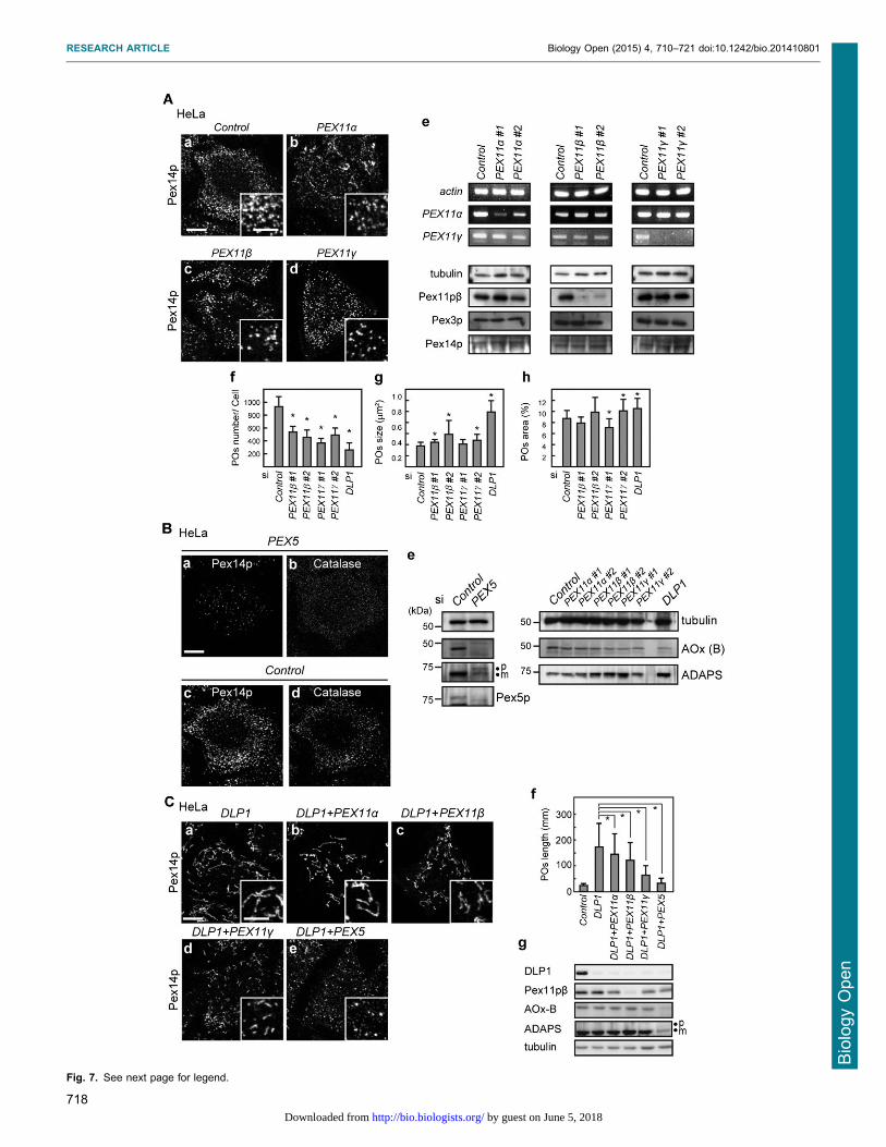
Fig. 7. See next page for legend.
RESEARCH ARTICLE Biology Open (2015) 4, 710–721 doi:10.1242/bio.201410801
718
Bio
log
yO
pe
n
by guest on June 5, 2018http://bio.biologists.org/Downloaded from

(PDPC), and 1-palmitoyl-2-docosahexaenoyl-sn-glycero-3-phosphoethanolamine
(PDPE) were purchased from Avanti Polar Lipids (Alabaster, AL). Bovine
brain L-a-phosphatidyl-L-serine (PS) was from Sigma (St Louis, MO). All
lipids were stored in chloroform at 220 C.
AntibodiesThe antibodies used were rabbit antisera to Pex14p (Shimizu et al., 1999),
PMP70 (Tsukamoto et al., 1990; Honsho et al., 1998), AOx (Tsukamoto
et al., 1990), ADAPS (Honsho et al., 2008), and Pex5p (Otera et al.,
2000). The antibody against Pex11pb was raised against recombinant
Pex11pb protein (amino acid residues 1–203), then affinity-purified as
described previously (Itoyama et al., 2012). Mouse monoclonal
antibodies to human DLP1 (BD Biosciences, Franklin Lake, NJ),
human tubulin (Abcam, Cambridge, UK), and c-Myc (Santa Cruz
Biotechnology, Santa Cruz, CA) were purchased.
Cell culture and siRNA transfectionHeLa cells were maintained in DMEM (Life Technologies, Grand Island,
NY) supplemented with 10% FCS under 5% CO2. DNA transfection of
HeLa cells was done with Lipofectamine (Life Technologies) according
to the manufacturer’s protocols. Knockdown experiments were performed
using Stealth siRNA duplexes (Life Technologies). The siRNA target
sequences were as follows: human Pex11pc #1, 59-AAGAGUCGCAAG-
AUGGUCCUGCAGU-39; human Pex11pc #2, 59-UUGCUUAGUGUAG-
ACAAACAUGGCC-39; and human Mff, 59-UUAUCACACUAGCAUU-
UGGAACUCC-39. Stealth RNAi Negative Control (Life Technologies)
was used as a control. The siRNAs for PEX11a, PEX11b, and DLP1 were
described previously (Itoyama et al., 2012). HeLa cells were trypsinized
and plated on cover slips. Cells were immediately transfected with 20 nM
of siRNA with Lipofectamine 2000 and cultured for 96 h.
Plasmid constructioncDNA encoding human Pex11pb was amplified by reverse-transcription
PCR using mRNA from HeLa cells as the template. The PCR product
was cloned into pcDNAZeo3.1 (Life Technologies) to generate the
untagged Pex11pb expression plasmid for HeLa cells. For the
construction of His-tagged Pex11pa, the EcoRI-SalI fragment of
Pex11pa was ligated into the corresponding sites of pCold1 (Takara,
Japan). For His-Pex11pb and His-Pex11pc, the BamHI-EcoRI fragments
were respectively amplified and inserted into pCold1. EGFP-fused
Pex11pb and Pex11pc were generated by the insertion of the KpnI-
BamHI fragment of plasmid encoding MBP and the BamHI-BglII
fragment of plasmid encoding EGFP into the KpnI-BamHI site of the
pCold1-Pex11pb and pCold1-Pex11pc, respectively. To construct EGFP-
Pex11pa, using MBP-EGFP as a template, KpnI-BamHI fragment was
amplified and ligated into pCold1 and the EcoRI-SalI fragment of the
plasmid encoding Pex11pa was subsequently inserted into the plasmid.
Introduction of point mutations in the N-terminal amphipathic helix of
Pex11pb was performed by site-directed mutagenesis using specific
primers: Pex11pb-P sense, 59-TGAGCCTTGGAAGAAAGCTTCCA-
CGCCTGGGTAACTCAGCACCTGCCCTTGAGTCAGCCAAAAG-39;
Pex11pb-P antisense, 59-CTTTTGGCTGACTCAAGGGCAGGTGCTGA-
GTTACCCAGGCGTGGAAGCTTTCTTCCAAGGCTCA-39; Pex11pb-wsense, 59-ACCTGAGCCTTGGAAGAAAGGAACTACGCGAGGGTA-
ACTCAGAAGATGCCCTTGAGTCAGCCAA-39; Pex11pb-w antisense,
59-TTGGCTGACTCAAGGGCATCTTCTGAGTTACCCTCGCGTAGT-
TCCTTTCTTCCAAGGCTCAGGT-39. Pex11pb-w contains L58E, L61E,
and A65E mutations, and Pex11pb-P has point mutations to proline
residues at L59 and D66. Pex14p was prepared as a GST-fusion as
described previously (Itoh and Fujiki, 2006).
Morphological analysisFor the observation of peroxisomal elongation by Pex11pb, untagged
Pex11pb was expressed in HeLa cells. Cells were fixed at 6 h and 24 h
after transfection. For the detection of peroxisomal fission induced by
Pex11pa, Pex11pb, Pex11pc, and mutant forms of Pex11pb, cells were
transfected with the corresponding plasmids for 24 h. To verify the effect
of these fission factors on Pex11pb-dependent peroxisomal proliferation,
Pex11p-expression plasmids were transfected into HeLa cells pre-treated
for 48 h with siRNA for DLP1 or MFF. Cells were fixed with 4%
paraformaldehyde for 15 min at room temperature. Peroxisomes were
visualized by indirect immunofluorescence staining with the indicated
antibodies. Primary antibodies were detected with goat anti-mouse and
anti-rabbit IgG conjugated to Alexa Fluor 488 or Alexa Fluor 568
(Molecular Probes/Life Technologies). Cells were observed under
confocal laser microscopy (LSM710; Carl Zeiss, Oberkochen,
Germany) with the Plan Apochromat 100 6/1.3 NA objective lens.
Line scanning was performed using the Plot profile tool in ImageJ
software (National Institutes of Health, Bethesda, Md). For the analysis
of number, size, and area of peroxisomes, images were taken of at least
30 cells by confocal microscopy. Each image was converted to a
threshold image, and the number of peroxisomes was measured using the
Analyze Particle command in ImageJ.
Fig. 8. A schematic model for the Pex11pb function inperoxisomal fission. Schematic drawing ofperoxisomes during fission process as maturation wasrepresented in red. The whole process consists of threesteps, maturation, constriction/elongation, and fission.Pex11pb is highlighted in green only in the constriction/elongation step, where it locates at the constriction sites.Note that the Pex11pb forms the active oligomer at thestage to deform peroxisomal membrane.
Fig. 7. Knockdown of PEX11 in HeLa cells. (A) HeLa cells were treated for96 h with a control siRNA (a) or siRNA for PEX11a (b), PEX11b (c), orPEX11c (d). Cells were then immunostained with an anti-Pex14p antibody.Scale bars: 10 mm and 5 mm (inset). The efficiency of knockdown wasevaluated by RT-PCR (e, upper panel) and western blotting (e, lower panel).PEX11a and PEX11c mRNA levels were assessed by RT-PCR using totalRNA. Actin was used as a loading control. Pex11pb protein levels wereverified by immunoblot analysis using the antibody to Pex11pb. Peroxisomalmarker proteins, Pex3p and Pex14p, were also assessed. Tubulin was usedas a loading control. Peroxisomal abundance per cell (f), averageperoxisomal size (g), and peroxisomal area per cell (h) in each siRNA-treatedcell type were determined as described in the Materials and Methods. Atleast 20 cells were randomly selected and analyzed at each time point. Datarepresent the means6SD. *p,0.05. (B) HeLa cells were treated with acontrol siRNA or siRNA for PEX5 for 48 h and immunostained withantibodies to Pex14p (a,c) and catalase (b,d). Scale bar: 10 mm. Cells werelysed and analyzed by SDS-PAGE and immunoblotting using the antibodiesindicated on the right in the panel (e). ‘p’ and ‘m’ in the panel of ADAPSindicates the larger ADAPS precursor harboring PTS2 and its mature form,respectively. (C) HeLa cells were transfected for 96 h with siRNAs each forDLP1 (a) in combination with the indicated genes (b–e). Cells were thensubjected to immunostaining with an anti-Pex14p antibody. Scale bars:10 mm and 5 mm (inset). The average lengths of peroxisomes of the cellswere measured and plotted in (f). Cells were lysed and analyzed by SDS-PAGE and immunoblotting using the antibodies indicated on the left in thepanel (g). Data represent the means6SD. *p,0.05.
RESEARCH ARTICLE Biology Open (2015) 4, 710–721 doi:10.1242/bio.201410801
719
Bio
log
yO
pe
n
by guest on June 5, 2018http://bio.biologists.org/Downloaded from
In silico analysisMultiple sequence alignment was prepared as described previously
(Opalinski et al., 2011). The hydrophobic moments of the sequences of
Pex11pa, Pex11pb, and Pex11pc were calculated using the EMBOSS
server (http://mobyle.pasteur.fr/cgi-bin/portal.py?form5hmoment). The
helical wheel representation of the Pex11p amphipathic region was
prepared using the Heliquest server (http://heliquest.ipmc.cnrs.fr).
Preparation of small unilamellar liposomeLiposomes were prepared essentially as described previously (Takei et al.,
2001). The lipid mixture was dried under nitrogen stream to form a thin
lipid film, and then rehydrated in Hepes-KOH buffer (20 mM Hepes-KOH,
pH 7.4, 150 mM NaCl). Liposomes were formed by vortexing, then
extruded by passing through a 100-nm filter 11 times. The lipid composition
was designed to mimic mammalian peroxisomal membranes (Hardeman
et al., 1990). DHA levels in human fibroblasts were determined (Itoyama
et al., 2012). DHA liposomes contained DOPC/DOPE/PDPC/PDPE/PI/PS/
Rhodamine-PE at a ratio of 29:25:25:11:5:5:1% (w/w).
Protein purificationE. coli BL21(DE3) was transformed with pCold-Pex11p and protein
expression was induced at 15 C for 24 h with 0.1 mM isopropyl b-D-
thiogalactoside (IPTG). Cells were harvested and lysed in sarcosine
buffer 50 mM Tris-HCl, pH 7.4, 0.5 M NaCl, 2 M Urea, 50 mM
imidazole, 0.5% sodium N-lauroylsarcosine. After 15-min incubation on
ice, the lysate was ultracentrifuged at 42,000 rpm for 30 min at 4 C using
a Hitachi TLA100.3 rotor (Hitachi, Tokyo, Japan). The supernatant was
incubated with Ni-NTA beads (Qiagen, Dusseldorf, Germany) at 4 C for
1 h, then the beads were collected and washed with wash buffer 50 mM
Tris-HCl, pH 7.4, 150 mM NaCl, 0.5% sodium N-lauroylsarcosine.
Protein was eluted by washing buffer containing 0.3 M imidazole. The
elution buffer was used for protein reconstitution in the control of the
experiment. For purification of MBP-EGFP-Pex11p proteins, cells were
lysed in buffer containing n-dodecyl-b-D-maltoside (Dojindo, Kumamoto,
Japan), and protein was affinity-purified by amylose resin (New England
BioLabs, Hertfordshire, UK) according to the manufacturer’s instructions.
We named MBP-EGFP-Pex11p protein as ‘‘EGFP-Pex11p’’ and used as
such, since the MBP-tag was not readily cleaved off by PreScission
protease (GH Healthcare, Little Chalfont, UK).
Protein reconstitutionPurified protein (up to 1 mg) or the same volume of the elution buffer was
mixed with 50 ml of liposomes, and the mixture was frozen at 280 C for
at least 5 min. Then, the mixture was thawed and diluted in 3 ml of
Hepes-KOH buffer to form proteo-liposomes. Liposomes were collected
by ultracentrifugation at 100,000 rpm for 1 h at 4 C, using a Hitachi
TLA100.3 rotor. For the separation of protein precipitate after
ultracentrifugation, the liposomal pellet was mixed with an equal
volume of 60% sucrose (,100 ml). One ml of 20% sucrose was added
to the liposomes, and 100 ml of Hepes-KOH buffer was loaded on the top
of the sucrose gradient. Then, the sample was centrifuged at 100,000 rpm
for 1 h at 4 C using a Hitachi TLA100.3 rotor. Three serial fractions were
harvested from the top of the tube by pipetting.
Observation of protein-reconstituted proteo-liposomesPrecipitated proteo-liposomes were mounted on glass slides by mixing
with PermaFluor (Thermo Scientific, Bremen, Germany), and covered
with glass coverslips. Samples were observed under confocal laser-
scanning microscopy with a Plan Apochromat 1006/1.3 NA objective
lens. Photobleaching was performed in a 1-mm circle placed at the edge
of a cluster of liposomes. Images were taken before and after 30 rounds
of photobleaching, and the total fluorescence intensity of the liposomal
clusters was analyzed. In the control experiment, an adjacent region to
the liposome clusters was photobleached and subjected to analysis.
Electron microscopyFor negative staining electron microscopy, proteo-liposomes were
adsorbed onto a formvar-coated 300-mesh copper grid (Nisshin EM
Co., Ltd, Tokyo, Japan) that had been glow-discharged just prior to use.
The grid was negatively stained with 2% uranyl acetate and observed
under a transmission electron microscope at 200 kV (Tecnai-20, FEI
Company, Eindhoven, The Netherlands). To detect the localization of
Pex11pb, proteo-liposomes were stained with antibodies to Pex11pb for
1 h at room temperature (RT), and labeled for 1 h with a goat anti-rabbit
IgG secondary antibody conjugated to 10 nm gold particles (EY
Laboratories, San Mateo, CA). The liposomes were then collected by
ultracentrifugation and subjected to negative staining electron
microscopy. For ultra-thin sectioning, liposomes were pelleted and
fixed with 3% glutaraldehyde, and further processed for embedding in
Epon and staining, as described previously (Meyerholz et al., 2005).
Detection of mRNA by RT-PCRTotal RNA was extracted from HeLa cells using TRIzol reagent (Life
Technologies), and cDNA was obtained by reverse transcription (RT),
(PrimeScript RT-PCR kit; Takara, Tokyo, Japan) according to the
manufacturer’s instructions. RT-PCR was performed using a set of
specific primers: Pex11pc sense, 59-TGATGACCTGGCCATGTTTGT-
CTA-39, and antisense, 59-AAGTGACAGCGCCTCCGACTGCAT-39.
Primer sets for Pex11pa and actin have been described previously
(Itoyama et al., 2012).
AcknowledgementsWe thank R. Ugawa and M. Sasaki in the Laboratory for Technical Support andthe Medical Institute of Bioregulation, Kyushu University, for their technicalsupport with the electron microscopy. We also thank K. Shimizu for preparingfigures and the other members of our laboratory for discussions.
Competing interestsThe authors declare no competing or financial interests.
Author contributionsY.Y. conceived the study, performed the experiments and wrote the manuscript,H.N. provided several reagents and contributed to manuscript writing,M.H. performed some of the experiments and contributed to manuscript writing,A.I. performed some of the experiments, Y.F. conceived the study and wrote themanuscript.
FundingThis work was supported in part by Grants-in-Aid for Scientific Research[25440030 to H.N.; 23570236 and 26440102 to M.H.; and 19058011, 20370039,24247038, 25112518, 25116717, and 26116007 to Y. F.]; Global COE (Centers ofExcellence) Program; and The Grants for Excellent Graduate Schools from theMinistry of Education, Culture, Sports, Science, and Technology of Japan; aCREST grant (to Y.F.) from the Science and Technology Agency of Japan; KyushuUniversity Interdisciplinary Programs in Education and Projects in ResearchDevelopment (to M.H. and Y.F.); and grants from Takeda Science Foundation (toM.H. and Y.F.); and Japan Foundation for Applied Enzymology (to Y.F.).
ReferencesAbe, I. and Fujiki, Y. (1998). cDNA cloning and characterization of a constitutively
expressed isoform of the human peroxin Pex11p. Biochem. Biophys. Res.Commun. 252, 529-533.
Abe, I., Okumoto, K., Tamura, S. and Fujiki, Y. (1998). Clofibrate-inducible, 28-kDa peroxisomal integral membrane protein is encoded by PEX11. FEBS Lett.431, 468-472.
Bonekamp, N. A., Grille, S., Cardoso, M. J., Almeida, M., Aroso, M., Gomes,S., Magalhaes, A. C., Ribeiro, D., Islinger, M. and Schrader, M. (2013). Self-interaction of human Pex11pb during peroxisomal growth and division. PLoSONE 8, e53424.
Chang, C. C., South, S., Warren, D., Jones, J., Moser, A. B., Moser, H. W. andGould, S. J. (1999). Metabolic control of peroxisome abundance. J. Cell Sci.112, 1579-1590.
Chang, C. R., Manlandro, C. M., Arnoult, D., Stadler, J., Posey, A. E., Hill, R. B.and Blackstone, C. (2010). A lethal de novo mutation in the middle domain ofthe dynamin-related GTPase Drp1 impairs higher order assembly andmitochondrial division. J. Biol. Chem. 285, 32494-32503.
Dawson, J. C., Legg, J. A. and Machesky, L. M. (2006). Bar domain proteins: arole in tubulation, scission and actin assembly in clathrin-mediated endocytosis.Trends Cell Biol. 16, 493-498.
Delille, H. K., Agricola, B., Guimaraes, S. C., Borta, H., Luers, G. H., Fransen,M. and Schrader, M. (2010). Pex11pbeta-mediated growth and division ofmammalian peroxisomes follows a maturation pathway. J. Cell Sci. 123, 2750-2762.
RESEARCH ARTICLE Biology Open (2015) 4, 710–721 doi:10.1242/bio.201410801
720
Bio
log
yO
pe
n
by guest on June 5, 2018http://bio.biologists.org/Downloaded from
Ebberink, M. S., Koster, J., Visser, G., Spronsen, F., Stolte-Dijkstra, I., Smit,G. P. A., Fock, J. M., Kemp, S., Wanders, R. J. A. and Waterham, H. R. (2012).A novel defect of peroxisome division due to a homozygous non-sense mutationin the PEX11b gene. J. Med. Genet. 49, 307-313.
Ford, M. G. J., Mills, I. G., Peter, B. J., Vallis, Y., Praefcke, G. J. K., Evans, P. R.and McMahon, H. T. (2002). Curvature of clathrin-coated pits driven by epsin.Nature 419, 361-366.
Gandre-Babbe, S. and van der Bliek, A. M. (2008). The novel tail-anchoredmembrane protein Mff controls mitochondrial and peroxisomal fission inmammalian cells. Mol. Biol. Cell 19, 2402-2412.
Ghaedi, K., Honsho, M., Shimozawa, N., Suzuki, Y., Kondo, N. and Fujiki, Y.(2000). PEX3 is the causal gene responsible for peroxisome membraneassembly-defective Zellweger syndrome of complementation group G. Am.J. Hum. Genet. 67, 976-981.
Grabenbauer, M., Satzler, K., Baumgart, E. and Fahimi, H. D. (2000). Three-dimensional ultrastructural analysis of peroxisomes in HepG2 cells. Absence ofperoxisomal reticulum but evidence of close spatial association with theendoplasmic reticulum. Cell Biochem. Biophys. 32, 37-49.
Hardeman, D., Versantvoort, C., van den Brink, J. M. and van den Bosch, H.(1990). Studies on peroxisomal membranes. Biochim. Biophys. Acta 1027, 149-154.
Hettema, E. H. and Motley, A. M. (2009). How peroxisomes multiply. J. Cell Sci.122, 2331-2336.
Honsho, M., Tamura, S., Shimozawa, N., Suzuki, Y., Kondo, N. and Fujiki, Y.(1998). Mutation in PEX16 is causal in the peroxisome-deficient Zellwegersyndrome of complementation group D. Am. J. Hum. Genet. 63, 1622-1630.
Honsho, M., Yagita, Y., Kinoshita, N. and Fujiki, Y. (2008). Isolation andcharacterization of mutant animal cel l l ine defect ive in alky l -dihydroxyacetonephosphate synthase: localization and transport ofplasmalogens to post-Golgi compartments. Biochim. Biophys. Acta 1783, 1857-1865.
Itoh, R. and Fujiki, Y. (2006). Functional domains and dynamic assembly of theperoxin Pex14p, the entry site of matrix proteins. J. Biol. Chem. 281, 10196-10205.
Itoh, T., Erdmann, K. S., Roux, A., Habermann, B., Werner, H. and De Camilli,P. (2005). Dynamin and the actin cytoskeleton cooperatively regulate plasmamembrane invagination by BAR and F-BAR proteins. Dev. Cell 9, 791-804.
Itoyama, A., Honsho, M., Abe, Y., Moser, A., Yoshida, Y. and Fujiki, Y. (2012).Docosahexaenoic acid mediates peroxisomal elongation, a prerequisite forperoxisome division. J. Cell Sci. 125, 589-602.
Itoyama, A., Michiyuki, S., Honsho, M., Yamamoto, T., Moser, A., Yoshida, Y.and Fujiki, Y. (2013). Mff functions with Pex11pb and DLP1 in peroxisomalfission. Biol. Open 2, 998-1006.
Kobayashi, S., Tanaka, A. and Fujiki, Y. (2007). Fis1, DLP1, and Pex11pcoordinately regulate peroxisome morphogenesis. Exp. Cell Res. 313, 1675-1686.
Koch, J. and Brocard, C. (2012). PEX11 proteins attract Mff and human Fis1 tocoordinate peroxisomal fission. J. Cell Sci. 125, 3813-3826.
Koch, A., Thiemann, M., Grabenbauer, M., Yoon, Y., McNiven, M. A. andSchrader, M. (2003). Dynamin-like protein 1 is involved in peroxisomal fission.J. Biol. Chem. 278, 8597-8605.
Koch, A., Schneider, G., Luers, G. H. and Schrader, M. (2004). Peroxisomeelongation and constriction but not fission can occur independently of dynamin-like protein 1. J. Cell Sci. 117, 3995-4006.
Koch, A., Yoon, Y., Bonekamp, N. A., McNiven, M. A. and Schrader, M. (2005).A role for Fis1 in both mitochondrial and peroxisomal fission in mammalian cells.Mol. Biol. Cell 16, 5077-5086.
Koch, J., Pranjic, K., Huber, A., Ellinger, A., Hartig, A., Kragler, F. andBrocard, C. (2010). PEX11 family members are membrane elongation factorsthat coordinate peroxisome proliferation and maintenance. J. Cell Sci. 123,3389-3400.
Lazarow, P. B. and Fujiki, Y. (1985). Biogenesis of peroxisomes. Annu. Rev. CellBiol. 1, 489-530.
Li, X. and Gould, S. J. (2002). PEX11 promotes peroxisome divisionindependently of peroxisome metabolism. J. Cell Biol. 156, 643-651.
Li, X. and Gould, S. J. (2003). The dynamin-like GTPase DLP1 is essential forperoxisome division and is recruited to peroxisomes in part by PEX11. J. Biol.Chem. 278, 17012-17020.
Li, X., Baumgart, E., Morrell, J. C., Jimenez-Sanchez, G., Valle, D. andGould, S. J. (2002a). PEX11 b deficiency is lethal and impairs neuronalmigration but does not abrogate peroxisome function. Mol. Cell. Biol. 22,4358-4365.
Li, X., Baumgart, E., Dong, G.-X., Morrell, J. C., Jimenez-Sanchez, G., Valle,D., Smith, K. D. and Gould, S. J. (2002b). PEX11a is required for peroxisomeproliferation in response to 4-phenylbutyrate but is dispensable for peroxisomeproliferator-activated receptor alpha-mediated peroxisome proliferation. Mol.Cell. Biol. 22, 8226-8240.
Matsuzono, Y., Kinoshita, N., Tamura, S., Shimozawa, N., Hamasaki, M.,Ghaedi, K., Wanders, R. J. A., Suzuki, Y., Kondo, N. and Fujiki, Y. (1999).Human PEX19: cDNA cloning by functional complementation, mutation analysisin a patient with Zellweger syndrome, and potential role in peroxisomalmembrane assembly. Proc. Natl. Acad. Sci. USA 96, 2116-2121.
Meyerholz, A., Hinrichsen, L., Groos, S., Esk, P. C., Brandes, G. andUngewickell, E. J. (2005). Effect of clathrin assembly lymphoid myeloidleukemia protein depletion on clathrin coat formation. Traffic 6, 1225-1234.
Muntau, A. C., Mayerhofer, P. U., Paton, B. C., Kammerer, S. and Roscher,A. A. (2000). Defective peroxisome membrane synthesis due to mutations inhuman PEX3 causes Zellweger syndrome, complementation group G. Am.J. Hum. Genet. 67, 967-975.
Opalinski, Ł., Kiel, J. A., Williams, C., Veenhuis, M. and van der Klei, I. J.(2011). Membrane curvature during peroxisome fission requires Pex11. EMBOJ. 30, 5-16.
Otera, H., Harano, T., Honsho, M., Ghaedi, K., Mukai, S., Tanaka, A., Kawai,A., Shimizu, N. and Fujiki, Y. (2000). The mammalian peroxin Pex5pL, thelonger isoform of the mobile peroxisome targeting signal (PTS) type 1transporter, translocates the Pex7p-PTS2 protein complex into peroxisomesvia its initial docking site, Pex14p. J. Biol. Chem. 275, 21703-21714.
Otera, H., Setoguchi, K., Hamasaki, M., Kumashiro, T., Shimizu, N. and Fujiki,Y. (2002). Peroxisomal targeting signal receptor Pex5p interacts with cargoesand import machinery components in a spatiotemporally differentiated manner:conserved Pex5p WXXXF/Y motifs are critical for matrix protein import. Mol.Cell. Biol. 22, 1639-1655.
Otera, H., Wang, C., Cleland, M. M., Setoguchi, K., Yokota, S., Youle, R. J. andMihara, K. (2010). Mff is an essential factor for mitochondrial recruitment ofDrp1 during mitochondrial fission in mammalian cells. J. Cell Biol. 191, 1141-1158.
Peter, B. J., Kent, H. M., Mills, I. G., Vallis, Y., Butler, P. J., Evans, P. R. andMcMahon, H. T. (2004). BAR domains as sensors of membrane curvature: theamphiphysin BAR structure. Science 303, 495-499.
Praefcke, G. J. K. and McMahon, H. T. (2004). The dynamin superfamily:universal membrane tubulation and fission molecules? Nat. Rev. Mol. Cell Biol.5, 133-147.
Schrader, M., Reuber, B. E., Morrell, J. C., Jimenez-Sanchez, G., Obie, C.,Stroh, T. A., Valle, D., Schroer, T. A. and Gould, S. J. (1998). Expression ofPEX11b mediates peroxisome proliferation in the absence of extracellularstimuli. J. Biol. Chem. 273, 29607-29614.
Shimizu, N., Itoh, R., Hirono, Y., Otera, H., Ghaedi, K., Tateishi, K., Tamura, S.,Okumoto, K., Harano, T., Mukai, S. et al. (1999). The peroxin Pex14p. cDNAcloning by functional complementation on a Chinese hamster ovary cell mutant,characterization, and functional analysis. J. Biol. Chem. 274, 12593-12604.
Shimozawa, N., Suzuki, Y., Zhang, Z., Imamura, A., Ghaedi, K., Fujiki, Y. andKondo, N. (2000). Identification of PEX3 as the gene mutated in a Zellwegersyndrome patient lacking peroxisomal remnant structures. Hum. Mol. Genet. 9,1995-1999.
Shupliakov, O., Low, P., Grabs, D., Gad, H., Chen, H., David, C., Takei, K., DeCamilli, P. and Brodin, L. (1997). Synaptic vesicle endocytosis impaired bydisruption of dynamin-SH3 domain interactions. Science 276, 259-263.
South, S. T. and Gould, S. J. (1999). Peroxisome synthesis in the absence ofpreexisting peroxisomes. J. Cell Biol. 144, 255-266.
Takei, K., Slepnev, V. I., Haucke, V. and De Camilli, P. (1999). Functionalpartnership between amphiphysin and dynamin in clathrin-mediatedendocytosis. Nat. Cell Biol. 1, 33-39.
Takei, K., Slepnev, V. I. and De Camilli, P. (2001). Interactions of dynamin andamphiphysin with liposomes. Methods Enzymol. 329, 478-486.
Tanaka, A., Okumoto, K. and Fujiki, Y. (2003). cDNA cloning andcharacterization of the third isoform of human peroxin Pex11p. Biochem.Biophys. Res. Commun. 300, 819-823.
Tanaka, A., Kobayashi, S. and Fujiki, Y. (2006). Peroxisome division is impairedin a CHO cell mutant with an inactivating point-mutation in dynamin-like protein1 gene. Exp. Cell Res. 312, 1671-1684.
Thoms, S. and Erdmann, R. (2005). Dynamin-related proteins and Pex11proteins in peroxisome division and proliferation. FEBS J. 272, 5169-5181.
Tsukamoto, T., Yokota, S. and Fujiki, Y. (1990). Isolation and characterization ofChinese hamster ovary cell mutants defective in assembly of peroxisomes.J. Cell Biol. 110, 651-660.
van den Bosch, H., Schutgens, R. B. H., Wanders, R. J. A. and Tager, J. M.(1992). Biochemistry of peroxisomes. Annu. Rev. Biochem. 61, 157-197.
Wanders, R. J. A. and Waterham, H. R. (2006). Peroxisomal disorders: the singleperoxisomal enzyme deficiencies. Biochim. Biophys. Acta 1763, 1707-1720.
Waterham, H. R., Koster, J., van Roermund, C. W. T., Mooyer, P. A. W.,Wanders, R. J. A. and Leonard, J. V. (2007). A lethal defect of mitochondrialand peroxisomal fission. N. Engl. J. Med. 356, 1736-1741.
Wu, M., Huang, B., Graham, M., Raimondi, A., Heuser, J. E., Zhuang, X. andDe Camilli, P. (2010). Coupling between clathrin-dependent endocytic buddingand F-BAR-dependent tubulation in a cell-free system. Nat. Cell Biol. 12, 902-908.
Yoon, Y., Pitts, K. R. and McNiven, M. A. (2001). Mammalian dynamin-likeprotein DLP1 tubulates membranes. Mol. Biol. Cell 12, 2894-2905.
Yoshida, Y., Kinuta, M., Abe, T., Liang, S., Araki, K., Cremona, O., Di Paolo,G., Moriyama, Y., Yasuda, T., De Camilli, P. et al. (2004). The stimulatoryaction of amphiphysin on dynamin function is dependent on lipid bilayercurvature. EMBO J. 23, 3483-3491.
Zhu, P. P., Patterson, A., Stadler, J., Seeburg, D. P., Sheng, M. andBlackstone, C. (2004). Intra- and intermolecular domain interactions of the C-terminal GTPase effector domain of the multimeric dynamin-like GTPase Drp1.J. Biol. Chem. 279, 35967-35974.
RESEARCH ARTICLE Biology Open (2015) 4, 710–721 doi:10.1242/bio.201410801
721
Bio
log
yO
pe
n
by guest on June 5, 2018http://bio.biologists.org/Downloaded from