PET/CT in Lymphoma - Human Health Campus · PET/CT in Lymphoma Tuesday, August 28, 2012 Session 5,...
37
PET/CT in Lymphoma Tuesday, August 28, 2012 Session 5, 11:30-12:10 FDG-avidity Staging (nodal & extra nodal) Response evaluation Early / interim Post-treatment Evaluation criteria
Transcript of PET/CT in Lymphoma - Human Health Campus · PET/CT in Lymphoma Tuesday, August 28, 2012 Session 5,...

PET/CT in Lymphoma
Tuesday, August 28, 2012
Session 5, 11:30-12:10
FDG-avidity
Staging (nodal & extra nodal)
Response evaluation
Early / interim
Post-treatment
Evaluation criteria

Lymphoma
Subtypes differ in
molecular characteristics
biologic behavior
aggressive
indolent
The WHO histologic classification
morphologic
immunohistochemical
genetic features
The most important factors
For therapy and prognosis
histologic subtype
extent of disease
Coloured scanning electron micrograph of dividing
Hodgkin's cells taken from the pleural effusions of a
55 year old, male patient with "mixed cellularity
Hodgkin disease

Based on size alone
- benign lymph node enlargement
may lead to overstaging
- malignant small lymph nodes may
be understaged
Limited detection of spleen, liver,
and bone marrow involvement
Equivocal lesions require additional
imaging or biopsy
Limitations of conventional imaging

FDG-avidity
Weiler-Sagie M et al
FDG Avidity in Lymphoma Readdressed:
A Study of 766 Patients JNM 2010
METHODS:
The reports from FDG PET/CT
studies performed in a single
center for staging of 1,093
patients with newly diagnosed
Hodgkin disease and non-Hodgkin
lymphoma were reviewed for the
presence of FDG avidity.
766 patients with a histopathologic
diagnosis verified according to the
WHO classification were included
in the final analysis.

FDG-avidity was lower in indolent disease
(83%) than in aggressive disease (97%).
Indolent subtypes (eg. plasmacytoma,
follicular lymphoma) are FDG-avid
Aggressive (enteropathy-type
T-cell lymphoma) has low FDG-uptake
FDG-avidity

Lymphoma

Lymphoma

Staging HL and aggresive NHL
FDG-PET detects more nodal
and extranodal disease sites,
than CT
The higher sensitivity leads to
significant upward stage
migration in 10-40%. In about
half of these patients treatment
strategy is changed
PET seems to be at least as
sensitive as blind bone marow
biopsy in HD
Bangerter M et al Ann Oncol 1998
Buchmann i et al Cancer 2001
Carr R Blood 1998
FDG-PET
Sagggittal
view

FDG-PET - “state of the art”
In HL and aggressive NHL FDG-PET is more accurate
for diagnosing both nodal and extranodal disease than
CT, thus having a strong potential impact on the staging
FDG-PET is more accurate
Gastric lymphoma (lesser curvature)

Staging
Whether the changes in treatment
strategy caused by FDG-PET will
eventually lead to improvement in
treatment outcome is at present unknown
and being tested in randomized trials

Response evaluation - general
• Tumor response serves as an important
surrogate for other measures of clinical benefit
such as progression-free and overall survival
• Tumor response also serves as an important
guide in decisions regarding continuation or
change of therapy
• Response has hitherto been based mainly on
morphological criteria with a reduction in tumor
size on CT as the most important factor
Surrogate endpoint and decision guide

PET-negative resopnders and PET-positive non-
responders after 2 cycles of chemotherapy
CT cannot predict
outcome
Early treatmemt evaluation

Early response evaluation
• Several studies, in Hodgkin lymphoma and
in aggressive non Hodgkin lymphoma,
have showed that an early FDG-PET
scan, after 1 to 3 cycles of chemotherapy,
is a strong predictor of treatment outcome.
Predictor of treatment outcome

• After completion of therapy CT will often reveal
residual masses. It is very difficult to assess
whether this represents viable lymphoma,
fibrotic scar tissue or necrosis in patients with
otherwise clinical complete response. To
perform a biopsy on all these lesions would be
impractical, and even if it were done it would be
too inaccurate.
• CRu – complete remission unconfirmed
Residual masses
Post treatment response evaluation

Post treatment evaluation
• FDG-PET distinguish between viable lymphoma and necrosis/fibrosis in residual masses (CT-scan) after treatment of HL and aggressive NHL
• Post-treatment FDG-PET is highly predictive of PFS and OS in HL and (aggresive) NHL
• FDG-PET is incoorporated into the definition of end-of-treatment response evaluation (the International Harmonization Project)
Cheson BD et al. Revised resopnse criteria for malignant lymphoma. J Clin oncol 2007
Juweid ME et al. Use of positron emissio tomography for response assessment of lymphoma:
consensus of the Imaging Subcommittee of International harmonization Projet in Lymphoma.J Clin
Oncol 2007

Clinical example

Response evaluation
• It is, however, clear that a negative FDG-PET
scan after therapy does not exclude the
presence of microscopic disease.
• The new recommendations for response criteria
are not as yet supported by clinical data, and
long-term follow-up of lymphoma patients
evaluated by these criteria is awaited with great
interest.
Microscopic disease

Treatmemt evaluation
Quantitative assessment
Semiquantitative analysis using
standardized uptake value (SUV) represents
the metabolic activity of the tumor compared
with that of surrounding tissue, corrected for
injected dose and (usually) patient weight.

SUV
Standardized uptake value
a widely used, simple PET
quantifier
SUV =
CPET(T) / (Injected dose / body weight)
Quantitative asessment

– tumor metabolism
– underestimation of true activity in small tumors
– heterogeneous tumors
– time (after inj) dependent
– plasma glucose dependent
– Body weight, BSA, LBM
– Scanning parameters and PET-scanner
Intraindividual variation in FDG uptake in serial PET-scans is low
(CV 10%). Changes by more than 20% ( 1 SUV) is significant
SUVs

International Harmonization Project 2007
London criteria 2010
Gallamini criteria 2007
Deauville criteria 2010, 2011
October 2012 in Menton
Response criteria
Deauville, France


Deauville 3 = FDG-positive
SUV only for research

The Deauville criteria

The Deauville criteria – clinical application
Baseline
Deauville 5
Deauville 1

The Deauville criteria – clinical application
Deauville 1

The Deauville criteria – clinical application

The Deauville criteria – clinical application

The Deauville criteria – clinical application

The Deauville criteria – clinical application

The Deauville criteria – clinical application
Deauville 1

Treatmemt evaluation
It is strongly recommended that
a baseline scan is available comparison
the scan is performed with either
low-dose or diagnostic CT
the time from chemotherapy to scan is
no less than 10 days

Ten days later: 11 cm absces,
communicating with dudenum.
Special case

Journal of Nuclear Medicine
2006
Summary

Routine long-term
follow up is not
recommended
(subclinical disease).
If transformation of
Indolent NHL is
suspected PET is
recommended
for biopsy guidance
Summary
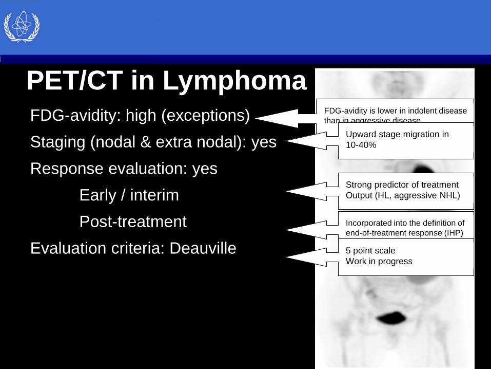
PET/CT in Lymphoma
Tuesday, August 28, 2012
Session 5, 11:30-12:10
FDG-avidity: high (exceptions)
Staging (nodal & extra nodal): yes
Response evaluation: yes
Early / interim
Post-treatment
Evaluation criteria: Deauville
FDG-avidity is lower in indolent disease
than in aggressive disease
Upward stage migration in
10-40%
Strong predictor of treatment
Output (HL, aggressive NHL)
Incorporated into the definition of
end-of-treatment response (IHP)
5 point scale
Work in progress

Tuesday, August 28, 2012
Session 5, 11:30-12:10
Ilulissat, Greenland, Denmark