PET/CT and PET/MR in Gynecological Malignancies
59
Nuclear Medicine University Hospital Zürich SWITZERLAND PET/CT and PET/MR in Gynecological Malignancies Irene A. Burger, MD Pärnu (Estonia), October 6 – 10, 2014 Radiology and Nuclear Medicine Department University Hospital Zurich Switzerland
Transcript of PET/CT and PET/MR in Gynecological Malignancies

Nuclear MedicineUniversity Hospital ZürichSWITZERLAND
PET/CT and PET/MR in Gynecological
Malignancies
Irene A. Burger, MDPärnu (Estonia), October 6 – 10, 2014
Radiology and Nuclear Medicine DepartmentUniversity Hospital Zurich
Switzerland
Nuclear Medicine, University Hospital Zurich, Switzerland
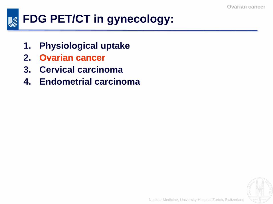
FDG PET/CT in gynecology:
1. Physiological uptake
2. Ovarian cancer
3. Cervical carcinoma
4. Endometrial carcinoma
PET/CT of the female genital tract
Physiological uptake

Nuclear Medicine, University Hospital Zurich, Switzerland
PET/CT of the female genital tract
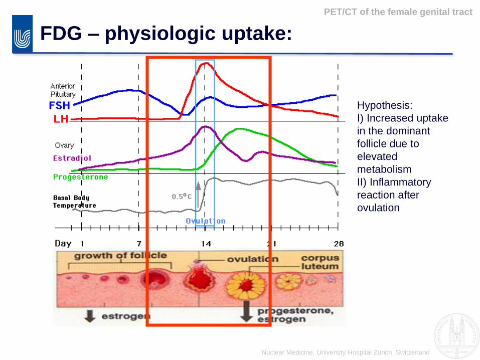
FDG – physiologic uptake:
Patient with Melanoma in
CR since 2 years:
FDG activity in the
uterine cave and right
adnex
=> Ovulation phase

Nuclear Medicine, University Hospital Zurich, Switzerland
PET/CT of the female genital tract
FDG – physiologic uptake:
56 yo Breast cancer since 8
years after several antihormonal
and chemo therapies:
Focal FDG activity in the Liver
Focal activity in both ovaries
(SUVmax 3.5)
Bilateral ovarian metastasis
Liver Metastasis
Nuclear Medicine, University Hospital Zurich, Switzerland
PET/CT of the female genital tract
FDG – physiologic uptake:
Hypothesis:
I) Increased uptake
in the dominant
follicle due to
elevated
metabolism
II) Inflammatory
reaction after
ovulation
Nuclear Medicine, University Hospital Zurich, Switzerland
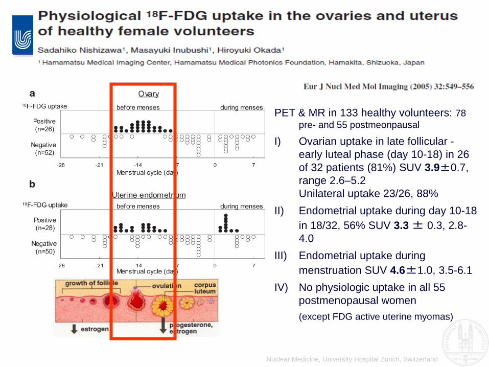
PET & MR in 133 healthy volunteers: 78
pre- and 55 postmeonpausal
I) Ovarian uptake in late follicular -
early luteal phase (day 10-18) in 26
of 32 patients (81%) SUV 3.9±0.7,
range 2.6–5.2
Unilateral uptake 23/26, 88%
II) Endometrial uptake during day 10-18
in 18/32, 56% SUV 3.3± 0.3, 2.8-
4.0
III) Endometrial uptake during
menstruation SUV 4.6±1.0, 3.5-6.1
IV) No physiologic uptake in all 55
postmenopausal women
(except FDG active uterine myomas)

Nuclear Medicine, University Hospital Zurich, Switzerland
PET/CT of the female genital tract
FDG – physiologic uptake:
Same patient as in case 1:
one year later (CR since 3
years):
FDG activity only in the
uterine cave with markedly
increased SUV=> During menses

Nuclear Medicine, University Hospital Zurich, Switzerland
PET/CT of the female genital tract
FDG – physiologic uptake:
Side note:
Vaginal tampons should
be removed or changed
after voiding
The high uptake is due to
urinary contamination,
high spill over could
obscure surrounding
structures

Nuclear Medicine, University Hospital Zurich, Switzerland
PET/CT of the female genital tract
FDG – possible interference:
25 y.o. woman
breast cancer:
FDG activity in the cervix,
SUVmax 4.3
Cervical cancer

Nuclear Medicine, University Hospital Zurich, Switzerland
FDG – physiologic uptake?:
PET/CT of the female genital tract
51 y.o. woman
Melanoma with
mediastinal and
pulmonary metastasis:
FDG activity right adnex
SUVmax 7.2
And milde activity in the
uterine cave (SUVmax 3.2)
Transvaginal
sonographie:
Bleeding into corpus
luteum cyst

Nuclear Medicine, University Hospital Zurich, Switzerland
FDG – physiologic uptake?:
PET/CT of the female genital tract
Nuclear Medicine, University Hospital Zurich, Switzerland
FDG PET/CT in gynecology:
1. Physiological uptake
2. Ovarian cancer
3. Cervical carcinoma
4. Endometrial carcinoma
Ovarian cancer
Ovarian cancer

Nuclear Medicine, University Hospital Zurich, Switzerland
Histology:
Ovarian cancer
WHO classification of surface epithelial-stromal tumors (>
90%)
Papillary serous tumors (42%)
Mucinous tumors
Endometrioid tumors
Clear-cell tumors
Transitional cell tumors (Brenner tumor)
Squamous cell tumors
Mixed epithelial tumors
Undifferentiated carcinoma
Germ cell tumors (Teratoma, gestational trophoblastic
tumors)

Nuclear Medicine, University Hospital Zurich, Switzerland
Adneocarcinomas:
Median age over 60 years
Lifetime risk 1 to 70
Risk factors:
Family history of ovarian
and breast cancer
Inherited mutation of
BRCA1 or BRCA2
Ovarian cancer
USA 2014 estimates:
new cases per year: 22’000
Cancer death per year:
14’000
Nuclear Medicine, University Hospital Zurich, Switzerland
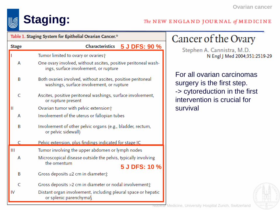
Staging:
Ovarian cancer
For all ovarian carcinomas
surgery is the first step.
-> cytoreduction in the first
intervention is crucial for
survival
5 J DFS: 90 %
5 J DFS: 10 %

Nuclear Medicine, University Hospital Zurich, Switzerland
Staging:
Ovarian cancer
FIGO:
Diagnosis: transvaginal sonography
Staging: intra operative

Nuclear Medicine, University Hospital Zurich, Switzerland
Staging:
Ovarian cancer
Nuclear Medicine, University Hospital Zurich, Switzerland
Staging:
Ovarian cancer
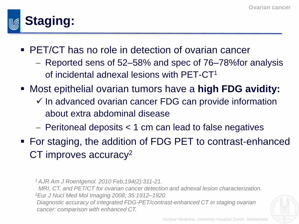
PET/CT has no role in detection of ovarian cancer
Reported sens of 52–58% and spec of 76–78%for analysis
of incidental adnexal lesions with PET-CT1
Most epithelial ovarian tumors have a high FDG avidity:
In advanced ovarian cancer FDG can provide information
about extra abdominal disease
Peritoneal deposits < 1 cm can lead to false negatives
For staging, the addition of FDG PET to contrast-enhanced
CT improves accuracy2
1 AJR Am J Roentgenol. 2010 Feb;194(2):311-21.
MRI, CT, and PET/CT for ovarian cancer detection and adnexal lesion characterization.2Eur J Nucl Med Mol Imaging 2008; 35:1912–1920.
Diagnostic accuracy of integrated FDG-PET/contrast-enhanced CT in staging ovarian
cancer: comparison with enhanced CT.

Nuclear Medicine, University Hospital Zurich, Switzerland
Ovarian cancer
Stage ?:
68 y.o. woman with
ovarian carcinoma:
Typical spread around
the liver dome
Stage IIIC
PET/CT:
Pathological FDG uptake
in LN metastasis in the
upper mediastinum
extra abdominal
disease

Nuclear Medicine, University Hospital Zurich, Switzerland
Staging:
Ovarian cancer
Comparing ceCT with PET/ceCT:
40 patients staged with ceCT and PET/ceCT, sensitivity and
accuracy improved significantly with p values of 5.6×10⁻⁷
and 1.2×10⁻⁷, respectively. (Kitajima et al. EJNMMI 2008, 35:1912-20)
Nuclear Medicine, University Hospital Zurich, Switzerland
Follow up:
Ovarian cancer
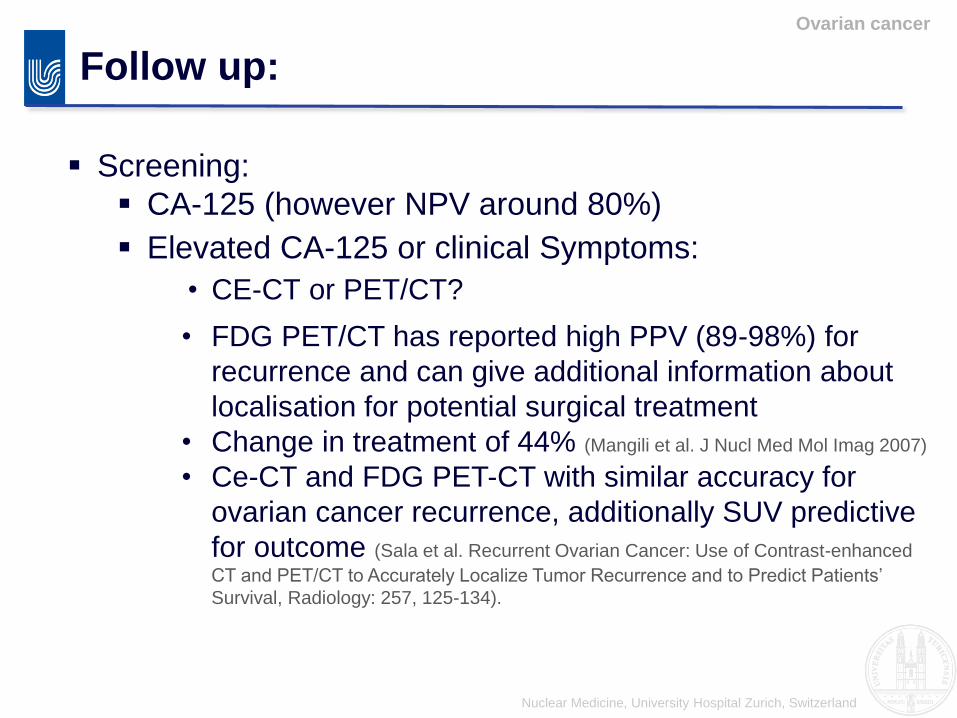
Screening:
CA-125 (however NPV around 80%)
Elevated CA-125 or clinical Symptoms:
• CE-CT or PET/CT?
• FDG PET/CT has reported high PPV (89-98%) for
recurrence and can give additional information about
localisation for potential surgical treatment
• Change in treatment of 44% (Mangili et al. J Nucl Med Mol Imag 2007)
• Ce-CT and FDG PET-CT with similar accuracy for
ovarian cancer recurrence, additionally SUV predictive
for outcome (Sala et al. Recurrent Ovarian Cancer: Use of Contrast-enhanced
CT and PET/CT to Accurately Localize Tumor Recurrence and to Predict Patients’
Survival, Radiology: 257, 125-134).

Nuclear Medicine, University Hospital Zurich, Switzerland
Ovarian cancer
Pitfalls:
In unenhanced CT peritoneal
carcinomatosis can mimic bowel loop
activity
Contrast enhanced CT of the abdomen
for ovarian cancer

Nuclear Medicine, University Hospital Zurich, Switzerland
Ovarian cancer
Pitfalls:
Peritoneal carcinomatosis
around the liver can
mimic hepatic
involvement:
This would over stage
the patient form IIIC to IV
Ovarian cancer should be
imaged with iv contrast.

Nuclear Medicine, University Hospital Zurich, Switzerland
Ovarian cancer
Summary:
For staging PET/CT with contrast (ce) is superior to ceCT
and ceMR- Sens ca. 65% and spec ca. 78%
- Limitation in assessing primary tumor extension in the
pelvis, but superior in detection of LN and distant
metastasis
PET/CT can distinguish metabolic responder and non-
responders
Restaging: higher sens and spec than CT or MR

Nuclear Medicine, University Hospital Zurich, Switzerland
Germ cell tumors:
Ovarian cancer
44 y.o. large lesion in the left
adnex
Teratoma
A mature teratoma is FDG
negative.
Nuclear Medicine, University Hospital Zurich, Switzerland
FDG PET/CT in gynecology:
1. Physiological uptake
2. Ovarian cancer
3. Cervical carcinoma
4. Endometrial carcinom
Cervical carcinoma
Cervical carcinoma
Nuclear Medicine, University Hospital Zurich, Switzerland
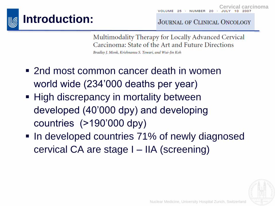
Introduction:
2nd most common cancer death in women
world wide (234’000 deaths per year)
High discrepancy in mortality between
developed (40’000 dpy) and developing
countries (>190’000 dpy)
In developed countries 71% of newly diagnosed
cervical CA are stage I – IIA (screening)
Cervical carcinoma
Nuclear Medicine, University Hospital Zurich, Switzerland
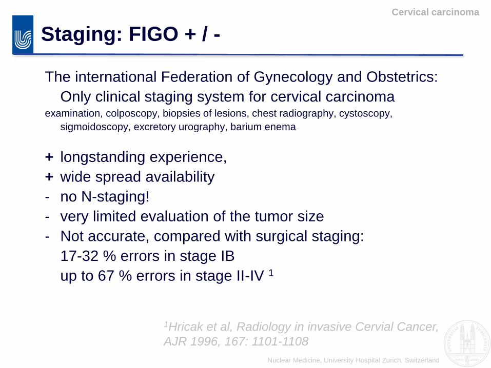
Staging: FIGO + / -
The international Federation of Gynecology and Obstetrics:
Only clinical staging system for cervical carcinomaexamination, colposcopy, biopsies of lesions, chest radiography, cystoscopy,
sigmoidoscopy, excretory urography, barium enema
+ longstanding experience,
+ wide spread availability
- no N-staging!
- very limited evaluation of the tumor size
- Not accurate, compared with surgical staging:
17-32 % errors in stage IB
up to 67 % errors in stage II-IV 1
1Hricak et al, Radiology in invasive Cervial Cancer,
AJR 1996, 167: 1101-1108
Cervical carcinoma
Nuclear Medicine, University Hospital Zurich, Switzerland
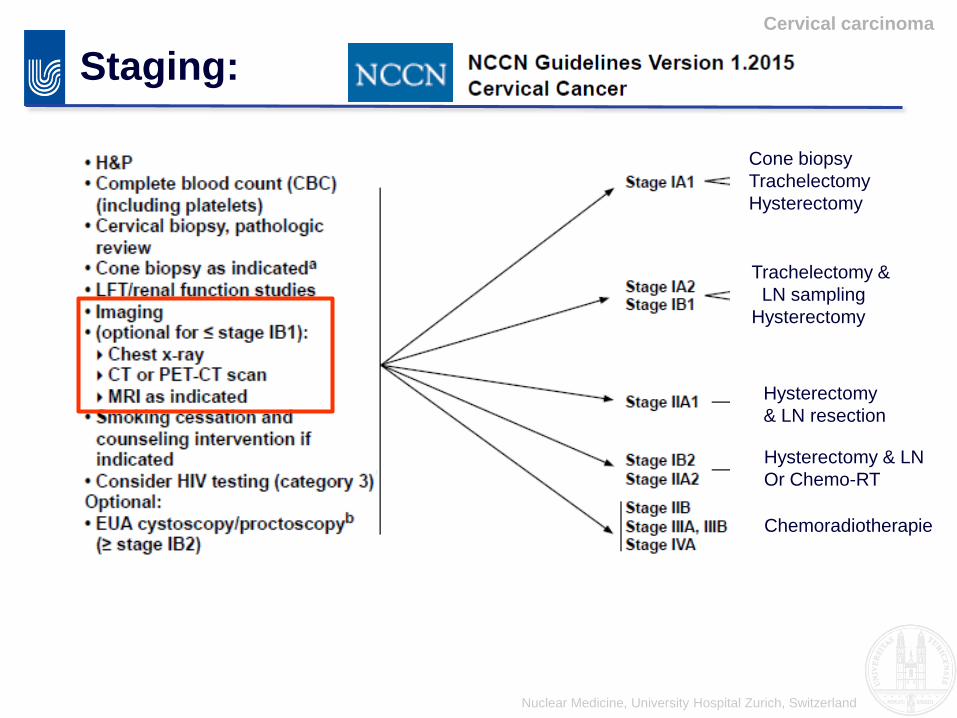
Staging:
Cervical carcinoma
Cone biopsy
Trachelectomy
Hysterectomy
Trachelectomy &
LN sampling
Hysterectomy
Hysterectomy
& LN resection
Hysterectomy & LN
Or Chemo-RT
Chemoradiotherapie

Nuclear Medicine, University Hospital Zurich, Switzerland
FIGO therapy:
Waggoner SE, Lancet 2003; 361:2217-25:
Cervical carcinoma
IIb or not IIb

Nuclear Medicine, University Hospital Zurich, Switzerland
Nodal involvement: It has an impact!
In all FIGO stages N1 clearly decreases the survival.
The extent of nodal involvement also has an impact on survival.
Cervical carcinoma
Nuclear Medicine, University Hospital Zurich, Switzerland
Staging today?
In the guidelines staging is still performed with
clinical examination.
Although imaging: CT, MRI, PET or PET-CT is
recommended for tumors stage > IB2
The additional information is added to the FIGO
stage: eg. FIGO IIB-2, cN1, cM1
Cervical carcinoma

Nuclear Medicine, University Hospital Zurich, Switzerland
Local staging: Early stages
Cervical carcinoma
MRI is superior to CT or PET/CT for the assessment of fertility
preserving therapy:
For CIN – Stage IA => Cone Biopsy
FIGO stage IA1 with lymphovascular invasion, IA2, and IB1 =>
Trachelectomy
• Tumor < 2 cm
• 2 - 4 cm abdominal
trachelectomy
• At least 1 cm from internal
cervical os
• Infiltrating < ½ of cervical stroma
thickness

Nuclear Medicine, University Hospital Zurich, Switzerland
Local staging:
Cervical carcinoma
MRI is superior to CT or PET/CT for local extent and especially
to delineate parametrial invasion

Nuclear Medicine, University Hospital Zurich, Switzerland
Lymph node staging:
Cervical carcinoma
PET/CT is superior for lymph node metastasis:
Patient A Patient B
• For high risk cervical cancer: sensitivities 75-86% and specificities 92-97 %• PET/CT detects lymph node metastases despite negative findings on CT/MRI
“PET/MRI identified all lesions depicted on PET/CT and significantly clarified the anatomic site of disease visualized on PET/CT.” Perry Grigsby, SNM 2013 (Washington University)

Nuclear Medicine, University Hospital Zurich, Switzerland
Combined pre-treatment MRI and 18F-FDG PET/CT parameters
as prognostic biomarkers in patients with cervical cancer :
DFS HR p
ADC 0.56 0.007*
PE 1.07 0.59
SUVmax1.12 0.18
TLG 1.03 0.024*
MTV 1.31 0.014*
Micco M, Vargas HA, Burger IA et al, Eur Radiol 2014 (Epub)
Two patients both with FIGO IIA cervical cancer:
1) ADC 0.81 mm2/s, SUV 11.1, MTV 18.4 ml, TLG 126 mg (DFS = 10 m; OS = 33.7 m)
2) ADC 0.97 mm2/s, SUV 8.7, MTV 10.4 ml, TLG 53 mg (alive free of disease)
Cervical cancer:
Nuclear Medicine, University Hospital Zurich, Switzerland
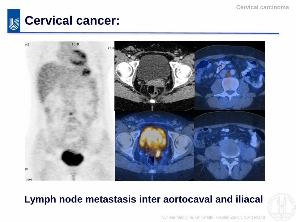
Cervical cancer:
Cervical carcinoma
Lymph node metastasis inter aortocaval and iliacal

Nuclear Medicine, University Hospital Zurich, Switzerland
Conclusion staging:
MRI for local tumor extent:
To evaluate for fertility spearing trachelectomy
To detect parametrial invasion
PET/CT: according to ACR guidelines highly
appropriate for nodal disease in Stage II or higher:
1.MRI seems to be inferior to PET in detecting lymph
node metastasis
2.PET/CT is superior for distant metastasis
Potential future: PET/MR, PET/CT-Nomogram?
Cervical carcinoma

Nuclear Medicine, University Hospital Zurich, Switzerland
Nomogram for staging:
Cervical carcinoma
FDG-PET-based prognostic nomograms for locally advanced cervical cancerElizabeth A. Kidd, Issam El Naqa, Barry A. Siegel, Farrokh Dehdashti, Perry W. Grigsby, 24. June 2012
Concordance index and SD

Nuclear Medicine, University Hospital Zurich, Switzerland
1. Therapy response assessment
2. Detection of recurrence
Restaging:
Cervical carcinoma

Nuclear Medicine, University Hospital Zurich, Switzerland
Therapy response assessment:
Cervical carcinoma
KM curves for Survival based on post
therapy FDG PET in 378 patients: 269
complete, 52 persistent abnormal FDG
uptake,57 new sites of disease. Schwarz et al: J Nucl Med 2009; 50:64S–73S
Nuclear Medicine, University Hospital Zurich, Switzerland
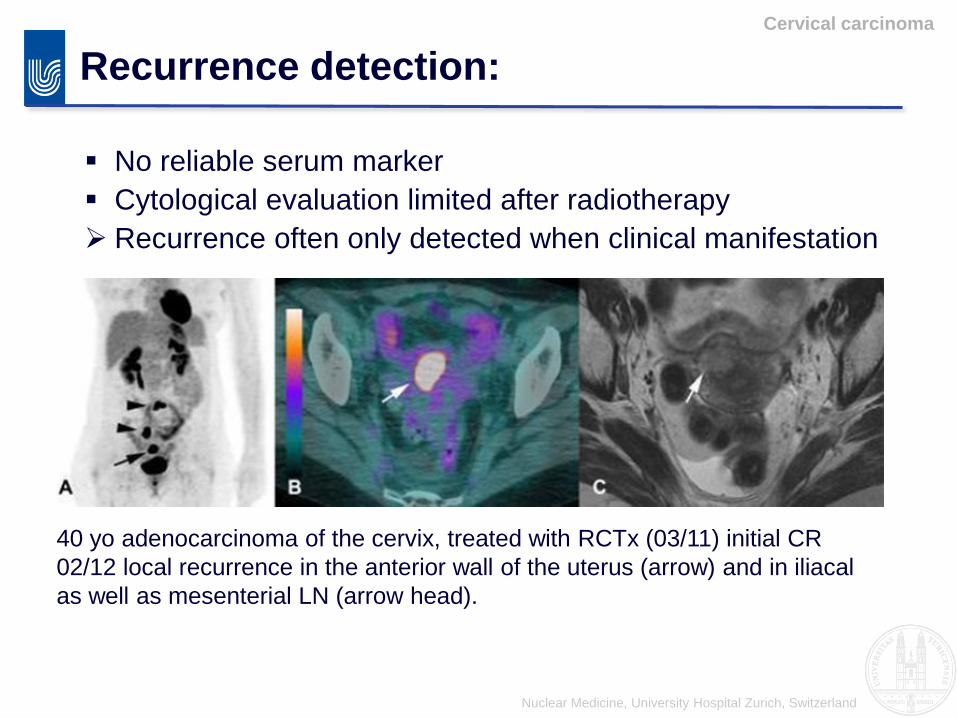
No reliable serum marker
Cytological evaluation limited after radiotherapy
Recurrence often only detected when clinical manifestation
Recurrence detection:
Cervical carcinoma
40 yo adenocarcinoma of the cervix, treated with RCTx (03/11) initial CR
02/12 local recurrence in the anterior wall of the uterus (arrow) and in iliacal
as well as mesenterial LN (arrow head).

Nuclear Medicine, University Hospital Zurich, Switzerland
If recurrence confined to the pelvis best therapy option:
Pelvic exenteration
PET/CT to rule out distant metastasis
MRI for local extent: for surgery planning organ or side wall
invasion is critical
Recurrence management:
Cervical carcinoma

Nuclear Medicine, University Hospital Zurich, Switzerland
MRI-PET superior?
Recurrence management:
Cervical carcinoma

Nuclear Medicine, University Hospital Zurich, Switzerland
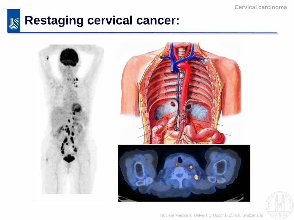
Restaging cervical cancer:
Cervical carcinoma
Supraclavicular lymph node metastasis
Nuclear Medicine, University Hospital Zurich, Switzerland
Restaging cervical cancer:
Cervical carcinoma
Nuclear Medicine, University Hospital Zurich, Switzerland
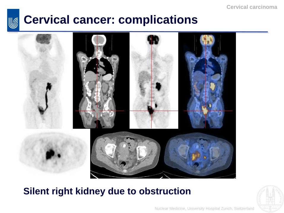
Cervical cancer: complications
Cervical carcinoma
Silent right kidney due to obstruction

Nuclear Medicine, University Hospital Zurich, Switzerland
Restaging recurrence?:
Cervical carcinoma
43 y.o. cervical Ca FIGO IIa
After surgery and radiation
- Two retroperitoneal cystic
lesions with mild FDG
uptake
- Recurrence?
- Ovariopexy

Nuclear Medicine, University Hospital Zurich, Switzerland
Conclusion restaging:
Therapy response assessment:
•PET/CT is superior to CT or MRI to predict response
•Complete metabolic response is the best predictor for a favorable
outcome
Cervical carcinoma
Recurrence management:
•No reliable blood or PAP smear screening
•PET/CT is superior to CT or MRI to predict extra pelvic disease
•MRI is superior to PET/CT for local extent
•PET/MRI high potential for this demanding situation

Nuclear Medicine, University Hospital Zurich, Switzerland
FDG PET/CT in gynecology:
1. Physiological uptake
2. Ovarian cancer
3. Cervical carcinoma
4. Endometrial carcinomaEndometrial carcinoma
Endometrial tumors

Nuclear Medicine, University Hospital Zurich, Switzerland
Endometrial carcinoma:
Endometrial carcinoma
Epidemiology:
Age: 55-65 y.o.
Incidence: 0.04%
Mortality: 2-5/100’000
Most present with an early stage:
postmenopausal or irregular bleeding
5 J-DFS: 80-90%
Advanced disease has high risk for relapse

Nuclear Medicine, University Hospital Zurich, Switzerland
Endometrial carcinoma:
Endometrial carcinoma
Staging:IA Tumor confined to the uterus, < ½ myometrium invasion
IB Tumor confined to the uterus, > ½ myometrium invasion
II Cervical stromal invasion
IIIATumor invades serosa or adnexa
IIIBTumor invades vagina
IIIC Pelvic or para-aortic lymph node involvement
IVABladder/bowel invasion
IVBDistant metastasis, inguinal lymph nodes
Nuclear Medicine, University Hospital Zurich, Switzerland
Staging:
Endometrial carcinoma
Transvaginal sonography
Surgery with lymphadenectomy for staging (Stadium IB
ore more, IA G1 & G2 no lymphadenectomy)
If further imaging: MRI
• Invasion of the myometrium?
• Invasion of the cervix?
• Invasion of adjacent organs?

Nuclear Medicine, University Hospital Zurich, Switzerland
Staging:
Endometrial carcinoma

Nuclear Medicine, University Hospital Zurich, Switzerland
Staging: Signorelli et al. 2009
Endometrial carcinoma
FDG-PET/CT can detect pelvic lymph node metastasis:
Sens 77.8%, spec 100%, PPV 100% and NPV 93.1%
FDG-PET/CT could be used to stage patients with high
risk endometrial cancer to select patients that benefit
from radical lymphadenectomy.
Nuclear Medicine, University Hospital Zurich, Switzerland
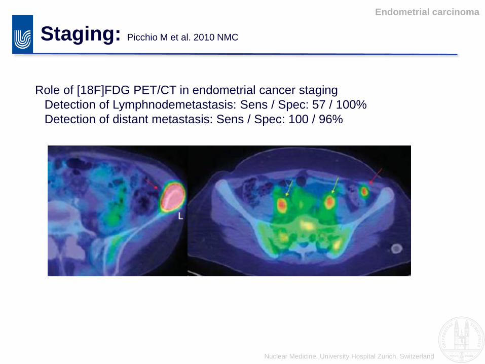
Staging: Picchio M et al. 2010 NMC
Endometrial carcinoma
Role of [18F]FDG PET/CT in endometrial cancer staging
Detection of Lymphnodemetastasis: Sens / Spec: 57 / 100%
Detection of distant metastasis: Sens / Spec: 100 / 96%

Nuclear Medicine, University Hospital Zurich, Switzerland
Restaging:
Endometrial carcinoma
65 y.o. Endometrial Ca
FIGO IIIc
After surgery and radiation
- Iliacal lymph node
metastasis with high FDG
activity

Nuclear Medicine, University Hospital Zurich, Switzerland
Uterine leiomyomas:
Endometrial tumors
Overview of different leiomyomas (Kitajima et al):
No uptake
(SUV 2.0)
High uptake
(SUV 8.5)
Moderate uptake
(SUV 3.9)

Nuclear MedicineUniversity Hospital ZürichSWITZERLAND
Thank you!