Opioid therapy vs. multimodal analgesia in head and neck ...
PET Monitoring of Therapy Response inHead and Neck Squamous Cell Carcinoma
Heiko Schoder1, Matthew Fury2, Nancy Lee3, and Dennis Kraus4
1Department of Radiology, Nuclear Medicine Service, Memorial Sloan-Kettering Cancer Center, New York, New York; 2Departmentof Medicine, Head and Neck Oncology Service, Memorial Sloan-Kettering Cancer Center, New York, New York; 3Department ofRadiation Oncology, Memorial Sloan-Kettering Cancer Center, New York, New York; and 4Department of Surgery, Head and NeckSurgery Service, Memorial Sloan-Kettering Cancer Center, New York, New York
In the Western world, more than 90% of head and neck cancersare head and neck squamous cell carcinomas (HNSCCs). Themost appropriate treatment approach for HNSCC varies withthe disease stage and disease site in the head and neck. Concur-rent chemoradiotherapy has become a widely used means for thedefinitive treatment of locoregionally advanced HNSCC. Al-though this multimodality treatment provides higher responserates than radiotherapy alone, the detection of residual viable tu-mor after the end of therapy remains an important issue and isone of the major applications of 18F-FDG PET. Studies haveshown that negative 18F-FDG PET or PET/CT results after con-current chemoradiotherapy have a high negative predictive value(.95%), whereas the positive predictive value is only about 50%.However, when applied properly, FDG PET/CT can exclude re-sidual disease in most patients, particularly patients with residualenlarged lymph nodes who would otherwise undergo neck dis-section. In contrast to other malignancies, data are limited onthe utility of 18F-FDG PET for monitoring the response to inductionchemotherapy in HNSCC or for assessing treatment responseearly during the course of definitive chemoradiotherapy. The prolif-eration marker 18F-39-deoxy-39fluorothymidine is currently understudy for this purpose. Beyond standard chemotherapy, newertreatment regimens inHNSCC take advantage of our improved un-derstanding of tumor biology. Two molecules important in the pro-gression of HNSCC are the epidermal growth factor receptor andthe vascular endothelial growth factor (VEGF) and its receptorVEGF-R. Drugs attacking these molecules are now under studyfor HNSCC. PET probes have been developed for imaging thepresence of these molecules in HNSCC and their inhibition by spe-cific drug interaction; the relevance of these probes for responseassessment in HNSCC will be discussed. Hypoxia is a commonphenomenon in HNSCC and renders cancers resistant to chemo-and radiotherapy. Imaging and quantification of hypoxia with PETprobes is under study and may become a prerequisite for over-coming chemo- and radioresistance using radiosensitizing drugsor hypoxia-directed irradiation techniques and for monitoring theresponse to these techniques in selected groups of patients. Al-though 18F-FDG PET/CT will remain the major clinical tool for mon-itoring treatment in HNSCC, other PET probes may have a role inidentifying patients who are likely to benefit from treatment strate-
gies that include biologic agents such as epidermal growth factorreceptor inhibitors or VEGF inhibitors.
Key Words: oncology; PET; PET/CT; head and neck cancer;treatment response
J Nucl Med 2009; 50:74S–88SDOI: 10.2967/jnumed.108.057208
Head and neck cancer is the sixth most common cancerworldwide. In the Western world, more than 90% of thesemalignancies are head and neck squamous cell carcinomas(HNSCCs). Approximately 47,000 new cases of HNSCCwere diagnosed in the United States in 2008 (1). The worstprognosis is seen in patients with unresectable advanceddisease, with a 5-y survival rate of less than 10%. Selection ofthe most appropriate treatment approach varies with diseasestage and site in the head and neck.
CURRENT CLINICAL PRACTICE
Patients with early-stage disease are generally treated withunimodality therapy consisting of either surgery or radio-therapy, and nearly 80% are cured. Postoperative radiotherapyis recommended when the risk for locoregional recurrence inthe head and neck exceeds 20%. For patients whose disease isnot controlled with definitive radiotherapy, salvage surgery isrecommended. In patients who undergo surgery for moreadvanced lesions, postoperative adjuvant radiotherapy isgenerally part of the treatment plan. Improvements in locore-gional control rates, progression-free survival (2), and overallsurvival (3) can be achieved at the expense of an increasedrate of acute treatment-related adverse events. The pooledanalysis of these 2 randomized trials suggested that concur-rent chemoradiotherapy should be offered when surgicalmargins are positive for tumor or when lymph nodes showextracapsular extension (4).
Patients with locoregionally advanced disease that issurgically unresectable, and patients in whom definitivetreatment is administered with an attempt at organ preserva-tion (e.g., oropharyngeal and laryngeal carcinomas), undergotreatment with concurrent chemoradiotherapy (5,6). Cis-
Received Jan. 27, 2009; revision accepted Mar. 25, 2009.For correspondence or reprints contact: Heiko Schoder, Departments
of Radiology and Nuclear Medicine, Memorial Sloan-Kettering CancerCenter, 1275 York Ave., Box 77, New York, NY 10065.
E-mail: [email protected] ª 2009 by the Society of Nuclear Medicine, Inc.
74S THE JOURNAL OF NUCLEAR MEDICINE • Vol. 50 • No. 5 (Suppl) • May 2009
platin is the drug with the most randomized clinical trial datato support its use as a drug enhancing the effects of radio-therapy in this setting. The larynx-preservation paradigm issupported by the results of a study that randomized 547patients with stage 3 or 4 supraglottic and glottic laryngealcancer into 3 treatment arms: concurrent chemoradiotherapy,induction chemotherapy consisting of cisplatin and 5-fluorouracil (5-FU) followed by radiotherapy, or radiother-apy alone. After a median follow-up of 3.5 y, the rates oflocoregional control were 78%, 61%, and 56%, respectively.The fraction of patients who maintained an intact larynx at2 y (and thus the ability to speak and swallow after the end oftherapy) was also better with the concurrent regimen (88%,75%, and 70%, respectively) (5). Overall survival rates weresimilar in all 3 groups. The utility of concurrent high-dosecisplatin for other subsites was established in a randomizedstudy of 295 patients with unresectable head and neck cancer(7). Patients were randomized to radiotherapy alone, radio-therapy plus concurrent high-dose cisplatin, or radiotherapy(split course) plus cisplatin and 5-FU. With a median follow-up of 41 mo, 3-y overall survival results were 23%, 37%, and27%, respectively. Improved locoregional control rates werealso reported for concurrent chemoradiotherapy with a drugcombination of carboplatin and 5-FU (6). In 226 patients withstage 3 or 4 oropharynx cancer, the 5-y rates of locoregionalcontrol were 48% for concurrent therapy, compared with24% for radiotherapy alone. Concurrent chemoradiotherapyis therefore now widely applied as the definitive treatment ofchoice for locoregionally advanced HNSCC. If residual dis-ease is detected after the end of therapy or during follow-up,salvage surgery (e.g., laryngectomy) may be offered.
The management of the neck when using an organ-preservation approach has remained somewhat controversial.Complete response rates in irradiated cervical lymph nodesvary between 59% and 83% and to some degree are related tonodal size, dose of radiotherapy, and time point whenresponse is determined: complete response rates are almost100% in N1 disease, higher in N2 than in N3 disease, andbetter when the largest metastatic node is smaller than 3 cm(8). In N2 or N3 disease, residual cancer in neck nodes hasbeen reported in 16%239% of patients achieving a clinicalcomplete response (no overt residual neck mass) (8–11).Early studies (12) demonstrated better outcomes when radi-otherapy was followed by neck dissection, leading to thepractice of ‘‘planned neck dissection’’ for all patients with N2or N3 disease on presentation (regardless of the response totreatment) and for patients with N1 disease and persistentpalpable lymph nodes after irradiation (13–15). More re-cently, however, the improved locoregional control rates withconcurrent chemoradiotherapy have prompted a debate onwhether planned neck dissection is still appropriate ornecessary in all patients with initial N2 or N3 disease.Proponents of this approach argue that because clinicalexamination and structural imaging cannot reliably identifyresidual viable tumor, neck dissection is the only means toeradicate all residual disease in the neck (9). In contrast,
opponents suggest that neck dissection be performed only inhigh-risk patients, whereas close clinical follow-up andobservation may be appropriate in most cases. Several recentstudies have tried to define the utility of 18F-FDG PET in thispatient population. These will be discussed later in thisarticle.
Patients with recurrent or metastatic disease have a mediansurvival of approximately 6–9 mo. Therapeutic optionsinclude chemotherapy alone, irradiation or reirradiation withor without chemotherapy, salvage surgery, or best supportivecare for patients with a low performance status. Reirradiationwith concurrent chemotherapy is feasible for recurrentunresectable HNSCC but is associated with considerableacute and long-term toxicity (16–19).
In summary, the suboptimal disease control rates andsurvival figures in HNSCC emphasize the need for earlydisease detection in the primary and recurrent settings and aneed for better imaging tools for staging and response as-sessment. There is also a clear need to investigate new thera-peutic regimens and drugs, such as biologic and molecularagents.
NEWER BIOLOGIC THERAPIES
Antiangiogenesis Therapies
The sprouting of new vessels (angiogenesis) is essentialfor tumor growth and metastasis (20). Tumor cell prolifera-tion alone, in the absence of angiogenesis, may give rise todormant, microscopic tumors of about 1 mm3 or less, butthese in situ cancers are harmless to the host (21,22). Theterm angiogenic switch has been used to describe the stepwhen tumors acquire the ability to recruit their own bloodsupply to support growth beyond microscopic size (23).Preclinical research from the past 20 y suggests that completepharmacologic blockade of tumor angiogenesis will leaveonly residual microscopic lesions, which may be clinicallyharmless and manageable as a chronic condition (23). Ofnote, the newly formed tumor vessels are structurally abnor-mal (‘‘leaky’’) and dysfunctional, delivering less blood andoxygen and fewer nutrients than normal blood vessels ofsimilar caliber. Their leakiness (hyperpermeability) alsocauses increased interstitial pressure. Ultimately, these prop-erties limit the build-up of sufficient drug concentrationswithin the tumor (24) and promote the development ofhypoxic tumor subregions. Hypoxia in turn is one of thestrongest promoters of angiogenesis (25–31).
Antiangiogenic therapies are particularly directed againstthese newly formed tumor vessels and may thus reduce oreliminate the excess supply of nutrients that are needed fortumor growth (32). Paradoxically, these drugs may alsoimprove drug distribution within the tumor and reduce levelsof intratumoral hypoxia because they eliminate the dysfunc-tional, leaky tumor vessels and thereby reduce intratumoralinterstitial pressure (24). Angiogenesis is tightly regulated byseveral molecules (33); the best known and studied of these isthe vascular endothelial growth factor (VEGF). Increased
PET RESPONSE ASSESSMENT IN HNSCC • Schoder et al. 75S
levels of VEGF are found in many HNSCCs and in patientserum (34,35). High VEGF expression is a marker of poorprognosis, correlating with higher clinical stage (36), highrates of locoregional recurrence, and lower disease-free andoverall survival (37,38). Accordingly, there is great interest inexploiting antiangiogenic therapies for the treatment ofHNSCC.
Bevacizumab, a recombinant humanized, monoclonal IgGantibody against VEGF, has been studied in HNSCC incombination with chemotherapy (33). Trials testing thecombination of bevacizumab with cisplatin and radiotherapyare ongoing (39). VEGF receptor tyrosine kinase inhibitorshave been studied in smaller phase I trials (40), and combi-nation therapies of bevacizumab with tyrosine kinase inhib-itors are being developed (41). Experimentally, synergistic(supraadditive) effects have been observed for the combina-tion of bevacizumab with the epidermal growth factorreceptor (EGFR) tyrosine kinase inhibitor erlotinib, as wellas for the EGFR kinase inhibitor gefitinib and specific VEGFreceptor tyrosine kinase inhibitors in conjunction with irra-diation (42,43). The rationale for combining biologic agentswith radiotherapy is based on evidence from experimentalstudies. Contrary to the concern that antiangiogenic therapymay cause or promote tumor hypoxia and hence radio-resistance, it is indeed the newly formed tumor vessels thatcontribute to radioresistance. In tumor xenografts, externalirradiation induces a 2- to 3-fold increase in VEGF expres-sion and secretion that lasts for up to 14 d (44). This mec-hanism may contribute to protecting tumor blood vesselsfrom radiation-mediated cytotoxicity and thus in fact pro-motes radioresistance (the tumor protecting itself). Thisradiation-induced angiogenesis can be suppressed by bev-acizumab and erlotinib (43).
Therapies Targeting the EGFR
The EGFR is a member of the ErbB family of tyrosinekinase receptors. It is overexpressed or activated in mostHNSCCs relative to normal tissue (45), and high expressionis associated with poor disease control (46–49). EGFR copynumber as determined by quantitative reverse-transcriptasepolymerase chain reaction is inversely related to patientoutcome: in a study of 134 patients with primary HNSCC, the5-y survival in individuals with an increased EGFR copynumber was only 9%, compared with 71% in individuals witha normal copy number (50). Although these data suggest thatHNSCC is an ideal malignancy for treatment with EGFRinhibitors, the selection of appropriate patients for therapywith these agents remains challenging. In studies of anti-EGFR agents in patients with advanced HNSCC, the objec-tive response rates have been approximately 10%, with thepercentage depending on the specific agent (51).
Cetuximab, a chimeric IgG1 antibody against the extra-cellular domain of EGFR, is the most widely studied agent.Cetuximab receptor binding competes with the binding of thenatural EGFR ligands and blocks the activation of thereceptor tyrosine kinase (52). Cetuximab has been studied
in recurrent HNSCC in combination with cisplatin (53) or incombination with platinum plus 5-FU (54) as a first-linetreatment and also as a secondary treatment option in cases ofplatinum-refractory disease (55–57). In these trials includinga total of almost 400 patients, the response rates based on CTor MRI assessment of target lesions were in the range of10%226%, although the disease control rates (a parameterthat includes all patients with complete response, partial re-sponse, or stable disease) were as high as 46%253% (55,57).In the largest trial to date (EXTREME trial), 442 patientswith untreated metastatic or recurrent HNSCC were ran-domized to treatment with 5-FU plus either carboplatin orcisplatin, or the same chemotherapy regimen plus cetuximab.The addition of cetuximab increased the response rate from20% to 36% (P , 0.001) and improved the median overallsurvival slightly but significantly from 7.4 to 10.1 mo (58).
Cetuximab also enhances the efficacy of radiation therapy.In a large, randomized study of 420 patients with locore-gionally advanced HNSCC (59), the addition of cetuximab toradiotherapy improved locoregional tumor control and over-all survival without increasing mucositis and dysphagia,when compared with radiotherapy alone. The correspondingmedian progression-free survival was 24 versus 15 mo, andthe median overall survival was 49 versus 29 mo. Althoughthis study did establish concurrent radiotherapy plus cetux-imab as a viable treatment option in locoregionally advancedHNSCC, the study has also been criticized because the con-trol group was treated with radiotherapy alone whereas thepresent standard regimen would be concurrent chemoradio-therapy. This issue is being addressed in ongoing clinicaltrials. One study is randomizing patients to induction che-motherapy followed by cisplatin plus radiotherapy versusinduction chemotherapy followed by cetuximab with radio-therapy (60). Another randomized study is comparingstandard concurrent chemoradiotherapy versus concurrentchemoradiotherapy plus cetuximab (61).
Other EGFR-inhibiting drugs include the small-moleculetyrosine kinase inhibitors gefitinib and erlotinib, which havebeen tested as single agents (62,63) or in combination withcisplatin (64). When used in combination with cisplatin,erlotinib seemed to have an additive effect, leading to anobjective partial or complete response in 21% of patients anddisease stabilization in 49%. However, the median progression-free survival was only 3 mo.
In summary, there is a sound scientific rationale forapplying biologic agents against EGFR and VEGF in patientswith locoregionally advanced or metastatic HNSCC. Unfor-tunately, major clinical responses are relatively rare. Theselection of appropriate patients for therapy with these newand expensive drugs remains challenging.
RESPONSE ASSESSMENT WITH 18F-FDG PET
18F-FDG PET After Chemo- or Radiotherapy
Structural imaging with contrast-enhanced CT or MRIand functional imaging with 18F-FDG PET are now con-
76S THE JOURNAL OF NUCLEAR MEDICINE • Vol. 50 • No. 5 (Suppl) • May 2009
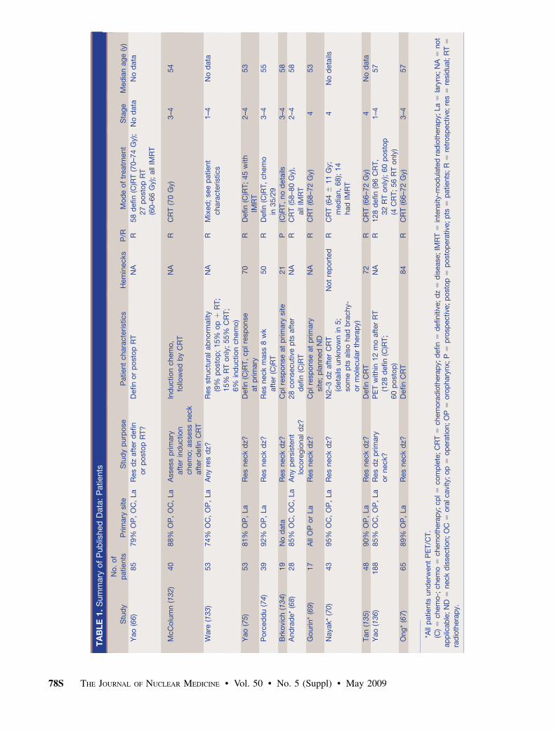
sidered standard for assessing the response to therapy inHNSCC. In this section, we shall focus on studies in which18F-FDG PET was used in patients treated with curativeintent. Response rates at the site of primary disease aregenerally high with concurrent chemoradiotherapy. There-fore, the main focus of posttherapy PET is the detection ofresidual disease in neck lymph nodes. Tables 1 and 2, asummary of published data, show that most studies onposttherapy 18F-FDG PET in HNSCC included heteroge-neous patient populations and that all the data come fromretrospective analyses:
• Patient selection criteria varied: Some studies includedconsecutive patients after chemo- or radiotherapy re-gardless of clinical or structural imaging findings. Somestudies enrolled only patients with residual structuralabnormality on clinical examination or CT/MRI. Somestudies excluded patients with suspected or provenresidual abnormality at the primary disease site andspecifically addressed the role of PET for detectingresidual cancer in neck lymph nodes.
• Disease sites varied: Most studies focused on the utilityof PET for response assessment after concurrent che-moradiotherapy (which is applied mostly for malignan-cies in the oropharynx and larynx), but some studies alsoincluded a large fraction of patients with other diseasesites (nasopharynx, paranasal sinuses, skin). Thisdifference is critical because tumor biology, clinicalbehavior, expected response rates, and the potential forfalse-positive findings differ between these latter can-cers and tumors of the oropharynx and larynx.
• Treatment strategies varied somewhat: One institution(65) reported on the utility of PET after inductionchemotherapy (which is currently not considered astandard approach) and after subsequent definitive con-current chemoradiotherapy, whereas all other studiesfocused on the outcome after definitive therapy.
• Treatment protocols varied somewhat: Most but not allpatients were treated with concurrent chemoradiother-apy. It is clear that greater posttreatment inflammation isexpected after concurrent therapy than after irradiationalone. Chemotherapy acts as a radiation sensitizer in theirradiated tumor but also in the surrounding normaltissue that is included in the radiation field. Studies alsodiffer in radiotherapy schedules, doses, and techniques.Only one study specifically addressed the role of PETafter intensity-modulated radiotherapy (66).
• Use of PET/CT versus only PET varied: Only a fewrecent studies specifically studied the role of combinedPET/CT in this setting (67–70). In some other studies,a fraction of patients was imaged with PET/CT, butother patients were imaged with PET only. Earlierstudies generally assessed the utility of PET only. Thisdifference between studies is important because com-bined PET/CT reduces the number of equivocalfindings and improves study accuracy (71,72).
• Appropriate time for PET in response assessmentvaried: Perhaps most important, the time between theend of therapy and the PET scan varied considerably,ranging from 4 wk to almost 1 y. In a study of 26 patients,Goerres et al. (73) observed a sensitivity and specificityof 91% and 93%, respectively, for PET scans performedas early as 6 wk after chemoradiotherapy. We and othershave not been able to reproduce these data (74,75) andinstead suggest that posttherapy PET after chemo-radiotherapy should not be performed before 10–12 wkafter the end of treatment. By that time, most of theposttreatment inflammatory changes will have sub-sided, reducing the number of potentially false-positiveinterpretations. In general, the rate of false-positivecases declines with the interval between end of therapyand PET (76). Therefore, it is not surprising that studiesthat include a large fraction of patients in whom PETwas performed as late as 6 mo after the end of therapywill have a lower false-positive rate. Some studies alsodemonstrated a higher false-negative rate when PETwasdone less than 4–8 wk after the end of therapy (68,77). Itis conceivable that small-volume residual disease at thisearly time escapes detection on PET. Of note, mostirradiated cells do not die instantaneously but insteadcan still undergo several cycles of cell division (onlycells irradiated in the mid to late S-phase showinstantaneous cell cycle blockade). Their subsequentfate varies: some cells may remain dormant for aprotracted time and die eventually, but some cells mayrecover and start dividing again (78,79). Indeed, exper-imental studies on irradiated cell cultures show a rapiddecline in 18F-FDG uptake, but the tracer uptake is notinstantaneously abolished. Dormant cancer cells main-tain their capability for glucose uptake and retention aslong as their cell membrane is intact and basic metabolicprocesses continue. Some of these cells eventually die,whereas other cells may recover their full metabolic andproliferative potential. With increasing volume, theresidual viable tumor cell nests may eventually becomedetectable on 18F-FDG PET.
We would like to highlight some of the studies listed in
Tables 1 and 2 to demonstrate the utility of 18F-FDG PET in
HNSCC after irradiation and chemoradiotherapy. Data are
based on the number of patients or on the number of
heminecks in which disease was initially diagnosed. The
first study included 53 patients (70 heminecks) who were
imaged with PET or PET/CT (75). The median interval
between end of therapy and PET was 15 wk. Twenty-eight of
the 70 heminecks harbored residual enlarged lymph nodes.
Using neck dissection or clinical follow-up as the standard of
reference, 18F-FDG PET showed a sensitivity of 100% and a
specificity of 94%, with a positive predictive value (PPV) of
43% and a negative predictive value (NPV) of 100%. The
second study evaluated 39 patients who achieved a complete
PET RESPONSE ASSESSMENT IN HNSCC • Schoder et al. 77S
TA
BL
E1.
Sum
mary
of
Pub
lished
Data
:P
atients
Stu
dy
No
.o
f
patients
Prim
ary
site
Stu
dy
purp
ose
Patient
chara
cte
ristics
Hem
inecks
P/R
Mo
de
of
treatm
ent
Sta
ge
Med
ian
ag
e(y
)
Yao
(66)
85
79%
OP
,O
C,
La
Res
dz
aft
er
defin
or
po
sto
pR
T?
Defin
or
po
sto
pR
TN
AR
58
defin
(C)R
T(7
0–7
4G
y);
27
po
sto
pR
T(6
0–6
6G
y);
all
IMR
T
No
data
No
data
McC
olu
mn
(132)
40
88%
OP
,O
C,
La
Ass
ess
prim
ary
aft
er
ind
uctio
nchem
o;
assess
neck
aft
er
defin
CR
T
Ind
uctio
nchem
o,
follo
wed
by
CR
T
NA
RC
RT
(70
Gy)
3–4
54
Ware
(133)
53
74%
OC
,O
P,
La
Any
res
dz?
Res
str
uctu
ralab
no
rmalit
y
(9%
po
sto
p;
15%
op
1R
T;
15%
RT
only
;55%
CR
T;
6%
ind
uctio
nchem
o)
NA
RM
ixed
;see
patient
chara
cte
ristics
1–4
No
data
Yao
(75)
53
81%
OP
,La
Res
neck
dz?
Defin
(C)R
T,
cp
lre
sp
onse
at
prim
ary
70
RD
efin
(C)R
T;
45
with
IMR
T2–4
53
Po
rced
du
(74)
39
92%
OP
,La
Res
neck
dz?
Res
neck
mass
8w
k
aft
er
(C)R
T
50
RD
efin
(C)R
T,
chem
o
in35/2
9
3–4
55
Brk
ovic
h(1
34)
19
No
data
Res
neck
dz?
Cp
lre
sp
onse
at
prim
ary
site
21
P(C
)RT
,no
deta
ils3–4
58
And
rad
e*
(68)
28
85%
OC
,O
C,
La
Any
pers
iste
nt
loco
reg
ionald
z?
28
co
nsecutive
pts
aft
er
defin
(C)R
T
NA
RC
RT
(58–8
0G
y),
all
IMR
T
2–4
58
Go
urin*
(69)
17
All
OP
or
La
Res
neck
dz?
Cp
lre
sp
onse
at
prim
ary
site;
pla
nned
ND
NA
RC
RT
(68–7
2G
y)4
53
Nayak*
(70)
43
95%
OC
,O
P,
La
Res
neck
dz?
N2–3
dz
aft
er
CR
T
(deta
ilsunkno
wn
in5;
so
me
pts
als
ohad
bra
chy-
or
mo
lecula
rth
era
py)
No
tre
po
rted
RC
RT
(64
611
Gy;
med
ian,
68);
14
had
IMR
T
4N
od
eta
ils
Tan
(135)
48
90%
OP
,La
Res
neck
dz?
Defin
CR
T72
RC
RT
(66–7
2G
y)4
No
data
Yao
(136)
188
85%
OC
,O
P,
La
Res
dz
prim
ary
or
neck?
PE
Tw
ithin
12
mo
aft
er
RT
(128
defin
(C)R
T;
60
po
sto
p)
NA
R128
defin
(96
CR
T,
32
RT
only
);60
po
sto
p(4
CR
T;
56
RT
only
)
1–4
57
Ong
*(6
7)
65
89%
OP
,La
Res
neck
dz?
Defin
CR
T84
RC
RT
(66–7
2G
y)3–4
57
*All
patients
und
erw
ent
PE
T/C
T.
(C)5
chem
o-;
chem
o5
chem
oth
era
py;
cp
l5
com
ple
te;
CR
T5
chem
ora
dio
thera
py;
defin
5d
efiniti
ve;
dz
5d
isease
;IM
RT
5in
tensi
ty-m
od
ula
ted
rad
ioth
era
py;
La
5la
rynx;
NA
5not
ap
plic
ab
le;
ND
5neck
dis
section;
OC
5ora
lcavi
ty;
op
5op
era
tion;
OP
5oro
phary
nx;
P5
pro
spective
;p
ost
op
5p
ost
op
era
tive
;p
ts5
patients
;R
5re
trosp
ective
;re
s5
resi
dual;
RT
5
rad
ioth
era
py.
78S THE JOURNAL OF NUCLEAR MEDICINE • Vol. 50 • No. 5 (Suppl) • May 2009

TA
BL
E2.
Sum
mary
of
Pub
lished
Data
:R
esults
Ind
ex
(%)*
Prim
ary
site
Necks
Stu
dy
PE
To
r
PE
T/C
TP
ET
crite
ria
Go
ldsta
nd
ard
Med
ian
F/U
aft
er
PE
T(m
onth
s:
rang
e)
Sen
Sp
ec
PP
VN
PV
Sen
Sp
ec
PP
VN
PV
Fra
ctio
no
fp
ts
with
po
sp
rim
ary
site
or
hem
inecks
(pre
vale
nce)
Dt
fro
mR
Tto
PE
T(w
eeks:
med
ian
or
rang
e)
Ded
icate
d
NM
read
er?
Yao
(66)
So
me
PE
T/C
T
Fo
cal
up
take
Bx,
ND
,clin
F/U
No
data
86
90
54
97
100
96
77
100
Prim
ary
:19%
;
neck:
15%
No
deta
ils
(‘‘m
ost
scans
3–5
mo
aft
er
RT
’’)
Yes
McC
olu
mn
(132)
PE
T2
NM
physic
ians
read
scans
as
po
so
r
neg
;no
deta
ils
Bx,
ND
,o
vert
clin
recurr
ence
20
(16–31)y
100
65
27
100
67
53
46
73
3/2
6(1
1%
)aft
er
ICT
;9/3
7(2
4%
)
aft
er
CR
T
4–12
Yes
67
z57
z33
z84
z
Ware
(133)
PE
TF
ocal
up
take
.b
ackg
round
;
no
np
hysio
log
ic
Bx,
clin
F/U
55
Lo
co
reg
ional
19/4
6(5
1%
)
.6
wk
aft
er
op
;
.2
mo
aft
er
RT
Yes
Yao
(75)
Either
Fo
cal
up
take
Bx,
clin
F/U
26
(12–57)
—§
—§
—§
—§
100
94
43
100
Neck:
3/7
0(4
%)
15
(5–29;
41%
12–20
wk)
Yes
Po
rced
du
(74)
PE
TN
ot
sp
ecifi
ed
Bx,
clin
F/U
34
—§
—§
—§
—§
83
93
71
97
Neck:
6/3
9(1
5%
)12
(8–32;
39%
8–12
wk)
Yes
Brk
ovic
h(1
34)
PE
TN
Mp
hysic
ian
read
scans
as
po
so
rneg
All
had
ND
—§
—§
—§
—§
76
65
33
92
Neck:
4/2
1(1
9%
)9
(7–12)
No
And
rad
e(6
8)
PE
T/C
TF
ocal:
mo
dera
te
or
inte
nse
Bx,
ND
,clin
F/U
17
(4.5
–34)
Lo
co
reg
ional:
13/2
8(4
6%
)
8(4
–16)
Yes
Go
urin
(69)
PE
T/C
TU
pta
ke
‘‘sig
nifi
cantly
hig
her
than
muscle
or
blo
od
po
ol,
fusin
g
toly
mp
hno
de
on
CT
’’
All
had
pla
nned
ND
NA
—§
—§
—§
—§
40
25
18
50
Neck:
5/1
7(2
9%
)8–10
Yes
Nayak
(70)
PE
T/C
T‘‘R
ad
iolo
gis
t
imp
ressio
n’’
ND
,clin
F/U
18
(5m
inim
um
)—
§—
§—
§—
§87
91
70
97
Neck:
10/4
3(2
3%
)8–26
Yes
Tan
(135)
PE
T‘‘R
esid
ual
hyp
erm
eta
bo
licup
take’’
ND
or
clin
F/U
20
(10–55)
—§
—§
—§
—§
25
83
15
90
Neck:
8/7
2(1
1%
)10
(5–22)
No
(uncle
ar)
Yao
(136)
Either
Fo
cal
up
take
ND
,B
x,
overt
clin
pro
gre
ssio
n
29
86
86
32
99
86
97
70
98
Prim
ary
:37/1
88
(20%
);neck:
14/1
88
(7.5
%)
15
(5.1
–44;
74%
in10–20
wk)
Yes
Ong
(67)
PE
T/C
TA
bno
rmal
(no
np
hysio
log
ic)
focalup
take,
fusin
g
tono
des
on
CT
ND
or
clin
F/U
37
(exclu
din
g
DO
Do
rR
D:
43
mo
)
NA
95
NA
97
71
89
38
97
Neck:
7/8
4(8
%)
12
(8–27)
Yes
80k
74k
40k
94k
*Only
loco
reg
ionalin
dex
valu
es
were
rep
ort
ed
by
Ware
(PP
V,
95%
;N
PV
,83%
)and
And
rad
e(s
ensitiv
ity,
76%
;sp
ecifi
city,
93%
;P
PV
,90%
;N
PV
,82%
).yF
ollo
w-u
ptim
efo
rp
atients
who
had
no
neck
dis
sectio
n.
zW
hen
co
nsid
ering
ND
and
clin
icalfo
llow
-up
.§N
ot
rep
ort
ed
or
no
tp
rim
ary
ob
jective.
k Resid
ualenla
rged
lym
ph
no
de
only
.
Bx
5b
iop
sy;clin
5clin
ical;
CR
T5
chem
ora
dio
thera
py;D
OD
5d
eath
of
dis
ease
;R
D5
recurr
ent
dis
ease
;D
t5
tim
ein
terv
al;
F/U
5fo
llow
-up
;IC
T5
ind
uctio
nchem
oth
era
py;N
A5
no
tap
plic
ab
le;
ND
5neck
dis
sectio
n;
neg
5neg
ative
;N
M5
nucle
ar
med
icin
e;
po
s5
po
sitiv
e;
pts
5p
atients
;R
T5
rad
ioth
era
py;
sen
5sensitiv
ity;
sp
ec
5sp
ecifi
city.
PET RESPONSE ASSESSMENT IN HNSCC • Schoder et al. 79S
response to concurrent chemoradiotherapy at the primary sitebut had a residual neck mass (74). 18F-FDG PET, performedat a median of 12 wk after the end of therapy, had a sensitivityof 83% and a specificity of 93%; the NPV was 97%. Residualnodal metastasis was eventually proven in 6 of the 39individuals; hence the PPV of structural imaging was only15%. Finally, Ong et al. (67) studied 65 patients (84 hemi-necks) after concurrent chemoradiotherapy. 18F-FDG PET/CTwas performed on all patients at least 8 wk after the end oftherapy. The standard of reference consisted of histopathol-ogy of neck dissection specimens or clinical and imagingfollow-up. In this study, the sensitivity and specificity were71% and 89%, respectively, with a PPVof 38% and an NPVof97%. All false-positive lymph nodes in neck dissectionspecimens showed either inflammation or granulomatousdisease, which are known causes of increased 18F-FDG up-take in lymph nodes. Nevertheless, the fraction of false-positive studies could be reduced from 27% to 10% when theneck was assessed by combined PET/CT rather than bystructural imaging alone, while maintaining a high NPV of97%. The true value of posttherapy 18F-FDG PET in patientstreated with current radio- or chemoradiotherapy is the highNPV. Many patients who might otherwise proceed to biopsyor planned neck dissection can in fact be observed withclinical follow-up and periodic imaging studies. Althoughthe PPV of PET after chemoradiotherapy is relatively low,most scans will in fact have negative findings when inter-preted properly. However, true prolonged, intense 18F-FDGuptake after definitive therapy indicates a poor treatmentoutcome or treatment-related complications: In an interest-ing study on patients with laryngeal cancer treated withintensity-modulated radiotherapy, Dornfeld et al. (80)showed an inverse relationship between the intensity of(persistent) 18F-FDG uptake at 12 mo after treatment andquality of life, the ability to speak, and the ability to swallowsolid foods. In other words, persistent 18F-FDG uptakeindicates either persistent disease or persistent treatment-induced structural and functional damage to the larynx.
Suggested PET Interpretation Criteria
Combined PET/CT, rather than PET only, should be usedbecause it is more accurate and avoids equivocal interpreta-tions, can distinguish between 18F-FDG uptake in normalstructures versus lymph nodes, can guide biopsies, and can
potentially guide planning of the target for adjuvant radio-therapy (71,72,81,82). 18F-FDG uptake in the treated neckshould decline within weeks, allowing an accurate reading tobe rendered at approximately 10–12 wk after the end ofchemoradiotherapy. However, as in other disease sites, traceruptake may occasionally persist (usually at mild to moderateintensity) for several months. The underlying reasons includepersistent cancer, inflammation, abscess formation, and ra-dionecrosis.
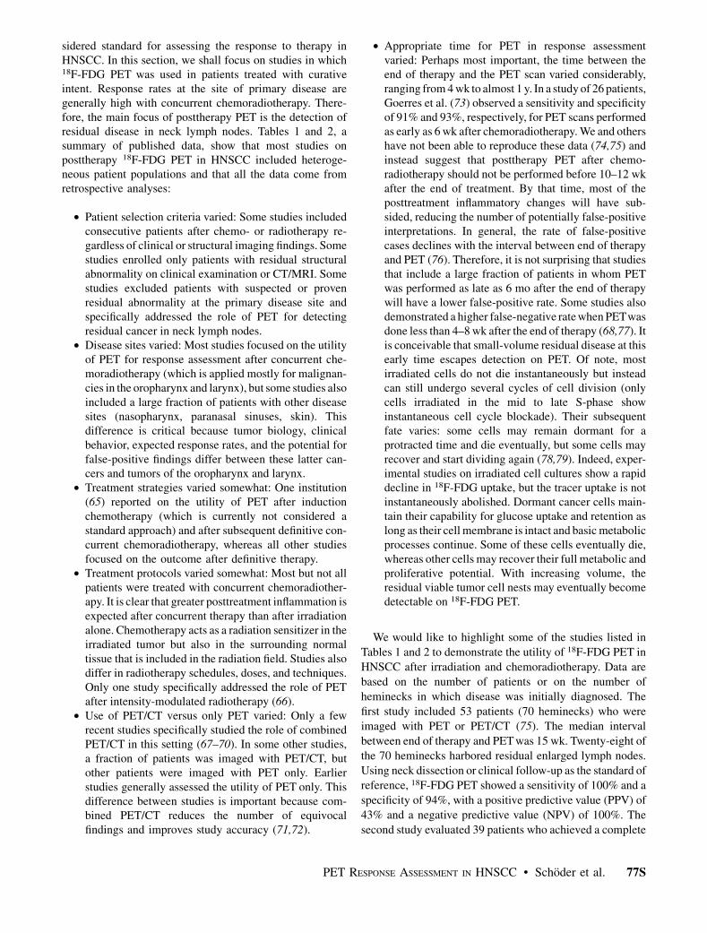
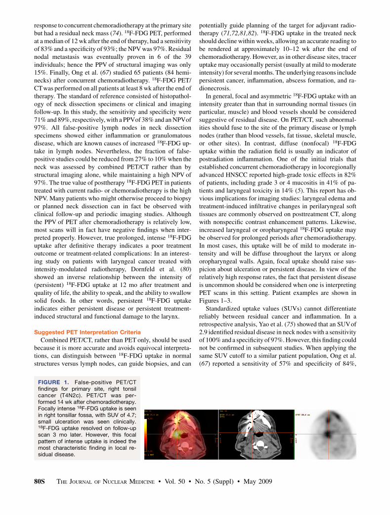
In general, focal and asymmetric 18F-FDG uptake with anintensity greater than that in surrounding normal tissues (inparticular, muscle) and blood vessels should be consideredsuggestive of residual disease. On PET/CT, such abnormal-ities should fuse to the site of the primary disease or lymphnodes (rather than blood vessels, fat tissue, skeletal muscle,or other sites). In contrast, diffuse (nonfocal) 18F-FDGuptake within the radiation field is usually an indicator ofpostradiation inflammation. One of the initial trials thatestablished concurrent chemoradiotherapy in locoregionallyadvanced HNSCC reported high-grade toxic effects in 82%of patients, including grade 3 or 4 mucositis in 41% of pa-tients and laryngeal toxicity in 14% (5). This report has ob-vious implications for imaging studies: laryngeal edema andtreatment-induced infiltrative changes in perilaryngeal softtissues are commonly observed on posttreatment CT, alongwith nonspecific contrast enhancement patterns. Likewise,increased laryngeal or oropharyngeal 18F-FDG uptake maybe observed for prolonged periods after chemoradiotherapy.In most cases, this uptake will be of mild to moderate in-tensity and will be diffuse throughout the larynx or alongoropharyngeal walls. Again, focal uptake should raise sus-picion about ulceration or persistent disease. In view of therelatively high response rates, the fact that persistent diseaseis uncommon should be considered when one is interpretingPET scans in this setting. Patient examples are shown inFigures 1–3.
Standardized uptake values (SUVs) cannot differentiatereliably between residual cancer and inflammation. In aretrospective analysis, Yao et al. (75) showed that an SUVof2.9 identified residual disease in neck nodes with a sensitivityof 100% and a specificity of 97%. However, this finding couldnot be confirmed in subsequent studies. When applying thesame SUV cutoff to a similar patient population, Ong et al.(67) reported a sensitivity of 57% and specificity of 84%,
FIGURE 1. False-positive PET/CTfindings for primary site, right tonsilcancer (T4N2c). PET/CT was per-formed 14 wk after chemoradiotherapy.Focally intense 18F-FDG uptake is seenin right tonsillar fossa, with SUV of 4.7;small ulceration was seen clinically.18F-FDG uptake resolved on follow-upscan 3 mo later. However, this focalpattern of intense uptake is indeed themost characteristic finding in local re-sidual disease.
80S THE JOURNAL OF NUCLEAR MEDICINE • Vol. 50 • No. 5 (Suppl) • May 2009
which were considerably worse than the 71% sensitivityand 89% specificity based on visual assessment using theaforementioned criteria. SUVs proposed as suitable cutoffsfor the identification of residual cancer based on single-institution studies on a specific set of patients may not beapplicable to other institutions with different equipment,patient populations, and clinical imaging protocols. It is alsounlikely that any sharp cutoff truly exists between malignantand benign SUVs. Looking on the bright side, though, it isreassuring that clinical image interpretation skills and judg-ment, acquired over many years, cannot be replaced by acomputer-generated number.
Impact on Patient Management
Because clinical parameters and structural imaging cannotreliably predict the presence of residual metastatic neckdisease, some investigators still advocate planned neckdissection in all patients with initial N2 or N3 disease (8–10,83,84). Historically, the risk for residual cancer in suchnodes has exceeded 20%, a number that has been accepted asthe lower threshold for surgical intervention. In light of thehigh NPVof posttreatment 18F-FDG PET, this approach mayno longer be justified. In one of the previously cited studies(67), planned neck dissection would have been considered in51 patients because of the presence of residual enlargedlymph nodes, but disease was in fact present in only 7 of
them. As shown in Tables 1 and 2, most posttreatment 18F-FDG PET scans even in this subset of patients with moreadvanced (N2 or N3) nodal disease will be negative withcurrent treatment protocols. Implementing a treatment strat-egy based on posttherapy PET/CT findings in the study byOng et al. (67) could have reduced the number of plannedneck dissections by 75% (from 51 to 13) while missingdisease in 2% (2/84 heminecks). Other investigators havesuggested that negative 18F-FDG PET/CT results afterchemoradiotherapy could reduce the number of plannedneck dissections by more than 80% (70). Although clinicalfactors, including the initial nodal stage, remain an importantconsideration, it would appear that a PET/CT-based strategymight reduce the element of arbitrary decision making inpatient management after chemoradiotherapy.
Whereas all of the aforementioned studies were retrospec-tive, the currently ongoing Radiation Therapy OncologyGroup Trial 0522 (85) is collecting these data prospectively.Patients with locoregionally advanced HNSCC will be ran-domized to treatment with cisplatin and radiotherapy, orcisplatin and radiotherapy plus cetuximab. This study pri-marily aims to determine whether disease-specific survivalcan be improved with the addition of cetuximab. However, asubstudy will also investigate the prognostic and diagnosticutility of 18F-FDG PET before and after the end of therapy,and in particular the correlation between posttreatment
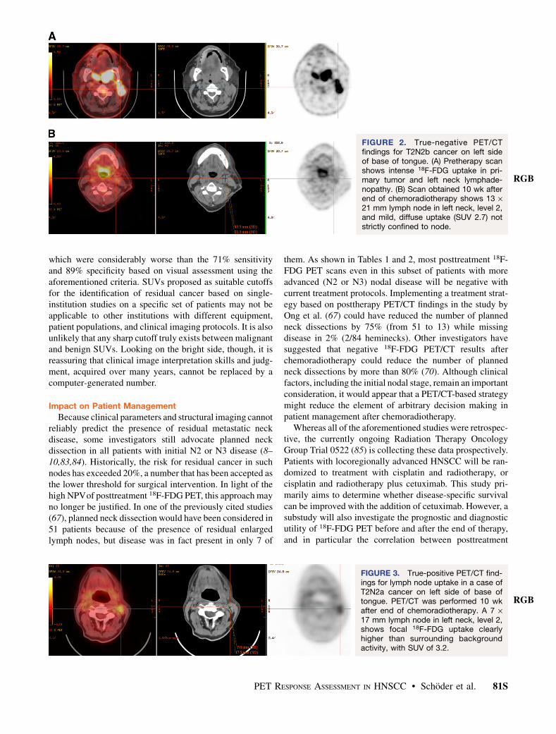
FIGURE 2. True-negative PET/CTfindings for T2N2b cancer on left sideof base of tongue. (A) Pretherapy scanshows intense 18F-FDG uptake in pri-mary tumor and left neck lymphade-nopathy. (B) Scan obtained 10 wk afterend of chemoradiotherapy shows 13 ·21 mm lymph node in left neck, level 2,and mild, diffuse uptake (SUV 2.7) notstrictly confined to node.
RGB
FIGURE 3. True-positive PET/CT find-ings for lymph node uptake in a case ofT2N2a cancer on left side of base oftongue. PET/CT was performed 10 wkafter end of chemoradiotherapy. A 7 ·17 mm lymph node in left neck, level 2,shows focal 18F-FDG uptake clearlyhigher than surrounding backgroundactivity, with SUV of 3.2.
RGB
PET RESPONSE ASSESSMENT IN HNSCC • Schoder et al. 81S

18F-FDG PET findings and nodal response or nodal relapserates. The organizers hypothesize that negative posttreatment18F-FDG PET results in patients with N2 or N3 diseaseindicates a pathologic complete response in more than 85%of treated necks and, conversely, a low overall nodal relapserate of 10% or less. Study results will likely not becomeavailable before 2010.
Several authors have proposed algorithms for patientmanagement after chemoradiotherapy (67,74,75,86). Thesealgorithms differ slightly in their proposed time points forearly structural imaging and in the implications of PETfindings for subsequent patient management. Some authors(75,86) have proposed the routine use of early (6–8 wk afterthe end of therapy) structural imaging to assess the treatmentresponse, followed by a second set of PET and CT studies at12 wk. We believe that a more measured approach can betaken and recommend early imaging of any kind only if thereis a strong clinical suspicion of lack of response or progres-sion (Fig. 4). In particular, it is the consensus opinion of themultidisciplinary head and neck cancer team at MemorialSloan-Kettering Cancer Center that a dedicated neck CT scanwith intravenous contrast material and 18F-FDG PET/CT(potentially in a single imaging session) should be performedabout 10–12 wk after the end of therapy, unless clinicalmanagement requires imaging earlier. This time point strikesa balance between the clinical desire for early, yet accurate,response assessment and the surgeon’s desire not to perform aneck dissection on tissues in which extensive fibrosis and scartissue have developed as the result of chemoradiotherapy.Shrinkage of large nodal masses will take time; early imagingafter the end of therapy only causes uncertainty and willrarely provide guidance for management. As shown in Figure 1,patients with no residual abnormal lymph nodes on CT (i.e.,diameter , 1 cm) can generally be observed, because CT
has a high NPV and 18F-FDG uptake in these normal-sizednodes is frequently false-positive. Patients with residuallymphadenopathy and abnormal PET findings should un-dergo neck dissection, possibly in the form of superselectiveneck dissection, which addresses only 1 or 2 neck levelsof concern (87). Management of patients with residualenlarged nodes and negative PET findings should beindividualized according to the following considerations:First, negative PET findings have a high NPV, and plannedneck dissection is thus unnecessary in most of these cases.However, close follow-up of these patients is required.Second, if close clinical follow-up cannot be guaranteed,or if extenuating circumstances indicate a higher likelihoodfor local recurrence (e.g., extranodal tumor extension), aneck dissection may be indicated. Third, in any event, therationale for the chosen approach should be discussed withthe patient.
Regarding the optimal time point for follow-up imaging,we believe that PET scans done later than 12 wk after theend of therapy will not be helpful to surgeons attempting tojudge the need for a neck dissection. Although these laterscans may still provide clinically useful information (e.g.,in the surveillance of high-risk patients or in the detectionof early recurrence), they do not qualify as a tool forresponse assessment.
Ideally, the concept that patient management can bedecided on the basis of posttherapy PET/CT findings wouldbe tested in a randomized trial comparing outcome data in 2groups of patients. In one group, planned neck dissectionwould be performed routinely, whereas in the other group,neck dissection would be performed only when the PETscan result was suggestive; all other patients would befollowed clinically. Although a randomized trial may bepreferred on scientific grounds, it is unlikely that this will
FIGURE 4. Suggested algorithm formanagement of HNSCC patients afterchemoradiotherapy, based on PET andCT findings obtained approximately10–12 wk after end of therapy. dz. 5
disease; ECE 5 extracapsular extension;iv. 5 intravenous; LN 5 lymph node;ND 5 neck dissection; PET - 5 PET-negative; PET 1 5 PET-positive.
82S THE JOURNAL OF NUCLEAR MEDICINE • Vol. 50 • No. 5 (Suppl) • May 2009
happen, because the available evidence has already begunto shape clinical practice. A reasonable alternative ap-proach would involve a medical team of head and neckcancer specialists (surgeons, oncologists, imaging physi-cians) that provide a structured plan for close follow-upbased on one of the proposed algorithms.
18F-FDG PET During Chemoradiotherapy or AfterInduction Chemotherapy
The potential clinical utility of PET for early responseassessment during chemoradiotherapy has not been studiedsystematically. Data from other malignancies (88,89) sug-gest that a significant decline in 18F-FDG uptake betweenbaseline and interim PET after a few cycles of chemo- orchemoradiotherapy may indicate a better prognosis and highlikelihood for achieving a complete response. Only a singlesmall study has attempted to address this question in HNSCC(90). Using coincidence camera imaging, that study noted anearly and significant decline in 18F-FDG uptake in 47 patientswith locally advanced disease after a cycle of chemotherapyor 24 Gy of radiotherapy. When dichotomized by the medianSUV, individuals with lower 18F-FDG uptake showed a betterrate of locoregional control. However, a closer analysis of thestudy reveals that similar prognostic information could alsobe derived from the baseline scan alone. Although interest-ing, the study remains therefore largely inconclusive. Inparticular, it remains unclear at what interim time pointduring the course of therapy a PET scan should be performedand how interim PET findings might alter patient manage-ment (good local control rates with concurrent chemoradio-therapy, lack of an established alternate therapy).
There are also limited data on the role of PET in assessingthe response to induction chemotherapy before subsequentconcurrent chemoradiotherapy. This is a topic of growinginterest to medical oncologists. Proponents of this approachbelieve that it may improve clinical outcome in locoregion-ally advanced HNSCC. For instance, in the DeCIDE trial,patients with N2 or N3 disease will be randomized totreatment with standard concurrent chemoradiotherapy orinduction chemotherapy followed by chemoradiotherapy.The effect on overall and progression-free survival, rate ofdistant metastases, and quality of life will be compared (91).Other currently ongoing studies in the United States andEurope will apply induction chemotherapy followed byradiotherapy in combination with cisplatin or a biologicagent such as cetuximab (92,93). In the future, it will beimportant to identify patients who might benefit from thisnew treatment approach. It is conceivable that PET witheither 18F-FDG or 18F-39-deoxy-39fluorothymidine (FLT)after induction chemotherapy might help in this decision.For instance, if a patient shows little or no metabolic responseafter induction chemotherapy, this might indicate a lowlikelihood for cure with subsequent chemoradiotherapy;perhaps such patients would benefit from immediate salvagesurgery after induction therapy or should be enrolled in moreaggressive chemoradiotherapy protocols.
18F-FDG PET After Surgery and Before Adjuvant Therapy
Shintani et al. (94) investigated the role of 18F-FDG PET/CT early after surgical resection and before planned adjuvantradiotherapy in a heterogeneous group of 91 patients (62 withsquamous cell carcinoma). PET/CTapproximately 28 d aftersurgery revealed findings suggestive of residual macroscopictumor in 24 of the 91 individuals (26%). Subsequent biopsiesproving residual cancer in 45% of these instances (PPV, 45%)led to a change in management in 14 patients (15%),including abandonment of adjuvant radiotherapy and aswitch to palliative chemotherapy, as well as changes inradiation field or dose. Imaging early after surgery obviouslycauses a high rate of false-positive findings; the exact role ofPET/CT, if any, remains to be defined. However, it is clearthat combined PET/CT (rather than PET only) should beperformed to enable precise target definition for radiotherapyin HNSCC (95).
A POTENTIAL ROLE FOR OTHER PET RADIOTRACERS
Amino Acid Transport and Protein Synthesis
PETwith labeled amino acids has been applied to head andneck cancer. Despite the excitement generated by earlierstudies, no tracer of amino acid transport or protein synthesishas been tested rigorously in HNSCC. 11C-methionine didnot provide any clear benefits over imaging with 18F-FDG.The short half-life of 11C requires a nearby cyclotron, and theimage quality is suboptimal. False-positive uptake at sites ofinflammation can occur with this agent and with other aminoacids. It is doubtful that any of these agents will have a futurein the clinical response assessment of patients with HNSCC.
18F-FLT18F-FLT uptake and retention require a functional nucle-
oside transporter in the plasma membrane and activity of thethymidine kinase-1 enzyme (96). In vitro, 18F-FLT retentioncorrelates with the fraction of cells in the S-phase of the cellcycle and with thymidine kinase-1 activity (97,98). Thymi-dine kinase-1 is inhibited by many chemotherapeutic drugsand by external-beam irradiation (97). When tumor cells areirradiated, they do not enter the cell cycle but instead remainin the S-phase (97). In vitro, a radiation dose–dependentdecrease in 18F-FLT uptake that paralleled the decline of thefraction of cells in the S-phase was shown (98). Decreases in18F-FLT uptake and thymidine kinase-1 activity were ob-served within 24 h after irradiation.
The clinical utility of 18F-FLT in clinical response assess-ment in HNSCC is under investigation. From studies on othermalignancies (99) it might be inferred that 18F-FLT imagingcould potentially permit an earlier or more accurate responseassessment in HNSCC than can imaging with 18F-FDG orCT/MRI. However, preliminary experimental studies haveshown mixed results. When nude mice bearing the humansquamous cell cancer graft (HNX-OE; nu/nu mice) wereirradiated, both 18F-FDG and 18F-FLT uptake declined frombaseline levels, but the decline in 18F-FDG uptake occurredmainly during the first week of treatment, whereas the
PET RESPONSE ASSESSMENT IN HNSCC • Schoder et al. 83S

greatest decline in 18F-FLT uptake was noted during thesecond treatment week (100). In another study, however, 18F-FLT PET showed promise in assessing the early response tocetuximab in a squamous cell cancer xenograft model (101).A significant decline in tumor SUV, tumor-to-muscle ratios,and thymidine kinase-1 activity occurred as early as 6 d aftersingle-agent therapy had been initiated with cetuximab. Inthe clinic, these early changes might be helpful in assessingthe response to cetuximab monotherapy during the inductionphase for subsequent combination chemotherapy or chemo-radiotherapy. Of note, false-positive 18F-FLT uptake can occurin reactive lymph nodes (102) and is a particular problemafter concurrent chemoradiotherapy. The frequency of false-positive 18F-FLT uptake during or after chemoradiotherapy inpatients with HNSCC has not been established. If this wereindeed a common phenomenon, it would severely limit theutility of this tracer for response assessment.
Hypoxia Imaging
Tumor hypoxia is a common phenomenon in HNSCC(103–105). Hypoxic cells are resistant to the cytotoxic effectsof chemotherapy and ionizing radiation (106–109) andrequire radiation doses up to 3 times higher than for thesame level of cell inactivation relative to the same cells undernormoxic conditions. A recent review of nearly 400 HNSCCpatients who underwent tumor oxygenation measurementdemonstrated that hypoxia was strongly associated withtreatment failure (locoregional recurrence and distant me-tastasis) independently of stage and therapeutic modality(105). This observation has led to a growing interest indiagnosing hypoxic HNSCC before therapy in the hope ofapplying novel treatment strategies that may overcome theresistance to conventional chemoradiotherapy. Several com-pounds are available for the imaging of hypoxia (109,110).Currently, 18F-fluoromisonidazole (18F-FMISO) is still con-sidered the standard PET tracer for hypoxia imaging. Onegoal of hypoxia imaging is the identification of patients withhypoxic tumors who may benefit from a combination therapyof irradiation with radiation sensitizers (111), vasodilators, orcarbogen breathing, as was used in the ARCON (acceleratedradiotherapy with carbogen and nicotinamide) trial in ad-vanced head and neck cancer (112), or hypoxic cell cyto-toxins such as tirapazamine. Whereas the nonselectiveaddition of tirapazamine to standard chemoradiotherapydid not improve clinical outcome in a randomized phase IIItrial (HEADSTART) (113), an improvement in locoregionalcontrol rates could be shown in selected patients withevidence of tumor hypoxia on 18F-FMISO PET (114). How-ever, some recent data suggest that hypoxia as shown onFMISO PET may be overcome in most patients even withstandard chemoradiotherapy (115). Four weeks into treat-ment, only 2 of 20 patients showed imaging evidence of per-sistent hypoxia on the 18F-FMISO scan, and local recurrencedid not develop in these 2 patients. With a median follow-upof 36 mo, the 3-y progression-free survival in this patientpopulation was 95%. The frequency and time course at which
tumor hypoxia persists after the initiation of chemoradio-therapy are unknown. It is conceivable that treatment out-come can be improved, despite the presence of hypoxia, withcertain combinations of standard chemotherapy and irradia-tion. However, if interventions aimed at overcoming tumorhypoxia are used as part of the experimental protocol, theyshould probably center on the first 4 wk of therapy.
Imaging EGFR and Response to EGFR Inhibitors
Neither clinical presentation nor immunohistochemicalanalysis of tumor specimens can reliably predict the thera-peutic response to EGFR inhibitors. A high EGFR gene copyin tumor specimens may identify patients with a increasedlikelihood of response, and a decline in phosphorylatedEGFR levels in skin biopsies during therapy has beensuggested as a potential surrogate marker for improvedclinical outcome (116). However, the latter reflects only asystemic drug effect or toxicity and does not provide anyinformation about local drug concentrations or about theeffect on the tumor itself.
Clinical response can certainly be assessed nonspecificallywith 18F-FDG PET (117,118). Downregulation of 18F-FDGuptake after treatment with small-molecule tyrosine kinaseinhibitors, such as gefitinib, seems to be an early phenome-non (119) that precedes changes on structural imaging. Morespecific imaging tests based on the increasing recognition ofthe molecular structure of receptors and tumor pathways arebeing developed. Signal transduction through EGFR re-quires ligand binding to the receptor, dimerization of ligand–receptor complexes, and autophosphorylation of the receptorat the intracellular tyrosine kinase domain. In principle, atleast 2 of these steps can be imaged, and such imaging mightbe helpful in predicting and monitoring treatment response.For instance, one radiolabeled ligand for EGFR, the chelatecomplex 64Cu–DOTA–cetuximab, has been tested in small-animal studies correlating tracer uptake on small-animal PETwith EGFR expression in Western blot analysis (120). An-other group of tracers, radiolabeled anilinoquinazolines, bindto the intracellular adenosine triphosphate binding pocket ofthe receptor kinase; agents that bind irreversibly at this siteand are labeled with 11C or 124I appear suitable for PET(121,122). It is hoped that clinical PET studies with these orsimilar compounds may help in selecting patients who arelikely to respond to EGFR inhibitors and in monitoring thedrug response in a specific manner.
Imaging of Angiogenesis and Angiogenesis Inhibitors
It is currently unclear which patients may particularlybenefit from treatment with angiogenesis-inhibiting drugs.Although elevated VEGF serum levels are a marker of poorprognosis in HNSCC, they cannot predict the clinical re-sponse to antiangiogenic therapies (123). First, VEGF isproduced not only by tumor cells but also by normal platelets,cells in the tumor-surrounding stroma, and other cells in thebody; second, VEGF receptors are found primarily on endo-thelial cells but also on various tumor cells; third, resistanceto VEGF inhibitors can exist intrinsically or may develop
84S THE JOURNAL OF NUCLEAR MEDICINE • Vol. 50 • No. 5 (Suppl) • May 2009

during therapy (e.g., due to upregulation of other angiogenicreceptors or an increase in circulating endothelial progenitorcells) (123). Because inhibition of angiogenesis per se maynot cause cell death and tumor shrinkage (but instead ‘‘only’’prevent further growth), it is also unclear how the efficacy ofangiogenesis inhibitors could best be shown and monitored inthe clinical setting (rather than inferring efficacy indirectlyfrom achieving stable disease).
Changes in tumor hyperemia can be documented oncontrast-enhanced CT and MRI or dedicated MRI perfusionsequences. However, more specific imaging tests document-ing drug targeting and effect are available. One example is theradiotracer 18F-RGD peptide (124,125), which binds specif-ically to anb3 integrins expressed at the surface of activatedendothelial cells during angiogenesis (126). The intensity of18F-RGD peptide accumulation correlates with the presenceof activated endothelial cells and, quantitatively, with micro-vessel density (124). 18F-RGD peptide imaging of HNSCC isfeasible (127): In a pilot study of 11 patients, tracer uptakewas detected in 10 of 12 primary lesions (SUV, 2.2–5.8) withreasonable contrast, based on a tumor-to-blood ratio of 2.8 6
1.1. Two tumors that were smaller than 5 mm were missed.Immunohistochemistry confirmed avb3 expression on mi-crovessels in all tumors. This or similar radiotracers mightpotentially be useful in identifying suitable patients fortreatment with antiangiogenic drugs and in monitoring theresponse to such therapies in a drug-specific manner (insteadof, or in addition to, tumor shrinkage or a decline in glucosemetabolism).
Treatment Options on the Horizon
Cancer gene therapy, whereby replication-incompetentviral vectors are used to transfer a therapeutic gene into thecancer cell, is under study in many malignancies, includingHNSCC (128). Another treatment option is the use ofoncolytic viruses, which infect, multiply within, and subse-quently lyse cancer cells. The attenuated adenovirus ONYX-015, which preferentially localizes in cells lacking the tumorsuppressor gene p53, has been tested clinically. In a phase IItrial, intratumoral ONYX-015 injection led to a complete orpartial (,50% reduction in tumor diameter) response in 20%of cases (129). Other oncolytic viruses are under investiga-tion. An attenuated, replication-competent, oncolytic herpessimplex virus (NV1023) has shown promise in experimentalstudies. NV 1023 delivery to the surgical bed after tumorresection caused viral infection of metastatic squamous cellsin cervical lymph nodes and improved the rate of locore-gional control and disease-free survival (130). Localization,distribution, and survival of this oncolytic virus was imagedwith 18F-29-fluoro-29-deoxy-1-b-D-b-arabinofuranosyl-5-ethyluracil PET, taking advantage of viral thymidine kinaseexpression (131). One can envision similar imaging studiesin the clinical environment, which may be particularly usefulwhen the agent is to be administered intravenously ratherthan into the tumor site.
CONCLUSION
Concurrent chemoradiotherapy is widely used as thedefinitive treatment for locoregionally advanced HNSCCbecause it provides higher response rates than radiotherapyalone. Negative 18F-FDG PET or PET/CT findings afterconcurrent chemoradiotherapy have a high NPV (.95%).The PPV is lower, but few studies will be considered positivewhen interpreted as suggested in this review. On the basis of apatient’s specific condition, neck dissection may be avoidedin many cases. The role of PET with either 18F-FDG or 18F-FLT in assessing response during induction or definitive che-moradiotherapy is under investigation. Experimental therapywith EGFR and VEGF inhibitors is under study in HNSCC;several PET probes appear promising for measuring theresponse to these new drugs and potentially for selecting pa-tients with suitable molecular targets for such experimentaltherapies. Hypoxia imaging may be helpful in designing al-ternate radiotherapy techniques that may overcome the radio-resistance of hypoxic tumors. For most clinical purposes,however, 18F-FDG PET/CT will remain the major clinicaltool for monitoring treatment in HNSCC in the near future.
ACKNOWLEDGMENTS
We thank John Humm, for helpful discussions onradiation biology, and Richard Wong, for helpful discus-sions on oncolytic virus treatment for head and neck cancer.
REFERENCES
1. Jemal A, Siegel R, Ward E, et al. Cancer statistics, 2008. CA Cancer J Clin.
2008;58:71–96.
2. Cooper JS, Pajak TF, Forastiere AA, et al. Postoperative concurrent radiother-
apy and chemotherapy for high-risk squamous-cell carcinoma of the head and
neck. N Engl J Med. 2004;350:1937–1944.
3. Bernier J, Domenge C, Ozsahin M, et al. Postoperative irradiation with or
without concomitant chemotherapy for locally advanced head and neck cancer.
N Engl J Med. 2004;350:1945–1952.
4. Bernier J, Cooper JS, Pajak TF, et al. Defining risk levels in locally advanced
head and neck cancers: a comparative analysis of concurrent postoperative
radiation plus chemotherapy trials of the EORTC (#22931) and RTOG (#
9501). Head Neck. 2005;27:843–850.
5. Forastiere AA, Goepfert H, Maor M, et al. Concurrent chemotherapy and
radiotherapy for organ preservation in advanced laryngeal cancer. N Engl J
Med. 2003;349:2091–2098.
6. Denis F, Garaud P, Bardet E, et al. Final results of the 94-01 French Head and
Neck Oncology and Radiotherapy Group randomized trial comparing radio-
therapy alone with concomitant radiochemotherapy in advanced-stage oro-
pharynx carcinoma. J Clin Oncol. 2004;22:69–76.
7. Adelstein DJ, Li Y, Adams GL, et al. An intergroup phase III comparison of
standard radiation therapy and two schedules of concurrent chemoradiotherapy
in patients with unresectable squamous cell head and neck cancer. J Clin
Oncol. 2003;21:92–98.
8. McHam SA, Adelstein DJ, Rybicki LA, et al. Who merits a neck dissection
after definitive chemoradiotherapy for N2-N3 squamous cell head and neck
cancer? Head Neck. 2003;25:791–798.
9. Brizel DM, Prosnitz RG, Hunter S, et al. Necessity for adjuvant neck dissection
in setting of concurrent chemoradiation for advanced head-and-neck cancer. Int
J Radiat Oncol Biol Phys. 2004;58:1418–1423.
10. Frank DK, Hu KS, Culliney BE, et al. Planned neck dissection after
concomitant radiochemotherapy for advanced head and neck cancer. Laryngo-
scope. 2005;115:1015–1020.
11. Argiris A, Stenson KM, Brockstein BE, et al. Neck dissection in the combined-
modality therapy of patients with locoregionally advanced head and neck
cancer. Head Neck. 2004;26:447–455.
PET RESPONSE ASSESSMENT IN HNSCC • Schoder et al. 85S

12. Mendenhall WM, Million RR, Cassisi NJ. Squamous cell carcinoma of the
head and neck treated with radiation therapy: the role of neck dissection for
clinically positive neck nodes. Int J Radiat Oncol Biol Phys. 1986;12:733–740.
13. Boyd TS, Harari PM, Tannehill SP, et al. Planned postradiotherapy neck
dissection in patients with advanced head and neck cancer. Head Neck.
1998;20:132–137.
14. Lee HJ, Zelefsky MJ, Kraus DH, et al. Long-term regional control after
radiation therapy and neck dissection for base of tongue carcinoma. Int J
Radiat Oncol Biol Phys. 1997;38:995–1000.
15. Lavertu P, Adelstein DJ, Saxton JP, et al. Management of the neck in a
randomized trial comparing concurrent chemotherapy and radiotherapy with
radiotherapy alone in resectable stage III and IV squamous cell head and neck
cancer. Head Neck. 1997;19:559–566.
16. Spencer SA, Harris J, Wheeler RH, et al. Final report of RTOG 9610, a multi-
institutional trial of reirradiation and chemotherapy for unresectable recurrent
squamous cell carcinoma of the head and neck. Head Neck. 2008;30:281–288.
17. Sulman EP, Schwartz DL, Le TT, et al. IMRT reirradiation of head and neck
cancer: disease control and morbidity outcomes. Int J Radiat Oncol Biol Phys.
2009;73:399–409.
18. Langer CJ, Harris J, Horwitz EM, et al. Phase II study of low-dose paclitaxel
and cisplatin in combination with split-course concomitant twice-daily
reirradiation in recurrent squamous cell carcinoma of the head and neck:
results of Radiation Therapy Oncology Group Protocol 9911. J Clin Oncol.
2007;25:4800–4805.
19. Lee N, Chan K, Bekelman JE, et al. Salvage re-irradiation for recurrent head
and neck cancer. Int J Radiat Oncol Biol Phys. 2007;68:731–740.
20. Folkman J. Angiogenesis in cancer, vascular, rheumatoid and other disease. Nat
Med. 1995;1:27–31.
21. Holmgren L, O’Reilly MS, Folkman J. Dormancy of micrometastases:
balanced proliferation and apoptosis in the presence of angiogenesis suppres-
sion. Nat Med. 1995;1:149–153.
22. Naumov GN, Akslen LA, Folkman J. Role of angiogenesis in human tumor
dormancy: animal models of the angiogenic switch. Cell Cycle. 2006;5:1779–
1787.
23. Folkman J. Angiogenesis. Annu Rev Med. 2006;57:1–18.
24. Jain RK. Normalization of tumor vasculature: an emerging concept in
antiangiogenic therapy. Science. 2005;307:58–62.
25. Arany Z, Foo SY, Ma Y, et al. HIF-independent regulation of VEGF and
angiogenesis by the transcriptional coactivator PGC-1alpha. Nature.
2008;451:1008–1012.
26. Denko NC. Hypoxia, HIF1 and glucose metabolism in the solid tumour. Nat
Rev Cancer. 2008.
27. Cao Y, Linden P, Shima D, Browne F, Folkman J. In vivo angiogenic activity
and hypoxia induction of heterodimers of placenta growth factor/vascular
endothelial growth factor. J Clin Invest. 1996;98:2507–2511.
28. Liao D, Johnson RS. Hypoxia: a key regulator of angiogenesis in cancer.
Cancer Metastasis Rev. 2007;26:281–290.
29. Liao D, Corle C, Seagroves TN, Johnson RS. Hypoxia-inducible factor-1alpha
is a key regulator of metastasis in a transgenic model of cancer initiation and
progression. Cancer Res. 2007;67:563–572.
30. Li XF, Carlin S, Urano M, Russell J, Ling CC, O’Donoghue JA. Visualization
of hypoxia in microscopic tumors by immunofluorescent microscopy. Cancer
Res. 2007;67:7646–7653.
31. Mizukami Y, Kohgo Y, Chung DC. Hypoxia inducible factor-1 independent
pathways in tumor angiogenesis. Clin Cancer Res. 2007;13:5670–5674.
32. Folkman J. Angiogenesis: an organizing principle for drug discovery? Nat Rev
Drug Discov. 2007;6:273–286.
33. Seiwert TY, Cohen EE. Targeting angiogenesis in head and neck cancer. Semin
Oncol. 2008;35:274–285.
34. Shemirani B, Crowe DL. Head and neck squamous cell carcinoma lines
produce biologically active angiogenic factors. Oral Oncol. 2000;36:61–66.
35. Riedel F, Gotte K, Schwalb J, Wirtz H, Bergler W, Hormann K. Serum levels of
vascular endothelial growth factor in patients with head and neck cancer. Eur
Arch Otorhinolaryngol. 2000;257:332–336.
36. Kyzas PA, Stefanou D, Batistatou A, Agnantis NJ. Prognostic significance of
VEGF immunohistochemical expression and tumor angiogenesis in head and
neck squamous cell carcinoma. J Cancer Res Clin Oncol. 2005;131:624–630.
37. Smith BD, Smith GL, Carter D, Sasaki CT, Haffty BG. Prognostic significance
of vascular endothelial growth factor protein levels in oral and oropharyngeal
squamous cell carcinoma. J Clin Oncol. 2000;18:2046–2052.
38. Kyzas PA, Cunha IW, Ioannidis JP. Prognostic significance of vascular
endothelial growth factor immunohistochemical expression in head and neck
squamous cell carcinoma: a meta-analysis. Clin Cancer Res. 2005;11:1434–
1440.
39. ClinicalTrials.gov. Cisplatin, bevacizumab, and intensity-modulated radiation
therapy in treating patients with stage III or stage IV head and neck cancer.
ClinicalTrials.gov identifier NCT00423930. Available at: http://clinicaltrials.
gov/ct2/show/NCT00423930?term5head1and1neck1cancer&recr5Open&
rank526. Accessed March 27, 2009.
40. Fury MG, Zahalsky A, Wong R, et al. A phase II study of SU5416 in patients
with advanced or recurrent head and neck cancers. Invest New Drugs.
2007;25:165–172.
41. Bozec A, Gros FX, Penault-Llorca F, et al. Vertical VEGF targeting: a
combination of ligand blockade with receptor tyrosine kinase inhibition. Eur J
Cancer. 2008;44:1922–1930.
42. Bozec A, Formento P, Lassalle S, Lippens C, Hofman P, Milano G. Dual
inhibition of EGFR and VEGFR pathways in combination with irradiation:
antitumour supra-additive effects on human head and neck cancer xenografts.
Br J Cancer. 2007;97:65–72.
43. Bozec A, Sudaka A, Fischel JL, Brunstein MC, Etienne-Grimaldi MC, Milano
G. Combined effects of bevacizumab with erlotinib and irradiation: a
preclinical study on a head and neck cancer orthotopic model. Br J Cancer.
2008;99:93–99.
44. Gorski DH, Beckett MA, Jaskowiak NT, et al. Blockage of the vascular
endothelial growth factor stress response increases the antitumor effects of
ionizing radiation. Cancer Res. 1999;59:3374–3378.
45. Grandis JR, Tweardy DJ. Elevated levels of transforming growth factor alpha
and epidermal growth factor receptor messenger RNA are early markers of
carcinogenesis in head and neck cancer. Cancer Res. 1993;53:3579–3584.
46. Ang KK, Berkey BA, Tu X, et al. Impact of epidermal growth factor receptor
expression on survival and pattern of relapse in patients with advanced head
and neck carcinoma. Cancer Res. 2002;62:7350–7356.
47. Hitt R, Ciruelos E, Amador ML, et al. Prognostic value of the epidermal growth
factor receptor (EGRF) and p53 in advanced head and neck squamous cell
carcinoma patients treated with induction chemotherapy. Eur J Cancer.
2005;41:453–460.
48. Chung CH, Ely K, McGavran L, et al. Increased epidermal growth factor
receptor gene copy number is associated with poor prognosis in head and neck
squamous cell carcinomas. J Clin Oncol. 2006;24:4170–4176.
49. Psyrri A, Yu Z, Weinberger PM, et al. Quantitative determination of nuclear
and cytoplasmic epidermal growth factor receptor expression in oropharyngeal
squamous cell cancer by using automated quantitative analysis. Clin Cancer
Res. 2005;11:5856–5862.
50. Temam S, Kawaguchi H, El-Naggar AK, et al. Epidermal growth factor receptor
copy number alterations correlate with poor clinical outcome in patients with head
and neck squamous cancer. J Clin Oncol. 2007;25:2164–2170.
51. Cohen EE. Role of epidermal growth factor receptor pathway-targeted therapy
in patients with recurrent and/or metastatic squamous cell carcinoma of the
head and neck. J Clin Oncol. 2006;24:2659–2665.
52. Mendelsohn J, Baselga J. Status of epidermal growth factor receptor antagonists
in the biology and treatment of cancer. J Clin Oncol. 2003;21:2787–2799.
53. Burtness B, Goldwasser MA, Flood W, Mattar B, Forastiere AA. Phase III
randomized trial of cisplatin plus placebo compared with cisplatin plus
cetuximab in metastatic/recurrent head and neck cancer: an Eastern Cooper-
ative Oncology Group study. J Clin Oncol. 2005;23:8646–8654.
54. Bourhis J, Rivera F, Mesia R, et al. Phase I/II study of cetuximab in
combination with cisplatin or carboplatin and fluorouracil in patients with
recurrent or metastatic squamous cell carcinoma of the head and neck. J Clin
Oncol. 2006;24:2866–2872.
55. Baselga J, Trigo JM, Bourhis J, et al. Phase II multicenter study of the
antiepidermal growth factor receptor monoclonal antibody cetuximab in
combination with platinum-based chemotherapy in patients with platinum-
refractory metastatic and/or recurrent squamous cell carcinoma of the head and
neck. J Clin Oncol. 2005;23:5568–5577.
56. Herbst RS, Arquette M, Shin DM, et al. Phase II multicenter study of the
epidermal growth factor receptor antibody cetuximab and cisplatin for recurrent
and refractory squamous cell carcinoma of the head and neck. J Clin Oncol.
2005;23:5578–5587.
57. Vermorken JB, Trigo J, Hitt R, et al. Open-label, uncontrolled, multicenter
phase II study to evaluate the efficacy and toxicity of cetuximab as a single
agent in patients with recurrent and/or metastatic squamous cell carcinoma of
the head and neck who failed to respond to platinum-based therapy. J Clin
Oncol. 2007;25:2171–2177.
58. Vermorken JB, Mesia R, Rivera F, et al. Platinum-based chemotherapy plus
cetuximab in head and neck cancer. N Engl J Med. 2008;359:1116–1127.
59. Bonner JA, Harari PM, Giralt J, et al. Radiotherapy plus cetuximab for
squamous-cell carcinoma of the head and neck. N Engl J Med. 2006;354:567–
578.
86S THE JOURNAL OF NUCLEAR MEDICINE • Vol. 50 • No. 5 (Suppl) • May 2009
60. ClinicalTrials.gov. TPF plus cisplatin and radiotherapy vs TPF plus cetuximab
and radiotherapy to treat head and neck cancer. ClinicalTrials.gov identifier
NCT00716391. Available at: http://clinicaltrials.gov/ct2/show/NCT00716391?
term5head1and1neck1cancer&recr5Open&rank510. Accessed March 27,
2009.
61. ClinicalTrials.gov. Radiation therapy and cisplatin with or without cetuximab
in treating patients with stage III or stage IV head and neck cancer.
ClinicalTrials.gov identifier NCT00265941. Available at: http://clinicaltrials.
gov/ct2/show/NCT00265941?term5head1and1neck1cancer&recr5Open&
rank548. Accessed March 27, 2009.
62. Soulieres D, Senzer NN, Vokes EE, Hidalgo M, Agarwala SS, Siu LL.
Multicenter phase II study of erlotinib, an oral epidermal growth factor receptor
tyrosine kinase inhibitor, in patients with recurrent or metastatic squamous cell
cancer of the head and neck. J Clin Oncol. 2004;22:77–85.
63. Cohen EE, Kane MA, List MA, et al. Phase II trial of gefitinib 250 mg daily in
patients with recurrent and/or metastatic squamous cell carcinoma of the head
and neck. Clin Cancer Res. 2005;11:8418–8424.
64. Siu LL, Soulieres D, Chen EX, et al. Phase I/II trial of erlotinib and cisplatin in
patients with recurrent or metastatic squamous cell carcinoma of the head and
neck: a Princess Margaret Hospital phase II consortium and National Cancer
Institute of Canada Clinical Trials Group Study. J Clin Oncol. 2007;25:2178–2183.
65. McCollum AD, Burrell SC, Haddad RI, et al. Positron emission tomography
with 18F-fluorodeoxyglucose to predict pathologic response after induction
chemotherapy and definitive chemoradiotherapy in head and neck cancer. Head
Neck. 2004;26:890–896.
66. Yao M, Graham MM, Smith RB, et al. Value of FDG PET in assessment of
treatment response and surveillance in head-and-neck cancer patients after
intensity modulated radiation treatment: a preliminary report. Int J Radiat
Oncol Biol Phys. 2004;60:1410–1418.
67. Ong SC, Schoder H, Lee NY, et al. Clinical utility of 18F-FDG PET/CT in
assessing the neck after concurrent chemoradiotherapy for locoregional
advanced head and neck cancer. J Nucl Med. 2008;49:532–540.
68. Andrade RS, Heron DE, Degirmenci B, et al. Posttreatment assessment of
response using FDG-PET/CT for patients treated with definitive radiation therapy
for head and neck cancers. Int J Radiat Oncol Biol Phys. 2006;65:1315–1322.
69. Gourin CG, Williams HT, Seabolt WN, Herdman AV, Howington JW, Terris
DJ. Utility of positron emission tomography-computed tomography in iden-
tification of residual nodal disease after chemoradiation for advanced head and
neck cancer. Laryngoscope. 2006;116:705–710.
70. Nayak JV, Walvekar RR, Andrade RS, et al. Deferring planned neck dissection
following chemoradiation for stage IV head and neck cancer: the utility of PET-
CT. Laryngoscope. 2007;117:2129–2134.
71. Schoder H, Yeung HW, Gonen M, Kraus D, Larson SM. Head and neck cancer:
clinical usefulness and accuracy of PET/CT image fusion. Radiology.
2004;231:65–72.
72. Branstetter BF IV, Blodgett TM, Zimmer LA, et al. Head and neck malignancy: is
PET/CT more accurate than PET or CT alone? Radiology. 2005;235:580–586.
73. Goerres GW, Schmid DT, Bandhauer F, et al. Positron emission tomography in
the early follow-up of advanced head and neck cancer. Arch Otolaryngol Head
Neck Surg. 2004;130:105–109.
74. Porceddu SV, Jarmolowski E, Hicks RJ, et al. Utility of positron emission
tomography for the detection of disease in residual neck nodes after
(chemo)radiotherapy in head and neck cancer. Head Neck. 2005;27:175–181.
75. Yao M, Smith RB, Graham MM, et al. The role of FDG PET in management of
neck metastasis from head-and-neck cancer after definitive radiation treatment.
Int J Radiat Oncol Biol Phys. 2005;63:991–999.
76. Lonneux M, Lawson G, Ide C, Bausart R, Remacle M, Pauwels S. Positron
emission tomography with fluorodeoxyglucose for suspected head and neck tumor
recurrence in the symptomatic patient. Laryngoscope. 2000;110:1493–1497.
77. Greven KM, Williams DW III, McGuirt WF Sr, et al. Serial positron emission
tomography scans following radiation therapy of patients with head and neck
cancer. Head Neck. 2001;23:942–946.
78. Forrester HB, Albright N, Ling CC, Dewey WC. Computerized video time-
lapse analysis of apoptosis of REC:Myc cells x-irradiated in different phases of
the cell cycle. Radiat Res. 2000;154:625–639.
79. Luce A, Courtin A, Levalois C, et al. Death receptor pathways mediate targeted
and non-targeted effects of ionizing radiations in breast cancer cells.
Carcinogenesis. 2009;30:432–439.
80. Dornfeld K, Hopkins S, Simmons J, et al. Posttreatment FDG-PET uptake in
the supraglottic and glottic larynx correlates with decreased quality of life after
chemoradiotherapy. Int J Radiat Oncol Biol Phys. 2008;71:386–392.
81. Connell CA, Corry J, Milner AD, et al. Clinical impact of, and prognostic
stratification by, F-18 FDG PET/CT in head and neck mucosal squamous cell
carcinoma. Head Neck. 2007;29:986–995.
82. Fleming AJ Jr, Smith SP Jr, Paul CM, et al. Impact of [18F]-2-fluorodeoxy-
glucose-positron emission tomography/computed tomography on previously
untreated head and neck cancer patients. Laryngoscope. 2007;117:1173–1179.
83. Sanguineti G, Corvo R, Benasso M, et al. Management of the neck after
alternating chemoradiotherapy for advanced head and neck cancer. Head Neck.
1999;21:223–228.
84. Velazquez RA, McGuff HS, Sycamore D, Miller FR. The role of computed
tomographic scans in the management of the N-positive neck in head and neck
squamous cell carcinoma after chemoradiotherapy. Arch Otolaryngol Head
Neck Surg. 2004;130:74–77.
85. Radiation Therapy Oncology Group (RTOG 0522): A randomized phase III
trial of concurrent accelerated radiation and cisplatin versus concurrent
accelerated radiation, cisplatin and cetuximab (C225) (followed by surgery
for selected patients) for stage III and IV head and neck carcinomas. Available
at: http://www.rtog.org/members/protocols/0522/0522.pdf. Accessed March 27,
2009.
86. Quon A, Fischbein NJ, McDougall IR, et al. Clinical role of 18F-FDG PET/CT
in the management of squamous cell carcinoma of the head and neck and
thyroid carcinoma. J Nucl Med. 2007;48(suppl 1):58S–67S.
87. Robbins KT, Shannon K, Vieira F. Superselective neck dissection after
chemoradiation: feasibility based on clinical and pathologic comparisons. Arch
Otolaryngol Head Neck Surg. 2007;133:486–489.
88. Weber WA, Wieder H. Monitoring chemotherapy and radiotherapy of solid
tumors. Eur J Nucl Med Mol Imaging. 2006;33(suppl 1):27–37.
89. Schoder H, Moskowitz C. PET imaging for response assessment in lymphoma:
potential and limitations. Radiol Clin North Am. 2008;46:225–241.
90. Brun E, Kjellen E, Tennvall J, et al. FDG PET studies during treatment:
prediction of therapy outcome in head and neck squamous cell carcinoma.
Head Neck. 2002;24:127–135.
91. ClinicalTrials.gov. Docetaxel based chemotherapy plus or minus induction
chemotherapy to decrease events in head and neck cancer (DeCIDE).
ClinicalTrials.gov identifier NCT00117572. Available at: http://clinicaltrials.
gov/ct2/show/NCT00117572?term5DECIDE&rank54. Accessed March 27,
2009.
92. ClinicalTrials.gov. TPF induction with concomitant chemoradiation to treat
patients with head and neck cancer (Condor). ClinicalTrials.gov identifier
NCT00774319. Available at: http://clinicaltrials.gov/ct2/show/NCT00774319?
term5head1and1neck1cancer&recr5Open&rank542. Accessed March 27,
2009.
93. ClinicalTrials.gov. Induction chemotherapy prior to radio-immunotherapy in
head and neck cancer stage III/IV: a methodical trial (ASOG-HNO1).
ClinicalTrials.gov identifier NCT00502463. Available at: http://clinicaltrials.
gov/ct2/show/NCT00502463. Accessed March 27, 2009.
94. Shintani SA, Foote RL, Lowe VJ, Brown PD, Garces YI, Kasperbauer JL.
Utility of PET/CT imaging performed early after surgical resection in the
adjuvant treatment planning for head and neck cancer. Int J Radiat Oncol Biol
Phys. 2008;70:322–329.
95. Heron DE, Andrade RS, Beriwal S, Smith RP. PET-CT in radiation oncology:
the impact on diagnosis, treatment planning, and assessment of treatment
response. Am J Clin Oncol. 2008;31:352–362.
96. Shields AF. PET imaging with 18F-FLT and thymidine analogs: promise and
pitfalls. J Nucl Med. 2003;44:1432–1434.
97. Rasey JS, Grierson JR, Wiens LW, Kolb PD, Schwartz JL. Validation of FLT
uptake as a measure of thymidine kinase-1 activity in A549 carcinoma cells. J
Nucl Med. 2002;43:1210–1217.
98. Schwartz JL, Tamura Y, Jordan R, Grierson JR, Krohn KA. Effect of p53
activation on cell growth, thymidine kinase-1 activity, and 39-deoxy-
39fluorothymidine uptake. Nucl Med Biol. 2004;31:419–423.
99. Herrmann K, Wieder HA, Buck AK, et al. Early response assessment using 39-
deoxy-39-[18F]fluorothymidine-positron emission tomography in high-grade
non-Hodgkin’s lymphoma. Clin Cancer Res. 2007;13:3552–3558.
100. Molthoff CF, Klabbers BM, Berkhof J, et al. Monitoring response to
radiotherapy in human squamous cell cancer bearing nude mice: comparison
of 29-deoxy-29-[18F]fluoro-D-glucose (FDG) and 39-[18F]fluoro-39-deoxythy
midine (FLT). Mol Imaging Biol. 2007;9:340–347.
101. Atkinson DM, Clarke MJ, Mladek AC, et al. Using fluorodeoxythymidine to
monitor anti-EGFR inhibitor therapy in squamous cell carcinoma xenografts.
Head Neck. 2008;30:790–799.
102. Troost EG, Vogel WV, Merkx MA, et al. 18F-FLT PET does not discriminate
between reactive and metastatic lymph nodes in primary head and neck cancer
patients. J Nucl Med. 2007;48:726–735.
103. Brizel DM, Sibley GS, Prosnitz LR, Scher RL, Dewhirst MW. Tumor hypoxia
adversely affects the prognosis of carcinoma of the head and neck. Int J Radiat
Oncol Biol Phys. 1997;38:285–289.
PET RESPONSE ASSESSMENT IN HNSCC • Schoder et al. 87S
104. Becker A, Hansgen G, Bloching M, Weigel C, Lautenschlager C, Dunst J.
Oxygenation of squamous cell carcinoma of the head and neck: comparison of
primary tumors, neck node metastases, and normal tissue. Int J Radiat Oncol
Biol Phys. 1998;42:35–41.
105. Nordsmark M, Bentzen SM, Rudat V, et al. Prognostic value of tumor
oxygenation in 397 head and neck tumors after primary radiation therapy: an
international multi-center study. Radiother Oncol. 2005;77:18–24.
106. Barendsen GW, Koot CJ, Van Kersen GR, Bewley DK, Field SB, Parnell CJ.
The effect of oxygen on impairment of the proliferative capacity of human cells
in culture by ionizing radiations of different LET. Int J Radiat Biol Relat Stud
Phys Chem Med. 1966;10:317–327.
107. Hockel M, Schlenger K, Mitze M, Schaffer U, Vaupel P. Hypoxia and radiation
response in human tumors. Semin Radiat Oncol. 1996;6:3–9.
108. Gabalski EC, Adam M, Pinto H, Brown JM, Bloch DA, Terris DJ. Pretreatment
and midtreatment measurement of oxygen tension levels in head and neck
cancers. Laryngoscope. 1998;108:1856–1860.
109. Tatum JL, Kelloff GJ, Gillies RJ, et al. Hypoxia: importance in tumor biology,
noninvasive measurement by imaging, and value of its measurement in the
management of cancer therapy. Int J Radiat Biol. 2006;82:699–757.
110. Schoder H, Ong SC. Fundamentals of molecular imaging: rationale and appli-
cations with relevance for radiation oncology. Semin Nucl Med. 2008;38:119–128.
111. Rosenberg A, Knox S. Radiation sensitization with redox modulators: a
promising approach. Int J Radiat Oncol Biol Phys. 2006;64:343–354.
112. Kaanders JH, Pop LA, Marres HA, et al. ARCON: experience in 215 patients
with advanced head-and-neck cancer. Int J Radiat Oncol Biol Phys.
2002;52:769–778.
113. Rischin D, Peters L, O’Sullivan B, et al. Phase III study of tirapazamine, cisplatin
and radiationversus cisplatin and radiation for advanced squamous cell carcinoma
of the head and neck [abstract]. J Clin Oncol. 2008;26:(suppl):LBA6008.
114. Rischin D, Hicks RJ, Fisher R, et al. Prognostic significance of [18F]-
misonidazole positron emission tomography-detected tumor hypoxia in patients
with advanced head and neck cancer randomly assigned to chemoradiation with
or without tirapazamine: a substudy of Trans-Tasman Radiation Oncology
Group Study 98.02. J Clin Oncol. 2006;24:2098–2104.
115. Lee N, Nehmeh S, Schoder H, et al. Prospective trial of incorporating pre-/
mid-treatment [18F]-misonidazole positron emission tomography for head-and-
neck cancer patients undergoing concurrent chemoradiotherapy. Int J Radiat
Oncol Biol Phys. February 7, 2009 [Epub ahead of print].
116. Agulnik M, da Cunha Santos G, Hedley D, et al. Predictive and pharmaco-
dynamic biomarker studies in tumor and skin tissue samples of patients with
recurrent or metastatic squamous cell carcinoma of the head and neck treated
with erlotinib. J Clin Oncol. 2007;25:2184–2190.
117. Inoue A, Suzuki T, Fukuhara T, et al. Prospective phase II study of gefitinib for
chemotherapy-naive patients with advanced non-small-cell lung cancer with epi-
dermal growth factor receptor gene mutations. J Clin Oncol. 2006;24:3340–3346.
118. Bastholt L, Specht L, Jensen K, et al. Phase I/II clinical and pharmacokinetic
study evaluating a fully human monoclonal antibody against EGFr (HuMax-
EGFr) in patients with advanced squamous cell carcinoma of the head and
neck. Radiother Oncol. 2007;85:24–28.
119. Su H, Bodenstein C, Dumont RA, et al. Monitoring tumor glucose utilization by
positron emission tomography for the prediction of treatment response to
epidermal growth factor receptor kinase inhibitors. Clin Cancer Res.
2006;12:5659–5667.
120. Cai W, Chen K, He L, Cao Q, Koong A, Chen X. Quantitative PET of EGFR
expression in xenograft-bearing mice using 64Cu-labeled cetuximab, a chimeric
anti-EGFR monoclonal antibody. Eur J Nucl Med Mol Imaging. 2007;34:850–
858.
121. Mishani E, Abourbeh G, Jacobson O, et al. High-affinity epidermal growth
factor receptor (EGFR) irreversible inhibitors with diminished chemical
reactivities as positron emission tomography (PET)-imaging agent candidates
of EGFR overexpressing tumors. J Med Chem. 2005;48:5337–5348.
122. Pal A, Glekas A, Doubrovin M, et al. Molecular imaging of EGFR kinase
activity in tumors with 124I-labeled small molecular tracer and positron
emission tomography. Mol Imaging Biol. 2006;8:262–277.
123. Kerbel RS. Tumor angiogenesis. N Engl J Med. 2008;358:2039–2049.
124. Haubner R, Weber WA, Beer AJ, et al. Noninvasive visualization of the
activated alphavbeta3 integrin in cancer patients by positron emission
tomography and [18F]galacto-RGD. PLoS Med. 2005;2:e70.
125. Beer AJ, Haubner R, Goebel M, et al. Biodistribution and pharmacokinetics of
the avb3-selective tracer 18F-galacto-RGD in cancer patients. J Nucl Med.
2005;46:1333–1341.
126. Clezardin P. Recent insights into the role of integrins in cancer metastasis. Cell
Mol Life Sci. 1998;54:541–548.
127. Beer AJ, Grosu AL, Carlsen J, et al. [18F]galacto-RGD positron emission
tomography for imaging of alphavbeta3 expression on the neovasculature in
patients with squamous cell carcinoma of the head and neck. Clin Cancer Res.
2007;13:6610–6616.
128. Nemunaitis J, O’Brien J. Head and neck cancer: gene therapy approaches. Part
1: adenoviral vectors. Expert Opin Biol Ther. 2002;2:177–185.
129. Nemunaitis J, Ganly I, Khuri F, et al. Selective replication and oncolysis in p53
mutant tumors with ONYX-015, an E1B-55kD gene-deleted adenovirus, in
patients with advanced head and neck cancer: a phase II trial. Cancer Res.
2000;60:6359–6366.
130. Wong RJ, Joe JK, Kim SH, Shah JP, Horsburgh B, Fong Y. Oncolytic
herpesvirus effectively treats murine squamous cell carcinoma and spreads by
natural lymphatics to treat sites of lymphatic metastases. Hum Gene Ther.
2002;13:1213–1223.
131. Gil Z, Kelly KJ, Brader P, Shah JP, Fong Y, Wong RJ. Utility of a herpes
oncolytic virus for the detection of neural invasion by cancer. Neoplasia.
2008;10:347–353.
132. McCollum AD, Burrell SC, Haddad RI, et al. Positron emission tomography
with 18F-fluorodeoxyglucose to predict pathologic response after induction
chemotherapy and definitive chemoradiotherapy in head and neck cancer. Head
Neck. 2004;26:890–896.
133. Ware RE, Matthews JP, Hicks RJ, et al. Usefulness of fluorine-18
fluorodeoxyglucose positron emission tomography in patients with a residual
structural abnormality after definitive treatment for squamous cell carcinoma of
the head and neck. Head Neck. 2004;26:1008–1017.
134. Brkovich VS, Miller FR, Karnad AB, Hussey DH, McGuff HS, Otto RA.
The role of positron emission tomography scans in the management of the
N-positive neck in head and neck squamous cell carcinoma after chemo-
radiotherapy. Laryngoscope. 2006;116:855–858.
135. Tan A, Adelstein DJ, Rybicki LA, et al. Ability of positron emission
tomography to detect residual neck node disease in patients with head and
neck squamous cell carcinoma after definitive chemoradiotherapy. Arch
Otolaryngol Head Neck Surg. 2007;133:435–440.
136. Yao M, Smith R, Hoffman H, et al. Clinical significance of postradiotherapy
[18F]-fluorodeoxyglucose positron emission tomography imaging in manage-
ment of head and neck cancer: a long term outcome report. Int J Radiat Oncol
Biol Phys. In press.
88S THE JOURNAL OF NUCLEAR MEDICINE • Vol. 50 • No. 5 (Suppl) • May 2009