
Personalizing mechanical ventilation according to ... · REVIEW Open Access Personalizing...
21
REVIEW Open Access Personalizing mechanical ventilation according to physiologic parameters to stabilize alveoli and minimize ventilator induced lung injury (VILI) Gary F. Nieman 1 , Joshua Satalin 1,5* , Penny Andrews 2 , Hani Aiash 1 , Nader M. Habashi 3 and Louis A. Gatto 4 * Correspondence: [email protected] 1 Department of Surgery, SUNY Upstate Medical University, Syracuse, NY, USA 5 Cardiopulmonary Critical Care Lab, Department of Surgery, Upstate Medical University, 750 East Adams Street, Syracuse, NY 13210, USA Full list of author information is available at the end of the article Abstract It has been shown that mechanical ventilation in patients with, or at high-risk for, the development of acute respiratory distress syndrome (ARDS) can be a double-edged sword. If the mechanical breath is improperly set, it can amplify the lung injury associated with ARDS, causing a secondary ventilator-induced lung injury (VILI). Conversely, the mechanical breath can be adjusted to minimize VILI, which can reduce ARDS mortality. The current standard of care ventilation strategy to minimize VILI attempts to reduce alveolar over-distension and recruitment-derecruitment (R/D) by lowering tidal volume (Vt) to 6 cc/kg combined with adjusting positive-end expiratory pressure (PEEP) based on a sliding scale directed by changes in oxygenation. Thus, Vt is often but not always set as a “one-size-fits-all” approach and although PEEP is often set arbitrarily at 5 cmH 2 O, it may be personalized according to changes in a physiologic parameter, most often to oxygenation. However, there is evidence that oxygenation as a method to optimize PEEP is not congruent with the PEEP levels necessary to maintain an open and stable lung. Thus, optimal PEEP might not be personalized to the lung pathology of an individual patient using oxygenation as the physiologic feedback system. Multiple methods of personalizing PEEP have been tested and include dead space, lung compliance, lung stress and strain, ventilation patterns using computed tomography (CT) or electrical impedance tomography (EIT), inflection points on the pressure/volume curve (P/V), and the slope of the expiratory flow curve using airway pressure release ventilation (APRV). Although many studies have shown that personalizing PEEP is possible, there is no consensus as to the optimal technique. This review will assess various methods used to personalize PEEP, directed by physiologic parameters, necessary to adaptively adjust ventilator settings with progressive changes in lung pathophysiology. Keywords: ARDS, VILI, Personalizing mechanical ventilation, Open lung ventilation, PEEP Review Improvements in protective mechanical ventilation strategies have reduced mortality secondary to the acute respiratory distress syndrome (ARDS) from almost certain death (~70%) to the current mortality rate of ~40% [1] in the moderate to severe form of the disease [2]. Although some studies have shown a reduction in ARDS mortality [3], a recent review of the literature concluded that ARDS mortality rate remains Intensive Care Medicine Experimental © The Author(s). 2017 Open Access This article is distributed under the terms of the Creative Commons Attribution 4.0 International License (http://creativecommons.org/licenses/by/4.0/), which permits unrestricted use, distribution, and reproduction in any medium, provided you give appropriate credit to the original author(s) and the source, provide a link to the Creative Commons license, and indicate if changes were made. Nieman et al. Intensive Care Medicine Experimental (2017) 5:8 DOI 10.1186/s40635-017-0121-x
Transcript of Personalizing mechanical ventilation according to ... · REVIEW Open Access Personalizing...

REVIEW Open Access
Personalizing mechanical ventilationaccording to physiologic parameters tostabilize alveoli and minimize ventilatorinduced lung injury (VILI)Gary F. Nieman1, Joshua Satalin1,5* , Penny Andrews2, Hani Aiash1, Nader M. Habashi3 and Louis A. Gatto4
* Correspondence:[email protected] of Surgery, SUNYUpstate Medical University,Syracuse, NY, USA5Cardiopulmonary Critical Care Lab,Department of Surgery, UpstateMedical University, 750 East AdamsStreet, Syracuse, NY 13210, USAFull list of author information isavailable at the end of the article
Abstract
It has been shown that mechanical ventilation in patients with, or at high-risk for, thedevelopment of acute respiratory distress syndrome (ARDS) can be a double-edgedsword. If the mechanical breath is improperly set, it can amplify the lung injuryassociated with ARDS, causing a secondary ventilator-induced lung injury (VILI).Conversely, the mechanical breath can be adjusted to minimize VILI, which canreduce ARDS mortality. The current standard of care ventilation strategy to minimizeVILI attempts to reduce alveolar over-distension and recruitment-derecruitment (R/D)by lowering tidal volume (Vt) to 6 cc/kg combined with adjusting positive-endexpiratory pressure (PEEP) based on a sliding scale directed by changes inoxygenation. Thus, Vt is often but not always set as a “one-size-fits-all” approach andalthough PEEP is often set arbitrarily at 5 cmH2O, it may be personalized accordingto changes in a physiologic parameter, most often to oxygenation. However, thereis evidence that oxygenation as a method to optimize PEEP is not congruent withthe PEEP levels necessary to maintain an open and stable lung. Thus, optimal PEEPmight not be personalized to the lung pathology of an individual patient usingoxygenation as the physiologic feedback system. Multiple methods of personalizingPEEP have been tested and include dead space, lung compliance, lung stress andstrain, ventilation patterns using computed tomography (CT) or electrical impedancetomography (EIT), inflection points on the pressure/volume curve (P/V), and theslope of the expiratory flow curve using airway pressure release ventilation (APRV).Although many studies have shown that personalizing PEEP is possible, there is noconsensus as to the optimal technique. This review will assess various methods usedto personalize PEEP, directed by physiologic parameters, necessary to adaptivelyadjust ventilator settings with progressive changes in lung pathophysiology.
Keywords: ARDS, VILI, Personalizing mechanical ventilation, Open lung ventilation, PEEP
ReviewImprovements in protective mechanical ventilation strategies have reduced mortality
secondary to the acute respiratory distress syndrome (ARDS) from almost certain
death (~70%) to the current mortality rate of ~40% [1] in the moderate to severe form
of the disease [2]. Although some studies have shown a reduction in ARDS mortality
[3], a recent review of the literature concluded that ARDS mortality rate remains
Intensive Care MedicineExperimental
© The Author(s). 2017 Open Access This article is distributed under the terms of the Creative Commons Attribution 4.0 InternationalLicense (http://creativecommons.org/licenses/by/4.0/), which permits unrestricted use, distribution, and reproduction in any medium,provided you give appropriate credit to the original author(s) and the source, provide a link to the Creative Commons license, andindicate if changes were made.
Nieman et al. Intensive Care Medicine Experimental (2017) 5:8 DOI 10.1186/s40635-017-0121-x

unchanged and has not been reduced for almost 15 years [1, 4]. Thus, research em-
phasis has shifted from treating to preventing ARDS using preemptive ventilator strat-
egies applied to the normal lung in patients at high-risk [5, 6]. Preemptive ventilator
strategies, although not definitive, have been shown to reduce the complications of
mechanically ventilated patients with the believed mechanism to be maintaining an
open, homogeneously ventilated lung, and minimizing repetitive alveolar collapse and
expansion (RACE) with each breath. However, existing preemptive strategies use the
same “one-size-fits-all” approach that is currently used to treat established ARDS [7]
and have not yet shown a clear reduction in ARDS incidence. Many physicians do not
strictly stay with the recommended 6 cc/kg for all patients but make adjustment using
their clinical knowledge to adjust Vt to better match the need of the patient. Moreover,
PEEP and FiO2 are adjusted in reaction to changes in oxygenation, which has been
shown not to correlate well with pathologic changes in lung mechanics that are known
to cause ventilator-induced lung injury (VILI) [8, 9].
Optimization of the protective mechanical breath could be achieved if a closed-loop
feedback system existed, in which the physician analyzes changes in lung physiology
and uses this as feedback to adjust ventilator settings, with the goal to maintain an
open and stable lung regardless of the degree of lung pathology (Fig. 1) [10, 11]. Since
both alveolar opening and collapse time constants vary depending on lung injury
severity and evolve as the lung pathology improves or deteriorates, ventilator settings
must be constantly adjusted to fit the specific needs of the individual [12–19]. The
components that comprise the Mechanical Breath Profile (MBP) (i.e., airway pressures,
flows, volumes, rates, and the duration that they are applied during each breath) have
been targeted for personalization [20–22], but personalized PEEP has been the most
studied. Multiple studies have reviewed or tested methods to apply PEEP using the
Fig. 1 A schematic of a closed-loop feedback system that would adaptively modify ventilator settingsnecessary to maintain lung stability. The input is the key physiologic parameter that will be maintained bythe feedback system; in this case lung stability. The set point is the parameter on the ventilator that will beadjusted to maintain the input as required. The controller is what will be adjusted to maintain the set point;in this case ventilator setting such as tidal volume and PEEP. The output is the desired physiologic effect; inthis case actual lung stability. The key component of a functional feedback system is the presence of asensor that can identify if the output is less than desirable and readjust the set point to bring the outputback into compliance. Physiologic changes in lung function, such as oxygenation, dead space, lung compliance,infection points on the pressure/volume curve, stress index, imaging, or slope of the expiratory flow curve, canbe used as the sensor to maintain the desired input
Nieman et al. Intensive Care Medicine Experimental (2017) 5:8 Page 2 of 21

pathology of the lung [23–30]. However, no consensus has been reached on what that
optimal strategy is that can lead to the personalization of PEEP in the protective
mechanical breath.
In order to determine that strategy, the mechanism by which positive pressure
ventilation injures lung tissue must first be understood. Thus, this review will discuss
the current postulated mechanisms of VILI at the alveolar level. Using our understand-
ing of the dynamic pathophysiology that occurs in the microenvironment (i.e., alveoli
and alveolar ducts), we can form hypotheses on the optimal method of personalizing
PEEP necessary to prevent progressive acute lung injury (ALI). Setting the ideal PEEP
to stabilize the lung is an important parameter in reducing VILI and will be the focus
of this review, it must be remembered that the entire MBP must be adjusted properly
to maximize lung protection.
Mechanisms of VILI in the microenvironment—alveoli and alveolar ducts
Although there is still debate [31], there is a great deal of literature supporting three
mechanisms by which alveoli and alveolar ducts are injured during mechanical ventilation:
(1) over-distension (OD) [32]; 2) dynamic recruitment and derecruitment (R/D) causing a
significant dynamic strain with each breath; and (3) stress-concentration (S-C) that occurs
between open and collapse or edema-filled alveoli (Fig. 2) [33, 34]. Tissue damage,
secondary to these mechanical injuries, results in a secondary inflammatory injury
known as biotrauma [35], which exacerbates the primary mechanical injury. However,
it remains unknown which of these three mechanisms plays the greatest role in VILI
pathology. This critical information is needed to determine how PEEP should be applied
when attempting to block the most injurious VILI component(s). The following is a
review on the relative importance of each of the above VILI mechanisms.
Alveolar over-distension (OD)
It is well known that ARDS causes a heterogeneous injury with collapsed or edema-
filled lung adjacent to normal lung tissue. Ever since the publication of the clinical trial
showing that low tidal volume (Vt) reduced ARDS mortality, the presumed mechanism
for this protection was a reduction in over-distension of the normal lung tissue [7].
Gattinoni et al. reinforced this hypothesis using the term ‘Baby Lung’ for the remaining
normal lung tissue in patients with ARDS. They hypothesized that the majority of the
Vt would be delivered to the more compliant normal [baby] lung, thereby causing
tissue injury by over-distension [32]. Most of the data supporting alveolar OD as a
mechanism of VILI did not directly measure the change in alveolar size but rather the
change in lung tissue density measured using computerized tomography (CT) [36].
Using CT, lung parenchyma is classified as a gas/tissue ratio in four categories: (1) not-
inflated; (2) poorly inflated; (3) well-inflated; and (4) overinflated [33, 36]. Lung areas in
the overinflated ‘Baby Lung’ category are hypothesized to be the tissue damaged during
tidal ventilation, thus, reducing Vt would reduce tissue stretch and VILI and is believed
to be the mechanism for the reduced mortality using low Vt ventilation [7].
However, a great deal of literature supports the concept that over-distension in
normal lung tissue (i.e., Baby Lung) will not cause the histopathology typical of VILI,
although it may cause tears in airways leading to a pneumothorax. Direct assessment of
alveolar size change, using multiple techniques, have shown that alveoli do not expand
Nieman et al. Intensive Care Medicine Experimental (2017) 5:8 Page 3 of 21

Fig. 2 The three mechanical mechanisms of ventilator-induced lung injury (VILI) include: a over-distensionof tissue caused by excessive volume and pressure, b alveolar collapse and reopening with each breathsecondary to surfactant deactivation, which causes a dynamic strain-induced tissue trauma, and c stress-concentrators caused by heterogeneous ventilation with open alveoli adjacent to collapsed or edema-filledalveoli. a An alveolar duct (yellow) is shown surrounded by alveoli represented by hexagons. Low volume/pressure (small arrows) do not over-distend alveolar ducts or distort surrounding alveoli. High volume/pressure(large arrows) over-distend alveolar ducts and distort surrounding alveoli that can lead to stress-failure in thesetissues [40]. b Surfactant deactivation is a hallmark of ARDS and will result in alveolar collapse at end expirationand reopening during inspiration. Following loss of surfactant function at inspiration alveoli (hexagons) are fullyinflated. However, unless end expiratory pressure is increased alveoli collapse at expiration (hexagonssignificantly reduced in size). This alveolar recruitment/derecruitment with each breath causes severeshear stress-induced tissue trauma [116, 117]. c Homogeneous ventilation is represented by uniformlyopen alveoli (hexagons) and the interdependence of these alveoli with shared wall results in a very stablestructure [118]. Internal force lines (black arrows) are uniform across the homogeneously inflated lungtissue. [119]. Heterogeneous Ventilation, where isolated areas of alveolar collapse occur (blue arrows)disrupts the stability of alveolar interdependence such that stress is no longer evenly distributed acrossthe tissue. Thus, heterogeneous tissue inflation causes a significant concentration of stress in the areassurrounding the collapse. Internal force lines bow in toward the collapsed alveoli and concentrate thestress, represented by the black stress lines becoming closer together, around the area of collapse. Thisstress-concentration would exacerbate tissue damage in the area surrounding the collapse [33]
Nieman et al. Intensive Care Medicine Experimental (2017) 5:8 Page 4 of 21

significantly, as would a rubber balloon, with high volumes or pressures [37, 38]. Others
have shown heterogeneous changes in individual alveolar size and shape with lung
inflation but also did not show balloon-like overexpansion [39]. The site of over-
distension and potential rupture may be the alveolar duct, rather than the individual
alveoli (Fig. 2a) [40]. Early work by Dreyfuss et al. demonstrated that high lung volume
and airway pressure sufficient to cause over-distension, induced lung damage but did
not cause injury as long as dynamic alveolar strain secondary to alveolar recruitment/
derecruitment (R/D) was prevented with adequate PEEP [41]. Similarly, Seah et al.
showed that over-distension caused by high Vt did not cause lung histopathology
unless it was combined with high dynamic strain when PEEP is set at zero [42, 43].
Using a novel method of polarized gas inhalation, which can identify the dynamic
change in structures as small as alveoli and alveolar ducts, it was shown that increas-
ing lung volume with PEEP actually decreased alveolar size, while increasing alveolar
number [44]. Thus, the ‘hyper-inflated’ lung tissue seen on CT might not be caused
by over-distended alveoli but rather by an increase in the number of smaller, newly
recruited alveoli. In summary, the role of gross alveolar over-distension (i.e., balloon-
like overexpansion) as the primary mechanism of VILI is still in question with many
studies demonstrating that dynamic alveolar strain (i.e., R/D) and not OD is the
primary mechanism of VILI [45, 46]. These studies are further supported in the
clinically meta-analysis by Amato, which demonstrated ARDS outcome was associated
with driving pressure or dynamic tidal R/D rather than static end inspiratory tidal
volume/distension at given plateau pressure [22].
Alveolar recruitment/derecruitment (R/D)
The ability to adjust mechanical ventilator settings necessary to stabilize the lung dur-
ing expiration is seen as a crucial method of reducing R/D and thus lung damage. Most
studies have shown that a high static airway pressure (OD) with minimal dynamic
strain (i.e., alveolar collapse and reopening) will not cause VILI [41, 42, 47, 48]. Direct
measurement of alveolar R/D using in vivo microscopy demonstrated that stabilizing
alveoli with adequate PEEP significantly reduced ALI [49]. The pathologic role of R/D
was best evidenced in studies in which animals were ventilated at a high peak lung
volume (high static strain) associated with lung over-distension with and without high
dynamic strain (R/D). High static strain did not cause the histopathology and pulmonary
edema characteristic of ARDS unless combined with high dynamic strain. Increasing Vt
and reducing PEEP were used to cause high dynamic strain (Fig. 2b), while reducing Vt
and increasing PEEP were used to cause low dynamic strain [47, 48]. Combined, these
studies further demonstrate that dynamic strain caused by alveolar R/D, and not alveolar
over-distension as was originally thought, is the main mechanism of VILI, which drives
progressive ALI. Thus, if alveolar collapse during expiration can be prevented with
properly adjusted PEEP, VILI should be dramatically reduced.
Alveolar stress concentrators (SC)
Recent work has identified another VILI mechanism, which occurs during heterogeneous
ventilation when open alveoli are adjacent to collapsed or edema filled alveoli, which
sets up stress-concentrators generating excessive strain across alveolar walls (Fig. 2c)
[33, 34]. Retamal et al. demonstrated, in a novel heterogeneous rat lung injury model,
Nieman et al. Intensive Care Medicine Experimental (2017) 5:8 Page 5 of 21

that injurious stresses occur at the interface between collapsed and expanded [34].
They hypothesized that a local non-lobar atelectasis would act as a SC significantly
exacerbating tissue damage in these areas. Their data supported this hypothesis,
demonstrating increased inflammation and structural injury in the healthy tissue
that was adjacent to the collapsed tissue during mechanical ventilation [34].
Cressoni et al. hypothesized that the mechanism of VILI in lungs with ALI was
due to the presence of local inhomogeneities acting as SC [33]. The presence of
local inhomogeneities was identified using CT in patients with ARDS. Increased
lung inhomogeneity was correlated with the severity of ARDS and was the only
variable independently associated with mortality. Increasing PEEP reduced lung
inhomogeneity. Borges et al. showed increased inflammation in the lung tissue
associated with lung inhomogeneities using combined positron emission tomography
(PET) and CT, further supporting these studies [50]. Wellman et al. further supported
the work of Borges and demonstrated that regional tidal lung strain causes local
inflammation during mechanical ventilation in a sheep ARDS model [51].
The pathogenesis of ARDS can start when loss of surfactant function, caused by
ventilation (either spontaneous or mechanical ventilation), leads to collapsed alveoli
that act as SC in the tissue surrounding them [52, 53]. Thus, SC may be the first step
in ALI pathogenesis that if unchecked will result in ARDS. It has been shown that
VILI can result even with low Vt ventilation [54, 55]. It was hypothesized that the
mechanism of low Vt-induced VILI was lung collapse secondary to the small ventila-
tion volumes, resulting in heterogeneous alveolar ventilation causing SC and excessive
local strain [56–58]. This hypothesis was supported by Wellman et al., who showed in
early stages of ALI that: (1) high regional lung strain caused by SC may be present
even when global strain is not in the pathologic range; (2) local inflammation has a
positive linear relationship with tidal strain; (3) systemic inflammation (endotoxin
infusion) exacerbates this inflammation; and (4) homogenizing regional tidal strain
(reducing stress concentrators) by increasing PEEP and reducing Vt reduces local
inflammation [51].
In summary, emerging data strongly suggests that the presence of SC is a major
mechanism of VILI. The evidence also supports the hypothesis that dynamic strain
caused by alveolar R/D significantly contributes to VILI pathophysiology, whereas
high static strain (alveolar OD) is a less important VILI mechanism. Thus, this
review will focus on how PEEP can be personalized using physiologic signals to
reduce stress-concentrators (open the lung) and/or prevent dynamic strain
(stabilize the lung).
Methods and efficacy of personalizing PEEP
Introduction
Although the use of PEEP is the primary tool to stabilize the lung, decades of research
have not discovered the optimal approach to set PEEP [59]. Multiple attempts have been
made to personalize protective ventilation using changes in lung physiology. The current
standard of care is a set Vt based on patient weight, while PEEP is personalized by a
sliding scale based on changes in oxygenation [7]. The current methods and efficacy of
personalizing PEEP to individual lung physiology used clinically will be reviewed.
Nieman et al. Intensive Care Medicine Experimental (2017) 5:8 Page 6 of 21

Personalized PEEP overview
Application of PEEP, before the onset of lung injury, has prevented the development of
ALI in numerous animal studies [60]. This protection was effective in multiple injury
models including high endothelial permeability, high vascular pressure, high surface
tension, and high airway pressure [60]. Although multiple mechanisms, including alter-
ation of the Starling fluid flux equation (i.e., increased interstitial pressure) [61] and
preservation of surfactant function [52] played a role in PEEP-induced lung protection,
stabilizing alveoli is critical and has been shown to block progressive ALI [49]. These
studies suggest that properly adjusted PEEP may have a significant protective effect in
patients with or at high-risk of developing ARDS. However, there is currently no
consensus on the optimal method to set PEEP with the goal of reducing VILI and
blocking progressive ALI [62].
Caramez et al. compared the use of multiple physiologic parameters to set PEEP fol-
lowing a recruitment maneuver (RM) in a sheep saline lavage model [24]. They found
that dynamic tidal respiratory compliance, maximum PaO2, maximum PaO2 + PaCO2,
minimal shunt, lower inflection point (PFLEX), and the point of maximal compliance
increase (Pmci,i) on the inflation limb of the pressure-volume (P-V) curve all set a similar
level of PEEP. However, the PEEP obtained using the PFLEX on the deflation limb of
the P-V curve and the maximal compliance decrease on the deflation limb set a
significantly higher PEEP; the true inflection point on the inflation limb and minimum
PaCO2 set a significantly lower PEEP. They concluded that open-lung PEEP (PEEP
resulting in homogenous alveolar inflation) could be identified by a decremental PEEP
trial following a RM using multiple physiologic parameters (maximum dynamic tidal
respiratory compliance, maximum PaO2, maximum PaO2 + PaCO2, minimum shunt or
the inflation PFLEX, and Pmci,i).
The current standard of care uses oxygenation as the criteria to set PEEP in combination
with low Vt and Pplat < 30cmH2O, but no difference in outcome was observed between
high [63] and low PEEP [7] using this strategy. It has been shown that PEEP based on
changes in oxygenation, not on changes in lung mechanics, may result in under treatment
with end expiratory pressure insufficient to stabilize the lung [64]. Since oxygenation set
PEEP is the current standard of care, we will begin by reviewing the evidence of efficacy for
this strategy followed by other methods used to personalize PEEP that have a significant
publication database for analysis.
PEEP personalized by oxygenation
Since the primary function of the lung is to oxygenate and ventilate, the first attempts
to personalize PEEP used oxygenation to set the PEEP level. PEEP was increased with
the focus on treating the blood gases until oxygenation was normalized, regardless of
the impact on lung mechanics, which caused severe VILI with mortality rates between
50–75% [65]. It has been shown that oxygenation does not identify the presence of
alveolar R/D (i.e., dynamic strain) [8, 9], and improved oxygenation does not always
identify lung recruitment [66]. Furthermore, PEEP set to optimize oxygenation has
been shown to increase lung inflammation [67]. Although, as mentioned above, there
are concerns that oxygenation is not the optimal physiologic parameter by which to set
protective mechanical breath parameters, it remains the current clinical standard of
care [7, 68].
Nieman et al. Intensive Care Medicine Experimental (2017) 5:8 Page 7 of 21

Chiumello et al. compared PEEP set using lung mechanics (stress index), esophageal
pressure, and oxygenation and found that using oxygenation was the only method that
provided PEEP levels that corresponded with lung recruitability and gradually increased
with progressive lung injury [69]. This study was only designed to identify if PEEP
maintained lung recruitment and thus we do not know if this strategy reduced
mortality. Oxygenation may be beneficial as a physiologic feedback parameter,
when used in conjunction with a RM, to identify the level of PEEP necessary to
keep the newly recruited alveoli open. Borges et al. showed that following a RM, a
combined PaO2 + PaCO2 > 400 mmHg identified a fully inflated lung with minimal
shunt (Fig. 3) [70]. There have been three large clinical trials studying the role of
PEEP in ARDS: the ALVEOLI study [63] the LOV study [71], and the ExPress
study [72]. Of the three, only the ALVEOLI study used oxygenation to set PEEP, the LOV
and ExPress studies used open lung ventilation and lung mechanics, respectively.
Although there was no outcome difference in any of these studies, the LOV and ExPress
showed a survival benefit in severe ARDS patients when treated with higher PEEP [73].
In summary, a recent review suggests that mortality has not been reduced significantly
in the past 15 years (1998–2013) [4] suggesting that using other physiologic parameters to
adjust mechanical ventilator settings is necessary. Although using oxygenation to set
PEEP can be useful, especially when combined with a RM, the lack of direct correlation
between an open and stable lung and PaO2 renders this personalized PEEP strategy
questionable. With that said, it has been shown in a secondary analysis of the LOV and
ExPress studies that patients who improved oxygenation in response to PEEP had a lower
risk of death [74].
PEEP personalized by dead space
Another physiologic parameter that has been used to optimize PEEP is Dead Space
ventilation, often expressed as the dead space (VD) to tidal volume (Vt) ratio (VD/Vt).
Fig. 3 Methods used to set PEEP using a combined recruitment maneuver, and PEEP titration to a PO2 +PCO2 ≥ 400. This protocol was conducted as computed tomography (CT) was being performed tomeasure lung volume changes. PEEP was increased to 25 cmH2O with a driving pressure (ΔP) of15 cmH2O above PEEP. If a PO2 + PCO2 ≥ 400 was not obtained, PEEP was increased by 5 cmH2O for2 min, returned to PEEP 25 cmH2O for 2 min and repeated until PO2 + PCO2 ≥ 400 or a PEEP of45 cmH2O was obtained. CPAP continuous positive airway pressure, OLA open lung approach [70]
Nieman et al. Intensive Care Medicine Experimental (2017) 5:8 Page 8 of 21
Elevated VD/Vt is a hallmark of ARDS and has been shown to be independently
associated with increased mortality [75] and has also been shown to outperform any
oxygenation index parameter in predicting ARDS mortality [76]. In a review by
Suarez-Sipmann, it was shown that the recent advances in volumetric capnography
(VCap) make it a powerful bedside tool to assess inadequate lung protective ventilator
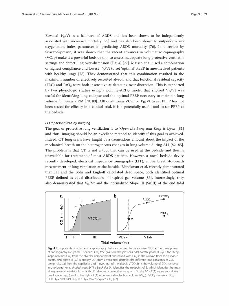
settings and detect lung over-distension (Fig. 4) [77]. Maisch et al. used a combination
of highest compliance and lowest VD/Vt to set ‘optimal’ PEEP in anesthetized patients
with healthy lungs [78]. They demonstrated that this combination resulted in the
maximum number of effectively recruited alveoli, and that functional residual capacity
(FRC) and PaO2 were both insensitive at detecting over-distension. This is supported
by two physiologic studies using a porcine-ARDS model that showed VD/Vt was
useful for identifying lung collapse and the optimal PEEP necessary to maintain lung
volume following a RM [79, 80]. Although using VCap or VD/Vt to set PEEP has not
been tested for efficacy in a clinical trial, it is a potentially useful tool to set PEEP at
the bedside.
PEEP personalized by imaging
The goal of protective lung ventilation is to ‘Open the Lung and Keep it Open’ [81]
and thus, imaging should be an excellent method to identify if this goal is achieved.
Indeed, CT lung scans have taught us a tremendous amount about the impact of the
mechanical breath on the heterogeneous changes in lung volume during ALI [82–85].
The problem is that CT is not a tool that can be used at the bedside and thus is
unavailable for treatment of most ARDS patients. However, a novel bedside device
recently developed, electrical impedance tomography (EIT), allows breath-to-breath
measurement of lung ventilation at the bedside. Blandkman et al. recently demonstrated
that EIT and the Bohr and Enghoff calculated dead space, both identified optimal
PEEP, defined as equal distribution of inspired gas volume [86]. Interestingly, they
also demonstrated that VD/Vt and the normalized Slope III (SnIII) of the end tidal
Fig. 4 Components of volumetric capnography that can be used to personalize PEEP. a The three phasesof capnography are: phase I contains CO2-free gas from the previous tidal breath; phase II (SII) is the steepslope contains CO2 from the alveolar compartment and mixed with CO2 in the airways from the previousbreath; and phase III (SIII) is entirely CO2 from alveoli and identifies the different time constants of CO2
being released from the capillaries and moved out of the alveoli. VTCO2,br is the volume of CO2 removedin one breath (grey shaded area). b The black dot (A) identifies the midpoint of SII which identifies the meanairway-alveolar interface from both diffusive and convective transports. To the left of (A) represents airwaydead space (VDaw) and to the right of (A) represents alveolar tidal volume (VTalv). PaCO2 = alveolar CO2;PETCO2 = end-tidal CO2; PECO2 =mixed-expired CO2 [77]
Nieman et al. Intensive Care Medicine Experimental (2017) 5:8 Page 9 of 21

CO2 curve (Fig. 4) did not identify lung inhomogeneity. However, EIT can be used to
identify the impact of PEEP on distribution of ventilation (Fig. 5) [87]. Although the
majority of studies to date have simply been EIT validation experiments, a few have
investigated the efficacy of EIT-guided PEEP. Muders et al., in a porcine oleic acid
and abdominal hypertension-induced ARDS model, showed that EIT was effective at
quantifying the amount of alveolar R/D at different PEEP levels [13]. They concluded
that EIT has the ability to identify dynamic changes in tidal recruitment and thus may
be an effective tool to titrate optimal PEEP. These findings were supported by Liu et
al. who demonstrated that EIT can identify lung overinflation and R/D at various
levels of PEEP in a porcine saline lavage-induced ARDS model [88]. Finally, Gerhard
et al. compared PEEP set to ARDSnet guidelines [7] with PEEP set by EIT-derived
compliance to maximize PEEP-induced lung recruitment in a porcine saline lavage
ARDS model [89]. They demonstrated that EIT-guided ventilation resulted in a higher
PEEP, improved global and regional compliance, improved oxygenation, and reduced
lung histopathology as compared with ARDSnet protocol set PEEP. Combined data
suggest that EIT may become an important tool in setting optimal PEEP.
PEEP personalized by lung mechanics: compliance/elastance
Retrospective analysis of the ARMA data [68] demonstrated that lung mechanics, in
the form of changes in compliance, are much more predictive of mortality than Vt
[90]. Although these data are very interesting, it must be remembered that the ARMA
trial was not designed to study lung mechanics and patient contribution to respiration
was not identified, which may confound these interpretation of these results. The use
of compliance to adjust PEEP is not a new concept. Indeed, Suter et al. first described
the use of compliance adjusted PEEP over 40 years ago (Fig. 6) [91]. This early work
has been supported by Chiew et al. who investigated a patient-specific, model-based,
PEEP optimization strategy analyzing the relationship between the constant lung
elastance (Elung) and the time-variant dynamic elastance (Edrs) in ARDS patients [92].
They found that PEEP set using the model-based changes in elastance was superior to
that of clinically set PEEP in maximizing lung recruitment and minimizing the work of
Fig. 5 Lung ventilation during a decremental PEEP (15–0 cmH2O) trial measured by electrical impedancetomography (EIT) in patients following cardiac surgery. The top row images from the cranial and thebottom row images from the caudal thoracic lung level. The optimal regional compliance was differentbetween the cranial (10 and 5 cmH2O) and caudal levels (15 and 10 cmH2O) suggesting that no singleoptimal PEEP may exist for all lung levels [87]
Nieman et al. Intensive Care Medicine Experimental (2017) 5:8 Page 10 of 21

breathing. Continuous monitoring of dynamic compliance (Cdyn) as a tool to personalize
PEEP was studied by Suarez-Sipmann et al. in a porcine saline lavage-induced ARDS
model [93]. They compared changes in Cdyn, oxygenation, and lung inflation, measured
by CT following a RM plus PEEP titration trial. Initially, there was an increase in Cdyn with
each reduced PEEP level. The beginning of lung collapse was defined as the PEEP level at
which Cdyn began to fall. The PEEP value selected by Cdyn was compared with that
selected by oxygenation and CT measurements. Both oxygenation and CT confirmed that
the PEEP set by Cdyn maintained a fully open lung and concluded that Cdyn might be a
valuable bedside tool to set optimal PEEP. The use of lung compliance to identify the
optimally protective mechanical breath has recently been reassessed in a retrospective
paper analyzing the parameters associated with increased mortality. In this statistical
analysis by Amato et al., 3562 patients enrolled in nine previous ARDSnet studies were
studied, and it was shown that higher plateau pressure (Pplat) was not always associated
with increased mortality nor was higher PEEP always protective, whereas driving pressure
(ΔP = tidal volume/respiratory-system compliance) was strongly associated with survival
[22]. This study reaffirms the importance of lung compliance in identifying the optimally
protective mechanical breath.
Pressure/volume curve
Evidence supporting the clinical use of the whole lung pressure volume (P/V) curve
as a tool to identify optimal PEEP was demonstrated by Amato et al. in two clinical
trials [94, 95]. Using the P/V curve to set PEEP requires a maneuver which slowly
inflates the lung, with the goal being to identify the upper and lower infection points
(PFLEX) on this P/V curve (Fig. 7). The lower PFLEX is postulated to identify the
pressure at the beginning of alveolar recruitment and the upper PFLEX is at the point
of lung over-distension. They showed that a Vt of 6 cc/kg with PEEP set above the
lower PFLEX improved 28-day survival as compared with a ventilation strategy using a
Fig. 6 Use of the pressure/volume (P/V) curve to personalize PEEP. The shape of the P/V curve changes fromnormal (N) and differs greatly with emphysema (E) or acute pulmonary failure (APF). The P/V relationship duringtidal ventilation is depicted in the shaded area with and without PEEP. RV regional volume at which alveolicollapse, FRC functional residual capacity, and TLC total lung capacity. Central drawing of alveoli size changesalong the P/V curve [91]
Nieman et al. Intensive Care Medicine Experimental (2017) 5:8 Page 11 of 21

Vt of 12 cc/kg combined with lowest set PEEP to maintain acceptable oxygenation
[95]. However, subsequent studies demonstrated that alveoli recruit continually
throughout the entire inflation curve in both animal models [96, 97] and in humans.
In addition, it was shown that there was a higher amount of aerated lung tissue above
the point of maximum curvature on the deflation curve [98] and a higher number of
alveoli above the upper PFLEX on the deflation curve [97] as compared with the lower
PFLEX on the inflation curve. These studies suggest that using the deflation limb of
the P/V curve to set PEEP may be superior, in terms of the amount of recruited lung,
as compared to the inflation limb. Although using the P/V curve as a physiologic tool
to personalize PEEP can be effective, generating the P/V curve is a complex procedure
and there is a risk of causing hemodynamic compromise and injuring the lung during
the inflation procedure. Also, ALI is always evolving and thus even if the PEEP is set
properly using the P/V curve, it must be reset as the lung improves or deteriorates in
function.
Transpulmonary pressure
Patients at risk of developing ARDS often have a decrease in chest wall compliance
secondary to fluid overload and/or increased intra-abdominal pressure. Therefore, it is
Fig. 7 Pressure/volume (P/V) curve from an ARDS patient showing both the lower and upper inflection points(PFLEX). The hypothesis is that the lower PFLEX is the critical alveolar opening point and the upper PFLEX the pointat which alveoli begin to over-distend, however, this hypothesis has been challenged [97, 98]. In this patient,ventilation with a high tidal volume (Vt = 10 ml/kg plus PEEPIDEAL = 15 cmH2O) would cause over-distensionsince ventilation is well above the upper PFLEX. Ventilation with low Vt and PEEPIDEAL was below the upper PFLEX.The calculated lung compliance was increased from 31.6 to 40 with low Vt ventilation [94]
Nieman et al. Intensive Care Medicine Experimental (2017) 5:8 Page 12 of 21

possible that PEEP could be set too low unless the transpulmonary pressure (Ptp) is
known. Clinically, esophageal pressure (Pes) is used as a surrogate for pleural pressure
(Ppl) and used to calculate Ptp. It has shown that PEEP set to maintain Ptp above
0 cmH2O had positive impact in both animal [99, 100] and human [101, 102] studies.
Personalizing PEEP using Ptp is a physiologically sound concept since Ptp is the force that
distends the lung. However, Pes is not the perfect surrogate of Ppl and thus the calculated
Ptp might not be accurate. In a recent study, Huang et al. compared open-lung PEEP
guided by CT to Ptp guided PEEP (i.e., PEEP set to always keep end Ptp above 0 cmH2O)
in a porcine saline lavage lung injury model [103]. They found that Ptp-guided PEEP was
unable to maintain recruited lung open with a hypothesized mechanism for this failure
being an increase in the superimposed pressure between the esophageal plane and dorsal
lung level. However, using Ptp to guide PEEP is a physiologically sound technique and,
there is a multicenter randomized clinical trial using Ptp-guided ventilation that should
help determine the efficacy of this ventilation strategy [104].
Stress index
In 2000, Ranieri et al. demonstrated that the pressure-time (P-t) curve generated using
constant flow ventilation could be used to identify protective mechanical ventilation
(Fig. 8) [105]. They used the shape of the curve to identify the stress being directed into
the lung during mechanical ventilation. These studies demonstrated that if the P-t curve
was straight there was minimal stress, whereas, if the curve had a downward concavity,
there was increasing compliance, and if there is an upward concavity, there is decreasing
compliance. The term b in the curve-fitting equation (Fig. 8) describes the shape of
the P-t curve, and they found that a coefficient b of 1.00 was associated with lung
protection, determined by reduced histopathology and inflammatory mediators. An
extension of this work showed that a coefficient of b < 1 correlated with tidal recruitment,
Fig. 8 Pressure-time (P-t) curves demonstrating the concept of using stress index to personalize PEEP. Usingthe power equation PL = a •tb + c, b describes the shape of the P-t curve. When b < 1, the shape of the curve isa downward concavity as compliance increases over time. When b > 1, the curve has an upward concavity ascompliance decreases over time. When b = 1, the P-t curve is straight and compliance is constant. Adjustingtidal volume (Vt) and PEEP so that b = 1 produces minimal lung stress, if b < 1 would produce low-lung volumestress and b > 1 would cause high-lung volume stress [105]
Nieman et al. Intensive Care Medicine Experimental (2017) 5:8 Page 13 of 21

and a b > 1 correlated with hyperinflation, with b = 1 correlating with non-injurious
mechanical ventilation, confirming the predictive power of this stress-index to identify
injurious mechanical ventilation [26]. The accuracy of the stress-index to identify
injurious mechanical ventilator settings likely to cause VILI was recently confirmed in
humans [106]. CT was used to identify morphological markers of VILI including tidal
hyperinflation, hyperinflated lung at expiration, and tidal recruitment. Results demonstrated
that the Pplat currently considered not to cause VILI (≤30 cmH2O) was shown to cause
tidal hyperinflation, whereas stress-index suggested a Pplat of <25 cmH2O (b < 1.05) would
not and was confirmed by CT. Stress-index was also superior to Pplat at identifying the
optimally protective ventilator settings in the presence of decreased chest wall compliance.
Grasso et al. [107] demonstrated in ARDS patients that using stress-index to set PEEP
reduced alveolar hyperinflation as compared to PEEP set using standard of care [7],
although these findings have been challenged [108]. Although use of stress index to set
PEEP has several physiologic advantages over using oxygenation, which is the standard of
care, and there is a commercially available ventilator that can measure stress index, this
technique has not been shown conclusively superior to the current strategy of adjusting
PEEP using oxygenation.
Time controlled PEEP
A novel physiologic tool to set PEEP is the slope of the expiratory flow curve (SlEFC)
in conjunction with airway pressure release ventilation (APRV) (Fig. 9) [109, 110].
Although this method is not widely utilized throughout the country, it is intensely
used in some hospitals and can be used as a primary mode of ventilation [109]. This
time-controlled PEEP is not directly set but rather the time during expiration (TLOW)
is sufficiently brief to prevent the lung from fully emptying. Thus, both lung volume
and pressure (i.e., PEEP) remain at the beginning of lung reinflated. The shorter the
duration of expiration, the higher the retained-end expiratory lung volume and
positive-end release pressure (PERP) that remains in the lung.
It is important to understand that the personalized APRV (P-APRV) used to set
time-controlled PEEP [109, 110] and inverse inspiratory:expiratory (I:E) ratio are
not at all the same mechanical breath. Although there are many differences between
P-APRV and inverse I:E, I will focus on the large difference in the time at expiration
between these two ventilation strategies. It was shown by Neumann et al. that the
ARDS lung collapses very rapidly, their data showing collapse in 0.6 s after the
initiation of expiration [17]. Our work using direct observation of alveoli during
mechanical ventilation has also shown a very rapid alveolar collapse in the ARDS
lung [40, 111–113]. Unlike APRV, inverse ratio ventilation does not allow direct and
independent adjustments of the expiratory and inspiratory times. Thus, if the ex-
piratory duration with inverse I:E is not less than 0.4–0.6 s, which is most often the
case, alveoli would have sufficient time to collapse with each breath and inverse I:E
would not be defined as time controlled PEEP, since the lung was allowed time to
empty.
An example of how the SlEFC will change with progressive ALI and how we can use
the SlEFC to set the expiratory duration necessary to stabilize the lung is as follows: a
hypothetical normal lung has been given a SlEFC of ~45° and an ARDS lung a SlEFC of
~30° (Fig. 9b). To set the optimal expiratory duration necessary to stabilize the lung,
Nieman et al. Intensive Care Medicine Experimental (2017) 5:8 Page 14 of 21

the end expiratory flow to peak expiratory flow ratio (EEF/PEF) is used. It has been
shown that the ratio that best stabilizes the lung but is still able to adequately ventilate
the patient or animal is 75% [40, 111, 113, 114]. In the example, the PEF is −60 L/min
so to determine when to stop expiration take −60 × 0.75 = 45 L/min, and thus the
clinician would terminate exhalation and reapply the continuous positive airway
pressure (CPAP) at −45 L/min. In the normal lung, the expiratory duration would be
0.5 s (Fig. 9b). With the development of ALI or ARDS, the lung becomes noncompliant
Fig. 9 a Typical airway pressure release ventilation (APRV) airway pressure and flow curves. Correctly setAPRV has a very brief duration at expiration (time at low pressure, TLow) and extended inspiratory duration(time at high pressure, THigh) [109]. The THigh is ~90% of each breath. The two other ARPV settings are thepressure at inspiration (PHigh) and at expiration (PLow). PHigh is set sufficiently high to recruit and open alveoliand PLow is always set at 0 cmH2O to facilitate expiratory flow. However, TLow is sufficiently short such thatend-expiratory pressure (PLow) never reaches 0 cmH2O identified by the tracheal pressure (green line)maintaining a level of PEEP. b This figure summarizes our novel method to maintain alveolar stability byadaptively adjusting the expiratory duration as directed by the expiratory flow curve. The rate of lungcollapse is seen in the normal (slope 45°) and acutely injured lung (ARDS, slope 30°). ARDS causes a morerapid lung collapse due to decreased lung compliance. Our preliminary studies have shown that if theratio of the peak expiratory flow (PEF, −60 L/min) to when we end expiratory flow (EEF, −45 L/min) (EEF/PEF) is equal to 75% that this expiratory duration (0.5 s) is sufficient to stabilize alveoli [40, 111]. The lungwith ARDS collapses more rapidly such that the EEF/PEF-75% identifies an expiratory duration of 0.45 snecessary to stabilize alveoli. Although the EEF/PEF is fixed, the expiratory duration is not, but ratheradaptive and will stabilize alveoli regardless of lung injury severity. Thus, this method of setting expiratoryduration is adaptive to changes in lung pathophysiology and personalizes the mechanical breath to eachindividual patient
Nieman et al. Intensive Care Medicine Experimental (2017) 5:8 Page 15 of 21

and collapses very rapidly decreasing the SlEFC to ~30°. Using the same equation used
in the normal lung, but with a steeper slope, we see that the expiratory time has been
reduced from 0.5 to 0.45 s in order to prevent alveolar collapse in this noncompliant
lung (Fig. 9b). This method results in a time-controlled PEEP, effectively minimizing
dynamic strain (Fig. 9b) by stabilizing alveoli that uses two mechanisms: time and
pressure [110].
Multiple studies have shown that this combined method of PEEP plus a brief release time
is very effective at stabilizing alveoli and alveolar ducts, reducing tissue strain [40, 111],
blocking progressive ALI, and reducing ARDS incidence in a clinically applicable,
high-fidelity, porcine model of sepsis and gut ischemia/reperfusion-induced ARDS
[113], and in a trauma patient statistical analysis [114]. More clinical studies are
necessary to confirm the efficacy of this novel method to stabilize the lung.
ConclusionsIt is clear that a high level of dynamic strain caused by alveolar R/D is a major mechanism
of lung tissue damage associated with VILI. Application of PEEP is currently the primary
strategy by which to minimize dynamic strain for established ARDS. In addition, early
PEEP application has been effective at reducing the complication associated with
mechanical ventilation in both animal and human studies. It is also clear that in order
for PEEP to be effective, it must be personalized to the specific pathology of each
patient’s lung. The continued high mortality rate of ARDS supports the hypothesis
that the current PEEP strategies are not always effective [1]. Multiple methods to
personalize PEEP have been tested and have been shown to be capable of stabilizing
the lung. A large body of literature supports the use of RMs to open the lung, prior
to the application of PEEP, which is set based on physiologic feedback. Recent clinical
studies are attempting to personalize PEEP following RMs in humans [67, 115]. The
main problem with the use of RMs is that they cannot be given very often, due to
potential serious side effects, and the acutely injured lung is constantly changing.
Thus, if lung pathology increases following the initial PEEP setting, lung instability
would go unrecognized, causing additional VILI-induced lung damage. A novel
method of personalizing PEEP is the using of expiratory flow curve during APRV.
The advantage is that a RM is not required so that adjustments in expiratory duration
are adaptive with progressive changes in acute lung injury, regardless if these changes
are for the better or worse. Also, this method does not directly set PEEP but rather
uses a short expiratory duration to generate intrinsic PEEP, which is used as a tool to
stabilize the lung. Both animal studies and a human statistical analysis suggest that
adjusting the release time on a breath-to-breath basis may be the optimal mechanism
to adaptively personalize PEEP.
AbbreviationsALI: Acute lung injury; APRV: Airway pressure release ventilation; ARDS: Acute respiratory distress syndrome;CPAP: Continuous positive airway pressure; CT: Computed tomography; EEF/PEF: End expiratory flow to peakexpiratory flow ratio; EIT: Electrical impedance tomography; FRC: Functional residual capacity; OD: Over-distension; P/V: Pressure/volume; P-APRV: Personalized airway pressure release ventilation; PEEP: Positive-end expiratory pressure;Pes: Esophageal pressure; PFLEX: Lower inflection point; Pmci,I: Maximal compliance increase; Ppl: Pleural pressure;Ptp: Transpulmonary pressure; R/D: Recruitment and derecruitment; RACE: Rapid alveolar collapse and expansion;RM: Recruitment maneuver; S-C: Stress concentration; SC: Stress concentrators; SlEFC: Slope of the expiratory flow curve;VCap: Volumetric capnography; VILI: Ventilator-induced lung injury; Vt: Tidal volume
Nieman et al. Intensive Care Medicine Experimental (2017) 5:8 Page 16 of 21

FundingSalary support for JS is from NIH R01 HL131143.
Authors’ contributionsGFN drafted the manuscript. PLA, NMH, JS, and LAG critically revised the manuscript. GFN and LAG helped to conceivethe manuscript. All authors read and approved the final manuscript.
Competing interestsPLA, GFN, and NMH have presented and received honoraria and/or travel reimbursement at event(s) sponsored byDräger Medical Systems, Inc., outside of the published work. PLA, GFN, NMH, and LAG have lectured for Intensive CareOnline Network, Inc. (ICON). NMH is the founder of ICON, of which PLA is an employee. NMH holds patents on amethod of initiating, managing, and/or weaning airway pressure release ventilation, as well as, controlling a ventilatorin accordance with the same, but these patents are not commercialized, licensed nor royalty-producing. The authorsmaintain that industry had no role in the design and conduct of the study; the collection, management, analysis, or in-terpretation of the data; nor the preparation, review, or approval of the manuscript.
Author details1Department of Surgery, SUNY Upstate Medical University, Syracuse, NY, USA. 2Intensive Care Online (ICON), Baltimore,MD, USA. 3Department of Trauma Critical Care Medicine, R Adams Cowley Shock Trauma Center, University ofMaryland, Baltimore, MD, USA. 4Biological Sciences Department, Biological Sciences Department, SUNY Cortland,Cortland, NY, USA. 5Cardiopulmonary Critical Care Lab, Department of Surgery, Upstate Medical University, 750 EastAdams Street, Syracuse, NY 13210, USA.
Received: 7 October 2016 Accepted: 26 January 2017
References1. Villar J, Blanco J, Kacmarek RM (2016) Current incidence and outcome of the acute respiratory distress syndrome.
Curr Opin Crit Care 22:1–62. Force ADT, Ranieri VM, Rubenfeld GD, Thompson BT, Ferguson ND, Caldwell E, Fan E, Camporota L, Slutsky AS
(2012) Acute respiratory distress syndrome: the Berlin definition. JAMA 307:2526–25333. Guerin C, Reignier J, Richard JC (2013) Prone positioning in the acute respiratory distress syndrome. N Engl J Med
369:980–9814. Villar J, Sulemanji D, Kacmarek RM (2014) The acute respiratory distress syndrome: incidence and mortality, has it
changed? Curr Opin Crit Care 20:3–95. Neto AS, Simonis FD, Barbas CS, Biehl M, Determann RM, Elmer J, Friedman G, Gajic O, Goldstein JN, Linko R,
Pinheiro de Oliveira R, Sundar S, Talmor D, Wolthuis EK, Gama de Abreu M, Pelosi P, Schultz MJ, InvestigatorsPRVN (2015) Lung-protective ventilation with low tidal volumes and the occurrence of pulmonary complicationsin patients without acute respiratory distress syndrome: a systematic review and individual patient data analysis.Crit Care Med 43:2155–2163
6. Gong MN, Thompson BT (2016) Acute respiratory distress syndrome: shifting the emphasis from treatment toprevention. Curr Opin Crit Care 22:21–37
7. ARDSnet (2000) Ventilation with lower tidal volumes as compared with traditional tidal volumes for acute lunginjury and the acute respiratory distress syndrome. The Acute Respiratory Distress Syndrome Network. N Engl JMed 342:1301–1308
8. Andrews P, Sadowitz B, M. K-S, Satalin J, Roy S, Snyder K, Gatto L, Nieman G, Habashi N, (2015) Alveolar instability(atelectrauma) is not identified by arterial oxygenation predisposing the development of an occult ventilator-induced lung injury. Intensive Care Med Exp 3:1–12
9. Baumgardner JE, Markstaller K, Pfeiffer B, Doebrich M, Otto CM (2002) Effects of respiratory rate, plateau pressure, andpositive end-expiratory pressure on PaO2 oscillations after saline lavage. Am J Respir Crit Care Med 166:1556–1562
10. Branson RD, Johannigman JA, Campbell RS, Davis K Jr (2002) Closed-loop mechanical ventilation. Respir Care 47:427–451, discussion 451-423
11. Pomprapa A, Schwaiberger D, Pickerodt P, Tjarks O, Lachmann B, Leonhardt S (2014) Automatic protectiveventilation using the ARDSNet protocol with the additional monitoring of electrical impedance tomography.Crit Care 18:R128
12. Boehme S, Bentley AH, Hartmann EK, Chang S, Erdoes G, Prinzing A, Hagmann M, Baumgardner JE, Ullrich R,Markstaller K, David M (2015) Influence of inspiration to expiration ratio on cyclic recruitment and derecruitmentof atelectasis in a saline lavage model of acute respiratory distress syndrome. Crit Care Med 43:e65–74
13. Muders T, Luepschen H, Zinserling J, Greschus S, Fimmers R, Guenther U, Buchwald M, Grigutsch D, Leonhardt S,Putensen C, Wrigge H (2012) Tidal recruitment assessed by electrical impedance tomography and computedtomography in a porcine model of lung injury*. Crit Care Med 40:903–911
14. Albert SP, DiRocco J, Allen GB, Bates JH, Lafollette R, Kubiak BD, Fischer J, Maroney S, Nieman GF (2009) The roleof time and pressure on alveolar recruitment. J Appl Physiol 106:757–765
15. Smith BJ, Grant KA, Bates JH (2013) Linking the development of ventilator-induced injury to mechanical functionin the lung. Ann Biomed Eng 41:527–536
16. Henderson WR, Dominelli PB, Molgat-Seon Y, Lipson R, Griesdale DE, Sekhon M, Ayas N, Sheel AW (2016) Effect of tidalvolume and positive end-expiratory pressure on expiratory time constants in experimental lung injury. Phys Rep 4:1–11
17. Neumann P, Berglund JE, Fernandez Mondejar E, Magnusson A, Hedenstierna G (1998) Dynamics of lung collapseand recruitment during prolonged breathing in porcine lung injury. J Appl Physiol 85:1533–1543
18. Smith BJ, Lundblad LK, Kollisch-Singule M, Satalin J, Nieman G, Habashi N, Bates JH (2015) Predicting the responseof the injured lung to the mechanical breath profile. J Appl Physiol 118:932–940
Nieman et al. Intensive Care Medicine Experimental (2017) 5:8 Page 17 of 21

19. Schranz C, Becher T, Schadler D, Weiler N, Moller K (2014) Model-based setting of inspiratory pressure andrespiratory rate in pressure-controlled ventilation. Physiol Meas 35:383–397
20. Ferrando C, Suarez-Sipmann F, Gutierrez A, Tusman G, Carbonell J, Garcia M, Piqueras L, Compan D, Flores S, SoroM, Llombart A, Belda FJ (2015) Adjusting tidal volume to stress index in an open lung condition optimizesventilation and prevents overdistension in an experimental model of lung injury and reduced chest wallcompliance. Crit Care 19:9
21. Protti A, Maraffi T, Milesi M, Votta E, Santini A, Pugni P, Andreis DT, Nicosia F, Zannin E, Gatti S, Vaira V, Ferrero S,Gattinoni L (2016) Role of Strain rate in the pathogenesis of ventilator-induced lung edema. Crit Care Med 44:e838–845
22. Amato MB, Meade MO, Slutsky AS, Brochard L, Costa EL, Schoenfeld DA, Stewart TE, Briel M, Talmor D, Mercat A,Richard JC, Carvalho CR, Brower RG (2015) Driving pressure and survival in the acute respiratory distresssyndrome. N Engl J Med 372:747–755
23. Hess DR (2015) Recruitment Maneuvers and PEEP Titration. Respir Care 60:1688–170424. Caramez MP, Kacmarek RM, Helmy M, Miyoshi E, Malhotra A, Amato MB, Harris RS (2009) A comparison of
methods to identify open-lung PEEP. Intensive Care Med 35:740–74725. Hata JS, Togashi K, Kumar AB, Hodges LD, Kaiser EF, Tessmann PB, Faust CA, Sessler DI (2014) The effect of the
pressure-volume curve for positive end-expiratory pressure titration on clinical outcomes in acute respiratorydistress syndrome: a systematic review. J Intensive Care Med 29:348–356
26. Grasso S, Terragni P, Mascia L, Fanelli V, Quintel M, Herrmann P, Hedenstierna G, Slutsky AS, Ranieri VM (2004)Airway pressure-time curve profile (stress index) detects tidal recruitment/hyperinflation in experimental acutelung injury. Crit Care Med 32:1018–1027
27. Takeuchi M, Goddon S, Dolhnikoff M, Shimaoka M, Hess D, Amato MB, Kacmarek RM (2002) Set positive end-expiratory pressure during protective ventilation affects lung injury. Anesthesiology 97:682–692
28. Soroksky A, Esquinas A (2012) Goal-directed mechanical ventilation: are we aiming at the right goals? A proposalfor an alternative approach aiming at optimal lung compliance, guided by esophageal pressure in acuterespiratory failure. Crit Care Res Prac 2012:597932
29. Sundaresan A, Yuta T, Hann CE, Chase JG, Shaw GM (2009) A minimal model of lung mechanics and model-basedmarkers for optimizing ventilator treatment in ARDS patients. Comput Methods Programs Biomed 95:166–180
30. Talmor D, Sarge T, O'Donnell CR, Ritz R, Malhotra A, Lisbon A, Loring SH (2006) Esophageal and transpulmonarypressures in acute respiratory failure. Crit Care Med 34:1389–1394
31. Hubmayr RD (2002) Perspective on lung injury and recruitment: a skeptical look at the opening and collapsestory. Am J Respir Crit Care Med 165:1647–1653
32. Gattinoni L, Pesenti A (2005) The concept of "baby lung". Intensive Care Med 31:776–78433. Cressoni M, Cadringher P, Chiurazzi C, Amini M, Gallazzi E, Marino A, Brioni M, Carlesso E, Chiumello D, Quintel M,
Bugedo G, Gattinoni L (2014) Lung inhomogeneity in patients with acute respiratory distress syndrome. Am JRespir Crit Care Med 189:149–158
34. Retamal J, Bergamini BC, Carvalho AR, Bozza FA, Borzone G, Borges JB, Larsson A, Hedenstierna G, Bugedo G,Bruhn A (2014) Non-lobar atelectasis generates inflammation and structural alveolar injury in the surroundinghealthy tissue during mechanical ventilation. Crit Care 18:505
35. Santos CC, Zhang H, Liu M, Slutsky AS (2005) Bench-to-bedside review: biotrauma and modulation of the innateimmune response. Crit Care 9:280–286
36. Gattinoni L, Pesenti A, Avalli L, Rossi F, Bombino M (1987) Pressure-volume curve of total respiratory system inacute respiratory failure. Computed tomographic scan study. Am Rev Respir Dis 136:730–736
37. Carney DE, Bredenberg CE, Schiller HJ, Picone AL, McCann UG, Gatto LA, Bailey G, Fillinger M, Nieman GF (1999)The mechanism of lung volume change during mechanical ventilation. Am J Respir Crit Care Med 160:1697–1702
38. Namati E, Thiesse J, de Ryk J, McLennan G (2008) Alveolar dynamics during respiration: are the pores of Kohn apathway to recruitment? Am J Respir Cell Mol Biol 38:572–578
39. Perlman CE, Bhattacharya J (2007) Alveolar expansion imaged by optical sectioning microscopy. J Appl Physiol103:1037–1044
40. Kollisch-Singule M, Emr B, Smith B, Ruiz C, Roy S, Meng Q, Jain S, Satalin J, Snyder K, Ghosh A, Marx W, Andrews P,Habashi N, Nieman G, Gatto LA (2014) Airway pressure release ventilation reduces conducting airway micro-strainin lung injury. J Am Coll Surg 219:9
41. Dreyfuss D, Soler P, Basset G, Saumon G (1988) High inflation pressure pulmonary edema. Respective effects ofhigh airway pressure, high tidal volume, and positive end-expiratory pressure. Am Rev Respir Dis 137:1159–1164
42. Seah AS, Grant KA, Aliyeva M, Allen GB, Bates JHT (2011) Quantifying the roles of tidal volume and PEEP in thepathogenesis of ventilator-induced lung injury. Ann Biomed Eng 39:1505–1516
43. Steinberg J, Schiller HJ, Halter JM, Gatto LA, Dasilva M, Amato M, McCann UG, Nieman GF (2002) Tidal volumeincreases do not affect alveolar mechanics in normal lung but cause alveolar overdistension and exacerbatealveolar instability after surfactant deactivation. Crit Care Med 30:2675–2683
44. Cereda M, Xin Y, Kadlecek S, Hamedani H, Rajaei J, Clapp J, Rizi RR (2014) Hyperpolarized gas diffusion MRI for thestudy of atelectasis and acute respiratory distress syndrome. NMR Biomed 27:1468–1478
45. Chen ZL, Song YL, Hu ZY, Zhang S, Chen YZ (2015) An estimation of mechanical stress on alveolar walls duringrepetitive alveolar reopening and closure. J Appl Physiol 119:190–201
46. Albaiceta GM, Blanch L (2011) Beyond volutrauma in ARDS: the critical role of lung tissue deformation.Crit Care 15:304
47. Protti A, Andreis DT, Monti M, Santini A, Sparacino CC, Langer T, Votta E, Gatti S, Lombardi L, Leopardi O, MassonS, Cressoni M, Gattinoni L (2013) Lung stress and strain during mechanical ventilation: any difference betweenstatics and dynamics? Crit Care Med 41:1046–1055
48. Protti A, Andreis DT, Milesi M, Iapichino GE, Monti M, Comini B, Pugni P, Melis V, Santini A, Dondossola D, Gatti S,Lombardi L, Votta E, Carlesso E, Gattinoni L (2015) Lung anatomy, energy load, and ventilator-induced lung injury.Intensive Care Med Exp 3:34
Nieman et al. Intensive Care Medicine Experimental (2017) 5:8 Page 18 of 21
49. Steinberg JM, Schiller HJ, Halter JM, Gatto LA, Lee HM, Pavone LA, Nieman GF (2004) Alveolar instability causesearly ventilator-induced lung injury independent of neutrophils. Am J Respir Crit Care Med 169:57–63
50. Borges JB, Costa EL, Suarez-Sipmann F, Widstrom C, Larsson A, Amato M, Hedenstierna G (2014) Earlyinflammation mainly affects normally and poorly aerated lung in experimental ventilator-induced lung injury*. CritCare Med 42:e279–287
51. Wellman TJ, Winkler T, Costa EL, Musch G, Harris RS, Zheng H, Venegas JG, Vidal Melo MF (2014) Effect of localtidal lung strain on inflammation in normal and lipopolysaccharide-exposed sheep*. Crit Care Med 42:e491–500
52. Albert RK (2012) The role of ventilation-induced surfactant dysfunction and atelectasis in causing acute respiratorydistress syndrome. Am J Respir Crit Care Med 185:702–708
53. Fanelli V, Mascia L, Puntorieri V, Assenzio B, Elia V, Fornaro G, Martin EL, Bosco M, Delsedime L, Fiore T, Grasso S,Ranieri VM (2009) Pulmonary atelectasis during low stretch ventilation: “open lung” versus “lung rest” strategy. CritCare Med 37:1046–1053
54. Serpa Neto A, Cardoso SO, Manetta JA, Pereira VG, Esposito DC, Pasqualucci Mde O, Damasceno MC, Schultz MJ(2012) Association between use of lung-protective ventilation with lower tidal volumes and clinical outcomesamong patients without acute respiratory distress syndrome: a meta-analysis. JAMA 308:1651–1659
55. Wolthuis EK, Vlaar AP, Choi G, Roelofs JJ, Juffermans NP, Schultz MJ (2009) Mechanical ventilation using non-injuriousventilation settings causes lung injury in the absence of pre-existing lung injury in healthy mice. Crit Care 13:R1
56. Fuld MK, Easley RB, Saba OI, Chon D, Reinhardt JM, Hoffman EA, Simon BA (2008) CT-measured regional specificvolume change reflects regional ventilation in supine sheep. J Appl Physiol 104:1177–1184
57. Reinhardt JM, Ding K, Cao K, Christensen GE, Hoffman EA, Bodas SV (2008) Registration-based estimates of locallung tissue expansion compared to xenon CT measures of specific ventilation. Med Image Anal 12:752–763
58. Kaczka DW, Cao K, Christensen GE, Bates JH, Simon BA (2011) Analysis of regional mechanics in canine lung injuryusing forced oscillations and 3D image registration. Ann Biomed Eng 39:1112–1124
59. Slutsky AS, Villar J, Pesenti A (2016) Happy 50th birthday ARDS! Intensive Care Med 42:637–63960. Nieman GF, Gatto LA, Bates JH, Habashi NM (2015) Mechanical ventilation as a therapeutic tool to reduce ARDS
incidence. Chest 148:1396–140461. Fernandez Mondejar E, Vazquez Mata G, Cardenas A, Mansilla A, Cantalejo F, Rivera R (1996) Ventilation with
positive end-expiratory pressure reduces extravascular lung water and increases lymphatic flow in hydrostaticpulmonary edema. Crit Care Med 24:1562–1567
62. Ochiai R (2015) Mechanical ventilation of acute respiratory distress syndrome. J Intensive Care 3:2563. Brower RG, Lanken PN, MacIntyre N, Matthay MA, Morris A, Ancukiewicz M, Schoenfeld D, Thompson BT, National
Heart L, Blood Institute ACTN (2004) Higher versus lower positive end-expiratory pressures in patients with theacute respiratory distress syndrome. N Engl J Med 351:327–336
64. Briel M, Meade M, Mercat A, Brower RG, Talmor D, Walter SD, Slutsky AS, Pullenayegum E, Zhou Q, Cook D,Brochard L, Richard JC, Lamontagne F, Bhatnagar N, Stewart TE, Guyatt G (2010) Higher vs lower positive end-expiratory pressure in patients with acute lung injury and acute respiratory distress syndrome: systematic reviewand meta-analysis. JAMA 303:865–873
65. Nelson LD (1996) High-inflation pressure and positive end-expiratory pressure. Injurious to the lung? No. Crit CareClin 12:603–625
66. Gattinoni L, Carlesso E, Brazzi L, Caironi P (2010) Positive end-expiratory pressure. Curr Opin Crit Care 16:39–4467. Ambrosio AM, Luo R, Fantoni DT, Gutierres C, Lu Q, Gu WJ, Otsuki DA, Malbouisson LM, Auler JO Jr, Rouby JJ,
Experimental ASG (2012) Effects of positive end-expiratory pressure titration and recruitment maneuver on lunginflammation and hyperinflation in experimental acid aspiration-induced lung injury. Anesthesiology 117:1322–1334
68. Kallet RH, Campbell AR, Dicker RA, Katz JA, Mackersie RC (2006) Effects of tidal volume on work of breathingduring lung-protective ventilation in patients with acute lung injury and acute respiratory distress syndrome. CritCare Med 34:8–14
69. Chiumello D, Cressoni M, Carlesso E, Caspani ML, Marino A, Gallazzi E, Caironi P, Lazzerini M, Moerer O, Quintel M,Gattinoni L (2014) Bedside selection of positive end-expiratory pressure in mild, moderate, and severe acuterespiratory distress syndrome. Crit Care Med 42:252–264
70. Borges JB, Okamoto VN, Matos GF, Caramez MP, Arantes PR, Barros F, Souza CE, Victorino JA, Kacmarek RM, BarbasCS, Carvalho CR, Amato MB (2006) Reversibility of lung collapse and hypoxemia in early acute respiratory distresssyndrome. Am J Respir Crit Care Med 174:268–278
71. Meade MO, Cook DJ, Guyatt GH, Slutsky AS, Arabi YM, Cooper DJ, Davies AR, Hand LE, Zhou Q, Thabane L, AustinP, Lapinsky S, Baxter A, Russell J, Skrobik Y, Ronco JJ, Stewart TE, Lung open ventilation study I (2008) Ventilationstrategy using low tidal volumes, recruitment maneuvers, and high positive end-expiratory pressure for acutelung injury and acute respiratory distress syndrome: a randomized controlled trial. JAMA 299:637–645
72. Mercat A, Richard JC, Vielle B, Jaber S, Osman D, Diehl JL, Lefrant JY, Prat G, Richecoeur J, Nieszkowska A, Gervais C,Baudot J, Bouadma L, Brochard L, Expiratory Pressure Study G (2008) Positive end-expiratory pressure setting in adultswith acute lung injury and acute respiratory distress syndrome: a randomized controlled trial. JAMA 299:646–655
73. Gattinoni L, Caironi P (2008) Refining ventilatory treatment for acute lung injury and acute respiratory distresssyndrome. JAMA 299:691–693
74. Goligher EC, Kavanagh BP, Rubenfeld GD, Adhikari NK, Pinto R, Fan E, Brochard LJ, Granton JT, Mercat A, MarieRichard JC, Chretien JM, Jones GL, Cook DJ, Stewart TE, Slutsky AS, Meade MO, Ferguson ND (2014) Oxygenationresponse to positive end-expiratory pressure predicts mortality in acute respiratory distress syndrome. Asecondary analysis of the LOVS and ExPress trials. Am J Respir Crit Care Med 190:70–76
75. Nuckton TJ, Alonso JA, Kallet RH, Daniel BM, Pittet JF, Eisner MD, Matthay MA (2002) Pulmonary dead-spacefraction as a risk factor for death in the acute respiratory distress syndrome. N Engl J Med 346:1281–1286
76. Cepkova M, Kapur V, Ren X, Quinn T, Zhuo H, Foster E, Liu KD, Matthay MA (2007) Pulmonary dead space fraction andpulmonary artery systolic pressure as early predictors of clinical outcome in acute lung injury. Chest 132:836–842
77. Suarez-Sipmann F, Bohm SH, Tusman G (2014) Volumetric capnography: the time has come. Curr Opin Crit Care20:333–339
Nieman et al. Intensive Care Medicine Experimental (2017) 5:8 Page 19 of 21

78. Maisch S, Reissmann H, Fuellekrug B, Weismann D, Rutkowski T, Tusman G, Bohm SH (2008) Compliance and deadspace fraction indicate an optimal level of positive end-expiratory pressure after recruitment in anesthetizedpatients. Anesth Analg 106:175–181, table of contents
79. Tusman G, Suarez-Sipmann F, Bohm SH, Pech T, Reissmann H, Meschino G, Scandurra A, Hedenstierna G (2006)Monitoring dead space during recruitment and PEEP titration in an experimental model. Intensive Care Med32:1863–1871
80. Yang Y, Huang Y, Tang R, Chen Q, Hui X, Li Y, Yu Q, Zhao H, Qiu H (2014) Optimization of positive end-expiratorypressure by volumetric capnography variables in lavage-induced acute lung injury. Respiration 87:75–83
81. Lachmann B (1992) Open up the lung and keep the lung open. Intensive Care Med 18:319–32182. Gattinoni L, Pesenti A, Bombino M, Baglioni S, Rivolta M, Rossi F, Rossi G, Fumagalli R, Marcolin R, Mascheroni D et
al (1988) Relationships between lung computed tomographic density, gas exchange, and PEEP in acuterespiratory failure. Anesthesiology 69:824–832
83. Gattinoni L, Bombino M, Pelosi P, Lissoni A, Pesenti A, Fumagalli R, Tagliabue M (1994) Lung structure andfunction in different stages of severe adult respiratory distress syndrome. JAMA 271:1772–1779
84. Caironi P, Langer T, Gattinoni L (2008) Acute lung injury/acute respiratory distress syndrome pathophysiology:what we have learned from computed tomography scanning. Curr Opin Crit Care 14:64–69
85. Caironi P, Cressoni M, Chiumello D, Ranieri M, Quintel M, Russo SG, Cornejo R, Bugedo G, Carlesso E, Russo R,Caspani L, Gattinoni L (2010) Lung opening and closing during ventilation of acute respiratory distress syndrome.Am J Respir Crit Care Med 181:578–586
86. Blankman P, Shono A, Hermans BJ, Wesselius T, Hasan D, Gommers D (2016) Detection of optimal PEEP for equaldistribution of tidal volume by volumetric capnography and electrical impedance tomography during decreasinglevels of PEEP in post cardiac-surgery patients. Br J Anaesth 116:862–869
87. Bikker IG, Leonhardt S, Reis Miranda D, Bakker J, Gommers D (2010) Bedside measurement of changes in lungimpedance to monitor alveolar ventilation in dependent and non-dependent parts by electrical impedancetomography during a positive end-expiratory pressure trial in mechanically ventilated intensive care unit patients.Crit Care 14:R100
88. Liu S, Tan L, Moller K, Frerichs I, Yu T, Liu L, Huang Y, Guo F, Xu J, Yang Y, Qiu H, Zhao Z (2016) Identification ofregional overdistension, recruitment and cyclic alveolar collapse with electrical impedance tomography in anexperimental ARDS model. Crit Care 20:119
89. Wolf GK, Gomez-Laberge C, Rettig JS, Vargas SO, Smallwood CD, Prabhu SP, Vitali SH, Zurakowski D, Arnold JH(2013) Mechanical ventilation guided by electrical impedance tomography in experimental acute lung injury. CritCare Med 41:1296–1304
90. Deans KJ, Minneci PC, Cui X, Banks SM, Natanson C, Eichacker PQ (2005) Mechanical ventilation in ARDS: one sizedoes not fit all. Crit Care Med 33:1141–1143
91. Suter PM, Fairley B, Isenberg MD (1975) Optimum end-expiratory airway pressure in patients with acutepulmonary failure. N Engl J Med 292:284–289
92. Chiew YS, Chase JG, Shaw GM, Sundaresan A, Desaive T (2011) Model-based PEEP optimisation in mechanicalventilation. Biomed Eng Online 10:111
93. Suarez-Sipmann F, Bohm SH, Tusman G, Pesch T, Thamm O, Reissmann H, Reske A, Magnusson A, Hedenstierna G(2007) Use of dynamic compliance for open lung positive end-expiratory pressure titration in an experimentalstudy. Crit Care Med 35:214–221
94. Amato MB, Barbas CS, Medeiros DM, Schettino Gde P, Lorenzi Filho G, Kairalla RA, Deheinzelin D, Morais C,Fernandes Ede O, Takagaki TY et al (1995) Beneficial effects of the “open lung approach” with low distendingpressures in acute respiratory distress syndrome. A prospective randomized study on mechanical ventilation. Am JRespir Crit Care Med 152:1835–1846
95. Amato MB, Barbas CS, Medeiros DM, Magaldi RB, Schettino GP, Lorenzi-Filho G, Kairalla RA, Deheinzelin D, MunozC, Oliveira R, Takagaki TY, Carvalho CR (1998) Effect of a protective-ventilation strategy on mortality in the acuterespiratory distress syndrome. N Engl J Med 338:347–354
96. Schiller HJ, Steinberg J, Halter J, McCann U, DaSilva M, Gatto LA, Carney D, Nieman G (2003) Alveolar inflationduring generation of a quasi-static pressure/volume curve in the acutely injured lung. Crit Care Med 31:1126–1133
97. DiRocco JD, Carney DE, Nieman GF (2007) Correlation between alveolar recruitment/derecruitment and inflectionpoints on the pressure-volume curve. Intensive Care Med 33:1204–1211
98. Albaiceta GM, Taboada F, Parra D, Luyando LH, Calvo J, Menendez R, Otero J (2004) Tomographic study of theinflection points of the pressure-volume curve in acute lung injury. Am J Respir Crit Care Med 170:1066–1072
99. Loring SH, Pecchiari M, Della Valle P, Monaco A, Gentile G, D'Angelo E (2010) Maintaining end-expiratorytranspulmonary pressure prevents worsening of ventilator-induced lung injury caused by chest wall constrictionin surfactant-depleted rats. Crit Care Med 38:2358–2364
100. Staffieri F, Stripoli T, De Monte V, Crovace A, Sacchi M, De Michele M, Trerotoli P, Terragni P, Ranieri VM, Grasso S(2012) Physiological effects of an open lung ventilatory strategy titrated on elastance-derived end-inspiratorytranspulmonary pressure: study in a pig model*. Crit Care Med 40:2124–2131
101. Talmor D, Sarge T, Malhotra A, O'Donnell CR, Ritz R, Lisbon A, Novack V, Loring SH (2008) Mechanical ventilationguided by esophageal pressure in acute lung injury. N Engl J Med 359:2095–2104
102. Grasso S, Terragni P, Birocco A, Urbino R, Del Sorbo L, Filippini C, Mascia L, Pesenti A, Zangrillo A, Gattinoni L,Ranieri VM (2012) ECMO criteria for influenza A (H1N1)-associated ARDS: role of transpulmonary pressure.Intensive Care Med 38:395–403
103. Huang Y, Tang R, Chen Q, Pan C, Liu S, Hui X, Li Y, Yang Y, Ranieri VM, Qiu H (2016) How much esophagealpressure-guided end-expiratory transpulmonary pressure is sufficient to maintain lung recruitment in lavage-induced lung injury? J Trauma Acute Care Surg 80:302–307
104. Fish E, Novack V, Banner-Goodspeed VM, Sarge T, Loring S, Talmor D (2014) The Esophageal Pressure-GuidedVentilation 2 (EPVent2) trial protocol: a multicentre, randomised clinical trial of mechanical ventilation guided bytranspulmonary pressure. BMJ Open 4:e006356
Nieman et al. Intensive Care Medicine Experimental (2017) 5:8 Page 20 of 21

105. Ranieri VM, Zhang H, Mascia L, Aubin M, Lin CY, Mullen JB, Grasso S, Binnie M, Volgyesi GA, Eng P, Slutsky AS(2000) Pressure-time curve predicts minimally injurious ventilatory strategy in an isolated rat lung model.Anesthesiology 93:1320–1328
106. Terragni PP, Filippini C, Slutsky AS, Birocco A, Tenaglia T, Grasso S, Stripoli T, Pasero D, Urbino R, Fanelli V,Faggiano C, Mascia L, Ranieri VM (2013) Accuracy of plateau pressure and stress index to identify injuriousventilation in patients with acute respiratory distress syndrome. Anesthesiology 119:880–889
107. Grasso S, Stripoli T, De Michele M, Bruno F, Moschetta M, Angelelli G, Munno I, Ruggiero V, Anaclerio R, Cafarelli A,Driessen B, Fiore T (2007) ARDSnet ventilatory protocol and alveolar hyperinflation: role of positive end-expiratorypressure. Am J Respir Crit Care Med 176:761–767
108. Formenti P, Graf J, Santos A, Gard KE, Faltesek K, Adams AB, Dries DJ, Marini JJ (2011) Non-pulmonary factorsstrongly influence the stress index. Intensive Care Med 37:594–600
109. Habashi NM (2005) Other approaches to open-lung ventilation: airway pressure release ventilation. Crit Care Med33:S228–240
110. Jain SV, Kollisch-Singule M, Sadowitz B, Dombert L, Satalin J, Andrews P, Gatto LA, Nieman GF, Habashi NM (2016)The 30-year evolution of airway pressure release ventilation (APRV). Intensive Care Med Exp 4:11
111. Kollisch-Singule M, Emr B, Smith B, Roy S, Jain S, Satalin J, Snyder K, Andrews P, Habashi N, Bates J, Marx W,Nieman G, Gatto LA (2014) Mechanical breath profile of airway pressure release ventilation: the effect on alveolarrecruitment and microstrain in acute lung injury. JAMA Surg 149:1138–1145
112. Kollisch-Singule M, Jain S, Andrews P, Smith BJ, Hamlington-Smith KL, Roy S, DiStefano D, Nuss E, Satalin J, MengQ, Marx W, Bates JH, Gatto LA, Nieman GF, Habashi NM (2015) Effect of Airway pressure release ventilation ondynamic alveolar heterogeneity. JAMA Surg 151:1–9
113. Roy S, Habashi N, Sadowitz B, Andrews P, Ge L, Wang G, Roy P, Ghosh A, Kuhn M, Satalin J, Gatto LA, Lin X, DeanDA, Vodovotz Y, Nieman G (2013) Early airway pressure release ventilation prevents ARDS-a novel preventiveapproach to lung injury. Shock 39:28–38
114. Andrews PL, Shiber JR, Jaruga-Killeen E, Roy S, Sadowitz B, O'Toole RV, Gatto LA, Nieman GF, Scalea T, Habashi NM(2013) Early application of airway pressure release ventilation may reduce mortality in high-risk trauma patients: asystematic review of observational trauma ARDS literature. J Trauma Acute Care Surg 75:635–641
115. Kacmarek RM, Villar J, Sulemanji D, Montiel R, Ferrando C, Blanco J, Koh Y, Soler JA, Martínez D, Hernández M,Tucci M, Borges JB, Lubillo S, Santos A, Araujo JB, Amato MBP, Suárez-Sipmann F, the Open Lung Approach N(2016) Open lung approach for the acute respiratory distress syndrome: a pilot, randomized controlled trial*. CritCare Med 44:32–42
116. Fung YC (1975) Stress, deformation, and atelectasis of the lung. Circ Res 37:481–496117. Taskar V, John J, Evander E, Robertson B, Jonson B (1997) Surfactant dysfunction makes lungs vulnerable to
repetitive collapse and reexpansion. Am J Respir Crit Care Med 155:313–320118. Mead J, Takishima T, Leith D (1970) Stress distribution in lungs: a model of pulmonary elasticity. J Appl Physiol
28:596–608119. Pilkey WD, Peterson RE (1997) Peterson's stress concentration factors. Wiley, New York
Submit your manuscript to a journal and benefi t from:
7 Convenient online submission
7 Rigorous peer review
7 Immediate publication on acceptance
7 Open access: articles freely available online
7 High visibility within the fi eld
7 Retaining the copyright to your article
Submit your next manuscript at 7 springeropen.com
Nieman et al. Intensive Care Medicine Experimental (2017) 5:8 Page 21 of 21