Peripheral T-cell Lymphoma,...
41
Peripheral T-cell Lymphoma, unspecified
Transcript of Peripheral T-cell Lymphoma,...

Peripheral T-cell Lymphoma, unspecified

Peripheral T-Cell Lymphoma Unspecified
T-cell lymphomas that don’t meet the criteria for the more specific types
About 50% of the T-cell lymphomas
Mostly adults, but may occur in children
Usually nodal, but may be extranodal
Usually advanced stage at diagnosis

Peripheral T-Cell Lymphoma Unspecified
Patients present with lymphadenopathy
Constitutional symptoms often present
Paraneoplastic features: eosinophilia, pruritus, hemophagocytic syndrome
Aggressive clinical course
– Patients respond poorly to treatment
– Relapses are frequent
– Overall 5 year survival 20-30%

Peripheral T-Cell Lymphoma
Scaly plaque Large tumors

Peripheral T-Cell Lymphoma Unspecified
Diffuse infiltration with effacement of lymph node architecture
Broad cytologic spectrum: usually predominance of medium-sized or large cells with irregular nuclei
Clear cells and Reed-Sternberg-like cells
High endothelial venules increased
Polymorphous inflammatory background
Peripheral T-Cell Lymphoma Unspecified
T-zone variant
– Interfollicular growth pattern with preserved or even hyperplastic follicles
– Tumor cells predominantly small or medium-sized without nuclear pleomorphism
Lymphoepithelial variant (Lennert lymphoma)
– Diffuse or interfollicular
– Numerous small clusters of epitheliod histiocytes

Peripheral T-Cell Lymphoma Unspecified

Peripheral T-Cell Lymphoma Unspecified

Peripheral T-Cell Lymphoma Unspecified
Immunophenotype
– T-cell associated antigens (CD3, CD5, CD7)
– Often show loss of normal antigen expression
– Most nodal cases are CD4+, CD8-
– CD30 may be positive, but not cytotoxic granule associated proteins
– Some cases may express CD56, usually extranodal with cytotoxic T-cell phenotype
Genetics
– TCR genes clonally rearranged in most cases

Peripheral T-Cell Lymphoma Unspecified
Typically positive in reactive B cells

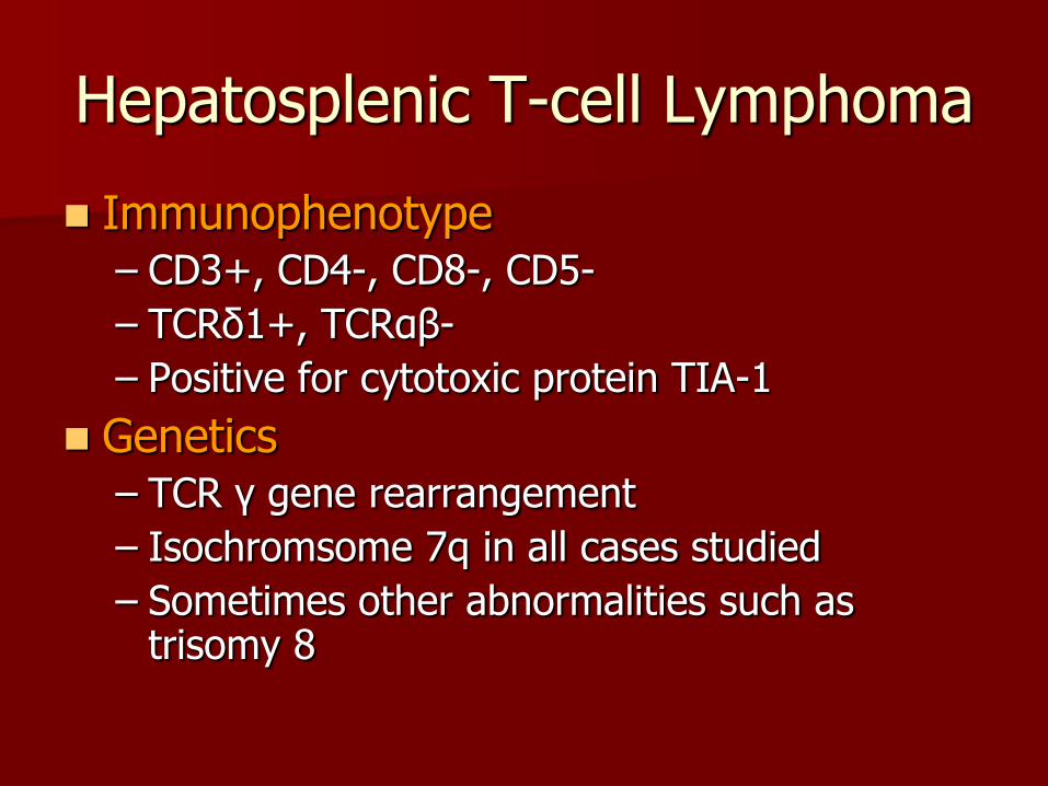
Hepatosplenic T-cell Lymphoma

Hepatosplenic T-cell Lymphoma
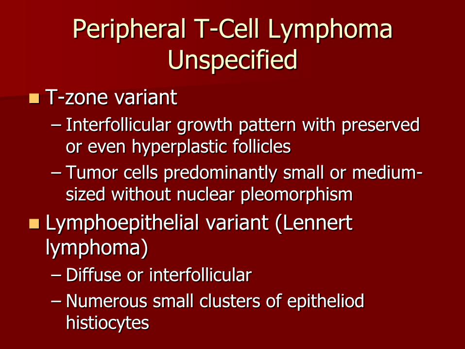
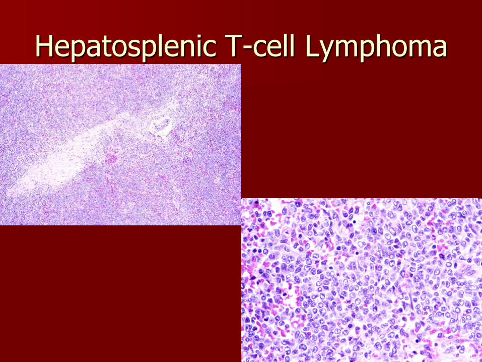
Extranodal and systemic lymphoma usually of cytotoxic T-cells of the γδ type
Marked sinusoidal infiltration seen in the spleen, liver and bone marrow
Patients present with marked hepatosplenomegaly but no lymphadenopathy
Bone marrow almost always involved
More common in immunosuppressed pts

Sinusoidal infiltrate


Hepatosplenic T-cell Lymphoma
59y/o woman with fatigue.
Hepatosplenic T-cell Lymphoma
Hepatosplenic T-cell Lymphoma
Hepatosplenic T-cell Lymphoma
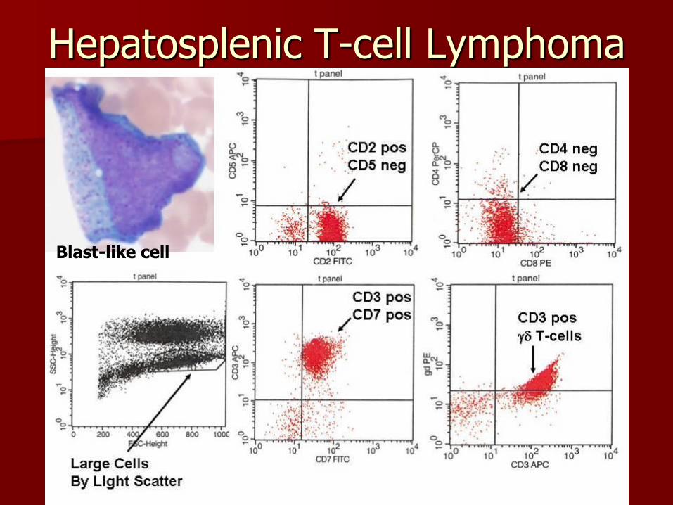
Immunophenotype – CD3+, CD4-, CD8-, CD5-
– TCRδ1+, TCRαβ-
– Positive for cytotoxic protein TIA-1
Genetics – TCR γ gene rearrangement
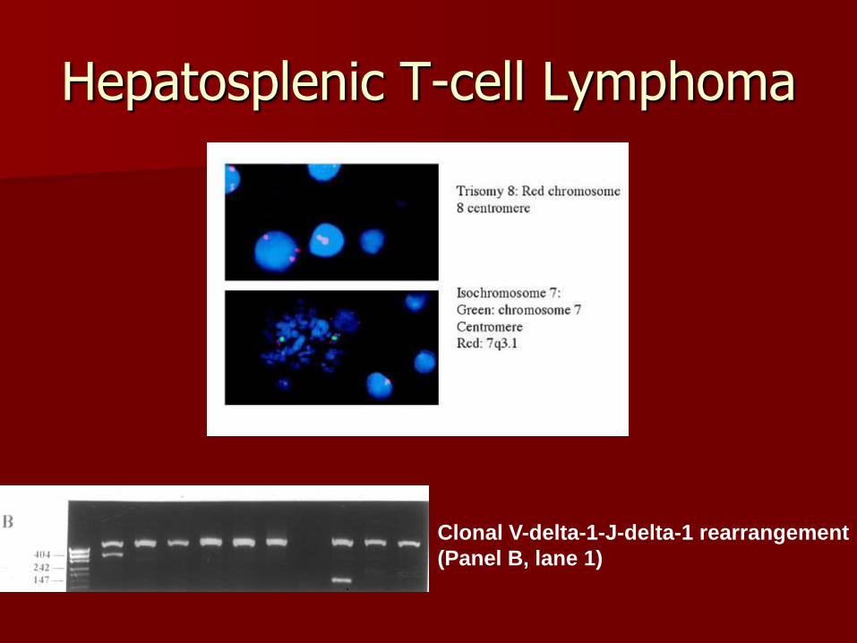
– Isochromsome 7q in all cases studied
– Sometimes other abnormalities such as trisomy 8
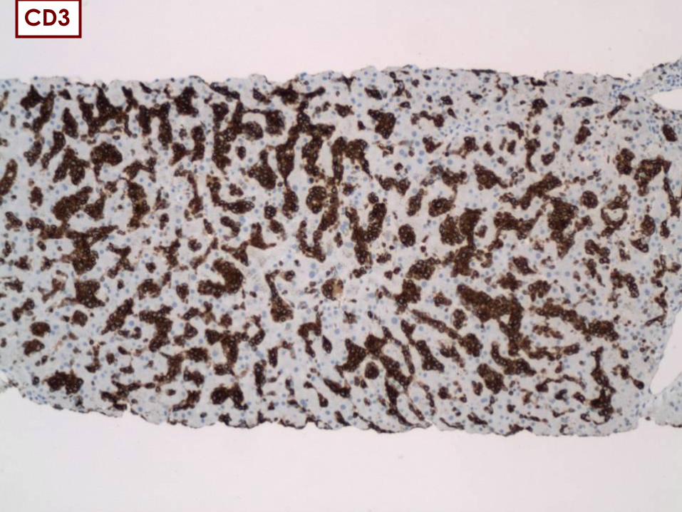
CD3

CD3

Hepatosplenic T-cell Lymphoma
Hepatosplenic T-cell Lymphoma
Blast-like cell
Hepatosplenic T-cell Lymphoma
Liver
Clonal V-delta-1-J-delta-1 rearrangement
(Panel B, lane 1)

Extranodal NK/T-cell Lymphoma, Nasal Type

Extranodal NK/T-cell Lymphoma, Nasal Type
Predominantly extranodal lymphoma with broad morphologic spectrum
Nasal cavity most common site, but may occur anywhere
More prevalent in Asia, Mexico and Central and South America
M>F
May occur in immunosuppressed and post-transplant patients

Extranodal NK/T-cell Lymphoma, Nasal Type
Present with nasal obstruction or epistaxis due to mass lesion or extensive midfacial destructive lesions
Variable presentation outside nasal cavity, e.g., skin ulceration, intestinal perforation
May disseminate rapidly
May have associated hemophagocytic syndrome
May overlap with aggressive NK cell leukemia

Extranodal NK/T-cell Lymphoma, Nasal Type
Mucosal sites show extensive ulceration
Diffuse infiltrate
Angiocentric and angiodestructive pattern common with fibrinoid changes in vessels
Coagulative necrosis and apoptotic bodies
Cytologic spectrum broad from small to large anaplastic cells

Expansion of the nasal bridge
CT: tumor extends into the orbit

Lymphocytic infiltrare with destruction of an artery
The lymphoma in skin with angiocentric angiodestructive property

Extranodal NK/T: A-predominantly small cells B-predominantly medium-sized cells C-predominantly large cells with apoptosis D-pleomorphic large cells with small cells E-touch prep showing azurophilic granules
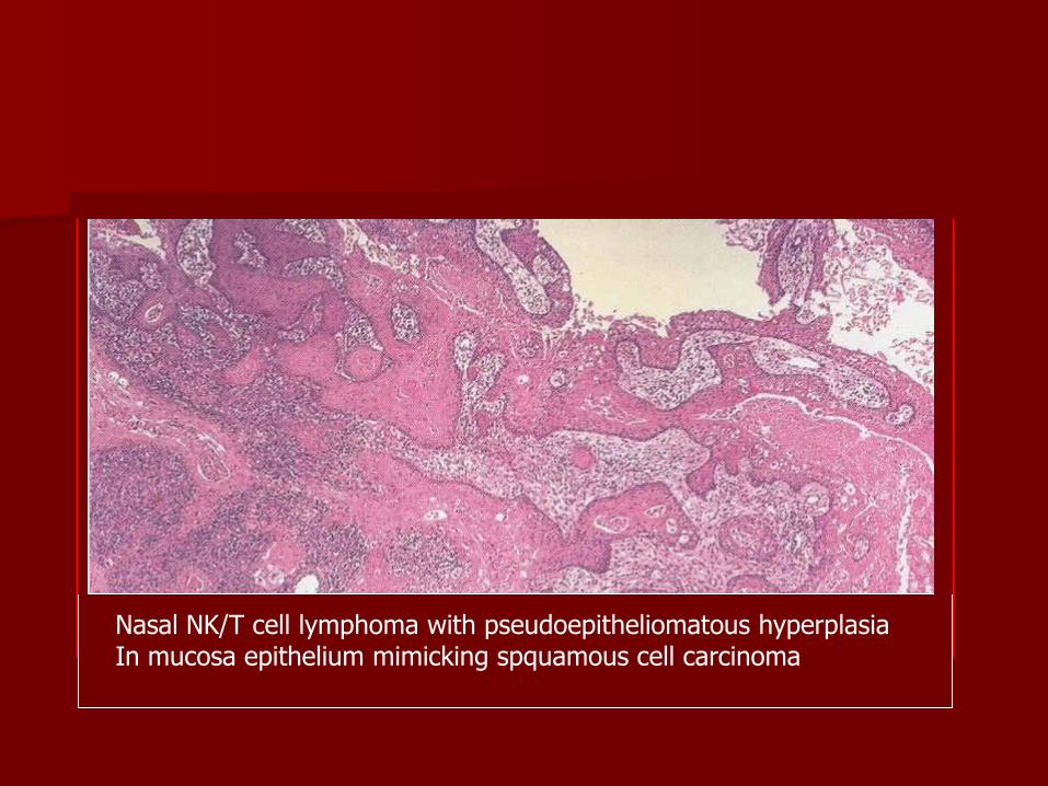
Nasal NK/T cell lymphoma with pseudoepitheliomatous hyperplasia In mucosa epithelium mimicking spquamous cell carcinoma

Extranodal NK/T-cell Lymphoma, Nasal Type
Nasal Cavity

Extranodal NK/T-cell Lymphoma, Nasal Type

Extranodal NK/T-cell Lymphoma, Nasal Type
Bowel
Extranodal NK/T-cell Lymphoma, Nasal Type
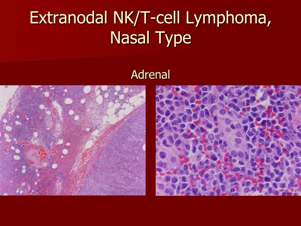
Adrenal

Extranodal NK/T-cell Lymphoma, Nasal Type
Immunophenotype
– CD2+, CD56+, sCD3-, cCD3€+
– Positive for cytotoxic proteins
– Positive for EBV
– Other T-cell and NK-cell antigens negative
Genetics
– Usually TCR and Ig genes are germline
Extranodal NK/T-cell Lymphoma, Nasal Type
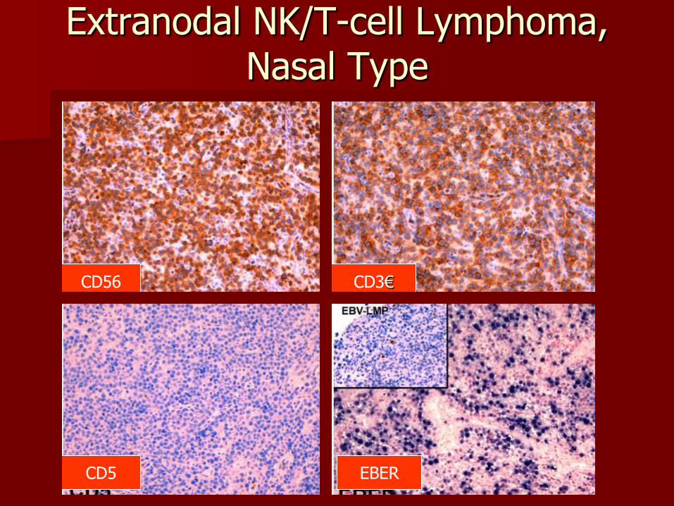
CD56 CD3€
CD5 EBER

Most cases also show cytotoxic granule associated proteins (granzyme B) and EBER
Granzyme B EBER

Cell of Origin
Activated NK cells or (rarely) cytotoxic T cells

Prognosis
Variable
Some pts respond well to therapy and others die of disseminated disease despite aggressive therapy