Rhythmic Blueprints: A tutorial on Design and Evaluation of Rhythmic Interaction

Periodic and Rhythmic Patterns
Suzette M LaRoche, MD
Mission Health Epilepsy Center
Asheville, North Carolina

Continuum of EEG Activity
Triphasics
GPDs LRDA
Burst-SuppressionSIRPIDs
?
NCSE
Clinical Correlate
NCS
Frequency, Duration, Rhythmicity, Location
LPDs +
LPDs
Neuronal Injury
BRDs

American Clinical Neurophysiology Society (ACNS): Critical Care EEG Terminology
Main Term #2
Periodic Discharges
PD s
Rhythmic Delta Activity
RDA
Spike-wave
SW
Main Term #1
Generalized
G
Lateralized
L
Bilateral Independent
BI
Multifocal
Mf

Main Term 1: Location
• Generalized
– Symmetric in both hemispheres
• Lateralized
– Seen in only one hemisphere: unilateral
– Seen in both hemispheres but asymmetric: Bilateral asymmetric
• Bilateral Independent
– Seen in both hemispheres but Asynchronous
• Multifocal

Main term 2: Pattern Type• Periodic Discharge (PD)
– Repetition of a waveform with uniform morphology
– Quantifiable interval between waveforms
• Rhythmic Delta Activity (RDA)
- Repetition of a waveform with uniform morphology
- No interval between consecutive waveforms

Modifiers
• Amplitude
• Frequency
• Prevalence (how much of the recording?)
• Plus (superimposed Fast, Rhythmic, Sharp)
• Stimulus Induced (SIRPIDs)
• Triphasic Morphology

Lateralized Periodic Discharges: LPDs
Etiologies:
Stroke
Encephalitis (HSV)
Tumor
Intracranial hemorrhage
Clinical Correlates:
Acute injury
Encephalopathy
Seizures in 50-90%
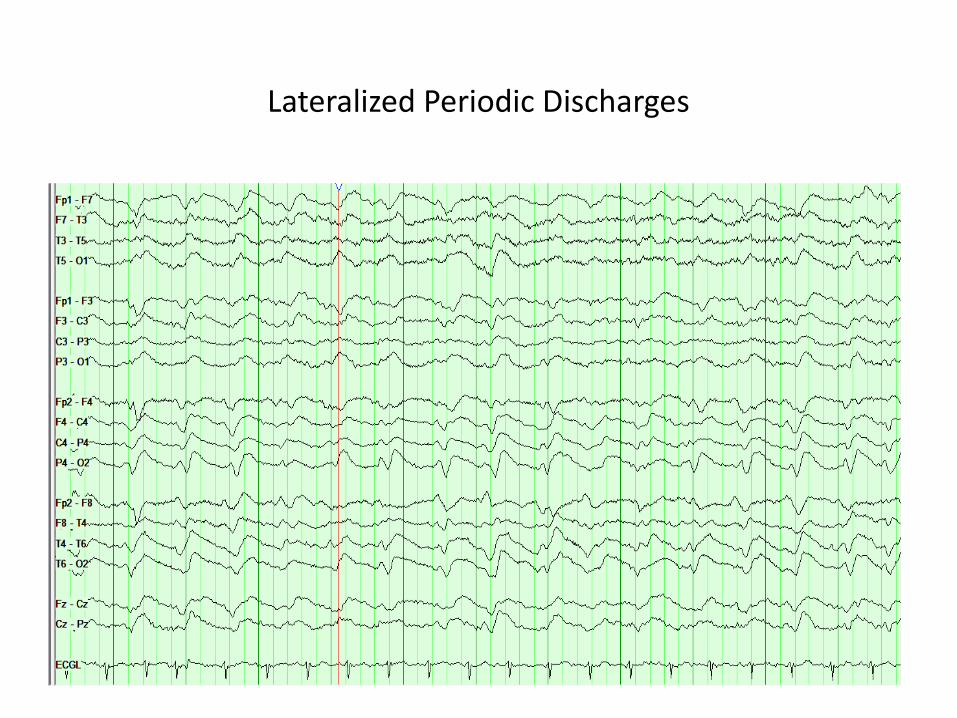
Lateralized Periodic Discharges

Lateralized Periodic Discharges Plus

Bilateral Independent Periodic Discharges (BIPDs)

Left Face Twitch: “ictal” LPDs

Generalized Periodic Discharges: GPDs
Clinical Correlate:
SEIZURES??
Outcome?
Etiologies:
Anoxia
Toxic metabolic
Infections (CJD)
Focal structural lesions

Generalized Periodic Discharges:Relationship to Seizures and Prognosis
• Patients with GPDs were matched by age, etiology, level of consciousness to patients without GPDs (200
each)
Foreman et al, Neurology 2012
GPDs Control p value
Any seizure during hospitalization 46% 34% 0.014
Non-convulsive seizure 27% 8% <0.001
NCSE 22% 7% <0.001
Mortality (univariate)* 36.8% 26.9% 0.049
*Multivariate predictors of worse outcome were cardiac arrest, coma, nonconvulsive status epilepticus, and sepsis, but not generalized periodic
discharges.

GPDs with Triphasic Morphology (old term: Triphasic Waves)
• High amplitude, positive discharge
• Each phase longer than the preceding
• Frontally predominant +/- A-P lag
• Hepatic or renal encephalopathy, anoxia, seizures?

Generalized Periodic Discharges:Clinical Significance of Triphasic Morphology
Foreman et al, Clin Nphys, 2015
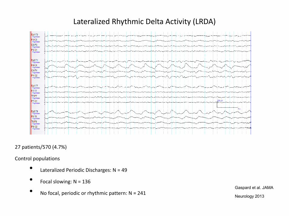
Lateralized Rhythmic Delta Activity (LRDA)
Gaspard et al. JAMA
Neurology 2013
27 patients/570 (4.7%)
Control populations
• Lateralized Periodic Discharges: N = 49
• Focal slowing: N = 136
• No focal, periodic or rhythmic pattern: N = 241

LRDA: Risk of acute seizures
Gaspard et al, JAMA Neurology, 2013

Continuous EEG Monitoring Consortium Training Module
Generalized Rhythmic Delta Activity (GRDA)
• High amplitude, bisynchronous slow waves
• Typical frequency of 2- 2.5 Hz
• Typically seen in toxic-metabolic disturbances
• May see with large midline structural lesions or increased ICP with herniation

Brief (potentially ictal)Rhythmic Discharges
B(i)RDs, BRDs
“Evolving rhythmic patterns…. less than 10 seconds”
Tsuchida et al, Neonatal ICU EEG Terminology, J Clin Neurophys 2013

B(i)RDs and Association with Seizures
Yoo et al, JAMA Neurology, 2014
• 20 adult patients with B(i)RDs and compared to control groups

Continuous EEG Monitoring Consortium Training Module
Hirsch et al, Epilepsia 2004
SIRPIDS= Stimulus induced rhythmic, periodic or ictal discharges
• 33 of 150 pts. undergoing cEEG (22%)
• 50% experienced clinical or subclinical seizures during hospitalization
• Reactivity? Pathophysiology?

Term % Agreement (SD) Kappa (95% CI) Agreement
Main Term 1 Generalized
Lateralized
Bilateral Independent
Multifocal
96% (7%) .87 (.75-.98) Almost Perfect
Main Term 2 Periodic Discharges
Rhythmic Delta Activity
Spike-Wave
98% (3%) .92 (.78-.98) Almost Perfect
Modifiers Amplitude 93% (12%) n/a
Frequency 80% (20%) n/a
+ Fast 83% (18%) .54 (.16-.87) Fair
+ Rhythmic Activity 88% (20%) .62 (.41-.87) Moderate
+ Sharp or Spike 82% (20%) .16 (.10-.28) Poor
Mani et al Journal Clin Neurophysiol 2012

Periodic and Rhythmic Patterns: Association with Seizures
• Retrospective, 3-center review of 4772 critically ill adults undergoing CEEG
• Seizures were documented in 719 (12.5%) of which 530 (74%) also had a periodic or rhythmic pattern
Rodriguez et al, presented at AES, December 2015

Pattern Frequency
Sei
zure
Ris
k
Significant Risk
GRDA - GRDA +
1.5 Hz
LPD +
Periodic and Rhythmic Patterns:
Association with Seizures
Rodriguez et al, presented at AES, December 2015

Case
• 66 yo man with 4 months of cognitive decline and gait instability
• Medications: Methadone, Diazepam
• Neurological Exam: Oriented to person only, bilateral visual field deficits, strength intact, reflexes brisk throughout,
multifocal myoclonus, unable to stand
• Initial Diagnostic Tests:
• MRI partially obscured due to movement artifact but essentially unremarkable
• CSF – Protein 35, Glucose 67, WBC 3/ 4

Lateralized Periodic Discharges (LPDs)- Not Ictal
Load Fosphenytoin 20 mg/kg/PELoad Fosphenytoin 20 mg/kg/PE

2 weeks later….
CSF Analysis: 14, 3, 3 Protein Tau/Theta Positive
Clinical Diagnosis: Creutzfeldt – Jacob Disease
Deceleration of Care
CSF Analysis: 14, 3, 3 Protein Tau/Theta Positive
Clinical Diagnosis: Creutzfeldt – Jacob Disease
Deceleration of Care

Case
• 85 yo admitted for fever, productive cough and confusion
• Medications: Albuterol, Lisinopril
• Neurological Exam: Lethargic, oriented to person only, unable to follow commands, otherwise non focal
neurological exam
• Chest X ray: Bilateral pulmonary infiltrates
• Brain MRI: Mild generalized volume loss

EEG 2 Days later: No improvement in mental status

Generalized Periodic Discharges (GPDs), 1.5-2 Hz, with triphasic morphology

5 min after Lorazepam 2 mg: Awake, Follows Commands

Case
• 44 yo woman with shortness of breath and confusion
• Medications: Oxycodone
• Urine Drug Screen: Positive for opiates, benzodiazepines
• Head CT: Unremarkable
• Intubated and sedated (with propofol) secondary to respiratory distress, possible overdose
• Neurology consult 2 days later for persistent confusion
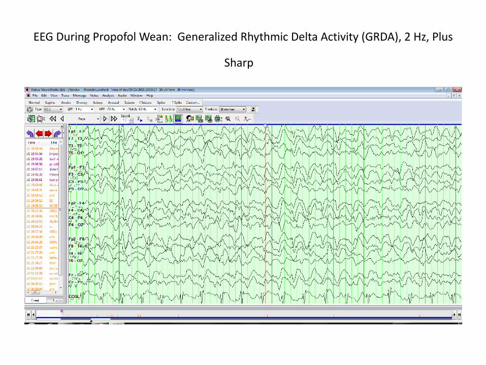
EEG During Propofol Wean: Generalized Rhythmic Delta Activity (GRDA), 2 Hz, Plus
Sharp

Also…. Fluctuating
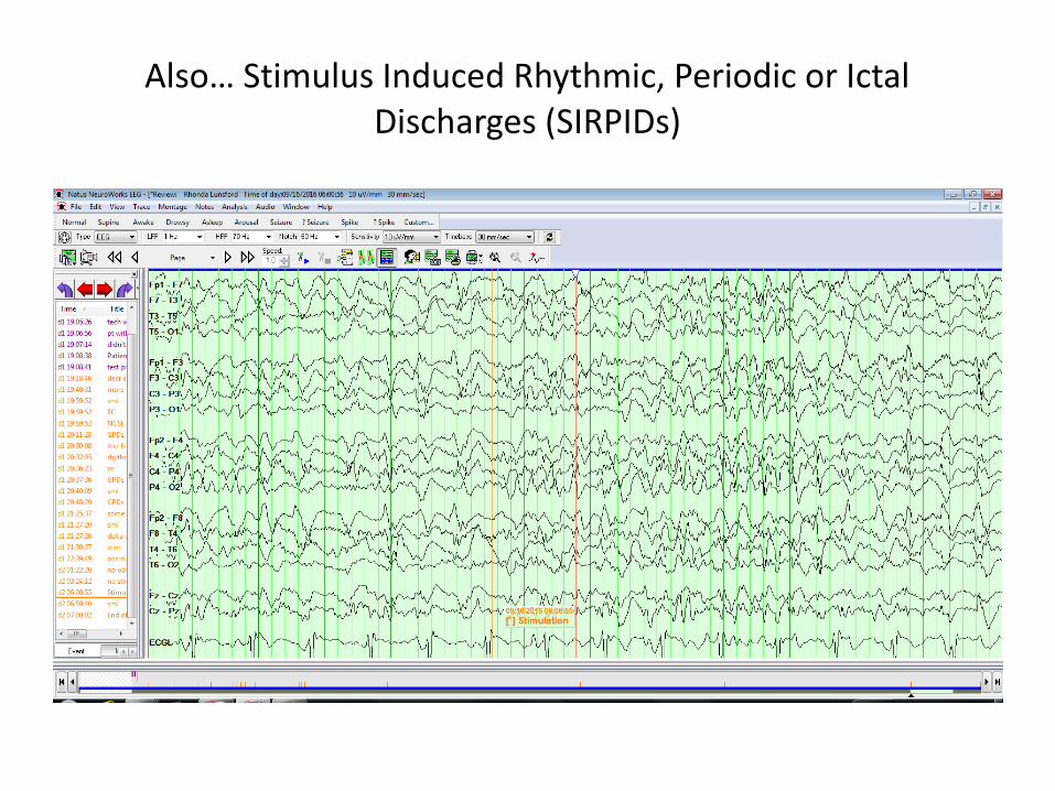
Also… Stimulus Induced Rhythmic, Periodic or Ictal Discharges (SIRPIDs)

Next day, propofol off…..transferred out of ICU

Summary
• Periodic and rhythmic patterns are common in the critically ill, many of which have increased association with seizures
• Standardized terminology is critical for consistency in clinical reporting and research
• Medical decisions need to take into account the EEG pattern AND clinical history