Perinatal and Delivery Management of Infants with ... 17-Dra Sandra Perinatal... · Perinatal and...
17
Perinatal and Delivery Management of Infants with Congenital Heart Disease Laura Sanapo, MD a , Anita J. Moon-Grady, MD b , Mary T. Donofrio, MD a,c, * Video content accompanies this article at http://www.perinatology.theclinics. com/ INTRODUCTION Advances in prenatal imaging and increasing experience in fetal cardiology have improved the examination of the fetal cardiovascular system. 1 Fetal echocardiography Disclosure: The authors have nothing to disclose. a Division of Fetal and Transitional Medicine, Children’s National Health System, 111 Michigan Avenue, Northwest, Suite M3-118, Washington, DC 20010, USA; b Fetal Cardiovascular Pro- gram, UCSF Benioff Children’s Hospitals, University of California San Francisco, 550 16th Street, 5th Floor, Box 0544, San Francisco, CA 94158, USA; c Fetal Heart Program, Division of Cardiol- ogy, Children’s National Health System, 111 Michigan Avenue, Northwest, Washington, DC 20010, USA * Corresponding author. Fetal Heart Program, Division of Cardiology, Children’s National Health System, 111 Michigan Avenue, Northwest, Washington, DC 20010. E-mail address: [email protected] KEYWORDS Fetal echocardiography Fetal cardiology Congenital heart disease Neurodevelopment Biophysical profile Obstetric management Antenatal surveillance KEY POINTS Prenatal diagnosis has improved neonatal outcomes of congenital heart disease (CHD) but perinatal morbidity and mortality are still significant in some cases. Fetal echocardiography can facilitate delivery and perinatal planning for infants with CHD. Antenatal surveillance of fetuses with CHD can identify prenatal progression of the lesion and decompensation, and may improve perinatal and postnatal outcomes. Successful perinatal management of neonates with a prenatal diagnosis of CHD requires close collaboration between obstetric, neonatal, and cardiology services. Delivery of infants prenatally diagnosed with CHD in most cases should not be sched- uled before 39 weeks unless there is an obstetric indication or concern regarding fetal well-being. Clin Perinatol 43 (2016) 55–71 http://dx.doi.org/10.1016/j.clp.2015.11.004 perinatology.theclinics.com 0095-5108/16/$ – see front matter Ó 2016 Elsevier Inc. All rights reserved.
Transcript of Perinatal and Delivery Management of Infants with ... 17-Dra Sandra Perinatal... · Perinatal and...
Perinatal and DeliveryManagement of Infants
with Congenital Heart DiseaseLaura Sanapo, MDa, Anita J. Moon-Grady, MDb,Mary T. Donofrio, MDa,c,*
KEYWORDS
� Fetal echocardiography � Fetal cardiology � Congenital heart disease� Neurodevelopment � Biophysical profile � Obstetric management� Antenatal surveillance
KEY POINTS
� Prenatal diagnosis has improved neonatal outcomes of congenital heart disease (CHD)but perinatal morbidity and mortality are still significant in some cases.
� Fetal echocardiography can facilitate delivery and perinatal planning for infants with CHD.
� Antenatal surveillance of fetuses with CHD can identify prenatal progression of the lesionand decompensation, and may improve perinatal and postnatal outcomes.
� Successful perinatal management of neonates with a prenatal diagnosis of CHD requiresclose collaboration between obstetric, neonatal, and cardiology services.
� Delivery of infants prenatally diagnosed with CHD in most cases should not be sched-uled before 39 weeks unless there is an obstetric indication or concern regarding fetalwell-being.
Video content accompanies this article at http://www.perinatology.theclinics.com/
Discla DivAvengram5th Fogy,2001* CoHealE-ma
Clinhttp0095
INTRODUCTION
Advances in prenatal imaging and increasing experience in fetal cardiology haveimproved the examination of the fetal cardiovascular system.1 Fetal echocardiography
osure: The authors have nothing to disclose.ision of Fetal and Transitional Medicine, Children’s National Health System, 111 Michiganue, Northwest, Suite M3-118, Washington, DC 20010, USA; b Fetal Cardiovascular Pro-, UCSF Benioff Children’s Hospitals, University of California San Francisco, 550 16th Street,loor, Box 0544, San Francisco, CA 94158, USA; c Fetal Heart Program, Division of Cardiol-Children’s National Health System, 111 Michigan Avenue, Northwest, Washington, DC0, USArresponding author. Fetal Heart Program, Division of Cardiology, Children’s Nationalth System, 111 Michigan Avenue, Northwest, Washington, DC 20010.il address: [email protected]
Perinatol 43 (2016) 55–71://dx.doi.org/10.1016/j.clp.2015.11.004 perinatology.theclinics.com-5108/16/$ – see front matter � 2016 Elsevier Inc. All rights reserved.
Sanapo et al56
is able to obtain precise details of cardiac structural and hemodynamic alterations infetuses with congenital heart disease (CHD). Sequential examinations through gesta-tion can predict the evolution of disease in utero and during the transition to postnatalcirculation at delivery. This approach allows detailed prenatal counseling and enablesplanning to define perinatal management, selecting the fetuses at risk of postnatal he-modynamic instability who might require a specialized delivery plan.2,3 The prenataldiagnosis andmanagement of critical neonatal CHD has been shown to play an impor-tant role in improving the outcome of newborns with these conditions, allowing timelystabilization of the disease before cardiac surgery4 and reducing the risk of perioper-ative morbidity,4–9 including the risk of perioperative neurologic insults.10 Despite ev-idence that fetal diagnosis has improved the outcome of some CHDs, critical formsmay still be associated with significant morbidity and mortality caused by hemody-namic instability that occurs after birth, often shortly after separation from theplacental circulation.11 Therefore, prenatal assessment of the severity of the lesionand disease-specific delivery recommendations has been suggested to ensure thebest care and avoid delays in treatment.11–14 Perinatal management of neonateswith a prenatal diagnosis of CHD requires a close collaboration between obstetric,neonatal, and cardiology services, and a well-delineated network with communicationbetween the adult hospital and pediatric tertiary care center.3
This article reviews the most recent recommendations for the perinatal and deliverymanagement of infants with a prenatal diagnosis of CHD.
FETAL ECHOCARDIOGRAPHY AND RISK OF HEMODYNAMIC INSTABILITY AT BIRTH
Most CHD is well tolerated in utero, does not present a risk of hemodynamic instabilityat birth or in the first days of life, and does not require specialized delivery care. How-ever, some critical CHDs have an increased risk of hemodynamic instability after de-livery and may require maintenance of patency of the fetal shunts and/or immediateintervention.2 In order to identify the fetuses with CHD at risk of hemodynamic insta-bility at birth, it is important to understand the physiology of the fetal circulation andthe transition to the extrauterine circulation and how this process is compromised innewborns with a cardiac defect.
Fetal and Transitional Circulation
The fetal circulation is a highly efficient system that provides blood to the fetal body andthe placenta. Fetal shunts allow the more highly oxygenated and nutrient-rich bloodfrom the umbilical vein to be preferentially delivered to the left ventricle, thereforeentering the systemic circulation. The remainder of the umbilical vein blood mixeswith less oxygenated blood from the fetal body and passes to the right heart, and viathe ductus arteriosus is directed into the descending aorta supplying the lower bodyand the placenta.15 The fetoplacental circulation is characterized by low resistance,whereas the circulation to the fetal lungs is limited by high resistance. At delivery, mul-tiple important changes occur. Cord clamping interrupts the low-resistance placentalcirculation, whereas initiation of respiration decreases the pulmonary vascular resis-tance and increases pulmonary blood flow and ultimately the blood volume returningto the left atrium through the pulmonary veins. Consequently, the left atrial pressure in-creases and functional closure of the foramenovale occurs.With the closure of theduc-tus venosus (within the first week of life) and the ductus arteriosus (usually within 12–72 hours),16 the fetal circulation transitions to the postnatal circulation in series.17
In specific cases of CHD, the presence of in utero cardiac shunting permits redis-tribution of blood flow to maintain cardiac output and adequate oxygen delivery to
Perinatal Management of Fetal Congenital Heart Disease 57
the fetal body to maintain a normal fetoplacental circulation. For this reason, mostCHDs are well tolerated in utero,17 although the altered circulatory patternmay impair systemic oxygenation and affect fetal growth and brain develop-ment.18–21 The risk of hemodynamic instability after birth depends on 3 factors:(1) the type of CHD, including whether the defect is shunt dependent; (2) whetherthere is cardiac dysfunction either from a primary cardiomyopathy, structuralanomaly leading to an excessive pressure or volume load to the heart, or a cardiacrhythm abnormality; and (3) whether there is an associated abnormality of the res-piratory system.
Models of Risk Assessment of Hemodynamic Instability at Birth
Several postnatal risk stratification protocols for babies diagnosed in utero with CHDhave been proposed11,12,14 and more recently summarized as part of the AmericanHeart Association Statement on Fetal Cardiology2 (Table 1). The risk of potentialcompromise at birth is most often determined prenatally by the fetal cardiologist, tak-ing into account the specific CHD as well as patient-specific findings noted on fetalechocardiogram (Table 2). A recent evaluation of a risk assessment protocol appliedprospectively to a patient population at the Children’s National Health System showedhigh accuracy with a sensitivity ranging from 0.83 to 0.99 for prediction of postnatalcare and need for specialized intervention at birth.12
CHDs can be divided into 3 main categories, according to the predicted risk of he-modynamic instability at birth: (1) CHD without risk of hemodynamic instability duringor immediately after delivery, or in the neonatal period; (2) CHD with minimal risk ofhemodynamic instability; and (3) CHD with high risk of instability.
Congenital heart disease without predicted risk of hemodynamic instability at birthThis group includes left-to-right shunt lesions such as ventricular septal defects, atrialseptal defects, atrioventricular septal defects, and mild valve abnormalities. Left-to-right shunt lesions most often become hemodynamically unstable weeks after birthwhen the decrease in the pulmonary vascular resistance causes significant left-to-right shunting and associated pulmonary overcirculation.15 Similarly, infants prenatallydiagnosed with a mild isolated valve abnormality and normal cardiac function are usu-ally stable after birth. These conditions do not require specialized care in the deliveryroom, and babies often can be delivered at local hospitals and be evaluated either inthe nursery or as an outpatient.12,22
Congenital heart disease with minimal risk of hemodynamic instability at birthThis group mainly includes CHDs that depend on the patency of the ductusarteriosus for maintenance of the systemic or pulmonary circulation after birth.The ductus arteriosus does not generally close immediately at birth, but after 12to 72 hours,16 and therefore these babies are not expected to be compromisedin the delivery room or immediate perinatal period.12,13 In these cases, babiesmay be delivered at a location that can institute therapy with prostaglandin E1for maintenance of ductal patency. After initial stabilization, transport of thenewborn to the tertiary care cardiac center for anticipated intervention and/or sur-gery should be arranged.The prediction of the need for patency of ductus arteriosus after birth may be diffi-
cult but there are several criteria available (see Table 2). In case of pulmonary outflowtract obstruction, such as critical pulmonary stenosis or atresia, severe tricuspid valvestenosis or atresia without or with a small ventricular septal defect, or severe tetralogyof Fallot (TOF), the following findings have been shown to be fairly reliable predictors of
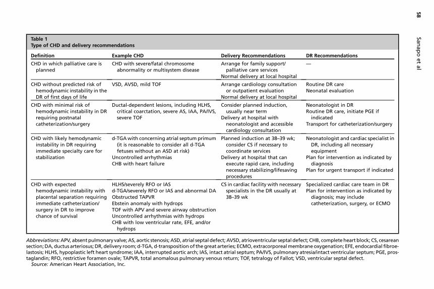
Table 1Type of CHD and delivery recommendations
Definition Example CHD Delivery Recommendations DR Recommendations
CHD in which palliative care isplanned
CHD with severe/fatal chromosomeabnormality or multisystem disease
Arrange for family support/palliative care services
Normal delivery at local hospital
—
CHD without predicted risk ofhemodynamic instability in theDR of first days of life
VSD, AVSD, mild TOF Arrange cardiology consultationor outpatient evaluation
Normal delivery at local hospital
Routine DR careNeonatal evaluation
CHD with minimal risk ofhemodynamic instability in DRrequiring postnatalcatheterization/surgery
Ductal-dependent lesions, including HLHS,critical coarctation, severe AS, IAA, PA/IVS,severe TOF
Consider planned induction,usually near term
Delivery at hospital withneonatologist and accessiblecardiology consultation
Neonatologist in DRRoutine DR care, initiate PGE if
indicatedTransport for catheterization/surgery
CHD with likely hemodynamicinstability in DR requiringimmediate specialty care forstabilization
d-TGAwith concerning atrial septum primum(it is reasonable to consider all d-TGAfetuses without an ASD at risk)
Uncontrolled arrhythmiasCHB with heart failure
Planned induction at 38–39 wk;consider CS if necessary tocoordinate services
Delivery at hospital that canexecute rapid care, includingnecessary stabilizing/lifesavingprocedures
Neonatologist and cardiac specialist inDR, including all necessaryequipment
Plan for intervention as indicated bydiagnosis
Plan for urgent transport if indicated
CHD with expectedhemodynamic instability withplacental separation requiringimmediate catheterization/surgery in DR to improvechance of survival
HLHS/severely RFO or IASd-TGA/severely RFO or IAS and abnormal DAObstructed TAPVREbstein anomaly with hydropsTOF with APV and severe airway obstructionUncontrolled arrhythmias with hydropsCHB with low ventricular rate, EFE, and/orhydrops
CS in cardiac facility with necessaryspecialists in the DR usually at38–39 wk
Specialized cardiac care team in DRPlan for intervention as indicated by
diagnosis; may includecatheterization, surgery, or ECMO
Abbreviations:APV, absentpulmonary valve; AS, aortic stenosis; ASD, atrial septal defect; AVSD, atrioventricular septal defect; CHB, completeheart block; CS, cesareansection;DA, ductus arteriosus;DR, delivery room;d-TGA,d-transpositionof thegreat arteries; ECMO, extracorporealmembraneoxygenation; EFE, endocardial fibroe-lastosis; HLHS, hypoplastic left heart syndrome; IAA, interrupted aortic arch; IAS, intact atrial septum; PA/IVS, pulmonary atresia/intact ventricular septum; PGE, pros-taglandin; RFO, restrictive foramen ovale; TAPVR, total anomalous pulmonary venous return; TOF, tetralogy of Fallot; VSD, ventricular septal defect.
Source: American Heart Association, Inc.
Sanapoetal
58
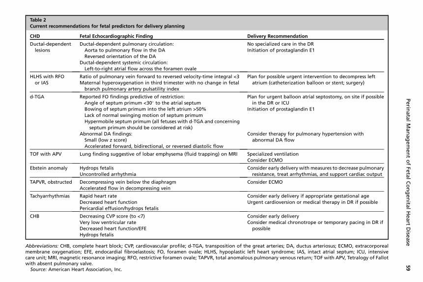
Table 2Current recommendations for fetal predictors for delivery planning
CHD Fetal Echocardiographic Finding Delivery Recommendation
Ductal-dependentlesions
Ductal-dependent pulmonary circulation:Aorta to pulmonary flow in the DAReversed orientation of the DA
Ductal-dependent systemic circulation:Left-to-right atrial flow across the foramen ovale
No specialized care in the DRInitiation of prostaglandin E1
HLHS with RFOor IAS
Ratio of pulmonary vein forward to reversed velocity-time integral <3Maternal hyperoxygenation in third trimester with no change in fetalbranch pulmonary artery pulsatility index
Plan for possible urgent intervention to decompress leftatrium (catheterization balloon or stent; surgery)
d-TGA Reported FO findings predictive of restriction:Angle of septum primum <30� to the atrial septumBowing of septum primum into the left atrium >50%Lack of normal swinging motion of septum primumHypermobile septum primum (all fetuses with d-TGA and concerning
septum primum should be considered at risk)
Plan for urgent balloon atrial septostomy, on site if possiblein the DR or ICU
Initiation of prostaglandin E1
Abnormal DA findings:Small (low z score)Accelerated forward, bidirectional, or reversed diastolic flow
Consider therapy for pulmonary hypertension withabnormal DA flow
TOF with APV Lung finding suggestive of lobar emphysema (fluid trapping) on MRI Specialized ventilationConsider ECMO
Ebstein anomaly Hydrops fetalisUncontrolled arrhythmia
Consider early delivery withmeasures to decrease pulmonaryresistance, treat arrhythmias, and support cardiac output
TAPVR, obstructed Decompressing vein below the diaphragmAccelerated flow in decompressing vein
Consider ECMO
Tachyarrhythmias Rapid heart rateDecreased heart functionPericardial effusion/hydrops fetalis
Consider early delivery if appropriate gestational ageUrgent cardioversion or medical therapy in DR if possible
CHB Decreasing CVP score (to <7)Very low ventricular rateDecreased heart function/EFEHydrops fetalis
Consider early deliveryConsider medical chronotrope or temporary pacing in DR if
possible
Abbreviations: CHB, complete heart block; CVP, cardiovascular profile; d-TGA, transposition of the great arteries; DA, ductus arteriosus; ECMO, extracorporealmembrane oxygenation; EFE, endocardial fibroelastosis; FO, foramen ovale; HLHS, hypoplastic left heart syndrome; IAS, intact atrial septum; ICU, intensivecare unit; MRI, magnetic resonance imaging; RFO, restrictive foramen ovale; TAPVR, total anomalous pulmonary venous return; TOF with APV, Tetralogy of Fallotwith absent pulmonary valve.
Source: American Heart Association, Inc.
Perin
atalManagementofFe
talCongenita
lHeart
Dise
ase
59
Sanapo et al60
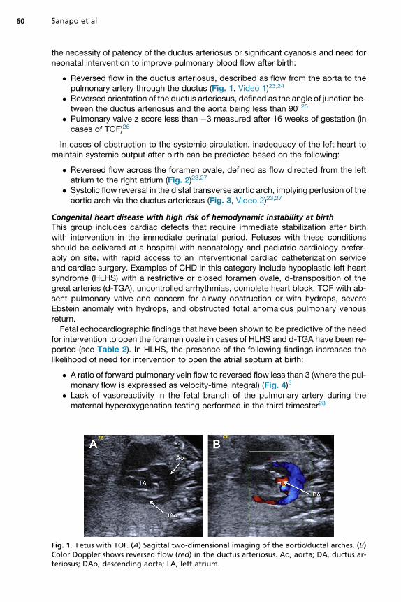
the necessity of patency of the ductus arteriosus or significant cyanosis and need forneonatal intervention to improve pulmonary blood flow after birth:
� Reversed flow in the ductus arteriosus, described as flow from the aorta to thepulmonary artery through the ductus (Fig. 1, Video 1)23,24
� Reversed orientation of the ductus arteriosus, defined as the angle of junction be-tween the ductus arteriosus and the aorta being less than 90�25
� Pulmonary valve z score less than �3 measured after 16 weeks of gestation (incases of TOF)26
In cases of obstruction to the systemic circulation, inadequacy of the left heart tomaintain systemic output after birth can be predicted based on the following:
� Reversed flow across the foramen ovale, defined as flow directed from the leftatrium to the right atrium (Fig. 2)23,27
� Systolic flow reversal in the distal transverse aortic arch, implying perfusion of theaortic arch via the ductus arteriosus (Fig. 3, Video 2)23,27
Congenital heart disease with high risk of hemodynamic instability at birthThis group includes cardiac defects that require immediate stabilization after birthwith intervention in the immediate perinatal period. Fetuses with these conditionsshould be delivered at a hospital with neonatology and pediatric cardiology prefer-ably on site, with rapid access to an interventional cardiac catheterization serviceand cardiac surgery. Examples of CHD in this category include hypoplastic left heartsyndrome (HLHS) with a restrictive or closed foramen ovale, d-transposition of thegreat arteries (d-TGA), uncontrolled arrhythmias, complete heart block, TOF with ab-sent pulmonary valve and concern for airway obstruction or with hydrops, severeEbstein anomaly with hydrops, and obstructed total anomalous pulmonary venousreturn.Fetal echocardiographic findings that have been shown to be predictive of the need
for intervention to open the foramen ovale in cases of HLHS and d-TGA have been re-ported (see Table 2). In HLHS, the presence of the following findings increases thelikelihood of need for intervention to open the atrial septum at birth:
� A ratio of forward pulmonary vein flow to reversed flow less than 3 (where the pul-monary flow is expressed as velocity-time integral) (Fig. 4)5
� Lack of vasoreactivity in the fetal branch of the pulmonary artery during thematernal hyperoxygenation testing performed in the third trimester28
Fig. 1. Fetus with TOF. (A) Sagittal two-dimensional imaging of the aortic/ductal arches. (B)Color Doppler shows reversed flow (red) in the ductus arteriosus. Ao, aorta; DA, ductus ar-teriosus; DAo, descending aorta; LA, left atrium.
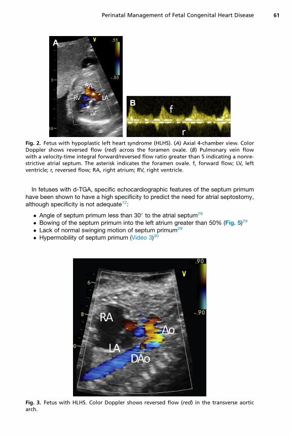
Fig. 2. Fetus with hypoplastic left heart syndrome (HLHS). (A) Axial 4-chamber view. ColorDoppler shows reversed flow (red) across the foramen ovale. (B) Pulmonary vein flowwith a velocity-time integral forward/reversed flow ratio greater than 5 indicating a nonre-strictive atrial septum. The asterisk indicates the foramen ovale. f, forward flow; LV, leftventricle; r, reversed flow; RA, right atrium; RV, right ventricle.
Perinatal Management of Fetal Congenital Heart Disease 61
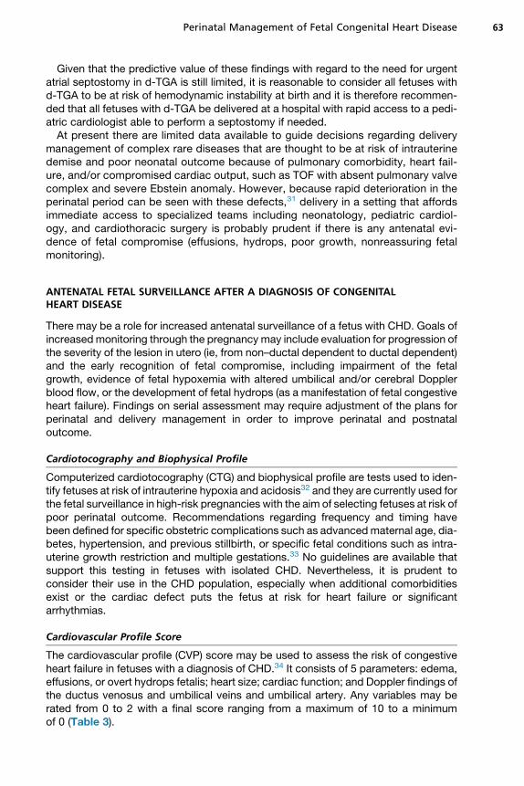
In fetuses with d-TGA, specific echocardiographic features of the septum primumhave been shown to have a high specificity to predict the need for atrial septostomy,although specificity is not adequate12:
� Angle of septum primum less than 30� to the atrial septum29
� Bowing of the septum primum into the left atrium greater than 50% (Fig. 5)29
� Lack of normal swinging motion of septum primum29
� Hypermobility of septum primum (Video 3)30
Fig. 3. Fetus with HLHS. Color Doppler shows reversed flow (red) in the transverse aorticarch.
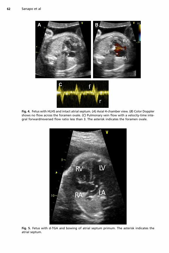
Fig. 4. Fetus with HLHS and intact atrial septum. (A) Axial 4-chamber view. (B) Color Dopplershows no flow across the foramen ovale. (C) Pulmonary vein flow with a velocity-time inte-gral forward/reversed flow ratio less than 3. The asterisk indicates the foramen ovale.
Fig. 5. Fetus with d-TGA and bowing of atrial septum primum. The asterisk indicates theatrial septum.
Sanapo et al62
Perinatal Management of Fetal Congenital Heart Disease 63
Given that the predictive value of these findings with regard to the need for urgentatrial septostomy in d-TGA is still limited, it is reasonable to consider all fetuses withd-TGA to be at risk of hemodynamic instability at birth and it is therefore recommen-ded that all fetuses with d-TGA be delivered at a hospital with rapid access to a pedi-atric cardiologist able to perform a septostomy if needed.At present there are limited data available to guide decisions regarding delivery
management of complex rare diseases that are thought to be at risk of intrauterinedemise and poor neonatal outcome because of pulmonary comorbidity, heart fail-ure, and/or compromised cardiac output, such as TOF with absent pulmonary valvecomplex and severe Ebstein anomaly. However, because rapid deterioration in theperinatal period can be seen with these defects,31 delivery in a setting that affordsimmediate access to specialized teams including neonatology, pediatric cardiol-ogy, and cardiothoracic surgery is probably prudent if there is any antenatal evi-dence of fetal compromise (effusions, hydrops, poor growth, nonreassuring fetalmonitoring).
ANTENATAL FETAL SURVEILLANCE AFTER A DIAGNOSIS OF CONGENITALHEART DISEASE
There may be a role for increased antenatal surveillance of a fetus with CHD. Goals ofincreasedmonitoring through the pregnancy may include evaluation for progression ofthe severity of the lesion in utero (ie, from non–ductal dependent to ductal dependent)and the early recognition of fetal compromise, including impairment of the fetalgrowth, evidence of fetal hypoxemia with altered umbilical and/or cerebral Dopplerblood flow, or the development of fetal hydrops (as a manifestation of fetal congestiveheart failure). Findings on serial assessment may require adjustment of the plans forperinatal and delivery management in order to improve perinatal and postnataloutcome.
Cardiotocography and Biophysical Profile
Computerized cardiotocography (CTG) and biophysical profile are tests used to iden-tify fetuses at risk of intrauterine hypoxia and acidosis32 and they are currently used forthe fetal surveillance in high-risk pregnancies with the aim of selecting fetuses at risk ofpoor perinatal outcome. Recommendations regarding frequency and timing havebeen defined for specific obstetric complications such as advancedmaternal age, dia-betes, hypertension, and previous stillbirth, or specific fetal conditions such as intra-uterine growth restriction and multiple gestations.33 No guidelines are available thatsupport this testing in fetuses with isolated CHD. Nevertheless, it is prudent toconsider their use in the CHD population, especially when additional comorbiditiesexist or the cardiac defect puts the fetus at risk for heart failure or significantarrhythmias.
Cardiovascular Profile Score
The cardiovascular profile (CVP) score may be used to assess the risk of congestiveheart failure in fetuses with a diagnosis of CHD.34 It consists of 5 parameters: edema,effusions, or overt hydrops fetalis; heart size; cardiac function; and Doppler findings ofthe ductus venosus and umbilical veins and umbilical artery. Any variables may berated from 0 to 2 with a final score ranging from a maximum of 10 to a minimumof 0 (Table 3).
Table 3The cardiovascular profile score
Category 2 Points 1 Point 0 Point
Hydrops None Ascites or pericardialor pleural effusion
Skin edema
Heart size(HA/CA)
>0.2 and �0.35 0.35–0.5 <0.2 or >0.5
Cardiacfunction
Normal MV andTV, biphasic diastolicfilling, LV or RV SFs >0.28
Holosystolic TR or LVor RV SFs <0.28
Holosystolic MR orTR dP/dt <400, monophasicdiastolic filling
ArterialumbilicalDoppler
Normal AEDV REDV
VenousDoppler
Normal DV atrial reversal UV pulsations
Abbreviations: AEDV, absent end-diastolic velocity; DV, ductus venosus; HA/CA, heart to chest arearatio; LV, left ventricle; MR, mitral valve regurgitation; MV, mitral valve; REDV, reversed end-diastolic velocity; RV, right ventricle; SF, ventricular shortening fraction; TR dP/dt, change in pres-sure over time of TR jet; TR, tricuspid valve regurgitation; TV, tricuspid valve; UV, umbilical vein.
FromWieczorek A, Hernandez-Robles J, Ewing L, et al. Prediction of outcome of fetal congenitalheart disease using a cardiovascular profile score. Ultrasound Obstet Gynecol 2008;31:285; withpermission.
Sanapo et al64
The use of the CVP score has been evaluated in CHD and fetal rhythm abnormal-ities,35–37 with a score less than or equal to 7 correlating with a higher risk of perinatalcompromise or death. Among the 5 parameters, the presence of hydrops fetalis andsevere cardiomegaly, defined as heart/chest area ratio greater than 0.5, have showedthe most statistically significant association with mortality. The presence of hydrops orCVP less than 7 may represent an indication for urgent delivery and planning for po-tential immediate postnatal intervention.
DELIVERY PLANNING FOR NEONATES PRENATALLY DIAGNOSED WITH CONGENITALHEART DISEASE
Delivery planning should take into account 3 main factors: (1) the risk of hemodynamicinstability at birth, (2) the resources of the region, (3) the presence of obstetriccomplications.
Location of Delivery and Transportation of the Newborn
Most newborns with CHD do not need any specialized care in the perinatal period andrecommendations are made to deliver at the local hospital and be followed as outpa-tients.2 However, if the presence of a specialized cardiac team after delivery is antic-ipated (see Table 1), the location of delivery should take into account these specialneeds. Determination of the site of delivery may depend on the availability of a pedi-atric cardiac unit in close proximity. In some regions where the number of specializedcardiac centers is limited, strategies have been developed to safely plan the deliveryand perinatal management of infants prenatally diagnosed with CHD.14,22 These plansinclude either creation of highly specialized neonatal units locally or maternal-fetaltransport with delivery nearer to a cardiac center in patients determined to be athigh risk of hemodynamic instability. In order to improve the neonatal outcome of
Perinatal Management of Fetal Congenital Heart Disease 65
the infants with CHD associated with hemodynamic instability with placental separa-tion and avoid transportation of critically ill newborns, some freestanding children’shospitals have planned deliveries in their facilities to accommodate these particularlyhigh-risk patients, thus minimizing time to lifesaving intervention.11,38
Timing of Delivery
Recent studies have shown that infants prenatally diagnosed with critical forms ofCHD tend to be delivered earlier than neonates in whom the diagnosis of CHD ismade after birth.10,39,40 This finding is particularly worrisome given that otherwisehealthy neonates born at 37 to 38 weeks have increased risk of worse outcomescompared with those born at later term (39–40 weeks).41 This finding has also beenobserved in babies with CHD. Studies have shown that babies with CHD have longerpostoperative lengths of stay and higher mortality if delivered before 39 weeks.40,42,43
Therefore, in the absence of fetal or maternal indications for earlier delivery, the poten-tial advantages of an elective delivery of fetuses with CHD early should be carefullyconsidered.In addition to an increased mortality, there is growing evidence that the decision
about timing of delivery of infants with CHD should also consider the potential effectof the gestational age at birth on the neurologic outcome. It has been shown that fe-tuses with critical CHDs, such as d-TGA or single-ventricle physiology, have delays inbrain maturation that manifest as alteration of brain growth, brain metabolism, and themicrostructure of the white matter of the pyramidal tract44–47 and that suggest thatbrain development may lag by as much as 4 weeks in term neonates with CHD beforeheart surgery. Given these findings, it has been suggested that planning for the deliv-ery of babies with CHD near or at full term may improve brain development anddecrease susceptibility to injury postnatally46; however, further studies are neededto establish whether this will translate to improvement in long-term neurologicoutcome.Planning of the delivery of babies with CHD is also affected by the overall risk of pre-
term birth (occurring in 12.8% of all US births).48 To date, although there are effectivepredictors of preterm birth before 34 weeks, such as cervicovaginal fetal fibronectinand cervical length based on ultrasonography evaluations, there is no way to identifythe exact timing of the delivery among women with risk factors or symptoms of pre-term labor.49,50 For these reasons, close communication between the obstetricianand the pediatric cardiologist is necessary if spontaneous preterm birth seems likely.
Mode of Delivery
Data from retrospective studies show that prenatal diagnosis of a major CHD, such asHLHS, d-TGA, double-outlet right ventricle, or TOF, increases the likelihood forplanned delivery and cesarean section. In fetuses with CHD, the mode of deliveryhas not been shown to affect the Apgar score, presurgical and postsurgical morbidityincluding the risk of hemodynamic instability, metabolic acidosis, and end-organdysfunction, the length of hospitalization, or survival to surgery or discharge.51,52
Two retrospective studies have concluded that labor is safe for fetuses with CHD inmost cases,53,54 but the impact on the long-term functional and neurodevelopmentaloutcomes is largely unknown.
Fetal Surveillance During Labor
The decision to perform a vaginal delivery in women with a prenatal diagnosis of fetalCHD opens a debate regarding how to monitor these fetuses during labor with the aimof promptly identifying and intervening for those at risk of hypoxemia and acidosis in
Sanapo et al66
order to minimize risk of hypoxic ischemic encephalopathy and adverse long-termneurologic outcome.The use of CTG to record the fetal heart rate and the uterine contractile activity can
stratify the risk of intrapartum hypoxia and neonatal acidosis,55,56 but this test in prac-tice has low positive predictive value for neonatal hypoxia and acidosis.57 The intro-duction of continuous CTG in labor has not been shown to decrease the incidenceof cerebral palsy or infant death among low-risk or high-risk pregnancies but hasbeen associated with a decrease in the incidence of neonatal seizures. However, itis also associated with a higher percentage of cesarean and instrumental vaginaldeliveries.57
The fetal heart rate is determined by the interaction between the cardiovascularcenter in the medulla oblongata, the vagus nerve, and the heart. It has been hypoth-esized that fetal anomalies involving the central nervous system or the heart may alterthe fetal heart rate patterns without correlating with a hypoxic or acidotic state.58 Thefew retrospective studies evaluating the use of CTG in labor of fetuses with CHD53,58,59
have shown that these fetuses show a higher percentage of nonreassuring fetal heartrate tracings, but no characteristic fetal heart rate patterns have been related to spe-cific heart defects. As with normal fetuses, the use of continuous CTG in labor for fe-tuses with CHD has been associated with an increased rate of emergent cesareandelivery.58 In addition, CTG is limited in the assessment of the heart rate in fetuseswith significant arrhythmias. To overcome these limitations, the fetal electrocardio-gram, performed with scalp electrode, has been proposed in labor to monitor fetuseswith a prenatal diagnosis of congenital heart block. Despite encouraging results, theonly available data are based on case reports.60,61
Other types of fetal surveillance during labor, such as abdominal fetal electrocardio-gram, pulse oximetry, and fetal scalp blood sampling for estimation of lactate, mayserve as tools to select those fetuses with nonreassuring fetal heart rate tracings sug-gesting a risk of hypoxemia and acidosis; however, there are no data to support theirroutine use in fetuses with CHD.62 Given the lack of data, the CTG remains the maintool for routine surveillance during labor for fetuses with CHD, with the interpretation ofthe tracings presently based on the classification systems proposed for allpregnancies.55,56
SUMMARY
The prenatal diagnosis of severe CHD is associated with improvement in preoperativecondition, with a reduction in morbidity, including neonatal hypoxemia, need for inva-sive respiratory support, and metabolic acidosis, and an increased survival in selectdefects, including HLHS and d-TGA. Furthermore, detection of CHD in utero allowsbetter parental counseling and delivery planning, especially when the need for urgentpostnatal intervention is anticipated based on available predictive models. Perinatalmanagement should be tailored to the specific needs of the mother and fetus, andshould include decisions regarding location, timing, and mode of delivery that in gen-eral minimize the risk of early or operative delivery. In selected cases, there may becompelling maternal or fetal indications for earlier delivery, including a variety of ob-stetric indications, such as spontaneous onset of labor, maternal comorbidities, preg-nancy complications, or nonreassuring results of fetal testing. Collaboration betweenobstetric and pediatric specialty services and careful attention to perinatal manage-ment and delivery planning after a prenatal diagnosis of CHD is made can improvethe perinatal status of newborns with potential for improvement in both survival andlong-term functional and neurodevelopmental outcome.
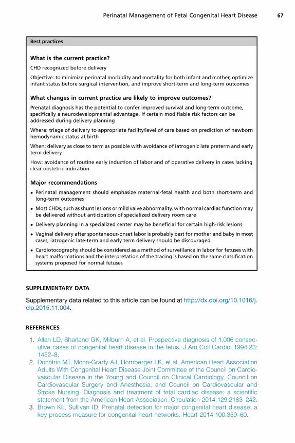
Best practices
What is the current practice?
CHD recognized before delivery
Objective: to minimize perinatal morbidity and mortality for both infant and mother, optimizeinfant status before surgical intervention, and improve short-term and long-term outcomes
What changes in current practice are likely to improve outcomes?
Prenatal diagnosis has the potential to confer improved survival and long-term outcome,specifically a neurodevelopmental advantage, if certain modifiable risk factors can beaddressed during delivery planning
Where: triage of delivery to appropriate facility/level of care based on prediction of newbornhemodynamic status at birth
When: delivery as close to term as possible with avoidance of iatrogenic late preterm and earlyterm delivery
How: avoidance of routine early induction of labor and of operative delivery in cases lackingclear obstetric indication
Major recommendations
� Perinatal management should emphasize maternal-fetal health and both short-term andlong-term outcomes
� Most CHDs, such as shunt lesions ormild valve abnormality, with normal cardiac functionmaybe delivered without anticipation of specialized delivery room care
� Delivery planning in a specialized center may be beneficial for certain high-risk lesions
� Vaginal delivery after spontaneous-onset labor is probably best for mother and baby in mostcases; iatrogenic late-term and early term delivery should be discouraged
� Cardiotocography should be considered as a method of surveillance in labor for fetuses withheart malformations and the interpretation of the tracing is based on the same classificationsystems proposed for normal fetuses
Perinatal Management of Fetal Congenital Heart Disease 67
SUPPLEMENTARY DATA
Supplementary data related to this article can be found at http://dx.doi.org/10.1016/j.clp.2015.11.004.
REFERENCES
1. Allan LD, Sharland GK, Milburn A, et al. Prospective diagnosis of 1,006 consec-utive cases of congenital heart disease in the fetus. J Am Coll Cardiol 1994;23:1452–8.
2. Donofrio MT, Moon-Grady AJ, Hornberger LK, et al, American Heart AssociationAdults With Congenital Heart Disease Joint Committee of the Council on Cardio-vascular Disease in the Young and Council on Clinical Cardiology, Council onCardiovascular Surgery and Anesthesia, and Council on Cardiovascular andStroke Nursing. Diagnosis and treatment of fetal cardiac disease: a scientificstatement from the American Heart Association. Circulation 2014;129:2183–242.
3. Brown KL, Sullivan ID. Prenatal detection for major congenital heart disease: akey process measure for congenital heart networks. Heart 2014;100:359–60.
Sanapo et al68
4. Tworetzky W, McElhinney DB, Reddy VM, et al. Improved surgical outcome afterfetal diagnosis of hypoplastic left heart syndrome. Circulation 2001;103:1269–73.
5. Divanovi�c A, Hor K, Cnota J, et al. Prediction and perinatal management ofseverely restrictive atrial septum in fetuses with critical left heart obstruction: clin-ical experience using pulmonary venous Doppler analysis. J Thorac CardiovascSurg 2011;141:988–94.
6. Tzifa A, Barker C, Tibby SM, et al. Prenatal diagnosis of pulmonary atresia:impact on clinical presentation and early outcome. Arch Dis Child Fetal NeonatalEd 2007;92:F199–203.
7. Franklin O, Burch M, Manning N, et al. Prenatal diagnosis of coarctation of theaorta improves survival and reduces morbidity. Heart 2002;87:67–9.
8. Eapen RS, Rowland DG, Franklin WH. Effect of prenatal diagnosis of critical leftheart obstruction on perinatal morbidity and mortality. Am J Perinatol 1998;15:237–42.
9. Kumar RK, Newburger JW, Gauvreau K, et al. Comparison of outcome when hy-poplastic left heart syndrome and transposition of the great arteries are diag-nosed prenatally versus when diagnosis of these two conditions is made onlypostnatally. Am J Cardiol 1999;83:1649–53.
10. Kipps AK, Feuille C, Azakie A, et al. Prenatal diagnosis of hypoplastic left heartsyndrome in current era. Am J Cardiol 2011;108:421–7.
11. Donofrio MT, Levy RJ, Schuette JJ, et al. Specialized delivery room planning forfetuses with critical congenital heart disease. Am J Cardiol 2013;111:737–47.
12. Donofrio MT, Skurow-Todd K, Berger JT, et al. Risk-stratified postnatal care ofnewborns with congenital heart disease determined by fetal echocardiography.J Am Soc Echocardiogr 2015;28(11):1339–49.
13. Johnson BA, Ades A. Delivery room and early postnatal management of neonateswho have prenatally diagnosed congenital heart disease. Clin Perinatol 2005;32:921–46.
14. Berkley EM, Goens MB, Karr S, et al. Utility of fetal echocardiography in postnatalmanagement of infants with prenatally diagnosed congenital heart disease. Pre-nat Diagn 2009;29:654–8.
15. RudolphAM. The fetal circulation andpostnatal adaptation. In: RudolphAM, editor.Congenital diseases of the heart: clinical-physiological considerations. Armonk(NY): Futura Publishing Company; 2001. p. 3–44.
16. Lim MK, Hanretty K, Houston AB, et al. Intermittent ductal patency in healthynewborn infants: demonstration by colour Doppler flow mapping. Arch Dis Child1992;67:1217–8.
17. Friedman AH, Fahey JT. The transition from fetal to neonatal circulation: normalresponses and implications for infants with heart disease. Semin Perinatol1993;17:106–21.
18. Rosenthal GL. Patterns of prenatal growth among infants with cardiovascularmalformations: possible fetal hemodynamic effects. Am J Epidemiol 1996;143:505–13.
19. Limperopoulos C, Tworetzky W, McElhinney DB, et al. Brain volume and meta-bolism in fetuses with congenital heart disease: evaluation with quantitative mag-netic resonance imaging and spectroscopy. Circulation 2010;121:26–33.
20. Donofrio MT, Duplessis AJ, Limperopoulos C. Impact of congenital heart diseaseon fetal brain development and injury. Curr Opin Pediatr 2011;23:502–11.
21. Manzar S, Nair AK, Pai MG, et al. Head size at birth in neonates with transpositionof great arteries and hypoplastic left heart syndrome. Saudi Med J 2005;26:453–6.
Perinatal Management of Fetal Congenital Heart Disease 69
22. Anagnostou K, Messenger L, Yates R, et al. Outcome of infants with prenatallydiagnosed congenital heart disease delivered outside specialist paediatric car-diac centres. Arch Dis Child Fetal Neonatal Ed 2013;98:F218–21.
23. Berning RA, Silverman NH, Villegas M, et al. Reversed shunting across the duc-tus arteriosus or atrial septum in utero heralds severe congenital heart disease.J Am Coll Cardiol 1996;27:481–6.
24. Quartermain MD, Glatz AC, Goldberg DJ, et al. Pulmonary outflow tract obstruc-tion in fetuses with complex congenital heart disease: predicting the need forneonatal intervention. Ultrasound Obstet Gynecol 2013;41:47–53.
25. Hinton R, Michelfelder E. Significance of reverse orientation of the ductus arterio-sus in neonates with pulmonary outflow tract obstruction for early intervention. AmJ Cardiol 2006;97:716–9.
26. Arya B, Levasseur SM, Woldu K, et al. Fetal echocardiographic measurementsand the need for neonatal surgical intervention in tetralogy of Fallot. Pediatr Car-diol 2014;35:810–6.
27. Makikallio K, McElhinney DB, Levine JC, et al. Fetal aortic valve stenosis and theevolution of hypoplastic left heart syndrome: patient selection for fetal interven-tion. Circulation 2006;113:1401–5.
28. Szwast A, Tian Z, McCann M, et al. Vasoreactive response to maternal hyperox-ygenation in the fetus with hypoplastic left heart syndrome. Circ Cardiovasc Im-aging 2010;3:172–8.
29. Jouannic JM, Gavard L, Fermont L, et al. Sensitivity and specificity of prenatalfeatures of physiological shunts to predict neonatal clinical status in transpositionof the great arteries. Circulation 2004;110:1743–6.
30. Punn R, Silverman NH. Fetal predictors of urgent balloon atrial septostomyin neonates with complete transposition. J Am Soc Echocardiogr 2011;24:425–30.
31. Freud LR, Escobar-Diaz MC, Kalish BT, et al. Outcomes and predictors of peri-natal mortality in fetuses with Ebstein anomaly or tricuspid valve dysplasia inthe current era: a multi-center study. Circulation 2015;132(6):481–9.
32. Manning FA, Harman CR, Morrison I, et al. Fetal assessment based on fetal bio-physical profile scoring. IV. An analysis of perinatal morbidity and mortality. Am JObstet Gynecol 1990;162:703–9.
33. ACOG practice bulletin. Antepartum fetal surveillance. Number 9, October 1999(replaces technical bulletin number 188, January 1994). Clinical managementguidelines for obstetrician-gynecologists. Int J Gynaecol Obstet 2000;68:175–85.
34. Falkensammer CB, Paul J, Huhta JC. Fetal congestive heart failure: correlation ofTei-index and cardiovascular-score. J Perinat Med 2001;29:390–8.
35. Wieczorek A, Hernandez-Robles J, Ewing L, et al. Prediction of outcome of fetalcongenital heart disease using a cardiovascular profile score. Ultrasound ObstetGynecol 2008;31:284–8.
36. Huhta JC. Right ventricular function in the human fetus. J Perinat Med 2001;29:381–9.
37. Donofrio MT, Gullquist SD, Mehta ID, et al. Congenital complete heart block: fetalmanagement protocol, review of the literature, and report of the smallest suc-cessful pacemaker implantation. J Perinatol 2004;24:112–7.
38. Howell LJ. The Garbose Family Special Delivery Unit: a new paradigm formaternal-fetal and neonatal care. Semin Pediatr Surg 2013;22:3–9.
39. Levey A, Glickstein JS, Kleinman CS, et al. The impact of prenatal diagnosis ofcomplex congenital heart disease on neonatal outcomes. Pediatr Cardiol 2010;31:587–97.
Sanapo et al70
40. Costello JM, Polito A, Brown DW, et al. Birth before 39 weeks’ gestation is asso-ciated with worse outcomes in neonates with heart disease. Pediatrics 2010;126:277–84.
41. Cheng YW, Nicholson JM, Nakagawa S, et al. Perinatal outcomes in low-risk termpregnancies: do they differ by week of gestation? Am J Obstet Gynecol 2008;199:370.e1–7.
42. Cnota JF, Gupta R, Michelfelder EC, et al. Congenital heart disease infant deathrates decrease as gestational age advances from 34 to 40 weeks. J Pediatr 2011;159:761–5.
43. Costello JM, Pasquali SK, Jacobs JP, et al. Gestational age at birth and outcomesafter neonatal cardiac surgery: an analysis of the society of thoracic surgeonscongenital heart surgery database. Circulation 2014;129:2511–7.
44. Miller SP, McQuillen PS, Hamrick S, et al. Abnormal brain development in new-borns with congenital heart disease. N Engl J Med 2007;357:1928–38.
45. Partridge SC, Vigneron DB, Charlton NN, et al. Pyramidal tract maturation afterbrain injury in newborns with heart disease. Ann Neurol 2006;59:640–51.
46. Licht DJ, Shera DM, Clancy RR, et al. Brain maturation is delayed in infants withcomplex congenital heart defects. J Thorac Cardiovasc Surg 2009;137:529–36.
47. Clouchoux C, du Plessis AJ, Bouyssi-Kobar M, et al. Delayed cortical develop-ment in fetuses with complex congenital heart disease. Cereb Cortex 2013;23:2932–43.
48. Martin JA, Hamilton BE, Sutton PD, et al, Centers for Disease Control and Preven-tion National Center for Health Statistics National Vital Statistics System. Births:final data for 2006. Natl Vital Stat Rep 2009;57:1–104.
49. Honest H, Forbes CA, Duree KH, et al. Screening to prevent spontaneous pre-term birth: systematic reviews of accuracy and effectiveness literature with eco-nomic modelling. Health Technol Assess 2009;13:1–627.
50. Lockwood CJ. Predicting premature delivery–no easy task. N Engl J Med 2002;346:282–4.
51. Peterson AL, Quartermain MD, Ades A, et al. Impact of mode of delivery onmarkers of perinatal hemodynamics in infants with hypoplastic left heart syn-drome. J Pediatr 2011;159:64–9.
52. Trento LU, Pruetz JD, Chang RK, et al. Prenatal diagnosis of congenital heart dis-ease: impact of mode of delivery on neonatal outcome. Prenat Diagn 2012;32:1250–5.
53. Walsh CA, MacTiernan A, Farrell S, et al. Mode of delivery in pregnancies compli-cated by major fetal congenital heart disease: a retrospective cohort study.J Perinatol 2014;34:901–5.
54. Reis PM, Punch MR, Bove EL, et al. Obstetric management of 219 infants withhypoplastic left heart syndrome. Am J Obstet Gynecol 1998;179:1150–4.
55. American College of Obstetricians and Gynecologists. ACOG practice bulletinNo. 106: intrapartum fetal heart rate monitoring: nomenclature, interpretation,and general management principles. Obstet Gynecol 2009;114:192–202.
56. Parer JT, Ikeda T, King TL. The 2008 National Institute of Child Health and HumanDevelopment report on fetal heart rate monitoring. Obstet Gynecol 2009;114:136–8.
57. Alfirevic Z, Devane D, Gyte GML. Continuous cardiotocography (CTG) as a formof electronic fetal monitoring (EFM) for fetal assessment during labour. CochraneDatabase Syst Rev 2013;(5):CD006066.
58. Ueda K, Ikeda T, Iwanaga N, et al. Intrapartum fetal heart rate monitoring in casesof congenital heart disease. Am J Obstet Gynecol 2009;201:64.e1–6.

Perinatal Management of Fetal Congenital Heart Disease 71
59. Morikawa M, Endo D, Yamada T, et al. Electronic fetal heart rate monitoring in fivefetuses with Ebstein’s anomaly. J Obstet Gynaecol Res 2014;40:424–8.
60. Katz AR, Ross PJ, Bekheit-Saad S. Utilization of fetal scalp pH and direct fetalelectrocardiogram in the intrapartum management of congenital complete heartblock in an adolescent primigravida. J Adolesc Health Care 1980;1:152–4.
61. Friedman DM, Zervoudakis I, Buyon JP. Perinatal monitoring of fetal well-being inthe presence of congenital heart block. Am J Perinatol 1998;15:669–73.
62. East CE, Leader LR, Sheehan P, et al. Intrapartum fetal scalp lactate sampling forfetal assessment in the presence of a non-reassuring fetal heart rate trace. Co-chrane Database Syst Rev 2015;(5):CD006174.