Pericardial disease and other acquired heart diseases · MCQ 3 Pericardiocentesis –everything is...
39
Pericardial disease and other acquired heart diseases Royal Brompton & Harefield NHS Foundation Trust Sylvia Krupickova Exam oriented Echocardiography course, 8th November 2018
Transcript of Pericardial disease and other acquired heart diseases · MCQ 3 Pericardiocentesis –everything is...
Pericardial disease
and
other acquired heart diseases
Royal Brompton & Harefield NHS Foundation Trust
Sylvia Krupickova
Exam oriented Echocardiography course, 8th November 2018

2 pericardial sinuses –
(not continuous with one another):
• Transverse sinus – between in front aorta and pulmonary
artery and posterior vena cava superior
• Oblique sinus - posterior to the heart, with the vena cava
inferior on the right side and left pulmonary veins on the left
side
Normal pericardium is not seen usually on normal echocardiogram,
neither the pericardial fluid
2 layers – fibrous
- serous – visceral and parietal layer
Normal Pericardium:
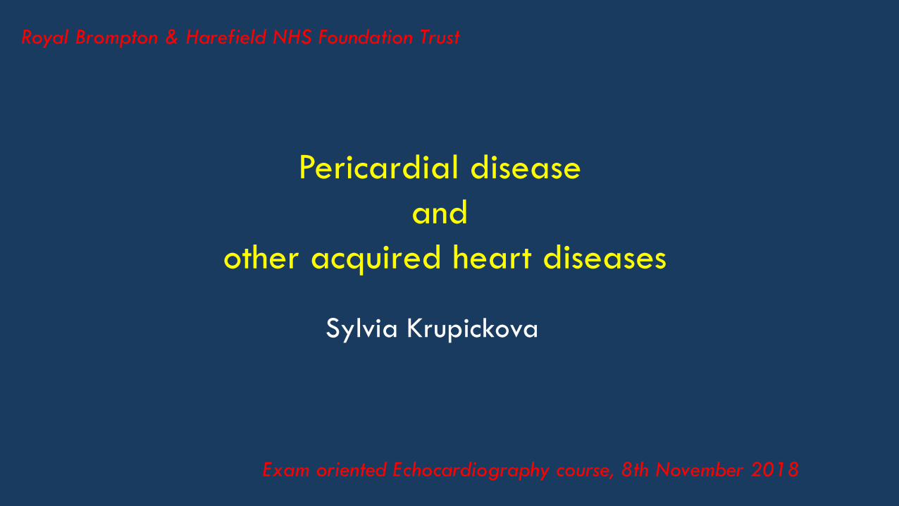
Small effusion – <10mm, black space posterior to the heart in parasternal
short and long axis views, seen only in systole
Moderate – 10-20 mm, more than 25 ml in adult, echo free space is all
around the heart throughout the cardiac cycle
Large – >20 mm, swinging motion of the heart in the pericardial cavity
• How big is the effusion? (always measure in diastole)
• Where is it? (appears first behind the LV)
• Is it causing haemodynamic compromise?
Acute Pericarditis:

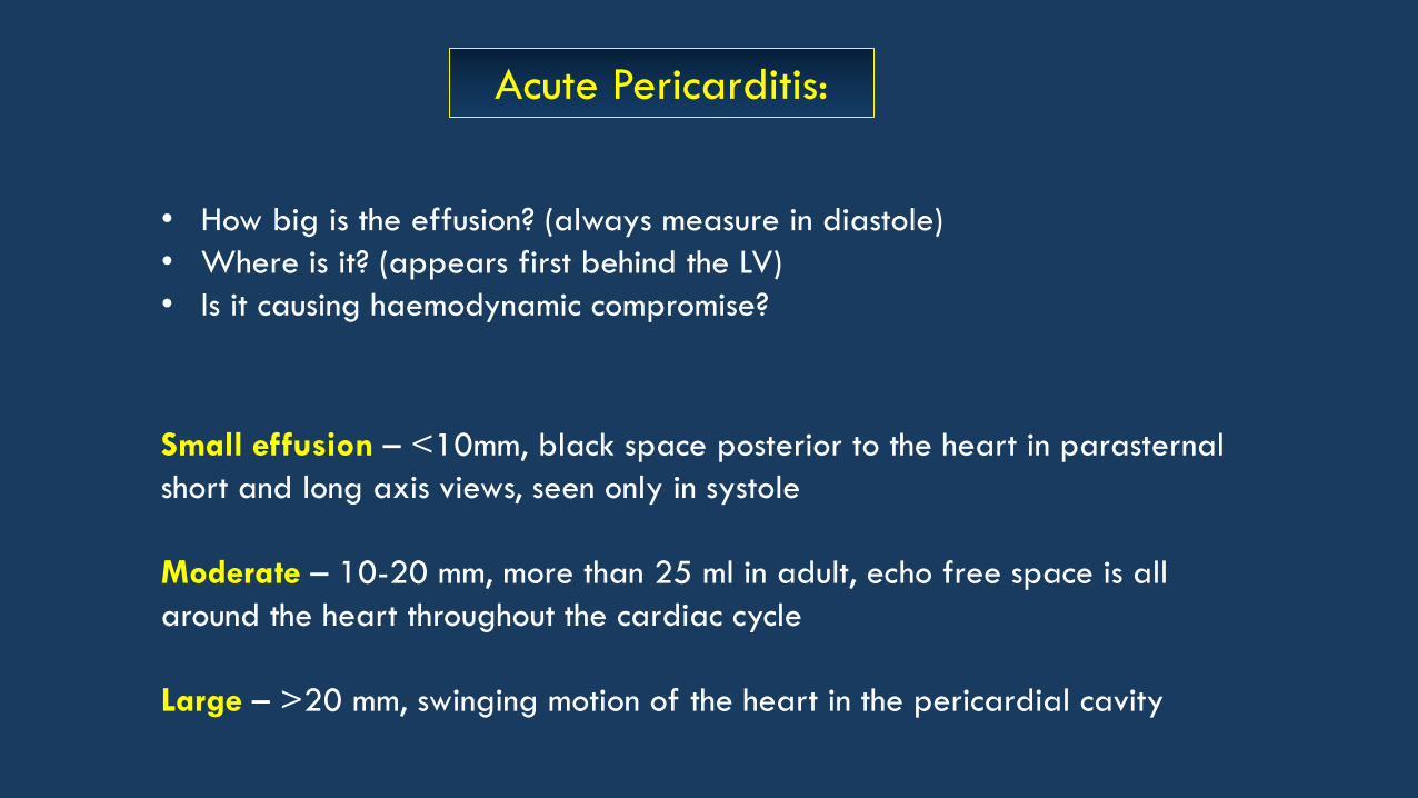
Pericardiocentesis

Constrictive pericarditis
Constriction of LV filling by pericardium
Restrictive cardiomyopathy
Impaired relaxation of LV
Restriction versus Constriction:

Constriction
E´ velocity is normal as there is no impediment to relaxation of the left ventricle.
Restriction
E´ velocity is low (less than 5 cm/s) due to impaired filling of the ventricle
(impaired relaxation)
Both have affected left ventricular filling
Constriction versus Restriction
Pathophysiology:
• Endocardial lesion
• Activation of prothrombic cascade - sterile platelet-fibrin deposition
• Bacterial colonisation
The highest risk of IE:
• Prosthetic cardiac valve
• CHD – unrepaired, with previous palliation, after corrective surgery
(residual findings, pacemaker)
• Catheters in newborn – structurally normal heart
Infective Endocarditis:

• Determine the location, size, extent, mobility and number of IE lesions
• Assess the severity of intracardiac damage:
Severity of valvular regurgitation
Perforation of valve leaflets
Development of paravalvular abscess
Dehiscence of prosthetic valves
• Assess the haemodynamic sequelae:
Valvular function
Myocardial function
Pericardial effusion
Echocardiography Imaging of Infective Endocarditis:
Echocardiography Guidelines:

Infective Endocarditis in Hypertrophic Cardiomyopathy:

Fever persisting 5 or more days
+ at least 4 clinical features:
• Changes on extremities (erytema, edema, desquamation)
• Polymorphous exanthema
• Nonexsudative conjunctivitis
• Changes in the lips and oral mucosa
• Cervical lymphadenopathy
Kawasaki Disease
Mucocutaneous Lymphnode Syndrome

Kawasaki Disease
Acute febrile phase
- diffuse microvascular angiitis - coronary arteries
- pericarditis - pericardial effusion
- myocarditis – myocardial dysfunction
- endocarditis – valvular regurgitation
Subacute phase – pancarditis, ectasia, aneurysm, thrombosis of the coronary arteries
Chronic phase – intimal thickening of the coronary arteries
– stenosis, calcification, thrombosis

Coronary arteries – ectasia, aneurysm, thrombus
Location – frequently LAD and RCA affected
Ectasia – diffuse dilation of coronary artery
Aneurysm – localised dilation – saccular - rounded
- fusiform – larger in one direction
Kawasaki Disease

Coronary arteries supplying myocardial segments
• Acute inflammatory illness that occurs following an upper respiratory infection
with group A beta hemolytic streptococci
• Affects joints, central nervous system, skin, musculoskeletal system and heart
• Acute rheumatic fever – mitral regurgitation, aortic regurgitation
• Chronic rheumatic fever – mitral regurgitation and/or stenosis,
aortic regurgitation and/or stenosis
Rheumatic Heart Disease

The most common manifestation of rheumatic heart disease in young
Echocardiography criteria:
• Pathologic mitral regurgitation: Seen in two views, jet length at least 2cm,
pan-systolic jet in at least one view
• Combination of morphological changes (thickening and restricted motion
of the anterior MV leaflet)
Rheumatic Heart Disease
Mitral valve regurgitation

Mitral valve regurgitation
Normal MV Rheumatic MV with abnormal
coaptation and regurgitationProlapse with billowing
Rheumatic Heart Disease
Mitral stenosis
Rheumatic fever is the most common cause of the mitral stenosis worldwide.
Echocardiography criteria:
• Mean gradient at least 4 mmHg
• At least 2 morphological changes (thickened leaflets and relatively immobile
posterior leaflet, chordal shortening or fusion)
Differential diagnosis:
Congenital mitral valve stenosis – frequently associated with abnormal papillary
muscles

The
Mitral valve stenosis
Normal MV in diastoleRheumatic MV in with thickened
anterior and posterior leaflets
Rheumatic Heart Disease
Aortic regurgitation
Less frequently affected
Echocardiography criteria:
• Pathologic aortic regurgitation: seen in two views,
jet length at least 1 cm, pan-diastolic jet in at least 1 view
• Pathologic features: thickened leaflets, coaptation defect, prolapse
Differential diagnosis: Bicuspid aortic valve and aortic root dilation

A. Should be measured always in systole
B. Should be measured always in diastole
C. Not only the size of effusion, also the rate of accumulation is
very important
D. A small or moderate effusion may cause haemodynamic
compromise, if accumulated rapidly
E. Appears usually first behind the left ventricle
Pericardial Effusion:MCQ 1

A. Diastolic collapse of the free walls of right atrium and right
ventricle
B. Typical finding is increased ventricular interdependence
C. Typical finding is decreased ventricular interdependence
D. Inspiratory increase in tricuspid velocity
E. Inspiratory decrease in tricuspid velocity
Cardiac Tamponade:MCQ 2

A. May be used for diagnosis of neoplastic effusions or purulent pericarditis
B. Ultrasound plays a crutial role for guidance of the needle
C. The most common approach is subxiphoid
D. Is life-saving procedure in case of cardiac tamponade
E. There has to be effusion behind left atrium, otherwise
the pericardiocentes cannot be performed
Pericardiocentesis – everything is right, EXCEPT:MCQ 3

A. Septal bounce is sign of ventricular interdependence
B. The effect of the size, shape and compliance of one ventricle
on the size, shape and compliance of the other ventricle
C. Is pronounced in constrictive pericarditis
D. Is pronounced in restrictive cardiomyopathy
E. Is pronounced in dilated cardiomyopathy
MCQ 4 Ventricular Interdependence

MCQ 5
A. Most probably infective endocarditis of mitral valve
B. Severe mitral stenosis
C. Congenital dysplastic mitral valve
D. Thrombus of MV
E. Tumor of MV – most probably rhabdomyoma or myxoma of MV
This patient has history of fever and high CRP.
The most probable diagnosis is:

A. The major role in pathogenesis plays chronic endocardial trauma
of the interventricular septum by the anterior septal leaflet - common finding
in the obstructive form
B. The same incidence is in obstructive and non-obstructive form
C. Patients with obstructive form are affected much more often than non-obstructive
D. Patients with HCM are not at higher risk of IE than healthy patients
E. Aortic valve is the most often affected valve in hypertrophic cardiomyopathy
Infective Endocarditis in Hypertrophic Cardiomyopathy:MCQ 6

A. TOE has the same sensitivity and specificity for detecting IE as TTE
B. TOE is especially important in diagnosis of IE affecting prosthetic valves
and should be always considered when clinical suspicion of IE
C. If TOE is negative, IE is excluded
D. Dehiscence of the prosthetic valve appears very often with IE and can
be seen easier on TTE than TOE
E. TOE can diagnose paravalvular abscess easier than TTE
MCQ 7 Role of TOE in suspected IE:

A. The most common sites for coronary artery aneurysms are LAD and RCA
B. Pericardium is never affected
C. Myocardium is never affected
D. Valvar involvement is never present
E. Thrombosis of the coronary arteries can be present in all phases
Kawasaki Disease:MCQ 8
A. Multiple aneurysms are quite common
B. Systolic function of the LV may be impaired due to myocarditis
C. It is crucial to assess regional wall motion abnormalities
D. Coronary artery imaging should be performed at high transducer frequency
E. Apical windows are the best for coronary arteries imaging
Kawasaki Disease:
MCQ 9

MCQ 10

A. Dilated coronary sinus indicating left SVC entering the CS
B. Dilated left coronary artery suggestive of Kawasaki disease
C. The best view for showing left coronary artery
D. Dilated right coronary artery
E. Coronary sinus defect
What si the rounded structure in 4-chamber:

MCQ 11

A. Ectasia of left LCA, LAD, LCX and RCA
B. Kawasaki disease is the most probable diagnosis
C. A thrombus is seen in the LCA
D. These findings can be see in any phase of Kawasaki disease
E. Myocardial involvement cannot be excluded
Everything is right EXCEPT:

MCQ12


A. This patient’s diagnosis is most probably rheumatic heart disease
B. This patient has mixed mitral and aortic valve disease
C. There is prosthetic valve seen in the mitral valve position
D. Tricuspid valve is not affected
E. This is most probably chronic rheumatic disease
Which statements are true:

Thank you!

Mitral valve inflow