Voyager Observations of Galactic and Anomalous Cosmic Rays in the Heliosheath

NASA Technical Paper 3473
Performance Study of Galactic Cosmic RayShield MaterialsMyung-Hee Y. Kim, John W. Wilson, Sheila A. Thibeault, John E. Nealy, Francis F. Badavi,and Richard L. Kiefer
November 1994

NASA Technical Paper 3473
Performance Study of Galactic Cosmic RayShield MaterialsMyung-Hee Y. KimCollege of William and Mary � Williamsburg, Virginia
John W. Wilson, Sheila A. Thibeault, and John E. NealyLangley Research Center � Hampton, Virginia
Francis F. BadaviChristopher Newport University � Newport News, Virginia
Richard L. KieferCollege of William and Mary � Williamsburg, Virginia
National Aeronautics and Space AdministrationLangley Research Center � Hampton, Virginia 23681-0001
November 1994

The use of trademarks or names of manufacturers in this
report is for accurate reporting and does not constitute an
o�cial endorsement, either expressed or implied, of such
products or manufacturers by the National Aeronautics and
Space Administration.
This publication is available from the following sources:
NASA Center for AeroSpace Information National Technical Information Service (NTIS)
800 Elkridge Landing Road 5285 Port Royal Road
LinthicumHeights, MD 21090-2934 Spring�eld, VA 22161-2171
(301) 621-0390 (703) 487-4650

Summary
The space program is faced with two di�cult ra-diation protection issues for future long-term oper-ations. First, retro�t of shield material or conser-vatism in shield design is prohibitively expensive andoften impossible. Second, shielding from the cos-mic heavy ions is faced with limited knowledge onthe physical properties and biological responses ofthese radiations. The current status of space shield-ing technology and its impact on radiation healthis discussed herein in terms of conventional protec-tion practice and a test biological response model.The impact of biological response on the selection ofoptimum materials for cosmic ray shielding is pre-sented in terms of the transmission characteristics ofthe shield material. The transmission properties are,in turn, related to the nuclear cross sections of thecosmic heavy ions, for which an inadequate experi-mental database exists. Clearly, these physical andbiological issues must be resolved before an adequateshield design can be de�ned.
The choice of structural materials composition isa means of reducing astronaut exposure risk fromspace radiations in future NASA missions. The useof a performance index for shield materials related tothe change in biological protection at constant shieldmass and varying shield composition indicates perfor-mance indices up to a factor of 20. Although the sys-tematics of nuclear cross sections are able to demon-strate the relation of exposure risk to shield-materialcomposition, the current uncertainty in nuclear crosssections will not allow an accurate evaluation of riskreduction. Even so, the unique role of hydrogenousmaterials used as high-performance shields is clear.Shinn et al. suggested that polyethylene with itsshort nuclear absorption lengths is an e�ective shieldmaterial in spite of the favoring of massive projectilefragments, and this is demonstrated herein for mono-energetic ion beams. This paper presents a theoret-ical study of risk-related factors and a pilot exper-iment to study the e�ectiveness of choice of shieldmaterials to reduce the risk in space operations.
Introduction
In the past exploratory manned space missionslasting up to several weeks, only the more intensesources of space radiation, such as solar cosmic raysand trapped radiations, were considered to be theprimary radiation hazards. The principal radiationprotection issues were the control of early somatic ef-fects of radiation exposure and their impact on mis-sion safety. Few astronauts, if any, were expected tomake more than one high-pro�le trip to the Moon sothat career exposures were of secondary importance.
In this context, the galactic cosmic ray (GCR) back-ground exposures at rates of 150 to 200 mGy/yr werenot of great concern (refs. 1 and 2).
With the advent of the Space Shuttle, the con-text of an astronaut changed from space explorer tospace worker and career exposure limits came intofocus with late somatic e�ects seen as the ultimatelimiting factor on mission activity (ref. 3). Such aradical shift in astronaut exposure patterns led toa reevaluation of the importance of low-level GCRbackground exposures. (A detailed review is given inref. 2.)
Within a few years of the discovery of particlesof high charge and energy (HZE) as componentsof the GCR, the unique pattern of energy depositon the microscopic scale raised issues with respectto e�ects on living cells (ref. 4). Also, the light ashes induced by proton reactions and HZE ionpassage through the vitreous humor observed byastronauts in space had already been predicted inthe infancy of the space program (ref. 5). Althoughradiobiological knowledge has greatly improved, ourability to estimate risk to the astronaut from suchexposures is still quite uncertain (ref. 6). Even acrude estimate using the linear energy transfer (LET)dependent quality factor (ref. 2) results in as muchas 1.2 Sv/yr exposures, depending on shielding nearsolar minimum. This shows a large potential impacton the career of a space worker or a deep-spaceexplorer.
Clearly, 1.2 Sv/yr is an important number, butone must hesitate in applying it to astronaut risk be-cause it implies extrapolation from the human data-base for late somatic e�ects that are based primarilyon X-ray and -ray exposures (refs. 7 and 8). Evi-dence is growing of biological end points which arepeculiar to high-LET HZE exposures that are notproduced by X-rays or -rays for which the relativebiological e�ectiveness (RBE) is in�nite or unde�ned.Evidence that the usual extrapolation of risk fromthe -ray database is inadequate has been providedby the measurement of sister chromatid exchangesin resting human lymphocytes irradiated with 238Pu�-particles (ref. 9), by the observation of abnormali-ties in stem cell colonies surviving similar �-particleirradiation (ref. 10), and by the partial disintegrationof chromosomes after irradiation with high-energyheavy ion beams to simulate space radiation (ref. 11).In these examples, a quality factor related to RBEbecomes meaningless because at doses comparableto that delivered by one particle (or a few parti-cles), and for radiation e�ects that are not manifestfor low-LET radiation (e.g., X-rays), the RBE be-comes in�nite. Thus, new methods to predict the

risk resulting from exposure to GCR radiation mustbe developed.
The biological response of living tissues depends(in part) on the temporal and spatial uctuations ofthe energy deposits within the tissue system. Such uctuations depend not only on the speci�c environ-ment to which the astronaut is exposed but also onhow that environment is modi�ed by interaction withthe astronaut's body in reaching the speci�c tissues.Only by knowledge of the speci�c radiation types andtheir physical properties at the tissue site can a ba-sis for estimating astronaut risk be found. Even ifthe environment to which the astronaut is exposedis known precisely, the energy deposit within speci�ctissues deep in the astronaut's body are, for the mostpart, known only through theoretical estimates andare, therefore, limited by the uncertainty in the cal-culational models. Clearly, an accurate conversion ofthe astronaut's environment to estimates of exposure�elds at speci�c tissue sites is a high priority in thespace-radiation protection problem.
Apart from the issues of the astronaut's self-shielding factors and uncertainty in human responseto the HZE particles, radiation shielding impliessome control over the radiation environment to whichthe astronaut is exposed. The traditional structuralmaterial within the space program has been alu-minum. The absorbed dose at solar minimum froman annual GCR exposure behind an aluminum shieldis shown in sketch A. The absorbed dose increases toa maximum at 3 to 4 g/cm2 and declines to the free-space value at about 30 g/cm2. Clearly, no shieldingadvantage is found in reducing the energy absorbedby the astronaut, and if any protection is provided,it results from changes in the microscopic pattern ofthe energy-absorption events (ref. 12).
Herein, we examine the modi�cation of the phys-ical parameters of the attenuated GCR environmentin various materials to develop an understandingof the qualitative changes in environmental compo-nents as a function of shield composition (includingtissue-equivalent shields). In this context, one be-gins to appreciate the role of nuclear cross sectionsin modifying the local environment and the associ-ated microscopic uctuation in the energy-absorptionevents. Furthermore, we will begin to understandthe e�ects of nuclear cross-section uncertainty asit applies to the change in the microscopic energy-absorption uctuations. We shall assess the impor-tance of these local environmental modi�cations onbiological systems in terms of conventional dosime-try by using de�ned quality factors and a biologicalmodel that is dependent on track structure.
250
200
150
100
50
0 10 20 30
D(x
), m
Gy/
yr
x, g/cm2
Sketch A
Although the human risk associated with such ex-posure is uncertain, radiobiology experiments withimmortal cell populations (cell cultures that can besustained inde�nitely) have yielded biological datasuitable for estimating GCR exposure e�ects onthose speci�c cell populations. The response of theC3H10T1/2 mouse cell cultures (ref. 13) has beenused to evaluate shield properties for the biologicalend points of clonogenic death and neoplastic trans-formation (ref. 12). Clonogenic death is closely asso-ciated with the early response of radiation sickness(nausea, vomiting, erythema, etc.), and neoplastictransformation is related to cancer induction. A cell-repair kinetics model including track-structure e�ectsfor the C3H10T1/2 system (refs. 13{15) provides abasis for studying shield performance.
In the present paper, we �rst discuss the problemof radiation risk assessment in the context of micro-dosimetry. We then examine the shield parametersrelated to shield performance and evaluate the per-formance on the basis of conventional risk assessmentand the C3H10T1/2 cell model. On this basis we ex-amine the e�ects of shield-material selection on shielddesign. Light hydrogenous compounds are shown tohold great promise as high-performance shield mate-rials. Encouraged by this prospect, we then examinethe e�ects of hydrogen-bearing compounds as poten-tial space structural components. A pilot experimentto study such e�ects is described.
The importance of hydrogenous materials in mod-ifying the biologically important components of ionbeams makes these studies important in evaluating
2

the therapeutic value of heavy ion beams in medicalapplications. Indeed, the computational proceduresused, the quality of the nuclear database, and thebiological response models should be useful in thedesign of therapeutic procedures.
Microscopic Fluctuations and Biological
Response Models
The response of living tissue (refs. 3 and 8) to adose D with low LET is represented by a sensitivitycoe�cient k and a quadratic coe�cient Do as
R = k D
�1 +
D
Do
�(1)
where R is either the risk of inducing a speci�c endpoint or the level of severity. The parameter Do isdose-rate dependent and is on the order of 1.2 Gy fordose rates larger than 50 mGy/day (refs. 3 and 8).We assume a low dose rate herein so that D2
may beneglected, where
R = k D (2)
The concept of dose as a physical or chemical insultper unit mass of tissue is a carryover from the con-cepts of pharmacology and assumes that dose is ameasure of e�ects on individual cells (ref. 16). Tis-sue cells are, in fact, not all equal at low exposuresbecause the energy deposits are quantized and en-ergy is deposited in only a fraction of cells; similarly,volumes within a given cell are not all equally sensi-tive. In general, the absorbed dose D is not a goodmeasure of biological damage because this averagequantity can be decomposed (ref. 16) as follows:
D =
P�i
V NE
=
P�i
V NH
NH
NE
(3)
where V is the sensitive site volume (unit density),�i is the energy absorbed per site hit (referred toas the \hit size" of the ith event), and NE is thenumber of exposed sites. At a low dose, not all sitesare hit, and so the number of site hits NH is less thanthe number of sites exposed. Only when NH ! NE
is D meaningful in terms of individual cell response(ref. 16). The fraction of sites that are hit at lowexposure (that is, NH � NE) is
NH
NE
� �g� (4)
where �g is the site geometric cross section and � isthe charged-particle uence within the tissue system.In reality, the cross section can be larger than the
geometric cross section because of the �-ray di�usionfor which the number of site hits is increased by siteshit far from the ionizing particles path. The uence �is related to the macroscopic absorbed dose D andto the value of the unrestricted LET (L) as
� = 6:24D
L(5)
where � is given in particles/�m2, D in Gy, and L
in keV/�m. For -rays, L corresponds to thesecondary electrons generated and has a value ofabout 0.25 keV/�m; the corresponding � is ane�ective secondary electron uence that is dependenton the photoabsorption coe�cient and the -ray uence.
The average hit size (��) is given as
�� =Xi
�i
NH
(6)
and is known from basic physical principles and spec-i�cations of the site volume V . The mean number ofhits per exposed site is then
NH
NE
=DV
��(7)
and is related to the number of hit sites assumingPoisson statistics. We have estimated �� from the the-ory of Xapsos et al. (ref. 17) for various ion typesas shown in �gure 1(a) for a 1-Gy exposure and0.1-�m site size corresponding approximately to thewidth of a single chromatin strand and its immedi-ate environment. In �gure 1 we have ignored con-tributions from fragmenting nuclei of the biologicaltarget. The e�ect of site size is shown by compar-ing the 0.1-�m site size with the 0.5-�m site sizein �gure 1(b). Note that the hit size and averagenumber of hits increase with the site size. The re-cently de�ned quality factors (ref. 7) are also shownin �gure 1(c). The region of unit quality factor forthis 1-Gy exposure is marked by a sizable fractionof hit sites with a fraction of keV hit size, and thecorresponding excess fatal cancer risk to this expo-sure would be about 3 percent. In distinction, the100-keV/�m exposure has a quality factor near 20to 30 and would result in an estimated excess cancerrisk of 60 to 90 percent. The mean hit size in this caseis several tens of keV, and a small fraction (less than1 percent) of the sites are, in fact, hit. The HZE par-ticles show a smaller hit size because of their rangeand �-ray di�usion than the smaller ions at the sameLET. A corresponding increase occurs in the numberof sites hit. A further distinction of HZE exposure is
3

that a clustered group of contiguous cells (or sites)is a�ected by a single ion passage because of theirrange and �-ray di�usion (ref. 18) in distinction tosmaller ions of the same LET.
Figure 1 aptly illustrates the great variability ofthe microscopic uctuations expressed previously asthe mean hit size and the fraction of sites hit forvarious radiation �eld components. Although themeaning of this variability is somewhat representedby the quality factor, as noted in �gure 1, an addeddistinctive feature of the HZE exposures is that largeclusters of contiguous cells are a�ected. We do notyet understand the radiation response of many ofthe GCR components, but it is surely the changeswrought by shield materials on these microscopic uctuations that will serve as the primary means ofradiation protection and not a decline in the energyabsorbed with the addition of shield material. (Seesketch A.)
Conventional Risk Assessment
According to equation (1), excess cancer risks forhumans are estimated based on coe�cients derivedfrom X-ray and -ray exposures. The conventionalmethod of extrapolating the human database to high-LET exposures is to replace D in equation (1) bythe dose equivalent H given by
H = QD (8)
where Q is the LET-dependent quality factor shownin �gure 1(c). Equation (8) follows from analogy withthe relative biological e�ectiveness given for -rayand ion exposure levels D and Di which result inthe same biological end point by
RBE =D
Di(9)
We note that the quality factor is a de�ned quantity(not given by a measurement) and represents trendsof measured RBE in cell culture, plant, and animalexperiments. The RBE values depend on end point,dose, dose rate, and quality of the radiation usu-ally represented by LET. Usually, RBE is assumedto reach a maximum value (denoted by (RBE)M) atsu�ciently low dose as related to the initial slopesof the response curves of each radiation type (refs. 3and 19). Furthermore, the dose at which (RBE)Mis achieved is assumed to be dose-rate dependent asshown in �gure 2. The values of RBE from which Q isde�ned as a function of LET are largely for high doserates at the 0.1-Gy level of exposure for which �ssionneutrons have Q = 25 corresponding to a -ray ex-posure of 2.5 Gy. The RBE values for lower levels
of exposure and/or lower dose rate are much larger(ref. 19), as shown in table 1, and occur for lower ex-posure and dose rates than were used in deriving Q.In that the achieving of (RBE)M is accelerated ata low dose rate, the RBE values in table 1 may, infact, be more appropriate for space exposures. This isone source of the rather large uncertainties in space-radiation exposure risks. The second source of un-certainty concerns the response to HZE exposuresfor which little is known. The assumption is madethat single-ion track e�ects for which -ray exposureshave no analog are possible. One such mechanismwas suggested by Todd (ref. 18) in which the cellsexposed at 0.25 Gy have a high probability of beingtransformed whereas the dead cells of the track coremust be replaced, thus causing promotion to a cancergrowth by this one event. (See �g. 3.) The RBE forsuch results is unde�ned (in�nite), and extrapolationfrom the human database is not possible.
Table 1. (RBE)Mfor Fission Neutrons
Tumor induction (approximate) . . . . . . . . . . 3{200
Life shortening . . . . . . . . . . . . . . . . . 15{45
Transformation . . . . . . . . . . . . . . . . . 35{70
Cytogenic studies . . . . . . . . . . . . . . . . 40{50
Genetic end points in mammalian systems . . . . . 10{45
Other end points:
Lens opaci�cation . . . . . . . . . . . . . . 25{200
Micronucleus assay . . . . . . . . . . . . . . 6{60
Testes weight loss . . . . . . . . . . . . . . . 5{20
The use of an LET-dependent quality factor as re-lated to dose equivalent implies additivity of diversecomponents in estimating risk. Such assumptionsmay underestimate the actual risk as was discussedby Scott (ref. 20). Furthermore, risks associated withdi�erent time intervals are likewise not additive, es-pecially if radiation proves to be an e�ective promo-tion factor in carcinogenic response (ref. 21). Forlow-LET exposures, substantial repair is often oper-ative and results in reduced risk. For high-LET ex-posures, dose-rate enhancement e�ects are possiblein which risk is substantially increased at lower doserates (ref. 22) as shown in �gure 4.
The uncertainties in radiation-induced risk havebeen estimated in the NASA Radiation Health Pro-gram (ref. 6) and are presented in �gure 5. In theapproximation used here, the risk is assumed to berelated to the total value of dose equivalent. This as-sumes that the dose-response curve is of similar shapefor each radiation component which is linear at low
4

dose and dose rate. The excess risk (the added riskdue to exposure) is then given by
R = k H = k (Hx +Hz) (10)
where H is the dose equivalent (given in Sv), Hx isthe component of dose equivalent due to low-LETradiation, and Hz is the dose equivalent due tothe HZE component of the radiation. By mak-ing the further approximation that the uncertaintiesin k and Hx are negligible in comparison with theuncertainty in Hz, we obtain
�R = k �Hz
Hz
Hz � k UHz (11)
so that the net e�ect of the uncertainty in R is toincrease the relative risk, which becomes
R +�R = k H + k UHz = k Hu (12)
This equation de�nes an e�ective dose equivalent(Hu) which corresponds to the increased risk due touncertainties. If a limit L is de�ned on the basis ofexcess risk R, then it is required that
R +�R � L (13)
where L is the de�ned limit of acceptable risk. Asafety factor (S) can be de�ned with reference toequation (12). Let S be an upper bound on the es-timated value of the uncertainty in HZE dose equiv-alent (that is, S = nU), where n = 1; 2 : : : corre-sponds to the number of standard deviations requiredto establish an acceptable safety margin. Then,equation (12) becomes
R +�R = k H + k SHz = k Hs (14)
where the e�ective dose equivalent, including thesafety factor, is given by
Hs = H + SHz
Alternatively, the HZE component in equation (10)can be increased according to
H 0
z= Hz + SHz =(1 + S)Hz
This formulation suggests the possibility of using theratio between experimental values of RBE (as appro-priate for GCR exposure) and Q as an approxima-tion for 1 + S; for example, the measured RBE forlife shortening in mice has been reported to be aslarge as 80 for �ssion neutrons (ref. 22), whereas the
estimated value of Q is on the order of 20. Thus,an estimate for the value of S would be 3 (whichcorresponds to an e�ective dose equivalent that is300 percent greater for HZE exposure than would beobtained from currently accepted conventional dosi-metric analyses). Such a value (300 percent) mightbe considered reasonable from a radiobiological pointof view and may not be too restrictive on missiondesign and operations (ref. 23).
In the present study we will ignore the uncer-tainty in risk estimates (S � 0) and apply the qual-ity factor Q in estimating the dose equivalent thatis assumed to be linearly related to risk. The vari-ation of dose equivalent with shield thickness andcomposition will be one means of estimating shielde�ectiveness.
Track-Structure Repair Model
Although the use of quality factors may givesome indication of the attenuation of biologically im-portant components, their use in space protectionagainst HZE particles has speci�cally not been rec-ommended (ref. 3), and we consider herein a testbiological system for the study of shield properties.Ionizing radiation interacts with matter through theformation and interaction of radicals which we callthe nascent lesions. These highly active chemicalspecies may result in structural change or restore thecell to its initial state, but they are �nally consumed.If these structural changes occur within the DNA andcannot be repaired by enzymatic processes, then sub-sequent generations may exhibit new characteristicsor the cell may be unable to undergo cell division forwhich clonogenic death occurs.
Many ways exist in which the DNA can bechanged to cause cell death, but only a few spe-ci�c changes are allowed to reach other biological endpoints. First, we treat those lesions that lead to celldeath and write kinetic equations (ref. 13) for thetime development of the cell population ni(t) withi-fold lesions as
_n0=
1X
i=1
�rini � kn0
(15)
_ni =
i�1X
j=0
ki�jnj � kni � �ini (16)
_nd =
1X
i=1
�mini (17)
where ki is proportional to the charged-particle ux(primary and secondary), �ri is the repair rate,
5

�miis the misrepair rate, and nd is the population
of misrepaired cells. Conservation of cells within agiven cell cycle requires that k = k1+ k2+ : : : and�i = �ri+ �mi
. The ratio �ri��1
i is the kinetic re-pair e�ciency and md is the smallest i for which therepair e�ciency is zero.
The ki kinetic coe�cients are related to theKatz model (ref. 24) for the highly repair e�cient,stationary G1 phase cells as
k1 =(md!)1=md
_D
D0
(18)
kmd= �� (19)
where all other values of ki are taken as zero (refs. 25and 26) and the remaining quantities are all given byKatz as
_D =
�1�
�
�0
�L� (20)
in which � is the local charged particle ux (primaryand secondary), L is their corresponding LET, and �is approximated by using the Katz model. (Seeref. 13.)
The cellular track model of Katz et al. (ref. 24)attributes biological damage from energetic ions tothe secondary electrons (�-rays) produced along thepath of the ion. The e�ects caused by energeticions are correlated with those of -rays by assumingthat the response in sensitive sites near the path ofthe ion is part of a larger system irradiated with -rays at the same dose. The response due to ione�ects is then approximately related to the -rayresponse and the �-ray dose surrounding the pathof the ion. For a multitarget response with targetnumber m, the inactivation of cells by -rays isassumed to follow a Poisson distribution re ectingthe random accumulation of sublethal damage, witha radiosensitivity parameter D0.
For the inactivation of cells by ions, two modesare identi�ed: \ion-kill" which corresponds to intra-track e�ects and \gamma-kill" which corresponds tointertrack e�ects. Here, the ion-kill mode is uniqueto ions corresponding to single-particle inactivationof cells described by the cross section �. The inacti-vation cross section for a sensitive site whose responseto radiation is ahistoric is determined as
� =
Z1
0
2�t dt�1� e�D=D0
�m (21)
where D is the average dose at the sensitive sitefrom the �-rays of the ion. The evaluation of thecross section is separated by Katz et al. (ref. 24) into
a so-called grain-count regime (where inactivationoccurs randomly along the path of the particle) andinto the so-called track-width regime (where manyinactivations occur and are said to be distributed likea \hairy-rope"). In the grain-count regime, � may beparameterized as
� = �0
�1� e�Z
�2=��2
�m (22)
where �0 is the saturation value of the cross section,the e�ective charge number is given by
Z� = Z
�1� e�125�=Z
2=3�
(23)
and � is a parameter related to the radius of thesensitive site (a0) by
D0a2
0
�� 2� 10�7erg=cm (24)
The transition from the grain-count regime to thetrack-width regime is observed to take place at a
value of Z�2
=��2 of about 4; we are in the grain-count regime at lower values and in the track-widthregime at higher values.
The fraction of the cells damaged in the ion-kill mode is P = �=�0; note that in the track-widthregime, � > �0, and the assumption is made thatP = 1. The track model assumes that a fraction ofthe dose of the ion (1�P ) acts cumulatively withthat for other particles to inactivate cells in thegamma-kill mode.
The repair coe�cients are found to be cell-phasedependent, and the G1-phase repair e�ciencies arenear maximum for i < md and near zero other-wise. The exponential population showed relativelyhigh single-lesion repair e�ciency and much lowermultiple-lesion repair e�ciencies (see table 2) in an-alyzing the repair-dependent experiments of Yanget al. (ref. 15). As examples, the G1 repair-enhancedexposures (made by delayed plating, the process bywhich G1 exposed cells are delayed in the G1 phasefor 24 hours after exposure) and exponential phaserepair exposures (made by immediate plating, theprocess whereby G1 exposed cells are separated andimmediately introduced to nutrients after exposure)are compared with the present results in �gure 6 forvarious ions (ref. 14) and with fractionated exposuresfrom 225-kVp X-rays (ref. 15) in sketch B. We willuse this model to study the functional dependence ofRBE at low total dose and low dose rate for G1 phaseand exponential phase repair processes.
6

Table 2. Parameters for Track-Structure Repair Model
(a) Survival repair rates and repair e�ciencies
G1phase for| Exponential phase for|
Rate and e�ciency i=1 i=2 i=1 i=2 �md
�i (rate), per hr . . . . . . . 0.25 0:125 0.25 0.125 <0.08
�ri��1
i(e�ciency) . . . . . . >0.97 >0.84 0.7 0.118 �0
(b) Katz C3H10T1/2 cell parameters
Biological response �0, cm2 k md D0, Gy
Survival . . . . . . . . . . . . . . 5�10�7 750 3 2.8
Transformation . . . . . . . . . . . 7�10�11 475 3 150
(c) Transformation repair rates and repair e�ciencies
G1phase for| Exponential phase for|
Rate and e�ciency i=1 i=2 i=1 i=2 �md
�i (rate), per hr . . . . . . . 0.25 0.125 0.25 0.125 �0.08
�ri��1
i(e�ciency) . . . . . . 1.0 1.0 0.99 0.70 0
100
20
5
10 2 4 6 8 12
Dose, Gy
Surv
ival
, per
cent
10
Split
2
10
50
SingleExperimentPresent
Sketch B
We consider now a special solution of equa-tions (15) to (17) for an exposure �eld with a lowconstant dose rate (�i � kj for all i; j). At lowdose rates the populations of cells with lesions can beapproximated as
n1(t)�k1n0(t)
�1(25)
n2(t)�k21n0(t)
�1�2(26)
n3(t)�
k31
�1�2�3+
k3�3
!n0(t) (27)
In the case of low total exposure, n0(t) may be takenas constant and the accumulation of misrepaired cellsis written as
nm(t)
n0�
�m1�1
61=3(1� P )D
D0
+�m2�2
62=3(1�P )2 _DD
D2
0�1
+�m3�3
6(1�P )3 _D2D
D3
0�1�2
+�m3�3
�
LD (28)
where _D is the dose rate and P = �=�0. In the caseof an exponential population, �m1=�1 � 0:3 so thatthe �rst term is always dominant over the secondand third terms for very low dose-rate exposures( _D��1i � D0). The (RBE)M is found to be
(RBE)M = 1� P + 6�1=3�m3�3
�1�m1
�
LD0 (29)
as was found for our earlier result (ref. 25). Ifthe repair e�ciency of the G1 phase is high
7

(�m1=�1�_D=�iD0), then the higher order terms of
equation (28) cannot be ignored in determining theRBE for which important dose-rate-dependent fac-tors exist whenever _D � �iD0 � 0:01 Gy/min. At
much lower dose rates ( _D � 0:01�m1=�1 Gy/min),the (RBE)M given by equation (29) is obtained. Aparameter study using the data in �gure 6 shows that�m1=�1 < 0:03, which corresponds to a 97-percentrepair e�ciency as noted in table 2.
In exposures by galactic cosmic rays, the dose rateis very small:
_D � 0:5m Gy=min� �iD0 � 10m Gy=min (30)
for which the nonsurviving fraction is
nm(t)
n0�
�m1�1
61=3(1� P )D
D0
+�m3�3
�
LD (31)
One may similarly show that the fraction of trans-formed cells is given by the same functional formas nm(t)=n0, with the kinetic parameters associatedwith transformation as given in table 2.
Galactic Cosmic Ray Transport
To predict the propagation and interactions ofthe deep-space nucleons and heavy ions through var-ious media, the galactic cosmic ray (GCR) transportcode, HZETRN (ref. 27), that was developed at theLangley Research Center is used. This code includesthe transport of high-energy heavy ions up to anatomic number (Z) of 28 and solves the fundamentalBoltzmann transport equation. With the straight-ahead approximation and the target secondary frag-ments neglected, the transport equation is writtenas �
@
@x�
@
@EeSj(E) + �j(E)
��j(x;E)
=Xk�j
1ZE
�jk�E;E 0
��k�x;E 0
�dE 0 (32)
where
�j(x;E) ux of ions of type j with atomicmass Aj having energy E (in units
of MeV/amu) at spatial location x
�j macroscopic total nuclear-absorptioncross sections
eSj change in E per unit distance
�jk di�erential nuclear-interaction crosssections
To evaluate the ux of particles of type j withenergy E, the input database required consists of thestopping power, the macroscopic total nuclear crosssections, and the di�erential nuclear-interaction crosssections. The di�erential cross sections �jk describethe production of type j particles with energy E bytype k particles of energies E 0 > E. These data arethose compiled for the present HZETRN code system(refs. 2 and 27).
The absorbed dose D due to energy depositionat given location x by all particles is calculatedaccording to
D(x)=Xj
1Z0
Sj(E)�j(x;E) dE (33)
For human exposure, the dose equivalent is de�nedby the quality factor Q which relates the biologicaldamage incurred because of any ionizing radiation tothe damage produced by soft X-rays. In general, Q isa function of linear energy transfer which depends onboth particle type and energy. For dose-equivalentcalculations, the quality factors used are those de-�ned by the International Commission on Radiolog-ical Protection in 1990 (ref. 7). The values of doseequivalent H are computed as
H(x)=Xj
1Z0
Qj(E)Sj(E)�j(x;E) dE (34)
In addition to the standard dosimetric techniquesused to evaluate health risks due to high-energy,low-dose-rate exposure from the GCR heavy ions,the fractions of cell destruction and transformationare calculated by using radiosensitivity parametersderived from biological experiments (ref. 13). Thenonsurviving fraction is found by using
nm(t)
n0=Xj
1Z0
(61=3
�m1�1
�1� Pj(E)
�Sj(E)
D0
+ �(E)
)�j(x;E) dE (35)
where �(E) is the appropriate Katz cross section forion j. A similar expression applies for the evaluationof the fraction of transformed cells. The cellularparameters used in the present analyses are given intable 2.
8

Characteristics of Shield Materials
Shielding the work area of an astronaut crew willalways result in a wall thickness (given in cm) thatis small in comparison with the linear dimension ofthe crew compartment. The shield mass is thenproportional to the areal density (given in g/cm2)which we use as the appropriate measure of shieldthickness.
The shield properties depend on the basic atomic/molecular and nuclear cross sections. Atomic/molecular stopping cross sections depend on thenumber of electrons per unit volume, the electronicmean excitation energy, and tight binding correctionsfor the inner shell electrons. The stopping range inunits of areal density are shown in �gure 7 for severalions and greatly di�ering materials. Materials withthe most electrons per unit mass, the least mean exci-tation energy, and the least tight binding correctionsmake the best energy absorbers. Thus, liquid hydro-gen is a favored material and lead is less e�cient asan energy absorber.
The nuclear cross sections relate not only to thefree paths for nuclear reaction but to the nature ofthe reaction products. The projected nuclear crosssection per unit mass of material is the appropriateparameter as shown in �gure 8. Equally importantis the nature of the reaction products produced. Theproduction cross sections per unit mass of shield athigh energy are shown in �gure 9. Although thelow Z shields are favored by the short free pathsof �gure 8, the e�ects of the products produced in�gure 9 are unclear.
The microscopic uctuations in the energy-absorption events of several ions are representedparametrically as a function of LET in �gure 1. Al-though LET is a less-than-perfect indicator of themicroscopic patterns, it is a useful physical quantityto indicate radiation quality; it remains the focusof many biological investigations and serves as thebasis of conventional radiation protection practice(ref. 7). The transmitted di�erential LET spectrathrough four shield materials are shown in �gure 10.The left-hand discontinuities are associated with theminimum ionization at relativistic energies for eachion type. The far-left discontinuity consists of hydro-gen isotopes followed by helium isotopes and so onthrough Ni isotopes. The smaller right-hand discon-tinuities are associated with maximum ionization inthe stopping region. At one time these stopping ionswere suspected of being the primary hazard (ref. 4).One should keep in mind that an uncertainty factorof 2 to 3 exists for the LET region above 100 keV/�mbecause of an uncertainty in the nuclear cross sec-
tions (ref. 28). Even adding energy dependence inthe nuclear cross sections resulted in a 50-percent in-crease above 100 keV/�m (refs. 29 and 30).
In each case, we see the attenuation of the high-est LET components in each material with liquid hy-drogen being the most e�cient and lead the leaste�cient. When viewing the transmission curves foraluminum (�g. 10(c)), one notes that the spectralchanges are minimum in the range of several keV/�mand that the LET spectrum attenuates at higher LETand ampli�es at lower LET. This pivotal LET value,which is a function of the shield composition, in-creases to 40 to 50 keV/�m for lead and decreasesto less than 1 keV/�m for liquid hydrogen. Thepivotal LET value is associated with the loss of agiven species because of attenuation being matchedby the production of that same species in nuclearevents. The location of the pivotal LET value is crit-ical to the changes in the microscopic uctuations inenergy-absorption events which ultimately a�ect thebiological response. Clearly, the shield e�ectivenessis intimately related to the nature of the nuclear crosssections through the change in the microscopic uc-tuations in biological exposure, but selection of theshield material must wait for improved knowledge ofthe biological response.
Illustrations of Shield E�ectiveness
We examine the aforementioned concepts in termsof two biological models. The �rst model is theconventional risk-assessment method (ref. 7) usingthe quality factor as a function of LET. The sec-ond model is a track-structure-repair kinetic model(ref. 13) for the C3H10T1/2 mouse cell for which alarge body of experimental data exist with variousions in which repair kinetic studies have been made(refs. 14 and 15). We will evaluate the e�ectivenessof these materials to reduce the biological e�ects asa function of shield mass.
The distribution of particle uence at 5 g/cm2 isconverted (ref. 30) to the distribution of an absorbeddose over the same LET intervals in �gure 11. Alsoin �gure 11 is the dose-equivalent distribution ob-tained by multiplying the absorbed dose at each LETby the corresponding quality factor as shown in �g-ure 1(c). A large contribution to the dose equivalentresults from ions in the LET interval ranging from 10to 103 keV/�m. Shown in �gure 12 are the geometrichit frequency, the initial level of cell injury, and theunrepaired cell injury leading to clonogenic death ina C3H10T1/2 mouse cell population as calculated inreference 13.
The attenuation of dose equivalent as a func-tion of areal density is shown in �gure 13(a). The
9

modi�cation of the LET distribution as it depends onshield composition is obviously a critical issue. Leadshielding with the LET pivot point near the peak ofthe LET contributions to dose equivalent is a poorshield material for the GCR environment. Clearly,the lowering of the LET pivot point enhances theshield performance of the materials, with liquid hy-drogen being an optimum selection. Liquid hydro-gen, is of course, a di�cult material to use because itis a liquid with a very low temperature. Evaluationof the relative gain made by the use of o�-optimumshield materials that are more useful in construc-tion is a critical issue. Furthermore, the adequacyof results derived by using quality factors to repre-sent biological systems is still questionable for HZEparticles.
A second illustration is found using a model forneoplastic transformation of the C3H10T1/2 mousecell for which su�cient experimental data exist fordeveloping a reasonable model (ref. 13). The repairkinetics model was solved at a low dose rate for a1-year exposure behind the shields in �gure 10. Fig-ure 12 shows that although the cell is most often hitby protons and helium ions, the probability of injuryis small and the repair e�ciency is high with littlepermanent injury. Conversely, a high probability ofinjury and near-zero e�ciency of repair occur fromhits of silicon and iron ions. As a consequence, mostclonogenic death from GCR exposure comes fromions with an LET above 10 keV/�m (ions above rel-ativistic carbon). Radiation injury from these ionsshows minimal cellular repair. As a result, dose pro-traction (an extended exposure period at the sameaccumulated dose) for GCR exposure will be lesse�ective in reducing the biological response.
The change in radiation-induced transformationsfor a 1-year exposure in space is shown in �g-ure 13(b). Although the attenuation characteristicsfor various shield materials are qualitatively simi-lar to the attenuation of dose equivalent shown in�gure 13(a), important quantitative di�erences ex-ist. This is best seen in terms of the attenuationof the transformation rate in a given material com-pared with the attenuation of the dose equivalent inthe same material. The relative attenuation for thetransformation rate and dose equivalent are shown in�gure 14 for the data shown in �gure 13.
The rates of attenuation of biological e�ects asestimated by the two risk models are similar onlyfor the liquid hydrogen shield. This implies that thequality factor in ICRP-60 (ref. 7) represents in someway the dependence on radiation quality in this case.The quality factor is less useful for shields containingnonhydrogenous components and is a poor indicator
for lead shields. Very similar results are found as wellfor clonogenic death of the C3H10T1/2 cells (ref. 13).What is very clear from �gure 14 is that the use oflocal materials (such as regolith) for a lunar base orfor martian exploration shielding designs based onquality factors remains in great doubt. A meaningfuldesign can be made only when improved risk modelsand the nuclear fragmentation parameters becomeavailable.
Proposed Shield-Performance Index
In an attempt to assign a quantitative measure ofshield performance, we consider a track-structure ki-netics model of the C3H10T1/2 cell system for clono-genic death and transformation (ref. 12). Results ofthis model for a 1-year exposure behind a 5 g/cm2
aluminum shield is shown in �gure 12. We have fur-ther evaluated this model for various shield materialsused in the present study at the various depths in �g-ure 13(b). We note that the depths in units of arealdensity are proportional to the total shield mass ofa large shielded region. The exposure conditions as-sume a stationary G1 phase exposure for a constantdose rate over the 1-year period. We compare the celltransformation behind an aluminum shield (TAl(x))of areal density x with the cell transformation for adi�erent material (Tm(x)) of the same areal density.Thus,
Cell-transformation ratio =TAl(x)
Tm(x)(36)
as a measure of relative biological protection of thetwo materials.
As shown previously, the cell-transformation ratiodoes not correlate well with the dose equivalent. (Seeref. 12 and �g. 14 herein.) The separation of physicaland biological factors is accomplished by using basicconcepts in microdosimetry. The physical factorsare the moments of the LET distribution and aredetermined by the shield properties (ref. 12). A newquantity that correlates well with cell transformationbehind various shield thicknesses and materials isde�ned from the postulate of Bond, Varma, andSondhaus (ref. 16). The risk function within a cellpopulation for the radiation of the LET (L) value isapproximated as
RL = 6:24 �gDL
L
XBi
D�iE
(37)
Because average lineal energy is numerically equalto the LET (that is, h�ni / hLni) in a mixed
10

Table 3. Moments of LET Behind Various ShieldMaterials for a 1-Year Exposure
of GCR at Solar Minimum and Their CorrelatedQuantities
Shield Moments of LET, (MeV/cm)icm�2, for|
Thickness,
Material g/cm2 i=0 i=1 i=2 i=3 i=4 pm(x) Pm(x)
Free space 1:29�108 1:00�109 1:70�1012 3:70�1016 118�1019
Al 2 1:32�108 0:916�109 0:47�1012 0:278�1016 4:84�1019 10:6�1015 1
5 1.35 .897 .365 .201 3.42 8.78 1
10 1.38 .866 .253 .124 2.05 6.57 1
Fe 2 1:34�108 0:938�109 0:493�1012 0:303�1016 5:41�1019 12�1015 0.88
5 1.35 .942 .407 .235 4.14 10.4 .85
10 1.38 .923 .302 .158 2.72 8.11 .81
Polyethylene 2 1:31�108 0:849�109 0:4�1012 0:22�1016 3:65�1019 8:33�1015 1.27
5 1.33 .787 .261 .128 2.03 6.05 1.45
10 1.34 .716 .143 .0586 .864 3.65 1.80
environment, the total risk R is the sum over all LETcomponents as (ref. 12)
R =
Zk
�L+ a0
2L2+ a0
3L3+ : : :
��L dL
= k hLi�+ k
nXi=2
a0
i
DLiE� (38)
Here, k is the -ray response at the limit of lowLET, the zeroth-order moment is the total particle ux, the �rst-order moment is the locally absorbeddose, and the second-order moment is related to thedose equivalent. A correlation of cell transformationwas found in terms of the square of the ratio of thefourth moment to the second moment (ref. 12)
pm(x)=
24DL4E
L2�352
(39)
The relative performance index is de�ned as
Pm(x)=pAl(x)
pm(x)(40)
The cell-transformation ratio does correlate well withthe relative performance index (ref. 12), which isshown with the �ve lowest moments of LET in ta-ble 3. The material dependence of cell transforma-tion is characteristic of the higher LETmoments, anda relative performance index is proposed (ref. 12)for evaluation of GCR shield materials. The cell-transformation ratio is shown as a function of areal
density for di�erent shields and is relative to the alu-minum standard in �gure 15. The comparison of cell-transformation ratios for liquid hydrogen, lithiumhydride, and lead is shown in �gure 15. In this �g-ure, the cell-transformation ratio for liquid hydrogenshows a linear relationship to its areal density x witha best �t of
TAl(x)
TH2(x)= 1 + 0:383976x (41)
The ratio has an exponential relationship to x witha best �t of
TAl(x)
TLiH(x)= exp
�0:07176x� 0:0014999x2
�(42)
for lithium hydride and
TAl(x)
TPb(x)= exp
��0:08366x+ 0:001965x2
�(43)
for lead. The liquid hydrogen shows great promiseas a high-performance shield material with an in-creasing shield depth x. This value can provide therelative performance index for all shield materialsbecause of the excellent linearity between the cell-transformation ratio and the relative performance in-dex (ref. 12). We can only presume that such an ad-vantage applies to astronaut exposure risks but mustawait a clearer understanding of the essential radio-biological factors. Furthermore, the required nuclearcross sections are uncertain and must await furtherdevelopment of the nuclear database and validation
11

of the shielding codes. This must be accomplishedthrough experimentation at high-energy heavy ionaccelerator facilities.
Nuclear Attenuation and Shield
Performance
The analysis of shield performance in prior sec-tions has been cast in terms of the microscopic uc-tuations of the energy deposit in the exposed bio-logical systems. The range of such uctuations isdetermined by the particle type and energy. (See�g. 1.) Relating any particular LET interval withany particular species of the radiation �eld or to thespeci�c nuclear processes by which the �eld compo-sition is altered is di�cult. The nuclear data are rep-resented by two aspects as they a�ect the radiation�eld. The �rst aspect is the mean free paths of indi-vidual species to a nuclear reaction site given in �g-ure 8, and the second aspect is the array of secondaryproducts of the reactions as given in �gure 9.
The nuclear free paths are among the best-knownnuclear parameters. Although the physical mea-surements of free paths are limited in the numberof projectile-target combinations and beam energies,theoretical calculations can be made without a de-tailed knowledge of the nuclear excitation spectraand corresponding wave functions because free pathsare calculated from the elastic channel amplitudesand are little a�ected by coupling to inelastic pro-cesses (ref. 31). Con�dence is gained in that the lim-ited experimental nuclear-absorption cross sectionsagree well with theoretical calculation (ref. 32).
In distinction, the nuclear breakup depends onthe details of the nuclear excitation spectra (bothdiscrete and continuous) and theoretical calculationsare not possible (with the exception of very light nu-clei). Fortunately, the charge distribution of any par-ticular fragment mass is dominated by the nuclearbinding and not so much by the means by which thefragments are produced. Such charge distributionsfor proton-induced reactions have been studied ex-tensively by Rudstam (ref. 33). The mass-removalcross section could be estimated by a semiempiricalliquid drop model in which the surface energy hasan empirical correction for highly misshapen nuclei(ref. 34). The semiempirical correction is adjustedto �t the available experimental data, but because ofthe paucity of experimental data, the validity of thismodel is in question. Current estimates are shown in�gure 9.
In viewing the nuclear free paths in �gure 8, thehydrogen shield clearly presents the greatest crosssection per unit mass. In addition, the lighter mass
shields are more e�ective in reacting with the heavierions. Still, the fragment distributions produced alsoa�ect the results as shown by Shinn, Townsend, andWilson (ref. 30).
The e�ects of the fragment distributions can bestudied by looking at the physical limits of the frag-mentation event. These limits are expressed as anextreme peripheral collision in which a single nucleonis removed per collision to extreme central collisionsin which the nucleus is completely dissociated intonucleonic components. The e�ects of these physi-cal limits on several shield types are shown in �g-ure 16. The uncertainty in the nuclear fragmentationevents has a great e�ect on the transformation ratesof the C3H10T1/2 cell system. This uncertainty isundoubtedly due to the dependence of the trans-formation rates on the higher moments of the LETdistribution that are sensitive to the distribution offragments produced in the nuclear events (ref. 12).
Although the LET distribution is closely relatedto the energy uctuation within speci�c target sitesin the tissue system, LET is not directly related toparticle type and, thus, relating the LET distributionto the fragmentation process is di�cult. An alternatemeans of representing the biological response datais to use contributions of biological change fromeach charge group of the environment as shown in�gure 17.
Figure 17 clearly shows that the e�ciency of theliquid hydrogen shield comes from its rapid attenua-tion of the HZE components. For example, the iron ux in free space accounts for nearly 30 percent ofthe cell transformations, and this ux is reduced byseveral orders of magnitude in the 30 g/cm2 liquidhydrogen shield compared with a reduction factor ofonly 3 behind an equivalent mass of lead shielding. Inthe liquid hydrogen shield, all components are atten-uated to some degree, whereas in the lead shield, thelight ions tend to increase as the heavier ions slowlyattenuate. In addition, the neutron, hydrogen ions,and helium ions are greatly enhanced over their free-space values, partly because of the secondary pro-duction from the target nuclei. These charge dis-tributions are intimately related to the reduction ofthe high-LET moments and are closely related to theshield parameters studied in laboratory experimentswith HZE beams. Clearly, hydrogen-bearing mate-rials will play an important role in shielding fromlong-term space exposure. In the next section, weexamine several possible choices in space construc-tion and begin an evaluation of their e�ectiveness.
12

Table 4. Values of Atomic Parameters for Pure Epoxy With �=1:32 g/cm3
Parameter Hydrogen Carbon Nitrogen Oxygen Sulfur
Atomic number, Z . . . . . . . . . . . . 1 6 7 8 16
Mass number,A . . . . . . . . . . . . . 1 12 14 16 32
Number of atoms in each repeat unit . . . . 42 37 4 6 1
Weight in each repeat unit . . . . . . . . . 42 444 56 96 32
Atomdensity, 1022atoms/gm . . . . . . . 3.77 3.32 0.37 0.54 0.09
Table 5. Values of Atomic Parameters for Lunar RegolithWith �=1:5 g/cm3
Parameter Oxygen Silicon Aluminum Iron Magnesium
Atomic number, Z . . . . . . . . . . . . 8 14 13 26 12
Mass number,A . . . . . . . . . . . . . 16 28 27 56 24
Normalized weight, percent . . . . . . . . . 44.7 24.5 9.3 15.4 6.0
Atomdensity, 1021atoms/gm . . . . . . . 16.8 5.28 2.05 1.67 1.50
Table 6. Values of Atomic Parameters for Lunar-Regolith/Epoxy Composites
With �f=1:5 g/cm3and �e=1:32 g/cm3
Atomic parameters Atomicdensity, 1021atoms/gm, for|
Atomic Mass Wt=0:1 epoxy; Wt=0:2 epoxy;
Elements number, Z number,A �c=1:48 g/cm3 �c=1:46 g/cm3
H 1 1 3.78 7.53
C 6 12 3.32 6.65
N 7 14 .359 .72
O 8 16 .539 19.57
S 16 32 .09 .179
Si 14 28 3.74 3.32
Al 13 27 1.51 1.34
Fe 26 56 .59 .525
Mg 12 24 1.24 1.10
Potential Materials for Space
Construction
The calculation is extended herein to more com-plex polymer molecular structures that are hydrogencontaining and which may be fabricated and suppliedas shield media. The model (ref. 35) of tetragly-cidyl 4,40 diamino diphenyl methane (TG 4,40 DDM)epoxy that is cured with diamino diphenyl sulfone(DDS) is among those considered. Figure 18 showsthis epoxy model, in which the dashed line enclosesthe cured repeat unit. Table 4 contains the valuesof the atomic parameters for the pure epoxy with adensity (�) of 1.32 g/cm3.
For more speci�c extended-duration lunar mis-sions, a lunar-soil model by Nealy, Wilson, andTownsend (ref. 36) is used to predict the uxes ofenergetic galactic cosmic rays in the internal environ-
ment after passing through the thick regolith shieldfor the protection of the lunar inhabitant. In thecase of a lunar-soil model, the �ve most abundantelements, comprising up to 99.9 percent of the re-golith samples, are chosen. The lunar-soil compo-sition, which is normalized to the measured abun-dances of SiO2, Al2O3, FeO, and MgO, has theelemental percentages given by Nealy, Wilson, andTownsend (ref. 36). Table 5, which contains the val-ues of atomic parameters for lunar regolith with anaverage soil mass density of 1.5 g/cm3, is used basedon the density range reported of 0.8 to 2.15 g/cm3.Table 6 contains the values of atomic parameters forlunar-regolith/epoxy composites.
The properties of one group of condensation poly-mers, the aromatic polyetherimides, are well known.These materials have an unusually high melting
13

Table 7. Values of Atomic Parameters for Polyetherimide, Polysulfone, and Polyimide
Atomic parameters
Atomic Mass Atomdensity, Density,
Polymers Elements number, Z number, A 1022atoms/gm g/cm3
Polyetherimide H 1 1 2.44
C 6 12 3.76
N 7 14 .203 1.27
O 8 16 .61
Polysulfone H 1 1 3.0
C 6 12 3.68
O 8 16 .545 1.24
S 16 32 .136
Polyimide H 1 1 1.58
C 6 12 3.47
N 7 14 .315 1.42
O 8 16 .788
point, are easy to process, and possess outstandingthermal stabilities. The commercial polyetherimideUltem from the General Electric Company (ref. 37)is evaluated as a shield material.
Many polyethers are amorphous, rigid, toughthermoplastics with high second-order transitions,glass transition temperatures (Tg), and notewor-thy electrical properties. One of the aromaticpolyethers, polysulfone Udell P-1700 (ref. 38) fromthe Union Carbide Corporation, is also investigatedfor shielding.
Aromatic polypyromellitimides are materials withexcellent thermal, oxidative, and hydrolytic stability.One of the polyimides from the Du Pont Corporation,the thermoset Kapton, is also investigated as a shieldmaterial. Films of the aromatic polypyromellitimideswith a thickness of 2.0 mils have shown outstandingresistance to irradiation from high-energy electronsand from thermal neutrons (ref. 39). Table 7 containsthe values of the atomic parameters for polyetherim-ide, polysulfone, and polyimide, and the repeat unitsof these polymers are shown in �gure 19.
The addition of boron powder to a polymer al-lows the material to absorb low-energy neutrons(ref. 40) because neutrons have a high probabilityof reacting with a nucleus in a process called neu-tron capture when the neutrons have been sloweddown to very low energies. Neutron thermalizationis a natural consequence of transport through thehydrogen-bearing polymers. Low-energy neutronsreact with a stable isotope of boron (10B), whichconstitutes 19.6 percent of the naturally occurringelement. The products of the reaction, 4He and 7Li,are not radioactive. Thus, various weight fractions
of boron in �lms of these polymers are studied tocompare their neutron-absorbing capability. Natu-ral boron, which has an atomic number (Z) of 5, isused in the form of an amorphous submicron pow-der with a density of 2.59 g/cm3. Table 8 containsthe values of the atomic parameters for the polymer-boron composites. We next evaluate the e�ects ofthe shield composition on the astronaut environmentand ultimately on astronaut risk.
Experimental and Theoretical Studies
With the straight-ahead approximation and thetarget secondary fragments neglected, the transportequation is written as (refs. 1 and 2)
�@
@x�
@
@EeSj + �j
��j(x;E) =
Xk�j
mjk�k �k(x;E)
(44)where
�j(x;E) ux of ions of type j with atomicmass Aj at x moving along x-axis
at energy E (in units of MeV/amu)
�j corresponding macroscopic nuclearabsorption cross sections
eSj change in E per unit distance
mjk fragmentation parameter for ion j
produced in collision by ion k
The primary beams were taken as 56Fe at605 MeV/amu or 20Ne at 425 MeV/amu. An ini-tial range for the primary ion beam for a mate-rial with known density is calculated by using the
14

Table 8. Values of Atomic Parameters for Various PolymersContaining Boron and Hydrogen
Atomic parameters Atom density, 1022atoms/gm, for|
Atomic Mass
Polymers Elements number, Z number,A 5 percent B 10 percent B 15 percent B 20 percent B
Polyetherimide H 1 1 23.2 22.0 20.7 19.5
C 6 12 35.8 33.8 32.0 30.1
N 7 14 1.93 1.83 1.73 1.63
O 8 16 5.80 5.49 5.18 4.88
B 5 11 2.23 4.46 6.69 8.93
B 5 10 .558 1.11 1.67 2.23
Polysulfone H 1 1 28.6 27.0 25.5 24.1
C 6 12 35.0 33.1 31.3 29.6
O 8 16 5.19 4.90 4.63 4.38
S 16 32 1.30 1.22 1.16 1.10
B 5 11 2.11 4.46 6.66 8.76
B 5 10 .527 1.12 1.66 2.20
Polyimide H 1 1 15.0 14.2 13.4 12.6
C 6 12 33.0 31.2 29.4 27.7
N 7 14 3.0 2.84 2.67 2.52
O 8 16 7.52 7.10 6.69 6.31
B 5 11 2.15 4.46 6.82 8.9
B 5 10 .538 1.12 1.7 2.23
Bethe formula where the linear energy transfer per
unit mass eSj is quite accurate at high energy. Thesolution (ref. 41) to equation (44) is
�j(x; E)= �(0)j (x;E)+ �
(1)j (x;E)+ �
(2)j (x;E) (45)
where �(0)j (x;E) is the attenuated primary ion u-
ence, �(1)j (x;E) is the �rst collision term, and
�(2)j (x;E) is the second collision term. The results of
the �rst collision term �(1)j (x;E) and of the second
collision term �(2)j (x;E) are integrated numerically
over their entire energy spectrum.
The total integral ux associated with each termis evaluated as
�(1)j (x) =
1Z
0
�(1)j (x;E) dE �
Xi
�(1)j (x;Ei) (�E)
(46)and
�(2)j (x) =
1Z
0
�(2)j (x;E) dE �
Xi
�(2)j (x;Ei) (�E)
(47)
For a three-term perturbation expansion, the totalion uence is
�j(x)= �(0)j (x)+ �
(1)j (x)+ �
(2)j (x) (48)
To compare the ux of each identi�ed nucleuswith charge Z, �z(x;E) is de�ned as
�z(x;E)=XAj
�z;Aj(x;E) (49)
where �z;Aj(x; E) is the same as �j(x;E) of equa-
tion (45) for all the isotopes of projectile fragmentcharge Z with di�erent atomic mass Aj. Equa-tion (49) is integrated numerically over the entireenergy spectrum and the total integral ux for eachcharge Z is approximated as
�z(x) =
1Z
0
�z(x;E) dE �Xi
�z(x;Ei) (�E) (50)
The high-energy heavy ion radiation componentsare usually attenuated to lower LET as a resultof nuclear interactions between projectile and tar-get nuclei, and these processes become more signi�-cant as the particles penetrate further into the shieldmedium. Recall that LET is proportional to the
15
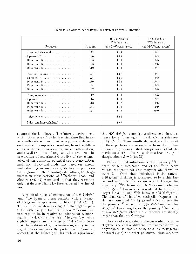
Table 9. Calculated Initial Range for Di�erent Polymeric Materials
Initial range of Initial range of56Fe beam at 20Ne beam at
Polymers �, g/cm3 605 MeV/amu, g/cm2 425 MeV/amu, g/cm2
Pure polyetherimide . . . . . . . . 1.27 13.8 19.2
5 percent B . . . . . . . . . . . 1.30 13.9 19.3
10 percent B . . . . . . . . . . . 1.33 14.0 19.5
15 percent B . . . . . . . . . . . 1.36 14.0 19.6
20 percent B . . . . . . . . . . . 1.40 14.1 19.7
Pure polysulfone . . . . . . . . . 1.24 13.7 19.1
5 percent B . . . . . . . . . . . 1.27 13.8 19.2
10 percent B . . . . . . . . . . . 1.30 13.9 19.3
15 percent B . . . . . . . . . . . 1.34 14.0 19.5
20 percent B . . . . . . . . . . . 1.37 14.0 19.5
Pure polyimide . . . . . . . . . . 1.42 14.1 19.6
5 percent B . . . . . . . . . . . 1.45 14.1 19.7
10 percent B . . . . . . . . . . . 1.48 14.2 19.8
15 percent B . . . . . . . . . . . 1.51 14.3 19.9
20 percent B . . . . . . . . . . . 1.54 14.4 20.0
Polyethylene . . . . . . . . . . . 12.2
Poly(tetra uoroethylene) . . . . . . 15.7
square of the ion charge. The internal environmentwithin the spacecraft or habitat structure that inter-acts with onboard personnel or equipment dependson the shield composition resulting from the di�er-ences in atomic cross sections, nuclear attenuation,and the distribution of fragmentation products. Inpreparation of experimental studies of the attenu-ation of ion beams in potential space constructionmaterials, theoretical predictions based on currentunderstanding are used as a guide to an experimen-tal program. In the following calculations, the frag-mentation cross sections of Silberberg, Tsao, andShapiro (ref. 42) were used in that they were theonly database available for these codes at the time ofuse.
The initial range of penetration of a 605-MeV/amu 56Fe beam in lunar regolith with a densityof 1.5 g/cm3 is approximately 10 cm (15.4 g/cm2).The calculations show (see �g. 20) that lighter par-ticles with energies lower than 605 MeV/amu arepredicted to be in relative abundance for a lunar-regolith brick with a thickness of 16 g/cm2, which isslightly larger than the range of 15.4 g/cm2. Notethat the addition of hydrogen-bearing epoxy to theregolith brick increases the protection. Figure 21shows that the lighter particles with energies lower
than 605 MeV/amu are also predicted to be in abun-dance for a lunar-regolith brick with a thicknessof 18 g/cm2. These results demonstrate that mostof these particles are secondaries from the nuclearinteraction processes. Most conspicuous is that themaximum contribution comes from a broad range ofcharges above Z = 3 (for Li).
The calculated initial ranges of the primary 56Febeam at 605 MeV/amu and of the 20Ne beamat 425 MeV/amu for each polymer are shown intable 9. From these calculated initial ranges,a 10 g/cm2 thickness is considered to be a thin tar-get and an 18 g/cm2 thickness is a thick target fora primary 56Fe beam at 605 MeV/amu, whereasan 18 g/cm2 thickness is considered to be a thintarget for a primary 20Ne beam at 425 MeV/amu.The uences of identi�ed projectile fragment nu-clei are compared for 18 g/cm2 thick targets forthe primary 56Fe beam at 605 MeV/amu and for20 g/cm2 thick targets for the primary 20Ne beamat 425 MeV/amu where the thicknesses are slightlylarger than the initial ranges.
Because of the greater hydrogen content of poly-ethylene, the charge di�erence in fragmentation bypolyethylene is smaller than that by poly(tetra- uoroethylene) and other polymers. However, thin
16

polyethylene enhances the high Z fragment. Thesecond fragmentation event occurs more often in thethicker polyethylene shields because the nuclear at-tenuation rate is higher in polyethylene than in theother polymer shields. The second charge di�erencegreatly reduces the uence for a polyethylene shield(ref. 30).
The lighter material such as polyethylene en-hances the high-energy heavy ion uence relativeto poly(tetra uoroethylene) for thin shields (see�g. 22) and reduces the uence more e�cientlythan poly(tetra uoroethylene) and other polymersfor thick shields (see �g. 23). In fact, the succes-sion of curves in �gures 22 and 23 is governed by theamount of hydrogen per unit mass, and polyethy-lene is the most abundant in hydrogen. Studies onthe e�ect of shield composition on LET distributionat several depths has already shown that polyethy-lene is the most e�ective high-LET degrader beyond5 g/cm2 at solar minimum (ref. 30). Again, polyethy-lene is the most e�ective shield material amongthese polymers beyond an 18 g/cm2 thickness for theprimary 56Fe beam at 605 MeV/amu.
The addition of boron (B) powder to a materialallows the material to absorb low-energy neutronswithout any degradation in glass transition temper-ature or Young's modulus in the polymeric materials(ref. 40). The uence for a polyetherimide containingvarious weight fractions of boron is shown in �gure 24for a primary 56Fe beam at 605 MeV/amu and in �g-ure 25 for a primary 20Ne beam at 425 MeV/amu.These results show no signi�cant di�erence for var-ious weight fractions of boron. For thick shields,the pure polymer shows a slightly better attenua-tion of fragments at Z > 3 than a composite contain-ing 20 percent boron. As the fraction of B increasesfrom 5 to 20 percent by weight, both the densityand the initial range increase because boron has ahigher atomic number (Z) than hydrogen. Similarresults are obtained for the polysulfone and the poly-imide. Hence, pure polymers with slightly shorterinitial ranges are expected to attenuate fragments atZ > 3 better than materials containing any fractionof boron. The laboratory code used does not includelight fragments of Z < 3 in any realistic way becausea greater knowledge of nuclear fragmentation pro-cesses and a corresponding transport theory are re-quired for these fragments.
A target with a high percentage of lighter atomssuch as hydrogen would, therefore, be an e�ectiveshield material for thick shields, whereas a targetwith a heavier atom composition might yet proveto be more e�ective in thin shields for energetic ionbeams. Pilot experiments to validate these theoreti-
cal results have been performed, but data reductionis not yet complete.
Concluding Remarks
Radiation risks to astronauts depend on themicroscopic uctuations of energy-absorption eventsin speci�c tissues. These uctuations depend notonly on the space environment but also on the modi�-cations of that environment by the shielding of the as-tronaut's surrounding structures and the attenuationcharacteristics of the astronaut's body. The e�ects ofattenuation of the shield and body depend on the tis-sue biological response to the microscopic uctuatione�ects. A great deal of uncertainty presently existsin estimating astronaut risk because of uncertaintyin the nuclear properties and risk models. Clearly,these uncertainties must be reduced before the shielddesign can be made.
Using current estimates for nuclear cross sectionshas shown that the high charge and energy (HZE)ions in space pose a signi�cant hazard to biologicalsystems and that the linear energy transfer (LET)distribution above about 10 keV/�m is an impor-tant indicator of biological damage. Furthermore,the LET distribution is a function of shield compo-sition, even with materials of the same areal den-sity. Shinn et al. suggested that polyethylene with itsshort nuclear absorption lengths is an e�ective shieldmaterial in spite of the favoring of massive projectilefragments, and this has been demonstrated herein formonoenergetic ion beams. The establishment of a re-liable nuclear fragmentation database, astronaut riskmethodology, suitable polymer materials, and struc-tural design methods remain as critical issues in thelong-term exposure to space radiations.
NASALangley Research Center
Hampton, VA 23681-0001August 29, 1994
References
1. Wilson, John W.: Environmental Geophysics and SPS
Shielding. Workshop on the Radiation Environment of
the Satellite Power System, Walter Schimmerling and
Stanley B. Curtis, eds., LBL-8581 (Contract W-7405-
ENG-48), Univ. of California, Sept. 15, 1978, pp. 33{116.
2. Wilson, JohnW.; Townsend, LawrenceW.; Schimmerling,
Walter; Khandelwal, Govind S.; Khan, Ferdous; Nealy,
JohnE.; Cucinotta, Francis A.; Simonsen, Lisa C.; Shinn,
Judy L.; and Norbury, John W.: Transport Methods and
Interactions for Space Radiations. NASARP-1257, 1991.
3. Guidance on Radiation Received in Space Activities.
NCRP Rep. No. 98, National Council on Radiation
Protection andMeasurements, 1989.
17

4. Schaefer, Hermann J.: Evaluation of Present-DayKnowl-
edge of Cosmic Radiation at Extreme Altitude in Terms
of the Hazard toHealth. J. Aviation Med., vol. 21, no. 5,
Oct. 1950, pp. 375{394, 418.
5. Tobias, Cornelius A.: RadiationHazards inHighAltitude
Aviation. J. Aviation Med., vol. 23, no. 4, Aug. 1952,
pp. 345{372.
6. Schimmerling, Walter: Radiobiological Problems in
Space|An Overview. Radiat. & Environ. Biophys.,
vol. 31, 1992, pp. 197{203.
7. 1990 Recommendations of the International Commission
on Radiological Protection. ICRP Publ. 60, Pergamon
Press Inc., 1991.
8. Committee on the Biological E�ects of Ionizing Radia-
tions: Health E�ects of Exposure toLowLevels of Ionizing
Radiation. BEIRV, National Academy Press, 1990.
9. Aghamohammadi, S. Z.; Goodhead, D. T.; and Savage,
J. R.: Induction of Sister Chromatid Exchanges (SCE)
in GO Lymphocytes by Plutonium-238 Alpha-Particles.
Int. J. Radiat. Biol. &Relat. Stud. Phys., Chem. &Med.,
vol. 53, no. 6, June 1988, pp. 909{915.
10. Kadhim, M. A.; Macdonald, D. A.; Goodhead, D. T.;
Lorimore, S. A.; Marsden, S. J.; and Wright, E. G.:
Transmission of Chromosomal Instability After Pluto-
nium �-Particle Irradiation. Nature, vol. 355, no. 6362,
Feb. 20, 1992, pp. 738{740.
11. Kraft, G.: Radiobiological E�ects of Very Heavy Ions:
Inactivation, Induction of Chromosome Aberrations and
Strand Breaks. Nucl. Sci. Appl., sect. A, vol. 3, no. 1,
1987, pp. 1{28.
12. Wilson, John W.; Wood, J. S.; Shinn, Judy L.;
Cucinotta, Francis A.; and Nealy, John E.: A Proposed
Performance Index for Galactic Cosmic Ray Shielding
Materials. NASA TM-4444, 1993.
13. Wilson, John W.; Cucinotta, F. A.; and Shinn, J. L.:
Cell Kinetics and Track Structure. Biological E�ects
and Physics of Solar and Galactic Cosmic Radiation,
Part B, Charles E. Swenberg, Gerda Horneck, and E. G.
Stassinopoulos, eds., PlenumPress, 1993, pp. 295{338.
14. Yang, T. C.; and Tobias, C. A.: Neoplastic Cell Trans-
formation by Energetic Heavy Ions and Its Modi�cation
With Chemical Agents. Adv. Space Res., vol. 4, no. 10,
1984, pp. 207{218.
15. Yang, Tracy Chui-Hsu; Craise, Laurie M.; Mei,
Man-Tong; and Tobias, Cornelius A.: Neoplastic Cell
Transformation by High-LET Radiation: Molecular
Mechanisms. Adv. Space Res., vol. 9, no. 10, 1989,
pp. (10)131{(10)140.
16. Bond, V. P.; Varma, M. N.; and Sondhaus, C. A.: The
RBE Concept, Its Inadequacies and a Suggested Re-
placement. Mechanisms of Radiation Interaction With
DNA: Potential Implications for Radiation Protection,
CONF-870163, U.S. Dep. of Energy, 1988, pp. 31{38.
17. Xapsos, M. A.; Burke, E. A.; Shapiro, P.; and Summers,
G. P.: Energy Deposition and Ionization Fluctuations
Induced by Ions inSmall Sites|AnAnalytical Approach.
Radiat. Res., vol. 137, no. 2, Feb. 1994, pp. 152{161.
18. Todd, Paul: Unique Biological Aspects of Radiation
Hazards|An Overview. Adv. Space Res., vol. 3, no. 8,
1983, pp. 187{194.
19. The Quality Factor in Radiation Protection. ICRU
Rep. 40, International Commission on Radiation Units
andMeasurements, Apr. 4, 1986.
20. Scott, B. R.: Methodologies for Predicting the Expected
Combined Stochastic Radiobiological E�ects of Di�erent
Ionizing Radiations and SomeApplications. Radiat. Res.,
vol. 98, no. 1, Apr. 1984, pp. 182{197.
21. Burns, F. J.; and Albert, R. E.: Dose-Response for
Radiation-Induced Cancer in Rat Skin. Radiation
Carcinogenesis and DNA Alterations, F. J. Burns,
A. C. Upton, and G. Silini, eds., Plenum Press, 1986,
pp. 51{70.
22. Thomson, John F.; and Grahn, Douglas: Life Shortening
inMice Exposed toFissionNeutrons and Rays. VII. Ef-
fects of60Once-WeeklyExposures. Radiat. Res., vol. 115,
1988, pp. 347{360.
23. Wilson, John W.; Nealy, John E.; Schimmerling, Walter;
Cucinotta, Francis A.; and Wood, James S.: E�ects of
Radiobiological Uncertainty on Vehicle and Habitat Shield
Design for Missions to the Moon and Mars. NASA
TP-3312, 1993.
24. Katz, R.; Ackerson, B.; Homayoonfar, M.; and Sharma,
S. C.: Inactivation of Cells by Heavy Ion Bombardment.
Radiat. Res., vol. 47, 1971, pp. 402{425.
25. Wilson, John W.; and Cucinotta, Francis A.: Cellular
Repair/Misrepair Track Model. NASA TP-3124, 1991.
26. Wilson, John W.; Cucinotta, Francis A.; and Shinn,
Judy L.: Multiple Lesion Track Structure Model. NASA
TP-3185, 1992.
27. Wilson, John W.; Chun, Sang Y.; Badavi, Forooz F.;
Townsend, LawrenceW.; andLamkin, StanleyL.: HZETRN:
A Heavy Ion/Nucleon Transport Code for Space Radia-
tions. NASA TP-3146, 1991.
28. Townsend, L. W.; Cucinotta, F. A.; and Wilson, J. W.:
HZE Reactions and Data-Base Development. Biologi-
cal E�ects and Physics of Solar and Galactic Cosmic
Radiation, 1993, pp. 787{809.
29. Shinn, Judy L.; John, Sarah; Tripathi, Ram K.;
Wilson, JohnW.; Townsend, LawrenceW.; and Norbury,
JohnW.: Fully Energy-Dependent HZETRN (AGalactic
Cosmic-Ray Transport Code). NASATP-3243, 1992.
30. Shinn, Judy L.; Townsend, Lawrence W.; and Wilson,
JohnW.: GalacticCosmicRayRadiationLevels inSpace-
craft on Interplanetary Missions. Book of Abstracts|
TheWorld SpaceCongress, 43rdCongress of the Interna-
tional Astronautical Federation and 29th PlenaryMeeting
18

of the Committee on Space Research, International As-
tronautical Federation and Committee on Space Research,
Aug.{Sept. 1992, pp. 567{568.
31. Wilson, John W.: Composite Particle Reaction Theory.
Ph.D. Diss., College of WilliamandMary, June 1975.
32. Wilson, J. W.; and Townsend, L. W.: An Optical Model
for Composite Nuclear Scattering. Canadian J. Phys.,
vol. 59, no. 11, 1981, pp. 1569{1576.
33. Rudstam,G.: SystematicsofSpallationYields. Zeitschrift
fur Naturforschung, vol. 21a, no. 7, July 1966,
pp. 1027{1041.
34. Wilson, John W.; Townsend, LawrenceW.; and Badavi,
F. F.: A Semiempirical Nuclear Fragmentation Model.
Nucl. Instrum. & Methods Phys. Res., vol. B18, no. 3,
Feb. 1987, pp. 225{231.
35. Long, Edward R., Jr.: Electron and Proton Absorp-
tion Calculations for a Graphite/EpoxyCompositeModel.
NASA TP-1568, 1979.
36. Nealy, John E.; Wilson, John W.; and Townsend,
Lawrence W.: Solar-Flare Shielding With Regolith at a
Lunar-Base Site. NASATP-2869, 1988.
37. Johnson, R. N.; Farnham, A. G.; Clendinning, R. A.;
Hale, W. F.; and Merriam, C. N.: Poly(aryl Ethers)
by Nucleophilic Aromatic Substitution. I. Synthesis and
Properties. J. Polym. Sci.: pt. A-1, vol. 5, 1967,
pp. 2375{2398.
38. White, D. M.; Takekoshi, T.; Williams, F. J.; Relles,
H. M.; Donahue, P. E.; Klopfer, H. J.; Loucks, G. R.;
Manello, J. S.; Mathews, R. O.; and Schluenz, R. W.:
Polyetherimides Via Nitro-Displacement Polymerization:
Monomer Synthesis and 13C-NMRAnalysis of Monomers
and Polymers. J. Polym. Sci.: Polymer Chemistry Ed.,
vol. 19, 1981, pp. 1635{1658.
39. Sroog, C. E.; Endrey, A. L.; Abramo, S. V.; Berr, C. E.;
Edwards, W. M.; and Olivier, K. L.: Aromatic Polypy-
romellitimides FromAromaticPolyamicAcids. J. Polym.
Sci., pt. A, vol. 3, no. 4, Apr. 1965, pp. 1373{1390.
40. Kraus, W. B.; Glasgow, M. B.; Kim, M. Y.;
Olmeijer, D. L.; Kiefer, R. L.; Orwoll, R. A.; and
Thibeault, S. A.: Boron Containing Polymers for Radi-
ation Shielding. Polym. Prep., vol. 34, no. 1, Mar. 1993,
pp. 592{593.
41. Wilson, John W.; Lamkin, Stanley L.; Farhat,
Hamidullah; Ganapol, Barry D.; and Townsend,
LawrenceW.: A Hierarchy of Transport Approximations
for High Energy Heavy (HZE) Ions. NASA TM-4118,
1989.
42. Silberberg, R.; Tsao, C. H.; and Shapiro, M. M.: Semi-
empirical Cross Sections, and Applications to Nuclear
InteractionsofCosmicRays. Spallation NuclearReactions
andTheirApplications, B. S. P. ShenandM.Merker, eds.,
D. Reidel Publ. Co., 1976, pp. 49{81.
19

10 -3
10 -2
10-1
10 0
10 1
10 2
103
10 -1 10 0 10 1 10 2 10 3 10 4
1H
16 O56 Fe
L, keV/µm
Mea
n hi
t siz
e, k
eV
10 -5
10 -4
10 -3
10 -2
10 -1
100
10 -1 10 0 10 1 10 2 10 3 10 4
1 H16
O56 Fe
L, keV/µm
Fra
ctio
n of
sit
es h
it
(a) 0.1-�m site size.
Frac
tion
of s
ites
hit
10 -3
10 -2
10-1
10 0
101
10 2
10 3
10 4
10 -1 10 0 10 1 10 2 10 3 10 4
4 He26 Mg
56 Fe
L, keV/µm L, keV/µm
Mea
n hi
t siz
e, k
eV
10 -5
10 -4
10 -3
10 -2
10 -1
10 0
10 -1 10 0 10 1 10 2 10 3 10 4
4 He26 Mg56
Fe
(b) 0.5-�m site size.
30
25
20
15
10
10–1 100
Q
L, keV/µm
5
103102101 1040
(c) Quality factor.
Figure 1. Fraction of hit sites, hit size, and quality factor as a function of LET (L).
20

Lowdoserate
Highdoserate
Log Neutron dose
Log
RB
E
Figure 2. Schematic of RBE for neutron exposure at
di�erent dose rates.
Fe(Z = 26)
U(Z = 92)
Trackcore50 µm
Trackcore
≥10 cells
LET > 200 keV/µmD > 0.25 Gy
10 µm
Trackcore
Deltarays
Figure 3. Single-particle e�ect proposed by Todd
(ref. 18).
2460 Single Fractions
Single
24
60
200
100
0 2 4Dose, Gy
Lif
e sh
orte
ning
, day
s
Fission neutronsγ-rays
Fractions
Figure 4. Dose protraction e�ects in mice with repair
for low LET ( -ray) and enhancement at high
LET for �ssion neutron (ref. 22).
1992 1997Year
2010
Factor of 2Factor of 1.25
Factor of 1030
20
10
0Exc
ess
canc
er p
roba
bilit
y, p
erce
nt
Figure 5. Estimates for current and projected risk
uncertainties in NASA Space Radiation Program.
21

102
101
100
10–1
Surv
ival
, per
cent
0 2 4 6 8 10Dose, Gy
Katz modelPresent modelDelayed platingImmediate plating
102
101
100
10–1
Surv
ival
, per
cent
0 2 4 6 8 10Dose, Gy
(a) 20Ne with 425 MeV/amu�1 at 32 keV/�m. (b) 28Si with 670 MeV/amu at 50 keV/�m.
102
101
100
10–1
Surv
ival
, per
cent
0 2 4 6 8 10Dose, Gy
102
101
100
10–1
Surv
ival
, per
cent
0 2 4 6 8 10Dose, Gy
(c) 28Si with 320 MeV/amu at 82 keV/�m�1. (d) 56Fe with 600 MeV/amu at 190 keV/�m.
Figure 6. Cell survival of C3H10T1/2 for delayed plating and immediate plating.
22

-4
-3
-2
-1
0
1
2
3
4
5
10
10
10
10
10
10
10
10
10
10
Ran
ge, g
/cm
2
-4
-3
-2
-1
0
1
2
3
4
5
10
10
10
10
10
10
10
10
10
10
10 -2 10 -1 10 0 10 1 10 2 10 3 10 4 10 510 -2 10 -1 10 0 10 1 10 2 10 3 10 4 10 5
Energy, MeV/nucleon
1251422
Z
Ran
ge, g
/cm
2
Energy, MeV/nucleon
(a) Liquid hydrogen. (b) Water.
10 -2 10 -1 10 0 10 1 10 2 10 3 10 4 10 5 10 -2 10 -1 10 0 10 1 10 2 10 3 10 4 10 5-4
-3
-2
-1
0
1
2
3
4
5
10
10
10
10
10
10
10
10
10
10
-4
-3
-2
-1
0
1
2
3
4
5
10
10
10
10
10
10
10
10
10
10
Ran
ge, g
/cm
2
Ran
ge, g
/cm
2
Energy, MeV/nucleon Energy, MeV/nucleon
(c) Aluminum. (d) Lead.
Figure 7. Stopping ranges of selected ions in four diverse materials.
23

Z p
2620862
1
10 -3
10 -2
10 -1
100
σ abs, c
m2 /g
Z p
262086
2
1
10 1 10 2 10 3 10 4
Energy, MeV/nucleon10 1 10 2 10 3 10 410 -3
10 -2
10 -1
100
σ abs, c
m2 /g
Energy, MeV/nucleon
(a) Liquid hydrogen. (b) Water.
10 1 10 2 10 3 10 4
Z p
2620862
1
26208621
Z p
10 1 10 2 10 3 10 410 -3
10 -2
10 -1
100
10 -3
10 -2
10 -1
100
σ abs, c
m2 /g
σ abs, c
m2 /g
Energy, MeV/nucleon Energy, MeV/nucleon
(c) Aluminum. (d) Lead.
Figure 8. Nuclear absorption cross sections per unit mass for selected ions in four diverse materials.
24

(a) Liquid hydrogen. (b) Water.
(c) Aluminum. (d) Lead.
Figure 9. Fragment production cross sections per unit mass for ions transported in the shielding code in four
diverse materials.
25

101
102
103
104
105
106
107
108
109
L
, c
m–2
dN dL
Free space
5
15
30
101
102
103
104
105
106
107
108
109
10-1 100 101 102 103
L, keV/µm
10-1 100 101 102 103
x, g/cm2
L
, c
m–2
dN dL
L, keV/µm
(a) Liquid hydrogen. (b) Water.
10-1 100 101 102 103101
102
103
104
105
106
107
108
109
101
102
103
104
105
106
107
108
109
10-1 100 101 102 103
L
, c
m–2
dN dL
L
, c
m–2
dN dL
L, keV/µm L, keV/µm
(c) Aluminum. (d) Lead.
Figure 10. Annual transmitted GCR di�erential LET spectrum in four diverse shield materials.
26
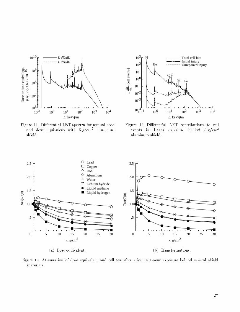
L dD/dLL dH/dL
10-1 100 101 102 103 104
1010
109
108
107
106
L, keV/µm
103
104
Total cell hitsInitial injuryUnrepaired injury102
101
100
10-1
10-2
10-3
10-410310210110010-1
H
He
CNO
Si Fe
L
(
cell
even
ts)
dn dL
Dos
e or
dos
e eq
uiva
lent
,(G
y, S
v)/3
.68
× 10
–10
L, keV/µm
Figure 11. Di�erential LET spectra for annual dose
and dose equivalent with 5-g/cm2 aluminum
shield.
Figure 12. Di�erential LET contributions to cell
events in 1-year exposure behind 5-g/cm2
aluminum shield.
LeadCopperIronAluminumWaterLithium hydrideLiquid methaneLiquid hydrogen
0
.5
1.0
1.5
2.0
2.5
5 10 15 20 25 30
H(x
)/H
(0)
5 10 15 20 25 30
T(x
)/T
(0)
0
.5
1.0
1.5
2.0
2.5
x, g/cm2x, g/cm2
(a) Dose equivalent. (b) Transformations.
Figure 13. Attenuation of dose equivalent and cell transformation in 1-year exposure behind several shield
materials.
27

0
.5
1.0
1.5
2.0
2.5
.5 1.0 1.5 2.0
LeadAluminumWaterLiquid hydrogen
T(x
)/T
(0)
H(x)/H(0)
Figure 14. Correlation of cell transformation and dose equivalent behind several shield materials.
302010
x, g/cm20
2.5
2.0
1.5
1.0
.5
TA
l(x)/
Tm
(x)
Liquid methaneLithium hydrideWaterRegolith/10% epoxyRegolithIronCopperLead
302010
x, g/cm20
TA
l(x)/
Tm
(x)
Liquid hydrogenLithium hydrideLead
2
4
6
8
10
12
14
Figure 15. Cell-transformation ratio as a function of areal density relative to aluminum standard.
28

302010
x, g/cm20
2.5
2.0
1.5
1.0
.5
T(x
)/T
(0)
Peripheral collisionNominalCentral collision
3020100
2.5
2.0
1.5
1.0
.5T
(x)/
T(0
)
x, g/cm2
(a) Liquid hydrogen. (b) Water.
302010
x, g/cm20
2.5
2.0
1.5
1.0
.5
T(x
)/T
(0)
3020100
2.5
2.0
1.5
1.0
.5
T(x
)/T
(0)
x, g/cm2
(c) Aluminum. (d) Lead.
Figure 16. E�ects of physical limits on several shield types.
29

10–9
10–7
10–5
10–3
10–1
1 6 11 16 21 26Z
101
Tz(x
)/T
tot(0
)
1
2
5
10
15
20
30
Free space
x, g/cm2
31
(a) Liquid hydrogen.
10–5
10–4
10–3
10–2
10–1
1 6 11 16 21 26Z
100
Tz(x
)/T
tot(0
)
31
(b) Water.
Figure 17. Contributions of biological change from each charge group of environment.
30

10–4
10–3
10–2
10–1
1 6 11 16 21 26Z
100
Tz(x
)/T
tot(0
)
31
(c) Aluminum.
12510152030
Free space
x, g/cm2
10–5
10–4
10–3
10–2
10–1
1 6 11 16 21 26Z
100
Tz(x
)/T
tot(0
)
31
(d) Lead.
Figure 17. Concluded.
31

CHS
O
O
NNCH2
OH
CH2
NCH
S
O
O
N NCH2
OH
CH2
CH2
N
CHS
O
O
NNCH2
OH
CH2CHS
O
O
N NCH2
OH
CH2
Figure 18. Tetraglycidyl 4; 40 diamino diphenyl methane epoxy cured with diamino diphenyl sulfone.
CO
CH3
CH3
n
C
O
NO
O
C
C
O
N
O
C
(a) Polyetherimide.
C O S O
CH3
CH3
O
O
n
C
O
N
O
C
C
O
N
O
CO
n
(b) Polysulfone. (c) Polyimide.
Figure 19. Repeat units of three polymers studied.
32

0 5 10 15 20 25 30
.04
.03
.02
.01
Flue
nce,
par
ticle
s/cm
2
Projectile fragment charge
Pure lunar soil model
Regolith/10% epoxy brick
Regolith/20% epoxy brick
Pure epoxy
Figure 20. Attenuation of 605 MeV/amu iron beams in lunar construction materials of 16 g/cm2 thickness.
0 5 10 15 20 25 30
.04
.03
.02
.01
Flue
nce,
par
ticle
s/cm
2
Projectile fragment charge
Pure lunar soil model
Regolith/10% epoxy brick
Regolith/20% epoxy brick
Pure epoxy
Figure 21. Attenuation of 605 MeV/amu iron beams in lunar construction materials of 18 g/cm2 thickness.
33

0 5 10 15 20 25 30
.08
.06
.04
.02
Flue
nce,
par
ticle
s/cm
2
Projectile fragment charge
Polyethylene
Poly(tetrafluoroethylene)
Polyetherimide
Polysulfone
Polyimide
Figure 22. Attenuation of 605 MeV/amu iron beams in various polymer construction materials of 5 g/cm2
thickness.
0 5 10 15 20 25 30
.016
.012
.008
.004
Flue
nce,
par
ticle
s/cm
2
Projectile fragment charge
Polyethylene
Poly(tetrafluoroethylene)
Polyetherimide
Polysulfone
Polyimide
Figure 23. Attenuation of 605 MeV/amu iron beams in various polymer construction materials of 18 g/cm2
thickness.
34

20% B
15% B
10% B
5% B
Pure polyetherimide
.0100
.0075
.0050
.0025
0 5 10 15 20 25 30
Projectile fragment charge
Flue
nce,
par
ticle
s/cm
2
Figure 24. Attenuation of 605 MeV/amu iron beams in various weight fractions of boron-containing
polyetherimide of 18 g/cm2 thickness.
20% B
15% B
10% B
5% B
Pure polyetherimide
.07
.06
.05
.04
.03
.02
.01
03 4 5 6 7 8 9 10
Projectile fragment charge
Flue
nce,
par
ticle
s/cm
2
Figure 25. Attenuation of 425 MeV/amu neon beams in various weight fractions of boron-containing
polyetherimide of 20 g/cm2 thickness.
35

REPORT DOCUMENTATION PAGEForm Approved
OMB No. 0704-0188
Public reporting burden for this collection of information is estimated to average 1 hour per response, including the time for reviewing instructions, searching existing data sources,gathering and maintaining the data needed, and completing and reviewing the collection of information. Send comments regarding this burden estimate or any other aspect of thiscollection of information, including suggestions for reducing this burden, to Washington Headquarters Services, Directorate for Information Operations and Reports, 1215 Je�ersonDavis Highway, Suite 1204, Arlington, VA 22202-4302, and to the O�ce of Management and Budget, Paperwork Reduction Project (0704-0188), Washington, DC 20503.
1. AGENCY USE ONLY(Leave blank) 2. REPORT DATE 3. REPORT TYPE AND DATES COVERED
November 1994 Technical Paper
4. TITLE AND SUBTITLE
Performance Study of Galactic Cosmic Ray Shield Materials
6. AUTHOR(S)
Myung-Hee Y. Kim, John W. Wilson, Sheila A. Thibeault,
John E. Nealy, Francis F. Badavi, and Richard L. Kiefer
7. PERFORMING ORGANIZATION NAME(S) AND ADDRESS(ES)
NASA Langley Research Center
Hampton, VA 23681-0001
9. SPONSORING/MONITORING AGENCY NAME(S) AND ADDRESS(ES)
National Aeronautics and Space Administration
Washington, DC 20546-0001
5. FUNDING NUMBERS
WU 199-45-16-11
8. PERFORMING ORGANIZATION
REPORT NUMBER
L-17389
10. SPONSORING/MONITORING
AGENCY REPORT NUMBER
NASA TP-3473
11. SUPPLEMENTARY NOTES
Kim and Kiefer: College of William and Mary, Williamsburg, VA; Wilson, Thibeault, and Nealy: LangleyResearch Center, Hampton, VA; Badavi: Christopher Newport University, Newport News, VA.
12a. DISTRIBUTION/AVAILABILITY STATEMENT 12b. DISTRIBUTION CODE
Unclassi�ed{Unlimited
Subject Category 93
13. ABSTRACT (Maximum 200 words)
The space program is faced with two di�cult radiation protection issues for future long-term operations. First,retro�t of shield material or conservatism in shield design is prohibitively expensive and often impossible.Second, shielding from the cosmic heavy ions is faced with limited knowledge on the physical properties andbiological responses of these radiations. The current status of space shielding technology and its impact onradiation health is discussed herein in terms of conventional protection practice and a test biological responsemodel. The impact of biological response on the selection of optimum materials for cosmic ray shielding ispresented in terms of the transmission characteristics of the shield material. Although the systematics ofnuclear cross sections are able to demonstrate the relation of exposure risk to shield-material composition, thecurrent uncertainty in nuclear cross sections will not allow an accurate evaluation of risk reduction. This paperpresents a theoretical study of risk-related factors and a pilot experiment to study the e�ectiveness of choiceof shield materials to reduce the risk in space operations.
14. SUBJECT TERMS 15. NUMBER OF PAGES
Galactic cosmic rays; Shielding; Nucleon; Biology 36
16. PRICE CODE
A0317. SECURITY CLASSIFICATION 18. SECURITY CLASSIFICATION 19. SECURITY CLASSIFICATION 20. LIMITATION
OF REPORT OF THIS PAGE OF ABSTRACT OF ABSTRACT
Unclassi�ed Unclassi�ed Unclassi�ed
NSN 7540-01-280-5500 Standard Form 298(Rev. 2-89)Prescribed by ANSI Std. Z39-18298-102