Perforator preservation technologies (PPT) based on a new ...
9
RESEARCH Open Access Perforator preservation technologies (PPT) based on a new neuro-interventional classification in endovascular treatment of perforator involving aneurysms (piANs) Chen Li 1† , Ao-Fei Liu 1† , Han-Cheng Qiu 1† , Xianli Lv 2 , Ji Zhou 1 , Yi-Qun Zhang 1 , Jin Lv 1 , Ying-Ying Zhang 1 , Sushan Hu 1 , Fang Liu 1 , Yun-e Liu 1 , Min Jin 1* and Wei-Jian Jiang 1* Abstract Background: Treatment of perforator involving aneurysm (piAN) remains a challenge to open and endovascular neurosurgeons. Our aim is to demonstrate a primary outcome of endovascular therapy for piANs with the use of perforator preservation technologies (PPT) based on a new neuro-interventional classification. Methods: The piANs were classified into type I: aneurysm really arises from perforating artery, type II: saccular aneurysm involves perforating arteries arising from its neck (IIa) or dome (IIb), and type III: fusiform aneurysm involves perforating artery. Stent protection technology of PPT was applied in type I and III aneurysms, and coil- basket protection technology in type II aneurysms. An immediate outcome of aneurysmal obliteration after treatment was evaluated (satisfactory obliteration: the saccular aneurysm body is densely embolized (I), leaving a gap in the neck (IIa) or dome (IIb) where the perforating artery arising; fusiform aneurysm is repaired and has a smooth inner wall), and successful perforating artery preservation was defined as keeping the good antegrade flow of those perforators on postoperative angiography. The periprocedural complication was closely monitored, and clinical and angiographic follow-ups were performed. Results: Six consecutive piANs (2 ruptured and 4 unruptured; 1 type I, 2 type IIa, 2 type IIb, and 1 type III) in 6 patients (aged from 43 to 66 years; 3 males) underwent endovascular therapy between November 2017 and July 2019. The immediate angiography after treatment showed 6 aneurysms obtained satisfactory obliteration, and all of their perforating arteries were successfully preserved. During clinical follow-up of 13–50 months, no ischemic or hemorrhagic event of the brain occurred in the 6 patients, but has one who developed ischemic event in the territory of involving perforators 4 h after operation and completely resolved within 24 h. Follow-up angiography at 3 to 10M showed patency of the parent artery and perforating arteries of treated aneurysms, with no aneurysmal recurrence. (Continued on next page) © The Author(s). 2021 Open Access This article is licensed under a Creative Commons Attribution 4.0 International License, which permits use, sharing, adaptation, distribution and reproduction in any medium or format, as long as you give appropriate credit to the original author(s) and the source, provide a link to the Creative Commons licence, and indicate if changes were made. The images or other third party material in this article are included in the article's Creative Commons licence, unless indicated otherwise in a credit line to the material. If material is not included in the article's Creative Commons licence and your intended use is not permitted by statutory regulation or exceeds the permitted use, you will need to obtain permission directly from the copyright holder. To view a copy of this licence, visit http://creativecommons.org/licenses/by/4.0/. The Creative Commons Public Domain Dedication waiver (http://creativecommons.org/publicdomain/zero/1.0/) applies to the data made available in this article, unless otherwise stated in a credit line to the data. * Correspondence: [email protected]; [email protected] † Chen Li, Ao-Fei Liu, and Han-Cheng Qiu contributed equally to this work. 1 New Era Stroke Care and Research Institute, PLA Rocket Force Characteristic Medical Center, 18 Xinjiekouwai Street, Beijing 100088, China Full list of author information is available at the end of the article Li et al. Chinese Neurosurgical Journal (2021) 7:26 https://doi.org/10.1186/s41016-021-00243-3 CHINESE MEDICAL ASSOCIATION 中华医学会神经外科学分会 CHINESE NEUROSURGICAL SOCIETY
Transcript of Perforator preservation technologies (PPT) based on a new ...

RESEARCH Open Access
Perforator preservation technologies (PPT)based on a new neuro-interventionalclassification in endovascular treatment ofperforator involving aneurysms (piANs)Chen Li1†, Ao-Fei Liu1†, Han-Cheng Qiu1†, Xianli Lv2, Ji Zhou1, Yi-Qun Zhang1, Jin Lv1, Ying-Ying Zhang1,Sushan Hu1, Fang Liu1, Yun-e Liu1, Min Jin1* and Wei-Jian Jiang1*
Abstract
Background: Treatment of perforator involving aneurysm (piAN) remains a challenge to open and endovascularneurosurgeons. Our aim is to demonstrate a primary outcome of endovascular therapy for piANs with the use ofperforator preservation technologies (PPT) based on a new neuro-interventional classification.
Methods: The piANs were classified into type I: aneurysm really arises from perforating artery, type II: saccularaneurysm involves perforating arteries arising from its neck (IIa) or dome (IIb), and type III: fusiform aneurysminvolves perforating artery. Stent protection technology of PPT was applied in type I and III aneurysms, and coil-basket protection technology in type II aneurysms. An immediate outcome of aneurysmal obliteration aftertreatment was evaluated (satisfactory obliteration: the saccular aneurysm body is densely embolized (I), leaving agap in the neck (IIa) or dome (IIb) where the perforating artery arising; fusiform aneurysm is repaired and has asmooth inner wall), and successful perforating artery preservation was defined as keeping the good antegrade flowof those perforators on postoperative angiography. The periprocedural complication was closely monitored, andclinical and angiographic follow-ups were performed.
Results: Six consecutive piANs (2 ruptured and 4 unruptured; 1 type I, 2 type IIa, 2 type IIb, and 1 type III) in 6patients (aged from 43 to 66 years; 3 males) underwent endovascular therapy between November 2017 and July2019. The immediate angiography after treatment showed 6 aneurysms obtained satisfactory obliteration, and all oftheir perforating arteries were successfully preserved. During clinical follow-up of 13–50 months, no ischemic orhemorrhagic event of the brain occurred in the 6 patients, but has one who developed ischemic event in theterritory of involving perforators 4 h after operation and completely resolved within 24 h. Follow-up angiography at3 to 10M showed patency of the parent artery and perforating arteries of treated aneurysms, with no aneurysmalrecurrence.
(Continued on next page)
© The Author(s). 2021 Open Access This article is licensed under a Creative Commons Attribution 4.0 International License,which permits use, sharing, adaptation, distribution and reproduction in any medium or format, as long as you giveappropriate credit to the original author(s) and the source, provide a link to the Creative Commons licence, and indicate ifchanges were made. The images or other third party material in this article are included in the article's Creative Commonslicence, unless indicated otherwise in a credit line to the material. If material is not included in the article's Creative Commonslicence and your intended use is not permitted by statutory regulation or exceeds the permitted use, you will need to obtainpermission directly from the copyright holder. To view a copy of this licence, visit http://creativecommons.org/licenses/by/4.0/.The Creative Commons Public Domain Dedication waiver (http://creativecommons.org/publicdomain/zero/1.0/) applies to thedata made available in this article, unless otherwise stated in a credit line to the data.
* Correspondence: [email protected]; [email protected]†Chen Li, Ao-Fei Liu, and Han-Cheng Qiu contributed equally to this work.1New Era Stroke Care and Research Institute, PLA Rocket Force CharacteristicMedical Center, 18 Xinjiekouwai Street, Beijing 100088, ChinaFull list of author information is available at the end of the article
Li et al. Chinese Neurosurgical Journal (2021) 7:26 https://doi.org/10.1186/s41016-021-00243-3
CHINESE MEDICAL ASSOCIATION
中华医学会神经外科学分会 CHINESE NEUROSURGICAL SOCIETY

(Continued from previous page)
Conclusions: Our perforator preservation technologies on the basis of the new neuro-interventional classificationseem feasible, safe, and effective in protecting involved perforators while occluding aneurysm.
Keywords: Intracranial aneurysm, Classification, Perforator, Endovascular treatment, Stroke, Subarachnoidhemorrhage
BackgroundThe standard methods of treatment for intracranial an-eurysms are surgical clipping and endovascular treat-ment, both of which are very mature [1, 2]. However,treatment of perforator involving aneurysm (piAN) re-mains a challenge to open and endovascular neurosur-geons [3, 4], because the success of aneurysm surgerieslies in the complete clipping of the aneurysm neck andin the preservation of branching and perforating arteries[3]. The same is true for endovascular treatment ofintracranial aneurysms. Injury to perforating arteries hasalways been one of the major causes of postoperativemorbidity in aneurysm treatment [4]. Under the premiseof ensuring complete treatment of aneurysm neck, bettertechniques are needed to protect the perforators. Re-searchers try to classify piANs and formulate corre-sponding treatments to improve the success rate ofsurgery. Satti et al. proposed a three-point classificationbased on the exact anatomical origin of basilar arteryperforator aneurysms (BAPAs) and present this uniqueclassification system to enable future papers tostandardize descriptions: type I—the aneurysm arisesfrom the basilar trunk adjacent to the perforating arterialbranch but not involving a perforating artery; type IIa—aneurysms incorporating the origin of the perforating ar-teries; type IIb—aneurysms having the perforating arteryarising from the dome of the aneurysm; and type III—fu-siform aneurysms arising beyond the parent vessel (basi-lar artery) [3, 4]. However, this classification method isonly applicable to BAPAs, not completely applicable toall intracranial piANs. We improved the classificationmethod based on the characteristics and feasibility ofinterventional therapy and also proposed some perfor-ator preservation technologies (PPT) on basis of thisclassification, which can protect the blood supply of theperforator artery on the premise of ensuring the satisfac-tory aneurysm packing. Here, we report the preliminarytreatment results.
MethodsPatient populationFrom November 2017 to July 2019, 6 consecutive pa-tients with 6 piANs received endovascular treatment in
our center. All of the demographic data, clinical presen-tation, aneurysm size and location, therapeutic interven-tion, immediate angiographic, and clinical result, as wellas clinical and radiological follow-up information wererecorded and analyzed.We proposed a three-point classification based on the
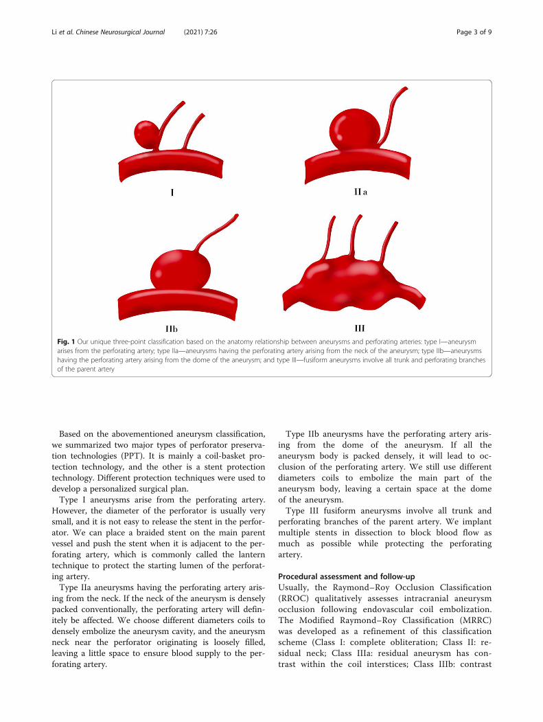
anatomy relationship between aneurysms and perforat-ing arteries after summarizing the characteristics ofthese piANs (Fig. 1):
� Type I—aneurysm arises from the perforating artery� Type IIa—aneurysms having the perforating artery
arising from the neck of the aneurysm� Type IIb—aneurysms having the perforating artery
arising from the dome of the aneurysm� Type III—fusiform aneurysms involve all trunk and
perforating branches of the parent artery
Endovascular treatmentFor patients with unruptured intracranial aneurysms,their pre-medication was given oral doses of aspirin (100mg) and Clopidogrel (75 mg) every morning for at least4 days prior to the operation. For patients with SAH, wedo not give antiplatelet drugs before surgery. If a stentneeds to be implanted during the operation, aspirin300mg + Clopidogrel 300mg is given through a nasogas-tric tube at one time. The post-procedural antiplateletregimen is unified, consisted of aspirin (100 mg oncedaily) and Clopidogrel (75 mg once daily) continued for3 months following treatment and aspirin (100 mg oncedaily) continued for life.All 6 patients received intravascular treatment and all
the treatments were performed under general anesthesia.All procedures were performed via the right commonfemoral route using a 6Fr access system as standard. Allprocedures were performed under heparin anticoagula-tion with a 3000 IU bolus dose at the start of the pro-cedure and subsequent 1000 IU bolus doses every hourto maintain the activated clotting time between 2 and2.5 times the baseline. The selection of stents and coilswas based on operator preference as well as the size ofthe aneurysms and parent vessels.
Li et al. Chinese Neurosurgical Journal (2021) 7:26 Page 2 of 9
Based on the abovementioned aneurysm classification,we summarized two major types of perforator preserva-tion technologies (PPT). It is mainly a coil-basket pro-tection technology, and the other is a stent protectiontechnology. Different protection techniques were used todevelop a personalized surgical plan.Type I aneurysms arise from the perforating artery.
However, the diameter of the perforator is usually verysmall, and it is not easy to release the stent in the perfor-ator. We can place a braided stent on the main parentvessel and push the stent when it is adjacent to the per-forating artery, which is commonly called the lanterntechnique to protect the starting lumen of the perforat-ing artery.Type IIa aneurysms having the perforating artery aris-
ing from the neck. If the neck of the aneurysm is denselypacked conventionally, the perforating artery will defin-itely be affected. We choose different diameters coils todensely embolize the aneurysm cavity, and the aneurysmneck near the perforator originating is loosely filled,leaving a little space to ensure blood supply to the per-forating artery.
Type IIb aneurysms have the perforating artery aris-ing from the dome of the aneurysm. If all theaneurysm body is packed densely, it will lead to oc-clusion of the perforating artery. We still use differentdiameters coils to embolize the main part of theaneurysm body, leaving a certain space at the domeof the aneurysm.Type III fusiform aneurysms involve all trunk and
perforating branches of the parent artery. We implantmultiple stents in dissection to block blood flow asmuch as possible while protecting the perforatingartery.
Procedural assessment and follow-upUsually, the Raymond–Roy Occlusion Classification(RROC) qualitatively assesses intracranial aneurysmocclusion following endovascular coil embolization.The Modified Raymond–Roy Classification (MRRC)was developed as a refinement of this classificationscheme (Class I: complete obliteration; Class II: re-sidual neck; Class IIIa: residual aneurysm has con-trast within the coil interstices; Class IIIb: contrast
Fig. 1 Our unique three-point classification based on the anatomy relationship between aneurysms and perforating arteries: type I—aneurysmarises from the perforating artery; type IIa—aneurysms having the perforating artery arising from the neck of the aneurysm; type IIb—aneurysmshaving the perforating artery arising from the dome of the aneurysm; and type III—fusiform aneurysms involve all trunk and perforating branchesof the parent artery
Li et al. Chinese Neurosurgical Journal (2021) 7:26 Page 3 of 9

along the aneurysm wall) [5, 6]. However, MRRCdoes not seem to be suitable for the assessment ofsatisfactory embolization of perforator involving an-eurysms. Here, we propose a new definition of satis-factory obliteration based on our new classificationand perforator preservation technologies (PPT) forperforator involving aneurysms:
� Type I—the whole saccular aneurysm body isdensely embolized which arises from the perforatingartery;
� Type IIa—the saccular aneurysm body is denselyembolized leaving a gap in the neck where theperforating artery arising from;
� Type IIb—the saccular aneurysm body is denselyembolized leaving a gap in the dome where theperforating artery arising from;
� Type III—fusiform aneurysm is repaired and has asmooth inner wall.
Immediate outcome of aneurysmal obliterationafter treatment was evaluated. Patency and flowcharacteristics within the aneurysm and parent arterywere also assessed immediately after treatment ofthe aneurysms and during follow-up. Successful per-forating artery preservation was defined as keepinggood antegrade flow of those perforators on postop-erative angiography. All of the complications duringthe perioperative period, clinical data during follow-up, and imaging results were recorded and analyzed.Procedural follow-up was performed initially at 1–3months, again at 6–12 months, and then once peryear. Standard angiographic projections were used toassess the patency of the vessels and the aneurysms.
ResultsSix patients ranged in age from 43 to 66 years old.Half of the patients were male (n = 3, 50%). Eachpatient had a single aneurysm, and there were noaneurysms identified elsewhere in the intracranialcirculation. The size of the aneurysms was listed inTable 1. According to the classification of ourunique classification of piAN, one aneurysm wereclassified as type I, two as type IIa, two as type IIb,and the remaining one aneurysm was classified astype III. Two of the piANs were ruptured and theremaining four aneurysms were unruptured. All 6patients received intravascular treatment, and theoperations were very successful. Four patients withpiANs had used coil-basket protection technology,and the other two patients had used the stent pro-tection technology. All perforating arteries involvedby piANs were successfully preserved. All patientshad no cerebral hemorrhage or infarction caused by
occlusion of perforating artery during perioperativeperiod. Only one patient developed TIA symptoms 4h after surgery, mainly manifested as motor aphasiaand difficulty in expression. Postoperative head MRIrevealed no infarcts. The symptoms were completelyrelieved in 24 h. There was no evidence of perfor-ator infarction on the follow-up post-treatment im-aging. The clinical follow-up time distribution of 6patients ranges from 12 months to 50 months. Clin-ical follow-up data was available in 6 patients allachieving a good outcome (mRS ≤ 2) (100%). Theresults are summarized in Table 1, and demonstratedcases are showed in Figs. 2, 3, 4 and 5.
DiscussionIn this study, we proposed a unique three-classification method of piAN and perforator preser-vation technology. Six patients with piANs receivedendovascular therapy based on this new classificationmethod and perforator preservation technology. Allof 6 aneurysms obtained satisfactory obliteration,and all of their perforating arteries were successfullypreserved. In terms of safety, only one patient devel-oped an ischemic event in the territory of involvingperforators 4 h after operation and completely re-solved within 24 h. Follow-up angiography alsoshowed patency of the parent artery and perforatingarteries of treated aneurysms, with no aneurysmalrecurrence.Usually, treatment for intracranial aneurysms is
done to achieve complete occlusion of the aneurysmwithout a remnant sac [7]. However, a large part ofthe complications in the treatment of aneurysms isdue to the excessive pursuit of complete occlusionof the aneurysm and neglect of the protection of theperipheral perforating artery [8]. Such perforator in-volving aneurysms (piAN) are not uncommon inintracranial aneurysms due to the anatomical charac-teristics of intracranial vessels [1]. Pritz et al. foundthat perforators were present in 7% of basilar artery(BA) bifurcations, 17% of internal carotid artery(ICA) bifurcation aneurysms, 12% of middle cerebralaneurysms, and 11% of anterior communicating an-eurysms [2]. Thus, preserving blood flow in thebranches and perforators of a parent artery is veryimportant for the successful treatment of piAN with-out postoperative morbidity and mortality [9, 10].How to avoid this kind of situation is paid more andmore attention by surgeons and constantly improvethe surgical skills and methods to protect the bloodsupply of the perforating artery [11–13]. Sung-PilJoo et al. discussed the consequences of perforatorinjury and how to avoid this phenomenon inaneurysm surgeries using intraoperative monitoring
Li et al. Chinese Neurosurgical Journal (2021) 7:26 Page 4 of 9

Table
1Dem
ograph
ic,treatmen
t,andfollow-updata
foreach
ofthepatients
Dem
ographics
Ane
urysm
characteristics
Ane
urysm
state
Trea
tmen
tPo
st-opan
giographic
result
Follo
w-up
Com
plications
Patien
tno
.Age
Gen
der
Size
(mm)
Location
Perforating
artery
invo
lving
Type
Rupture
orno
tFisher’s
grade
Tech
nique
type
Material
used
Deg
reeof
aneu
rysm
tamping
Protection
ofPA
Time
Ang
iographic
result
Infarction
Repea
tSA
H
147
F4.14×4.52
N:2.5
LC7
Anteriorchoroidal
artery
IIaYes
2②
LVIS
4.5*20
Coils
36.5cm
Satisfy
Yes
29M
Satisfy
No
No
243
M10.0×10.8
N:10.0
BAbifurcation
BApe
rforatin
gbranches
IIbYes
1②
Enterprise
4.5*28
Coils
181cm
Satisfy
Yes
26M
Satisfy
No
No
354
F6.0×
5.0
N:6.0
BAbifurcation
BApe
rforatin
gbranches
IIbNo
0②
Enterprise
4.5*28
Coils
63cm
Satisfy
Yes
18M
Satisfy
No
No
454
F2.0×
3.3
N:1.41
Lenticulostriate
arteries
Lenticulostriate
arteries
INo
0①
LVIS
4.5*20
LVISJr
2.5*17
Coils
10cm
Satisfy
Yes
50M
Satisfy
No
No
566
ML:20
W:5
RM1
Lenticulostriate
arteries
IIINo
0①
LVIS*2
3.5*20
3.5*15
Satisfy
Yes
34M
Satisfy
No
No
658
M3.2×
4.1
N:4.1
LC7
Anteriorchoroidal
artery
IIaNo
0②
LVIS
3.5*20
Coils
27.5cm
Satisfy
Yes
13M
Satisfy
TIA
No
Li et al. Chinese Neurosurgical Journal (2021) 7:26 Page 5 of 9

Fig. 3 a The angiography of patient 1 showed a lobulated LC7 aneurysm with the anterior choroidal artery origin from its neck. b According tothe measured diameter, different coils are used to densely embolize the two parts of the aneurysm respectively. Leave a little space (yellow circle)at the neck to ensure blood supply to the anterior choroidal artery. c Postoperative angiography showed satisfactory embolization of theaneurysm and perfect preservation of anterior choroidal artery (white arrow). d Follow-up angiography after 3 months showed no aneurysmalrecurrence and perfect preservation of the lenticulostriate artery (white arrow)
Fig. 2 a The angiography of patient 4 showed a small aneurysm located at one of the lenticulostriate arteries. b We used small coils to embolizethe aneurysm and planted a stent on the RM1. When the stent is adjacent to the aneurysm, we pushed it gently ensure the blood supply of thelenticulostriate artery. Postoperative angiography showed satisfactory embolization of the aneurysm and perfect preservation of thelenticulostriate artery (yellow arrow). c Follow-up angiography after 9 months showed no aneurysmal recurrence and perfect preservation of thelenticulostriate artery (yellow arrow)
Li et al. Chinese Neurosurgical Journal (2021) 7:26 Page 6 of 9

devices [1]. For surgical open craniotomy, the perfor-ating artery can be observed under direct vision, andthe damage to the perforating artery can be avoidedby improving the shape of the aneurysm clip or thedirection of the aneurysm clip.However, there are still few scholars conducting re-
search to propose how to protect the perforating ar-tery in interventional treatment of intracranialaneurysms [14–16]. The advantage of interventionaltherapy is that it can intuitively observe the bloodsupply of the perforating artery. Our three-point clas-sification is based on the anatomical relationship be-tween the perforator artery and aneurysm which wasfirst proposed. This classification method is moresuitable for the endovascular treatment of piANs. Weproposed two major perforator preservation technolo-gies (PPT) based on this classification for the firsttime to help neurointerventional doctors to developbetter surgical plans.However, the classification and technologies we pro-
posed still have certain flaws. Because we are still in
the exploratory stage and the number of piAN casesenrolled in our center is still small, the two types ofperforator preservation technology (PPT) based onour unique classification may not necessarily meet allpiANs. Perhaps there are some complex piANs thatcannot be classified in our proposed classification.Maybe some piANs need to combine two protectiontechnologies and even need to design new perforatorpreservation technology to treatment. We proposedthis unique three-classification method of piAN andperforator preservation technology and hoped toprovide new ideas for the neurointerventionists. Inthe future case expansion and exploration, we hopeto continuously improve the new classificationmethod and perforator preservation technology totreat piANs.
ConclusionsThe primary results showed that our perforator pres-ervation technologies on basis of the new neuro-interventional classification seem to be feasible, safe,
Fig. 4 a, b Patient 2 presented with acute SAH and the initial CTA showed a huge aneurysm at the tip of the basilar artery. c The 3D rotationalangiogram showed many perforating arteries on the dome of the aneurysm (yellow circle). d We used different diameters coils to embolize themain part of the aneurysm body, leaving a certain space at the dome of the aneurysm. e Postoperative angiography showed satisfactoryembolization of the aneurysm and perfect preservation of perforating arteries on the dome of the aneurysm (white circle). f Follow-upangiography after 4 months showed no aneurysmal recurrence and perfect preservation of the lenticulostriate artery
Li et al. Chinese Neurosurgical Journal (2021) 7:26 Page 7 of 9

and effective for endovascular treatment of perfor-ator involving aneurysm. It helps to evaluate the sur-gical risk and design an appropriate surgical plan,and new ideas are provided for the interventionaltreatment of the perforator involving aneurysms.
AbbreviationsPPT: Perforator preservation technology; piAN: Perforator involved aneurysms;ICA: Internal carotid artery; BAPAs: Basilar artery perforator aneurysms;LC7: The C7 segment of the left internal carotid artery; BA: Basilar artery;RM1: The M1 segment of the right middle cerebral artery
AcknowledgementsNot applicable
Authors’ contributionsWJJ has designed this technology and performed the surgeries. CL, AFL, andXLL drafted and revised the manuscript. HCQ, JZ, SSH, FL, and MJ assistedWJJ for the surgery. JL, YYZ, YQZ, and YEL revised the English language. Thedata were analyzed by CL and JL. The authors read and approved the finalmanuscript.
FundingThis work was funded by the National Key Basic Research Program of China(973 program) (grant No. 2013CB733805) and National Natural ScienceFoundation of China (grant No. 81271536, 81070925, 81371540, 81101033,and 81471767)
Availability of data and materialsThe datasets supporting the conclusion of this article are included within thearticle and its supplemental files.
Declarations
Ethics approval and consent to participateThe study was conducted according to the Declaration of Helsinki andapproved by the local ethics committee of PLA Rocket Force CharacteristicMedical Center (0077-3-14-HX(X)). All patients were admitted to theDepartment of Neurosurgery at this hospital, where all procedures tookplace.
Consent for publicationNot applicable.
Fig. 5 a The angiography of patient 5 revealed a fusiform aneurysm of the right middle cerebral artery, involving the entire trunk and all oflenticulostriate arteries. b We used telescopic stenting technology and implanted 2 LVIS stents (3.5×20mm, 3.5×15mm) in the right middlecerebral artery. c Postoperative angiography showed that the forward blood flow was satisfactory, and the lenticulostriate arteries were notaffected. d Follow-up angiography after 5 months showed satisfactory repair of fusiform aneurysm
Li et al. Chinese Neurosurgical Journal (2021) 7:26 Page 8 of 9

Competing interestsThe authors declare that this work does not involve competing interests.
Author details1New Era Stroke Care and Research Institute, PLA Rocket Force CharacteristicMedical Center, 18 Xinjiekouwai Street, Beijing 100088, China. 2NeurosurgeryDepartment, Beijing Tsinghua Changgung Hospital, School of ClinicalMedicine, Tsinghua University, Beijing, China.
Received: 10 November 2020 Accepted: 5 April 2021
References1. Brilstra EH, Rinkel GJ, van der Graaf Y, van Rooij WJ, Algra A. Treatment of
intracranial aneurysms by embolization with coils: a systematic review.Stroke. 1999;30(2):470–6. https://doi.org/10.1161/01.STR.30.2.470.
2. Currie S, Mankad K, Goddard A. Endovascular treatment of intracranialaneurysms: review of current practice. Postgrad Med J. 2011;87(1023):41–50.https://doi.org/10.1136/pgmj.2010.105387.
3. Satti SR, Vance AZ, Fowler D, Farmah AV, Sivapatham T. Basilar arteryperforator aneurysms (BAPAs): review of the literature and classification. JNeurointerv Surg. 2017;9(7):669–73. https://doi.org/10.1136/neurintsurg-2016-012407.
4. Pritz MB. Cerebral aneurysm classification based on angioarchitecture. JStroke Cerebrovasc Dis. 2011;20(2):162–7. https://doi.org/10.1016/j.jstrokecerebrovasdis.2009.11.018.
5. Stapleton CJ, Torok CM, Rabinov JD, Walcott BP, Mascitelli JR, Leslie-MazwiTM, et al. Validation of the Modified Raymond-Roy classification forintracranial aneurysms treated with coil embolization. J Neurointerv Surg.2016;8(9):927–33. https://doi.org/10.1136/neurintsurg-2015-012035.
6. Mascitelli JR, Moyle H, Oermann EK, Polykarpou MF, Patel AA, Doshi AH,et al. An update to the Raymond-Roy Occlusion Classification of intracranialaneurysms treated with coil embolization. J Neurointerv Surg. 2015;7(7):496–502. https://doi.org/10.1136/neurintsurg-2014-011258.
7. Hou K, Li G, Wang X, Xu K, Yu J. Endovascular treatment for peripheralsuperior cerebellar artery aneurysms: current state and futureconsiderations. World Neurosurg. 2019;127:423–33. https://doi.org/10.1016/j.wneu.2019.04.145.
8. Pritz MB. Perforator and secondary branch origin in relation to the neck ofsaccular, cerebral bifurcation aneurysms. World Neurosurg. 2014;82(5):726–32. https://doi.org/10.1016/j.wneu.2013.02.052.
9. Hino A, Fujimoto M, Iwamoto Y, Oka H, Echigo T. Surgery of proximalanterior cerebral artery aneurysms. Acta Neurochir. 2002;144(12):1291–6;discussion 6. https://doi.org/10.1007/s00701-002-1014-6.
10. Friedman JA, Pichelmann MA, Piepgras DG, Atkinson JLD, Maher CO, MeyerFB, et al. Ischemic complications of surgery for anterior choroidal arteryaneurysms. J Neurosurg. 2001;94(4):565–72. https://doi.org/10.3171/jns.2001.94.4.0565.
11. Kashimura H, Mase T, Ogasawara K, Ogawa A, Endo H. Trapping andvascular reconstruction for ruptured fusiform aneurysm in the proximal A1segment of the anterior cerebral artery. Neurol Med Chir (Tokyo). 2006;46(7):340–3. https://doi.org/10.2176/nmc.46.340.
12. Hirao J, Okamoto H, Watanabe T, Asano S, Teraoka A. Dissecting aneurysmsat the A1 segment of the anterior cerebral artery--two case reports. NeurolMed Chir (Tokyo). 2001;41(5):271–8. https://doi.org/10.2176/nmc.41.271.
13. Nomura M, Kida S, Kita D, Higashi R, Hasegawa M, Matsui O, et al. Fusiformaneurysm of the proximal anterior cerebral artery (A1). Acta Neurochir. 2000;142(10):1163–4. https://doi.org/10.1007/s007010070046.
14. Chalouhi N, Jabbour P, Starke RM, et al. Treatment of a basilar trunkperforator aneurysm with the pipeline embolization device: case report.Neurosurgery. 2014;74:E697–701 discussion.
15. Sivakanthan S, Carlson AP, van Loveren H, Agazzi S. Surgical clipping of abasilar perforator artery aneurysm: a case of avoiding perforator sacrifice. JNeurol Surg A Cent Eur Neurosurg. 2015;76(1):79–82. https://doi.org/10.1055/s-0033-1356488.
16. Ding D, Starke RM, Jensen ME, Evans AJ, Kassell NF, Liu KC. Perforatoraneurysms of the posterior circulation: case series and review of theliterature. J Neurointerv Surg. 2013;5(6):546–51. https://doi.org/10.1136/neurintsurg-2012-010557.
Li et al. Chinese Neurosurgical Journal (2021) 7:26 Page 9 of 9