peptides NIH Public Access Polina Feldman neuropathic pain ...
23
Challenging the catechism of therapeutics for chronic neuropathic pain: targeting CaV2.2 interactions with CRMP2 peptides Polina Feldman a and Rajesh Khanna a,b,c,d,* a Sophia Therapeutics LLC, 351 West 10th Street, Indianapolis, IN 46202, USA b Department of Pharmacology and Toxicology, 635 Barnhill Drive, Indianapolis, IN 46202, USA c Department of Biochemistry and Molecular Biology, 635 Barnhill Drive, Indianapolis, IN 46202, USA d Program in Medical Neurosciences, Paul and Carole Stark Neurosciences Research Institute, 950 West Walnut Street, Indianapolis, IN 46202, USA Abstract Chronic neuropathic pain management is a worldwide concern. Pharmaceutical companies globally have historically targeted ion channels as the therapeutic catechism with many blockbuster successes. Remarkably, no new pain therapeutic has been approved by European or American regulatory agencies over the last decade. This article will provide an overview of an alternative approach to ion channel drug discovery: targeting regulators of ion channels, specifically focusing on voltage-gated calcium channels. We will highlight the discovery of an anti-nociceptive peptide derived from a novel calcium channel interacting partner – the collapsin response mediator protein 2 (CRMP2). In vivo administration of this peptide reduces pain behavior in a number of models of neuropathic pain without affecting sympathetic-associated cardiovascular activity, memory retrieval, sensorimotor function, or depression. A CRMP2- derived peptide analgesic, with restricted access to the CNS, represents a completely novel approach to the treatment of severe pain with an improved safety profile. As peptides now represent one of the fastest growing classes of new drugs, it is expected that peptide targeting of protein interactions within the calcium channel complex may be a paradigm shift in ion channel drug discovery. 1. Introduction The Institute of Medicine of the National Academies in 2011 estimated that ~116 million adults in the USA (~1 in 3) suffer from chronic pain every year [50]. Chronic pain costs the nation up to $635 billion each year in medical treatment and lost productivity. Although some chronic pain conditions can be treated adequately with existing drugs, a large number of patients fail to achieve adequate pain relief, even with polypharmacy. Furthermore, currently available opioid pain therapies, which are generally only partially effective, are © 2013 Elsevier Ireland Ltd. All rights reserved. * Corresponding author:950 West Walnut Street, R2-Room 478, Indianapolis, Indiana 46202, USA Office phone: (317) 278-6531; Fax: (317) 278-5849; [email protected]. Publisher's Disclaimer: This is a PDF file of an unedited manuscript that has been accepted for publication. As a service to our customers we are providing this early version of the manuscript. The manuscript will undergo copyediting, typesetting, and review of the resulting proof before it is published in its final citable form. Please note that during the production process errors may be discovered which could affect the content, and all legal disclaimers that apply to the journal pertain. NIH Public Access Author Manuscript Neurosci Lett. Author manuscript; available in PMC 2014 December 17. Published in final edited form as: Neurosci Lett. 2013 December 17; 557(0 0): . doi:10.1016/j.neulet.2013.06.057. NIH-PA Author Manuscript NIH-PA Author Manuscript NIH-PA Author Manuscript
Transcript of peptides NIH Public Access Polina Feldman neuropathic pain ...
Challenging the catechism of therapeutics for chronicneuropathic pain: targeting CaV2.2 interactions with CRMP2peptides
Polina Feldmana and Rajesh Khannaa,b,c,d,*
aSophia Therapeutics LLC, 351 West 10th Street, Indianapolis, IN 46202, USAbDepartment of Pharmacology and Toxicology, 635 Barnhill Drive, Indianapolis, IN 46202, USAcDepartment of Biochemistry and Molecular Biology, 635 Barnhill Drive, Indianapolis, IN 46202,USAdProgram in Medical Neurosciences, Paul and Carole Stark Neurosciences Research Institute,950 West Walnut Street, Indianapolis, IN 46202, USA
AbstractChronic neuropathic pain management is a worldwide concern. Pharmaceutical companiesglobally have historically targeted ion channels as the therapeutic catechism with manyblockbuster successes. Remarkably, no new pain therapeutic has been approved by European orAmerican regulatory agencies over the last decade. This article will provide an overview of analternative approach to ion channel drug discovery: targeting regulators of ion channels,specifically focusing on voltage-gated calcium channels. We will highlight the discovery of ananti-nociceptive peptide derived from a novel calcium channel interacting partner – the collapsinresponse mediator protein 2 (CRMP2). In vivo administration of this peptide reduces painbehavior in a number of models of neuropathic pain without affecting sympathetic-associatedcardiovascular activity, memory retrieval, sensorimotor function, or depression. A CRMP2-derived peptide analgesic, with restricted access to the CNS, represents a completely novelapproach to the treatment of severe pain with an improved safety profile. As peptides nowrepresent one of the fastest growing classes of new drugs, it is expected that peptide targeting ofprotein interactions within the calcium channel complex may be a paradigm shift in ion channeldrug discovery.
1. IntroductionThe Institute of Medicine of the National Academies in 2011 estimated that ~116 millionadults in the USA (~1 in 3) suffer from chronic pain every year [50]. Chronic pain costs thenation up to $635 billion each year in medical treatment and lost productivity. Althoughsome chronic pain conditions can be treated adequately with existing drugs, a large numberof patients fail to achieve adequate pain relief, even with polypharmacy. Furthermore,currently available opioid pain therapies, which are generally only partially effective, are
© 2013 Elsevier Ireland Ltd. All rights reserved.*Corresponding author:950 West Walnut Street, R2-Room 478, Indianapolis, Indiana 46202, USA Office phone: (317) 278-6531; Fax:(317) 278-5849; [email protected].
Publisher's Disclaimer: This is a PDF file of an unedited manuscript that has been accepted for publication. As a service to ourcustomers we are providing this early version of the manuscript. The manuscript will undergo copyediting, typesetting, and review ofthe resulting proof before it is published in its final citable form. Please note that during the production process errors may bediscovered which could affect the content, and all legal disclaimers that apply to the journal pertain.
NIH Public AccessAuthor ManuscriptNeurosci Lett. Author manuscript; available in PMC 2014 December 17.
Published in final edited form as:Neurosci Lett. 2013 December 17; 557(0 0): . doi:10.1016/j.neulet.2013.06.057.
NIH
-PA Author Manuscript
NIH
-PA Author Manuscript
NIH
-PA Author Manuscript
often associated with many side effects that limit their clinical efficacy, including toleranceand addiction. Hence, new research and therapies are critically needed to decipher painmechanisms and open new avenues for specific and more effective treatments. Severalexcellent reviews describing the roles of voltage-gated calcium [94, 100, 113, 115] andsodium [36, 37] channels in pain have appeared in the literature; thus, in this review, we willfocus on a new peptide-based therapeutic approach for chronic pain.
2. Rationale for targeting calcium channels for pain researchN-type voltage-gated calcium channels (CaV2.2) are multiprotein complexes comprised of apore-forming α-subunits and auxiliary α2/δ, β, and γ subunits [39, 102]. CaV2.2 channelsare localized to primary afferent terminals in laminae 1 and 2 of the dorsal horn [108] wheretheir activation results in the influx of calcium and release of neurotransmitters such asglutamate, substance P, and calcitonin gene related peptide (CGRP). Following discovery ofCaV2.2-specific conotoxins, the biological role of this channel was evaluated in animalsthrough the use of isoform-specific inhibitors. Specific interest in the role of CaV2.2 insensation arose from the observation that omega-conotoxin (ω-CTX) was able to blocktransmitter release from sensory neurons and spinal nerve terminals [46, 61]. CaV2.2channels are also critical for pain transduction as block of these channels relieveshyperalgesia [8, 47, 56], mice lacking CaV2.2 show an increased threshold for pain [84],and expression of CaV2.2 is upregulated following a chronic constrictive nerve injury [34].Spinal administration of CaV2.2 blocking peptides has been shown to ameliorate painfulbehavior in rodent models of neuropathic and inflammatory pain [87]. Interestingly, severalCNS effects were also observed in the mice including; decreased anxiety behavior, changesin vigilance, and enhanced aggressive behavior [15, 55, 84]. The importance of CaV2.2 inpain is further underscored by the demonstration of a naturally occurring alternative spliceform of CaV2.2 (i.e. exon 37a) in small-diameter nociceptive neurons [14] which are criticalfor basal thermal nociception, and thermal and mechanical hyperalgesia [8]. The role ofCaV2.2 in neurotransmitter release and pain sensation is further reinforced by studiesdemonstrating that analgesic opioids and adrenergic agonists inhibit CaV2.2 [89]. In sum, byvirtue of their ability to control the regulated release of neurotransmitters from nociceptiveafferents, N-type Ca2+ channels are a prime target for the development of novel analgesics[86, 94, 113, 115].
3. Examples, limitations, and pipeline of CaV2.2-targeted drugsSeveral subtypes of neuronal voltage-gated calcium channels are expressed in thenociceptive pathway and are crucial for shaping action potentials and controlling cellularexcitability and synaptic transmission [115]. Recently, Ziconitide (trade-name Prialt®) wasapproved by the Food and Drug Administration (FDA) for the treatment of severe pain thatwas refractory to other therapies [38]. This drug is a synthetic version of the naturallyoccurring cone snail toxin μ-conotoxin MVIIA and is a highly potent and selective peptideblocker of CaV2.2, validating N-type calcium channels as a promising target of novelanalgesics. However, due to the profound side effects and a lack of efficacy when deliveredby more common routes (i.e. intravenous or oral), the use of Prialt® is limited to intrathecalpump route of administration, i.e. direct application to the spinal synapse between sensoryneurons and spinal neurons. That intrathecal application is required and sufficient foranalgesia also supports the notion that presynaptic blockade of CaV2.2 is responsible fortargeting pain. The use of Prialt® via this route is not without complications as theintrathecal route of administration is by far the most expensive and invasive method of drugdelivery. Many side effects are accompanied by the use of the drug that often manifest asconfusion, depression, hallucinations, decreased alertness, somnolence, orthostatichypotension and nausea [79, 85, 92, 99]. Prialt® is also contraindicated in patients with pre-
Feldman and Khanna Page 2
Neurosci Lett. Author manuscript; available in PMC 2014 December 17.
NIH
-PA Author Manuscript
NIH
-PA Author Manuscript
NIH
-PA Author Manuscript
existing mental disorders due to thoughts of suicide and worsening depression or paranoia[62]. Subsequently, Prialt® therapy in practice for neuropathic pain treatment, is warrantedfor relatively few patients who are intolerant or refractory to other treatments [109], such assystemic analgesics, adjunctive therapies or intrathecal morphine [6, 76, 77]. Prialt® is astate-independent CaV2.2 blocker and therefore inhibits the activity of the channel in boththe hyperpolarized state (closed) and the depolarized state (open or inactivated) [42]. Thus,the side effects presumably occur through a non-selective blockade of N-channels in alltissues [1, 98].
Despite the shortcomings of Prialt®, blockade of the presynaptic, CaV2.2 and its affiliatedsubunits remains an important therapeutic target for a number of chronic pain conditions[39, 94]. One commonly used drug that targets the α2δ1 subunit of CaV2.2 is theantiepileptic drug, gabapentin (Neurotin®). Neurotin® is FDA-approved for a number ofconditions including postherpetic neuralgia, diabetic neuropathy [12, 33, 45, 68],neuropathic pain [3, 18, 88], phantom limb pain [9, 16, 53, 71, 83], fibromyalgia [116] andtrigeminal neuralgia [67, 74]. Consistent, though less compelling clinical evidence supportsits use for a variety of other neuropathic pain syndromes, including cancer pain syndromes,pain associated with HIV infection and chronic back pain. Controlled clinical trials indiabetic neuropathy and postherpetic neuralgia [82] demonstrated that gabapentin at2400-3600 mg/day has an efficacy similar to the tricyclic antidepressants [2]. Thoughgabapentin has a favorable pharmacokinetic profile, it is only available in oral form andrequires at least three times daily dosing in order to achieve mean plasma levels throughouta 24-hour period to achieve efficacy [13]. However, one benefit of the drug is that, unlikePrialt, it does not require laboratory monitoring [103, 104].
Neurotin® is associated with only moderate to substantial benefits of pain relief in 31-43%of patients [67]. Adverse events are experienced by about two-thirds of people takinggabapentin, mainly dizziness, somnolence (27%), dizziness (24%), and ataxia (7%) and it isnot an uncommon finding that patients are not able to tolerate dosing in the therapeuticrange due to common side effects [67]. Gabapentin was also reported to disrupt theinteraction between α2δ-1 and thrombospondins in vitro and, as a result, interferes withsynaptogenesis, although it does not affect pre-formed synapses [41], a finding that couldencumber prolonged use of this class of drug.
There remains strong interest in the pharmaceutical and academic worlds toward thedevelopment of orally acting, state-dependent blockers of CaV2.2. Several research anddevelopment groups have made substantial progress in developing small organic moleculestargeting ‘hot spots’ of the N-type calcium channel. One group in particular led, by scientificinvestigator Terrance P. Snutch, has targeted chemical moieties that possess calcium channelblocking activity, such as the diphenylmethylpiperazine basic skeleton [17, 72, 73, 114].Through extensive structure-activity relationship (SAR) investigations two small organicmolecules NP118809 and NP078585 have been discovered and optimized as a N-typecalcium channel blockers with good selectivity over L-type calcium channels [114]. In doingso, Snutch and colleagues have gone on to commercialize several of these small moleculesselective for N-type or T-type calcium channel blockers, two of which are steadilyprogressing through the FDA regulatory pathway by Zalicus, Inc, a biopharmaceuticalcompany that discovers and develops novel treatments for patients suffering from pain.Specifically, Zalicus, Inc. is developing an orally available selective N-type calcium channelblocker (Z160) for the treatment of chronic neuropathic pain associated with lumbosacralradiculopathy and post-herpetic neuralgia. Z160 is in currently being tested in Phase IIclinical trials (www.zalicus.com). Convergence Pharmaceuticals, LLC has also developedan orally available small molecule (CNV2197944) state-dependent N-type calcium channelblocker for the treatment of neuropathic pain, currently in Phase IIa clinical trials
Feldman and Khanna Page 3
Neurosci Lett. Author manuscript; available in PMC 2014 December 17.
NIH
-PA Author Manuscript
NIH
-PA Author Manuscript
NIH
-PA Author Manuscript
(www.convergencepharma.com). Newron Pharmaceuticals SpA is perhaps the furthest alongthe pipeline, as their small molecule drug Ralfinamide is in Phase IIB/III trials, and isbelieved to mediate its potent analgesic effect through the inhibition of sodium channels,including NaV1.7, N-type calcium channels and NMDA receptor for the treatment of lowerback pain (www.newron.com). Small molecule equivalents of omega-conotoxin CVID (e.g.AM336/CNSB004/Leconotide), the selective N-type calcium channel blocker, have alsobeen engineered and are being tested in early stage preclinical studies for pain associatedwith diabetes and cancer [58, 59, 93].
In addition to direct block of calcium channels, targeting of protein-protein interaction (PPI)interfaces within a PPI complex is gaining momentum as a potentially new manner of drugdevelopment [27]. Several small molecule PPI inhibitors are currently in clinicaldevelopment by pharmaceutical companies Abbott, Genentech, Johnson & Johnson, orRoche; two drugs are already on the market (Tirofiban/Aggrastat® and Maraviroc/Selzentry®) [44]. The notion of PPIs altering CaV2.2 channel function was proposed almosttwo decades ago by William Catterall and colleagues who discovered that interruption of theCaV2.2-syntaxin interaction with SYNaptic Protein INTeraction (synprint) peptides inhibitsfast, synchronous transmitter release [66, 80, 90, 91]. Despite this early report, thesepatented [28] peptides have not advanced further. Additional peptides that attenuate Gprotein modulation of Ca2+ channels have also been reported and may be of use in probingcalcium channel mediated presynaptic mechanisms [25, 26]; however, none of thesepeptides have been translated beyond. As will be described in the following sections, SophiaTherapeutics, LLC in collaboration with Indiana University has developed a novel peptideplatform that target CaV2.2 by modulating channel activity through protein/proteininteractions for the treatment of neuropathic pain. Sophia Therapeutics is currentlyperforming preclinical in vivo studies and is actively seeking funding and partnershipopportunities to move the project into Phase I clinical trials.
4. Discovery of CRMP2, a novel regulator of CaV2.2Alternative strategies that target proteins interacting with calcium channels represent anemerging theme in drug discovery for treatment of clinical neuropathic pain. A proteomicscreen identified the collapsin response mediator protein 2 (CRMP2) as a putativeinteracting partner of CaV2.2 [54, 69]. Enriched at functional release sites, CaV2.2 formspart of a large macromolecular complex, which facilitates efficient neurotransmitter release.Identification and analyses of a litany of protein-protein interactions within the nerveterminal have demonstrated a functional coupling between presynaptic Ca2+ channels andthe transmitter release machinery [31, 35, 57, 95]. Although neurotransmission was notaffected in CRMP1 knockout mice [97], long-term potentiation, spatial learning andmemory were affected in both CRMP1 and CRMP3 knockout mice [78, 97], suggesting apossible role for CRMPs in neurotransmission as well as synaptogenesis. As potentialbinding partners, CRMPs could affect neurotransmission by either a physical interactionwith the synaptic machinery or CaV2.2 itself.
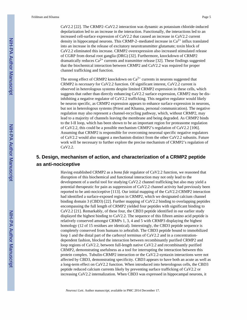
Our study showed that CRMP2 is part of the CaV2.2 proteome [22, 32, 54].Immunocytochemistry revealed a strong colocalization between CRMP2 and CaV2.2 withinhippocampal neurons that was supported by reciprocal co-immunoprecipitation of the twoproteins. CRMP2 localized to both extrasynaptic and synaptic fractions, suggesting that itmay traffic CaV2.2 within these regions (Figure 1). CRMP2 was present in both cytosolicand membrane fractions. CRMP2 was found to localize to the soma, dendrites, and axonsand therefore may interact with CaV2.2 in any of these regions. Traditional in vitro bindingexperiments [22] and isothermal titration calorimetric analyses (Khanna, M. and Khanna, R.;unpublished data) mapped the interaction to two domains within the cytoplasmic loops of
Feldman and Khanna Page 4
Neurosci Lett. Author manuscript; available in PMC 2014 December 17.
NIH
-PA Author Manuscript
NIH
-PA Author Manuscript
NIH
-PA Author Manuscript
CaV2.2 [22]. The CRMP2–CaV2.2 interaction was dynamic as potassium chloride-induceddepolarization led to an increase in the interaction. Functionally, the interactions led to anincreased cell-surface expression of CaV2.2 that caused an increase in CaV2.2 currentdensity in hippocampal neurons. This CRMP-2–mediated increase in Ca2+ influx translatedinto an increase in the release of excitatory neurotransmitter glutamate; toxin block ofCaV2.2 eliminated this increase. CRMP2 overexpression also increased stimulated releaseof CGRP from dorsal root ganglia (DRG) [32]. Furthermore, knockdown of CRMP2dramatically reduces Ca2+ currents and transmitter release [32]. These findings suggestedthat the biochemical interaction between CRMP2 and CaV2.2 was required for properchannel trafficking and function.
The strong effect of CRMP2 knockdown on Ca2+ currents in neurons suggested thatCRMP2 is necessary for CaV2.2 function. Of significant interest, CaV2.2 current isobserved in heterologous systems despite limited CRMP2 expression in these cells, whichsuggests that rather than directly enhancing CaV2.2 surface expression, CRMP2 may be dis-inhibiting a negative regulator of CaV2.2 trafficking. This negative regulator would likelybe neuron specific, as CRMP2 expression appears to enhance surface expression in neurons,but not in heterologous systems (Priest and Khanna, personal communication). The negativeregulation may also represent a channel-recycling pathway, which, without CRMP2, maylead to a majority of channels leaving the membrane and being degraded. As CRMP2 bindsto the I-II loop, which has been shown to be an important region for proteasome regulationof CaV2.2, this could be a possible mechanism CRMP2’s regulation of CaV2.2 [106].Assuming that CRMP2 is responsible for overcoming neuronal specific negative regulatorsof CaV2.2 would also suggest a mechanism distinct from the other CaV2.2 subunits. Futurework will be necessary to further explore the precise mechanism of CRMP2’s regulation ofCaV2.2.
5. Design, mechanism of action, and characterization of a CRMP2 peptideas anti-nociceptive
Having established CRMP2 as a bona fide regulator of CaV2.2 function, we reasoned thatdisruption of this biochemical and functional interaction may not only lead to thedevelopment of a useful tool for studying CaV2.2 channel trafficking but also may yield apotential therapeutic for pain as suppression of CaV2.2 channel activity had previously beenreported to be anti-nociceptive [113]. Our initial mapping of the CaV2.2:CRMP2 interactionhad identified a surface-exposed region in CRMP2, which we designated calcium channelbinding domain 3 (CBD3) [22]. Further mapping of CaV2.2 binding to overlapping peptidesencompassing the full length of CRMP2 yielded four peptides with significant binding toCaV2.2 [21]. Remarkably, of these four, the CBD3 peptide identified in our earlier studydisplayed the highest binding to CaV2.2. The sequence of this fifteen amino acid peptide isrelatively conserved amongst CRMPs 1, 3, 4 and 5 with CRMP3 displaying the highesthomology (12 of 15 residues are identical). Interestingly, the CBD3 peptide sequence iscompletely conserved from humans to zebrafish. The CBD3 peptide bound to immobilizedloop 1 and the distal part of the carboxyl terminus of CaV2.2 and in a concentration-dependent fashion, blocked the interaction between recombinantly purified CRMP2 andloop regions of CaV2.2, between full-length native CaV2.2 and recombinantly purifiedCRMP2, demonstrating usefulness as a tool for interrupting the interaction between thisprotein complex. Tubulin-CRMP2 interaction or the CaV2.2-syntaxin interactions were notaffected by CBD3, demonstrating specificity. CBD3 appears to have both an acute as well asa long-term effect on CaV2.2 function. When introduced into heterologous cells, the CBD3peptide reduced calcium currents likely by preventing surface trafficking of CaV2.2 orincreasing CaV2.2 internalization. When CBD3 was expressed in hippocampal neurons, it
Feldman and Khanna Page 5
Neurosci Lett. Author manuscript; available in PMC 2014 December 17.
NIH
-PA Author Manuscript
NIH
-PA Author Manuscript
NIH
-PA Author Manuscript
antagonized CRMP2-induced enhancement of Ca2+ channel currents. As CBD3 disrupts theinteraction between CRMP2 and CaV2.2, this supports the conclusion that the interactionbetween CRMP2 and CaV2.2 is likely responsible for the observed increase in Ca2+
currents.
Subsequent creation of TAT-CBD3 (tagged with the HIV-1 transactivator of transcription(TAT) cell penetrating motif) allowed evaluation of this peptide directly in cells as well as inanimal models of pain. TAT-CBD3 peptide interfered with CRMP2-CaV2.2 interactionsresulting in acute inhibition of CaV2.2 currents in sensory and hippocampal neurons; acuteinhibition of frequency of spontaneous excitatory postsynaptic currents (sEPSCs) in spinalcord slices as well as layer V pyramidal neurons suggesting reduction in probability ofglutamate release from stimulated presynaptic terminals; and inhibition of evoked calcitoningene-related peptide (CGRP) in sensory neurons in culture (acute and long-term inhibitionobserved) and in spinal cord slices.
Our results converged on the possibility that TAT-CBD3 might act as a novel antagonist ofCaV2.2 function prompting us to investigate its usefulness as a therapeutic for pain using anin vivo model of hypersensitivity. TAT-CBD3 was tested in a battery of behavioral assaysincluding formalin-induced nocifensive behavior, capsaicin-induced nocifensive behavior,and chronic neuropathic models including distal sensory polyneuropathy induced by anti-retroviral drugs, lysophosphotidylcholine-induced sciatic nerve focal demyelination (LPC),and traumatic tibial nerve injury model. In all of these models, TAT-CBD3 reducednocifensive behaviors or induced reversal of hypersensitivity. Notably, these effects wereobserved with systemic injections of TAT-CBD3 in stark contrast to the intrathecal route ofdelivery espoused for Prialt ®.
Importantly, in a further string of rodent behavioral tests to examine off-target effects, TAT-CBD3 was mildly anxiolytic without affecting memory retrieval, sensorimotor function, ordepression at doses at least 50 fold higher than needed to achieve anti-nociception.Sympathetic-associated cardiovascular activity was also not affected by TAT-CBD3 [110].Thus, peptide analgesics, such as TAT-CBD3, with restricted access to the CNS represent acompletely novel approach to the treatment of severe pain with a likely improved safetyprofile.
6. CRMP2 peptides: good, better, best?While we have documented great success with TAT-CBD3, challenges remain including thetransiency (≤4 hours) of pain reversal with TAT-CBD3 [21], the lack of efficacy in the tibialnerve injury model of persistent neuropathic pain [110], coupled with lack of knowledge ofthe time course (i.e., pharmacokinetics/bioavailability) of TAT-CBD3’s action, additionalpossible targets and a safety profile have not been fully investigated. In an effort to increaseTAT-CBD3’s duration of action and utility in a broader range of neuropathic conditions, wehave converged our initial efforts on optimizing the CBD3 platform by selected single sitemutagenesis and using alternative cell penetrating motifs.
To address the first issue, a peptide array harboring systematic amino acid scans of theCBD3 coupled with a Far-Western approach identified three peptides with point mutationsat positions 6 (A6K), 9 (R9L) and 14 (G14F) with greater binding to Ca2+ channels than theparent CBD3 peptide [75]. One of these peptides, TAT-CBD3-A6K, was modeled as an α-helix and molecular dynamics (MD) simulations were performed to explore atomisticflexibility of wild type and A6K mutant peptides in solution. This analysis showed that likethe wild type peptide, the C-terminus residues are less stable than N-terminus residues,which consistently maintain an α-helical structure throughout the MD simulation. Increased
Feldman and Khanna Page 6
Neurosci Lett. Author manuscript; available in PMC 2014 December 17.
NIH
-PA Author Manuscript
NIH
-PA Author Manuscript
NIH
-PA Author Manuscript
conformational change of the mutant peptide may result in greater efficacy for this peptide.Greater conformational change suggests that the peptide may sample alternativeconformational states that are more prone to bind to the calcium channel and inhibit itsinteraction with CRMP2. The wild type peptide appears more prone to undergo structuralchanges that are not seen in the A6K mutant simulations. Based on these structuralpredictions, we tested the mechanism of action and efficacy of TAT-CBD3-A6K and foundthat (i) this peptide exhibited greater anti-nociception in a rodent model of AIDS therapy-induced peripheral neuropathy when compared to the parent TAT-CBD3 peptide; (ii)intraperitoneal administration of TAT-CBD3A6K produced none of the minor side effects(i.e. tail kinking, body contortion) observed with the parent peptide; (iii) excitability ofdissociated small diameter sensory neurons isolated from rats was also reduced by TAT-CBD3A6K; and (iv) suppression of excitability may be due to inhibition of T- and R-typeCa2+ channels (Figure 4). These results suggested that structural modifications of the CBD3scaffold peptide might result in peptides with selectivity against a particular subclass ofvoltage-gated calcium channels resulting in a multi-pharmacology of action on the target.
To improve CBD3’s utility to broader range of neuropathic conditions, CBD3 was grafted toa stretch of nine arginines ((R9)-CBD3); the cell penetrating peptide (CPP) R9 motif waschosen due to its superior cell penetrating abilities [107]: it is the most efficacious ofcurrently known protein transduction domains with at least a 20–fold better penetrabilityinto cells than TAT [107] or other homopolymeric amino acids [65]; it is well tolerated bycells, with low short- and long-term toxicological effects [101]; and its the mechanism oftransduction into cells is well understood and involves binding to cell surface heparansulfate proteoglycans, heparin sulfate-mediated endocytosis into vesicles, release of R9 fromheparin sulfate upon cleavage by heparanases, culminating in release of unbound R9 into thecytosol due to vesicular leakage [43]. Three endocytic pathways – macropinocytosis,clathrin-mediated endocytosis and caveolae/lipid-raft-mediated endocytosis – are thought tobe involved [40]. Similar to TAT-CBD, the R9-conjugated CBD3 peptide interfered with theCaV2.2–CRMP2 interaction. The interference was specific, as the tubulin-CRMP2interaction was not disrupted by R9-CBD3. The functional consequence of the disruptedCaV2.2–CRMP2 interaction was a significantly higher extent (i.e. efficacy) of inhibition ofcalcium influx in sensory neurons. This increased efficacy may possibly contribute to themechanism of action of this peptide. That R9-CBD3 did not affect Ca2+ influx activated byvanilloid receptor activators resiniferatoxin and capsaicin rules out targeting of thesereceptors as a potential mechanism of action. These results are entirely consistent with ourprevious data that showed no effect of TAT-CBD3 on TRPV1 current recordings followingcapsaicin challenge [21] demonstrating that TAT-CBD3 does not work through directinhibition of TRPV1 channels. We found that R9-CBD3, like TAT-CBD3 [21], waseffective at reversing mechanical hypersensitivity induced by NRTI-associated distalsymmetrical polyneuropathy. Importantly, R9-CBD3 reversed mechanical hypersensitivityassociated with nerve-injury possibly due to its superior cell transduction attributes [107](Figure 5).
Future efforts will explore alternative routes of dosing including continuous subcutaneousinfusions, intramuscular, sublingual and or oral routes. Clinically, ~2-4 grams/day ofgabapentin are needed to achieve significant reduction in average daily pain intensity scorescompared with placebo [13]; but at this dosage the drug is not without significant sideeffects. Even taking into account allometric scaling, TAT-CBD3 reverses NRTI-inducedhypersensitivity at 0.1 mg/kg compared with 200 mg/kg of gabapentin needed to achievesimilar levels of reversal [21]. One therapeutic challenge for many neurologically actingmedications, including TAT-CBD3, is penetration of the blood brain barrier (BBB). Thepeptide, like Prialt®, appears to have a somewhat restricted penetration into the CNS [21],however disruption of the BBB due to direct injury to the spinal cord/brain or disease
Feldman and Khanna Page 7
Neurosci Lett. Author manuscript; available in PMC 2014 December 17.
NIH
-PA Author Manuscript
NIH
-PA Author Manuscript
NIH
-PA Author Manuscript

pathologies may allow the peptide to overcome the CNS’s physicochemical barriers andalter synapse functioning within higher order systems [20]. More importantly, thatperipheral nerve injury or inflammatory pain states may be maintained by CNS gliosis(activated glial cells that release inflammatory mediator) suggests that there could bechronic disruptions in BBB permeability and transport of TAT-CBD3 into the brain. So,despite the relatively restricted penetration across the BBB by TAT-CBD3 it could still havecognitive effects following chronic dosing. Thus, in the future we will also evaluate whetherthe peptides affect memory and learning, locomotion, social interaction as well as examinehistopathological changes in various tissues.
7. Getting over the “small-molecule fever”: peptide market, limitations andoptimization
Peptide based therapeutics for pain and a number of other chronic conditions are on the rise.The number of peptides approved in the past few years falls third in the line with smallmolecules (45% of the total) and ‘proteins’ (24% of the total). Between 2009-2011, eightpeptides were accepted by the FDA, which represents 11% of the total drugs accepted in thisperiod. The probability of regulatory approval for peptides is over 20%, a rate which isdouble that of small molecules [60]. Although these peptides have a broad range ofstructures, size, and complexity, it has not limited their place in the market [5]. The peptidemarket consists of a relatively small portion of all drug product sales (~1.5%). However,peptide drug product sales are increasing with an annual growth of 7.5-10% [60]. Currently,there are 80 peptides on the market, approximately 200 more in clinical trials and 400 inadvanced preclinical states [105], which have demonstrated diverse applications across anumber of clinical fields [11, 96].
Nature continues to be the main source and best inspiration for the drug discovery process[70]. The clear advantage that drug peptides offer include, a significantly high structuralrelationship between physiological active parent molecules thereby reducing the risk of off-target reactions [60] and lack of immunogenicity [64]. Overall, peptides have severaladvantages as drug; including their high biological activity (given their natural source), highspecificity, and low toxicity [5]. Peptides also have the ability to penetrate further intotissues due to their smaller size and require substantially less quantity to activate their target[48]. Compared to small organic molecules, peptides typically offer greater efficacy,selectivity and specificity [49].
In general, it is believed that peptides have several features that prevent activepharmaceutical ingredients; such as, lack of oral bioavailability, low stability underphysiological conditions [24, 117], short half-life because of their rapid degradation byproteolytic enzymes of the digestive system, and hydrophobicity (limits their ability to crossphysiological barriers) [5]. However, robust peptide chemistry strategies and ‘modern’solution organic synthetic chemistry provide for straightforward analog analysis for lead-candidate optimization. The process is quite efficient and economical when compared withsmall molecules [5].
The half-life of many peptides is primarily dependent on their proteolytic susceptibility.Orally available peptides inevitably battle gram quantities of proteolytic activity in thelumen of the small intestine, which are secreted by the pancreas (e.g. alpha chymotrypsin,trypsin, pancreatic elastase, carboxypeptidases A, etc.). Additionally, the membrane ofepithelial cells contains at least 15 peptidases. Other proteolyic sources include lysosomalpeptidases and matrix metalloproteinases [105, 112]. Thus, peptide modifications havebecome necessary strategies to optimize their function as an ideal drug-delivery system andbypass many of the limitations associated with natural peptide kinetics. Chemical
Feldman and Khanna Page 8
Neurosci Lett. Author manuscript; available in PMC 2014 December 17.
NIH
-PA Author Manuscript
NIH
-PA Author Manuscript
NIH
-PA Author Manuscript
optimization strategies are based on structure-activity relationship and/or quantitativestructure-activity relationship; including cyclization of peptide sequences, substitution withan unnatural amino acids, isosteric amide bond replacement, N-terminal esterification (toenhance plasma stability), peptide conjugation with cell penetrating peptides (i.e. TATconjugation) just to name a few [4, 105]. For example, Ahrens and co-workers developed‘intelligent peptides’ which have smart linkers that increase specificity and blood plasmastability [4]. With the sophistication currently possible in peptide modifications, a promisingfuture for innovative synthetic therapeutic peptides may provide the next generation ofpharmaceutical drugs.
8. ConclusionsIt is clear that CaV2.2 is a nidus for neurotransmitter release [10, 29, 30] and nociceptivetransmission [115]. However, use of CaV2.2 blockers in pain therapeutics (Prialt® orgabapentin) is limited by side-effects resulting from inhibition of the physiological functionsof CaV2.2 within the CNS. To circumvent this, we have advanced a strategy for targetingprotein interactions that regulate voltage-gated calcium channels as an alternative to directchannel block. In this regard, we identified CRMP2 as a bona fide modulator of CaVchannels[22, 32] with peptides from CRMP2 proving to be anti-nociceptive[21, 51, 81].Remarkably, interrupting the CaV-CRMP2 interaction with a peptide derived from the CaVchannel reversed pain behavior associated with peripheral neuropathy [111], furthervalidating the CaV-CRMP2 axis as a novel node for development of pain therapeutics.Importantly, targeting channel regulation may potentially avert many of the adverse sideeffects associated with direct channel block. Indeed, the CRMP2-derived TAT-CBD3peptide has a problem-free profile with at least a 500-fold therapeutic window [21]. Thatreplacement of a lysine with an alanine residue at the sixth residue of CBD3 allowed theresulting peptide (TAT-CBD3-A6K) to reverse tibial nerve-injury-induced pain which wasrefractory to TAT-CBD3 [110], as well as bestowed upon the new peptide an ability toblock T- and R-type calcium channels (Figure 4) suggests opportunities to tailor peptides forspecific pain indications and will be addressed in the future. In conclusion, the TAT-CBD3peptide is efficacious in both acute and chronic pain models when administered eithertopically or systemically; CBD3, interfering with the function of CaV2.2, producedanalgesic effects in rodents subjected to formalin-induced nocifensive behavior, capsaicin-induced nocifensive responses to ocular administration and neurogenic inflammation of themeninges, and reversed the chronic tactile hypernociceptive behavior observed inantiretroviral toxic neuropathy. Collectively, the antinociceptive activity of TAT-CBD3across a number of pain models suggests that N-type voltage calcium channels on thepresynaptic terminals of afferent sensory neurons play a central role in both inflammatoryand neuropathic pain behaviors.
AcknowledgmentsThe authors thank colleagues at the Stark Neurosciences Research Institute (SNRI), Dr. Joel M. Brittain forfractionation data, Professor Fletcher A. White (Anesthesia, Indiana University) for collaborations on pain models,Dr. May Khanna for isothermal titration calorimetric experiments, and Dr. Joe Trebley at the Indiana UniversityResearch and Technology Commercialization for help with Sophia Therapeutics, LLC. This work was supported, inpart, by grants from the Indiana Clinical and Translational Sciences Institute (CTSI) funded, in part by a ProjectDevelopment Team Grant Number (RR025761) from the National Institutes of Health, National Center forResearch Resources, Clinical and Translational Sciences Award, the Indiana State Department of Health – SpinalCord and Brain Injury Fund (A70-9-079138 to R.K.), NIH/NINDS (NS049136-06 to F.A.W.), the IndianaUniversity Biomedical Committee – Research Support Funds (2286501 to R.K), a National Scientist Developmentfrom the American Heart Association (SDG5280023 to R.K.), a Research Inventions and ScientificCommercialization grant (to R.K.) from the Indiana CTSI, a Funding Opportunities for ResearchCommercialization and Economic Success (FORCES) grant initiative from the Indiana CTSI (to R.K.), and theElwert Award in Medicine to R.K. We also acknowledge funding from the BioCrossroads New Venturecompetition. R.K. is a shareholder of Sophia Therapeutics, LLC.
Feldman and Khanna Page 9
Neurosci Lett. Author manuscript; available in PMC 2014 December 17.
NIH
-PA Author Manuscript
NIH
-PA Author Manuscript
NIH
-PA Author Manuscript
Abbreviations
CGRP calcitonin gene related peptide
CaV2.2 N-type voltage-gated Ca2+ channel
CBD Ca2+ channel binding domain
CRMP2 collapsin response mediator protein 2
CPP cell penetrating peptide
CNS central nervous system
ω-CTX omega-conotoxin
DIV days in vitro
DRG dorsal root ganglion
PWT paw withdrawal threshold
R9 nona-arginine
ST Sophia Therapeutics
TAT HIV-1 transactivator of transcription domain
TNI tibial nerve injury
CTX conotoxin
LPC lysophosphotidylcholine
sEPSCs spontaneous excitatory postsynaptic currents
NRTI nucleoside reverse transcriptase inhibitor
BBB blood brain barrier
REFERENCES[1]. Abbadie C, McManus OB, Sun SY, Bugianesi RM, Dai G, Haedo RJ, Herrington JB,
Kaczorowski GJ, Smith MM, Swensen AM, Warren VA, Williams B, Arneric SP, Eduljee C,Snutch TP, Tringham EW, Jochnowitz N, Liang A, Euan MD, McGowan E, Mistry S, WhiteVV, Hoyt SB, London C, Lyons KA, Bunting PB, Volksdorf S, Duffy JL. Analgesic effects of asubstituted N-triazole oxindole (TROX-1), a state-dependent, voltage-gated calcium channel 2blocker. J.Pharmacol.Exp.Ther. 2010; 334:545–555. [PubMed: 20439438]
[2]. Achar A, Chakraborty PP, Bisai S, Biswas A, Guharay T. Comparative study of clinical efficacyof amitriptyline and pregabalin in postherpetic neuralgia. Acta dermatovenerologica Croatica :ADC. 2012; 20:89–94. [PubMed: 22726281]
[3]. Ahn SH, Park HW, Lee BS, Moon HW, Jang SH, Sakong J, Bae JH. Gabapentin effect onneuropathic pain compared among patients with spinal cord injury and different durations ofsymptoms. Spine. 2003; 28:341–346. discussion 346-347. [PubMed: 12590206]
[4]. Ahrens VM, Bellmann-Sickert K, Beck-Sickinger AG. Peptides and peptide conjugates:therapeutics on the upward path. Future medicinal chemistry. 2012; 4:1567–1586. [PubMed:22917246]
[5]. Albericio F, Kruger HG. Therapeutic peptides. Future medicinal chemistry. 2012; 4:1527–1531.[PubMed: 22917241]
[6]. Alicino I, Giglio M, Manca F, Bruno F, Puntillo F. Intrathecal combination of ziconotide andmorphine for refractory cancer pain: a rapidly acting and effective choice. Pain. 2012; 153:245–249. [PubMed: 22082570]
Feldman and Khanna Page 10
Neurosci Lett. Author manuscript; available in PMC 2014 December 17.
NIH
-PA Author Manuscript
NIH
-PA Author Manuscript
NIH
-PA Author Manuscript
[7]. Almagor L, Chomsky-Hecht O, Ben-Mocha A, Hendin-Barak D, Dascal N, Hirsch JA. The role ofa voltage-dependent Ca2+ channel intracellular linker: a structure-function analysis. J Neurosci.2012; 32:7602–7613. [PubMed: 22649239]
[8]. Altier C, Dale CS, Kisilevsky AE, Chapman K, Castiglioni AJ, Matthews EA, Evans RM,Dickenson AH, Lipscombe D, Vergnolle N, Zamponi GW. Differential role of N-type calciumchannel splice isoforms in pain. The Journal of neuroscience : the official journal of the Societyfor Neuroscience. 2007; 27:6363–6373. [PubMed: 17567797]
[9]. Alviar MJ, Hale T, Dungca M. Pharmacologic interventions for treating phantom limb pain.Cochrane Database Syst Rev. 2011 CD006380.
[10]. Atlas D. The Voltage-Gated Calcium Channel Functions as the Molecular Switch of SynapticTransmission. Annual review of biochemistry. 2013
[11]. Ayoub M, Scheidegger D. Peptide drugs, overcoming the challenges, a growing business. ChimOggi. 2006; 24:46–48.
[12]. Backonja M, Beydoun A, Edwards KR, Schwartz SL, Fonseca V, Hes M, LaMoreaux L,Garofalo E. Gabapentin for the symptomatic treatment of painful neuropathy in patients withdiabetes mellitus: a randomized controlled trial. JAMA : the journal of the American MedicalAssociation. 1998; 280:1831–1836. [PubMed: 9846777]
[13]. Beal B, Moeller-Bertram T, Schilling JM, Wallace MS. Gabapentin for once-daily treatment ofpost-herpetic neuralgia: a review. Clinical interventions in aging. 2012; 7:249–255. [PubMed:22866002]
[14]. Bell TJ, Thaler C, Castiglioni AJ, Helton TD, Lipscombe D. Cell-specific alternative splicingincreases calcium channel current density in the pain pathway. Neuron. 2004; 41:127–138.[PubMed: 14715140]
[15]. Beuckmann CT, Sinton CM, Miyamoto N, Ino M, Yanagisawa M. N-type calcium channelalpha1B subunit (Cav2.2) knock-out mice display hyperactivity and vigilance state differences.The Journal of neuroscience : the official journal of the Society for Neuroscience. 2003;23:6793–6797. [PubMed: 12890773]
[16]. Bone M, Critchley P, Buggy DJ. Gabapentin in postamputation phantom limb pain: arandomized, double-blind, placebo-controlled, cross-over study. Regional anesthesia and painmedicine. 2002; 27:481–486. [PubMed: 12373695]
[17]. Borzenko A, Pajouhesh H, Morrison JL, Tringham E, Snutch TP, Schafer LL. Modular, efficientsynthesis of asymmetrically substituted piperazine scaffolds as potent calcium channel blockers.Bioorganic & medicinal chemistry letters. 2013; 23:3257–3261. [PubMed: 23639535]
[18]. Bosnjak S, Jelic S, Susnjar S, Luki V. Gabapentin for relief of neuropathic pain related toanticancer treatment: a preliminary study. J Chemother. 2002; 14:214–219. [PubMed: 12017380]
[19]. Brittain, JM. Program in Medical Neuroscience Vol. Ph.D. Indiana University; 2012. Dualregulation of voltage-and ligand-gated calcium channels by collapsin response mediator protein2; p. 224
[20]. Brittain JM, Chen L, Wilson SM, Brustovetsky T, Gao X, Ashpole NM, Molosh AI, You H,Hudmon A, Shekhar A, White FA, Zamponi GW, Brustovetsky N, Chen J, Khanna R.Neuroprotection against traumatic brain injury by a peptide derived from the collapsin responsemediator protein 2 (CRMP2). The Journal of biological chemistry. 2011; 286:37778–37792.[PubMed: 21832084]
[21]. Brittain JM, Duarte DB, Wilson SM, Zhu W, Ballard C, Johnson PL, Liu N, Xiong W, RipschMS, Wang Y, Fehrenbacher JC, Fitz SD, Khanna M, Park CK, Schmutzler BS, Cheon BM, DueMR, Brustovetsky T, Ashpole NM, Hudmon A, Meroueh SO, Hingtgen CM, Brustovetsky N, JiRR, Hurley JH, Jin X, Shekhar A, Xu XM, Oxford GS, Vasko MR, White FA, Khanna R.Suppression of inflammatory and neuropathic pain by uncoupling CRMP-2 from the presynapticCa(2)(+) channel complex. Nature medicine. 2011; 17:822–829.
[22]. Brittain JM, Piekarz AD, Wang Y, Kondo T, Cummins TR, Khanna R. An atypical role forcollapsin response mediator protein 2 (CRMP-2) in neurotransmitter release via interaction withpresynaptic voltage-gated calcium channels. The Journal of biological chemistry. 2009;284:31375–31390. [PubMed: 19755421]
Feldman and Khanna Page 11
Neurosci Lett. Author manuscript; available in PMC 2014 December 17.
NIH
-PA Author Manuscript
NIH
-PA Author Manuscript
NIH
-PA Author Manuscript
[23]. Brittain JM, Wang Y, Eruvwetere O, Khanna R. Cdk5-mediated phosphorylation of CRMP-2enhances its interaction with CaV2.2. FEBS letters. 2012; 586:3813–3818. [PubMed: 23022559]
[24]. Bruckdorfer T, Marder O, Albericio F. From production of peptides in milligram amounts forresearch to multi-tons quantities for drugs of the future. Curr Pharm Biotechno. 2004; 5:29–43.
[25]. Bucci G, Mochida S, Stephens GJ. Inhibition of synaptic transmission and G protein modulationby synthetic CaV2.2 Ca(2)+ channel peptides. The Journal of physiology. 2011; 589:3085–3101.[PubMed: 21521766]
[26]. Bucci, G.; Mochida, S.; Stephens, GJ. Use of Synthetic Ca2+ Channel Peptidesto StudyPresynaptic Function. In: Stephens, GJ.; Mochida, S., editors. Modulation of PresynapticCalcium Channels. Springer ScienceCBusiness Media Dordrecht; Germany: 2013. p. 223-240.
[27]. Buchwald P. Small-molecule protein-protein interaction inhibitors: therapeutic potential in lightof molecular size, chemical space, and ligand binding efficiency considerations. IUBMB life.2010; 62:724–731. [PubMed: 20979208]
[28]. Catterall, W.; Sheng, Z-H. Methods and compositions for screening for presynaptic calciumchannel blockers. USA: 1996.
[29]. Catterall WA. Signaling complexes of voltage-gated sodium and calcium channels. Neuroscienceletters. 2010; 486:107–116. [PubMed: 20816922]
[30]. Catterall WA. Voltage-gated calcium channels. Cold Spring Harbor perspectives in biology.2011; 3 a003947.
[31]. Catterall WA, Few AP. Calcium channel regulation and presynaptic plasticity. Neuron. 2008;59:882–901. [PubMed: 18817729]
[32]. Chi XX, Schmutzler BS, Brittain JM, Hingtgen CM, Nicol GD, Khanna R. Regulation of N-typevoltage-gated calcium (CaV2.2) channels and transmitter release by collapsin response mediatorprotein-2 (CRMP-2) in sensory neurons. J.Cell Sci. 2009; 23:4351–4362. [PubMed: 19903690]
[33]. Chong MS, Hester J. Diabetic painful neuropathy: current and future treatment options. Drugs.2007; 67:569–585. [PubMed: 17352515]
[34]. Cizkova D, Marsala J, Lukacova N, Marsala M, Jergova S, Orendacova J, Yaksh TL.Localization of N-type Ca2+ channels in the rat spinal cord following chronic constrictive nerveinjury. Exp.Brain Res. 2002; 147:456–463. [PubMed: 12444477]
[35]. Davies JN, Zamponi GW. Old proteinsdeveloping roles: The regulation of calcium channels bysynaptic proteins. Channels (Austin). 2008; 2:130–138. [PubMed: 18849653]
[36]. Dib-Hajj SD, Binshtok AM, Cummins TR, Jarvis MF, Samad T, Zimmermann K. Voltage-gatedsodium channels in pain states: role in pathophysiology and targets for treatment. Brain Res.Rev.2009; 60:65–83. [PubMed: 19150627]
[37]. Dib-Hajj SD, Cummins TR, Black JA, Waxman SG. Sodium channels in normal andpathological pain. Annu.Rev.Neurosci. 2010; 33:325–47. 325-347. [PubMed: 20367448]
[38]. Doggrell SA. Intrathecal ziconotide for refractory pain. Expert opinion on investigational drugs.2004; 13:875–877. [PubMed: 15212625]
[39]. Dolphin AC. Calcium channel auxiliary alpha2delta and beta subunits: trafficking and one stepbeyond, Nature reviews. Neuroscience. 2012; 13:542–555. [PubMed: 22805911]
[40]. Duchardt F, Fotin-Mleczek M, Schwarz H, Fischer R, Brock R. A comprehensive model for thecellular uptake of cationic cell-penetrating peptides. Traffic. 2007; 8:848–866. [PubMed:17587406]
[41]. Eroglu C, Allen NJ, Susman MW, O’Rourke NA, Park CY, Ozkan E, Chakraborty C, MulinyaweSB, Annis DS, Huberman AD, Green EM, Lawler J, Dolmetsch R, Garcia KC, Smith SJ, LuoZD, Rosenthal A, Mosher DF, Barres BA. Gabapentin receptor alpha2delta-1 is a neuronalthrombospondin receptor responsible for excitatory CNS synaptogenesis. Cell. 2009; 139:380–392. [PubMed: 19818485]
[42]. Feng ZP, Doering CJ, Winkfein RJ, Beedle AM, Spafford JD, Zamponi GW. Determinants ofinhibition of transiently expressed voltage-gated calcium channels by omega-conotoxins GVIAand MVIIA. The Journal of biological chemistry. 2003; 278:20171–20178. [PubMed: 12654924]
[43]. Fuchs SM, Raines RT. Pathway for polyarginine entry into mammalian cells. Biochemistry.2004; 43:2438–2444. [PubMed: 14992581]
Feldman and Khanna Page 12
Neurosci Lett. Author manuscript; available in PMC 2014 December 17.
NIH
-PA Author Manuscript
NIH
-PA Author Manuscript
NIH
-PA Author Manuscript
[44]. Fuller JC, Burgoyne NJ, Jackson RM. Predicting druggable binding sites at the protein-proteininterface. Drug discovery today. 2009; 14:155–161. [PubMed: 19041415]
[45]. Gorson KC, Schott C, Herman R, Ropper AH, Rand WM. Gabapentin in the treatment of painfuldiabetic neuropathy: a placebo controlled, double blind, crossover trial. Journal of neurology,neurosurgery, and psychiatry. 1999; 66:251–252.
[46]. Gruner W, Silva LR. Omega-conotoxin sensitivity and presynaptic inhibition of glutamatergicsensory neurotransmission in vitro. J.Neurosci. 1994; 14:2800–2808. [PubMed: 7910202]
[47]. Hatakeyama S, Wakamori M, Ino M, Miyamoto N, Takahashi E, Yoshinaga T, Sawada K, ImotoK, Tanaka I, Yoshizawa T, Nishizawa Y, Mori Y, Niidome T, Shoji S. Differential nociceptiveresponses in mice lacking the alpha(1B) subunit of N-type Ca(2+) channels. Neuroreport. 2001;12:2423–2427. [PubMed: 11496122]
[48]. Hruby VJ. Designing peptide receptor agonists and antagonists. Nat Rev Drug Discov. 2002;1:847–858. [PubMed: 12415245]
[49]. Hummel G, Reineke U, Reimer U. Translating peptides into small molecules. MolecularBioSystems. 2006; 2:499–508. [PubMed: 17216031]
[50]. C. Institute of Medicine Report from the Committee on Advancing Pain Research, Education,Relieving Pain in America, A Blueprint for Transforming Prevention, Care, Education andResearch. The National Academies Press; 2011.
[51]. Ju W, Li Q, Allette YM, Ripsch MS, White FA, Khanna R. Suppression of pain-related behaviorin two distinct rodent models of peripheral neuropathy by a homopolyarginine-conjugatedCRMP2 peptide. Journal of neurochemistry. 2012; 124:869–879. [PubMed: 23106100]
[52]. Ju W, Li Q, Wilson SM, Brittain JM, Meroueh L, Khanna R. SUMOylation alters CRMP2regulation of calcium influx in sensory neurons. Channels. 2013; 3
[53]. Ketz AK. Pain management in the traumatic amputee. Critical care nursing clinics of NorthAmerica. 2008; 20:51–57. [PubMed: 18206584]
[54]. Khanna R, Zougman A, Stanley EF. A proteomic screen for presynaptic terminal N-type calciumchannel (CaV2.2) binding partners. Journal of biochemistry and molecular biology. 2007;40:302–314. [PubMed: 17562281]
[55]. Kim C, Jeon D, Kim YH, Lee CJ, Kim H, Shin HS. Deletion of N-type Ca2+ channel Cav2.2results in hyperaggressive behaviors in mice. J.Biol.Chem. 2008
[56]. Kim C, Jun K, Lee T, Kim SS, McEnery MW, Chin H, Kim HL, Park JM, Kim DK, Jung SJ,Kim J, Shin HS. Altered nociceptive response in mice deficient in the alpha(1B) subunit of thevoltage-dependent calcium channel. Mol.Cell Neurosci. 2001; 18:235–245. [PubMed: 11520183]
[57]. Kisilevsky AE, Zamponi GW. Presynaptic calcium channels: structure, regulators, and blockers.Handbook of experimental pharmacology. 2008:45–75. [PubMed: 18064411]
[58]. Kolosov A, Aurini L, Williams ED, Cooke I, Goodchild CS. Intravenous injection of leconotide,an omega conotoxin: synergistic antihyperalgesic effects with morphine in a rat model of bonecancer pain. Pain Med. 2011; 12:923–941. [PubMed: 21539704]
[59]. Kolosov A, Goodchild CS, Cooke I. CNSB004 (Leconotide) causes antihyperalgesia without sideeffects when given intravenously: a comparison with ziconotide in a rat model of diabeticneuropathic pain. Pain Med. 2010; 11:262–273. [PubMed: 20002322]
[60]. Lax R. The Future of Peptide Development in the Pharmaceutical Industry. Pharmaufacturing:The International Peptide Review. 2010:10–15.
[61]. Maggi CA, Patacchini R, Santicioli P, Lippe IT, Giuliani S, Geppetti P, Del Bianco E, Selleri S,Meli A. The effect of omega conotoxin GVIA, a peptide modulator of the N-type voltagesensitive calcium channels, on motor responses produced by activation of efferent and sensorynerves in mammalian smooth muscle. Naunyn-Schmiedeberg’s archives of pharmacology. 1988;338:107–113.
[62]. Maier C, Gockel HH, Gruhn K, Krumova EK, Edel MA. Increased risk of suicide underintrathecal ziconotide treatment? - a warning. Pain. 2011; 152:235–237. [PubMed: 21041028]
[63]. Majava V, Loytynoja N, Chen WQ, Lubec G, Kursula P. Crystal and solution structure, stabilityand post-translational modifications of collapsin response mediator protein 2. FEBS J. 2008;275:4583–4596. [PubMed: 18699782]
Feldman and Khanna Page 13
Neurosci Lett. Author manuscript; available in PMC 2014 December 17.
NIH
-PA Author Manuscript
NIH
-PA Author Manuscript
NIH
-PA Author Manuscript
[64]. McGregor DP. Discovering and improving novel peptide therapeutics. Curr Opin Pharmacol.2008; 8:616–619. [PubMed: 18602024]
[65]. Mitchell DJ, Kim DT, Steinman L, Fathman CG, Rothbard JB. Polyarginine enters cells moreefficiently than other polycationic homopolymers. J Pept.Res. 2000; 56:318–325. [PubMed:11095185]
[66]. Mochida S, Sheng ZH, Baker C, Kobayashi H, Catterall WA. Inhibition of neurotransmission bypeptides containing the synaptic protein interaction site of N-type Ca2+ channels. Neuron. 1996;17:781–788. [PubMed: 8893034]
[67]. Moore RA, Wiffen PJ, Derry S, McQuay HJ. Gabapentin for chronic neuropathic pain andfibromyalgia in adults. Cochrane Database Syst Rev. 2011 CD007938.
[68]. Morello CM, Leckband SG, Stoner CP, Moorhouse DF, Sahagian GA. Randomized double-blindstudy comparing the efficacy of gabapentin with amitriptyline on diabetic peripheral neuropathypain. Archives of internal medicine. 1999; 159:1931–1937. [PubMed: 10493324]
[69]. Muller CS, Haupt A, Bildl W, Schindler J, Knaus HG, Meissner M, Rammner B, Striessnig J,Flockerzi V, Fakler B, Schulte U. Quantitative proteomics of the Cav2 channel nano-environments in the mammalian brain. Proceedings of the National Academy of Sciences of theUnited States of America. 2010; 107:14950–14957. [PubMed: 20668236]
[70]. Newman DJ, Cragg GM. Natural Products As Sources of New Drugs over the 30 Years from1981 to 2010. J Nat Prod. 2012; 75:311–335. [PubMed: 22316239]
[71]. Nikolajsen L, Finnerup NB, Kramp S, Vimtrup AS, Keller J, Jensen TS. A randomized study ofthe effects of gabapentin on postamputation pain. Anesthesiology. 2006; 105:1008–1015.[PubMed: 17065896]
[72]. Pajouhesh H, Feng ZP, Ding Y, Zhang L, Pajouhesh H, Morrison JL, Belardetti F, Tringham E,Simonson E, Vanderah TW, Porreca F, Zamponi GW, Mitscher LA, Snutch TP. Structure-activity relationships of diphenylpiperazine N-type calcium channel inhibitors. Bioorganic &medicinal chemistry letters. 2010; 20:1378–1383. [PubMed: 20117000]
[73]. Pajouhesh H, Feng ZP, Zhang L, Pajouhesh H, Jiang X, Hendricson A, Dong H, Tringham E,Ding Y, Vanderah TW, Porreca F, Belardetti F, Zamponi GW, Mitscher LA, Snutch TP.Structure-activity relationships of trimethoxybenzyl piperazine N-type calcium channelinhibitors. Bioorganic & medicinal chemistry letters. 2012; 22:4153–4158. [PubMed: 22579422]
[74]. Pexton T, Moeller-Bertram T, Schilling JM, Wallace MS. Targeting voltage-gated calciumchannels for the treatment of neuropathic pain: a review of drug development. Expert opinion oninvestigational drugs. 2011; 20:1277–1284. [PubMed: 21740292]
[75]. Piekarz AD, Due MR, Khanna M, Wang B, Ripsch MS, Wang R, Meroueh SO, Vasko MR,White FA, Khanna R. CRMP-2 peptide mediated decrease of high and low voltage-activatedcalcium channels, attenuation of nociceptor excitability, and anti-nociception in a model of AIDStherapy-induced painful peripheral neuropathy. Molecular pain. 2012; 8:54. [PubMed: 22828369]
[76]. Prommer E. Ziconotide: a new option for refractory pain. Drugs Today (Barc). 2006; 42:369–378. [PubMed: 16845440]
[77]. Prommer EE. Ziconotide: can we use it in palliative care? The American journal of hospice &palliative care. 2005; 22:369–374. [PubMed: 16225359]
[78]. Quach TT, Massicotte G, Belin MF, Honnorat J, Glasper ER, Devries AC, Jakeman LB, BaudryM, Duchemin AM, Kolattukudy PE. CRMP3 is required for hippocampal CA1 dendriticorganization and plasticity. FASEB journal : official publication of the Federation of AmericanSocieties for Experimental Biology. 2008; 22:401–409. [PubMed: 17785607]
[79]. Rauck RL, Wallace MS, Burton AW, Kapural L, North JM. Intrathecal ziconotide forneuropathic pain: a review. Pain practice : the official journal of World Institute of Pain. 2009;9:327–337. [PubMed: 19682321]
[80]. Rettig J, Sheng ZH, Kim DK, Hodson CD, Snutch TP, Catterall WA. Isoform-specific interactionof the alpha1A subunits of brain Ca2+ channels with the presynaptic proteins syntaxin andSNAP-25. Proc.Natl.Acad.Sci.U.S.A. 1996; 93:7363–7368. [PubMed: 8692999]
[81]. Ripsch MS, Ballard CJ, Khanna M, Hurley JH, White FA, Khanna R. A PEPTIDEUNCOUPLING CRMP-2 FROM THE PRESYNAPTIC Ca(2+) CHANNEL COMPLEXDEMONSTRATES EFFICACY IN ANIMAL MODELS OF MIGRAINE AND AIDS
Feldman and Khanna Page 14
Neurosci Lett. Author manuscript; available in PMC 2014 December 17.
NIH
-PA Author Manuscript
NIH
-PA Author Manuscript
NIH
-PA Author Manuscript
THERAPY-INDUCED NEUROPATHY. Translational neuroscience. 2012; 3:1–8. [PubMed:22662308]
[82]. Rowbotham M, Harden N, Stacey B, Bernstein P, Magnus-Miller L. Gabapentin for the treatmentof postherpetic neuralgia: a randomized controlled trial. JAMA : the journal of the AmericanMedical Association. 1998; 280:1837–1842. [PubMed: 9846778]
[83]. Rusy LM, Troshynski TJ, Weisman SJ. Gabapentin in phantom limb pain management inchildren and young adults: report of seven cases. Journal of pain and symptom management.2001; 21:78–82. [PubMed: 11223317]
[84]. Saegusa H, Kurihara T, Zong S, Kazuno A, Matsuda Y, Nonaka T, Han W, Toriyama H, TanabeT. Suppression of inflammatory and neuropathic pain symptoms in mice lacking the N-typeCa2+ channel. EMBO J. 2001; 20:2349–2356. [PubMed: 11350923]
[85]. Schmidtko A, Lotsch J, Freynhagen R, Geisslinger G. Ziconotide for treatment of severe chronicpain. Lancet. 2010; 375:1569–1577. [PubMed: 20413151]
[86]. Schroeder CI, Doering CJ, Zamponi GW, Lewis RJ. N-type calcium channel blockers: noveltherapeutics for the treatment of pain. Med Chem. 2006; 2:535–543. [PubMed: 17017994]
[87]. Scott DA, Wright CE, Angus JA. Actions of intrathecal omega-conotoxins CVID, GVIA,MVIIA, and morphine in acute and neuropathic pain in the rat. Eur.J.Pharmacol. 2002; 451:279–286. %20. [PubMed: 12242089]
[88]. Serpell MG. Gabapentin in neuropathic pain syndromes: a randomised, double-blind, placebo-controlled trial. Pain. 2002; 99:557–566. [PubMed: 12406532]
[89]. Seward E, Hammond C, Henderson G. Mu-opioid-receptor-mediated inhibition of the N-typecalcium-channel current. Proc.Biol.Sci. 1991; 244:129–135. [PubMed: 1679547]
[90]. Sheng ZH, Rettig J, Cook T, Catterall WA. Calcium-dependent interaction of N-type calciumchannels with the synaptic core complex. Nature. 1996; 379:451–454. [PubMed: 8559250]
[91]. Sheng ZH, Rettig J, Takahashi M, Catterall WA. Identification of a syntaxin-binding site on N-type calcium channels. Neuron. 1994; 13:1303–1313. [PubMed: 7993624]
[92]. Skov MJ, Beck JC, de Kater AW, Shopp GM. Nonclinical safety of ziconotide: an intrathecalanalgesic of a new pharmaceutical class. International journal of toxicology. 2007; 26:411–421.[PubMed: 17963128]
[93]. Smith MT, Cabot PJ, Ross FB, Robertson AD, Lewis RJ. The novel N-type calcium channelblocker, AM336, produces potent dose-dependent antinociception after intrathecal dosing in ratsand inhibits substance P release in rat spinal cord slices. Pain. 2002; 96:119–127. [PubMed:11932068]
[94]. Snutch TP. Targeting chronic and neuropathic pain: the N-type calcium channel comes of age.NeuroRx. 2005; 2:662–670. [PubMed: 16489373]
[95]. Stanley EF. The calcium channel and the organization of the presynaptic transmitter release face.Trends Neurosci. 1997; 20:404–409. [PubMed: 9292969]
[96]. Stevenson CL. Advances in Peptide Pharmaceuticals. Curr Pharm Biotechno. 2009; 10:122–137.
[97]. Su KY, Chien WL, Fu WM, Yu IS, Huang HP, Huang PH, Lin SR, Shih JY, Lin YL, Hsueh YP,Yang PC, Lin SW. Mice deficient in collapsin response mediator protein-1 exhibit impairedlong-term potentiation and impaired spatial learning and memory. J.Neurosci. 2007; 27:2513–2524. [PubMed: 17344389]
[98]. Swensen AM, Herrington J, Bugianesi RM, Dai G, Haedo RJ, Ratliff KS, Smith MM, WarrenVA, Arneric SP, Eduljee C, Parker D, Snutch TP, Hoyt SB, London C, Duffy JL, KaczorowskiGJ, McManus OB. Characterization of the Substituted N-triazole oxindole, TROX-1, a SmallMolecule, State-dependent Inhibitor of CaV2 Calcium Channels. Mol.Pharmacol. 2011
[99]. Thompson JC, Dunbar E, Laye RR. Treatment challenges and complications with ziconotidemonotherapy in established pump patients. Pain physician. 2006; 9:147–152. [PubMed:16703976]
[100]. Todorovic SM, Jevtovic-Todorovic V. T-type voltage-gated calcium channels as targets for thedevelopment of novel pain therapies. Br.J.Pharmacol. 2011; 163:484–495. [PubMed: 21306582]
[101]. Tunnemann G, Ter-Avetisyan G, Martin RM, Stockl M, Herrmann A, Cardoso MC. Live-cellanalysis of cell penetration ability and toxicity of oligo-arginines. J Pept.Sci. 2008; 14:469–476.[PubMed: 18069724]
Feldman and Khanna Page 15
Neurosci Lett. Author manuscript; available in PMC 2014 December 17.
NIH
-PA Author Manuscript
NIH
-PA Author Manuscript
NIH
-PA Author Manuscript
[102]. Turner RW, Anderson D, Zamponi GW. Signaling complexes of voltage-gated calciumchannels. Channels (Austin). 2011; 5:440–448. [PubMed: 21832880]
[103]. Udall M, Harnett J, Mardekian J. Costs of pregabalin or gabapentin for painful diabeticperipheral neuropathy. Journal of medical economics. 2012; 15:361–370. [PubMed: 22181052]
[104]. Udall M, Mardekian J, Cabrera J. Identification of Patients with Painful Diabetic PeripheralNeuropathy Who Have a Favorable Cost Profile with Pregabalin Treatment. Pain practice : theofficial journal of World Institute of Pain. 2012
[105]. Vlieghe P, Lisowski V, Martinez J, Khrestchatisky M. Synthetic therapeutic peptides: scienceand market. Drug discovery today. 2010; 15:40–56. [PubMed: 19879957]
[106]. Waithe D, Ferron L, Page KM, Chaggar K, Dolphin AC. Beta-subunits promote the expressionof Ca(V)2.2 channels by reducing their proteasomal degradation. The Journal of biologicalchemistry. 2011; 286:9598–9611. [PubMed: 21233207]
[107]. Wender PA, Mitchell DJ, Pattabiraman K, Pelkey ET, Steinman L, Rothbard JB. The design,synthesis, and evaluation of molecules that enable or enhance cellular uptake: peptoid moleculartransporters. Proc.Natl.Acad.Sci.U.S.A. 2000; 97:13003–13008. [PubMed: 11087855]
[108]. Westenbroek RE, Hell JW, Warner C, Dubel SJ, Snutch TP, Catterall WA. Biochemicalproperties and subcellular distribution of an N-type calcium channel alpha 1 subunit. Neuron.1992; 9:1099–1115. [PubMed: 1334419]
[109]. Williams JA, Day M, Heavner JE. Ziconotide: an update and review. Expert opinion onpharmacotherapy. 2008; 9:1575–1583. [PubMed: 18518786]
[110]. Wilson SM, Brittain JM, Piekarz AD, Ballard CJ, Ripsch MS, Cummins TR, Hurley JH,Khanna M, Hammes NM, Samuels BC, White FA, Khanna R. Further insights into theantinociceptive potential of a peptide disrupting the N-type calcium channel-CRMP-2 signalingcomplex. Channels (Austin). 2011; 5:449–456. [PubMed: 21829088]
[111]. Wilson SM, Schmutzler BS, Brittain JM, Dustrude ET, Ripsch MS, Pellman JJ, Yeum TS,Hurley JH, Hingtgen CM, White FA, Khanna R. Inhibition of Transmitter Release andAttenuation of AIDS Therapy-Induced and Tibial Nerve Injury-Related Painful PeripheralNeuropathy by Novel Synthetic Ca2+ Channel Peptides. The Journal of biological chemistry.2012; 287:35065–35077. [PubMed: 22891239]
[112]. Woodley JF. Enzymatic Barriers for Gi Peptide and Protein Delivery. Crit Rev Ther Drug.1994; 11:61–95.
[113]. Yaksh TL. Calcium channels as therapeutic targets in neuropathic pain. J.Pain. 2006; 7:S13–S30. [PubMed: 16426997]
[114]. Zamponi GW, Feng ZP, Zhang L, Pajouhesh H, Ding Y, Belardetti F, Pajouhesh H, Dolphin D,Mitscher LA, Snutch TP. Scaffold-based design and synthesis of potent N-type calcium channelblockers. Bioorganic & medicinal chemistry letters. 2009; 19:6467–6472. [PubMed: 19815411]
[115]. Zamponi GW, Lewis RJ, Todorovic SM, Arneric SP, Snutch TP. Role of voltage-gated calciumchannels in ascending pain pathways. Brain research reviews. 2009; 60:84–89. [PubMed:19162069]
[116]. Zareba G. New treatment options in the management of fibromyalgia: role of pregabalin.Neuropsychiatr.Dis.Treat. 2008; 4:1193–1201. [PubMed: 19337459]
[117]. Zompra AA, Galanis AS, Werbitzky O, Albericio F. Manufacturing peptides as activepharmaceutical ingredients. Future medicinal chemistry. 2009; 1:361–377. [PubMed: 21425973]
Feldman and Khanna Page 16
Neurosci Lett. Author manuscript; available in PMC 2014 December 17.
NIH
-PA Author Manuscript
NIH
-PA Author Manuscript
NIH
-PA Author Manuscript
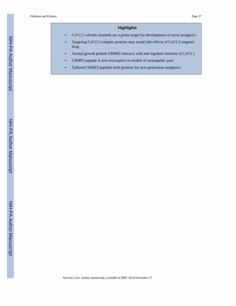
Highlights
• CaV2.2 calcium channels are a prime target for development of novel analgesics
• Targeting CaV2.2 complex proteins may avoid side-effects of CaV2.2-targeteddrug
• Axonal growth protein CRMP2 interacts with and regulates function of CaV2.2
• CRMP2 peptide is anti-nociceptive in models of neuropathic pain
• Tailored CRMP2 peptides hold promise for next generation analgesics
Feldman and Khanna Page 17
Neurosci Lett. Author manuscript; available in PMC 2014 December 17.
NIH
-PA Author Manuscript
NIH
-PA Author Manuscript
NIH
-PA Author Manuscript
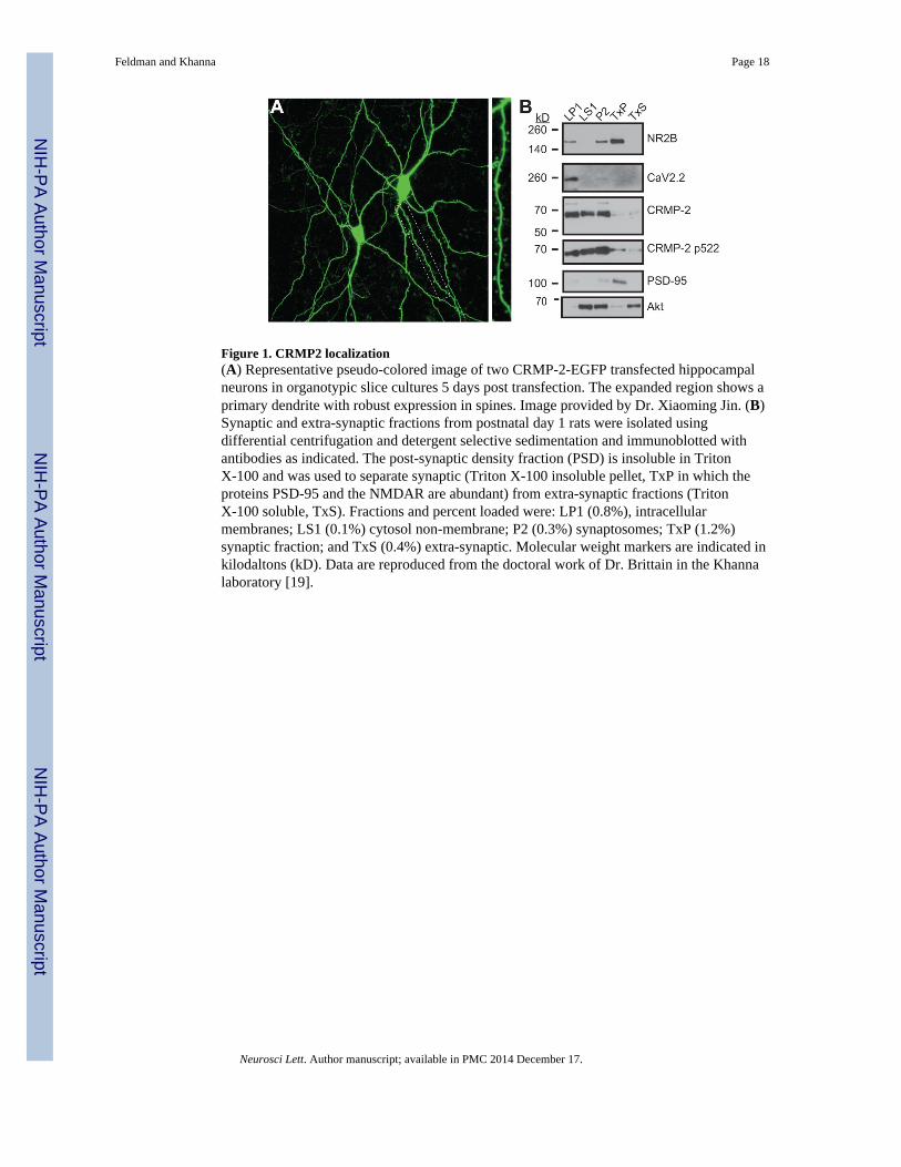
Figure 1. CRMP2 localization(A) Representative pseudo-colored image of two CRMP-2-EGFP transfected hippocampalneurons in organotypic slice cultures 5 days post transfection. The expanded region shows aprimary dendrite with robust expression in spines. Image provided by Dr. Xiaoming Jin. (B)Synaptic and extra-synaptic fractions from postnatal day 1 rats were isolated usingdifferential centrifugation and detergent selective sedimentation and immunoblotted withantibodies as indicated. The post-synaptic density fraction (PSD) is insoluble in TritonX-100 and was used to separate synaptic (Triton X-100 insoluble pellet, TxP in which theproteins PSD-95 and the NMDAR are abundant) from extra-synaptic fractions (TritonX-100 soluble, TxS). Fractions and percent loaded were: LP1 (0.8%), intracellularmembranes; LS1 (0.1%) cytosol non-membrane; P2 (0.3%) synaptosomes; TxP (1.2%)synaptic fraction; and TxS (0.4%) extra-synaptic. Molecular weight markers are indicated inkilodaltons (kD). Data are reproduced from the doctoral work of Dr. Brittain in the Khannalaboratory [19].
Feldman and Khanna Page 18
Neurosci Lett. Author manuscript; available in PMC 2014 December 17.
NIH
-PA Author Manuscript
NIH
-PA Author Manuscript
NIH
-PA Author Manuscript
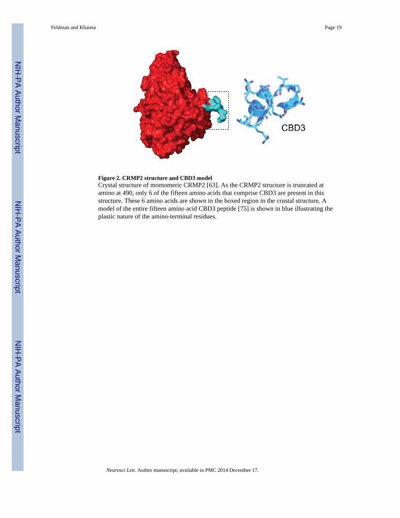
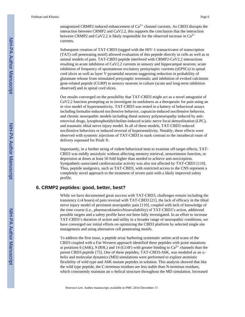
Figure 2. CRMP2 structure and CBD3 modelCrystal structure of momomeric CRMP2 [63]. As the CRMP2 structure is truncated atamino at 490, only 6 of the fifteen amino acids that comprise CBD3 are present in thisstructure. These 6 amino acids are shown in the boxed region in the crustal structure. Amodel of the entire fifteen amino acid CBD3 peptide [75] is shown in blue illustrating theplastic nature of the amino-terminal residues.
Feldman and Khanna Page 19
Neurosci Lett. Author manuscript; available in PMC 2014 December 17.
NIH
-PA Author Manuscript
NIH
-PA Author Manuscript
NIH
-PA Author Manuscript
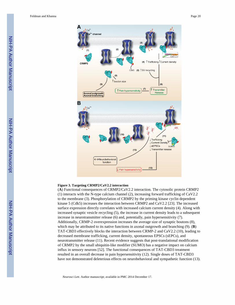
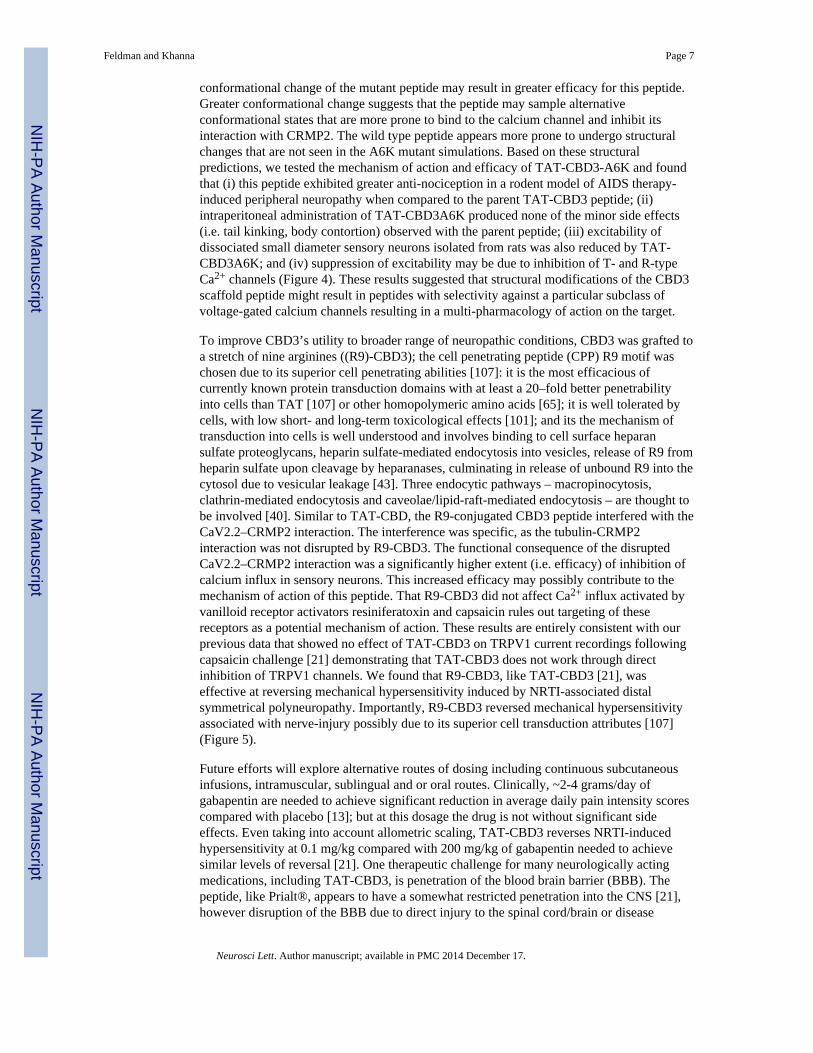
Figure 3. Targeting CRMP2/CaV2.2 interaction(A) Functional consequences of CRMP2/CaV2.2 interaction. The cytosolic protein CRMP2(1) interacts with the N-type calcium channel (2), increasing forward trafficking of CaV2.2to the membrane (3). Phosphorylation of CRMP2 by the priming kinase cyclin dependentkinase 5 (Cdk5) increases the interaction between CRMP2 and CaV2.2 [23]. The increasedsurface expression directly correlates with increased calcium current density (4). Along withincreased synaptic vesicle recycling (5), the increase in current density leads to a subsequentincrease in neurotransmitter release (6) and, potentially, pain hypersensitivity (7).Additionally, CRMP-2 overexpression increases the average size of synaptic boutons (8),which may be attributed to its native functions in axonal outgrowth and branching (9). (B)TAT-CBD3 effectively blocks the interaction between CRMP-2 and CaV2.2 (10), leading todecreased membrane trafficking, current density, spontaneous EPSCs (sEPCs), andneurotransmitter release (11). Recent evidence suggests that post-translational modificationof CRMP2 by the small ubiquitin-like modifier (SUMO) has a negative impact on calciuminflux in sensory neurons [52]. The functional consequences of TAT-CBD3 treatmentresulted in an overall decrease in pain hypersensitivity (12). Single doses of TAT-CBD3have not demonstrated deleterious effects on neurobehavioral and sympathetic function (13).
Feldman and Khanna Page 20
Neurosci Lett. Author manuscript; available in PMC 2014 December 17.
NIH
-PA Author Manuscript
NIH
-PA Author Manuscript
NIH
-PA Author Manuscript
Similar mechanistic effects on trafficking and overall in vivo effects on relief of chronic painare observed with a peptide (structure shown [7]) from the calcium channel itself [111].
Feldman and Khanna Page 21
Neurosci Lett. Author manuscript; available in PMC 2014 December 17.
NIH
-PA Author Manuscript
NIH
-PA Author Manuscript
NIH
-PA Author Manuscript
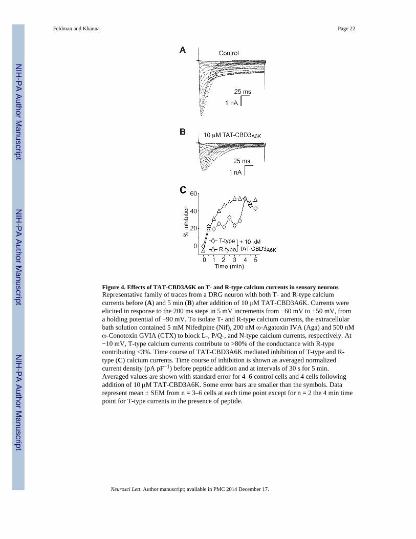
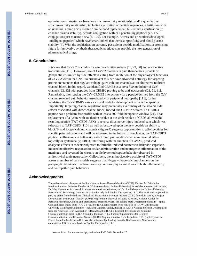
Figure 4. Effects of TAT-CBD3A6K on T- and R-type calcium currents in sensory neuronsRepresentative family of traces from a DRG neuron with both T- and R-type calciumcurrents before (A) and 5 min (B) after addition of 10 μM TAT-CBD3A6K. Currents wereelicited in response to the 200 ms steps in 5 mV increments from −60 mV to +50 mV, froma holding potential of −90 mV. To isolate T- and R-type calcium currents, the extracellularbath solution contained 5 mM Nifedipine (Nif), 200 nM ω-Agatoxin IVA (Aga) and 500 nMω-Conotoxin GVIA (CTX) to block L-, P/Q-, and N-type calcium currents, respectively. At−10 mV, T-type calcium currents contribute to >80% of the conductance with R-typecontributing <3%. Time course of TAT-CBD3A6K mediated inhibition of T-type and R-type (C) calcium currents. Time course of inhibition is shown as averaged normalizedcurrent density (pA pF−1) before peptide addition and at intervals of 30 s for 5 min.Averaged values are shown with standard error for 4–6 control cells and 4 cells followingaddition of 10 μM TAT-CBD3A6K. Some error bars are smaller than the symbols. Datarepresent mean ± SEM from n = 3–6 cells at each time point except for n = 2 the 4 min timepoint for T-type currents in the presence of peptide.
Feldman and Khanna Page 22
Neurosci Lett. Author manuscript; available in PMC 2014 December 17.
NIH
-PA Author Manuscript
NIH
-PA Author Manuscript
NIH
-PA Author Manuscript
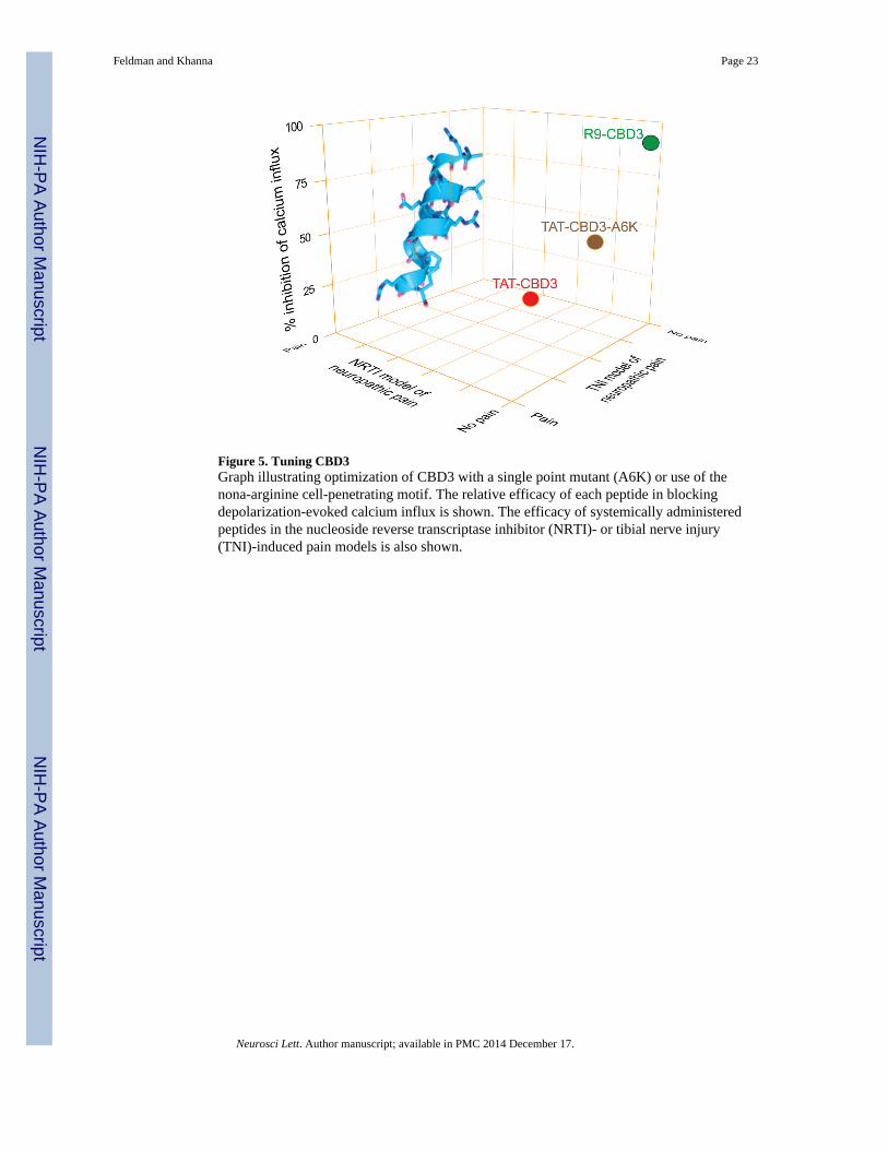
Figure 5. Tuning CBD3Graph illustrating optimization of CBD3 with a single point mutant (A6K) or use of thenona-arginine cell-penetrating motif. The relative efficacy of each peptide in blockingdepolarization-evoked calcium influx is shown. The efficacy of systemically administeredpeptides in the nucleoside reverse transcriptase inhibitor (NRTI)- or tibial nerve injury(TNI)-induced pain models is also shown.
Feldman and Khanna Page 23
Neurosci Lett. Author manuscript; available in PMC 2014 December 17.
NIH
-PA Author Manuscript
NIH
-PA Author Manuscript
NIH
-PA Author Manuscript