Peptide-based inhibitors of protein–protein interactions ...
17
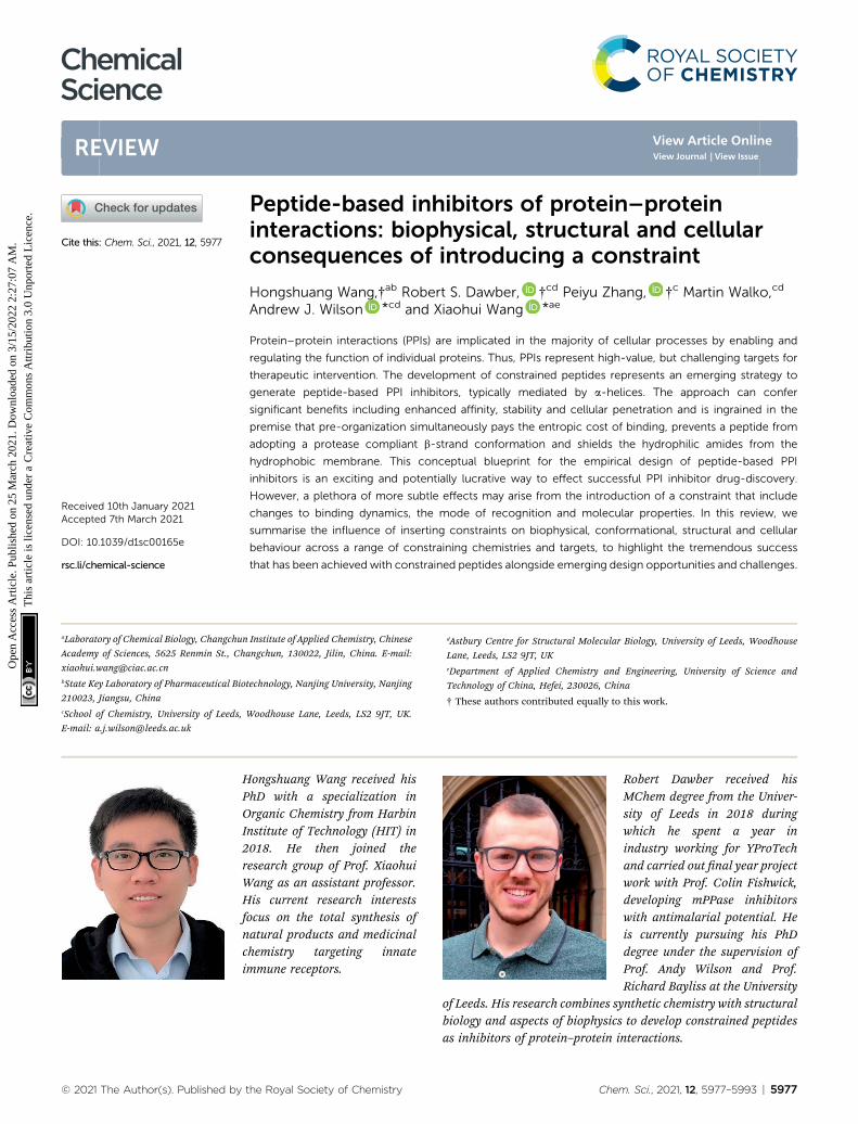
Peptide-based inhibitors of protein–protein interactions: biophysical, structural and cellular consequences of introducing a constraint Hongshuang Wang,† ab Robert S. Dawber, † cd Peiyu Zhang, † c Martin Walko, cd Andrew J. Wilson * cd and Xiaohui Wang * ae Protein–protein interactions (PPIs) are implicated in the majority of cellular processes by enabling and regulating the function of individual proteins. Thus, PPIs represent high-value, but challenging targets for therapeutic intervention. The development of constrained peptides represents an emerging strategy to generate peptide-based PPI inhibitors, typically mediated by a-helices. The approach can confer significant benefits including enhanced affinity, stability and cellular penetration and is ingrained in the premise that pre-organization simultaneously pays the entropic cost of binding, prevents a peptide from adopting a protease compliant b-strand conformation and shields the hydrophilic amides from the hydrophobic membrane. This conceptual blueprint for the empirical design of peptide-based PPI inhibitors is an exciting and potentially lucrative way to effect successful PPI inhibitor drug-discovery. However, a plethora of more subtle effects may arise from the introduction of a constraint that include changes to binding dynamics, the mode of recognition and molecular properties. In this review, we summarise the influence of inserting constraints on biophysical, conformational, structural and cellular behaviour across a range of constraining chemistries and targets, to highlight the tremendous success that has been achieved with constrained peptides alongside emerging design opportunities and challenges. Hongshuang Wang received his PhD with a specialization in Organic Chemistry from Harbin Institute of Technology (HIT) in 2018. He then joined the research group of Prof. Xiaohui Wang as an assistant professor. His current research interests focus on the total synthesis of natural products and medicinal chemistry targeting innate immune receptors. Robert Dawber received his MChem degree from the Univer- sity of Leeds in 2018 during which he spent a year in industry working for YProTech and carried out nal year project work with Prof. Colin Fishwick, developing mPPase inhibitors with antimalarial potential. He is currently pursuing his PhD degree under the supervision of Prof. Andy Wilson and Prof. Richard Bayliss at the University of Leeds. His research combines synthetic chemistry with structural biology and aspects of biophysics to develop constrained peptides as inhibitors of protein–protein interactions. a Laboratory of Chemical Biology, Changchun Institute of Applied Chemistry, Chinese Academy of Sciences, 5625 Renmin St., Changchun, 130022, Jilin, China. E-mail: [email protected] b State Key Laboratory of Pharmaceutical Biotechnology, Nanjing University, Nanjing 210023, Jiangsu, China c School of Chemistry, University of Leeds, Woodhouse Lane, Leeds, LS2 9JT, UK. E-mail: [email protected] d Astbury Centre for Structural Molecular Biology, University of Leeds, Woodhouse Lane, Leeds, LS2 9JT, UK e Department of Applied Chemistry and Engineering, University of Science and Technology of China, Hefei, 230026, China † These authors contributed equally to this work. Cite this: Chem. Sci. , 2021, 12, 5977 Received 10th January 2021 Accepted 7th March 2021 DOI: 10.1039/d1sc00165e rsc.li/chemical-science © 2021 The Author(s). Published by the Royal Society of Chemistry Chem. Sci. , 2021, 12, 5977–5993 | 5977 Chemical Science REVIEW Open Access Article. Published on 25 March 2021. Downloaded on 3/15/2022 2:27:07 AM. This article is licensed under a Creative Commons Attribution 3.0 Unported Licence. View Article Online View Journal | View Issue
Transcript of Peptide-based inhibitors of protein–protein interactions ...
ChemicalScience
REVIEW
Ope
n A
cces
s A
rtic
le. P
ublis
hed
on 2
5 M
arch
202
1. D
ownl
oade
d on
3/1
5/20
22 2
:27:
07 A
M.
Thi
s ar
ticle
is li
cens
ed u
nder
a C
reat
ive
Com
mon
s A
ttrib
utio
n 3.
0 U
npor
ted
Lic
ence
.
View Article OnlineView Journal | View Issue
Peptide-based in
HPOI2rWHfnci
aLaboratory of Chemical Biology, Changchun
Academy of Sciences, 5625 Renmin St., Ch
[email protected] Key Laboratory of Pharmaceutical Bio
210023, Jiangsu, ChinacSchool of Chemistry, University of Leeds,
E-mail: [email protected]
Cite this: Chem. Sci., 2021, 12, 5977
Received 10th January 2021Accepted 7th March 2021
DOI: 10.1039/d1sc00165e
rsc.li/chemical-science
© 2021 The Author(s). Published by
hibitors of protein–proteininteractions: biophysical, structural and cellularconsequences of introducing a constraint
Hongshuang Wang,†ab Robert S. Dawber, †cd Peiyu Zhang, †c Martin Walko,cd
Andrew J. Wilson *cd and Xiaohui Wang *ae
Protein–protein interactions (PPIs) are implicated in the majority of cellular processes by enabling and
regulating the function of individual proteins. Thus, PPIs represent high-value, but challenging targets for
therapeutic intervention. The development of constrained peptides represents an emerging strategy to
generate peptide-based PPI inhibitors, typically mediated by a-helices. The approach can confer
significant benefits including enhanced affinity, stability and cellular penetration and is ingrained in the
premise that pre-organization simultaneously pays the entropic cost of binding, prevents a peptide from
adopting a protease compliant b-strand conformation and shields the hydrophilic amides from the
hydrophobic membrane. This conceptual blueprint for the empirical design of peptide-based PPI
inhibitors is an exciting and potentially lucrative way to effect successful PPI inhibitor drug-discovery.
However, a plethora of more subtle effects may arise from the introduction of a constraint that include
changes to binding dynamics, the mode of recognition and molecular properties. In this review, we
summarise the influence of inserting constraints on biophysical, conformational, structural and cellular
behaviour across a range of constraining chemistries and targets, to highlight the tremendous success
that has been achieved with constrained peptides alongside emerging design opportunities and challenges.
ongshuang Wang received hishD with a specialization inrganic Chemistry from Harbinnstitute of Technology (HIT) in018. He then joined theesearch group of Prof. Xiaohuiang as an assistant professor.is current research interestsocus on the total synthesis ofatural products and medicinalhemistry targeting innatemmune receptors.
Robert Dawber received hisMChem degree from the Univer-sity of Leeds in 2018 duringwhich he spent a year inindustry working for YProTechand carried out nal year projectwork with Prof. Colin Fishwick,developing mPPase inhibitorswith antimalarial potential. Heis currently pursuing his PhDdegree under the supervision ofProf. Andy Wilson and Prof.Richard Bayliss at the University
of Leeds. His research combines synthetic chemistry with structuralbiology and aspects of biophysics to develop constrained peptidesas inhibitors of protein–protein interactions.
Institute of Applied Chemistry, Chinese
angchun, 130022, Jilin, China. E-mail:
technology, Nanjing University, Nanjing
Woodhouse Lane, Leeds, LS2 9JT, UK.
dAstbury Centre for Structural Molecular Biology, University of Leeds, Woodhouse
Lane, Leeds, LS2 9JT, UKeDepartment of Applied Chemistry and Engineering, University of Science and
Technology of China, Hefei, 230026, China
† These authors contributed equally to this work.
the Royal Society of Chemistry Chem. Sci., 2021, 12, 5977–5993 | 5977
Chemical Science Review
Ope
n A
cces
s A
rtic
le. P
ublis
hed
on 2
5 M
arch
202
1. D
ownl
oade
d on
3/1
5/20
22 2
:27:
07 A
M.
Thi
s ar
ticle
is li
cens
ed u
nder
a C
reat
ive
Com
mon
s A
ttrib
utio
n 3.
0 U
npor
ted
Lic
ence
.View Article Online
1 Introduction
Protein–protein interactions (PPIs) mediate virtually all biolog-ical processes and are associated with many diseases. PPIs havehistorically represented challenging targets for competitive(orthosteric) inhibitor discovery; they typically involve interac-tion of comparatively large and less featured protein surfaces, incomparison to established drug targets.1
The a-helix has been shown to have a relatively high preva-lence in the human PPI interactome2,3 and represents a genericpharmacophore for ligand design.4 As a result, a-helix mediatedPPIs have attracted signicant attention for the development ofselective probes and drug candidates to meet a plethora ofunmet therapeutic needs.5 At such PPI interfaces, the a-helix ofone protein is bound within a groove on the binding partner. Asignicant number of such interactions involve short peptidemotifs;6 these are typically located within intrinsically
Peiyu Zhang received his Bach-elor of Science degree in BasicPharmacy from Shenyang Phar-maceutical University (She-nyang, China) in 2016. Hereceived his Master of Sciencedegree in Medicinal Chemistry,also from Shenyang Pharma-ceutical University, in 2019.From 2017–2019, he workedwith Prof. Keliang Liu at theBeijing Institute of Pharma-cology and Toxicology as
a visiting student. He is a PhD student in the group of Prof. AndyWilson in the School of Chemistry, University of Leeds. Hisresearch focuses on the discovery of protein–protein interactioninhibitors using peptidomimetics approaches.
Martin Walko received his MScdegree from P. J. SafarikUniversity in Kosice (Slovakia)and completed his PhD degreewith B. L. Feringa at Universityof Groningen working on molec-ular switches. Aer time workingas an independent researcher atP. J. Safarik University in Kosiceand research in protein channelgating with A. Kocer at Univer-sity of Groningen, he is nowa research fellow at the Univer-
sity of Leeds. His current research focuses on development ofchemical biology tools to study amyloid aggregation and protein–protein interactions.
5978 | Chem. Sci., 2021, 12, 5977–5993
disordered regions (IDRs),7–9 thus the a-helix is transientlystabilized on formation of the PPI. Crystallographic and struc-tural analyses of these interfaces can facilitate the developmentof peptides (or judiciously designed small molecules) thatmimic the helix both topologically and/or topographically; suchmimetics (termed ‘peptidomimetics’) offer a promising startingpoint in the development of PPI inhibitors and have led toclinical candidates.10 Constrained peptides represent a branchof peptidomimetics that have emerged as powerful tools forperturbing PPIs; chemically constraining (or ‘stapling’)a peptide in its bioactive a-helical conformation has been re-ported to confer numerous benets such as enhanced proteaseresistance, stability in cells, increased cellular uptake andimproved biophysical properties in comparison to wild-typesequences.11–13
Just as the position of a constraint within a sequence haseffects on peptide helicity, the nature of chemical linker can
Andrew (Andy) J. Wilson isProfessor of Organic Chemistryat the University of Leeds. Hecompleted a PhD at WarwickUniversity supervised by Prof.David Leigh FRS before post-doctoral research with Prof.Andrew Hamilton FRS at YaleUniversity and with Prof. E.Meijer and Prof. Rint Sijbesmaat Technische Universiteit Eind-hoven. Andy has 20 years expe-rience working at the interface
between chemistry and biology, with signicant focus on under-standing and modulating protein–protein interactions (PPIs). Andywas recognized through the Royal Society of Chemistry (RSC) BobHay Lectureship (2012) and the RSC Norman Heatley Award(2016).
Xiaohui Wang is a Professor ofLaboratory of Chemical Biology,Changchun Institute of AppliedChemistry (CIAC), ChineseAcademy of Sciences (CAS). Hereceived his BE in Bioengi-neering from Beijing Technologyand Business University and hisPhD in inorganic chemistryunder the direction of Prof.Xiaogang Qu from CIAC. Aerhis post-doc training under thesupervision with Prof. Hang Yin
and Linda R Watkins at University of Colorado, he returned to hisalma mater CIAC to establish the Chemical Biology and DrugDiscovery Group. His research interests involve the discovery ofmodulators of innate immune receptors and integral membraneproteins.
© 2021 The Author(s). Published by the Royal Society of Chemistry
Review Chemical Science
Ope
n A
cces
s A
rtic
le. P
ublis
hed
on 2
5 M
arch
202
1. D
ownl
oade
d on
3/1
5/20
22 2
:27:
07 A
M.
Thi
s ar
ticle
is li
cens
ed u
nder
a C
reat
ive
Com
mon
s A
ttrib
utio
n 3.
0 U
npor
ted
Lic
ence
.View Article Online
also inuence its structure and function. Since the pioneeringwork of Grubbs14 and Verdine15 to develop hydrocarbonstapling, an extensive toolkit has been elaborated to chemicallyconstrain peptides.16,17 These tools include: disulde bonds,lactam bridges, hydrogen bond surrogates, alkanediyl tethers,bridges from thiol–ene coupling, triazole-staples from “click”chemistry and supramolecular approaches, with a numberdeveloped to allow further functionalization via the constrain-ing linker.17–25 With such synthetic diversity on offer, it isreasonable to ask how can one predict which staple type will bemost suitable? Different chemical linkers induce helicity ina given sequence to different extents and, when constraininga peptide in its bioactive a-helical conformation, the optimalexibility is target dependent.26
In this review, we provide an overview of constrainedpeptides demonstrating non-classical biophysical or structuralbehaviour. We focus predominantly on a-helix mediated PPIsalthough note the approach has been broadened to otherclasses of PPIs.27,28 We collate recent and unusual observationsthat highlight the different inuences staple types can have onboth peptide conformation and target binding. Finally, wediscuss the most recent literature on mechanisms of cellpenetration and studies to ascertain the characteristics of con-strained peptides that enhance cellular uptake. Ultimately, weintend that this review will aid the rational design and devel-opment of highly potent and cell penetrating constrainedpeptides as PPI inhibitors.
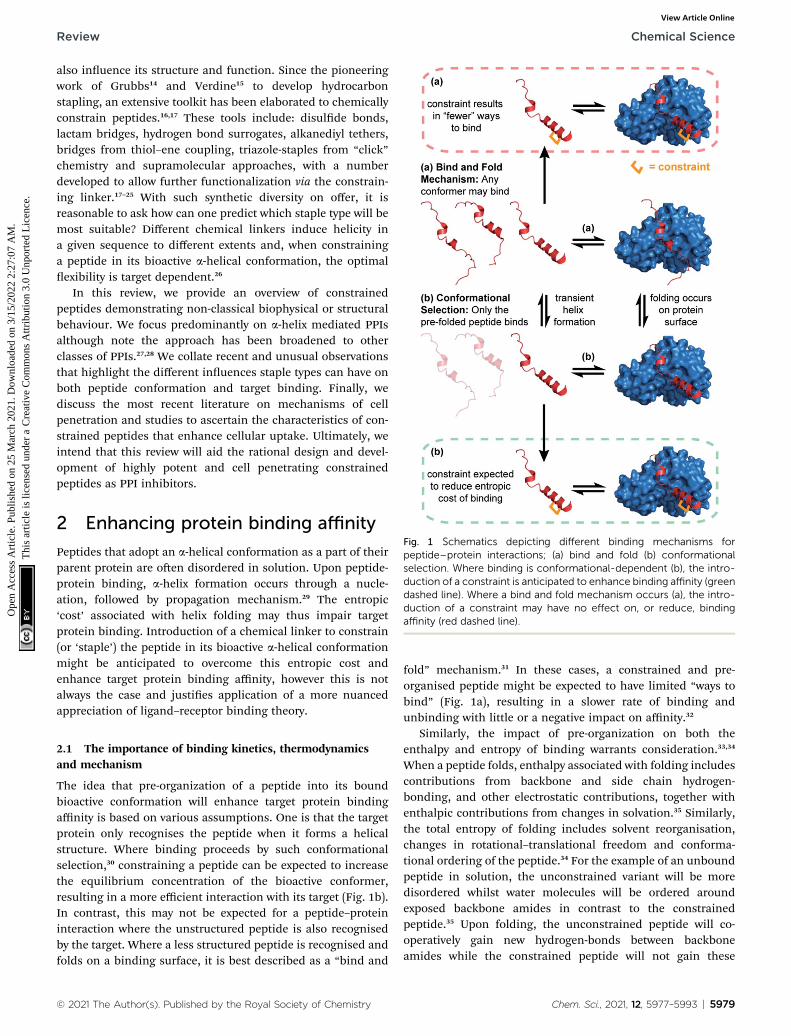
Fig. 1 Schematics depicting different binding mechanisms forpeptide–protein interactions; (a) bind and fold (b) conformationalselection. Where binding is conformational-dependent (b), the intro-duction of a constraint is anticipated to enhance binding affinity (greendashed line). Where a bind and fold mechanism occurs (a), the intro-duction of a constraint may have no effect on, or reduce, bindingaffinity (red dashed line).
2 Enhancing protein binding affinity
Peptides that adopt an a-helical conformation as a part of theirparent protein are oen disordered in solution. Upon peptide-protein binding, a-helix formation occurs through a nucle-ation, followed by propagation mechanism.29 The entropic‘cost’ associated with helix folding may thus impair targetprotein binding. Introduction of a chemical linker to constrain(or ‘staple’) the peptide in its bioactive a-helical conformationmight be anticipated to overcome this entropic cost andenhance target protein binding affinity, however this is notalways the case and justies application of a more nuancedappreciation of ligand–receptor binding theory.
2.1 The importance of binding kinetics, thermodynamicsand mechanism
The idea that pre-organization of a peptide into its boundbioactive conformation will enhance target protein bindingaffinity is based on various assumptions. One is that the targetprotein only recognises the peptide when it forms a helicalstructure. Where binding proceeds by such conformationalselection,30 constraining a peptide can be expected to increasethe equilibrium concentration of the bioactive conformer,resulting in a more efficient interaction with its target (Fig. 1b).In contrast, this may not be expected for a peptide–proteininteraction where the unstructured peptide is also recognisedby the target. Where a less structured peptide is recognised andfolds on a binding surface, it is best described as a “bind and
© 2021 The Author(s). Published by the Royal Society of Chemistry
fold” mechanism.31 In these cases, a constrained and pre-organised peptide might be expected to have limited “ways tobind” (Fig. 1a), resulting in a slower rate of binding andunbinding with little or a negative impact on affinity.32
Similarly, the impact of pre-organization on both theenthalpy and entropy of binding warrants consideration.33,34
When a peptide folds, enthalpy associated with folding includescontributions from backbone and side chain hydrogen-bonding, and other electrostatic contributions, together withenthalpic contributions from changes in solvation.35 Similarly,the total entropy of folding includes solvent reorganisation,changes in rotational–translational freedom and conforma-tional ordering of the peptide.34 For the example of an unboundpeptide in solution, the unconstrained variant will be moredisordered whilst water molecules will be ordered aroundexposed backbone amides in contrast to the constrainedpeptide.35 Upon folding, the unconstrained peptide will co-operatively gain new hydrogen-bonds between backboneamides while the constrained peptide will not gain these
Chem. Sci., 2021, 12, 5977–5993 | 5979
Chemical Science Review
Ope
n A
cces
s A
rtic
le. P
ublis
hed
on 2
5 M
arch
202
1. D
ownl
oade
d on
3/1
5/20
22 2
:27:
07 A
M.
Thi
s ar
ticle
is li
cens
ed u
nder
a C
reat
ive
Com
mon
s A
ttrib
utio
n 3.
0 U
npor
ted
Lic
ence
.View Article Online
enthalpically favourable contributions to the same extent, yetless impact from the entropically favourable release of water canbe anticipated.36 Thus, the opposing entropic and enthalpiccontributions (enthalpy–entropy compensation) oen result inonly marginal increase in stability of a folded form i.e. the netDG of folding may be comparable to the magnitude of a singlenon-covalent interaction (i.e. �<5 kJ mol�1) and this might beconsidered the maximum accessible gain arising from pre-organization (in the absence of additional interactions beingintroduced between constrained peptide and target protein; seelater). Such a framework ultimately represents an over-simplication; making the assumption that constraininga peptide changes the energetic state relative to the wild-typesequence is not valid as the sequences are different. Con-straining a peptide simply increases the stability/energy of itsunfolded form.
In a study to explore how pre-organization of BH3-familypeptides affects their inhibitory potency of the BH3/BCL2-family PPIs,37 specically BID and BIM BH3 domains withMCL-1 and BCL-xL, Miles et al. found that whilst the introduc-tion of a hydrocarbon constraint increased the population ofthe bioactive a-helical conformation of the peptides in solution,this did not enhance their potency.35 In fact, some of the con-strained peptides exhibited a signicant loss of potency. Co-crystal structures of the constrained BH3 peptides bound tothe BCl-2 family proteins showed that the constraint induced nosignicant differences in the orientation of hot-spot side chainsor registry of the peptide, nor did the constraint overtly intro-duce a steric clash with the target protein. Thus, the unexpecteddrop in potency could not be explained by the static structure ofthe bound complexes. Surface plasmon resonance (SPR) assaysrevealed that the rates of binding and unbinding differedbetween wild-type and constrained sequences; in all cases, theintroduction of a hydrocarbon constraint resulted in a signi-cant decrease in both on and off rates consistent with a bindand fold mechanism of binding. Van't Hoff analyses of uo-rescence anisotropy direct binding experiments allowed thecontributions of enthalpy and entropy to the interaction to bedetermined; these data revealed that the entropic cost ofbinding was indeed reduced for the constrained peptide.However, the favourable change in entropy was compensatedfor by an opposing change in the enthalpic contribution tobinding.35 These observations are consistent with what mightbe expected when constraining a peptide that interacts with itstarget through a bind and fold mechanism of interaction.
Ochsenbein and co-workers also observed enthalpy–entropycompensation in studies on the histone H3/ASF1 (anti-silencingfunction 1) interaction.38 In this work, variant H3 peptides (res118–135) bearing i, i + 4 hydrocarbon staples in multiple posi-tions within the sequence were investigated.39 In addition tohighlighting the above discussed limitations in correlating pre-organization with binding affinity, biophysical analyses alsorevealed that promotion of a-helix formation is dependent onthe position at which the constraint is introduced in thesequence. This is unsurprising, since different peptidesequences have different propensities for a-helix formation andreplacing different amino acids in a native sequence with
5980 | Chem. Sci., 2021, 12, 5977–5993
unnatural amino acids (used to incorporate a constraint) willinuence this natural propensity to differing extents.
Important insight into the effects of constraining peptides40
has been obtained using stapled peptide ligands for theEukaryotic translation initiation factor 4E (eIF4E); a protein thatregulates cap-dependent mRNA translation via interactionswith competing binding partners, 4E-BP1 and eIF4G.41,42 Both4E-BP1 and eIF4G bind to the same canonical groove of eIF4Eand bind via intrinsically disordered regions (IDRs) which forma-helical motifs when bound. Using molecular dynamics (MD)simulations it was shown that hydrocarbon constrained eIF4Gpeptides experienced changes in structural dynamics whenbound or unbound to eIF4E. Although stabilization of theunbound peptide in a helical conformation could be readilyachieved, binding could also be impeded by favouring meta-stable conformations that had to change on target binding toallow key side chains to adopt the orientation required formolecular recognition. These analyses were supported byexperimental and structural studies, highlighting the impor-tance of stabilizing solution conformations that match thebound conformation. Rational design subsequently led tostapled-peptides with enhanced target residence time targetingan unexploited patch on the surface of eIF4E.43 Similar obser-vations were made by Gallagher et al.44 Initially, the groupmeasured the helical propensities of the linear wild-type 4E-BP1and eIF4G peptides and found that the 4E-BP1 sequenceexhibits greater helicity in solution than the eIF4G. SPR wasused to prole the temperature- and salt-dependence of thepeptide–protein interactions; whilst eIF4G binding was foundto depend on electrostatic contributions and vary in bindingkinetics with temperature, 4E-BP1 was relatively unaffected. Thedata indicated that although 4E-BP1 and eIF4G form similarbound structures, the two peptides adopt distinct bindingmechanisms with the 4E-BP1 peptide exhibiting a greatercomplementarity for eIF4E and forming a more stable boundcomplex.44 By comparing the effects of hydrocarbon stapling onthe two sequences, Garner and co-workers demonstrateda correlation of the linear 4E-BP1 and eIF4G binding kineticswith the corresponding stapled peptide properties. Whilststapling the more complementary 4E-BP1 sequence improvedaffinity and bioactivity (indicating conformational selectionoperates for 4E-BP1), stapling eIF4G produced a nonhelicalmacrocyclic peptide with poorer affinity for eIF4E than its linearcounterpart. Therefore, this study again emphasized that theanalysis of binding mechanism and kinetics can play a crucialrole in understanding whether an IDR peptide will benet fromthe introduction of a conformational constraint.
Jamieson and co-workers adopted a constrained peptideapproach to target the a-helix mediated PPI of mitotic kinaseAurora-A with microtubule-associated protein TPX2.45 In thiswork, a hydrocarbon stapled TPX2 peptide demonstratedimproved binding affinity for Aurora-A and was even shown tomimic the function of TPX2 in activating autophosphorylationof the kinase. However, a closer inspection of the thermody-namic determinants of binding, as measured by IsothermalTitration Calorimetry (ITC), revealed a surprising nding;although the introduction of the constraint increased the
© 2021 The Author(s). Published by the Royal Society of Chemistry

Review Chemical Science
Ope
n A
cces
s A
rtic
le. P
ublis
hed
on 2
5 M
arch
202
1. D
ownl
oade
d on
3/1
5/20
22 2
:27:
07 A
M.
Thi
s ar
ticle
is li
cens
ed u
nder
a C
reat
ive
Com
mon
s A
ttrib
utio
n 3.
0 U
npor
ted
Lic
ence
.View Article Online
population of the bioactive a-helix and enhanced the bindingaffinity, the entropy of binding for the constrained peptide wasmore unfavourable when compared to the wild-type. Inconcordance, the constrained peptide had a more favourableenthalpy of binding, suggesting it made more favourableinteractions with the Aurora-A protein when bound. Thishypothesis was later conrmed via crystallographic analysis, bysuperposing the existing structure of Aurora-A in complex withnative TPX2 (PDB: 1OL5) onto a crystal structure of the boundstapled TPX2 peptide (PDB: 5LXM; Fig. 2a). Although thehydrocarbon staple itself makes no contacts with the Aurora-Asurface and the crucial hot-spot residues remain in almostidentical orientations between the two structures (Fig. 2b andc), the staple extends the length of the helix by an additionalturn and alters the conformation of the bound helix, allowingtwo additional charged side chains, Glu36TPX2 and Lys38TPX2, tocontribute to hydrogen-bonding interactions with Aurora-A(Fig. 2d). Thus the less favourable entropy can be accountedfor by the requirement to form a longer more ordered helix so asto make the additional enthalpically favourable non-covalentcontacts.
Hetherington et al. prepared and investigated a series ofconstrained peptides to target HIF-1a/p300;46 a PPI which
Fig. 2 Comparison of unconstrained/constrained Aurora-A boundTPX2 peptides; (a) view of Aurora-A (blue) bound to stapled TPX2 (red,hydrocarbon staple: orange, PDB: 5LXM) with native TPX2 (purple,PDB: 1OL5) overlaid. TPX2 residues known to be crucial for binding toAurora-A are shown as sticks to highlight the conserved binding modebetween stapled and native TPX2; (b) Tyr8, Tyr10, Asp11, Trp34 andPhe35; (c) Phe16 and Phe19; (d) orientations of residues Lys38 andGlu36 differ, promoting additional electrostatic interactions (dashedlines) between peptide and Aurora-A.45
© 2021 The Author(s). Published by the Royal Society of Chemistry
regulates oxygen levels in cells and is oen hijacked by cancer tosupply growing tumours with oxygen.47–49 Here, the peptideswere constrained through reaction of dibromomaleimide with iand i + 4 Cys residues.21,50 One of the dibromomaleimide (DBM)stapled HIF-1a peptides demonstrated a signicant enhance-ment of binding affinity for p300 in comparison to the uncon-strained native peptide. However, contrary to expectation, theenhanced inhibition did not correlate with an increase in a-helicity as shown by circular dichroism (CD) experiments. In anattempt to explain the benecial effects of introducing the DBMstaple, the group compared MD simulations of the HIF-1avariants both in solution and in complex with p300. MDsimulations indicated that both the constrained and uncon-strained peptides showed greater helical character in the boundstate when compared to the unbound peptides, with a moredramatic increase observed for the DBM stapled peptide. Thus,the staple-induced affinity enhancement was proposed to occuras a result of stabilizing the bound state of the peptide incomplex with p300 46 and was supported by experimental CDdifference experiments. In a similar vein, Grossmann and co-workers also observed that changes in the behaviour of boundstate as a consequence of introducing a constraint are impor-tant for interaction of Exoenzyme S (Exo S) derived peptideswith 14–3–3 protein although in this case, increased affinity wasattributed to increased dynamics in the peptide-receptorcomplex.51
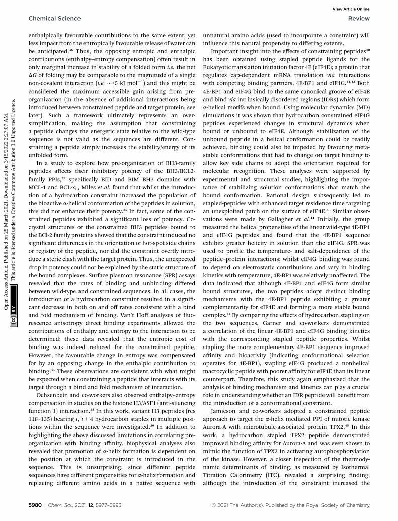
In studies by Strizhak et al. a library of peptide analogues,based on a known p53/MDM2 peptide discovered by phagedisplay,53 were stapled with a photoisomerizable diarylethene(DAE) moiety between the i, i + 7 residues using azide–alkyne“click” chemistry.52 Each analogue possessed two photo-isomers, referred to as “open” or “closed”, depending onexposure to visible or UV light, respectively (Fig. 3a). Analoguesalso differed in linker length (n ¼ 1 or 2) and/or N-methylation(R ¼ H or Me). By utilizing competition assays based on tryp-tophan uorescence quenching, an interesting observation wasnoted; analogues constrained with “open” DAE photoisomerswere consistently stronger binders to MDM2 than their “closed”counterparts. Moreover, ITC measurements, performed to gaininsight into the thermodynamic parameters of binding,revealed that binding of the “closed” forms were mostlyenthalpy-driven, whilst the “open” isomers bound with a greaterentropic contribution. Attempts to crystalize the highest affinityanalogue (R ¼ Me, n ¼ 1) bound to MDM2 were successful forthe “open” form (Fig. 3b). The peptide bound to MDM2 as ex-pected, forming an a-helix, with the Phe3p53, Trp7p53 andLeu10p53 hot-spot residues occupying the known lipophilicpockets on the protein surface (Fig. 3b). The linker was shown todirectly interact with the target protein by edge-to-face p-stacking interactions between the triazole and thiophenemoieties of the linker and Phe55MDM2. The triazole ring alsoforms hydrogen bonds to a local water molecule, which, in turn,forms a hydrogen bond with Gln59MDM2. Although the “open”isomer has greater conformational freedom, given the struc-tural data illustrate additional non-covalent interactions canform, the rationale for the observed greater entropic contribu-tion to binding in comparison to the “closed” form is not fully
Chem. Sci., 2021, 12, 5977–5993 | 5981
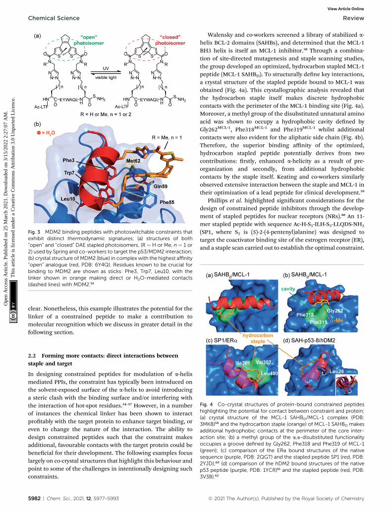
Fig. 3 MDM2 binding peptides with photoswitchable constraints thatexhibit distinct thermodynamic signatures; (a) structures of both“open” and “closed” DAE stapled photoisomers, (R ¼ H or Me, n ¼ 1 or2) used by Spring and co-workers to target the p53/MDM2 interaction;(b) crystal structure of MDM2 (blue) in complex with the highest affinity“open” analogue (red, PDB: 6Y4Q). Residues known to be crucial forbinding to MDM2 are shown as sticks: Phe3, Trp7, Leu10, with thelinker shown in orange making direct or H2O-mediated contacts(dashed lines) with MDM2.52
Chemical Science Review
Ope
n A
cces
s A
rtic
le. P
ublis
hed
on 2
5 M
arch
202
1. D
ownl
oade
d on
3/1
5/20
22 2
:27:
07 A
M.
Thi
s ar
ticle
is li
cens
ed u
nder
a C
reat
ive
Com
mon
s A
ttrib
utio
n 3.
0 U
npor
ted
Lic
ence
.View Article Online
clear. Nonetheless, this example illustrates the potential for thelinker of a constrained peptide to make a contribution tomolecular recognition which we discuss in greater detail in thefollowing section.
Fig. 4 Co-crystal structures of protein-bound constrained peptideshighlighting the potential for contact between constraint and protein;(a) crystal structure of the MCL-1 SAHBD/MCL-1 complex (PDB:3MK8)58 and the hydrocarbon staple (orange) of MCL-1 SAHBD makesadditional hydrophobic contacts at the perimeter of the core inter-action site; (b) a methyl group of the a,a-disubstituted functionalityoccupies a groove defined by Gly262, Phe318 and Phe319 of MCL-1(green); (c) comparison of the ERa bound structures of the nativesequence (purple, PDB: 2QGT) and the stapled peptide SP1 (red, PDB:2YJD);60 (d) comparison of the hDM2 bound structures of the nativep53 peptide (purple, PDB: 1YCR)61 and the stapled peptide (red, PDB:3V3B).62
2.2 Forming more contacts: direct interactions betweenstaple and target
In designing constrained peptides for modulation of a-helixmediated PPIs, the constraint has typically been introduced onthe solvent-exposed surface of the a-helix to avoid introducinga steric clash with the binding surface and/or interfering withthe interaction of hot-spot residues.54–57 However, in a numberof instances the chemical linker has been shown to interactprotably with the target protein to enhance target binding, oreven to change the nature of the interaction. The ability todesign constrained peptides such that the constraint makesadditional, favourable contacts with the target protein could bebenecial for their development. The following examples focuslargely on co-crystal structures that highlight this behaviour andpoint to some of the challenges in intentionally designing suchconstraints.
5982 | Chem. Sci., 2021, 12, 5977–5993
Walensky and co-workers screened a library of stabilized a-helix BCL-2 domains (SAHBs), and determined that the MCL-1BH3 helix is itself an MCL-1 inhibitor.58 Through a combina-tion of site-directed mutagenesis and staple scanning studies,the group developed an optimized, hydrocarbon stapled MCL-1peptide (MCL-1 SAHBD). To structurally dene key interactions,a crystal structure of the stapled peptide bound to MCL-1 wasobtained (Fig. 4a). This crystallographic analysis revealed thatthe hydrocarbon staple itself makes discrete hydrophobiccontacts with the perimeter of the MCL-1 binding site (Fig. 4a).Moreover, a methyl group of the disubstituted unnatural aminoacid was shown to occupy a hydrophobic cavity dened byGly262MCL-1, Phe318MCL-1 and Phe319MCL-1 whilst additionalcontacts were also evident for the aliphatic side chain (Fig. 4b).Therefore, the superior binding affinity of the optimized,hydrocarbon stapled peptide potentially derives from twocontributions: rstly, enhanced a-helicity as a result of pre-organization and secondly, from additional hydrophobiccontacts by the staple itself. Keating and co-workers similarlyobserved extensive interaction between the staple and MCL-1 intheir optimization of a lead peptide for clinical development.59
Phillips et al. highlighted signicant considerations for thedesign of constrained peptide inhibitors through the develop-ment of stapled peptides for nuclear receptors (NRs).60 An 11-mer stapled peptide with sequence Ac-H-S5-ILH-S5-LLQDS-NH2
(SP1, where S5 is (S)-2-(4-pentenyl)alanine) was designed totarget the coactivator binding site of the estrogen receptor (ER),and a staple scan carried out to establish the optimal constraint.
© 2021 The Author(s). Published by the Royal Society of Chemistry

Fig. 5 Comparison of constrained p53/hDM2 structures; (a) thebinding site formed in the crystallographically unique M06 complex iscapped by the ‘hinge’ helix of the hDM2 lid (PDB: 4UMN); (b) thebinding pockets that form when hDM2 interacts with SAH-8 (PDB:3V3B);62 (c) The ‘hinge’ helix caps the bottom of the pocket; (d) Tyr100(green) adopts the ‘closed’ conformation and forms a hydrogen bondinteraction with Asn29.65
Review Chemical Science
Ope
n A
cces
s A
rtic
le. P
ublis
hed
on 2
5 M
arch
202
1. D
ownl
oade
d on
3/1
5/20
22 2
:27:
07 A
M.
Thi
s ar
ticle
is li
cens
ed u
nder
a C
reat
ive
Com
mon
s A
ttrib
utio
n 3.
0 U
npor
ted
Lic
ence
.View Article Online
Structures of these complexes and of the peptides in isolationwere studied. For the most potent stapled peptide (SP1), boundto the ligand binding domain of ERa, inclusion of the hydro-carbon staple signicantly increased the helicity of the peptideas judged by CD spectroscopy and NMR. However, the crystalstructure of the SP1/ERa complex revealed that the hydrophobicstaple itself formed favorable contacts with the hydrophobicsurface dened by Val307ERa, Ile310ERa, and Leu490ERa (Fig. 4c).Comparison with the coactivator protein–peptide complex(2QGT) revealed a difference of a quarter turn of the helix,shiing the binding site residues out of register by one position.Therefore, the introduction of the hydrocarbon staple not onlyconformationally restrained the peptide, but induced a non-canonical mode of interaction with the potential to lead tonon-specic effects.
Later, Baek et al. reported a crystal structure of a hydro-carbon stapled p53 peptide (SAH-p53-8)63 in complex with itsbinding partner, hDM2.62 The crystal structure revealed thatthe staple occupied a hydrophobic region on the rim of thep53 binding site on hDM2, contributing approximately 10% ofthe total surface contact area between peptide and protein,likely enhancing binding affinity. Stapling of the peptideimposed perfectly helical angles (�58�/�45�) on Leu26p53 inthe structure of the constrained peptide resulting in a morehelical bound-state when compared to the wild-type, uncon-strained counterpart. Moreover, the resulting conformationalchange adjusted the Leu26p53 side chain orientation, leadingto a stronger interaction with hDM2 (Fig. 4d). Similar obser-vations were made for ATSP-7041 – a clinical candidatepeptide-based inhibitor of hDM2 and hDMX developed byAileron.64
Ghadessy and co-workers reported a crystal structure of thehydrocarbon stapled peptide M06 – a variant of one previouslyreported by Brown et al.66 – in complex with hDM2.65 The staplewas shown to pack favourably against hydrophobic residues ofhDM2. In addition, theM06/hDM2 crystal structure showed thatthe peptide binding groove was capped by the helical ‘hinge’region of hDM2 (Fig. 5a), near to the C-terminus of the boundpeptide. Comparison with the SAH-p53-8/hDM2 crystal struc-ture reported by Baek et al., reveals signicant differences(Fig. 5b).62 In the M06 complex the shorter helix of M06 does notll the hDM2 pocket as completely as SAH-p53-8, such that thehinge region of hDM2 adopts a helical conformation that capsthe pocket, accommodating a tight t of M06 (Fig. 5c). In theSAH-p53-8/hDM2 structure, Tyr100 of hDM2 points intoa ‘pocket’, stabilized by a hydrogen bond interaction withAsn29p53. This results in occlusion of the pocket, and in turn,shields Leu26p53 from solvent (Fig. 5d). It is clear that thismovement of the hinge can be accommodated by Tyr100hDM2
either projecting into (‘closed’ conformation) or out (‘open’conformation) of the p53 binding site on hDM2. Thus eventhough a constraint can form interactions with the targetprotein, this example highlights that in doing so, it can induceconformational changes adding to the challenge of designingconstraints so as to optimize non-covalent interactions withtarget proteins.
© 2021 The Author(s). Published by the Royal Society of Chemistry
2.3 The staple structure: inuence on peptide conformationand target binding
As discussed in the preceding section, the chemical staple usedto constrain peptides can, in certain cases, interact directly withthe target protein to enhance binding affinity. Minor alterationsto the constraint, such as stereochemistry at the a-position, a-substituent groups and additional branching on the hydro-carbon e.g. a g-Me group, insert-site and linker length can thussignicantly impact on the behaviour of constrained peptides.Indeed, during the development of the hydrocarbon staple itwas noted that small changes in linker length resulted insignicant effects on the efficiency of stapling reactions andconsequently the effects on peptide structure.15 Below wediscuss several examples that showcase the diverse inuence ofstaple structure on peptide conformation and binding.
Traditionally, incorporation of unnatural a-methyl, a-alkenyldisubstituted amino acids at i, i + 4, or i, i + 7 positions, is usedto introduce hydrocarbon staples. The a-methyl group wasintroduced to the unnatural amino acids for its added helix-stabilizing effect.15,67 Yeo et al. explored this using the BCL-2/BH3 family PPIs as a model; comparison of a BID sequenceconstrained using a-methyl, a-alkenyl amino acids (BID-DM)with the a-alkenyl monosubstituted variant (BID-MM) revealedcomparable helicity, resistance to proteolysis and potency forboth.68 However, such behaviour may not hold in every case (cf.the role of the methyl group in recognizing MCL-1, Section 2.2and Fig. 4b).
Chem. Sci., 2021, 12, 5977–5993 | 5983

Fig. 6 Effects of branching on staple behavior; (a) crystal structure ofhydrocarbon cross-linked macrocyclic peptide (red, hydrocarbonstaple: orange, PDB: 4N7Y) in complex with 14–3–3 (blue): compu-tational analysis of the 14–3–3 surface reveals cavities 1 and 2 (green)in proximity to amino acids X(Me)R3 and X(Me)S6 that may be suitablesites through which to optimize affinity; (b) crystal structures of ESp(purple) and bRS8 (PDB: 4N7G, red) bound to 14–3–3 (PDB: 4N7Y); (c)crystal structures of ESp (purple, PDB: 4N7G) and bSS12 (red) in boundto 14–3–3 (PDB: 4N84);28 (d) structure of branched stapling aminoacids S5, lS and lR; (e) SRC2-SP1 (salmon, PDB: 5DXB), SRC2-SP2(orange, PDB: 5HYR), and SRC2-SP3 (purple, PDB: 5DX3) adoptconformations to alleviate syn-pentane interactions between the a-and g-methyl groups.74
Chemical Science Review
Ope
n A
cces
s A
rtic
le. P
ublis
hed
on 2
5 M
arch
202
1. D
ownl
oade
d on
3/1
5/20
22 2
:27:
07 A
M.
Thi
s ar
ticle
is li
cens
ed u
nder
a C
reat
ive
Com
mon
s A
ttrib
utio
n 3.
0 U
npor
ted
Lic
ence
.View Article Online
Grossman and co-workers recently highlighted the profoundeffects that a-methylation can have on binding behaviour ofconstrained peptides that target the trimeric nuclear tran-scription factor Y (NF-Y) complex.69 Initially, incorporation of ani, i + 4 a-methyl, a-alkenyl hydrocarbon staple was shown toreduce the binding affinity of a native, 19-residue NF-YA peptidefor the NF-YB/C dimer, despite increasing the helicity of the freepeptide in solution from 13% to 47%.70 In contrast a shorter 16-residue analogue bearing the exact same constraint hadsignicantly higher affinity. Since the precise implications of a-methylation remain unclear, the group expanded their study toexplore whether the a-methyl groups caused the loss of affinityupon peptide elongation. Switching the N-terminal disubsti-tuted amino acid for its monosubstituted counterpart68 resultedin >10-fold affinity enhancement. Both NMR and CD analysisrevealed that the removal of this methyl group had negligibleimpact on peptide structure in solution, whilst crystallographicdata revealed that the methyl group did not make directcontacts with the target protein. The intriguing result was foundto derive exclusively from the conformational characteristics ofthe bound peptides; a combination of 2D 1H–1H TOCSY andtransfer-NOE NMR experiments of the constrained peptides inthe presence and absence of NF-YB/C dimer revealed that the a-methyl group caused the N-terminus of the peptide to deviateconsiderably from its bound form.70 The thermodynamicparameters for binding reect the structural differences; thefully a-methylated variant exhibited a reduced entropic cost ofbinding which was countered by a decrease in binding enthalpy,whereas the stronger binding mono-methylated variant hada much more unfavourable entropy of binding that wascompensated by a large enhancement in binding enthalpy.Such results underscore the importance of biophysical andstructural considerations in the development of potent pepti-domimetic PPI inhibitors.
The Grossmann group also investigated the effects ofaltering the smaller alkyl substituent on the a-carbon ofdisubstituted amino acids.71 The group previously reporteda hydrocarbon cross-linked macrocyclic peptide, derived fromthe pathogenic protein ExoS, to target the protein interactionsite of the human adaptor protein 14–3–3.28 Originally, thispeptide comprised 11 key amino acids72,73 and contained an R-and an S-congured a-methyl, a-alkenyl amino acid at positions3 and 6, X(Me)R3 and X(Me)S6, which were connected to form aneight membered hydrocarbon linker (Fig. 6a) and found toadopt an irregular structure when bound to human adaptorprotein 14–3–3.28 This work highlighted the potential to exploitstapling to stabilize recognition motifs other than the a-helix.Aer truncation studies identied a minimal sequence whichmaintained binding efficiency, the group systematicallyreplaced the a-methyl group of the disubstituted amino acids,X(Me)R3 and X(Me)S6, with either hydrogen or a more hydro-phobic ethyl substituent. These alterations were applied to bothdisubstituted amino acids giving a total of seven variants.Interestingly, all variants with at least one hydrogen-substituentexperienced a loss in binding affinity, whilst those with a-ethylsubstituents were stronger binders. Whilst ethyl-modication atposition 3 resulted in pronounced affinity enhancement, the
5984 | Chem. Sci., 2021, 12, 5977–5993
same modication at position 6 had little impact; thus, theeffects of introducing ethyl groups at both positions were notadditive. An extensive investigation of the conformationaldiversity of the free peptides in solution was used to rationalizethe results. MD simulations identied two predominantconformer populations of the macrocyclic peptide in solutionand, crucially, the alkyl substituent was observed to play a rolein biasing the conformation towards the higher affinity of thetwo dominant conformers. A degree of correlation with log Dvalues also indicated a role of hydrophobicity consistent withthe larger alkyl substituents making direct contact with theprotein. A co-crystal structure of the optimized macrocyclicpeptide in complex with 14–3–3 revealed that the a-ethylsubstituent in position 3 was able to insert into a hydrophobiccavity on the protein surface (cavity 1) more so than the smallera-methyl group (Fig. 6a). Thus, the minor structural alterationfrom a-methyl to a-ethyl had a synergistic stabilizing inuenceon both the free and bound peptide.71 Constrained peptideswith different conguration and length of linkers (i, i + 3 andbRS8, bSS12) were also developed with increased affinity for 14–
© 2021 The Author(s). Published by the Royal Society of Chemistry
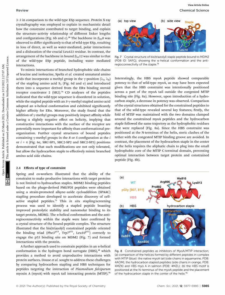
Fig. 7 Crystal structure of bis(triazolyl) staple peptide bound to MDM2(PDB ID: 5AFG), showing the a-helical conformation and the anti-regioconnectivity of the staple.75
Review Chemical Science
Ope
n A
cces
s A
rtic
le. P
ublis
hed
on 2
5 M
arch
202
1. D
ownl
oade
d on
3/1
5/20
22 2
:27:
07 A
M.
Thi
s ar
ticle
is li
cens
ed u
nder
a C
reat
ive
Com
mon
s A
ttrib
utio
n 3.
0 U
npor
ted
Lic
ence
.View Article Online
3–3 in comparison to the wild-type ESp sequence. Protein X-raycrystallography was employed to explain in mechanistic detailhow the constraint contributed to target binding, and explainthe structure–activity relationship of different linker lengthsand congurations (Fig. 6b and c).28 The backbone in bRS8 wasobserved to differ signicantly to that of wild-type ESp, resultingin loss of direct, as well as water-mediated, polar interactionsand a dislocation of the crucial Leu423 residue. In contrast, thearrangement of the backbone in bound bSS12 was similar to thatof the wild-type ESp peptide, including water mediatedinteractions.
To mimic interactions of branched hydrophobic side chainsof leucine and isoleucine, Speltz et al. created unnatural aminoacids that incorporate a methyl group in the g-position (lS, lR)of the stapling amino acid S5 (Fig. 6d and e) and introducedthem into a sequence derived from the ERa binding steroidreceptor coactivator 2 (SR2).74 CD analyses of the peptidesindicated that the wild-type sequence is disordered in solution,while the stapled peptide with an S-g-methyl stapled amino acidadopted an a-helical conformation and exhibited signicantlyhigher affinity for ERa. Moreover, the study found that theaddition of g-methyl groups may positively impact affinity whilehaving a slightly negative effect on helicity, implying thatconstructive interactions with the surface of the receptor arepotentially more important for affinity than conformational pre-organization. Further crystal structures of bound peptidescontaining g-methyl groups in the R or S conguration at the ior i + 4 (Fig. 6e, SRC-SP1, SRC2-SP2 and SRC2-SP3) positionsdemonstrated that such modications are not only tolerated,but allow the hydrocarbon staple to effectively mimic branchedamino acid side chains.
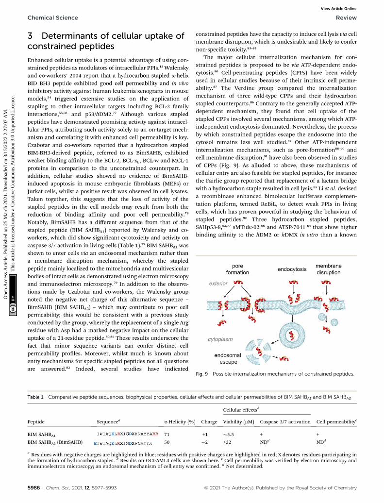
Fig. 8 Constrained peptides as inhibitors of MyoA/MTIP interaction;(a) comparison of the helices formed by different peptides in complexwith MTIP (blue): the native myoA tail (side chains in aquamarine, PDB:4AOM), the hydrocarbon stapled peptides (side chains in orange, PDB:4MZK) and HBS myo A in salmon (PDB: 4MZL); (b) the HBS motif ispositioned at the N-terminus of the myoA peptide and the placementof the hydrocarbon staple in the center of the helix.76
2.4 Effects of type of constraint
Spring and co-workers illustrated that the ability of theconstraint to make productive interactions with target proteinsis not limited to hydrocarbon staples. MDM2 binding peptidesbased on the phage-derived PMI/PDI peptides were obtainedusing a strain-promoted alkyne–azide cycloaddition (SPAAC)stapling procedure developed to accelerate discovery of cell-active stapled peptides.75 This in situ stapling/screeningprocess was used to identify a stapled peptide boastingimproved proteolytic stability and nanomolar binding to itstarget protein, MDM2. The a-helical conformation and the anti-regioconnectivity within the staple were later conrmed bya crystal structure of the bound-peptide complex. The structureillustrated that the bis(triazolyl) constrained peptide orientedthe binding triad (Phe3p53, Trp7p53, Leu10p53) correctly toengage the p53 binding site on MDM2 (Fig. 7) and formedinteractions with the protein.
A further approach used to constrain peptides in an a-helicalconformation is the hydrogen bond surrogate (HBS),18 whichprovides a method to avoid unproductive interactions withprotein surfaces. Douse et al. sought to address these challengesby comparing hydrocarbon stapling and HBS techniques forpeptides targeting the interaction of Plasmodium falciparummyosin A (myoA) with myoA tail interacting protein (MTIP).76
© 2021 The Author(s). Published by the Royal Society of Chemistry
Interestingly, the HBS myoA peptide showed comparablepotency to that of wild-type myoA, as may have been expectedgiven that the HBS constraint was intentionally positionedacross a part of the myoA tail outside the congested MTIPbinding site (Fig. 8a). However, upon introduction of a hydro-carbon staple, a decrease in potency was observed. Comparisonof the crystal structures obtained for the constrained peptides tothat of the wild-type revealed several key features; rstly, thefold of MTIP was maintained with the two domains clampedaround the constrained myoA peptides and the hydrocarbonstaple followed the same trajectory as the hydrophobic residuesthat were replaced (Fig. 8a). Since the HBS constraint waspositioned at the N-terminus of the helix, steric clashes of thetether with the congested MTIP binding groove are avoided. Incontrast, the placement of the hydrocarbon staple in the centerof the helix requires the aliphatic chain to plug into the smallhydrophobic core of the MTIP C-terminal domain, preventingoptimal interaction between target protein and constrainedpeptide (Fig. 8b).
Chem. Sci., 2021, 12, 5977–5993 | 5985
Chemical Science Review
Ope
n A
cces
s A
rtic
le. P
ublis
hed
on 2
5 M
arch
202
1. D
ownl
oade
d on
3/1
5/20
22 2
:27:
07 A
M.
Thi
s ar
ticle
is li
cens
ed u
nder
a C
reat
ive
Com
mon
s A
ttrib
utio
n 3.
0 U
npor
ted
Lic
ence
.View Article Online
3 Determinants of cellular uptake ofconstrained peptides
Enhanced cellular uptake is a potential advantage of using con-strained peptides as modulators of intracellular PPIs.13 Walenskyand co-workers’ 2004 report that a hydrocarbon stapled a-helixBID BH3 peptide exhibited good cell permeability and in vivoinhibitory activity against human leukemia xenogras in mousemodels,54 triggered extensive studies on the application ofstapling to other intracellular targets including BCL-2 familyinteractions,55,58 and p53/hDM2.77 Although various stapledpeptides have demonstrated promising activity against intracel-lular PPIs, attributing such activity solely to an on-target mech-anism and correlating it with enhanced cell permeability is key.Czabotar and co-workers reported that a hydrocarbon stapledBIM-BH3-derived peptide, referred to as BimSAHB, exhibitedweaker binding affinity to the BCL-2, BCL-xL, BCL-w and MCL-1proteins in comparison to the unconstrained counterpart. Inaddition, cellular studies showed no evidence of BimSAHB-induced apoptosis in mouse embryonic broblasts (MEFs) orJurkat cells, whilst a positive result was observed in cell lysates.Taken together, this suggests that the loss of activity of thestapled peptides in the cell models may result from both thereduction of binding affinity and poor cell permeability.78
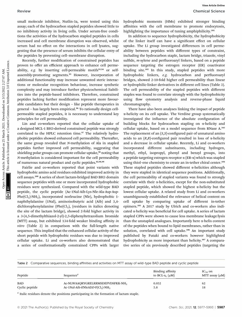
Notably, BimSAHB has a different sequence from that of thestapled peptide (BIM SAHBA1) reported by Walensky and co-workers, which did show signicant cytotoxicity and activity oncaspase 3/7 activation in living cells (Table 1).79 BIM SAHBA1 wasshown to enter cells via an endosomal mechanism rather thana membrane disruption mechanism, whereby the stapledpeptide mainly localized to the mitochondria and multivesicularbodies of intact cells as demonstrated using electron microscopyand immunoelectron microscopy.79 In addition to the observa-tions made by Czabotar and co-workers, the Walensky groupnoted the negative net charge of this alternative sequence –
BimSAHB (BIM SAHBA2) – which may contribute to poor cellpermeability; this would be consistent with a previous studyconducted by the group, whereby the replacement of a single Argresidue with Asp had a marked negative impact on the cellularuptake of a 21-residue peptide.80,81 These results underscore thefact that minor sequence variants can confer distinct cellpermeability proles. Moreover, whilst much is known aboutentry mechanisms for specic stapled peptides not all questionsare answered.82 Indeed, several studies have indicated
Table 1 Comparative peptide sequences, biophysical properties, cellula
Peptide Sequencea a-Helicity (%
BIM SAHBA1 71
BIM SAHBA2 (BimSAHB) 50
a Residues with negative charges are highlighted in blue; residues with pothe formation of hydrocarbon staples. b Results on OCI-AML3 cells are simmunoelectron microscopy; an endosomal mechanism of cell entry was
5986 | Chem. Sci., 2021, 12, 5977–5993
constrained peptides have the capacity to induce cell lysis via cellmembrane disruption, which is undesirable and likely to confernon-specic toxicity.83–85
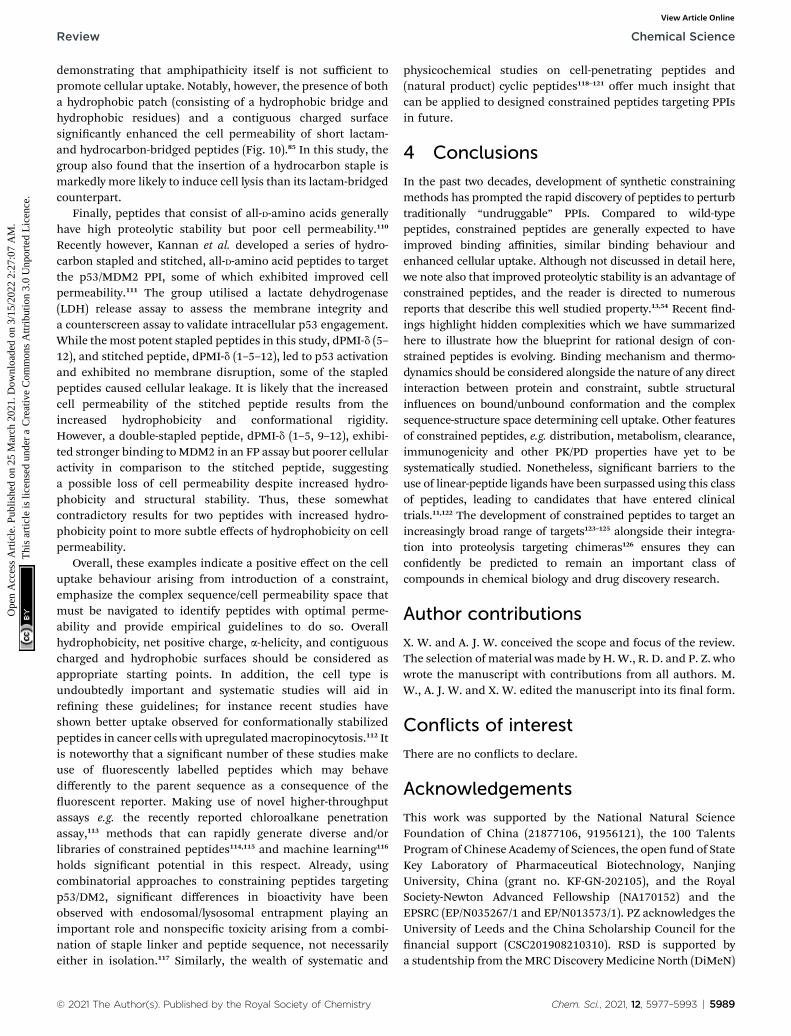
The major cellular internalization mechanism for con-strained peptides is proposed to be via ATP-dependent endo-cytosis.86 Cell-penetrating peptides (CPPs) have been widelyused in cellular studies because of their intrinsic cell perme-ability.87 The Verdine group compared the internalizationmechanism of three wild-type CPPs and their hydrocarbonstapled counterparts.86 Contrary to the generally accepted ATP-dependent mechanism, they found that cell uptake of thestapled CPPs involved several mechanisms, among which ATP-independent endocytosis dominated. Nevertheless, the processby which constrained peptides escape the endosome into thecytosol remains less well studied.82 Other ATP-independentinternalization mechanisms, such as pore-formation88–90 andcell membrane disruption,91 have also been observed in studiesof CPPs (Fig. 9). As alluded to above, these mechanisms ofcellular entry are also feasible for stapled peptides, for instancethe Fairlie group reported that replacement of a lactam bridgewith a hydrocarbon staple resulted in cell lysis.85 Li et al. deviseda recombinase enhanced bimolecular luciferase complemen-tation platform, termed ReBiL, to detect weak PPIs in livingcells, which has proven powerful in studying the behaviour ofstapled peptides.92 Three hydrocarbon stapled peptides,SAHp53-8,63,77 sMTide-02 66 and ATSP-7041 64 that show higherbinding affinity to the hDM2 or hDMX in vitro than a known
r effects and cellular permeabilities of BIM SAHBA1 and BIM SAHBA2
) Charge
Cellular effectsb
Cell permeabilitycViability (mM) Caspase 3/7 activation
+1 �5.5 + +
�2 >32 NDd NDd
sitive charges are highlighted in red; X denotes residues participating inhown here. c Cell permeability was veried by electron microscopy andconrmed. d Not determined.
Fig. 9 Possible internalization mechanisms of constrained peptides.
© 2021 The Author(s). Published by the Royal Society of Chemistry
Review Chemical Science
Ope
n A
cces
s A
rtic
le. P
ublis
hed
on 2
5 M
arch
202
1. D
ownl
oade
d on
3/1
5/20
22 2
:27:
07 A
M.
Thi
s ar
ticle
is li
cens
ed u
nder
a C
reat
ive
Com
mon
s A
ttrib
utio
n 3.
0 U
npor
ted
Lic
ence
.View Article Online
small molecule inhibitor, Nutlin-3a, were tested using thisassay; each of the hydrocarbon stapled peptides showed little tono inhibitory activity in living cells. Under serum-free condi-tions the activities of the hydrocarbon stapled peptides in cellsincreased and cell membrane disruption was observed, whilstserum had no effect on the interactions in cell lysates, sug-gesting that the presence of serum inhibits the cellular entry ofthe peptides by preventing cell–membrane disruption.
Recently, further modication of constrained peptides hasproven to offer an efficient approach to enhance cell perme-ability, e.g. by inserting cell-permeable motifs93,94 or self-assembly-promoting segments.95 However, incorporation ofadditional functionality may increase unwanted steric interac-tions or molecular recognition behaviour, increase syntheticcomplexity and may introduce further physicochemical liabili-ties into the peptide-based inhibitors. Therefore, constrainedpeptides lacking further modication represent more favour-able candidates but their design – like peptide therapeutics ingeneral – has largely been empirical.96 To rationally design cell-permeable stapled peptides, it is necessary to understand keyprinciples for cell permeability.
Lin and co-workers reported that the cellular uptake ofa designed MCL-1 BH3-derived constrained peptide was stronglycorrelated to the HPLC retention time.97 The relatively hydro-phobic peptides showed enhanced cell permeability. Thereaer,the same group revealed that N-methylation of Ala in stapledpeptides further improved cell permeability, suggesting thatshielding polar groups can promote cellular uptake,98 noting thatN-methylation is considered important for the cell permeabilityof numerous natural product and cyclic peptides.82,96,99
Fairlie and co-workers reported that point variants withhydrophobic amino acid residues exhibited improved activity incell assays.100 A series of short lactam-bridged BAD BH3 domainsequence peptides with one or more incorporated hydrophobicresidues were synthesized. Compared with the wild-type BADpeptide, the cyclic peptide (Ac-1Nal-Aib-Lys-Nle-Ala-Asp-Asp-Phe(Cl2)-NH2), incorporated norleucine (Nle), hydrophobic 1-naphthylalanine (1Nal), aminoisobutyric acid (Aib) and 3,4-dichlorophenylalanine (Phe(Cl2), (residues in italics denotingthe site of the lactam bridge), showed 2-fold higher activity ina 3-(4,5-dimethylthiazol-2-yl)-2,5-diphenyltetrazolium bromide(MTT) assay, but exhibited 134-fold weaker binding affinity invitro (Table 2) in comparison with the full-length nativesequence. This implied that the enhanced cellular activity of theshort peptide with hydrophobic residues was due to improvedcellular uptake. Li and co-workers also demonstrated thata series of conformationally constrained CPPs with larger
Table 2 Comparative sequences, binding affinities and activities on MTT
Peptide Sequencea
BAD Ac-NLWAAQRYGRELRRMSDEFVDSFKK-NCyclic peptide Ac-1Nal-Aib-KNleADD-F(Cl2)-NH2
a Italic residues denote the positions participating in the formation of lac
© 2021 The Author(s). Published by the Royal Society of Chemistry
hydrophobic moments (HMs) exhibited stronger bindingaffinities with the cell membrane to promote endocytosis,highlighting the importance of tuning amphiphilicity.101
In addition to sequence hydrophobicity, the hydrophobicityof the linker itself can have a signicant effect on cellularuptake. The Li group investigated differences in cell perme-ability between peptides with different types of constraint,including the hydrocarbon staple, lactam bridge, triazole, vinylsulde, m-xylene and peruoroaryl linkers, based on a peptidesequence targeting the estrogen receptor (ER) coactivatorbinding site.102 In this study, stapled peptides with morehydrophobic linkers, e.g. hydrocarbon and peruoroarylbridges, showed 2-10-fold higher cell permeability than linearor hydrophilic-linker derivatives in different cell lines (Table 3).The cell permeability of the stapled peptides with differentstaples was found to correlate strongly with the hydrophobicityusing ow cytometry analysis and reverse-phase liquidchromatography.
There have also been analyses linking the impact of peptidea-helicity on its cell uptake. The Verdine group systematicallyinvestigated the inuence of the absolute conguration ofbuilding blocks for hydrocarbon stapling on a-helicity andcellular uptake, based on a model sequence from RNase A.103
The replacement of an (S,S)-congured pair of unnatural aminoacids to an (R,R)-congured pair resulted in loss of a-helicityand a decrease in cellular uptake. Recently, Li and co-workersincorporated different substituents, including hydrogen,methyl, ethyl, isopropyl, phenyl and benzyl groups, intoa peptide targeting estrogen receptor a (ER-a) which was stapledusing thiol–ene chemistry to create an in-tether chiral center.104
These stapled peptides showed different a-helicities althoughthey were stapled in identical sequence positions. Additionally,the cell permeability of stapled variants was found to stronglycorrelate with their a-helicities, except for the non-substitutedstapled peptide, which showed the highest a-helicity but thelowest cellular uptake. A related study from Li and co-workersunambiguously established the relevance of helical content oncell uptake by comparing uptake of different in-tetherepimers.105 A 2017 study by Ulrich and co-workers also indi-cated a-helicity was benecial for cell uptake. A series of lactamstapled CPPs were shown to cause less membrane leakage/lysisthan the unstapled analogues. Importantly here a-helix contentof the peptides when bound to lipid membranes, rather than insolution, correlated with cell uptake.106 An important studypublished by Futaki and co-workers however highlightedhydrophobicity as more important than helicity.107 A compara-tive series of six previously described peptides (targeting the
assay of wild-type BAD peptide and cyclic peptide
Binding affinityto BCL-xL (mM)
IC50 onMTT assay (mM)
H2 0.052 625.8 18
tam staple.
Chem. Sci., 2021, 12, 5977–5993 | 5987

Fig. 10 Helical wheel diagrams illustrating subtle staple placement; (a)at the boundary between hydrophobic and hydrophilic surfaces whichis likely to result in improved uptake due to expansion of hydrophobicsurface area; (b) to form a contiguous hydrophobic surface which islikely to confer superior uptake. Hydrophobic residues and staples areshown in orange, and hydrophilic and positively charged residues are
Table 3 Comparative a-helicity, hydrophobicity and cell permeability orders of the model peptide targeting the ER coactivatora
Helicity order Hydrophobicity order Cell permeability orderb
Hydrocarbon 1 1 4Lactam 1 6 6Triazole 3 5 5Vinyl sulde 4 4 3m-Xylene 5 3 2Peruoroaryl 7 2 1None (linear) 6 6 7
a The same numbers denote the two stapled peptide showed similar specic properties. b The cell-permeabilities were detected using FAM-labelledstapled peptides in T47D, MCF-7, Hela, HEK293T cell lines via confocal microscopy, which showed concordant results in this study.
Chemical Science Review
Ope
n A
cces
s A
rtic
le. P
ublis
hed
on 2
5 M
arch
202
1. D
ownl
oade
d on
3/1
5/20
22 2
:27:
07 A
M.
Thi
s ar
ticle
is li
cens
ed u
nder
a C
reat
ive
Com
mon
s A
ttrib
utio
n 3.
0 U
npor
ted
Lic
ence
.View Article Online
p53/DM2 interaction) and their unstapled parent were reportedto enter cells via endocytosis. The unstapled peptides withgreater hydrophobicity were shown to have greater cell uptake.Non-helical cell-permeable stapled peptides have also been re-ported.108 Spring and co-workers reported a series of double-triazole stapled peptides based on a nuclear localizationsignal (NLS) sequence targeting the interaction between HNF1band importin a1, some of which penetrated the cellmembrane.108
Net charge has also been shown to play an important role indening the cellular uptake properties of stapled peptides (seeabove). In Verdine's study, increasing net charge from �1 to +5was found to improve the cellular uptake of both hydrocarbonstapled and stitched peptides86 (a unique form of doublestapled peptides with two contiguous staples bridging threeresidues109). However, the constrained peptides with net posi-tive charge over +7 showed a signicant decrease in cellpermeability.
The cellular uptake of constrained peptides therefore clearlydepends on several driving factors, e.g. hydrophobicity, a-hel-icity, net charge and the presence of serum. However, cellularstudies have usually focused on one of the key factors inde-pendently. To provide comprehensive guidelines for design ofcell-permeable constrained peptides, Walensky and co-workersdeveloped a high-throughput uorescence microscopyapproach tomeasure the cellular uptake of hydrocarbon stapledpeptides with N-terminal FITC labels.84 In this work, the groupsystematically tested three classes of hydrocarbon stapledpeptides, including hydrocarbon stapled BIM BH3 helices withdifferent stapling positions, or diverse single-residue variants,and hydrocarbon stapled peptides based on another sequence(RAS binding SOS1). Unbiased quantitative analytical protocols,used to clarify key biophysical factors inuencing the cellularuptake of these peptides, revealed that overall hydrophobicity,net charge, a-helicity, and staple placement are key determi-nants of cell penetration. The overall hydrophobicity of thestapled peptides, which was determined by HPLC retentiontime, correlates with cellular uptake. The stapled peptides withhigh, but not overwhelming hydrophobicity, showed optimal
5988 | Chem. Sci., 2021, 12, 5977–5993
cell permeability. The position of a staple also plays an impor-tant role in modulation of cellular uptake of the stapledpeptides. Subtle changes in position of a hydrocarbon staplebetween a hydrophobic surface and a hydrophilic surface werefound to improve the cellular uptake of the stapled peptides dueto the expansion of the hydrophobic surface (Fig. 10).84
Furthermore, a-helicity and isoelectric point (pI) were identiedas inuences on cell permeability in Walensky's study.84 Incombination with high a-helicity (61–86%), the relativelyhydrophobic stapled peptides showed excellent cell perme-ability. However, excessive hydrophobicity coupled with highlyacidic pI was shown to confer a high tendency to induce celllysis.
Recently, the Fairlie group tested the cell permeability ofa series of FITC-labelled stapled peptides quantitively via owcytometry in living cells without serum.85 A cell-penetratingpeptide, TAT, was selected as a control for 100% of cell entry.The cell permeability was found to be correlated to the con-nected hydrophobic surface area (cHSA) and the hydrophobicmoment (mH), which were determined computationally. Theconstrained peptides lacking amphipathic structures were notcell-permeable. In addition, amphipathic peptides lackingpositively charged residues showed poor cell permeability,
shown in blue.
© 2021 The Author(s). Published by the Royal Society of Chemistry
Review Chemical Science
Ope
n A
cces
s A
rtic
le. P
ublis
hed
on 2
5 M
arch
202
1. D
ownl
oade
d on
3/1
5/20
22 2
:27:
07 A
M.
Thi
s ar
ticle
is li
cens
ed u
nder
a C
reat
ive
Com
mon
s A
ttrib
utio
n 3.
0 U
npor
ted
Lic
ence
.View Article Online
demonstrating that amphipathicity itself is not sufficient topromote cellular uptake. Notably, however, the presence of botha hydrophobic patch (consisting of a hydrophobic bridge andhydrophobic residues) and a contiguous charged surfacesignicantly enhanced the cell permeability of short lactam-and hydrocarbon-bridged peptides (Fig. 10).85 In this study, thegroup also found that the insertion of a hydrocarbon staple ismarkedly more likely to induce cell lysis than its lactam-bridgedcounterpart.
Finally, peptides that consist of all-D-amino acids generallyhave high proteolytic stability but poor cell permeability.110
Recently however, Kannan et al. developed a series of hydro-carbon stapled and stitched, all-D-amino acid peptides to targetthe p53/MDM2 PPI, some of which exhibited improved cellpermeability.111 The group utilised a lactate dehydrogenase(LDH) release assay to assess the membrane integrity anda counterscreen assay to validate intracellular p53 engagement.While themost potent stapled peptides in this study, dPMI-d (5–12), and stitched peptide, dPMI-d (1–5–12), led to p53 activationand exhibited no membrane disruption, some of the stapledpeptides caused cellular leakage. It is likely that the increasedcell permeability of the stitched peptide results from theincreased hydrophobicity and conformational rigidity.However, a double-stapled peptide, dPMI-d (1–5, 9–12), exhibi-ted stronger binding to MDM2 in an FP assay but poorer cellularactivity in comparison to the stitched peptide, suggestinga possible loss of cell permeability despite increased hydro-phobicity and structural stability. Thus, these somewhatcontradictory results for two peptides with increased hydro-phobicity point to more subtle effects of hydrophobicity on cellpermeability.
Overall, these examples indicate a positive effect on the celluptake behaviour arising from introduction of a constraint,emphasize the complex sequence/cell permeability space thatmust be navigated to identify peptides with optimal perme-ability and provide empirical guidelines to do so. Overallhydrophobicity, net positive charge, a-helicity, and contiguouscharged and hydrophobic surfaces should be considered asappropriate starting points. In addition, the cell type isundoubtedly important and systematic studies will aid inrening these guidelines; for instance recent studies haveshown better uptake observed for conformationally stabilizedpeptides in cancer cells with upregulatedmacropinocytosis.112 Itis noteworthy that a signicant number of these studies makeuse of uorescently labelled peptides which may behavedifferently to the parent sequence as a consequence of theuorescent reporter. Making use of novel higher-throughputassays e.g. the recently reported chloroalkane penetrationassay,113 methods that can rapidly generate diverse and/orlibraries of constrained peptides114,115 and machine learning116
holds signicant potential in this respect. Already, usingcombinatorial approaches to constraining peptides targetingp53/DM2, signicant differences in bioactivity have beenobserved with endosomal/lysosomal entrapment playing animportant role and nonspecic toxicity arising from a combi-nation of staple linker and peptide sequence, not necessarilyeither in isolation.117 Similarly, the wealth of systematic and
© 2021 The Author(s). Published by the Royal Society of Chemistry
physicochemical studies on cell-penetrating peptides and(natural product) cyclic peptides118–121 offer much insight thatcan be applied to designed constrained peptides targeting PPIsin future.
4 Conclusions
In the past two decades, development of synthetic constrainingmethods has prompted the rapid discovery of peptides to perturbtraditionally “undruggable” PPIs. Compared to wild-typepeptides, constrained peptides are generally expected to haveimproved binding affinities, similar binding behaviour andenhanced cellular uptake. Although not discussed in detail here,we note also that improved proteolytic stability is an advantage ofconstrained peptides, and the reader is directed to numerousreports that describe this well studied property.13,54 Recent nd-ings highlight hidden complexities which we have summarizedhere to illustrate how the blueprint for rational design of con-strained peptides is evolving. Binding mechanism and thermo-dynamics should be considered alongside the nature of any directinteraction between protein and constraint, subtle structuralinuences on bound/unbound conformation and the complexsequence-structure space determining cell uptake. Other featuresof constrained peptides, e.g. distribution, metabolism, clearance,immunogenicity and other PK/PD properties have yet to besystematically studied. Nonetheless, signicant barriers to theuse of linear-peptide ligands have been surpassed using this classof peptides, leading to candidates that have entered clinicaltrials.11,122 The development of constrained peptides to target anincreasingly broad range of targets123–125 alongside their integra-tion into proteolysis targeting chimeras126 ensures they cancondently be predicted to remain an important class ofcompounds in chemical biology and drug discovery research.
Author contributions
X. W. and A. J. W. conceived the scope and focus of the review.The selection of material was made by H.W., R. D. and P. Z. whowrote the manuscript with contributions from all authors. M.W., A. J. W. and X. W. edited the manuscript into its nal form.
Conflicts of interest
There are no conicts to declare.
Acknowledgements
This work was supported by the National Natural ScienceFoundation of China (21877106, 91956121), the 100 TalentsProgram of Chinese Academy of Sciences, the open fund of StateKey Laboratory of Pharmaceutical Biotechnology, NanjingUniversity, China (grant no. KF-GN-202105), and the RoyalSociety-Newton Advanced Fellowship (NA170152) and theEPSRC (EP/N035267/1 and EP/N013573/1). PZ acknowledges theUniversity of Leeds and the China Scholarship Council for thenancial support (CSC201908210310). RSD is supported bya studentship from theMRCDiscovery Medicine North (DiMeN)
Chem. Sci., 2021, 12, 5977–5993 | 5989

Chemical Science Review
Ope
n A
cces
s A
rtic
le. P
ublis
hed
on 2
5 M
arch
202
1. D
ownl
oade
d on
3/1
5/20
22 2
:27:
07 A
M.
Thi
s ar
ticle
is li
cens
ed u
nder
a C
reat
ive
Com
mon
s A
ttrib
utio
n 3.
0 U
npor
ted
Lic
ence
.View Article Online
Doctoral Training Partnership (MR/N013840/1). AJW holdsa Royal Society Leverhulme Trust Senior Fellowship (SRF/R1/191087).
Notes and references
1 M. R. Arkin, Y. Tang and J. A. Wells, Chem. Biol., 2014, 21,1102–1114.
2 B. N. Bullock, A. L. Jochim and P. S. Arora, J. Am. Chem. Soc.,2011, 133, 14220–14223.
3 A. L. Jochim and P. S. Arora, ACS Chem. Biol., 2010, 5, 919–923.4 M. Raj, B. N. Bullock and P. S. Arora, Bioorg. Med. Chem.,2013, 21, 4051–4057.
5 V. Azzarito, K. Long, N. S. Murphy and A. J. Wilson, Nat.Chem., 2013, 5, 161–173.
6 P. Tompa, N. E. Davey, T. J. Gibson and M. M. Babu, Mol.Cell, 2014, 55, 161–169.
7 J. M. Rogers, V. Oleinikovas, S. L. Shammas, C. T. Wong,D. De Sancho, C. M. Baker and J. Clarke, Proc. Natl. Acad.Sci. U. S. A., 2014, 111, 15420–15425.
8 A. Bah, R. M. Vernon, Z. Siddiqui, M. Krzeminski,R. Muhandiram, C. Zhao, N. Sonenberg, L. E. Kay andJ. D. Forman-Kay, Nature, 2015, 519, 106–109.
9 P. E. Wright and H. J. Dyson, Nat. Rev. Mol. Cell Biol., 2015,16, 18–29.
10 C. Morrison, Nat. Rev. Drug Discovery, 2018, 17, 531–533.11 P. M. Cromm, J. Spiegel and T. N. Grossmann, ACS Chem.
Biol., 2015, 10, 1362–1375.12 T. K. Sawyer, A. W. Partridge, H. Y. K. Kaan, Y.-C. Juang,
S. Lim, C. Johannes, T. Y. Yuen, C. Verma, S. Kannan,P. Aronica, Y. S. Tan, B. Sherborne, S. Ha, J. Hochman,S. Chen, L. Surdi, A. Peier, B. Sauvagnat, P. J. Dandliker,C. J. Brown, S. Ng, F. Ferrer and D. P. Lane, Bioorg. Med.Chem., 2018, 26, 2807–2815.
13 L. D. Walensky and G. H. Bird, J. Med. Chem., 2014, 57,6275–6288.
14 H. E. Blackwell and R. H. Grubbs, Angew. Chem., Int. Ed.,1998, 37, 3281–3284.
15 C. E. Schafmeister, J. Po and G. L. Verdine, J. Am. Chem.Soc., 2000, 122, 5891–5892.
16 N. S. Robertson and D. R. Spring, Molecules, 2018, 23, 959.17 X. Li, S. Chen, W.-D. Zhang and H.-G. Hu, Chem. Rev., 2020,
120, 10079–10144.18 A. Patgiri, A. L. Jochim and P. S. Arora, Acc. Chem. Res., 2008,
41, 1289–1300.19 Y. H. Lau, P. de Andrade, S.-T. Quah, M. Rossmann,
L. Laraia, N. Skold, T. J. Sum, P. J. E. Rowling,T. L. Joseph, C. Verma, M. Hyvonen, L. S. Itzhaki,A. R. Venkitaraman, C. J. Brown, D. P. Lane andD. R. Spring, Chem. Sci., 2014, 5, 1804–1809.
20 Y. H. Lau, P. de Andrade, Y. Wu and D. R. Spring, Chem. Soc.Rev., 2015, 44, 91–102.
21 C. M. Grison, G. M. Burslem, J. A. Miles, L. K. A. Pilsl,D. J. Yeo, Z. Imani, S. L. Warriner, M. E. Webb andA. J. Wilson, Chem. Sci., 2017, 8, 5166–5171.
5990 | Chem. Sci., 2021, 12, 5977–5993
22 J. M. Fletcher, K. A. Horner, G. J. Bartlett, G. G. Rhys,A. J. Wilson and D. N. Woolfson, Chem. Sci., 2018, 9,7656–7665.
23 L. Reguera and D. G. Rivera, Chem. Rev., 2019, 119, 9836–9860.
24 K. J. Skowron, T. E. Speltz and T. W. Moore, Med. Res. Rev.,2019, 39, 749–770.
25 D. A. Guarracino, J. A. Riordan, G. M. Barreto, A. L. Oldeld,C. M. Kouba and D. Agrinsoni, Chem. Rev., 2019, 119, 9915–9949.
26 R. Rezaei Araghi and A. E. Keating, Curr. Opin. Struct. Biol.,2016, 39, 27–38.
27 J. Iegre, P. Brear, D. J. Baker, Y. S. Tan, E. L. Atkinson,H. F. Sore, D. H. O' Donovan, C. S. Verma, M. Hyvonenand D. R. Spring, Chem. Sci., 2019, 10, 5056–5063.
28 A. Glas, D. Bier, G. Hahne, C. Rademacher, C. Ottmann andT. N. Grossmann, Angew. Chem., Int. Ed., 2014, 53, 2489–2493.
29 J. M. Scholtz and R. L. Baldwin, Annu. Rev. Biophys. Biomol.Struct., 1992, 21, 95–118.
30 A. D. Vogt, N. Pozzi, Z. Chen and E. Di Cera, Biophys. Chem.,2014, 186, 13–21.
31 A. D. Vogt and E. Di Cera, Biochemistry, 2012, 51, 5894–5902.32 D. H. Williams, E. Stephens, D. P. O'Brien and M. Zhou,
Angew. Chem., Int. Ed., 2004, 43, 6596–6616.33 K. Rajarathnam and J. Rosgen, Biochim. Biophys. Acta Bi,
2014, 1838, 69–77.34 J. A. Caro, K. W. Harpole, V. Kasinath, J. Lim, J. Granja,
K. G. Valentine, K. A. Sharp and A. J. Wand, Proc. Natl.Acad. Sci. U. S. A., 2017, 114, 6563–6568.
35 J. A. Miles, D. J. Yeo, P. Rowell, S. Rodriguez-Marin,C. M. Pask, S. L. Warriner, T. A. Edwards and A. J. Wilson,Chem. Sci., 2016, 7, 3694–3702.
36 M. L. Verteramo, O. Stenstrom, M. M. Ignjatovic,O. Caldararu, M. A. Olsson, F. Manzoni, H. Leffler,E. Oksanen, D. T. Logan, U. J. Nilsson, U. Ryde andM. Akke, J. Am. Chem. Soc., 2019, 141, 2012–2026.
37 P. E. Czabotar, G. Lessene, A. Strasser and J. M. Adams, Nat.Rev. Mol. Cell Biol., 2014, 15, 49–63.
38 P. V. Sauer, J. Timm, D. Liu, D. Sitbon, E. Boeri-Erba,C. Velours, N. Mucke, J. Langowski, F. Ochsenbein,G. Almouzni and D. Panne, eLife, 2017, 6, e23474.
39 M. Bakail, S. Rodriguez-Marin, Z. Hegedus, M. E. Perrin,F. Ochsenbein and A. J. Wilson, ChemBioChem, 2019, 20,891–895.
40 D. Lama, S. T. Quah, C. S. Verma, R. Lakshminarayanan,R. W. Beuerman, D. P. Lane and C. J. Brown, Sci. Rep.,2013, 3, 3451.
41 A. Modrak-Wojcik, M. Gorka, K. Niedzwiecka,K. Zdanowski, J. Zuberek, A. Niedzwiecka and R. Stolarski,FEBS Lett., 2013, 587, 3928–3934.
42 J. Marcotrigiano, A.-C. Gingras, N. Sonenberg andS. K. Burley, Mol. Cell, 1999, 3, 707–716.
43 D. Lama, A.-M. Liberatore, Y. Frosi, J. Nakhle, N. Tsomaia,T. Bashir, D. P. Lane, C. J. Brown, C. S. Verma andS. Auvin, Chem. Sci., 2019, 10, 2489–2500.
© 2021 The Author(s). Published by the Royal Society of Chemistry

Review Chemical Science
Ope
n A
cces
s A
rtic
le. P
ublis
hed
on 2
5 M
arch
202
1. D
ownl
oade
d on
3/1
5/20
22 2
:27:
07 A
M.
Thi
s ar
ticle
is li
cens
ed u
nder
a C
reat
ive
Com
mon
s A
ttrib
utio
n 3.
0 U
npor
ted
Lic
ence
.View Article Online
44 E. E. Gallagher, J. M. Song, A. Menon, L. D. Mishra,A. F. Chmiel and A. L. Garner, J. Med. Chem., 2019, 62,4967–4978.
45 Y. K. Rennie, P. J. McIntyre, T. Akindele, R. Bayliss andA. G. Jamieson, ACS Chem. Biol., 2016, 11, 3383–3390.
46 K. Hetherington, Z. Hegedus, T. A. Edwards, R. Sessions,A. Nelson and A. J. Wilson, Chem.–Eur. J., 2020, 26, 7638–7646.
47 G. M. Burslem, H. F. Kyle, A. Nelson, T. A. Edwards andA. J. Wilson, Chem. Sci., 2017, 8, 4188–4202.
48 I. K. Nordgren and A. Tavassoli, Chem. Soc. Rev., 2011, 40,4307–4317.
49 A. R. Martin, C. Ronco, L. Demange and R. Benhida,MedChemComm, 2017, 8, 21–52.
50 A. Lindsey-Crosthwait, D. Rodriguez-Lema, M. Walko,C. M. Pask and A. J. Wilson, Pept. Sci., 2021, 113, e24157.
51 A. Glas, E.-C. Wamhoff, D. M. Kruger, C. Rademacher andT. N. Grossmann, Chem.–Eur. J., 2017, 23, 16157–16161.
52 A. V. Strizhak, O. Babii, S. Afonin, I. Bakanovich,T. Pantelejevs, W. Xu, E. Fowler, R. Eapen, K. Sharma,M. O. Platonov, V. V. Hurmach, L. Itzhaki, M. Hyvonen,A. S. Ulrich, D. R. Spring and I. V. Komarov, Org. Biomol.Chem., 2020, 18, 5359–5369.
53 B. Hu, D. M. Gilkes and J. Chen, Cancer Res., 2007, 67, 8810–8817.
54 L. D. Walensky, A. L. Kung, I. Escher, T. J. Malia, S. Barbuto,R. D. Wright, G. Wagner, G. L. Verdine and S. J. Korsmeyer,Science, 2004, 305, 1466–1470.
55 L. D. Walensky, K. Pitter, J. Morash, K. J. Oh, S. Barbuto,J. Fisher, E. Smith, G. L. Verdine and S. J. Korsmeyer, Mol.Cell, 2006, 24, 199–210.
56 T. L. Joseph, D. P. Lane and C. S. Verma, PLoS One, 2012, 7,e43985.
57 S. Bhattacharya, H. Zhang, A. K. Debnath and D. Cowburn,J. Biol. Chem., 2008, 283, 16274–16278.
58 M. L. Stewart, E. Fire, A. E. Keating and L. D. Walensky, Nat.Chem. Biol., 2010, 6, 595–601.
59 R. Rezaei Araghi, G. H. Bird, J. A. Ryan, J. M. Jenson,M. Godes, J. R. Pritz, R. A. Grant, A. Letai, L. D. Walenskyand A. E. Keating, Proc. Natl. Acad. Sci. U. S. A., 2018, 115,E886.
60 C. Phillips, L. R. Roberts, M. Schade, R. Bazin, A. Bent,N. L. Davies, R. Moore, A. D. Pannifer, A. R. Pickford,S. H. Prior, C. M. Read, A. Scott, D. G. Brown, B. Xu andS. L. Irving, J. Am. Chem. Soc., 2011, 133, 9696–9699.
61 P. H. Kussie, S. Gorina, V. Marechal, B. Elenbaas, J. Moreau,A. J. Levine and N. P. Pavletich, Science, 1996, 274, 948–953.
62 S. Baek, P. S. Kutchukian, G. L. Verdine, R. Huber,T. A. Holak, K. W. Lee and G. M. Popowicz, J. Am. Chem.Soc., 2012, 134, 103–106.
63 F. Bernal, M. Wade, M. Godes, T. N. Davis, D. G. Whitehead,A. L. Kung, G. M. Wahl and L. D. Walensky, Cancer Cell,2010, 18, 411–422.
64 Y. S. Chang, B. Graves, V. Guerlavais, C. Tovar, K. Packman,K. H. To, K. A. Olson, K. Kesavan, P. Gangurde,A. Mukherjee, T. Baker, K. Darlak, C. Elkin, Z. Filipovic,F. Z. Qureshi, H. Cai, P. Berry, E. Feyfant, X. E. Shi,
© 2021 The Author(s). Published by the Royal Society of Chemistry
J. Horstick, D. A. Annis, A. M. Manning, N. Fotouhi,H. Nash, L. T. Vassilev and T. K. Sawyer, Proc. Natl. Acad.Sci. U. S. A., 2013, 110, E3445–E3454.
65 S. M. Q. Chee, J. Wongsantichon, Q. Soo Tng, R. Robinson,T. L. Joseph, C. Verma, D. P. Lane, C. J. Brown andF. J. Ghadessy, PLoS One, 2014, 9, e104914.
66 C. J. Brown, S. T. Quah, J. Jong, A. M. Goh, P. C. Chiam,K. H. Khoo, M. L. Choong, M. A. Lee, L. Yurlova,K. Zolghadr, T. L. Joseph, C. S. Verma and D. P. Lane, ACSChem. Biol., 2013, 8, 506–512.
67 A. W. Partridge, H. Y. K. Kaan, Y.-C. Juang, A. Sadruddin,S. Lim, C. J. Brown, S. Ng, D. Thean, F. Ferrer,C. Johannes, T. Y. Yuen, S. Kannan, P. Aronica, Y. S. Tan,M. R. Pradhan, C. S. Verma, J. Hochman, S. Chen,H. Wan, S. Ha, B. Sherborne, D. P. Lane and T. K. Sawyer,Molecules, 2019, 24, 2292.
68 D. J. Yeo, S. L. Warriner and A. J. Wilson, Chem. Commun.,2013, 49, 9131–9133.
69 A. Gurtner, I. Manni and G. Piaggio, Biochim. Biophys. Acta,Gene Regul. Mech., 2017, 1860, 604–616.
70 S. Jeganathan, M. Wendt, S. Kiehstaller, D. Brancaccio,A. Kuepper, N. Pospiech, A. Carotenuto, E. Novellino,S. Hennig and T. N. Grossmann, Angew. Chem., Int. Ed.,2019, 58, 17351–17358.
71 K. Wallraven, F. L. Holmelin, A. Glas, S. Hennig, A. I. Frolovand T. N. Grossmann, Chem. Sci., 2020, 11, 2269–2276.
72 C. Ottmann, L. Yasmin, M. Weyand, J. L. Veesenmeyer,M. H. Diaz, R. H. Palmer, M. S. Francis, A. R. Hauser,A. Wittinghofer and B. Hallberg, EMBO J., 2007, 26, 902–913.
73 L. Yasmin, J. L. Veesenmeyer, M. H. Diaz, M. S. Francis,C. Ottmann, R. H. Palmer, A. R. Hauser and B. Hallberg,Biochem. J., 2010, 427, 217–224.
74 T. E. Speltz, S. W. Fanning, C. G. Mayne, C. Fowler,E. Tajkhorshid, G. L. Greene and T. W. Moore, Angew.Chem., Int. Ed., 2016, 55, 4252–4255.
75 Y. H. Lau, Y. Wu, M. Rossmann, B. X. Tan, P. de Andrade,Y. S. Tan, C. Verma, G. J. McKenzie, A. R. Venkitaraman,M. Hyvonen and D. R. Spring, Angew. Chem., Int. Ed.,2015, 54, 15410–15413.
76 C. H. Douse, S. J. Maas, J. C. Thomas, J. A. Garnett, Y. Sun,E. Cota and E. W. Tate, ACS Chem. Biol., 2014, 9, 2204–2209.
77 F. Bernal, A. F. Tyler, S. J. Korsmeyer, L. D. Walensky andG. L. Verdine, J. Am. Chem. Soc., 2007, 129, 2456–2457.
78 T. Okamoto, K. Zobel, A. Fedorova, C. Quan, H. Yang,W. J. Fairbrother, D. C. S. Huang, B. J. Smith, K. Deshayesand P. E. Czabotar, ACS Chem. Biol., 2013, 8, 297–302.
79 A. L. Edwards, F. Wachter, M. Lammert, A. J. Huhn,J. Luccarelli, G. H. Bird and L. D. Walensky, ACS Chem.Biol., 2015, 10, 2149–2157.
80 G. H. Bird, E. Gavathiotis, J. L. LaBelle, S. G. Katz andL. D. Walensky, ACS Chem. Biol., 2014, 9, 831–837.
81 J. L. LaBelle, S. G. Katz, G. H. Bird, E. Gavathiotis,M. L. Stewart, C. Lawrence, J. K. Fisher, M. Godes,K. Pitter, A. L. Kung and L. D. Walensky, J. Clin. Invest.,2012, 122, 2018–2031.
82 P. G. Dougherty, A. Sahni and D. H. Pei, Chem. Rev., 2019,119, 10241–10287.
Chem. Sci., 2021, 12, 5977–5993 | 5991
Chemical Science Review
Ope
n A
cces
s A
rtic
le. P
ublis
hed
on 2
5 M
arch
202
1. D
ownl
oade
d on
3/1
5/20
22 2
:27:
07 A
M.
Thi
s ar
ticle
is li
cens
ed u
nder
a C
reat
ive
Com
mon
s A
ttrib
utio
n 3.
0 U
npor
ted
Lic
ence
.View Article Online
83 W. B. Kauffman, T. Fuselier, J. He andW. C. Wimley, TrendsBiochem. Sci., 2015, 40, 749–764.
84 G. H. Bird, E. Mazzola, K. Opoku-Nsiah, M. A. Lammert,M. Godes, D. S. Neuberg and L. D. Walensky, Nat. Chem.Biol., 2016, 12, 845–852.
85 S. R. Perry, T. A. Hill, A. D. de Araujo, H. N. Hoang andD. P. Fairlie, Org. Biomol. Chem., 2018, 16, 367–371.
86 Q. Chu, R. E. Moellering, G. J. Hilinski, Y. W. Kim,T. N. Grossmann, J. T. H. Yeh and G. L. Verdine,MedChemComm, 2015, 6, 111–119.
87 Z. Kang, G. Ding, Z. Meng and Q. Meng, Peptides, 2019, 121,170149.
88 N. Vedovato and G. Rispoli, Eur. Biophys. J., 2007, 36, 771–778.
89 N. Vedovato, C. Baldini, C. Toniolo and G. Rispoli, Chem.Biodiversity, 2007, 4, 1338–1346.
90 T.-L. Sun, Y. Sun, C.-C. Lee and H. W. Huang, Biophys. J.,2013, 104, 1923–1932.
91 J. Durzynska, Ł. Przysiecka, R. Nawrot, J. Barylski,G. Nowicki, A. Warowicka, O. Musidlak and A. Gozdzicka-Jozeak, J. Pharmacol. Exp. Ther., 2015, 354, 32–42.
92 Y. C. Li, L. W. Rodewald, C. Hoppmann, E. T. Wong,S. Lebreton, P. Safar, M. Patek, L. Wang, K. F. Wertmanand G. M. Wahl, Cell Rep., 2014, 9, 1946–1958.
93 Y. H. Lau, P. de Andrade, S. T. Quah, M. Rossmann,L. Laraia, N. Skold, T. J. Sum, P. J. E. Rowling,T. L. Joseph, C. Verma, M. Hyvonen, L. S. Itzhaki,A. R. Venkitaraman, C. J. Brown, D. P. Lane andD. R. Spring, Chem. Sci., 2014, 5, 1804–1809.
94 P. G. Dougherty, J. Wen, X. Pan, A. Koley, J.-G. Ren, A. Sahni,R. Basu, H. Salim, G. Appiah Kubi and Z. Qian, J. Med.Chem., 2019, 62, 10098–10107.
95 S. Hyun, Y. Choi, H. N. Lee, C. Lee, D. Oh, D.-K. Lee, C. Lee,Y. Lee and J. Yu, Chem. Sci., 2018, 9, 3820–3827.
96 L. Peraro and J. A. Kritzer, Angew. Chem., Int. Ed., 2018, 57,11868–11881.
97 A. Muppidi, K. Doi, C. P. Ramil, H. G. Wang and Q. Lin,Tetrahedron, 2014, 70, 7740–7745.
98 A. Muppidi, K. Doi, S. Edwardraja, E. J. Drake, A. M. Gulick,H. G. Wang and Q. Lin, J. Am. Chem. Soc., 2012, 134, 14734–14737.
99 A. F. B. Rader, F. Reichart, M. Weinmuller and H. Kessler,Bioorg. Med. Chem., 2018, 26, 2766–2773.
100 N. E. Shepherd, R. S. Harrison, G. Ruiz-Gomez,G. Abbenante, J. M. Mason and D. P. Fairlie, Org. Biomol.Chem., 2016, 14, 10939–10945.
101 Y. Tian, X. Zeng, J. Li, Y. Jiang, H. Zhao, D. Wang, X. Huangand Z. Li, Chem. Sci., 2017, 8, 7576–7581.
102 Y. Tian, Y. H. Jiang, J. X. Li, D. Y. Wang, H. Zhao andZ. G. Li, ChemBioChem, 2017, 18, 2087–2093.
103 Y. W. Kim and G. L. Verdine, Bioorg. Med. Chem. Lett., 2009,19, 2533–2536.
104 Y. Jiang, K. Hu, X. Shi, Q. Tang, Z. Wang, X. Ye and Z. Li,Org. Biomol. Chem., 2017, 15, 541–544.
105 K. Hu, W. Li, M. Yu, C. Sun and Z. Li, Bioconjugate Chem.,2016, 27, 2824–2827.
5992 | Chem. Sci., 2021, 12, 5977–5993
106 M. J. Klein, S. Schmidt, P. Wadhwani, J. Burck, J. Reichert,S. Afonin, M. Berditsch, T. Schober, R. Brock, M. Kansy andA. S. Ulrich, J. Med. Chem., 2017, 60, 8071–8082.
107 K. Sakagami, T. Masuda, K. Kawano and S. Futaki, Mol.Pharm., 2018, 15, 1332–1340.
108 M. M. Wiedmann, Y. S. Tan, Y. Wu, S. Aibara, W. Xu,H. F. Sore, C. S. Verma, L. Itzhaki, M. Stewart,J. D. Brenton and D. R. Spring, Angew. Chem., Int. Ed.,2017, 56, 524–529.
109 G. J. Hilinski, Y. W. Kim, J. Hong, P. S. Kutchukian,C. M. Crenshaw, S. S. Berkovitch, A. Chang, S. Ham andG. L. Verdine, J. Am. Chem. Soc., 2014, 136, 12314–12322.
110 R.-J. Lohman, D. S. Nielsen, W. M. Kok, H. N. Hoang,T. A. Hill and D. P. Fairlie, Chem. Commun., 2019, 55,13362–13365.
111 S. Kannan, P. G. Aronica, S. Ng, D. T. G. Lian, Y. Frosi,S. Chee, J. Shimin, T. Y. Yuen, A. Sadruddin andH. Y. K. Kaan, Chem. Sci., 2020, 11, 5577–5591.
112 D. Y. Yoo, S. A. Barros, G. C. Brown, C. Rabot, D. Bar-Sagiand P. S. Arora, J. Am. Chem. Soc., 2020, 142, 14461–14471.
113 L. Peraro, K. L. Deprey, M. K. Moser, Z. Zou, H. L. Ball,B. Levine and J. A. Kritzer, J. Am. Chem. Soc., 2018, 140,11360–11369.
114 L. Peraro, Z. Zou, K. M. Makwana, A. E. Cummings,H. L. Ball, H. Yu, Y.-S. Lin, B. Levine and J. A. Kritzer, J.Am. Chem. Soc., 2017, 139, 7792–7802.
115 J. Iegre, N. S. Ahmed, J. S. Gaynord, Y. Wu, K. M. Herlihy,Y. S. Tan, M. E. Lopes-Pires, R. Jha, Y. H. Lau, H. F. Sore,C. Verma, D. H. O' Donovan, N. Pugh and D. R. Spring,Chem. Sci., 2018, 9, 4638–4643.
116 J. M. Wolfe, C. M. Fadzen, Z.-N. Choo, R. L. Holden, M. Yao,G. J. Hanson and B. L. Pentelute, ACS Cent. Sci., 2018, 4,512–520.
117 Y. Wu, A. Kaur, E. Fowler, M. M. Wiedmann, R. Young,W. R. J. D. Galloway, L. Olsen, H. F. Sore,A. Chattopadhyay, T. T. L. Kwan, W. Xu, S. J. Walsh, P. deAndrade, M. Janecek, S. Arumugam, L. S. Itzhaki,Y. H. Lau and D. R. Spring, ACS Chem. Biol., 2019, 14,526–533.
118 C. N. Kelly, C. E. Townsend, A. N. Jain, M. R. Naylor,C. R. Pye, J. Schwochert and R. S. Lokey, J. Am. Chem.Soc., 2021, 143, 705–714.
119 A. Furukawa, J. Schwochert, C. R. Pye, D. Asano,Q. D. Edmondson, A. C. Turmon, V. G. Klein, S. Ono,O. Okada and R. S. Lokey, Angew. Chem., Int. Ed., 2020,59, 21571–21577.
120 D. S. Nielsen, H. N. Hoang, R.-J. Lohman, T. A. Hill,A. J. Lucke, D. J. Craik, D. J. Edmonds, D. A. Griffith,C. J. Rotter, R. B. Ruggeri, D. A. Price, S. Liras andD. P. Fairlie, Angew. Chem., Int. Ed., 2014, 53, 12059–12063.
121 D. M. Copolovici, K. Langel, E. Eriste and U. Langel, ACSNano, 2014, 8, 1972–1994.
122 A. M. Ali, J. Atmaj, N. Van Oosterwijk, M. R. Groves andA. Domling, Comput. Struct. Biotechnol. J., 2019, 17, 263–281.
© 2021 The Author(s). Published by the Royal Society of Chemistry

Review Chemical Science
Ope
n A
cces
s A
rtic
le. P
ublis
hed
on 2
5 M
arch
202
1. D
ownl
oade
d on
3/1
5/20
22 2
:27:
07 A
M.
Thi
s ar
ticle
is li
cens
ed u
nder
a C
reat
ive
Com
mon
s A
ttrib
utio
n 3.
0 U
npor
ted
Lic
ence
.View Article Online
123 Y. Lee, H. Yoon, S.-M. Hwang, M.-K. Shin, J. H. Lee, M. Oh,S.-H. Im, J. Song and H.-S. Lim, J. Am. Chem. Soc., 2017, 139,16056–16059.
124 Y. Wang, T. G. Ho, E. Franz, J. S. Hermann, F. D. Smith,H. Hehnly, J. L. Esseltine, L. E. Hanold, M. M. Murph,D. Bertinetti, J. D. Scott, F. W. Herberg and E. J. Kennedy,ACS Chem. Biol., 2015, 10, 1502–1510.
© 2021 The Author(s). Published by the Royal Society of Chemistry
125 Y. Teng, A. Bahassan, D. Dong, L. E. Hanold, X. Ren,E. J. Kennedy and J. K. Cowell, Cancer Res., 2016, 76, 965–973.
126 Y. Lee, J. Heo, H. Jeong, K. T. Hong, D. H. Kwon, M. H. Shin,M. Oh, G. A. Sable, G.-O. Ahn, J.-S. Lee, H. K. Song andH.-S. Lim, Angew. Chem., Int. Ed., 2020, 59, 17548–17555.
Chem. Sci., 2021, 12, 5977–5993 | 5993