Peptaibols from two unidentified fungi of the order ... · of the order Hypocreales with...
11
Peptaibols from two unidentified fungi of the order Hypocreales with cytotoxic, antibiotic, and anthelmintic activities Sloan Ayers, a Brandie M. Ehrmann, a Audrey F. Adcock, b David J. Kroll, b Esperanza J. Carcache de Blanco, c,d Qi Shen, e Steven M. Swanson, e Joseph O. Falkinham III, f Mansukh C. Wani, g Sheila M. Mitchell, h Cedric J. Pearce h and Nicholas H. Oberlies a * As part of an ongoing investigation of filamentous fungi for anticancer leads, an active culture was identified from the Mycosynthetix library (MSX 70741, of the order Hypocreales, Ascomycota). The fungal extract exhibited cytotoxic activity against the H460 (human nonsmall cell lung carcinoma) cell line, and bioactivity-directed fractionation yielded peptaibols 1–12 and harzianums A (13) and B (14). Structure elucidation of 1–12 was facilitated by high-resolution MS/MS using higher-energy collisional dissociation and by high field NMR (950 MHz). The absolute configuration was determined by Marfey’s analysis of the individual amino acids; the time required for such analysis was decreased via the development of a 10-min ultra performance liquid chromatography method. The isolated peptaibols (1–12), along with three other peptaibols isolated and elucidated from a different fungus (MSX 57715) of the same order (15–17), were examined for activity in a suite of biological assays, including those for cytotoxic, antibacterial, and anthelmintic activities. Copyright © 2012 European Peptide Society and John Wiley & Sons, Ltd. Supporting information may be found in the online version of this article. Keywords: peptaibols; cytotoxicity; anthelmintic; Hypocreales; higher-energy collisional dissociation (HCD) Introduction By most measures of scientific progress, peptaibols have been investigated rather extensively. An entire book [1] and an issue of a two separate journals [2,3] have been devoted to the subject, and various aspects have been reviewed extensively, especially when dealing with a common source (Trichoderma sp.) [4] or the most well-studied class of peptaibols (the alamethicins) [5]. However, in the course of a collaborative project to identify anticancer leads from diverse natural product sources [6,7], extracts of filamentous fungi from the Mycosynthetix library, representing over 55 000 accessions, have not yielded peptaibols to date. In fact, most of the compounds discovered in this program, which is driven by bioactivity-directed fractionation guided by a suite of cytotoxicity and mechanism of action-based assays, have been of a molecular weight (MW) well under 1000 amu [8–11]. Hence, uncovering a series of both new and known compounds of significantly greater MW was of interest, both from the standpoint of evaluating their biological activity in assays that pertain to anticancer activity and from examining their chemical diversity relative to the library of fungal isolates. In the course of this research, state-of-the-art technologies were applied to the structure elucidation processes, thereby developing tools that could be applied to research on peptaibols or related compounds. For example, determining the sequence of residues was facilitated via the use of ultra performance liquid chromatography (UPLC) coupled to high-resolution MS/MS using higher-energy collisional dissociation (HCD) on a Thermo LTQ Orbitrap XL (Thermo Fisher Scientific, Waltman, MA, USA). This was complemented by the resolution enhancements observed when analyzing the TOCSY and NOESY spectra on a 950-MHz * Correspondence to: Nicholas H. Oberlies, Department of Chemistry and Biochemistry, University of North Carolina at Greensboro, Greensboro, NC, USA. E-mail: [email protected] a Department of Chemistry and Biochemistry, University of North Carolina at Greensboro, Greensboro, NC, USA b Department of Pharmaceutical Sciences, BRITE, North Carolina Central University, Durham, NC, USA c Division of Pharmacy Practice and Administration, The Ohio State University, Columbus, OH, USA d Division of Medicinal Chemistry and Pharmacognosy, College of Pharmacy, The Ohio State University, Columbus, OH, USA e Department of Medicinal Chemistry and Pharmacognosy, University of Illinois at Chicago, Chicago, IL, USA f Department of Biological Sciences, Virginia Polytechnic Institute and State University, Blacksburg, VA, USA g Natural Products Laboratory, Research Triangle Institute, Research Triangle Park, NC, USA h Mycosynthetix, Inc, Hillsborough, NC, 27278, USA J. Pept. Sci. 2012 Copyright © 2012 European Peptide Society and John Wiley & Sons, Ltd. Research Article Received: 8 March 2012 Revised: 10 May 2012 Accepted: 11 May 2012 Published online in Wiley Online Library (wileyonlinelibrary.com) DOI 10.1002/psc.2425
Transcript of Peptaibols from two unidentified fungi of the order ... · of the order Hypocreales with...

Research Article
Received: 8 March 2012 Revised: 10 May 2012 Accepted: 11 May 2012 Published online in Wiley Online Library
(wileyonlinelibrary.com) DOI 10.1002/psc.2425
J. Pept. Sci. 2012
Peptaibols from two unidentified fungiof the order Hypocreales with cytotoxic,antibiotic, and anthelmintic activities
Sloan Ayers,a Brandie M. Ehrmann,a Audrey F. Adcock,b David J. Kroll,b
Esperanza J. Carcache de Blanco,c,d Qi Shen,e Steven M. Swanson,e
Joseph O. Falkinham III,f Mansukh C. Wani,g Sheila M. Mitchell,h
Cedric J. Pearceh and Nicholas H. Oberliesa*
As part of an ongoing investigation of filamentous fungi for anticancer leads, an active culture was identified from theMycosynthetix library (MSX 70741, of the order Hypocreales, Ascomycota). The fungal extract exhibited cytotoxic activityagainst the H460 (human nonsmall cell lung carcinoma) cell line, and bioactivity-directed fractionation yielded peptaibols1–12 and harzianums A (13) and B (14). Structure elucidation of 1–12 was facilitated by high-resolution MS/MS usinghigher-energy collisional dissociation and by high field NMR (950MHz). The absolute configuration was determined byMarfey’s analysis of the individual amino acids; the time required for such analysis was decreased via the development of a10-min ultra performance liquid chromatography method. The isolated peptaibols (1–12), along with three other peptaibolsisolated and elucidated from a different fungus (MSX 57715) of the same order (15–17), were examined for activity in a suiteof biological assays, including those for cytotoxic, antibacterial, and anthelmintic activities. Copyright © 2012 EuropeanPeptide Society and John Wiley & Sons, Ltd.
Supporting information may be found in the online version of this article.
Keywords: peptaibols; cytotoxicity; anthelmintic; Hypocreales; higher-energy collisional dissociation (HCD)
* Correspondence to: Nicholas H. Oberlies, Department of Chemistry andBiochemistry, University of North Carolina at Greensboro, Greensboro, NC, USA.E-mail: [email protected]
a Department of Chemistry and Biochemistry, University of North Carolina atGreensboro, Greensboro, NC, USA
b Department of Pharmaceutical Sciences, BRITE, North Carolina Central University,Durham, NC, USA
c Division of Pharmacy Practice and Administration, The Ohio State University,Columbus, OH, USA
d Division of Medicinal Chemistry and Pharmacognosy, College of Pharmacy,The Ohio State University, Columbus, OH, USA
e Department of Medicinal Chemistry and Pharmacognosy, University of Illinoisat Chicago, Chicago, IL, USA
f Department of Biological Sciences, Virginia Polytechnic Institute and StateUniversity, Blacksburg, VA, USA
g Natural Products Laboratory, Research Triangle Institute, Research TrianglePark, NC, USA
h Mycosynthetix, Inc, Hillsborough, NC, 27278, USA
Introduction
By most measures of scientific progress, peptaibols have beeninvestigated rather extensively. An entire book [1] and an issueof a two separate journals [2,3] have been devoted to the subject,and various aspects have been reviewed extensively, especiallywhen dealing with a common source (Trichoderma sp.) [4] orthe most well-studied class of peptaibols (the alamethicins) [5].However, in the course of a collaborative project to identifyanticancer leads from diverse natural product sources [6,7],extracts of filamentous fungi from the Mycosynthetix library,representing over 55 000 accessions, have not yielded peptaibolsto date. In fact, most of the compounds discovered in thisprogram, which is driven by bioactivity-directed fractionationguided by a suite of cytotoxicity and mechanism of action-basedassays, have been of a molecular weight (MW) well under1000 amu [8–11]. Hence, uncovering a series of both new andknown compounds of significantly greater MW was of interest,both from the standpoint of evaluating their biological activityin assays that pertain to anticancer activity and from examiningtheir chemical diversity relative to the library of fungal isolates.
In the course of this research, state-of-the-art technologieswere applied to the structure elucidation processes, therebydeveloping tools that could be applied to research on peptaibolsor related compounds. For example, determining the sequenceof residues was facilitated via the use of ultra performance liquidchromatography (UPLC) coupled to high-resolution MS/MS usinghigher-energy collisional dissociation (HCD) on a Thermo LTQ
Orbitrap XL (Thermo Fisher Scientific, Waltman, MA, USA). Thiswas complemented by the resolution enhancements observedwhen analyzing the TOCSY and NOESY spectra on a 950-MHz
Copyright © 2012 European Peptide Society and John Wiley & Sons, Ltd.

AYERS ET AL.
NMR spectrometer. Moreover, the time required to determine theabsolute configuration of the residues with the use of Marfey’sanalysis was decreased by the development of a 10-min UPLCprocedure, compared with 30- to 40-min run times for similaranalyses of peptaibols by HPLC [12,13]. In short, the bioactivity-directed fractionation study of fungus MSX 70741 resulted in theisolation and characterization of a series of peptaibols (1–12;compounds 1–7 and 12 being new) and two known trichotheceneanalogues [harzianum A (13) and harzianum B (14)]. The isolatedpeptaibols (1–12), along with three other known peptaibols (15–17)isolated from a different fungus of the same order (MSX 57715),were examined for cytotoxicity, antibacterial and anthelminticactivities, and activity in a mitochondrial transmembrane potentialassay. Figure 1 illustrates the structures/sequences for the isolatedpeptaibols (1–12 and 15–17).
Materials and Methods
General Experimental Procedures
NMR experiments were conducted in CD3OH with presaturation ofthe OH peak at dH 4.9 ppm. NMR instrumentation was a BrukerUltrashield Plus with Avance III console, Topspin software version2.1, and a QNP style Cryoprobe (operating at 950.30MHz for 1H;Bruker BioSpin Corp., Billerica, MA, USA). For comparison of theseNMR data with that of a 500-MHz spectrometer, a JEOL ECA-500(JEOL Ltd., Tokyo, Japan) was utilized. HRESI-MS was performedon a Thermo LTQ Orbitrap XL system equipped with HCD cell. UPLCwas carried out on a Waters Acquity system with data collected andanalyzed using Empower software (build 2154; Waters Corp., Milford,MA, USA). Preparative HPLC was performed on Varian Prostar HPLC
cneuqeSdnuopmoC1 Ac-Ala-Pro-Aib-Ala-Aib-Ala-Gln-Aib-Val-Aib-Gly-L
2 Ac-Aib-Pro-Aib-Ala-Aib-Ser-Gln-Aib-Val-Aib-Gly-L
3* Ac-Aib-Pro-Aib-Ala-Aib-Ser-Gln-Aib-Vxx-Aib-Gly-L
4 Ac-Aib-Pro-Aib-Ala-Aib-Gly-Gln-Aib-Val-Aib-Gly-L
5 Ac-Aib-Pro-Aib-Ser-Aib-Ala-Gln-Aib-Val-Aib-Gly-L
6 Ac-Aib-Pro-Aib-Ala-Aib-Ala-Gln-Aib-Val-Aib-Gly-V
7 Ac-Aib-Ser-Val-Ile-Aib-Pro-Leu-L
8 Ac-Aib-Pro-Aib-Ala-Aib-Ala-Gln-Aib-Val-Aib-Gly-L
9 Ac-Aib-Pro-Aib-Ala-Aib-Ala-Gln-Aib-Val-Aib-Gly-L
10 Ac-Aib-Pro-Aib-Ala-Aib-Aib-Gln-Aib-Val-Aib-Gly-L
11 Ac-Aib-Pro-Aib-Ala-Aib-Aib-Gln-Aib-Val-Aib-Gly-L
12 Ac-Aib-Pro-Aib-Ala-Aib-Aib-Gln-Aib-Val-Aib-Gly-L
15 Ac-Aib-Ala-Aib-Ala-Aib-Ala-Gln-Aib-Val-Aib-Gly-L
16 Ac-Aib-Ala-Aib-Ala-Aib-Ala-Gln-Aib-Val-Aib-Gly-L
17 Ac-Aib-Ala-Aib-Ala-Aib-Ala-Gln-Aib-Val-Aib-Gly-L*Structure tentative. Vxx = valine or isovaline.
Figure 1. Structure of alamethicin F50 (8) and sequences of the other isola
wileyonlinelibrary.com/journal/jpepsci Copyright © 2012 Europ
systems equippedwith Prostar 210 pumps and a Prostar 335 photodi-ode array detector, with data collected and analyzed using GalaxieChromatography Workstation software (version 1.9.3.2; Walnut Creek,CA, USA). For preparative HPLC, a Phenomenex Synergi Max-RP 80(4mm; 250� 21.2mm; Phenomenex, Inc., Torrance, CA, USA) columnwas used at a 15ml/min flow rate, whereas for UPLC, a BEH C18(1.7mm; 50� 2.1mm; Waters Corp.) column was used with a0.61ml/min flow rate (0.5ml/min for Marfey’s analysis), bothmonitored at 205nm (340nm for Marfey’s analysis). Flash chromatog-raphy was performed on a Teledyne ISCO CombiFlash Rf (Teledyne-Isco, Lincoln, NE, USA) using a 40-g Silica Gold column andmonitoredby UV and evaporative light-scattering detectors. Reference standardsof amino acids and Marfey’s reagent were obtained from Sigma-Aldrich (St. Louis, MO, USA). All other reagents and solvents wereobtained from Fisher Scientific and were used without furtherpurification.
Producing Organisms and Fermentations
Mycosynthetix fungal strain MSX 70741 was isolated in April 1993from wood collected in a humid mountain forest, and strain MSX57715 was isolated in October 1991 from leaf litter from apredominately oak, humid forest, both by Dr Barry Katz of MYCO-search and later acquired by Mycosynthetix. DNA analyses wereperformed by MIDI Labs, Inc. (Newark, DE), and the D2 variableregion of the large subunit (LSU) rRNA was sequenced and com-pared with their database; in both cases, the closest match couldonly determine that these fungi were of the order Hypocreales,Ascomycota; these data were deposited in Genbank (accessionnos. JN377382 and JN377381, respectively). The cultures werestored on malt extract slants and were transferred periodically.
emaNeeu-Aib-Pro-Val-Aib-Aib-Gln-Gln-Pheol Atroviridin D eu-Aib-Pro-Val-Aib-Aib-Gln-Gln-Pheol Atroviridin E eu-Aib-Pro-Vxx-Aib-Vxx-Gln-Gln-Pheol Atroviridin F eu-Aib-Pro-Val-Aib-Aib-Gln-Gln-Pheol Atroviridin G eu-Aib-Pro-Val-Aib-Aib-Gln-Gln-Pheol Atroviridin H al-Aib-Pro-Val-Aib-Aib-Gln-Gln-Pheol Atroviridin I
eu-Aib-Pro-Valol Trichobrachin D-I eu-Aib-Pro-Val-Aib-Aib-Gln-Gln-Pheol Alamethicin F50 eu-Aib-Pro-Val-Aib-Iva-Gln-Gln-Pheol Atroviridin B eu-Aib-Pro-Val-Aib-Aib-Gln-Gln-Pheol Polysporin B eu-Aib-Pro-Val-Aib-Aib-Glu-Gln-Pheol Alamethicin II eu-Aib-Pro-Val-Aib-Iva-Glu-Gln-Pheol Atroviridin J eu-Aib-Pro-Val-Aib-Aib-Gln-Gln-Pheol Trichokonin VI eu-Aib-Pro-Val-Aib-Iva-Gln-Gln-Pheol Trichokonin VII eu-Aib-Pro-Val-Aib-Iva-Glu-Gln-Pheol Longibranchin BIII
ted peptaibols.
ean Peptide Society and John Wiley & Sons, Ltd. J. Pept. Sci. 2012
PEPTAIBOLS WITH CYTOTOXIC, ANTIBIOTIC, AND ANTHELMINTIC ACTIVITIES
Fresh cultures were grown on a similar slant, and a piece wastransferred to a medium containing 2% soy peptone, 2%dextrose, and 1% yeast extract (YESD media). Following incuba-tion (7 days) at 22 �C with agitation, the cultures were used to in-oculate 50ml of a rice medium, prepared using rice to which wasadded a vitamin solution and twice the volume of rice with H2O,in a 250-ml Erlenmeyer flask. This was incubated at 22 �C until thecultures showed good growth (approximately 14days) to generatethe screening cultures. The scale-up cultures, used for isolation ofthe peptaibols, were grown in a 2.8-l Fernbach flask containing150-g rice and 300-ml H2O and were inoculated using a seed culturegrown in YESD medium. These were incubated at 22 �C for 14days.
Extraction and Isolation
To the large scale solid fermentation of MSX 70741 was added500ml of 1 : 1 MeOH/CHCl3. The mixture was shaken for 16 h thenfiltered, and the solvent was evaporated. The material (2.07 g)was then dissolved in 1 : 1 CHCl3/MeOH and adsorbed onto Celite545 (Sigma-Aldrich) and fractionated by flash silica gel chroma-tography. The solvent conditions were 100% hexane to 100%CHCl3 over 10 column volumes (CV), then 100% CHCl3 for 7 CV,followed by increasing amounts of MeOH in CHCl3 from 0 to10% over 20 CV, 10 to 20% over 5 CV, 20 to 100% over 2 CV,and 100% MeOH for the remaining 8 CV, all at 40ml/min. Thepeptaibol-enriched material (100% MeOH; 600mg) was purifiedvia six separate injections on preparative HPLC using a gradientthat initiated with 40 : 60 CH3CN/H2O and increased linearly to100 : 0 CH3CN/H2O over 30min.
UPLC-HRMS of 1–12 and 15–17
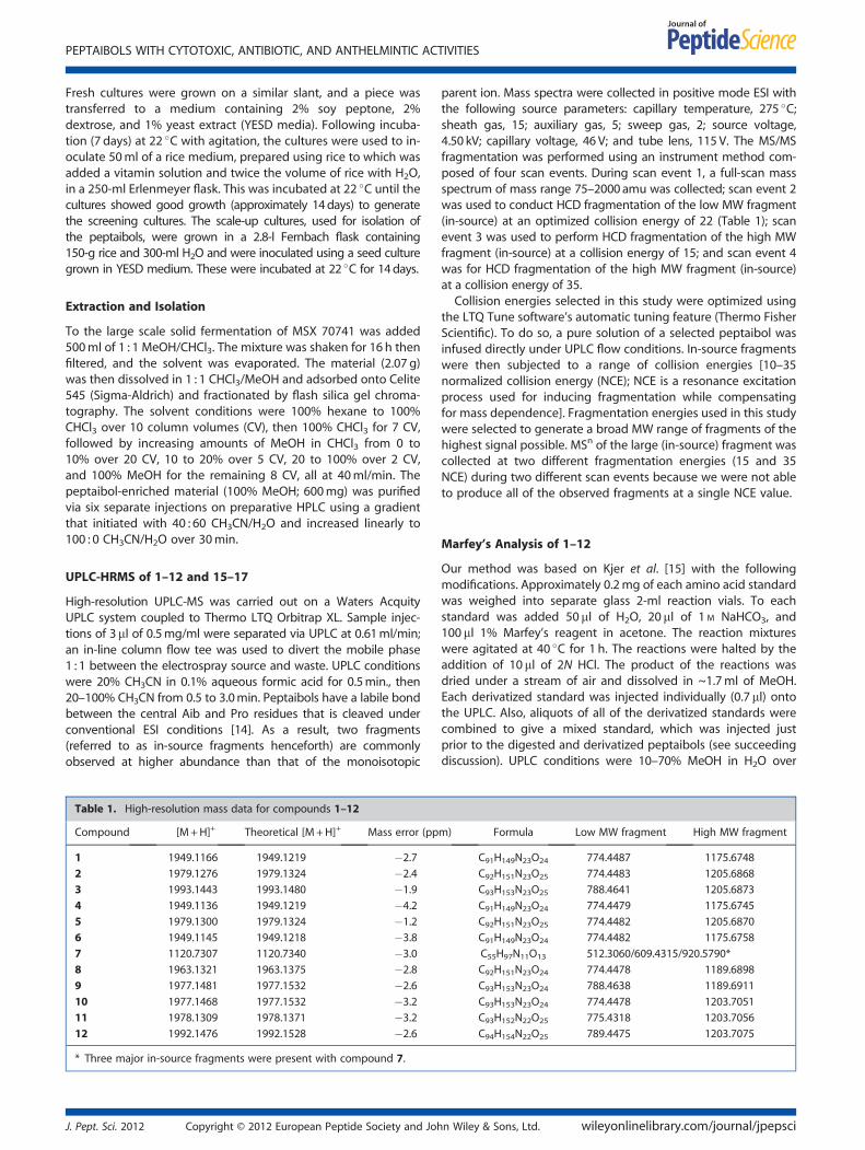
High-resolution UPLC-MS was carried out on a Waters AcquityUPLC system coupled to Thermo LTQ Orbitrap XL. Sample injec-tions of 3ml of 0.5mg/ml were separated via UPLC at 0.61ml/min;an in-line column flow tee was used to divert the mobile phase1 : 1 between the electrospray source and waste. UPLC conditionswere 20% CH3CN in 0.1% aqueous formic acid for 0.5min., then20–100% CH3CN from 0.5 to 3.0min. Peptaibols have a labile bondbetween the central Aib and Pro residues that is cleaved underconventional ESI conditions [14]. As a result, two fragments(referred to as in-source fragments henceforth) are commonlyobserved at higher abundance than that of the monoisotopic
Table 1. High-resolution mass data for compounds 1–12
Compound [M+H]+ Theoretical [M+H]+ Mass error (pp
1 1949.1166 1949.1219 �2.7
2 1979.1276 1979.1324 �2.4
3 1993.1443 1993.1480 �1.9
4 1949.1136 1949.1219 �4.2
5 1979.1300 1979.1324 �1.2
6 1949.1145 1949.1218 �3.8
7 1120.7307 1120.7340 �3.0
8 1963.1321 1963.1375 �2.8
9 1977.1481 1977.1532 �2.6
10 1977.1468 1977.1532 �3.2
11 1978.1309 1978.1371 �3.2
12 1992.1476 1992.1528 �2.6
* Three major in-source fragments were present with compound 7.
J. Pept. Sci. 2012 Copyright © 2012 European Peptide Society and Joh
parent ion. Mass spectra were collected in positive mode ESI withthe following source parameters: capillary temperature, 275 �C;sheath gas, 15; auxiliary gas, 5; sweep gas, 2; source voltage,4.50 kV; capillary voltage, 46 V; and tube lens, 115 V. The MS/MSfragmentation was performed using an instrument method com-posed of four scan events. During scan event 1, a full-scan massspectrum of mass range 75–2000 amu was collected; scan event 2was used to conduct HCD fragmentation of the low MW fragment(in-source) at an optimized collision energy of 22 (Table 1); scanevent 3 was used to perform HCD fragmentation of the high MWfragment (in-source) at a collision energy of 15; and scan event 4was for HCD fragmentation of the high MW fragment (in-source)at a collision energy of 35.
Collision energies selected in this study were optimized usingthe LTQ Tune software’s automatic tuning feature (Thermo FisherScientific). To do so, a pure solution of a selected peptaibol wasinfused directly under UPLC flow conditions. In-source fragmentswere then subjected to a range of collision energies [10–35normalized collision energy (NCE); NCE is a resonance excitationprocess used for inducing fragmentation while compensatingfor mass dependence]. Fragmentation energies used in this studywere selected to generate a broad MW range of fragments of thehighest signal possible. MSn of the large (in-source) fragment wascollected at two different fragmentation energies (15 and 35NCE) during two different scan events because we were not ableto produce all of the observed fragments at a single NCE value.
Marfey’s Analysis of 1–12
Our method was based on Kjer et al. [15] with the followingmodifications. Approximately 0.2mg of each amino acid standardwas weighed into separate glass 2-ml reaction vials. To eachstandard was added 50ml of H2O, 20ml of 1 M NaHCO3, and100 ml 1% Marfey’s reagent in acetone. The reaction mixtureswere agitated at 40 �C for 1 h. The reactions were halted by theaddition of 10ml of 2N HCl. The product of the reactions wasdried under a stream of air and dissolved in ~1.7ml of MeOH.Each derivatized standard was injected individually (0.7 ml) ontothe UPLC. Also, aliquots of all of the derivatized standards werecombined to give a mixed standard, which was injected justprior to the digested and derivatized peptaibols (see succeedingdiscussion). UPLC conditions were 10–70% MeOH in H2O over
m) Formula Low MW fragment High MW fragment
C91H149N23O24 774.4487 1175.6748
C92H151N23O25 774.4483 1205.6868
C93H153N23O25 788.4641 1205.6873
C91H149N23O24 774.4479 1175.6745
C92H151N23O25 774.4482 1205.6870
C91H149N23O24 774.4482 1175.6758
C55H97N11O13 512.3060/609.4315/920.5790*
C92H151N23O24 774.4478 1189.6898
C93H153N23O24 788.4638 1189.6911
C93H153N23O24 774.4478 1203.7051
C93H152N22O25 775.4318 1203.7056
C94H154N22O25 789.4475 1203.7075
n Wiley & Sons, Ltd. wileyonlinelibrary.com/journal/jpepsci

AYERS ET AL.
10min on the aforementioned BEH column and eluent monitoredat 340 nm.To generate the digested and derivatized peptaibols, approxi-
mately 0.2–0.3mg of compounds 1–12 were weighed separatelyinto 2-ml reaction vials, to which was added 0.5ml of 6N HCl. Thecompounds were hydrolyzed at 110 �C for 24 h, at which timethey were evaporated under a stream of air. To each hydrolysisproduct was then added 25 ml H2O, 10ml 1 M NaHCO3, and 50 mlof 1% Marfey’s reagent in acetone. The reaction mixtures wereagitated at 40 �C for 1h. The reactions were halted by the additionof 5ml of 2N HCl. The mixtures were dried under a stream of airand brought up in ~200ml of MeOH and injected onto the UPLC withthe use of the same conditions as for the standards.
Cytotoxicity Assays
The cytotoxicity measurements against the MCF-7 [16] humanbreast carcinoma (Barbara A. Karmanos Cancer Center), NCI-H460[17] human large cell lung carcinoma (HTB-177, American TypeCulture Collection (ATCC)), and SF-268 [18] human astrocytoma(NCI Developmental Therapeutics Program) cell lines wereperformed as described previously [19,20]. Moreover, a second cyto-toxicity assay was performed on only the isolated compoundsusing the MDA-MB-435 [21] human melanoma (HTB-129, ATCC) cellline as described previously [8] andwith the followingmodifications.
Table 2. Biological activities of peptaibols 1–12 and 15–17
Compound Cytotoxicity IC50 values (in mM)a
MCF-7 H460 SF268 IMR90 MDA-
Atroviridin D (1) >10 >10 >10 >10 N
Atroviridin E (2) 3.3 3.0 6.8 7.7 N
Atroviridin F (3) 3.8 3.2 5.3 >10 N
Atroviridin G (4) 2.7 1.3 1.9 5.9 N
Atroviridin H (5) 1.8 2.5 4.4 6.1 N
Atroviridin I (6) 2.1 2.3 5.1 9.8 N
Trichobrachin D-I (7) 3.4 4.1 7.0 >10 7
Alamethicin F50 (8) 2.2 3.4 2.3 4.8 8
Atroviridin B (9) 1.3 2.5 1.9 3.6 4
Polysporin B (10) 1.3 2.0 1.5 2.9 3
Alamethicin II (11) 1.1 1.6 1.7 4.0 6
Atroviridin J (12) 1.0 1.0 2.6 5.2 N
Trichokonin VI (15) 2.3 2.7 2.0 4.5 3
Trichokonin VII (16) 1.3 2.2 1.5 5.1 3
Longibranchin BIII (17) 0.8 0.8 1.4 4.6 N
Camptothecine 0.05 0.008 0.03 0.18 N
Vinblastinee NT NT NT NT
Vancomycine NT NT NT NT N
Ivermectine NT NT NT NT N
aIC50 values were determined as the concentration required to reduce cellucontinuous exposure (96 h for MDA-MB-435).
bMinimal inhibitory concentration is the lowest concentration of compouncIC50 values were determined as the concentration required to inhibit la
continuous exposure as expressed in mg/ml.dIndicates ‘not tested’.ePositive controls.fThe positive control for MDA-MB-435 was vinblastine tested at 2.0 an
respectively.
wileyonlinelibrary.com/journal/jpepsci Copyright © 2012 Europ
After treating the MDA-MB-435 cells with test substances and 96hincubation at 37 �C, the cells were evaluated for viability with acommercial absorbance assay (CellTiter 96 AQueous One Solution CellProliferation Assay, Promega Corp, Madison, WI, USA). The com-pounds were also tested in the IMR90 fibroblast cell line (ATCCCCL-186) [22], a normal diploid cell line that proliferates in culturefor approximately 58 generations prior to senescence. Positive con-trol data for all cell lines are provided in the legend of Table 2.
Antimicrobial Assay
The compounds were screened initially for antimicrobial activitywith the use of an agar plate diffusion assay. Overnight culturesof Escherichia coli, Pseudomonas aeruginosa, Staphylococcusaureus, Mycobacterium smegmatis, Candida albicans, and Bacillussubtilis were used to inoculate molten LB media or Middlebrook7H9 media (Difco Inc., Becton, Dickinson & Company, Sparks, MD,USA) with 1% glycerol, containing 1.5% agar and kept at 50 �C;these were then used to prepare assay plates. Samples (dissolvedin 10ml MeOH) were applied to the surface of the assay dish, andpositive controls were treated in a similar manner (penicillin G,gentamicin, novobiocin, and streptomycin; all from Sigma). Thebioassay plates were incubated overnight at 37 �C. Biologicalactivity of the standards could be detected to 1mg/ml (except thatpenicillin G was active against E. coli at 100mg/ml only).
MICb (mg/ml) IC50c (mg/ml)
MB-435 Staphylococcus aureus MRSA Haemonchus contortus
Td NT NT NT
T NT NT NT
T NT NT NT
T NT NT NT
T NT NT NT
T 44 44 NT
.3 113 113 NT
.9 35 140 0.2
.2 6 13 0.4
.2 102 102 NT
.2 12 23 NT
T 9 18 NT
.8 21 43 NT
.0 8 17 >8.3
T 4 8 3.0
T NT NT NTf NT NT NT
T 1.5 0.8 NT
T NT NT 0.006
lar proliferation by 50% relative to untreated controls following 72 h of
d completely inhibiting growth as expressed in mg/ml [19].
rval motility by 50% relative to untreated controls following 72 h of
d 1.0 nM, which resulted in 41 and 68% viable cells after treatment,
ean Peptide Society and John Wiley & Sons, Ltd. J. Pept. Sci. 2012

PEPTAIBOLS WITH CYTOTOXIC, ANTIBIOTIC, AND ANTHELMINTIC ACTIVITIES
Measurement of Antibiotic Activity against Methicillin-resistant Staphylococcus aureus
The samples were tested against a suite of methicillin-resistantS. aureus (MRSA) isolates (data not shown); only representativedata are shown in Table 2 against S. aureus (ATCC 6538) and anunrelated MRSA strain (ATCC 43300), which were both acquiredfrom Danville Community Hospital (Danville, VA, USA). Minimalinhibitory concentration (MIC) measurements were performedas described previously [23,24]. All measurements were made induplicate, and susceptibilities of the strains to vancomycin as apositive control were measured in parallel.
Anthelmintic Assay
The compounds were screened against Haemonchus contortusinfective larvae (HcL3) in an L3 motility assay; this assay evaluatesthe effect of the compounds on the body wall musculaturebecause this is not a feeding stage. Third stage larvae of an iso-late of H. contortus maintained in goats were obtained from thelaboratory of Dr Raymond Kaplan (University of Georgia, Athens,GA). The nematode L3 bioassay was as described [25] with slightmodifications. Briefly, in a 15-ml centrifugation tube, HcL3s wereincubated at room temperature in a 0.15% sodium hypochloritesolution for 25min, centrifuged at 100 rcf for 5min, and thenthe supernatant was removed and rinsed with distilled H2O.HcL3s were concentrated in phosphate buffered saline tocontain approximately 800 HcL3s/ml. The L3 motility assay wasconducted in triplicate in 96-well plates; each well had a totalvolume of 75ml and contained roughly 50 exsheathed HcL3s.Plates were incubated in a humidified chamber at 25–30 �C for72 h, and percent immobility was calculated. Ivermectin (positivecontrol) and DMSO only (negative control) wells were includedon every plate, and percent immobility was calculated using themean counts from three replicates. The peptaibols were dis-solved in DMSO and evaluated at 20 mg/ml in triplicate; com-pounds were considered active if they had any immobilizingeffects on HcL3s. Active compounds were evaluated further indose response, and IC50 values were calculated using a log probitregression analysis.
Mitochondria Transmembrane Potential Assay
The mitochondrial transmembrane potential assay was performedas described previously [8].
01008-44-1_110419104241#290 RT: 2.14 AV: 1 NL: 7.83E7F: FTMS + p ESI Full ms [75.00-2000.00]
400 600 800 100
m
0
20
40
60
80
100
Rel
ativ
e A
bund
ance
774.4487
975.564650.7113452.2489296.1599 922.5206
200
Figure 2. Full-scan HRMS (single scan) of atroviridin D (1). The two diagnos1175.6748 (high MW fragment). The multiply charged species are apparent a
J. Pept. Sci. 2012 Copyright © 2012 European Peptide Society and Joh
Results
The crude 1 : 1 CHCl3/MeOH extract of fungus MSX 70741 waspartitioned with 4 : 1 : 5 CHCl3/MeOH/H2O. The organic solublematerial was active (>95% growth inhibition of H460 cells at20 mg/ml) and was fractionated initially by flash silica gel chroma-tography. Harzianum A (13, 6.8mg) [26] and harzianum B (14,0.9mg) [27] were eluted in the 4–5% MeOH in CHCl3 fraction(>96% growth inhibition of H460 at 2mg/ml); these were purifiedby HPLC, and their NMR data were in excellent agreement withthe literature [Figure S1 for structures of 13 and 14 (SupportingInformation)] [27]. The peptaibols were eluted in the flashchromatography system in the 100% MeOH fraction (>98%growth inhibition of H460 at 20 mg/ml). Final purification of thepeptaibols was accomplished by preparative scale RP-HPLC(Figure S2). Compound 1 was eluted at 12.0min (1.1mg), 2 at13.1min (3.3mg), 3 at 14.0min (0.7mg), 4 at 14.3min (2.0mg),5 at 15.7min (2.6mg), 6 at 17.7min (6.7mg), 7 at 18.7min(12.4mg), 8 between 19 and 21min (242.5mg), 9 between 21.3and 22.3min (77.6mg), 10 between 22.3 and 23.5min(52.6mg), 11 between 23.5 and 24.5min (28.8mg), and 12 at25.3min (4.1mg). With the use of the same conditions as men-tioned previously, fungus MSX 57715 was extracted and fraction-ated to isolate peptaibols 15 (205.6mg), 16 (93.6mg), and 17(7.5mg) with HPLC elution times of 17–19, 19–20.5, and21.1min, respectively. Compounds 8–11 were the major isolatesand were known peptaibols, whereas 1–7 and 12 were new. Thenumbering of these compounds corresponds to their elution or-der on RP-HPLC: 1 eluted earliest and 12 eluted latest.
The high-resolution protonated monoisotopic precursor iondata, [M+H]+, and the resultant molecular formulae for com-pounds 1–12 are listed in Table 1; the structure for 8 and thesequence for the other peptaibols are displayed in Figure 1. An im-portant characteristic of the MS spectra for typical 20-residue pep-taibols was in-source fragmentation between the Aib13 andPro14 residues, leaving two major fragments (Figure 2) [14]. Otherdiagnostic peaks were the doubly and triply charged species, aswell as the triply charged dimer (Figure 2). The MS data for thetwo major fragments (termed here ‘low MW fragment’ and ‘highMW fragment’) of 1–12 are listed in Table 1; note that 7 had threemajor in-source fragments. These major fragments were then sub-jected to MS/MS to elucidate the sequence.
The MS/MS data, in conjunction with the NMR data, wereused to sequence the major metabolite (8), identified as the
0 1200 1400 1600 1800 2000
/z
1175.6748
61300.4180 1950.12131504.3147 1757.9775
1950 1952
m/z
0
20
40
60
80
100
Rel
ativ
e A
bund
ance
1950.1213
1949.1166
1951.1215
1952.1254
tic in-source fragments are apparent at 774.4487 (low MW fragment) andt 650.7113 [M+3H]3+, 975.5646 [M+2H]2+, and 1300.4180 [2M+3H]3+.
n Wiley & Sons, Ltd. wileyonlinelibrary.com/journal/jpepsci

AYERS ET AL.
known peptaibol alamethicin F50 (or atroviridin A) [28]. Theabsolute configuration of 8 was confirmed by Marfey’s analysis.As with all of the peptaibols isolated in this study, the individualamino acids had the L-configuration except for Iva, which hadD-configuration. Compounds 9–11 were likewise found to bethe known compounds atroviridin B (9) [29], polysporin B (10)[30], and alamethicin II (11) [31].The sequences and structures of compounds 1–7 and 12 were
elucidated in a similar fashion by MS/MS, NMR, and Marfey’s
100 200 300 400
m
0
20
40
60
80
100
Rel
ativ
e A
bund
ance
282.1802
367.2328
49197.1277393.2121
300 400 500 600 700 8m
0
20
40
60
80
100
Rel
ativ
e A
bund
ance
736.3967
523.2854452.2484651.3463
100 150 200 250m
0
20
40
60
80
100
Rel
ativ
e A
bund
ance 254.1491
2
211.1070
183.1121155.1172
Aib Ac+Ala+Pro (To 0)
-CO-CO
Ala+Pro+Aib
-C
Pro+Val/Iva (To 0)
VAib GlnAla
Aib Aib Gln
Figure 3. HR-MS/MS by HCD of atroviridin D (1): (A) HCD of 774 peaks, CE=
NH’s Pheol o/m/p
Gln NH’s
Figure 4. 1H NMR spectrum of atroviridin J (12) (CD3OH, 950MHz). Spectru
wileyonlinelibrary.com/journal/jpepsci Copyright © 2012 Europ
analysis. Examples of MS/MS analysis (Figure 3), 1H NMR andNOESY spectra (Figures 4 and 5, respectively), and Marfey’sanalysis (Figure 6) are provided. Because of paucity of material,quality NMR data for compounds 1 and 3 could not be obtained;however, MS/MS and Marfey’s analyses were sufficient for thecomplete structure elucidation for 1. Alternatively, structure 3should be considered tentative because the locations of Valversus Iva could not be determined unequivocally, because both werepresent in the Marfey’s analysis. It is highly likely that residues 9 and
500 600 700 800 900
/z
5.2905774.4497623.3491
00 900 1000 1100 1200 1300/z
920.5186
835.4658
977.5407 1090.6220 1175.6764
300 350 400 450/z
96.1596
367.1966339.2018
452.2488
A)
AibAla
C)
-COO
B) AibLeuGlyAibal
Gln Pheol
22; (B) HCD of 1175 peaks, CE= 15; (C) HCD of 1175 peaks, CE = 35.
Ac
Ala/Aib methyl groups
Leu/Val/Iva methyl groups
H’s
CD3OH
m was obtained with presaturation of the –OH peak at dH 4.9.
ean Peptide Society and John Wiley & Sons, Ltd. J. Pept. Sci. 2012

Q NH2’s
2D-NOESY: NH Region
U1
G11
U13
U10
U8
L12
E18
U6
U5
Q19
Q7
U3
U16
V15
A4
V9
Fol
20
J17
Figure 5. NOESY spectrum of atroviridin J (12), highlighting the correlations proving the position of the Iva (J) residue (CD3OH, 950MHz).
StandardsSamplehydrolysate
L-S
D-S
L-E
L-A
L-P
D-E
D-P
D-A U
L-V
L-F
ol
L-L
D-V
D-F
ol
D-L
Mar
fey’
s +
G
Figure 6. Marfey’s analysis of atroviridin E (2). Note that the peak in the sample hydrolysate at 3.4min that appears to indicate the presence of D-serineis an artifact of the derivatization reaction that was present in all hydrolysate chromatograms. The retention times, while close, do not match. Forprocedure and chromatographic conditions, see Section on Marfey’s Analysis of 1–12.
PEPTAIBOLS WITH CYTOTOXIC, ANTIBIOTIC, AND ANTHELMINTIC ACTIVITIES
15 were Val and residue 17 was Iva, because of similarity withknown peptaibols; however, this could not be proven.
In work of a similar vein from another fungus from the Myco-synthetix library (specifically, MSX 57715), other known peptaibolswere isolated and elucidated, along with trichodermin, which isa simple trichothecene. These peptaibols were identified astrichokonin VI (15) [32], trichokonin VII (16) [32], and longibranchinBIII (17) [33,34]. These peptaibols were evaluated in the samebioassays as 1–12 to determine the significance of Pro2 versus Ala2.
The isolated peptaibols were evaluated in a series of biologicalassays. With respect to anticancer activity, they were tested forcytotoxicity against a panel of human tumor cell lines and ahuman fibroblast cell line (Table 2). They were also examinedfor activity in a mitochondria transmembrane potential assaybut found inactive (data not shown). With literature precedentfor antimicrobial activity for some peptaibols [35], they weretested against a battery of assays, including those for Gram-positive and Gram-negative bacteria and C. albicans. The mostpromising antimicrobial activity was with respect to S. aureus,which spawned further examination against MRSA (Table 2).Finally, a few peptaibols have been reported to have anthelminticactivity [36,37], and thus the compounds were examined in an
J. Pept. Sci. 2012 Copyright © 2012 European Peptide Society and Joh
assay for larval motility against H. contortus infective stages.Larval motility was inhibited completely by four of the peptaibols(8, 9, 16, and 17) when tested at 20 mg/ml; these wereevaluated further for dose response with a top concentration of8.3 mg/ml (Table 2). In summary, the biological potential of thesecompounds were examined extensively.
Discussion
In this work, a single injection on UPLC-MS/MS using HCD on aThermo LTQ Orbitrap XL gave high-resolution fragmentation andnearly complete sequence data in a 3-min run. HCD fragmentationis becoming a routine tool in proteomics research, especially forquantitation studies that utilize various isotopic-labeling methodsand for peptide sequencing studies [38]; to the best of our knowl-edge, this technique has not been applied previously to researchon peptaibols. The advantages of the HCD collision cell in the LTQOrbitrap XL system include its ability to generate ‘rich’ fragmentationspectra that include low m/z values and Orbitrap (high-resolution/high mass accuracy) detection of mass fragments, allowing for mo-lecular formula assignment of a given m/z value [39].
n Wiley & Sons, Ltd. wileyonlinelibrary.com/journal/jpepsci

AYERS ET AL.
Compound 1 was identical to the major metabolite 8 exceptfor the substitution of Ala1 for Aib1. To our knowledge, theonly other peptaibol that has been reported to have Ala1 is tri-chokonin IIb [40], which was identical to 1 except for thereplacement of Pro2 with Ala2. The result of substituting Aib1with Ala1 on the cytotoxic activity was interesting, as 1 wasinactive compared with 8 (Table 2).Compounds 2–6 were also structurally related to alamethicin
F50 (8; Figure 1). Compounds 2 and 4–6 differed from 8 by asingle amino acid residue substitution. Residue 6 appeared tobe the most variable site, with substitution of Ser, Gly, or Aibfor Ala. Compound 2 was identical to 8 except for substitutionof Ser6 for Ala6. Compound 3 likely included Iva17 instead ofAib17 in addition to Ser6 versus Ala6, although the structureelucidation of 3 was incomplete because of paucity of material.
01008-44-7_110419134404#328-335 RT: 2.43-2.47 AV: F:FTMS + p ESI Full ms [75.00-2000.00]
200 400 60
0
20
40
60
80
100
Rel
ativ
e A
bund
ance
512.3060 609
409.2799201.1592128.0700
486.2909
[AcUSVIU]+
[PL
[PVol + 2H]+
[PLLU + H]+
[AcUSV]+
Figure 7. Full-scan HRMS (sum of eight mass spectra) of Trichobrachin D-I
Peptaibol_44_7_1H_500.esp
1.05 1.00 0.95
Chemica
0.050.100.150.200.250.300.350.400.450.500.550.600.650.700.75
Peptaibol_44_7_1H.esp
1.05 1.00 0.95
Chemica
0.05
0.10
0.15
0.20
0.25
0.30
0.35
0.40
0.45
0.50
Nor
mal
ized
Inte
nsity
Nor
mal
ized
Inte
nsity
I4
Vol11 L7
V3
V3 & L7
Figure 8. Comparison of the upfield methyl region of the 500-MHz versus 9
wileyonlinelibrary.com/journal/jpepsci Copyright © 2012 Europ
Compound 5 was identical to 8 except for substitution of Ser4for Ala4. Compound 6 was identical to 8 except for substitutionof Val12 for Leu12. Compound 12 was more closely related to ala-methicin II (11) and differed from 11 only by substitution of Iva17for Aib17. Because of their similarity to the known atroviridins,compounds 1–6 and 12 were ascribed the trivial names of atrovir-idin D through atroviridin J (Figure 1), respectively.
Compound 7 was unique with respect to the other peptaibolsisolated from MSX 70741 (Figure 1), in that 7 contained 11 aminoacid residues, whereas the others were 20-mers. Also, the HRMSof 7 showed three major in-source fragments instead of two, aswith the other peptaibols (Figure 7). The 1H NMR also showed amethyl triplet at dH 0.85 (Figure 8), and from 2D TOCSY andNOESY spectra, this triplet was due to Ile at residue 4. Ile wasnot present in any of the other peptaibols in this study.
3 NL: 4.70E7
0 800 1000
m/z
920.5790
1120.7307
.4315
788.4655 981.5657655.0483
860.4869 1077.1628
LUPVol + 2H]+
[AcUSVIUPLLU]+
[M + H]+
(7).
0.90 0.85 0.80
l Shift (ppm)
0.90 0.85 0.80 0.75
l Shift (ppm)
500 MHz
950 MHzI4 L8 L8
Vol11
50-MHz 1H NMR data of trichobrachin D-I (7).
ean Peptide Society and John Wiley & Sons, Ltd. J. Pept. Sci. 2012

PEPTAIBOLS WITH CYTOTOXIC, ANTIBIOTIC, AND ANTHELMINTIC ACTIVITIES
Compound 7 was most closely related to the trichobrachin[41,42], hypomurocin [43], and trichobrevin [44,45] subclasses of11-mer peptaibols. The effect of a shorter chain on cytotoxicactivity was negligible (Table 2), although it seemed to diminishantibacterial activity (Table 2) and was inactive in the HcL3motility assay at 20mg/ml. The NMR data for 7 illustrated thebenefit of higher field on resolution (Figure 8). The Ile4 methyltriplet for 7 was fully resolved at 950MHz, whereas at 500MHz,the triplet could not be identified because of overlap with theadjacent methyl doublets from the Leu8 residue. Four methyldoublets from dH 0.90 to 0.95 were also completely resolved at950MHz, whereas at 500MHz, there was too much overlap todistinguish the signals. The 950-MHz NMR data were not onlywell resolved for the methyl peaks of compound 7 but it alsoresulted in greatly improved resolution of NH–NH correlations,which were essential for sequencing. Although it is well appreci-ated in the natural products community that higher field pro-vides better resolution, the example of 500-MHz versus 950-MHzNMR spectra of compound 7 provides a cogent example ofhow this can be applied to structure elucidation of peptaibols.Compound 7 most closely resembled trichobrevin B-IIb [45],having the same ‘rough’ amino acid sequence; meaning thatthe Leu/Ile and Val/Iva residues were not unambiguously deter-mined for trichobrevin B-IIb. As compound 7 also closelyresembled the trichobrachin peptaibol series, it was ascribedthe trivial name trichobrachin D-I, to differentiate it from thetrichobrachin A series (which all have Asp2, Aib9, and Pro10),the trichobrachin B series (which contain Asp2, Val9, andPro10), and the trichobrachin C series (which have Gln2, Aib9,and Pro10). All trichobrachins, including 7, are also characterizedby Aib5 and Pro6 [41,42].
Compounds 2, 3, and 5 were novel in that they contain Ser atpositions 4 or 6, which, to the best of our knowledge, has notbeen reported previously in 20-mer peptaibols. Although Ser2has been reported in a number of classes of 18-mer peptaibols,such as the hypomurocin B series [43], the trichokindins [46],the trichorzin MA series [47], and the trichorzin PA series[33,34], the presence of Ser2 is rare for 11-mer peptaibols (as incompound 7), having only been reported in the trichobrevin Bclass [44,45]. The presence of Ser in 2, 3, 5, and 7, according tothe MS/MS fragmentation work, was confirmed by the TOCSYNMR spectra (except for 3) as well as the Marfey’s analyses, whichalso confirmed the stereochemistry as L-Ser (Figure 6).
Three known peptaibols were isolated from a second Myco-synthetix fungus (MSX 57715; Figure 1). Trichokonin VI (15) wasthe major peptaibol isolate and was identical to alamethicinF50 (8), except for replacement of Pro2 with Ala2; compounds8 and 15 had nearly the same cytotoxic activity profile (Table 2).Trichokonin VII (16) was analogous to atroviridin B (9) with thesame substitution of Ala2 for Pro2; these two compounds wereessentially equipotent in the cytotoxicity and antibacterial assays(Table 2). Longibranchin BIII (17) substituted Glu18 for Gln18 oftrichokonin VII (16); the former was slightly more potent in bothcytotoxicity and antibacterial assays (Table 2), although possiblywithin experimental error of the assays.
A range of bioactivities were observed for the isolated peptaibols(Table 2). Several of the peptaibols exhibited not only cytotoxicitybut also cancer cell selectivity (Table 2). Selectivity was assessedvia the IMR90 cell lines, and they are nontransformed but proliferat-ing cells. For example, the positive control camptothecin exhibited3.6-fold selectivity in MCF-7 cells to 23-fold selectivity in H460 cellsrelative to IMR90 cells. Alamethicin II (11), atroviridin J (12),
J. Pept. Sci. 2012 Copyright © 2012 European Peptide Society and Joh
trichokonin VII (16), and longibranchin BIII (17) weremost notewor-thy in exhibiting 3.6-fold, 5.2-fold, 3.9-fold, and 5.8-fold selectivity,respectively, in MCF-7 versus IMR90 cells. These data were consis-tent with a previous report of the growth inhibitory activity of tri-chokonin VI (15) against hepatocellular carcinoma cells, in which15 did not obviously affect normal liver cells at lower concentra-tions [48]. Three of these four compounds contained Iva17 andwere the more hydrophobic peptaibols. A general trend of higheractivity with higher hydrophobicity has been reported previously[42], although the effect was much less pronounced in our work.With respect to antibacterial activity, the compounds were essen-tially equipotent to both S. aureus and MRSA (or within experimen-tal error), except compound 8, which was approximately four timesmore potent against S. aureus than MRSA. Compound 8 was alsothe most potent in the assay for larval motility against H. contortusinfective stages, although still two orders of magnitude less potentthan the positive control.
In summary, a series of twelve structurally related peptaibols(1–12) were isolated from MSX 70741, including eight newpeptaibols, four of which featured the incorporation of serine.These peptaibols were sequenced by MS/MS using HCD. Themethod was designed to enable full-scan intact mass analysisfollowed by sequential fragmentation of the low and highMW fragments from a single UPLC injection. This streamlinedapproach enabled nearly complete amino acid sequencingcapabilities of peptaibols from a small volume of sample, over atenfold decrease in separation time, and accurate mass measure-ments to confirm amino acid identities. Fragmentation of peptaibolsby high resolutionwas important because some peptaibols containboth Leu (and/or Ile) as well as hydroxyproline residues, an exampleof which is clonostachin [49]. Leu, Ile, and hydroxyproline frag-ments have the same nominal mass (113); however, hydroxyprolinehas a different formula and therefore can only be distinguished inMS by high-resolution measurements. Leu and Ile have the sameformula and therefore have to be distinguished by NMR. TheNMR experiments confirmed the sequencing of the peptaibolsand were critical for distinguishing the locations of constitutionalisomers that were present in the same compound, such as Leuand Ile in compound 7 or Val and Iva in compound 12. The Marfey’sanalysis was also a good demonstration of the power of UPLC com-pared with HPLC, where analyses were completed in 10min withexcellent resolution of the amino acid-Marfey derivative standards.Finally, from a mycological perspective, both organisms were pro-lific producers of peptaibols, generating >400mg (MSX 70741)and >300mg (MSX 57715) of peptaibols per solid phase culturein a 2.8-l Fernbach flask, using standard techniques with no growthoptimization studies.
Acknowledgements
This research was supported by P01 CA125066 from the NationalCancer Institute/National Institutes of Health, Bethesda, MD, USA.The anthelmintic studies were supported in part by a CollaborativeFunding Grant (2011-CFG-8008) from the North Carolina Biotech-nology Center and the Kenan Institute for Engineering, Technology& Science. Mycology technical support was provided by BlaiseDarveaux and Maurica Lawrence. The authors acknowledge thetechnical assistance of Ms Myra D. Williams (Virginia Tech) in mea-suring MICs. The authors also thank Dr Kevin Knagge of the DavidH. Murdock Research Institute, Kannapolis, NC, for the 950-MHzNMR data and Ms Tamam El-Elimat for the helpful discussions.
n Wiley & Sons, Ltd. wileyonlinelibrary.com/journal/jpepsci

AYERS ET AL.
References1 Toniolo C, Bruckner H. Peptaibiotics: Fungal Peptides Containing a-Dialkyl
a-Amino Acids. Vrlag Helvetica Chimica Acta: Zurich, 2009; 702.2 Toniolo C, Bruckner H. Peptaibiotics. Chem. Biodivers. 2007; 4: 1021–1022.3 Bruckner H. Peptaibols/peptaibiotics – Editorial. J. Pept. Sci. 2003; 9: 659.4 Daniel JF, Filho ER. Peptaibols of Trichoderma. Nat. Prod. Rep. 2007; 24:
1128–1141.5 Leitgeb B, Szekeres A, Manczinger L, Vagvolgyi C, Kredics L. The history
of alamethicin: a review of the most extensively studied peptaibol.Chem. Biodivers. 2007; 4: 1027–1051.
6 Kinghorn AD, Carache de Blanco EJ, Chai HB, Orjala J, Farnsworth NR,Soejarto DD, Oberlies NH, Wani MC, Kroll DJ, Pearce CJ, Swanson SM,Kramer RA, Rose WC, Fairchild CR, Vite GD, Emanuel S, Jarjoura D,Cope FO. Discovery of anticancer agents of diverse natural origin. PureAppl. Chem. 2009; 81: 1051–1063.
7 Orjala J, Oberlies NH, Pearce CJ, Swanson SM, Kinghorn AD. Discoveryof potential anticancer agents from aquatic cyanobacteria, filamentousfungi, and tropical plants. In Bioactive Compounds from Natural Sources.Natural Products as Lead Compounds in Drug Discovery. Tringali C (ed.).Taylor & Francis: London, UK, 2012; 37–63.
8 Ayers S, Graf TN, Adcock AF, Kroll DJ, Matthew S, Carache de BlancoEJ, Shen Q, Swanson SM, Wani MC, Pearce CJ, Oberlies NH. Resorcylicacid lactones with cytotoxic and NF-kB inhibitory activities and theirstructure-activity relationships. J. Nat. Prod. 2011; 74: 1126–1131.
9 Ayers S, Graf TN, Adcock AF, Kroll DJ, Shen Q, Swanson SM, Wani MC,Darveaux BA, Pearce CJ, Oberlies NH, Obionin B. An o-pyrano-naphthoquinone decaketide from an unidentified fungus (MSX63619) from the Order Pleosporales. Tetrahedron Lett. 2011; 52:5128–5230.
10 Sy-Cordero AA, Graf TN, Adcock AF, Kroll DJ, Shen Q, Swanson SM,Wani MC, Pearce CJ, Oberlies NH. Cyclodepsipeptides, sesquiterpenoids,and other cytotoxic metabolites from the filamentous fungusTrichothecium sp. (MSX 51320). J. Nat. Prod. 2011; 74: 2137–2142.
11 Ayers S, Ehrmann BM, Adcock AF, Kroll DJ, Wani MC, Pearce CJ,Oberlies NH. Thielavin B methyl ester: a cytotoxic benzoate trimerfrom an unidentified fungus (MSX 55526) from the Order Sordariales.Tetrahedron Lett. 2011; 52: 5733–5735.
12 Summers MY, Kong F, Feng X, Siegel MM, Janso JE, Graziani EI, CarterGT. Septocylindrins A and B: peptaibols produced by the terrestrialfungus Septocylindrium sp. LL-Z1518. J. Nat. Prod. 2007; 70: 391–396.
13 Mitova MI, Murphy AC, Lang G, Blunt JW, Cole AL, Ellis G, Munro MH.Evolving trends in the dereplication of natural product extracts. 2. Theisolation of chrysaibol, an antibiotic peptaibol from a New Zealand sam-ple of the mycoparasitic fungus Sepedonium chrysospermum. J. Nat.Prod. 2008; 71: 1600–1603.
14 el Hajji M, Rebuffat S, Lecommandeur D, Bodo B. Isolation andsequence determination of trichorzianines A antifungal peptides fromTrichoderma harzianum. Int. J. Pept. Protein Res. 1987; 29: 207–215.
15 Kjer J, Debbab A, Aly AH, Proksch P. Methods for isolation of marine-derived endophytic fungi and their bioactive secondary products.Nat. Protoc. 2010; 5: 479–490.
16 Soule HD, Vazguez J, Long A, Albert S, Brennan M. A human cell linefrom a pleural effusion derived from a breast carcinoma. J. Natl.Cancer Inst. 1973; 51: 1409–1416.
17 Carney DN, Gazdar AF, Bunn PA, Jr., Guccion JG. Demonstration of thestem cell nature of clonogenic tumor cells from lung cancer patients.Stem Cells 1982; 1: 149–164.
18 Rosenblum ML, Gerosa MA, Wilson CB, Barger GR, Pertuiset BF, deTribolet N, Dougherty DV. Stem cell studies of human malignant braintumors. Part 1: development of the stem cell assay and its potential.J. Neurosurg. 1983; 58: 170–176.
19 Alali FQ, El-Elimat T, Li C, Qandil A, Alkofahi A, Tawaha K, Burgess JP,Nakanishi Y, Kroll DJ, Navarro HA, Falkinham JO, III, Wani MC, OberliesNH. New colchicinoids from a native Jordanian meadow saffron,Colchicum brachyphyllum: isolation of the first naturally occurringdextrorotary colchicinoid. J. Nat. Prod. 2005; 68: 173–178.
20 Li C, Lee D, Graf TN, Phifer SS, Nakanishi Y, Riswan S, Setyowati FM,Saribi AM, Soejarto DD, Farnsworth NR, Falkinham JO, III, Kroll DJ,Kinghorn AD, Wani MC, Oberlies NH. Bioactive constituents of the stembark of Mitrephora glabra. J. Nat. Prod. 2009; 72: 1949–1953.
21 Rae JM, Creighton CJ, Meck JM, Haddad BR, Johnson MD. MDA-MB-435 cells are derived from M14 melanoma cells—a loss for breastcancer, but a boon for melanoma research. Breast Cancer Res. Treat.2007; 104: 13–19.
wileyonlinelibrary.com/journal/jpepsci Copyright © 2012 Europ
22 Nichols WW, Murphy DG, Cristofalo VJ, Toji LH, Greene AE, Dwight SA.Characterization of a new human diploid cell strain, IMR-90. Science1977; 196: 60–63.
23 Williams AA, Sugandhi EW, Macri RV, Falkinham JO, 3rd, Gandour RD.Antimicrobial activity of long-chain, water-soluble, dendritic tricarboxy-lato amphiphiles. J. Antimicrob. Chemother. 2007; 59: 451–458.
24 Maisuria BB, Actis ML, Hardrict SN, Falkinham JO, 3rd, Cole MF,Cihlar RL, Peters SM, Macri RV, Sugandhi EW, Williams AA, PoppeMA, Esker AR, Gandour RD. Comparing micellar, hemolytic, and an-tibacterial properties of di- and tricarboxyl dendritic amphiphiles.Bioorg. Med. Chem. 2011; 19: 2918–2926.
25 Lopez-Aroche U, Salinas-Sanchez DO, de Gives PM, Lopez-ArellanoME, Liebano-Hernandez E, Valladares-Cisneros G, Arias-Ataide DM,Hernandez-Velazquez V. In vitro nematicidal effects of medicinalplants from the Sierra de Huautla, Biosphere Reserve, Morelos, Mexicoagainst Haemonchus contortus infective larvae. J. Helminthol. 2008; 82:25–31.
26 Corley DG, Millerwideman M, Durley RC. Isolation and structure ofharziazum A: a new trichothecene from Trichoderma harzianum. J.Nat. Prod. 1994; 57: 422–425.
27 Jin HZ, Lee JH, Zhang WD, Lee HB, Hong YS, Kim YH, Lee JJ. HarzianumsA and B produced by a fungal strain, Hypocrea sp F000527, and theircytotoxicity against tumor cell lines. J. Asian Nat. Prod. Res. 2007; 9:203–207.
28 Meyer CE, Reusser F. A polypeptide antibacterial agent isolated fromTrichoderma viride. Experientia 1967; 23: 85–86.
29 Oh SU, Lee SJ, Kim JH, Yoo ID. Structural elucidation of new antibioticpeptides, atroviridins A, B and C from Trichoderma atroviride.Tetrahedron Lett. 2000; 41: 61–64.
30 New AP, Eckers C, Haskins NJ, Neville WA, Elson S, HuesoRodriguez JA,RiveraSagredo A. Structures of polysporins A-D, four new peptaibolsisolated from Trichoderma polysporum. Tetrahedron Lett. 1996; 37:3039–3042.
31 Pandey RC, Cook JC, Rinehart KL. Peptaibophol antibiotics.3. High-resolution and field desorption mass-spectrometry studies and revisedstructures of alamethicin-I and alamethicin-II. J. Am. Chem. Soc. 1977;99: 8469–8483.
32 Huang Q, Tezuka Y, Kikuchi T, Nishi A, Tubaki K, Tanaka K. Studies onmetabolites of mycoparasitic fungi .2. Metabolites of Trichodermakoningii. Chem. Pharm. Bull. 1995; 43: 223–229.
33 Leclerc G, Rebuffat S, Bodo B. Directed biosynthesis of peptaibol anti-biotics in two Trichoderma strains II. Structure elucidation. J. Antibiot.1998; 51: 178–183.
34 Leclerc G, Rebuffat S, Goulard C, Bodo B. Directed biosynthesis ofpeptaibol antibiotics in two Trichoderma strains I. Fermentation andisolation. J. Antibiot. 1998; 51: 170–177.
35 Duclohier H. Peptaibiotics and peptaibols: an alternative to classicalantibiotics? Chem. Biodivers. 2007; 4: 1023–1026.
36 Thirumalachar MJ. Antiamoebin, a new antiprotozoal-anthelminticantibiotic. I. Production and biological studies. Hindustan Antibiot. Bull.1968; 10: 287–289.
37 Schiell M, Hofmann J, Kurz M, Schmidt FR, Vertesy L, Vogel M, Wink J,Seibert G. Cephaibols, new peptaibol antibiotics with anthelminticproperties from Acremonium tubakii DSM 12774. J. Antibiot. 2001;54: 220–233.
38 Olsen JV, Macek B, Lange O, Makarov A, Horning S, Mann M. Higher-energy C-trap dissociation for peptide modification analysis. Nat.Methods 2007; 4: 709–712.
39 Kim S, Rodgers RP, Marshall AG. Truly “exact”mass: elemental composi-tion can be determined uniquely frommolecular mass measurement atsimilar to 0.1 mDa accuracy for molecules up to similar to 500 Da. Int. J.Mass Spectrom. 2006; 251: 260–265.
40 Huang Q, Tezuka Y, Hatanaka Y, Kikuchi T, Nishi A, Tubaki K. Studieson metabolites of mycoparasitic fungi .5. Ion-spray ionization massspectrometric analysis of Trichokonin-II, a peptaibol mixture obtainedfrom the culture broth of Trichoderma koningii. Chem. Pharm. Bull.1996; 44: 590–593.
41 Mohamed-Benkada M, Montagu M, Biard JF, Mondeguer F, Verite P,Dalgalarrondo M, Bissett J, Pouchus YF. New short peptaibols from amarine Trichoderma strain. Rapid Commun. Mass Spectrom. 2006; 20:1176–1180.
42 Ruiz N, Wielgosz-Collin G, Poirier L, Grovel O, Petit KE, Mohamed-Benkada M, du Pont TR, Bissett J, Verite P, Barnathan G, Pouchus YF.New Trichobrachins, 11-residue peptaibols from a marine strain ofTrichoderma longibrachiatum. Peptides 2007; 28: 1351–1358.
ean Peptide Society and John Wiley & Sons, Ltd. J. Pept. Sci. 2012

PEPTAIBOLS WITH CYTOTOXIC, ANTIBIOTIC, AND ANTHELMINTIC ACTIVITIES
43 Becker D, Kiess M, Bruckner H. Structures of peptaibol antibiotics hypo-murocin A and B from the ascomycetous fungus Hypocrea muroianaHino et Katsumoto. Liebigs Annalen-Recueil. 1997; 767–772.
44 Degenkolb T, Dieckmann R, Nielsen KF, Grafenhan T, Theis C, Zafari D,Chaverri P, Ismaiel A, Bruckner H, vonDohrenH, ThraneU, Petrini O, SamuelsGJ. The Trichoderma brevicompactum clade: a separate lineage with newspecies, new peptaibiotics, and mycotoxins. Mycol Prog. 2008; 7: 177–219.
45 Degenkolb T, Grafenhan T, Nirenberg HI, Gams W, Bruckner H.Trichoderma brevicompactum complex: rich source of novel andrecurrent plant-protective polypeptide antibiotics (peptaibiotics).J. Agric. Food Chem. 2006; 54: 7047–7061.
46 Iida A, Sanekata M, Fujita T, Tanaka H, Enoki A, Fuse G, Kanai M,Rudewicz PJ, Tachikawa E. Fungal metabolites. XVI. Structures of new
J. Pept. Sci. 2012 Copyright © 2012 European Peptide Society and Joh
peptaibols, trichokindins I-VII, from the fungus Trichoderma harzianum.Chem. Pharm. Bull. 1994; 42: 1070–1075.
47 Goulard C, Hlimi S, Rebuffat S, Bodo B. Trichorzins HA and MA,antibiotic peptides from Trichoderma harzianum. I. Fermenta-tion, isolation and biological properties. J. Antibiot. 1995; 48:1248–1253.
48 ShiM, Wang HN, Xie ST, Luo Y, Sun CY, Chen XL, Zhang YZ. Antimicrobialpeptaibols, novel suppressors of tumor cells, targeted calcium-mediatedapoptosis and autophagy in human hepatocellular carcinoma cells.Mol.Cancer 2010; 9: 26.
49 Chikanishi T, Hasumi K, Harada T, Kawasaki N, Endo A. Clonostachin, anovel peptaibol that inhibits platelet aggregation. J. Antibiot. 1997;50: 105–110.
n Wiley & Sons, Ltd. wileyonlinelibrary.com/journal/jpepsci