PENDALA - EXPERIMENTAL DEFORMATION OF SULFIDE...
170
PENDALA - EXPERIMENTAL DEFORMATION OF SULFIDE ORES. 1 : ' l" 1
Transcript of PENDALA - EXPERIMENTAL DEFORMATION OF SULFIDE...

PENDALA - EXPERIMENTAL DEFORMATION OF SULFIDE ORES.
1 : '
l"
1

EXPERIMENTAL DEFORMATION OF
SULFIDE ORES
by
Penda1a Krishnamurthy
A thesis submitted to the Facu1ty of Graduate Studies and Research in partial fu1fi1ment of the requirements for the degree of Master of Science.
Department of Geo1ogica1 Sciences, McGi11 University, Montreal.
March 1967 •
. 0 Penda1a Krishnamurthy 1968

ABSTRACT
The deformation behaviour of chalcocite, galena, chalcopyrite,
pyrrhotite, sphalerite and pyrite were studied in a closed system subjected
to pressures and elevated temperatures.
Chalcocite was found to flow at 300°C. under a confining
pressure of 15,500 p.s.i.
Galena flowed plastically with translation gliding at around
40004under confining pressures up to 13,000 p.s.i.
Chalcopyrite readily flowed above 475°C. and below 650°C.
under confining pressures of 10,000 to 25,000 p.s.i. The mobility of
chalcopyrite was increasingly retarded as a result of loss of sul fur between
650°C. and SOOoC. under a constant confining pressure of 10,000 p.s.i.
Chalcopyrite recrystallized at and above 500°, 540°, and 562°C. under
confining pressures of 25,000, 20, 000, and 10, 000 p. s. i. respectively.
"Domains", developed during quenching from temperatures above 562°C., are
interpreted as evidence of a phase change from isometric to tetragonal at that
temperature.
The results on pyrrhotite single crystals, sphalerite and
pyrite substantiated earlier findings.

CONTENTS
INTRODUCTION
COLD SEAL BOMB EXPERIMENTS
Apparatus and general conditions
Results:
A. Deformation experiments on polycrystalline
chalcopyrite.
l
Page 1
2
2
15
15
B. Heating experiments on polycrystalline chalcopyrite. 30
C. Cold seal bomb and heating experiments on single
crystal chalcopyrite. 36
D. Exp1anations for the decrease in the amount of
intrusion with increase in tempe rature under a
confining pressure of 10,000 p.s.i. 42
E. Hardness tests. 44
EXPERIMENTS USING "TRI-AXIAL COMPRESSION" EQUIPMENT 50
Apparatus and general conditions 50
Resu1ts 55
DISCUSSION OF RESULTS 99
CONCLUSIONS 105
APPLICATION TO GEOLOGY 108
FURTHER WORK 110
ACKNOWLEDGEMENTS 111
BIBLIOGRAPHY 113
~PPENDIX 118
Previous experimenta1 work on the deformation of sulfides. 118
Polymorphism in chalcopyrite. 123

Number
1
II
Figure
4
5
43
Tables
Cold Seal Bomb and Heating Experiments
TriRAxial Compression Experiments
Graphs
II
A Plot of the amount of intrusion against temperature.
A plot of the amount of intrusion against confining
pressure.
A plot of mean Vickers Hardness number against
Page
3
56
16
17
temperature for a confining pressure of 25,000 p.s.i. 46
44 A plot of mean Vickers Hardenss number against
temperature for a confining pressure of 20,000 p.s.i. 46
45A A plot of the amount of intrusion against temperature
for a confining pressure of 10,000 p.s.i. 47
45B A plot of mean Vickers Hardness number against
temperature for a confining pressure of 10,000 p.s.i. 47
1
2
3
6
7
8
Photographs and Diagrams
Cold Seal Bomb apparatus in operating position.
A mullite tube between two samples. Sea1ed gold capsule.
Opened gold capsule after deformation.
Grain boundaries and mechanical twinning in original
polycrysta11ine chalcopyrite.
P-13. Fracturing in chalcopyrite before intruding into
the mullite tube.
P-l. Intrusion of chalcopyrite as a coherent masse
Il
11
13
19
19
19

III
Figure Page
9 P-13. Grain boundaries and mechanical twinning in
chalcopyrite. 20
10 P.l.a. Recrystallization and mechanical twinning in
chalcopyrite in the unintruded part. 20
11 P-l. Recrystallization and mechanical twinning in
chalcopyrite. 21
12 P-2. Recrystallization and increase in grain size in
chalcopyrite. 21
13 P .. 7. Mechanical twinning and grain boundaries in
chalcopyrite. 22
14 P-6.a. Recrystallization and mechanical twinning in
chalcopyri te. 22
15 P-12. a. Recrystallization and increase in grain size
in chalcopyrite. 23
16 P .. 16. Mechanical twinning and grain boundaries in
chalcopyri te. 23
17 P-15. Recrystallization and increase in grain size in
chalcopyrite. 24
18 P-10. a. Recrystallization and increase in grain size
in chalcopyrite. 24
19 2-24. Recrystallization and increase in grain size
in the intruded part in chalcopyrite. 25
20 P-24. Recrystallization and increase in grain size
in the unintruded part in chalcopyrite. 25
2l. P-32.a. Recrystallizatiol1 and increase in grain
size in chalcopyrite. 26

Figure
22
23
24
25
26
P-l. Intergrowths of cubanite and chalcopyrite.
P-ll. Reaction rim (cubanite-chalcopyrite inter-
growths) around a pyrrhotite grain.
P-17. Intergrowths of cubanite and chalcopyrite.
P-15. Domains in chalcopyrite.
P-2. Domains in chalcopyrite.
27A and B.X-ray diffraction photographs obtained from Guinier
camera for chalcopyrite at different temperatures.
28
29
30
31
32
33
34
35
36
37
P-2. Domain patterns in chalcopyrite.
P-29. Domains in chalcopyrite.
P-33. Domains in a chalcopyrite grain.
P .. 2·S.a. Domains in chalcopyrite.
P .. 31.b. Domains in chalcopyrite.
P-5. Fracturing in pyrite.
P-6.b. Shear fracturing in pyrite.
P .. 19. Intergrowths of cubanite and chalcopyrite.
P .. 18. n~mains in chalcopyrite.
P-IO. Domains and twinning in chalcopyrite.
38A and B.X-ray diffraction photographs obtained from Guinier
camera for polycrystalline chalcopyrite, and single
crystal chalcopyrite.
39
40
41
P-22. Recrystallization and mechanica1 twinning in
single crystal chalcopyrite.
P-20. Recrystallization and increase in grain size
in single crystal chalcopyrite.
P-20. Domains in single crystal chalcopyrite.
IV
Page
26
28
28
29
29
31
32
32
33
33
34
34
35
35
~7
37
38
40
40
41

Figure
42 P-2l. Domains in single crystal chalcopyrite.
v
Page
41
46 Tri-Axial Compression apparatus in the operating conditio~ 51
47 Bomb-fittings (Tri-Axial Compression apparatus). 51
48 Assembled bomb (Tri-Axial Compresa~~n apparatus). 52
49 Bomb-fittings with deformed specimen (Tri-Axial
50
51
52
53
54
55
56
57
58
59
60
Compression apparatus).
K-13. Undeformed specimen (sketch).
K-13. Deformed specimen.
K-13. Fracturing in galena.
~13. Streakingout of pyrite between two galena bands.
K-15. Undeformed specimen (sketch).
K-15. Deformed specimen.
K-15. Streakingout of pyrite between two galena bands.
K-17. Undeformed specimen (sketch).
K-17. Deformed specimen.
K-17. Curving of triangular pits in galena.
K-17. Contortion of triangular pits in galena along a
fracture zone.
61 K-17. Fracturing and granulation in galena in contact
with pyrite.
62 K-17. Streakingout of pyrite grains between two galena
63
64
65
66
bands.
K-12. Undeformed specimen (sketch).
K-12. Deformed specimen.
K-12. Curving of triangular pits in galena.
K-12. Curving of triangular pits in galena.
52
59
59
59
61
61
61
62
62
62
64
64
65
65
66
66
66
68

VI
Figure Page
67 K-12. A fracture in galena. 68
68 K-12. Chalcocite appears to intrude galena. 69
69 K-14. Undefonned specimen (sketch). 69
70 K-14. Defonned specimen. 69
71 K-14. Pinch and swell structure. 71
72 K-14. Fractures in galena 71
73 K-16. Undefonned specimen (sketch). 72
74 K-16. Defonned specimen. 72
75 K-16. Flow texture in galena. 72
76 K-16. Undulating boundary between galena and
chalcopyrite-bornite masse 74
77 K-18. Undefonned specimen (sketch). 74
78 K-18. Deformed specimen. 74
79 K-18. Undulations in pyrrhotite in contact with galena. 76
80 K-18. Undulations in pyrrhotite in contact with galena. 76
81 K-18. Chalcopyrite in troilite, parallel to pyrrhotite
basal parting. 77
82 K-8. Undeformed specimen (sketch). 77
83 K-8. Deformed specimen. 77
84 K-9. Undefonned specimen (sketch). 79
85 K-9. Deformed specimen. 79
86 K-9. Curving of triangular pits in galena. 79
87 K-9. Pyrrhotite grains drawn along the direction of flow
of lead and chalcocite. 80
88 K-lO. Undefonned specimen (sketch). 80
89 K-ll. Undeformed specimen (sketch). 83

Figure
90
91
92
93
94
95
96
97
98
99
100
101
102
103
104
105
106
107
VII
Page
K-ll. Deformed specimen. 83
K-ll. Pyrrhotite grains carried along the direction of
flow of galena. 83
K-19. Undèformed specimen (sketch). 85
K-19. Deformed specimen. 85
K-19. Fracturing and granulation in pyrrhotite. 85
K-19. Curving of pyrrhotite basal partings. 86
K-19. Undulating boundary between pyrrhotite and pyrite. 86
K-5. Undeformed specimen (sketch). 88
K-5. Grain boundaries~) in chalcocite. 88
K-l. Undeformed specimen (sketch). 90
K-l. Shear fracture, with granulation in the shear zone. 90
K-3. Undeformed specimen (sketch). 91
K-2. Undeformed specimen (sketch). 91
K-2. Deformed specimen. 91
K-4. Undeformed specimen (sketch). 92
K-4. Brecciation in chalcopyrite. 92
K-6. Undeformed specimen (sketch). 94
K-6. Bornite lamellae in chalcopyrite. 94
108 K-6. Bornite lamellae andtroilite stringers inchalcopyrite. 95
109 K-7. Undeformed specimen (sketch). 95

VIII
Abbreviations
Galena Gn
Chalcocite Cc
Chalcopyrite Cp
Pyrrhotite Po
Magnetite M
Pyrite Py
Lead Pb

INTRODUCTION
This thesis is based on experimenta1 work undertaken in an effort
to provide more precise information about the behaviour of su1fides when
subjected ta stress and e1evated temperatures, such as have affected many
rocks du ring metamorphism. Many controversies about the origin and histories
of ore deposits cou1d, it wou1d appear, be 'reso1ved if more facts were
avai1ab1e about the behaviour of su1fides individua11y and in mineraI
assemblages under different environmenta1 conditions. Outstanding e~p1es
of such controversies are those re1ating to the Ramme1sberg 1ead-zinc-copper
deposits (Llndgren and Irving, 1911; Newhouse and Flaherty, 1930; Ramdohr,
1953), the Broken Hill, New South Wa1es, lead-zinc deposits (Gustafson et al.,
1950; Ramdohr, 1953; King and Thomson, 1953; Gustafson, 1954), and the ,
Bathurst, New Brunswick 1ead-zinc-copper deposits (Lea and Rancourt, 1958;
Stanton, 1959, 1960; Dechow, 1960; Ka11iokoski, 1965).
The su1fides ga1ena, chalcopyrite, pyrrhotite and pyrite were
chosen for study because of their common occurrence.
Before the experimenta1 work was started, the literature was
searched for ear1ier work a10ng the same 1ines. The published information
is summarized in the appendix.
The present study is the continuation of the "closed system
differential pressure experiments" performed by Sotes (1959), Davies
(1964-65), and Roberts (1965) under the direction of J. E. Gill.

COLD SEAL BOMB EXPERIMENTS
These experiments were undertaken to supplement preliminary
investigations of the behaviour of chalcopyrite by ~ Davies (unpublished).
They are numbered·P-l to P-34 and are arranged according to the
characteristics of the chalcopyrite used and the conditions under which the
experiments were performed. The physical conditions under which the
experiments were carried out are given in table l. The results of the
experiments are described collectively.
Apparatus and general conditions
The experiments were performed in cold seal bombs heated in an
electric furnace. The cold seal type pressure bomb was described in detail
by OJa (1959, pp. 10-24). The method employed was devised by R. Davies and
has been described in detail by him (Davies, 1965a, pp. 101-103). Figure 1
shows j,the apparatus in operating condition. The essential features of the
technique are mentioned below.
2
An impervious single bore mullite tube (MacDane»was packed between
two cylindrical samples and placed in a gold tube. This was then sealed with
a carbon arc. Figure 2 shows a mullite tube between two sample pieces, and a
sealed gold capsule. The gold tube has a 0.15 inch internaI diameter, a
0.010 inch wallthickness and was cut to a length of one inch. The mullite
tube has an internaI diameter of 1.6 mm. It was ground on a rotating lap to
a length of a little over 5.5 mm. Small pieces of ore were ground on a
rotating lap to a cylindrical shape, in length a little over 2.5 mm., to fit
tightly into the gold tube. The gold tubewas not evacuated before sealing
because of the difficulties involved in sealing.
To insure that the gold capsule was completely sealed, the capsule
was tested in the bomb under low confining pressure (less than the pressure

e e
TABLE l,
Cold Seal Bomb and Heating Experiments
Exp.No. Confining Tempera" Amount of Intrusion MM. Mean Vickers Standard Observations pressure. ture. oC. End l End II Average Hardness Number Deviation and Remarks p.s.i.
Cold Seal Bomb Experiments: Polycrystalline chalcopyrite.
p .. l3 25,100 477 0.43 0.24 0.335 205.6 4.87 Mechanica1 twinning present.
P ... 3 25,800 502 0.57 0.54 0.55 212.2 7.04 Recrysta11ization in the intruded part and a minor portion outside the ~u11ite tube.
P .. 14 25,000 526 1.345 1.315 1.33 209.7 6.41 Recrysta11ization in the intruded part and a major portion outside the mullite tube.
P .. 26 25,000 540 2.06 2.00 2.03 218.7 13.14 Recrysta11ization with a few mechanica1 twins in one corner of the sample.
P .. 1 25,400 555 1.70 1.58 1.64 206.7 5.44 Recrysta11ization ':vith a few mechanical twins in one corner of the sample. Less intrusion th an predicted. Cause undeterminab1e.
P-1.a 25,000 550 2.30 2.88 2.59 246.7 8.44 Same as in P-26.
P-2 25,000 603 Complete intrusion 2.933 252.4 3.73 Recrysta11ization and increase in grain / size. Domains present.
P .. 7 20,000 500 0.075 0.18 0.127 207.5 4.50 Mechanica1 twinning.
P-ll 20,000 527 0.54 0.30 0.42 204.6 5.31 Mechanica1 twinning.
w

e e
TABLE l continued
Exp.No. Confining Tempera" Amount of Intrusion MM. Mean Vickers Standard Observations pressure. ture oC. End l End II Average Hardness Number Deviation and Remarks p.s.i.
Cold Seal Bomb Experiments: Polycrysta1line chalcopyrite.
P-27 20,000 540 1.06 0.62 0.84 Recrysta11ization in the intruded part and a major portion outside the mu11ite tube.
P-6.a 20,000 550 2.48 2.32 2.40 219.3 13.86 Recrysta1lization in the intruded part and a major portion outside the mul1ite tube. Samp1es are oblique with respect to the mul1ite tube. Go1d tube intruded into the mu11ite tube.
P-6. b 20,000 552 0 3.164 215.5 6.45 Recrysta11ization in the intruded part and a major portion outside the mu11ite tube. One end, pyrite, not intruded. Chalcopyrite intruded twice the amount with pyrite at one end when chalcopy-rite was present at both ends.
P-6. bb 20,000 550 0 1.48 Recrysta1lization in the intruded part and a major portion outside the mu11ite tube. (Repeat of P-6.b. One end, single crystal pyrite, not intruded.
P .. 6.c 20,100 550 1.52 1.20 1.36 214.8 7.98 Recrysta11ization in the intruded part and a major portion outside the mu11ite tube. 20% impurities.
P-6.d 20,200 550 1.70 1.56 1.63 Recrysta11ization in the intruded part and a major portion outside the mullite tube. 10% impurities.
.po.

Exp. No.
e
Confining Tempe rapressure. ture oC. p.s.i.
TABLE l continued
Amount of Intrusion MM. Mean Vickers Standard End l End II Average Hardness Number Deviation
Cold Seal Bomb Experiments: Polycrystalline chalcopyrite.
P ... 12.a
P .. 5
p .. 4
P .. 16
P .. 9
P-17
P-17. a
P-15
20,000
20,000
20,000
10,000
10,000
10,000
10,000
10,000
1 576
602
650
525
550
562
562 /
575 ,/
2.80 2.36 2.58 248.0 5.22
Complete Intrusion 2.86 229.5 9.50
Complete Intrusion 2.69 243.5 7.73
0.045 0.045 0.045 210.6 5.64
o o o 211.6 10.10
0.15 0.12 0.l35 2l0.5 5.02
0.36 0.12 0.24 235.9 7.58
1.64 1.50 1.57 248.5 5.96
Observations and Remarks
e
Recrystallization and increase in grain size. Domains present. At one end, sample oblique, gold tube penetrated the mullite tube, and intruded more.
Recrystallization and increase in grain size. Domains of same size as in P-12.a.
Recrystallization and increase in grain size. Twins are present. Domains of smaller size than in P-5.
Mechanical twinning.
Mechanical twinning.
Recrystallization in the intruded part and a minor portion outside the mullite tube. 30% pyrite at one end.
Recrystallization in the :lntruded part and a major portion outside the mullite tube. 20% impurities at one end.
/Recrystallization and increase in grain size. Domains present. More intrusion. Cause not known.
VI

- e
TABLE l continued
Exp. No. Confining Tempe ra- Amount of Intrusion MM. Mean Vickers Standard Observations pressure. ture oC. End l End II Average Hardness -Number Deviation and Remarks p. s. i.
Co1d Sea1 Bomb ExperilIlents =-_ Po1ycrys ta11.ine ~ha1c:cœyri te.
P-8.a 10,000 603 1.28 1.04 1.16 236.2 8.39 Recrysta11ization and increase in grain size. Twins are present. Domains of same size as in P-15.
P-29 10,000 626 1.48 1.48 1.48 244.1 6.82 Recrysta11ization and increase in grain size. Domains of sma11er size than in P-8. a. 20% impurities. Samp1e at one end oblique to the mul1ite tube.
P-34 10,200 640 2.34 1.18 1.76 250.8 6.71 Recrysta11ization and increase in grain size. Twins are present. Domains of same size as in P-29. More intruded end is great1y oblique to the mu11ite tube and contains 20% impurities.
P-34.a 10,000 640 1.82 1.27 1.545 243.5 2.66 Recrysta11ization and increase in grain size. Domains of same size as in PN34.
P-lO 10,000 650 2.00 2.36 2.18 245.3 8.97 Recrysta11ization and increase in grain size. Domains of same size as in P-34.a.
P-lO.a 10,000 650 1.91 1.17 1.54 245.73 5.24 Recrysta11ization and increase in grain size. Domains of same size as in P-10. More intruded end is oblique to the mullite tube.
P-lO. b. 10,000 650 1.67 1.57 1.62 245.40 9.19 Recrysta11ization and increase in grain size. Domains of same size as in P-10.a.
0\

e e
TABLE l continued
Exp.No. Confining Tempe ra- Amount of Intrusion ~ Mean Vickers Standard Observations pressure. ture oC. En'd l End II Average Hardness Number Deviation and Remarks p.s.i.
Co1d Sea1 Bomb Experiments: Po1ycrysta11ine chalcopyrite.
P-lO. c 10,200 650 1.30 1.12 1.21 247.10 8.20 Recrysta11ization and increase in grain size. Domains of same size as in P-10.b. Less intruded end has 30% pyrite. More intruded end has 15% pyrite and magnetite.
P-lO. d 10,000 650 0.97 0.82 0.90 243.50 4.15 Recrysta11ization and increase in grain size. Domains of same size as in P-IO.c.
P-33 10,200 660 1.54 1.33 1.44 250.60 5.40 Recrystal1ization and increase in grain size. Domains of sma11er size than in P.lO.d. More intruded end has 20% pyrite.
P-24 10,000 675 1.41 1.47 1.44 226.30 7.90 Recrysta11ization and increase in grain size. Domains present.
P-25 10,000 700 1.10 1.00 1.05 229.50 6.90 Recrysta11ization and increase in grain size. Domains of sma11er size than in P-33.
P-25.a 10,000 700 1.10 0.98 1.04 Recrysta11ization and increase in grain size. Domains of same size as in P-25.
P .. 30 10,000 725 1.33 0.98 1.15 231.00 8.20 Recrystal1ization and increase in grain size. Domains of same size as in P-25.a.
P-28 10,000 750 1.40 0.53 0.965 243.00 6.38 Recrysta11ization and increase in grain size. Domains of same size as in P-30.
'"

e
TABLE 1 continued
Exp. No. Confining Tempera- Amount of Intrusion ~ Mean Vickers pressure. ture oC. End 1 End II Average Hardness Number p. s. i.
Cold Seal Bomb Experiments: Polycrystalline chalcopyrite.
P-32 10,000 775 0.74 0.53 0.64 254.83
P-32.a 10,000 776 1.10 0.83 0.96 256.10
P-3l.a 10,000 800 0.96 0.70 0.83
P-3l.b 10,000 800 1.00 0.86 0.93 267.00
Heating Experiments: Polycrystallinechalcopyrite.
P-19 525
P-18 600
Cold Seal Bomb Experiments: Polycrystalline chalcopyrite.
P-23 25,000 575
P-6 20,000 550
204.9
248.4
Standard Deviation·
6.77
10.10
7.8.5
5.04
6.65
e
Observations and Remarks
Recrystallization and increase in grain size. Domains of same size as in PN28.
Recrystallization and increase in grain size. Domains of same size as in P-32.
Recrystallization and increase in grain size. Domains of same size as in P-32.a. Annealing time is 3~ hours.
Recrystallization and increase in grain size. Domains of same size as in P-3l.a.
Domains present and are of comparable size as in P-2. Sul fur dioxide was given off. Sul fur deposited on the silica tube wall.
Leak in gold capsule.
Leak in gold capsule.
ex>

Exp. No.
e
Confining Tempe rapressure. ture oC. p.s.i.
TABLE l continued
Amount of Intrusion MM. End l End II Average
Mean Vickers Hardness Number
Cold Seal Bomb Experiments: Polycrystalline chalcopyrite.
P-12 20,100 578
P-8 10,000 600
P .. 28.x 10,600 755 1.21 0.83 1.02 261.33
P .. 3l 10,000 800
Cold Seal Bomb Experiments: Single crystal chalcopyrite.
P .. 22 20,100 550 1.56 1.50 1.53 207.9
P .. 20 20,000 602 2.90 2.50 2.70 232.2
Heating Experiment: Single crystal chalcopyrite:
P-21 600 ...... 232.0
Standard Deviation
8.15
5.73
10.10
6.20
Observations and Remarks
Leak in gold capsule.
Leak in gold capsule.
e
Sample used previously and withdrawn when pressure 1eaked. Again spoiled under uncontrolled conditions.
Leak in gold capsule.
Recrystallization in the intruded part and a minor portion outside the mul1ite tube. Minor pyrite impurities.
Recrystallization and increase iri grain size. /Domains present and are of greater size than in P-2. Minor pyrite impurities.
li Domains are of same size as in P .. 20. Minor pyrite impurities.
\0

ct e
TABLE 1 continued
~ - -- - ---- --- -- - - - - - - ---- -- ------Note:
1) Annea1ing time is 2~ hours in a11 experiments.
2) Average is the arithmetic mean of the amount of intrusion at the two ends of the mu11ite tube.
3) Where significant quantities of pyrrhotite, pyrite, magnetite, and gangue are present the modal percentages
are stated. A11 samp1eshave minor impurities 5
4) The po1ycrysta11ine chalcopyrite sample was from Noranda Mines. Five, single crystals of chalcopyrite were
from French Creek Mines, Chester County, Pennsylvania.
~
1-' o

Fig. 1. Co1d Sea1 Bomb apparatus in operating positipn.
"tI
QI ......
~ CIl
QI ......
~ CIl
...... o 00
Fig. 2. A mu11ite tube between twa samp1es. Sea1ed gold capsule.
11
~-UVU.L ining

( )
Fig. 1. Cold Seal Bomb apparatus in operating positipn.
'0
Qi ..-1
~ CIl
CIl
..-1 o 0.0
Qi '0..-1 Qi ;::l
..-1 Ul CIl Q.. Qi CIl
CIl CJ
Fig. 2. A mullite tube between twa samples. Sealed gold capsule.
_..:....:..l~_.
11
ining
Seal

12
to be used in theexperiment) at room temperature for a few minutes; it was
then removed from the bomb and examined for 1eaks. The capsule was rep1aced
in the bomb and a confining pressure, 1ess than the required pressure, was
app1ied. The pressure circuit was tested for 1eaks. The temperature of the
bomb was raised to the required tempe rature of the experiment and the -
pressure was simu1taneous1y adjusted to the required pressure. An "on - off"
type temperature contro1ler was used to regu1ate the temperature. A Leeds-
Northrup potentiometer was used to measure the temperatures accurately to
within 20C. The maximum and minimum temperatures were noted in each rune
The greatest 'temperature drop was 100 below the maximum temperature.
The confining pressure was maintained within SOO p.s.i. of its
maximum throughout each experiment. After reaching the required temperature,
each samp1e was annealed for 2~ hours. The bomb was quenched in water. The
capsule was mounted in Quickmount (Fisher Co.) and then opened by grinding
on a rotating 1ap using fine grinding powders. The maximum distance of
intrusion on each end was measured under a ref1ecting microscope with an
ocular sca1e, calibrated with a micrometer. The specimen was then carefu1ly
poli shed and examined under the microscope. Figure 3 shows an opened sample.
The minerals were a1so identified from X-ray diffraction patterns
obtained from a Guinier focussing camera using COk radiation. The Guinier 0{
camera enab1ed the examination of smal1 amounts of the samp1e: it was used
to check smal1 impurîties in the chalcopyrite, and to check for the high
tempe rature po1ymorph of chalcopyrite.
Davies (196Sa) fu11y discussed the pressure gradient fram the
outside confining pressure to the pressure of trapped gas (air) inside the
mul1ite tube at the advancing end of the samp1e. He ca1cu1ated the pressure
of enc10sed air to be 88 p.s.i. at a temperature of 62SoC. before intrùsion.

·13
al ,..0 ::1 .j.J al
,..0 al ::1
al .j.J .j.J
.-1 .~
~ .-1 "tl .-1 .-1
tI) ~ 8
Fig. 3. Opened gold capsule after deformation.

o
o
14
The resistance of the pressure of the trapped gases increases as the sample
intrudes and becomes equal to the outside confining pressure at a certain
reduced volume limit.
Sulfur vapour pressure from the sulfide samples (dissociation
pressures) should be taken into consideration. Merwin and Lombard (1937,
p. 222) determined the dissociation pressures in terms of mm. of mercury for
certain copper and iron sulfides. The dissociation pressures for
chalcopyrite and pyrite are given below in terms of p.s.i.:
CHALCOPYRITE PYRITE
Temperature Oc Pressure (p.s.i.) Temperature Oc Pressure (p.s.i.)
627 12 682 10
597 3 670 6.5
575 1.2 655 3.7
560 0.5 635 1.4
625 0.75
610 0.24
590 0.08
The total pressure of a mixture of gases, according to Dalton's law
of partial pressures, is the sum of the partial pressures of each gas, where
the partial pressure of a component gas is the pressure exerted by that gas
if it alone occupied the container. The total pressure (P) of two gases
present in a container will be the air pressure plus the sulfur pressure (as
above). In the experiments below 650oC. it was less than 100 p.s.i.

14
The resistance of the pressure of the trapped gases inèreases as the sample
intrudes and becomes equal to theoutside confining pressure at a certain
reduced volume limit.
Sulfur vapour pressure from the sulfide samples (dissociation
pressures) should be taken into consideration. Merwin and Lombard (1937,
p. 222) determined the dissociation pressures in terms of mm. of mercury for
certain copper and iron sulfides. The dissociation pressures for
chalcopyrite and pyrite are given below in terms of p.s.i.:
CHALCOPYRITE
Temperature Oc Pressure (p.s.i.)
627 12
597 3
575 1.2
560 0.5
Temperature
682
670
655
635
625
610
590
Oc
PYRITE
Pressure (p.s.i.)
10
6.5
3.7
1.4
0.75
0.24
0.08
The total pressure of a mixture of gases, according to Dalton's law
of partial pressures, is the sum of the partial pressures of each gas, where
the partial pressure of a component gas is the pressure exerted by that gas
if it alone occupied the container. The total pressure (P) of two gases
present in a container will be the air pressure plus the sul fur pressure (as
above). In the experiments below 650oC. it was less th an 100 p.s.i.

Results
A, Deformation experiments on polycrystalline chalcopyrite:
In the experiments with tri-axial compression, chalcopyrite was
intensely fractured in the tempe rature ~ange from 250 to 350Gt:as shawn
in pp.89-98. Experiments were then made to find the limits under which
15
chalcopyrite flows readily in a solid state. These runs were made in a cold
seal type pressure bomb under differential pressures and elevated
temperatures. The essential principle in these experiments is that a sample
was forced to intrude into an open space. The method was described earlier
in detail (pp.2-l4), The experiments were performed at temperatures
ranging from 4750 to 800oC. and under confining pressures of 10,000,
20,000 and 25,000 p.s.i. Irregularities in the amount of intrusion between
the two ends was mainly due to impurities in the sample, and the loose fit
of the sample in the gold tube. The amount of intrusion in each run was
plotted against tempe rature and three curves were obtained for differential
pressures of 10,000, 20,000 and 25,000 p.s.i. (Fig.4). Also the amount
of intrusion was plotted against differential pressure, and curves were
obtained for temperatures of 5250 and 550oC. (Fig.5). AlI the curves
indicate that for a given amount of intrusion the temperature required may
be lowered with an increase of differential pressure and~ versa.
o Figure 4 indicates that the chalcopyrite flows readily above 475 C. and
below 6500 C. under confining pressures of 10,000 to 25,000 p.s.i.
In the following experiments, chalcopyrite contained pyrrhotite,
pyrite, magnetite and gangue as impurities (Fig.6).
In experiments with temperature at 477 oC. under a confining
pressure of 25,000 p.s.i., and with temperature at 500oC. under confining
pressures of 25,000 and 20,000 p.s.i., fracturing occurred similar to that

3.0
2.5
~ s:: o 2.0
-ri ltJ ::s J.I ~ \:
-ri
~ o QI 1.5 u t:: cu ~ ltJ
-ri o
1.0
0.5
o
-
g -ri ~ cu N
-ri .-1 .-f . cu ~ ltJ ~ J.I u QI ~
1 1
1
1 1 1 1
8 1 ,
(:j' 1 1 .... :.1
1 W 1 Qj
Ji.... 01 r 81 1 0 1
';j 1 CV! ll.'1\: 1
8' ·~;....,)\I 01 ~~
, 1
tQI ~. 1'71 , cvI -ri ~I
~ 1 ~ ÉI ~I
1
8 1 1 1 1
1
~ 1 ~I
1 1 1
El
QI co \: cu \: .c o u
-ri ~ QI cu ltJ
1/ 1
N cu -rI.e .-Ip-. .-1 cu ~ ltJ ~ /' / J.I U
JI 11 . 1
/ 1
8:/ El ...... / /1
1
" /
El /
'"
500 0
/
"....... (;) _~_.......... 1
1 1 1
. .1
iJ Q,,1
8/ 0, ~I 1
1 1
A El
~ El
Ci)
o e
,..c:>-o .... " if 0 'a.... ......
/ "
e > .......
Figure'~ is a plot of the amount of intrusion against temperature. The plotted curves indicate the amount of intrusion of the polycrystalline chalcopyrite into a mullite tube of 1.6 mm. internal diameter at various temperatures under confining pressures of 10,000, 20,000, and 25,000 p.s.i. in a closed system for 2 1/2 hours; and the relative degree of mobility of polycrystalline chalcopyrite.
A indicates amount of intrusion under 25,000 p.s.i.
El indicates amount of intrusion under 20,000 p.s.i.
~ indicates amount of intrusion for single crystals under 20,000 p.s.i.
o indicates amount of intrusion under 10,000 p. s. i.
/ " 1 ...... ,
1 <:) G, l "
? '0, 0
'-.
1 <:)
<:)
Temperature Oc
" , ............ ,
<:)
o <:)
', . ' ...
...... 0\
600° 7ar? 8000

3.0
2.5
2.0 ~
0.5
/ o
/
1 /
1
/
/ 1
1 /
vI 01
~Ol fI)/ 1
1
1
G 1 /
1 1
10 1
/
/ Pi.
///
/ /
////
------.--.'
1 ;'
1
1 1
1 1
1 1
1
/ /
/ ,1 ,
/ 1
1
Li
oVI !fJ1 .,1'
/ 1
/
1
1 /
1 1
/
Confining pressure p.s.i.
Figure ) is a plot of the amount of intrusion against confining pressure. The plotted curves indicate the amount of intrusion of the polycrystalline chalcopyrite into a mullite tube of 1.6 mm. internal diameter under various confinin g pressures at temperatures of 5250 and 5500 C in a closed system for 2 1/2 hours; and the relative degree of mobility of polycrystalline chalcopyrite.
~ _ indicates 525°C curve
o - indicates 5500 C curve
17

e.
(
18
observed in tri-axial compression experiments. The chalcopyrite was
beginning to intrude as a pellet broken from tbe parent mass (Fig.7). Above
5000 C. it intruded as a coherent mass (Fig.8).
Grain boundaries were brought out in chalcopyrite on etching with
.-- .-...
Under a confining pressure of 25,000 p.s.i., mechanical twinning
was observed at 477oC. (Fig.9); recrystallization took place at 5000 C. in
both the intruded part and a major portion outside the mullite tube (Figs.
10, 11); recrystallization and increase in grain size was observed at 6000
C.
(Fig.12), but the increase in grain size may occur between 5500 and 6000 C.
Under a confining pressure of 20,000 p.s.i., mechanical twinning
was observed at 5270 C. and below (Fig.13); recrystallization occurred at
5400 C. in both the intruded part and a major portion outside the mullite tube
(Fig.14); recrystallization and increase in grain size occurred at 5750 C.
(Fig. 15).
Under a confining pressure of 10,000 p.s.i., mechanical twinning
was present at 5500 C. and below (Fig.16); recrystallization occurred at
5620 C. in both the intruded part and a minor portion outside the mullite
tube; recrystallization and increase in graiïi. size occurred at 575OC. (Figs. C
17, 18, 19, 20, 21).
Grains produced by recrystallization and increase in grain size are
randomly oriented and varied in shape. Twins were observed in some grains.
On etching with acidic K2Cr207' no grain boundaries were brought
out in chalcopyrite in the experiments below 5750 C. (Fig.22). However,
pyrrhotite grains were revealed with reaction halos around them (Fig.23).
The chalcopyrite reacted with the pyrrhotite grains fonning intergrowths with
two types of lamellae (Fig.22). One type of lamellae had a brownish green

19
Fig. 6. Grain boundaries ,and mechanica1 twinning in original po1ycrysta11ine
chalcopyrite. Grains with high relief are pyrite and magnetite.
Etched with 1:1 H202 and NH40H. Plain ref1ected 1ight (X 40).
Fig. 8
Fig. 7
Fig. 7. P-13 (477 oC., 25,100 p.s.i.). Fracturing in chalcopyrite before
intruding ÏLlto the mu11i te tube. The fractures appear to be shear
fractures. Tbe intruding part appears to have broken as a pellet
fram the original masse ~tched with K2Cr207. Plain ref1ected
light (X 33).
Fig. 8. P-1 (555 0 C., 25,400 p.s.i.). Chalcopyrite intruded into the mu11ite
tube as 'a coherent mass without showing fractures. Etched with
K2Cr207• Plain reflec~ed 1ight (X 33).

(
Fig. 6.
19
Grain boundaries and mechanical twinning in original polycrystalline
chalcopyrite. Grains with high relief are pyrite and magnetite.
Etched with 1:1 H20 2 and NH40H. plain reflected light (X 40).
Fig. 8
Fig. 7
Fig. 7. P-13 (477°C., 2S,100 p.s.i.). Fracturing in chalcopyrite before
intruding into the mullite tube. The fractures appear to be shear
fractures. The intruding part appears to have broken as a pellet
from the original massa Etched with K2Cr207. Plain reflected
light (X 33).
Fig. 8. PMl (SSSoC., 25,400 p.s.i.). Chalcopyrite iùtruded into the mullite
tube as a coherent mass \vithout shO\ving fractures. Etched with
- ,

Fig. 9. P-13 (477°C., 25,100 p.s.i.). Figure shows grain bounda~ies in
cha1capyrite. Mechanica1 twinnin.g is present on the 1eft side.
20
Creamy white grains are pyrrhotite. Etched with 1:1 H202 and NH40H.
Plain ref1ected 1ight (X 40).
Fig. 10. P-1.a (550°C., 25,000 p.s.i.). Unintruded part. Chalcopyrite is
recrysta11ized towards mu11ite tube (right side). The chalcopyrite
shows comp1ex mechanica1 twinning away from the mu11ite tube (1eft 1
side). Pyrite (high relief, white) and magnetite (dark gray) grains
are unaffected. Etched with 1:1 H202 and NH40H. Plain ref1ected
light (X 40).

Fig. 9. P-13 (477°C., 25,100 p.s.i.). Figure shows grain boundaries in
chalcopyrite. Mechanical twinni Ig is present on the 1eft side.
20
Creamy white grains are pyrrhotite. Etched with 1:1 H202 and NH40H.
Plain reflected light (X 40) •
.,.. ... . -'.' #.
~ .... ~ ~ . .. '~' .. .... A
,:~, .. ~ • ,rl. ~ .
..; .... ~ ..... '
. ",~ ."& il
Fig. 10. P-l.a (550°C., 25,000 p.s.i.). Unintruded part. Chalcopyrite is
recrystallized towards mullite tube (right side). The chalcopyrite
shows complex mechanical twinning away from the mullite tube (left ,
side). Pyrite (high relief, white) and magnetite (dark gray) grains
are unaffected. Etched with 1:1 H202 and NH40H. Plain reflected
light (X 40).

21
Fig. 11. p-1 (5550
C, 25,400 p.s.i.). Intruded and unintruded parts.
Chalcopyrite is recrysta11ized in the intruded part and a minor
porti0n0utside the mu11ite tube. Mechanica1 twins are present
aw~~ ·from the mu11ite tube. Etched with 1:1 H202 and NH40H. Plain
ref1ected light . (X 40).
Fig. 12. P-2 (603()C., 25,000 p.s.i.). Intruded part. Recrystallization and
and increase in grain size in chalcopyrite. Domains are present in
some grains. Black areas are either pyrrhotite or pà1ishing pits.
Etched ~ith 1:1 H202 and NH40H. Plain ref1eèted 1ight (X 40).

~ ~
21
Fig. 11. P-l (555°C, 25,400 p.s.i.). Intruded and unintruded parts.
Chalcopyrite is recrystallized in the intruded part and a minor
portion outside the mullite tube. Mechanical twins are present
aw~y ·from the mullite tube. Etched with 1:1 H202 and NH40H. Plain
reflected light(X 40).
Fig. 12. P-2 (603 0 C., 25,000 p.s.i.). Intruded part. Recrystallization and
and increase in grain size in chalcopyrite. Domains are present in
some grains. Black areas are either pyrrhotite or pàlishing pits.
Etched with 1:1 H202 and NH40H. Plain reflected light (X 40).

22
Fig. 13. P-7 (500°C., 20,000 p.s.i.). Mechanical twinning in chalcopyrite.
Pyrite (high relief, white) and magnetite (high relief, dark gray)
are present. Etched with 1:1 H202 and NH40H. Plain reflected
light (X 40).
1 f " ,''1 'fT· ~ •
• 1. 1 ~ ... Co .: ,.~
Fig. 14. P-6.a (550°C., .20,000 p.s.i.). Chalcopyrite recrystallized in the
intruded part and a minor portion outside the mullite tube.
Chalcopyrite shows mechanical twinning away from the mullite tube.
Gold (white) intruded into the mullite tube. High relief white
grains are pyrite. Etc~~d with 1:1 H202 and NH40H. Plain
reflected light (X 40).

(
22
Fig. 13. P-7 (500°C., 20,000 p.s.i.). Mechanica1 twinning in chalcopyrite.
Pyrite (high relief, white) and magnetite (high relief, dark gray)
are present. Etched with 1:1 H202 and NH40H. Plain ref1ected
light (X 4û).
Fig. 14. P-6.a (550°C., 20,000 p.s.i.). Chalcopyrite recrysta1lized in the
intruded part and a minor portion outside the mullite tube.
Chalcopyrite shows mechanical twinning away fram the mul1ite tube.
Gold (white) intruded into the mu1lite tube. High relief white
grains are pyrite. Etched with 1:1 H202 ~nd NH40H. Plain
reflected light (X 40).

23
Fig. 15. P-12.a (576°C., 20,000 p.s.i.). lntruded and unintruded parts.
Recrystallization and increase in grain size in chalcopyrite. Some
grains show domains. High relief white grains are pyrite. Black
areas are either pyrrhotite or polishing pits. Etched with
1:1 H202 and NH40H. Plain ref1ected light (~ 40).
Fig. 16. P-16 (525°C., 10,000 p.s.i.). Grain boundaries and mechanical
twinning in chalcopyrite. High relief white grains are pyrite.
High relief dark gray grains are magnetite. Etched with
1:1 H202 and NH40H. Plain reflected light (X 40).

23
Fig. 15. P-12.a (576°C., 20,000 p.s.i.). Intruded and unintruded parts.
Recrystallization and increase in grain size in chalcopyrite. Some
grains show domains. High relief white grains are pyrite. Black
areas are either pyrrhotite or polishing pits. Etched with
1:1 H202 and NH40H. Plain ref1ected light (~ 40).
Fig. 16. P-16 (525°C., 10,000 p.s.i.). Grain boundaries and mechanical
twinning in chalcopyrite. High relief white grains are pyrite.
High relief dark gray gra~ns are magnetite. Etched with
1:1 H202 and NH40H. Plain reflected light (X 40).

: .~ Û ) .. "~'),...V'l Fig. 17. P-15 (5750 C., 10,000 'p.s.i.). Recrystallization and increase in
grain size in chalcopyrite. Seme grains show demains. One twin
is present on the right side bottem. Black areas are either
pyrrhotite or polishing pits. Etched with 1:1 H202 and NH40H.
Plain reflected light (X 40).
24
Fig. 18. P-10.a (6500 C., 10,000 p.s.i.). Recrystallization and increase in
grain size in chalcopyrit~. A few twins are present. Black areas
are either pyrrhotite or polishing pits. Etched with'l:l H202 and
NH40H. Plain reflected light (X 40).

(::'-~'. -;-) ...
" . Fig. 17. P-15 (575°C., 10,000 p.s.i.). Recrysta11ization and increase in
grain size in chalcopyrite. Sorne grains show domains. One twin
is present on the right side bottom. Black areas are either
pyrrhotite or po1ishing pits. Etched with 1:1 H202 and NH40H.
Plain ref1ected light (X 40).
24
Fig. 18. P-10.a (650°C., 10,000 p.s.i.). Recrysta11ization and increase in
grain size in chalcopyrite. A few twins are present. Black areas
are either pyrrhotite or po1ishing pits. Etched with-1:1 H202 and
NH40H. Plain ref1ected light (X 40).

25
Fig. 19. P-24 (675°C., 10,000 p.s.i.). Intruded part. Recrysta11ization
ànd increase in grain size in chalcopyrite. Black areas are either
pyrrhotite or po1ishing pits. A few twins are present. Etched with
1:1 H202 and NH40H. Plain ref1ected 1ight (X 40).
Fig. 20. P-24 (675°C., 10,000 p.s.i.). Unintruded part (continuation of
Fig. 19). Recrysta11ization and increase in grain size in
chalcopyrite. A few twins are present. High relief white grains
are pyrite. High relief dark gray grains are magnetïte. Black
areas a.re either pyrrhotite or polishing pit's. Etched with
1:1 H202 and NH40H. Plain ref1ected 1ight (X 40).

25
Fig. 19. P~24 (675°C., 10,000 p.s.i.). Intruded part. Recrystallization
and increase in grain size in chalcopyrite. Black areas are either
pyrrhotite or polishing pits. A few twins are present. Etched wil~
1:1 H202 and NH40H. Plain reflected light (X 40).
Fig. 20. P-24 (675°C., 10,000 p.s.i.). Unintruded part (continuation of
Fig. 19). Recrystallization and increase in grain size in
chalcopyrite. A few twins are present. High relief white grains
are pyrite. High relief dark gray grains are magnetlte. Black
areas are either pyrrhotite or polishing pits. Etched with
1:1 H202 and NH40H. Plain reflected light eX 40).

Fig. 21. P-32.a (776oC., 10,000 p.s.i.). lntruded and unintruded parts.
Recrystallization and increase in grain size in chalcopyrite. A
few twins are present. Black areas are either pyrrhotite or
polishing pits. Etched with 1:1 H20
2 and NH
40H. Plain reflected
1l.ght (X 40).
r~ . ~
.
~~~~""., ,': . ..
, '" lÎl ' • .". 1
~, . ~
.\ ,.',' . .,.=-,',
26
Fig. 22. P-l (555 0 C., 25,400 p,s.i.). Pyrrhotite (black) grains surrounded
by reaction rims. The r~action rim (intergrowths of cubanite and
chalcopyrite) has sharp boundary with chalcopyrite. The inter
growths of chalcopyrite and cubanite define the chalcopyrite grain
boundaries. The intergrowths are of triangular or orthogonal
patterns. Etched with acidic K2Cr207• Plain reflected light (X 92).

.... " @, . .~
Fig. 21. P-32.a (776oC., 10,000 p.s.i.). lntruded and unintruded parts.
Recrysta11ization and increase in grain size in chalcopyrite. A
few twins are present. Black areas are either pyrrho~ite or
po1ishing pits. Etched with 1:1 H20
2 and Nn
40H. Plain ref1ected
light (X 40). , ....
26
Fig. 22. P-1 (555 0 C., 25,400 p,s.i.). Pyrrhotite (black) grains surrounded
by reaction rims. The r~action rim (intergrowths of cubanite and
chalcopyrite) has sharp boundary with chalcopyrite •. The inter
growths of chalcopyrite and cubanite define the chalcopyrite grain
boundaries. The intergrowths are of triangu1ar or orthogonal
pa~terns~ Etched with acidic K2Cr207• Plain ref1ected light (X 92).

c)
Fig. 21. P-32.a (776oC., 10,000 p.s.i.). Intruded and unintruded parts.
Recrystallization and increase in grain size in chalcopyrite. A
few twins are present. Black areas are either pyrrhotite or
polishing pits. Etched with 1:1 H20
2 and Nn4~H. Plain reflected
light (X 40).
,',
"
'- -.
.oI',,:C
,1
26
Fig. 22. P-l (555 0 C., 25,400 p,s.i.). Pyrrhotite (black) grains surrounded
by reaction rims. The reaction rim (intergrowths of cubanite and
chalcopyrite) has sharp boundary with c~~lcopyrite •. The inter
growths of chalcopyrite and cubanite define the chalcopyrite grain
boundaries. The intergrowths are of triangular or orthogonal
patterns. Etched with acidic K2Cr207• plain reflected light (X 92).

27
colour when etched with K2Cr207 and was strongly anisotropic. The other type
had a pale brass yellow colour and was weakly anisotropic. The former
corresponds to cubanite and the latter to the surrounding chalcopyrite mass.
The lamellae occur as triangular and orthogonal intergrowths (Fig.22). They
end abruptly at the chalcopyrite grain boundaries (Fig.22). The reaction
rim had a sharp boundary with the surrounding chalcopyrite mass. The above
characteristics indicate that the growth of the cubanite lamellae were
controlled by the crystallographic planes of chalcopyrite. The lamellae at
the periphery of the halo were of larger size than the ones near to the
pyrrhotite grain. Partly altered pyrrhotite grains are intensely fractured
(Fig. 23). When the pyrrhotite reacted more intensely with chalcopyrite, no
fractures were noted and the pyrrhotite was changed to a black coloured mass.
In the experiments at 5750 C. and above, chalcopyrite showed domains
of two different optical directions when etched with acidic K2Cr207. The
domains appeared at 5750 C. but not at 562oC. (see figs.24, 25). These were
formed when the high-temperature cubic polymorph cooled to a low-temperature
tetragonal polymorphe The domains stop sharply at the grain boundaries in
chalcopyrite (Fig.25). (Note that the grain boundaries in chalcopyrite
cannot be recognized with this etching reagent without the aid of the
domains.) The domains occurred in alternate optical directions (c and a or
b) throughout the chalcopyrite mass and showed t~n relationships (poly-
synthetic twins). One had a brown colour with a greenish tinge (on etching
wi th K 2 Cr207) but the colour was not easily distinguishable from the colour
of cubanite. These were isotropic and were different from the strongly
anisotropic cubanite lamellae. The isotropic domains are those with the
"c" - crystallographic axis normal to the polished section. The other
domains had a pale brass yellow colour (on etching with K2Cr207) and were
28
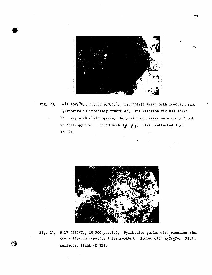
Fig. 23. P.ll (527 oC., 20,000 p.s.i.). Pyrrhotite grain with reaction rim.
Pyrrhotite is intensely fractured. The reaction rim has sharp
boundary with chalcopyrite. No grain boundaries were brought out
in chalcopyrite. Etched with K2Cr207. Plain reflected light
(X 92).
Fig. 24. P-17 (562oC., 10,000 p.s.i.). Pyrrhotite grains with reaction rims
(cubanite-chalcopyrite intergrowths). Etched.with K2Cr207. Plain
reflected light (X 92)0

28
'.~.,
• • ~ .~~ 1 ••
Fig. 23. P-ll (527°C., 20,000 p.s.i.). Pyrrhotite grain with reaction rime
Pyrrhotite is intensely fractured. The reaction rim has sharp
boundary with chalcopyrite. No grain boundaries were brought out
in chalcopyrite. Etched with K2Cr207' Plain reflected light
Fig. 24.
(X 92).
P-17 (562oC., 10,000 p.s.i.). Pyrrhotite grains with reaction rims
(cubanite-chalcopyrite intergrowths). Etched with K2Cr207' Plain
reflected light (X 92).

29
Fig. 25. P-15 ~5750C., 10,000 p.s.i.). Intruded part.' Domains in chalcopy
rite. These are isotropic (whitish gray) and weakly anisotropic
(grayish white). The domains end abruptly at the grain boundaries
in chalcopyrite. Domains reveal twinning in chalcopyrite.
Pyrrhotite (black). Pyrite (white). Magnetite (gray). Etched with
K2Cr207. Plain reflected light (X 92).
Fig. 26. P-2 (6030C., 25,000 p.s.i.). Domains in chalcopyrite. They end
at the grain boundaries in chalcopyrite. The domains are isotropic
(whitish gray) and weakly anisotropic (grayish-white). They appear
as rods. The domains a~e in either triangular or orthogonal
patterns. Pyrrhotite (black). Pyrite (white). Magnetite (gray).
Etched with K2Cr207. Plain reflccted light ,(X 92).

0" . ..: ~ . '~
29
Fig. 25. P-15 (5750 C., 10,000 p.s.i.). lntruded part. Domains in chalcopy
rite. These are isotropic (whitish gray) and weakly anisotropic
(grayish white). The domains end abruptly at the grain boundaries
in chalcopyrite. Domains reveal twinning in chalcopyrite.
Pyrrhotite (black). Pyrite (white). Magnetite (gray). Etched with
K2Cr207. Plain reflected light (X 92).
Fig. 26. P-2 (603 0 C., 25,000 p.s.i.). Domains in chalcopyrite. They end
at the grain boundaries in chalcopyrite. The domains are isotropic
(whitish gray) and weakly anisotropic (grayish·white). Theyappear
as rads. The domains are in either triangular or orthogonal
patterns. Pyrrhotite (black). Pyrite (white). Magnetite (gray).
Etched with K2Cr207. Plain reflected light (X 92).

, . . ~, " "
:~.
'. " ~, ,
30
weakly anisotropic. The domains appear as rods with a magnification of
92 X (Fig.26) and show spindle shapes under oil immersion with a
magnification of 380 X (Fig.28). The domains were oriented parallel to the
sphenoid (112) face edges, and appeared either as orthogonal or triangular
patterns, depending upon the orientation of the polished section in relation
to the grains. No cubanite was recognized at and above 575 0 C.
The X-ray diffraction pattern (using the Guinier camera) showed
the characteristics of the low tempe rature tetragonal polymorph. Figs.
27A and B shmoJ the X-ray patterns for different experiments. From this it
can be concluded that the cubic polymorph has reverted, on quenching, to a
stable tetragonal polymorph, leaving domains as evidence of the phase change.
There are four different sizes of domains which decreased as the
temperature increased and the changes were observed at 6000 , 6250 , 6600 and
7000 C. (see figs. 28, 29, 30 and 31). No apparent difference in the size
o 0 of the domains was found between 700 C. and 800 C. (see figs. 31 and 32).
The size of the domains did not change with differential pressure.
The polymorphic change has very little effect on the mobility of
chalcopyrite in spite of the volume expansion which occurs during trans-
formation from tetragonal to cubic form.
Magnetite, small pyrite grains, and gangue were carried along the
direction of chalcopyrite flow. The pyrite grains of larger size were
fractured (Fig.33). When the sample was completely pyrite, as in the
experiment P-6b (SS2 0 C. and 20,000 p.s.i.), it was fractured in contact
with the mullite hole (Fig.34). The magnetite, pyrite and gangue did not
react with chalcopyrite under the Experimental conditions.
B. Heating experiments on polycrystalline chalcopyrite:
The grmoJth of domains \oJas suspected to be due to the differential

30
weakly anisotropic. The domains appear as rods with a magnification of
92 X (Fig.26) and show spindle shapes under oil immersion with a
magnification of 380 ~ (Fig.28). The domains were oriented parallel to the
sphenoid (112) face edges, and appeared either as orthogonal or triangular
patterns, depending upon the orientation of the polished section in relation
to the grains. No cubanite was recognized at and above 5750 C.
The X-ray diffraction pattern (using the Guinier camera) showed
the characteristics of the low temperature ·'tetragonal polymorph. Figs.
27A and B show the X-ray patterns for different experiments. From this it
can be concluded that the cubic polymorph has reverted, on quenching, to a
stable tetragonal polymorph, leaving domains as evidence of the phase change.
There are four different sizes of domains which decreased as the
temperature increased and the changes were observed at 6000 , 6250 , 6600 and
7000 C. (see figs. 28, 29, 30 and 31). No apparent difference in the size
of the domains was found between 7000
C. and 8000 C. (see figs. 31 and 32).
The size of the domains did not change with differential pressure.
The polymorphic change has very little effect on the mobility of
chalcopyrite in spi te of the volume expansion which occurs during trans
formation from tetragonal to cubic form.
Magnetite, small pyrite grains, and gangue were carried along the
direction of chalcopyrite flow. The pyrite grains of larger size were
fractured (Fig.33). When the sample was completely pyrite, as in the
experiment P-6b (552oC. and 20,000 p.s.i.), it was fractured in contact
with the mullite hole (Fig.34). The magnetite, pyrite and gangue did not
react with chalcopyrite under the experimental conditions.
B. Heating experiments on polycrystalline chalcopyrite:
The growth of domains was suspected to be due to the differential

~ e
1 1 a- l ' i~ ~ 0 r "1 ;;: ,'" ~~I:
1 Il 1 Il '1 Il 1 1"! . - -1 m
r1 r- 1 11 . :f
'So·c t [10 i -fIIa.cl t !<!II
il!
Figs. 27 A and B. X-ray diffraction photographs for chalcopyrite obtained with the Guinier
focussing camera, using Co~~radia~ion. The diffraction patterns at different
temperatures show the characteristics of low-temperature polymorph. [chalCOPY-
rite (Cp), pyrrhotite (Po), pyrite (?y) , magnetite (M)J. .
Fig.27A
Fig. 27 B
w .....

32
Fig. 28. P-2 (603°C., 25,000 p.s.i.). Domains in chalcopyrite. Theyappear
as spindles. They occur as orthogonal pattern (centre) or tri
angular pattern (upper left corner). They are isotropic (grayish
black) and weakly anisotropic (grayish white). Etched with
K2Cr207. Plain reflected light. Oil imme~sion (X 380).
Fig. 29. P-29 (626°C., 10,000 p.s.i.). Domains in chalcopyrite. The size
of the domains decreased.'as compared in Fig. 28. Grain boundary in
chalcopyrite at the centre. Pyrite (white). Etched'with K2Cr207•
Plain r~flected light. Oil immersion (X 386).

32
Fig. 28. P-2 (603 0 C., 25,000 p.s.i.). Domains in chalcopyrite. Theyappear
as spindles. They occur as orthogonal pattern (centre) or tri
angular pattern (upper left corner). They are isotropic (grayish
black) and weakly anisotropic (grayish white). Etched with
K2Cr207. Plain reflected light. Oil imme~sion (X 380).
Fig. 29. P-29 (626 oC., 10,000 p.s.i.). Domains in chalcopyrite. The size
of the domains decreased'as compared in Fig. 28. Grain boundary in
chalcopyrite at the centre. Pyrite (white). Etchedwith K2Cr207•
Plain reflected light. Oil immersion (X 386).

Fig. 30. P-33 (660°C., 10,000 p.s.i.). Domains in a chalcopyrite grain.
The domains are of triangular pattern. The size of the domains
decreased as compared in Fig. 29. Etched with K2Cr207~ Plain
reflected light. Oil immersion (X 386).
33
Fig. 31. P-25.a (700°C., 10,000 p.s.i.). Domains in chalcopyrite. The
size of the domains decre~sed as compared in Fig. 30. Etched with
K2Cr207• Plain reflected light. Oil immersion (X 386).

Fig. 30. P·33 (660°C., 10,000 p.s.i.). Domains in a chalcopyrite grain.
The domains are of triangular pattern. The size of the domains
decreased as compared in Fig. 29. Etched with K2
Cr20
7• Plain
reflected light. Oil immersion (X 386).
33
Fig. 31. P.25.a (700°C., 10,000 p.s.i.). Domains in chalcopyrite. The
size of the domains decreased as compared in Fig. 30. Etched with
K2Cr20 7• Plain reflected light. Oil immersion (X 386).

34
Fig. 32. P-31.b (800°C., 10,000 p.s.i.). Domains in chalcopyrite grains.
The si~e of the domains appears to be comparable with the one in
Fig. 31. Etched wi~h K2Cri07- Plain ref1ected 1ight. Oi1
immersion (X 386).
Fig. 33. P-5 (602°C., 20,000 p.s.i.). Pyrite intensely fractured. Domains
are formed in chalcopyrite. Etched with K2C~207. Plain ref1ected
1ight (X 33).

(
34
Fig. 32. Pa 3l.b (800°C., 10,000 p.s.i.). Domains in chalcopyrite grains.
The size of the domains appears to be comparable with the one in
Fig. 31. Etched with K2CrZ0 7• Plain reflected light. Oil
immersion (X 386).
Fig. 33. P-5 (602°C., 20,000 p.s.i.). Pyrite intensely fractured. Domains
are formed in chalcopyrite. Etched with K2Cr207. Plain reflected
light (X 33).

35
Fig. 34. P-6.b (552oC., ~O,OOO p.s.i.). Sample is pyrite and fractured in
contact with the mullite tube (shear fracture). The vertical
fractures may belong to the original samplé. Plain reflected
light (X 33).
Fig. 35. P-19 (525 0 C.). Pyrrhotite grains with reaction rim (intergrowths
of cubanite and chalcopY~ite). The size of the intergrowth
lamellae greatly decreased as compared in Fig. 22. 'No domains in
chalcopyrite (grayish white). Pyrite (white). Magnetite (dark gray).
Etched with K2Cr207. Plain reflected light (X 92).

C)
(
35
Fig. 34. P-6.b (552°C., 20,000 p.s.i.). Sample is pyrite and fractured in
contact with the mullite tube (shear fracture). The vertical
fractures may belong to the original sample. Plain reflected
light (X 33).
Fig. 35. P-19 (525°C.). Pyrrhotite grains with reaction rim (intergrowths
of cubanite and chalcopyrite). The size of the intergrowth
lamellae greatly decreased as compared in Fig. 22. No domains in
chalcopyrite (grayish white). Pyrite (white). Magnetite (dark gray).
Etched with K2Cr207. Plain reflected light (X 92).
t~ , ..
I~
l'

36
pressure. Two heating experiments, without external pressure, were performed
at 5250 and 6000C. The samples were heated in evacuated silica tubes and
quenched in water.
At 5250C. chalcopyrite was not changed except for the formation
of reaction rims around the pyrrhotite grains. The reaction rims consist of
cubanite-chalcopyrite intergrowths as in the differential pressure
experiments. However, the size of the 1ame11ae was smal1er than in a
co1d sea1 bomb experiment at 5250C (Fig.35).
In the experiment at 6000C., when the si1ica tube was opened su1fur
dioxide was given off. The 10ss of su1fur cou1d not be checked in the case
of gold capsules because the sma11 amount of sul fur gas trapped in the
capsule escapes during opening the samp1e. Domains were observed in
chalcopyrite as they were in the differentia1 pressure experiments. The
size of the domains was great1y decreased (see figs. 36 and 26) and was
o comparable with that in the differentia1 pressure experiment at 650 C. (see
figs.36 and 37). The discrepancy in the size of the domains may be due to:
1) the coo1ing rate (si1ica tubes cool rapid1y when compared to the co1d
sea1 bombs), and 2) the 10ss of su1fur which may be greater in the heating
experiments than in the differentia1 pressure experiments. As a resu1t of
'rapid coo1ing of the si1ica tubes the su1fur re1eased did not re-enter the
crystal structure of the chalcopyrite comp1ete1y. The X-ray pattern was that
of the low-temperature tetragona1 po1ymorph (Fig.38A).
From the above two experiments it can be conc1uded that differential
pressure has very 1itt1e or no influence in the deve10pment of the domains;
and that a critica1 temperature was needed for causing them.
C. Co1d sea1 bomb and heating experiments on single crystal chalcopyrite:
It was suspected that the origin of the domains in chalcopyrite may

37
Fig. 36. p-18 (600°C.). Domains in chalcopyrite. The size of the domains
greatly decreased as compared in Fig. 26. Domains are isotropic
(grayish black) and weakly anistropic (white). Pyrite (grayish
white). Pyrrhotite (black). Etched with K2Cr207• Plain reflected
light (X 92).
Fig. 37. P-10 (650°C., 10,000 p.s.i.). Domains in chalcopyrite. The size
of the domains greatly decreased as compared in Fig. 26, but same as
in Fig. 36. Domains reveal grain boundaries and twinning in
chalcopyrite. Pyrrhotite (black). Etched with K2
Cr20
7• Plain
reflected light (X 92).

37
Fig. 36. P-18 (600oC.). Domains in chalcopyrite. The size of the domains
greatly decreased as compared in Fig. 26. Domains are isotropic
(grayish black) and weakly anistropic (white). Pyrite (grayish
white). Pyrrhotite (black). Etched with K2Cr207
• Plain reflected
light (X 92).
Fig. 37. P-10 (650oC., 10,000 p.s.i.). Domains in chalcopyrite. The size
of the domains greatly decreased as compared in Fig. 26, but same as
in Fig. 36. Domains reveal grain boundaries and twinning in
chalcopyrite. Pyrrhotite (black). Etched with K2
CrZ0
7• Plain
reflected light (X 92).

@
Figs. 38 A and B. X-ray diffraction photographs for chalcopyrite obtained with the Guinier
focussing camera, using COk radiation. The diffraction patterns for chalcopy.
e
Fig. 38 A
Fig. 38 B
~ rite show the characteristics of low~temperature polymorphe {chalcopyrite (Cp),
original chalcopyrite (ori. Cp), single crystal chalcopyrite (S.C.Cp),
pyrrhotite (Po), pyrite (Py), magnetite (M)}.
w 00

39
b~ due to the presence of impurities, especially pyrrhotite which readily
reacts with chalcopyrite. In order to ascertain their origin more clearly,
tbree experiments were performed with single crystals.
In all the crystals, only pyrite impurities were presen~
confirmed with X-ray diffrac.tion (Fig.40). The crystals have sphenoidal
habit [112]. In chalcopyrite planes parallel to sphenoid faces (112) are
the slip planes. In differential pressure experiments the crystals were
oriented so that the sphenoid (112) surfaces were oriented at approximately
o 45 to the differential pressure i.e., the c-axis of the crystal would be
parallel to the maximum pressure.
Differential pressure experiments were carried out at 5500 and
6000 C. under a differential pressure of 20,000 p.s.i. The distance of
intrusion of a single crystal, with proper orientation for easy glide, was
approximately the same as in the polycrystalline chalcopyrite.
On etching with 1:1 H202 and NH40H, polished sections showed that
chalcopyrite was composed of randomly oriented grains, which revealed
recrystallization in the in.truded part and a minor portion outside the
mullite tube (Fig.39). At 6000 C. it showed recrystallization and increase in
grain size (Fig.40).
On etching with acidic K2Cr207, chalcopyrite at 6000 C. exhibited
grain boundaries and domains (Fig. 41) and the grain boundaries were not
revealed in the absence of the domains, as in the experiment at 5500 C. The
characteristics of the domains were the same as in the polycrystalline
chalcopyrite except that 1) the colour of the isotropie domains was brown,
differing from that of the polycrystalline material which was brown with a
green tinge; and 2) the size of the domains had considerably increased. The
variation in size could be explained by the larger size of the single crystal

.8
40
Fig. 39. P-22 (550°C., 20,000 p.s.i.). Single crystal chalcopyrite.
Recrysta11ization in chalcopyrite in the intruded part and a minor
portion outside the mu11ite tube. Mechanica1 twinning present
away from the mu11ite tube. Black areas are po1ishing pits.
Etched with 1:1 H202 and NH40H. P1ail1 reflected light (xi!).
Fig. 40. P-20 (602°C., 20,000 p.s.i.). Single crystal cha1co~yrite.
Recrysta11ization and increase in grain size .in chalcopyrite. One
twin in'the intruded part (right side bottom). High relief
rounded w.hite grains are pyrite. Etched with 1:1 H202 and NH40H.
Plain ref1ected 1ight (X 40).

40
Fig. 39. P-22 (550°C., 20,000 p.s.i.). Single crystal chalcopyrite.
Recrystallization in chalcopyrite in the intruded part and a minor
portion outside the mullite tube. Mechanical twinning present
away from the mullite tube. Black areas are polishing pits.
Etched with 1:1 H202 and NH40H. Plaib reflected light (X~).
Fig. 40. P-20 (602°C., 20,000 p.s.i.). Single crystal chalco~yrite.
Recrystallization and increase in grain size in chalcopyrite. One
twin in the intruded part (right side bottom). High relief
rounded w.hite grains are pyrite. Etched with 1:1 H202 and NH40H.
Plain reflected light (X 40).

41
Fig. 41. P-20 (602oC., 20,000 p.s.i.). Single crystal chalcopyrite. Domains
in chalcopyrite. They occur either as triangu1ar or orthogonal
patterns. Domains are isotropic (grayish black) and weak1y
anisotropic (white). Domains revea1 grain boundaries and twinning
in chalcopyrite. Etched with K2C~207. Plain ref1ected 1ight (X 92).
Fig. 42. P-21 (600oC.). Single crystal chalcopyrite. Domains in chalcopy
rite. The domains occur as triangular patterns, each pattern
para11e1 to the sphenoid face (112) edge. One pattern is poorly
deve1oped. Domains are isotropic (grayish··black) and weakly
anisotropic (white). Etched withK2Cr207. Plain ref1ected light.
(X 92).

C' '.;
(
41
Fig. 41. P-20 (602oC., 20,000 p.s.i.). Single crystal chalcopyrite. Domains
in chalcopyrite. They occur either as triangular or orthogonal
patterns. Domains are isotropic (grayish black) and weakly
anisotropic (white). Domains reveal grain boundaries and twinning
in chalcopyrite. Etched with K2Cr207. Plain reflected light (X 92).
Fig. 42. P-21 (600oC.). Single crystal chalcopyrite. Domains in chalcopy
rite. The domains occur as triangular patterns, each pattern
parallel to the sphenoid face (112) edge. One pattern is poorly
developed. Domains are isotropic (grayish black) and weakly
anisotropic (white). Etched witn K2Cr207' Plain reflected light.
(X 92).

.... ,.
42
in which the domains grow more freely than in the polycrystalline mate rial
where the growth of the domains May have been retarded by the numerous grain
boundaries and the presence of impurities. The X-ray patterns, for the above
two experiments,.was that of the low-temperature tetragonal polymorph (Fig.
38B).
Together, the above two experiments reveal that the formation of
the domains was independant of impurities.
One heating experiment was performed at 6000 C. with a single
crystal to ascertain the cause of the discrepancy in the size of the domains
observed as compared with those in polycrystalline chalcopyrite between 6000
and 6250 C. The crystal was of sphenoidal habit [112J. The size of the
domains was the same as in the differential pressure experiment (see figs.
42 and 41).
When the sphenoid face was examined under the microscope,
3 patterns of domains were observed parallel to the sphenoid face edges
(Fig.42). One set was poorly developed. No sulfur odour was noticed, unlike
that in experim.ent with polycrystalline chalcopyrite. Because the size of
the sample was small compared to that of the polycrystalline material, no
vapour of sul fur was noted. This experiment shows that at a constant
temperature above the inversion temperature, differential pressure, cooling
rate and impurities (pyrite) have no apparent effect on the size of the
domains.
D. Explanations for the decrease in the amount of intrusion with increase
in temperature under a confining pressure of 10,000 p.s.i.
From the 10,000 p.s.i. curve (Fig.4) the amount of intrusion
appears to be a continuous function of the temperature until the temperature
reached 6500 C., beyond which the amount of intrusion decreased •. '. Chalcopyrite

43
deformed above and be10w 6500C. did not show differences·either in the
optica1 properties or in X-ray diffraction patterns (Fig.27A and B). The
decrease in the amount of intrusion with increasing temperature, from 6500C.
upwards, requires exp1anation.
In the experiments performed at 7000C. and above, su1fur gas or
su1fur dioxide was found in the gold capsules, which indicates that
considerable amounts of sul fur were re1eased from the chalcopyrite and not
fu11y resorbed on quenching.
In these experiments the amount of intrusion increased from 5250
to 650QC. and decreased from 65cPto 8000C. The chalcopyrite recrysta11ized
at 5620C. and 1arger grains resu1ted at and above 5750C. These changes in
structure shou1d have faci1itated intrusion.
According to Frueh (1959), sul fur 10ss begins in chalcopyrite
be10w 3000C. Hi11er and Probsthain (1956) found that the sul fur content of
chalcopyrite decreases, upon heating, continuous1y fram 4340 to 7500C. It
can be pointed out that the rate of 10ss of sul fur is great between 5800C.
and 6200C. It appears from the article that the sul fur is 10st permanent1y
fram the chalcopyrite. They have given the formula for chalcopyrite at
7200C. as CU17+x!e17+xS32 (X=0.6). However, in the present experiments, the
o rate of sulfur 10ss apparent1y was great only at about 650 C. as the samp1e
was under a confining pressure. At 7000C. marked 10ss of su1fur was evident
in the present experiments. Decreasein the amount of su1fur should decrease
the rate of f1owage, due to the concentration of meta1 ions in the structure.
If su1fur 10ss occurs between 5250C. and 6500C., the reduction in
mobility resu1ting from this must be increasing1y overshadowed by the effects
of recrystal1ization fram 5620 to 62SoC. as shown by the experimental results.
At 6500C. a reversa1 occursgiving a decrease in the amount of intrusion

44
with increased temperature. This appears to be accompanied by an increased
rate of sulfur loss. Also, in the present experiments, the chalcopyrite is
continuously under differential pressure which induces strain in the
recrystallized grains and the recrystallized grains deform plastically
throughout the experiment. A possible explanation of the relationships of
these phenomena is as follows:
When the sul fur vaporizes, the Cu and Fe atoms enter into the
chalcopyrite structure as interstitial atoms (impurities). Some adjustments
undoubtedly take place during recrystallization. Also, the individual
recrystallized grains tend to deform plastically with the movement of
dislocations. However, the Cu and Fe atoms obstruct the movement of the
dislocations in chalcopyrite and the chalcopyrite is ~~~~~. Thus
the resistance to deformation is increased and the amount of intrusion
decreases. In the temperature range from 5250 to 6250 C. loss of sulfur is
slight and the effects of recrystallization predominafe. Increasing sul fur
loss above 6500 C. leaves more cations in the structure, which retard the
deformation. This reveals why the amount of intrusion decreased continuously
as the temperature is increased from 6500 C. onwards.
A further factor appears to have some relevance. The sul fur
pressure from dissociation of chalcopyrite, building up in the gold tube
might oppose the intrusion. On page 14 it is shawn that the sul fur pressure
at 627 0 C. was on1y 12 p.s.i. but loss of sulfur becomes rapid at about 6500 C.
and this suggests that the dissociation pressures also were rapid. This
would retard the rate of intrusion. Thus the pressure inside the gold
capsule can aid in decreasing the amount of intrusion at higher temperatures.
E. Hardness tests:
The gradual decrease of the amount of intrusion was explained as

45
due to loss of sul fur. lt appeared probable that this wou1d be accompanied
by an increase in hardness. To examine this property, hardness tests were
made on al1 the samp1es. The resu1ts are given in Table 1. lt is evident
from figures 43, 44 and 45B that hardness and mobi1ity are not re1ated in
any simple manner. Specimens subjected to pressures of 10,000 p.s.i.,
20,000 p.s.i., or 25,000 p.s.i. a11 hardened abrupt1y at around 5750C • •
This is corre1ated with the phase change and with the development of
domains as described further be1ow. At pressures of 20,000 p.s.i. and
higher, chalcopyrite fi11ed the avai1ab1e space before temperatures of 6500C.
were reached. So the zone of decreased mobi1ity shown in the curve in
figure 45B for 10,000 p.s.i. confining pressure cou1d not be examined at
the higher pressures with the equipment used. The situation for 10,000
p.s.i. confining pressure was examined more fu11y and the relation shown in
figure 45B will be discussed in detai1.
The f1attening of the hardness curve (Fig.45B) between 5750 and
6600C. will be designated as Region A, the drop in hardness at 6750C as
Region B, and the curve beyond 6750C. as Region C.
Region A: The correlation of the sudden increase in hardness can be made
with the appearance of domains in the quenched samples. Domains.in
chalcopyrite were first observed and described by Frueh (1958). They were
formed as a resu1t of transformation from high temperature cubic form to
the tetragona1 form, on quenching. In the present experiments these were
observed at 57 SoC. When the domains are formed, the y cause strain which
hardens chalcopyrite. This a1so exp1ains the f1attening of the hardness
curve between 5750C. and 6600C. Harker (1944) noted thata simi1ar phase
change increased the hardness in a gold-copper a110y when it changed from
a face-~entered cubic structure to a base-centered tetragona1 structure.

,.. QI
~ ::J
260
z 240 !Il !Il QI C
-0 ,.. ~ !Il ,.. ~ 220 u
..-1 :>
'" CIl
~
e
l o
..-1 4-1 o;J N
..-1 .... .... o;J 4-1 !Il >. ,.. u ~
A r----~ ---1 1.!r-
t 1 1 1
1 , , 1 1
1 ____ A 1
A
A -T--A----i,-J 200 1 Temperature
i
Oc
4500 500 0 5500 600 0 6500
e
Fig. 43 is a plot of mean Vickers Hardness number against temperature. lIardness tests were made on samples from cold seal bomb experiments performed at various temperatures under a constant cor:fining pressure 25,000 p.s.i.
A - 25,000 p.s.i. confining pressure (Polycrystalline Chalcopyrite)
700Gl
,.. QI
'6 ~240
QI bO
l ~Œl _._ o CIl
~ ~----o
Fig. 44 is a plot of mean Vickers Hcrdness number against temperature. Hardness tests were made on samples from cold senl bomb experiments performed at various temperatures under a constant confining pressure
!Il !Il QI C
-0 ,.. CIl :x: ~ 220 QI ~ U
..-1 :> c CIl
I!]
N 1 ..-1 1 ::: 1
CIl 1 4-1 1 !Il 1 ~ 1 U 1
; El : @ )
----- ~
~ r::J
~ o ,
200 1 Tempera ture Oc
4500 5000 5500 6000 6500
..... ---------------------------------7000
20,000 P.s.!.
El - 20,000 p.s.i. confining pressure (Polycrystalline Chalcopyrite)
~ - 20,000 p. s. 1. confining pressure (Single Crystal Chalcopyrite)
.po 0\

2.0
~ s:; 0
1.5 .,-1
fil :l ,... .w s:;
H
4-1 0
-,
j (1) () s:;
1.0
i <-:: "-' fil
-..l e::::I
0.5
0 -..l .w CI! ~l
.,-1
.-1
.-1 cv .w fil :>... ,... () (1)
Il:::
c:y ,.,. ..... '6
o
1 . o 1
47
Increase in the rate of 10ss of sulfur~
o
Fig. 45A
o <:>
...... ..,.",-" 1
O--~--~--~~-+----------------------------__________ __ 260
240
220
(1) /
gj 0 0 / .c0 Region 1\., / ~ ____________ 0 __ ""\ /
{ 0 0~ :~ ;1 : 15 //
i 1 0 ~ / 'D, 00 c./
CI! '(1) Region //0 N l ,Il::: 0 ,.,. ~ 1 1 0 ,.,./ r;;l 1 Size (1) of Size (2) ,, __ .........
/ g
/ /
ri /
~ 1 the domains of the Size (3) 0:: :>... , domains the domain Size (4) of the domains ,... ,same.
~ g 1 ~ t:'\ Il::: ,
same. 1>2
same. 2 "73
same. 3)4
-- 0--- -,'L'-0..J Fig. 45B
200
5000
Fig. 45A
Fig. 45B
1
6000 Tempera ture Oc
is same as Fig. 4 showing the plot of intrusion against temperature under a confining pressure of 10,000 p.s.i. is a plot of mean Vickers Hardness numb~r against temperature. The hardness tests were made on the samples from cold seal bomb experiments performed at various temperatures under a constant confining pressure pf 10,000 p.s.i.

48
Region B: o
Unti1 660 C., the increase in hardness is exp1ainab1e by the
domains. At 6750C., the chalcopyrite hardness decreased.
On page 43 it is shawn that 10ss of sul fur becomes rapid at about
6500C. When the samp1e is coo1ed slow1y, the su1fur atoms shou1d recombine
with the Cu and Fe atoms, which were diffused into the chalcopyrite
structure as interstitia1 atoms, to restore the original structure. Above
6500C., the 10ss of su1fur atoms is so great that during the time of
quenching the y may not have time to recombine upon quenching to restore the
original structure. Most of the cations stay inside the still intact
chalcopyrite crysta1s. Some of the su1fur atoms are deposited around the
crysta1s 100se1y bound by a few cations migrating outward to form a softer
chalcopyrite. This resu1ts in a decrease in hardness as the temperature is
increased.
A further possible factor affecting hardness is suggested by the
fact that the domains formed in experiments at tempe ratures from 6000C. to
70'OoC. were sma11er with increase of temperature. The change in hardness
appears to be much more abrupt than the change in domain size. Whether or
not there is a cause-effect re1ationship between these two phenomena is a
prob1em still to be reso1ved.
o Region C: After a drop in hardness of chalcopyrite at 675 C., the hardness
increased progressive1y. (The amount of intrusion a1so decreased
progressively.) Here appears to be a direct correlation. The sul fur 10ss
continues to be greater and. greater as the temperature increased. The
progressive increase in hardness could be explained by the loss of sulfure
Due to loss of su1fur, chalcopyrite is work-hardened. With greater
amounts of sulfur 10ss as the temperature is increased,a gradual increase in
hardness occurs. As the size of the domains appears to be the same betWeen

49
00. 700 C. and 800 C., the strain energy caused by the doma1ns should be the same
as at 7000 C. and should not influence the increments in hardness. For this
reason, only the loss of sulfur should have caused increase in hardness.

50
EXPERIMENTS USING "TRI-AXIAL COMPRESSION" EQUIPMENT
This group of experiments were intended to explore the behaviour
of naturally occurring sulfides when subjected to differential pressures at
moderately elevated temperatures.
Apparatus and general conditions
A differential pressure bomb was externally heated and a uni axial
compressive force was applied. The apparatus was designed and built at
McGill by Cameron (1956) and Wolofsky (1957) for carrying out open and
closed system experiments on mineraIs and rocks. In the present
investigation the experiments were performed in. a closed system. The
apparatus and the operational procedures are fully described by Wolofsky
(1957). Figures 46, 47, 48 and 49 show the tri-axial compression apparatus
in the operating condition, bomb fittings, assembled bomb, and bomb fittings'
with a deformed specimen, respectively. Only the essential features are
described below.
The sample jacket was cut to a length of 2.4 inches from a
commercially available hard drawn copper tube, having 0.6 inch external
diameter and 0.03 inch wall thickness. To prepare the sample, a piece of
ore cut with parallel end faces was either drilled with a diamond core bit
or ground on a rotating lap so as to fit into the jacket. The latter method
became necessary when the samples were small (especially single crystals).
The samples were ground according to the necessity of the experiment; i.e. the
orientation of the crystallographic direction of the sample, whether
parallel, perpendicular or at an angle to the direction of the applied force.
One or more of the discs were fitted into a copper jacket and the
charge was then placed in the bombe The bomb was heated electrically through
an external winding. When the desired temperature was reached, the sample

Temperature ____ _ controller
51
-·----Confining pressure gauge
-----Bomb
.. -,-----Furnace
ack gauge
-----ack
Fig. 46. Tri-Axial Compression apparatus in the operating condition.
Fig. 47. Bomb fitt~ngs (Tri-Axial Compression apparatus).
Lower piston Teflon washer Copper washer Copper jacket

Tempera ture ____ _ controller
----Confining pressure gauge
------Bomb
.-------Furnace
ack gauge
ack
Fig. 46. Tri-Axial Compression apparatus in the operating condition.
Fig, 47. Bomb fittings (Tri-Axial Compression apparatus) •
. . "' .. " -_.
Lower piston Teflon washer
1ûasher
jacket
51

52
piston
Fig. 48. Assembled bomb (Tri-Axial Compression apparatus).
formed sample
Fig. 49. Bomb fittings with deformed specimen (Tri-Axial Compression
apparatus).

(
52
P"------L()Wer piston
Fig. 48. Assembled bomb (Tri-Axial Compression apparatus).
Deformed sample
Fig. 49. Bomb fittings with deformed specimen (Tri-Axial Compression
apparatus).

53
was subjected to the desired compressive force by a piston driven upwards by
a jack into the jacket. As the samp1e yie1ds under a constant compressive
force, the piston enters the bomb. The temperature of the bomb was main
tained with ±10oC. When the piston had entered the bomb to its possibie
1ength the experiment was terminated (The piston was made progressive1y
1arger in diameter towards the bot tom, so that the bomb admitted a11 but
approximate1y one cm. of its 1ength. This faci1itated taking out the piston
from the bomb.) The bomb was coo1ed in the air. The force exerted by the
jack on the samp1e was n.oted in each run.
The compressive stress was ca1cu1ated by dividing the force by the
initial cross-sectiona1.area of the samp1e (0.28 sq. inches). The actua1
compressive stress values were a1ways 1ess for two main reasons: first, the
cross-sectiona1 area of the samp1e increases with the shortenin.g a10ng the
axis; second, frictiona1 resistance (between jacket and piston, between
bomb and piston) decreases with increasing temperature. These two reasons
a1so have to be taken into consideration when the stress values of
different experiments are compared. Wo10fsky (1957, pp. 97-101) discussed
the complications invo1ved in the compressive stress ca1cu1ations. The
compressive stress difference was obtained by subtracting the confining
pressure from the compressive stress.
The shortening of the samp1e was measured in each run after opening
the samp1e and the percentage of shortening was ca1cu1ated in each run. Even
though the ca1cu1ated compressive stress and the percentage of shortening do
not represent the actua1 values, the y do indicate in a rough quantitative
way the physica1 behaviour of the samp1es.
Serious difficu1ties were encountered in maintaining a sea1 at the
moving end of the bomb because the tef10n washer me1ts and vo1ati1izes at

54
o about 300 C. When the teflon melted, the seal could not be maintained
under high confining pressures. To avoid this difficulty Many of the
experiments were performed at lower confining pressures.
At elevated temperatures the copper jacket reacted with the
sulfides. To avoid this the sample, in some experiments, was wrapped in
aluminum foil and placed inside the copper jacket. Even then, minor
reaction took place between the copper jacket and the sulfides. Possible
solutions to this problem might be bo have the copper jacket gold plated,
or to use a thin aluminum jacket. The gold plating would be worthwhile if
the lower piston seal could be perfected. In case the gold plating method
fails, gold foil could be wrapped around the specimen, but the cost of the
experiment will be high.
An aluminum jacket was tried, but it presented difficulties in
polishing the sample because of its high ductility. The aluminum jacket
could be used providing that a th in strip of the aluminum is cut away from
the edge of the section before it is set in the mounting medium. The
difficulty with this is that part of the sample at the boundary breaks away.
Difficulties were met in making polished sections from some of the
deformed samples because the y were brittle and fractured. To avoid this,
small holes were drilled in the sample and impregnated with plastic
(Quickmount, Fisher Co.) in a vacuum. The specimen was th en cut with a
diamond saw. To make sure that the fractured grains were welded together,
one section was soaked in household cement (Lepage's) diluted in acetone and
then polished.
The mineraIs were identified using a reflecting microscope. A
Phillips X-ray diffractometer, with Fek radiation, was used to identify ~
doubtful mineraIs.

Results
The maximum tempe rature deemed safe for the equipment was 5000 C.
and at such tempe ratures the confining pressure could not be raised much
above 15,000 p.s.i. without damage to the bombe
55
The experiments are numbered K-l to K-19, and are arranged
according to the mineraI or mineraIs and the physical conditions under which
the experiments were performed. The physical conditions and results of
each experiment are described in detail and are summarized in Table II.
Experiment K - 13
Material: Galena cut from a single crystal perpendicular to a cube face.
Pyrite cut from a single crystal perpendicular to a cube face.
Thick and thin layers alternately of galena and pyrite. Several
discs were used to provide comparisons, especially between pieces
in different positions (Fig. 50).
Conditions: See table II. Confining pressure: 4,000 p.s.i.; Temperature:
Room Temperature; Compressive stress difference: 31,000 p.s.i.
Results: Megascopically the deformed specimen showed dominant shear failure
(Fig. 51). The thick galena and pyrite bands were generally fractured parallel
to the direction of the applied force and granulation took place along the
fractures. In the thick galena bands some fractures followed cleavage
planes at an angle of 45 0 to the direction of the applied force. The galena
was finely crushed along the fracture zones (Fig. 52). The thinner galena
and pyrite bands were fractured and intensely granulated. The crushed pyrite
grains were drawn into streaks between the galena bands (Fig.53).
In this experiment both the galena and pyrite were deformed by
fracturing accompanied by granulation. Though fracturing and granulation
were intense, the original ban ding was retained.

8 e
TABLE II. Tri-Axial Compression Experiments
Exp.No. Samp1e Tempera- Confining Time of Jack Compress- Length Percentage Remarks ~ ture oC. pressure. rune force. ive stress shortened. of
p. s. i. Hours lbs. difference. cm. shortening. p.s.i.
K-13 cubic ga1ena. Room 4,000 l~ 9,800 31,000 4.00-2.10 47.5 cubic pyrite. tempera- =1.90
ture
K-15 cubic ga1ena. 200 4,000 1~ 8,500 26,350 4.00-2.00 50.0 A1uminum foi1 wrapped cubic pyri te. =2.00 around the samp1e.
cubic ga1ena. K .. l7 massive 300 4,000 2~ 8,500 26,350 3.85-2.40 38.0 A1uminum foi1 wrapped
pyrite. =1.45 around the samp1e.
K-12 cubic ga1ena. 400 4,000 ~ 7,000 21,000 3.80-1.80 52.5 A1uminum foi1 wrapped cubic pyrite. =2.00 around the samp1e.
octahedra1 K-14 galena. 400 4,000 Ji 7,500 22,800 4.05-1.65 59.0 A1uminum foi1 wrapped
cubic pyrite. =2.40 around the samp1e.
schistose K-16 ga1ena. 400 4,000 2 7,500 22,800 4.20-2.35 44.0 A1uminum foi1 wrapped
cubic pyrite. =1.85 around the samp1e.
cubic ga1ena. K-18 basai. 400 5,000 ~ 8,500 25,350 3.95-2.35 40.5 A1uminum foi1 wrapped
pyrrhotite. =1.60 around the samp1e four times.
K-8 galena with
200 15,000 4 19,000 52,850 4.00-2.30 42.5 pyrrhotite. =1.70
Ut 0\

8 e
TABLE II continued
Exp. No. Samp1e Tempera .. Confining Time of Jack Compress .. Length Percentage Remarks ture oC. pressure rune force. ive stress shortened. of
p.s.i. Hours lbs. difference. cm. shortening. p.s.i.
ga1ena with K-9 pyrrhotite. 385 13,000 1 14,500 37,800 3.45-1.45 58 Lead pellets present out-
cubic ga1ena. =2.00 side the copper jacket.
ga1ena with K-lO pyrrhotite. 475 18,000 2 18,000 46,300 4.20-1.90 55 Lead pellets present out-
spha1erite =2.30 side the copper jacket.
ga1ena with K-ll pyrrhotite. 300 7,000 1 12,000 35,850 4.20-1.70 59.5
massive =2.50 pyrite.
basal K-19 pyrrhoti te.
mas s ive pyrite 400 5,000 ~ 10,000 30,200 3.75 .. 2.25 40.0 A1uminum foi1 wrapped =1.50 around the samp1e twice.
K-5 chalcocite 300 15,500 ~ 18,000 48,800 4.00-2.15 46.0 with bornite. =1.85
chalcopyrite room K-1 ore. tempera .. 23,500 4~ 30,000 83,800 3.80-2.10 45.0
massive pyrite. ture =1.70
cha1copyri. te K-3 ore. 200 14,500 3 3/4 18,000 49,800 4.30 .. 2.75 36.0
massive pyrite. =1.55
VI ......

e
Exp.No. Samp1e Tempera- Confining Time of ture oC. pressure. run.
p.s.i. Hours.
chalcopyrite K-2 ore 200 14,000 3/4
massive pyrite.
chalcopyrite K-4 ore 200 15,000 4·
massive pyrite
K-6 chalcopyrite 350 15,000 1~ ore.
K-7 chalcopyrite 350 15,500 1 ore.
TABLE II continued
Jack Compress-force. ive stress lbs. difference.
p. s. i.
19,500 55,650
18,500 51,100
18,000 49,300
17,000 45,200
Length shortened. cm.
4.10-2.25 =1.85
4.25-3.25 =1.00
3.90-1.80 =2.10
3.80-1.50 =2.30
Percentage of shortening.
45.0
24.0
54.0
60.0
e
Remarks
A1uminum foi1 wrapped around the samp1e twice.
\.Il 00

..
CUBIC GALENA----CUBI C PYRITE-----
--
CUBIC GALENA--
tuBIC PYRITE
CUBIC GALENA--
CUBI C PYRITE---
CUBI C GALENA----
-
CUBIC PYRITE---
Fig. 50
oj
,
1
Fig. 51
Fig. 50. K-13 (Roam temperature). Undeformed specimen showing different
positions of samples in a copper jacket (X 1).
59
Fig. 51. K-13 (Room temperature). Deformed specimen. Galena and pyrite
single crystals were cut perpendicular to their cube faces. Thick
and thin bands alternately of galena and pyrite are deformed. The
samples failed along a shear fracture.
Fig. 52. K-13 (Rpom temperature). Galena is intensely crushed along a
fracture zone. Plain reflected light (X 33).

CUBIC GALENA···
eUBI e PYRITE··_·
CUBIC GALENA··_·
CUBle PYRITE···
eUBI e GALENA··-·
eUBI e PYRITE-···
eUBI e GALENA-·--
· ·
·
eUBI e PYRITE ._. -
Fig. 50
j s" :
i
Fig. 51
Fig. 50. K-13 (Room temperature). Undeformed specimen showing different
positions of samples in a copper jacket (X 1).
59
Fig. 51. K-13 (Room temperature). Deformed specimen. Galena and pyrite
single crystals were cut perpendicular to their cube faces. Thick
and thin bands alternately of galena and pyrite are deformed. The
samples failed along a shear fracture.
Fig. 52. K-l3 (R~om tempe rature). Galena is intensely crushed along a
fracture zone. Plain reflected light (X 33).

"e ". ,.
60
Experiment K - 15
Materia1: As in K - 13. Thick and thin 1ayers a1ternate1y of ga1ena and
pyrite. Severa1 discs were used to provide comparisons,
especia11y between pieces in different positions (Fig. 54).
Conditions: See Table II. Confining pressure: 4,000 p.s.i.; Temperature:
200oC.; Compressive stress difference: 26,350 p.s.i.
Resu1ts: The specimen was not uniformly deformed. The copper sheath
buck1ed on one side a110wing the specimen to move around the piston (Fig. 55).
Megascopica11y ga1ena and pyrite bands were fractured para11e1 to the
direction of the applied force.
Cleavage planes along which granulation took place developed in the
ga1ena; the galena was a1so granulated where it was in contact with the
pyrite bands. Rows of triangular pits in the galena were smoothly curved
due to translation gliding along the planes parallel to (100). Granulation
in galena is less here than in the experiment at room temperature. The
pyrite bands were fractured, granulated and individual pyrite grains were
locally drawn out into streaks embedded in the galena (Fig. 56).
In this experiment the galena was deformed largely by fracturing
but translation gliding was present to a small extent. Pyrite was fractured,
granulated and sometimes drawn into streaks.
Experiment K - 17
Material: Galena as in K - 13. Pyrite cut from a sample containing
pyrrhotite, chalcopyrite, magnetite and gangue. Thick and thin
layers alternately of galena and pyrite. Several discs were used
to provide comparisons, especially between pieces in different
positions (Fig. 57).
Conditions: See Table II. Confining Pressure: 4,000 p.s.i.; Temperature:

61
Fig. 53. K-13 (Room temperature). Pyrite is granu1ated and the grains are
drawn into a streak between two ga1ena bands. Etched with 1:1 HN03•
plain ref1ected 1ight (X 33).
cualc GALENA----
cualc PYRITE---
cualc GALENA-
cualc PYR!TE----
cualc GALENA--cualc PYRITE---
cualC GALENA----
cualc PYR/TE----
Fig. 54
- ~ !
Fig. 55
Fig. 54. K-15 (200oC.). Undeformed specimen showing different positions of
samp1es in a copper jacke~ (X 1).
Fig. 55. K-15 (200oC.). Deformed specimen. Ga1ena and pyrite. single crysta1s
were cut perpendicu1ar to their cube faces. Thick and thin bands
a1ternaté1y of galena and pyrite are deformed.

62
Fig. 56. K-15 (200°C.). Pyrite is granu1ated and the grains are drawn into
a streak between two ga1ena bands. Etched with 1:1 RN03• Plain
ref1ected 1ight (X 33).
eUBle GALENA----
.MASSIVE pyRITE---- 1---;
eUBIC GALENA----
MASSIVE PYRITE-----I---;
C UBIC GALENA-----
MASSIVE PYRITE-----======1
eUBIC GALENA----
MASSIVE PYRITE----
Fig. 57 Fig. 58
Plastic
Fig. 57. K-17 (300°C.). Undeformed specimen showing samp1es in different
positions in a copper jacket (X 1).
Fig. 58. K-17 (300°C.). Single crystal ga1ena cut perpendicu1ar to a cube
face. Pyrite was cut from a massive samp1e. Thick and thin 1ayers
a1ternate1y of ga1ena and pyrite.

63
3000 C.; Compressive stress difference:: 26,350 p.s.i.
Results: Megascopically the pyrite and some galena was fractured sub-parallel
to the direction of the applied force (Fig. 58). Most deformation in the
galena was due to translation gliding along planes parallel to the cube
faces (Fig. 59). The rows of triangular pits in galena were gently curved,
and became contorted along the fractures. The galena was intensely crushed
in the fracture zones (Fig.60), and along the contact with pyrite (Fig.6l).
The pyrite bands were either fractured parallel to the applied
force or granulated. Some pyrite grains were streaked into the galena
(Fig. 62).
The copper jacket reacted with pyrite to a minor extent to form
chalcopyrite, bornite, and chalcocite. The replacing minerals have
gradational contacts.
This experiment shows that galena deformed mainly by translation
gliding, although minor fracturing occurred. Pyrite was fractured and
granulated.
Experiment K-12
Material: As in K - 13. Thick and thin layers alternately of galena and
pyrite. Several discs were used to provide camparisons,
especially between pieces in different positions (Fig.63).
Conditions: See table II. Confining pressure: 4,000 p.s.i.; Temperature:
4000 C.; Compressive stress difference: 21,000 p.s.i.
Results: Megascopically the specimen showed uniform lateral elongation but
the jacket was buckled on one side (Fig. 64).
The deformed samples showed poorly developed pinch and swell
structure. The pyrite in the thick layers was broken into blocks which were,
in places, displaced (Fig.65). Fractures varied from parallel to 300 fram

64
Fig. 59. K-17 (300°C.). The rows of triangu1ar pits curved smooth1y which
indicate gliding along planes para11el to the cube faces. Etched
with 1:1 HN03. Plain ref1ected 1ight (X 92).
Fig. 60. K-17 (300oC.). A fracture in galena. Rows of triangular pits
contorted a10ng the fracture zone. Ga1ena is crushed in the
fracture zone. Etched with 1:1 HN03• Plain ref1ected 1ight eX 33).

()
(
.. ----_. ... .. __ ..... ~._--~---
64
Fig. 59. K-17 (300°C.). The rows of triangular pits curved smoothly which
indicate gliding along planes parallel to the cube faces. Etched
wi th 1: l RN03. Plain reflected light (X 92).
Fig. 60. K-17 (300°C.). A fracture in galena. Rows of triangular pits
contorted along the fracture zone. Galena is crushed in the
fracture zone. Etched with 1:1 RN03• Plain reflected light (X 33).

e·
65
Fig. 61. K-17 (3000~). Ga1ena shows fracturing and granulation in contact
with pyrite. Plain reflected 1ight (X 3~).
Fig. 62. K-17 (300oC.). Pyrite is granulated and the grains; are drawn into
a streak a10ng the direction of movement of ga1ena. Galena is
cleaved in the center. Plain reflected light (X 33).

( \
65
Fig. 61. K-17 (300°c.). Galena shows fracturing and granulation in contact
with pyrite. Plain reflected light (X 33).
10.
• G'T\. ,.- -'
Fig. 62. K-17 (300°C.). Pyrite is granulated and the grains are drawn into
a streak along the direction of movement of galena. Galena is
cleaved in the center. Plain reflected light (X 33).

Fig. 63.
Fig. 64.
CUBIC GALEU···-
CUBIC PYRITE-·-
CUBIC GALfNA-
CUBIC PYRITE·--
CUBIC GALENA·····
CUBIC PYRITE····
CUBIC GUEN.\-···
CUBIC . PYRITe-·_·
Fig. 63 Fig. 64
K-l2 (400oC.). Undeformed specimen showing samples in different
positions in a copper jacket (X·.~) •
K-12 (400oC.). Deformed specimen~ Single crystal galena and
pyrite were cut perpendicular to cube faces. Thick and thin
layers alternately of galena and pyrite.
. _. 4 ........ _ .•• .. .-" . , .... ~ ... ;'<'
' .. ~ .. " •.. ' '"~" o·
.••. . .~ Q"
.1
66
.. ~
Fig. 65. ~l2 (400oC.). Pyrite band is broken into two blocks and the
blocks are displaced. Galena flowed around py~ite without showing
granulation. The rows of triangular pits smoothly curved where
pyrite is offset. Plain reflected light (X 92).

Fig. 63.
Fig. 64.
CUBIC G4LENA···- , eUBle PYRITE-···
CUBIC GALENA--
CUBIC PYRITE·--
eUBle G4LEN4-····
eUBle PVRITE·_··
CUBle G4LEN4-·--
C:UBIC PYRITE-·---
Fig. 63 Fig. 64
K-12 (400oC. ). Undeformed specimen showing samples in different
positions in a copper jacket (X 1.).
K-l2 (400oc.). Deformed specimen. SiIigle crystal galena and
pyrite were cut perpendicular to cube faces. Thick and thin
layers alternately of galena and pyrite.
66
Fig. 65. K-l2 (400oC.). Pyrite band is broken into two blocks and the
blocks are displaced. Galena flowed around pyrite without showing
granulation. The rows of triangular pits smoothly curved where
pyrite is offset. Plain reflected light (X 92).

67
the direction of the applied force. The galena curved smoothly around the
pyrite fragments without showing granulation. In the galena the rows of
triangular pits were intensely curved (Fig.66) and generally were oriented
at an angle of 300 to 600 to the direction of the applied force. Note a
fracture in galena (Fig. 67).
Where the aluminum foil broke, the copper jacket reacted strongly
with pyrite to form chalcopyrite, bornite, and chalcocite. The replacing
mineraIs have gradational contacts. Close to the copper jacket bornite
lamellae were present in the chalcocite. In one place, the chalcocite
appeared to intrude into galena along a cleavage plane (Fig.68) where it was
crystallized into sub-rounded grains (seen on etching with 1:1 HN03). This
indicates that the chalcocite had greater mobility than the galena. The
galena did not appear to tear the aluminum foil and come into contact with
copper.
The galena deformed by translation gliding. Thick pyrite layers
were fractured and thin layers were granulated. The galena flowed around
the pyrite.
Experiment K - 14
Material: Galena cut from a single crystal perpendicular to an octahedral
plane. Pyrite as in K - 13. Thick and thin layers alternately
of galena and pyrite. Several discs were used to provide
comparisons, especially between pieces in different positions
(Fig.69).
Conditions: See table II. Confining pressure: 4,000 p.s.i.; Temperature:
4000C.; Compressive stress difference: 22,800 p.s.i.
Results: Megascopically the specimen was deformed symmetrical1y in aIl
directions and the sample pieces were elongated paral1e1 to shear

()
(
68
Fig. 66. K-12 (400°C.). Galena flowed around pyrite. The rows of triangular
pits are intensely curved. Pyrite is fractured at an angle 10° to
30° to the direction of the applied force. Plain reflected light
(X 33).
Fig. 67. K-12(4000C.). A fracture, parallel to the direction of the applied
force, distorted by a sub-horizontal fracture, acquiring step-like
arrangement. Plain reflected light (X 92).

jacket
Fig. 68. ~l2 (400°C.). Chalcocite (dark gray) appears to intrude into
galena along a cleavage plane. Plain reflected light (X 33).
OCTAHEDRAL
CUBIC
· ..
OCTAHEORAL GALENA····
CUBIC PYRITE·-
OCTAHERDAL GALENIr--
CUBIC PYRITE---
· ·
OCTAHEDRAL GALENA-·--CUBIC PYRITE-·-·
Fig. 69
,
Î ~ ...... '-.
Fig. 70
Fig. 69. K-l4 (400oC.). Undeformed specimen showing samples in different
positions in a copper jacket (X 1). 1
69
Fig. 70. K-l4 (400°C.). Deformed specimen. Single crystal ga~ena cut
perpendicular to an octahedral plane and pyrite cut perpendicular
to cube face. Thick and thin layers alternately of galena and

(-:~ ... . ':)
,.\ ..
....
69
'---,...-- Ga lena
r jacket
Fig. 68. K-12 (400°C.). Chalcocite (dark gray) appears to intrude into
galena along a cleavage plane. Plain reflected light (X 33).
, , ~,
OCTAH E 0 RAL GALENA----: 1
CUBIC PYRITE---' 1
OCT4HEDRAL
CUBIC
OCTAHERD4L
CUBIC
OCT4HEDR4L
CUBIC
GALENA-----~' 1 PYRITE----- •
GALENA-----
A PYRITE-----i
GALENA----
"""····m Fig. 69 Fig. 70
Fig. 69. K-14 (400°C.). Undeformed specimen showing samples in different
positions in a copper jac~et (X l).
Fig. 70. K-14 (400°C.). Deformed specimen. Single crystal galena eut
perpendieular to an oetahedral plane and pyrite eut perpendicular
to cube face. Thiek and thin layers alternately of galena and
pyrite •
. , .

70
directions (Fig.70).
Pinch and swe1l structures were well developed, galena occupying
the pinched portions of the pyrite and vice versa (Fig.7l). The galena
deformed plastically without showing granulation. The rows of triangular
pits were intensely curved. A few fractures were present in galena and the
cleavage planes were smoothly curved ~(Fig. 72). The pyrite was fractured at
o an angle of approximate1y 30 to the direction of the applied force.
The copper jacket reacted with pyrite forming chalcopyrite, bornite
and chalcocite, similar to experimen~K - 12.
In this experiment the specimen deformed symmetrica1ly, showing
translation glidingin the galena and fracturing in pyrite.
Experiment K - 16
Material: Galena dense and fine grained with a schistose appearance. The
sample cut parallel to ga1ena's schistosity. Pyrite as in K-13.
Thick and thin layers alternately of ga1ena and pyrite. Several
dis cs were used to provide comparisons, especially between pieces
in different positions (Fig.73).
Conditions: See table II. Confining pressure: 4,000 p.s.i.; Temperature:
4000C.; Compressive stress difference: 22,800 p.s.i.
Results: The specimen was non-uniformly deformed and was less shortened
than in the experiment K-12 (compare figs. 74, 64). Galena was, in
general, a coherent mass, although a few fractures were present. No
triangular pits were observed in the galena because it was a fine grained
material. The thin galena bands were intensely deformed (thinned and thicken-.
ed) and a f10w texture is revea1ed on etching with 1:1 HN03 (Fig.75). The
flow texture has resulted from individual crystals of galena e10ngated
perpendicular to the direction of the applied force. The pyrite bands
.,.

Fig. 71. K-14 (400°C.). Figure shows pinch and swe11 structure. Ga1ena
occupied pinched part of pyrite. Plain ref1ected 1ight (X 33).
Fig. 72. K-14 (400°C.). Fractures in ga1ena. The fractures are smoothly
curved. Plain ref1ected 1ight (X 33).
71

C" . . : "/
Fig. 71. K-14 (400°C.). Figure shows pinch and swell structure. Galena
occupied pinched part of pyrite. Plain reflected light (X 33).
,
~". Fig. 72. K-14 (400°C.). Fractures in galena. The fractures are smoothly
curved. Plain reflected light (X 33).
71

STEEL GALENA·---
·
· ·
CU81C pyRITE····
STEEL GALENA····
CU81C pyRITE····
STEEL GALENA-···
CU81C PYRITE-···
STEEL GALEN"'·· . CU81C PYRITE-···
Fig. 73
!
l .
Fig. 74
Fig. 73. K-16 (400oC.). Undeformed specimen showing samples in different
positions in a copper jacket (X.).).
72
Fig. 74. K-16 (400oC.). Deformed specimen. Galena'cut perpendicular to
schistosity and single crystal pyrite cut perpendicular to a cube
face. Thick and thin layers alternately of galena and pyrite •
. Fig. 75. K-16 (400oC.). Pyrite is intensely granulated. Galena smoothly
flowed around pyrite. Elongated individual ~alena crystals,
perpendicular to the direction of the applied force, reveal flow
texture. Etched with 1:1 RN03
• Plain reflected light (X 33).

STEEL GALENA-·-·
·1 -j
:1
CUBIC PlRITE---·
S TE EL GALEN A--·
CUBIC PYRIT E·_·
STEEL GALENA·_··
CUBIC PYRITE·--
STEEL GALENA--·-
CUBIC PYRITE----
Fig. 73
l
, . 1
..
. ~ --Fig. 74
Fig. 73. K-16 (400°C.). Undeformed specimen showing samp1es in different
positions in a copper jacket (X ~).
Fig. 74. K-16 (400°C.). Deformed specimen. Ga1ena ·cut perpendicu1ar to
72
schistosity and single crystal pyrite cut perpendicu1ar to a cube
face. Thick and thin 1ayers a1ternate1y of ga1ena and pyrite.
Fig. 75. K-16 (400°C.). Pyrite is intense1y granu1ated. Ga1ena smooth1y
f10wed around pyrite. E10ngated individua1 ga1ena crysta1s,
perpendicu1ar to the direction of the app1ied force, revea1 f10w
texture. Etched with 1:1 RN03
• Plain ref1ected 1ight (X 33).

73
showed intense fracturing sub-parallel to the direction of the applied force,
accompanied by granulation. The galena flowed around the pyrite grains. In
places, fractures in the pyrite extended into the galena.
The copper jacket reacted with pyrite forming chalcopyrite, bornite
(Fig.76) and minor chalcocite, similar to the experiment K - 12.
The galena in this experiment deformed plastically and the pyrite
was fractured and granulated. Flow texture was preserved in the galena.
There was less shortening of the specimen than in experiment K - 12.
Experiment K - 18
Material: Galena as in the experiment K - 13. Pyrrhotite sample cut fram a
single crystal perpendicular to the basal plane (0001). Thick
and thin layers alternately of galena and pyrrhotite. Several
discs were used to provide camparisons, especially between pieces
in different positions (Fig.77).
Conditions: See table II. Confining pressure: 4,000 p.s.i.; Temperature:
4000 C.; Compressive stress difference: 25,350 p.s.i.
Results: The specimen was buckled on one side (Fig.7S). Galena deformed by
translation gliding with minor fracturing especially along the contact with
the pyrrhotite. The rows of triangular pits in galena were gently curved.
The pyrrhotite bands were intensely fractured and granulated as
weIl as displaced by the fracture planes. These fracture planes make angles
from sub-parallel to 750 to the direction of the applied force. The fracture
planes making a 65 0 angle are Most pronounced and they cut across the basal
partings at an angle of approximately 45 0, as the basal planes themselves
make an angle less than 900 with the direction aD the applied force being
rotated, which was ohserved after deformation. The fracture planes displaced
the basal partings and produced undulating surfaces in pyrrhotite. The

Fig. 76. K-16 (400oC.). Galena has sharp but undulatory boundary with
chalcopyrite and bornite masse Plain reflected light (X 33).
',~""'I
LLI CUBIC GALENA'---'
BASAL PYRRHOTITE----.----I
ClJBIC GALENA----'
BASAL PYRHHOTITE---
CUBIC GALENA----
BASAL PYRRHOTITE---
CUBIC GALENA----
BASAl; P YRRHOTiTE-----I----f
Fig. 77 Fig. 78
Fig. 77. K-18 (400oC.). Undeformed specimen showing samples in different
positions in a copper jacket(X~).
74
Fig. 78. K-18 (400oC.). Deformed' specimen. Single crystal galena cut
perpendicular to a cube face and pyrrhotite perpendièular to a
basal p'arting (0001). Thick and thin layers alternately of galena
and pyrrhotite.

75
termination of basal partings along the fracture planes and undulations
produced an even continuous boundary (Fig.79). The above characteristic
feature indicates deformation in pyrrhotite by translation gliding along the
basal planes. The pyrrhotite granulated, in places, in contact with galena.
The galena flowed around the undulations in pyrrhotite. The basal partings
were moved fram perpendicular position to an angle of approximately 700 with
respect to the direction of the applied force. The deviation of the basal
partings was due to the differential movement of the specimen under the
deforming force. The basal partings, in places, were gently curved (Fig.80).
The copper jacket reacted with pyrrhotite forming troilite
(confirmed by X-ray diffraction), chalcopyrite, and minor bornite and
chalcocite. The chalcopyrite occurred as stringers (or blades) in troilite
parallel to the pyrrhotite basal planes (Fig.81). The troilite is yellower
than pyrrhotite and strongly anisotropie, showing optical continuity with
pyrrhotite. Chalcopyrite has a pale brass yellow colour, is weakly
anisotropie and extinguishes at an angle of approximately 300
fram that of
troilite. Bornite replaced chalcopyrite. The bornite showed recrystal
lization on etching with 1:1 RN03• Chalcocite replaced bornite.
This experiment shows that galena deformed by translation gliding,
and pyrrhotite mainly by fracturing accampanied by granulation. The
pyrrhotite, also, appeared to deform by gliding along its basal planes as
shown by smooth undulating surfaces in contact with galena.
Experiment K - 8
Material: Galena eut from a massive ore containing pyrrhotite grains, a few
Magnetite grains and gangue, the pyrrhotite grains appearing to
have a preferred orientation. The sample eut in such a way that
the observable orientation of the pyrrhotite grains would be

. -/
.-:~
76
Fig. 79. K-18 (400°C.). Fractures cut the basal partings in pyrrhotite at
approximately 45°. The termination of the basal partings along the
fracture 'planes and undulations form an even continuous boundary
which may indicate translation gliding in pyrrhotite along the
basal planes. Galena flowed around pyrrhotite undulating surfaces.
Plain reflected light (X 33).
Fig. 80. K-18 (400°C.). Pyrrhotite shows undulating surfaces. The basal
partings are gently curv,ed. Galena flowed around pyrrhoti te.
Plain reflected light (X 33).

® . \ ,~,
76
r ~
~ #" ~ , .
~.
• ~ .
Fig. 79. K-IB (400 0C.). Fractures cut the basal partings in pyrrhotite at
approximately 45 0 • The termination of the basal partings along the
fracture 'planes and undulations form an even continuous boundary
which may indicate translation gliding in pyrrhotite along the
basal planes. Galena flowed around pyrrhotite undulating surfaces.
Plain reflected light (X 33).
Fig. BO. K-IB (400 0 C.). Pyrrhotite shows undulating surfaces. The basal
partings are gently curved. Galena flowed around pyrrhotite.
Plain reflected light (X 33).

77
Fig. 81. K-18 (400°C.). Chalcopyrite stringers in troilite (FeS), parallel
to pyrrhotite basal partings. Plain reflected light (X 92).
1 III GALENA O.RE 1 Il
PYRRHOTITE GRAINS 1 ELONGATEO PARALLEL---- 1 TO THE AP PLI ED FORCE 1
MINOR SPHALERITE
1 Il
Fig. 82 Fig. 83
Fig. 82. K-8 (200°C.). Undeformed sample in a copper jacket OCO.
Fig. 83. K-8 (200°C.). Galena or~. contains pyrrhotite grains which appear
to have a preferred orientation. The specimen deformed by shear
fracturing.

(
77
Fig. 81. K-18 (400°C.). Chalcopyrite stringers in troilite (FeS), parallel
to pyrrhotite basal partings. Plain reflected light (X 92).
Il GALENA ORE 1: \ 1 1
PYRRHOTITE GRAINS
ELONGATED PARALLEL----, Il TO THE APPLIEO FORCE 1
MINOR SPHALERITE
1 Il
Fig. 82 Fig. 83
Fig. 82. K-8 (200°C.). Undeformed sample in a copper jacket~ ~).
Fig. 83. K-8 (200°C.). Galena ore. contains pyrrhotite grains which appear
to have a preferred orientation. The specimen deformed by shear
frac tu ring.

78
parallel to the direction of the applied force (Fig.82).
Conditions: See table II. Confining pressure: 15,000 p.s.i.; Temperature:
2000 C.; Compressive stress difference: 52,850 p.s.i.
Results: The sample was deformed by shear fractures and part of it moved up
around the piston (Fig.83). The galena was granulated into small cubic and
sub-rounded grains. The cleaved galena crystals curved around the pyrrhotite
grains. The pyrrhotite grains were broken into smaller sub-rounded grains.
This experiment shows that the massive galena ore was deformed by
fracturing and granulation.
Experiment K - 9
Material: Galena ore as in the experiment Ke 8, and the sample cut similar
to K - 8. Single crystal galena cut perpendicular to a cube face.
Thick sample of galena ore and thin sample of single crystal
galena (Fig.84).
Conditions: See table II. Confining pressure: 13,000 p.s.i.; Temperature:
3850 C.; Compressive stress difference: 37,800 p.s.i.
Results: The contact between the single crystal galena and galena ore is
smooth but undulatory (Fig.85). The rows of triangular pits in single
crystal galena were curved (Fig.86). The galena ore was a coherent mass,
and it also appears to have deformed plastically, though the movement planes
were difficult to distinguish, but it showed minor fracturing in contact with
the pyrrhotite grains. The pyrrhotite was broken into smaller grains which
were carried by galena along its flow direction.
The copper jacket reacted with galena forming lead and chalcocite
(Fig.87). Lead pellets were also present outside the copper jacket. The
formation of lead, under conditions such as those of this experiment, was
explained by Wagner and Wagner (1957) by a displacement reaction

Fig. 84.
Fig. 85.
CUB IC GAL ENA----
GAlENA ORE. PYRRHOTITE GRAINS
ElONGATED PARAllEL-TO THE APPLIED FORCE.
MIHOR SHPAlERITE.
Fig. 84
1
i
11//1 1/ /1
III , 1 1 / 1
K-9 (38s oe. ). Undeformed specimen
copper jacket (X 1).
K-9 (38Soe.). Deformed specimen.
79
Fig. 85
showing ga1ena samp1es in a
Single crystal ga1ena and ga1ena
ore defonned with e1ongation in one direction. The contact between
the two samp1es is smooth but undu1atory.
Fig. 86. K-9 (38Soe.). A fracture in single crystal ga1ena. Rows of
triangu1ar pits are intensely curved. Plain r~f1ected light (X 33).

80
Fig. 87. ~9 (38soe.). Pyrrhotite grains are drawn along the direction of
flow of lead and chalcocite. Plain reflected light (X 92).
SPH4LERITE·'" W ITH GALENA
GALENA ORE. Il Il PYRRHOTITE GRAINS 1 1 1
EL ONGArED PARALLEL-"-' 1 TO THE 4PPLIED FORCE 1
MlHOR SHP4LERITE@"
SPHAL ERIT E . WITH G4LEN4--
Fig. 88. K-lO (47SoC.). Undeformed specimen showing the positions of
samples in a copper jacket (X"~).

2 Cu(S) + PbS (S) ~ CU2S (S) + Pb (S,L).
They have shawn that the free energy change of the above displacement
reaction is zero at 279 ± 4°C. The free energy is negative ab ove this
temperature, thus supporting the feasibility of the reaction and stability
of the three phase system CU2S~.,Pb, PbS above 279 ± 4°C. They have also
shawn that the free energy of the displacement reaction is positive below
81
279 ± 4°C. and the system Cu, CU2S, PbS is stable. The present results follow
the above displacement reaction and lead was observed ab ove 3000C.
When aluminum foil was wrapped around the samples (K - 12 to K-18),
the copper did not react with the galena.
Pyrrhotite grains, present in the reaction zone, changed their
colour from brawnish-cream to pinkish-cream but possessed their original
anisotropism. Chalcocite invaded pyrrhotite grains along the openings
present in the grains. Magnetite grains were unaffected.
Under the conditions of this experiment the single crystal and
apparently the ga1ena ore deformed by translation gliding. Ga1ena carried
the pyrrhotite grains a10ng its direction of f10w. Lead and cha1cocite were
formed as a resu1t of the reaction between the copper and ga1ena.
Experiment K - 10
Materia1: Ga1ena ore as in K - 8, the samp1e cut in simi1ar fashion.
Massive sphalerite with considerable galena. Galena samp1e
between two sphalerite samp1es (Fig.88).
Conditions: See table II. Confining pressure: 18,000 p.s.i.; Temperature:
47SoC.; Compressive stress difference: 46,300 p.s.i.
Results: The type of deformation in the galena ore was difficult to
de termine. However, it appears to have deformed p1astically, and attained
a high mobility with movement at an angle of 400 to the direction of the

82
applied force. Sphalerite bands were crushed.
Lead and chalcocite were formed as in the experiment K - 9.
Chalcocite invaded the pyrrhotite along openings in grains. Chalcopyrite was
observed in two pyrrhotite grains, apparently formed as a result of reaction
between chalcocite and pyrrhotite.
The sphalerite in this experiment was crushed while the galena
appears to have deformed plastically.
Experiment K - 11
Material: Galena ore as in K - 8, the sample cut so that the observable
orientation of the pyrrhotite grains in galena would be
perpendicular to the direction of the applied force. Pyrite as
in K - 17. Galena and pyrite samples of the same size (Fig.89).
Conditions: See table II. Confining pressure: 7,000 p.s.i.; Temperature:
3000 C.; Compressive stress difference: 35,850 p.s.i.
Results: Megascopically, the galena was elongated along the shear fractures
(Fig.90), and the pyrite was fractured. The galena had a sinuous boundar&
in contact with pyrite.
Galena was intensely granulated along the shear fractures. The
remainder of it was coherent. Pyrrhotite, in galena, was broken into small
grains which were drawn along the shear planes. The straight rows of small
pyrrhotite grains in the undeformed galena ore were curved around the pyrite
fragments, indicating that galena flowed plastically and carried the broken
pyrrhotite grains along its direction of flow (Fig.9l).
Minor chalcocite was formed as a result of reaction between the
copper jacket and galena. No lead was observed. The chalcocite was beginning
to invade the pyrrhotite along the openings in its grains. Copper reacted
with pyrite forming bornite and chalcocite (chalcopyrite may have been lost

MASSIVE PYRITE----
GAL.ENA ORE. PYRRHOTITE GRAINS
EL.ONGATEO NORMAL. --- - - -TO THE APPL.IED FORCE.
MINOR SPHAL.ERITE----
r
Fig. 89
83
Fig. 90
Fig. 89. K-ll (300oC.). Undeformed specimen showin~positions of samples in
a copper jacket (X.~).
Fig. 90. K-ll (300oC.). Deformed specimen. Pyrite is crushed and lost.
Galena ore is elongated along two shear directions and granulated
along shear planes. The contact between pyrite and galena ore is
undulatory.
Fig. 91. K-ll (300oC.). Galena flowed around a pyritè fragment. Pyrrhotite
grains in galena are carried along the direction of flow of galena
an~ thus, 'assume the shape of the pyrite fragment. Plain reflected light (}Ç 33).

84
fram the specimen while cutting the sample).
Experiment K - 19
Material: Pyrrhotite cut fram a single crystal perpendicular to a basal
plane (0001). Pyrite cut from a massive specimen containing
pyrrhotite, chalcopyrite, magnetite and gangue. Thick and thin
layers alternately of pyrrhotite and pyrite. Several discs were
used to provide camparisons, especially between pieces in
different positions (Fig.92).
Conditions: See table II. Confining pressure: 5,000 p.s.i.; Temperature:
400°C.; Compressive stress difference: 30,200 p.s.i.
Results: The specimen was broken into two blocks along a fracture plane.
The fracture plane makes an angle of 20° with the direction of the applied
force (Fig.93). On the left side of the fracture the samples have straight
line contact and were less deformed. On the right side, the samples were
intensely compressed and elongated laterally, the central part being less
deformed.
In the less deformed block, fracturing and granulation in the
samples were less. The fractures were sub-parallel to the direction of the
applied force. The basal partings in pyrrhotite were less deformed, and were
granulated where the fractures intersected them (Fig.94).
In the highly deformed block, part of the sample was intensely
fractured and granulated. Pyrrhotite was traversed Dy fracture planes making
an angle of approximately 60° to the direction of the applied force: the
basal partings in pyrrhotite moved from perpendicular position to angles of
about 500 to 700• In places, the fracture planes and basal partings were
gently curved (Fig.95). In one place, the contact between pyrrhotite and
pyrite was undulatory but both minerais were crushed leaving no clear boundary

Fig. 92.
Fig. 93.
BASAL P'rRRHOTITE----
MASSIVE PYRITE···-t----I
BASAL PYRRHOT ITE--.-·
MASSIVE PYRITE-•• - I-----t
BASAL PYRRHOTlTE--.-.
MASSIVE PYRITE----I-----t
BASAL PYRRHOTITE----.
MASSIVE PYRITE-·-·I----I
Fig. 92 Fig. 93
85
o K-19 (400 C.). Undeformed specimen showing positions of samp1es in a copper jacket (X ~).
K-19 (400oC.). Deformed specimen. PYrrhoti·te cut perpendicu1ar to
a basal parting (0001). Pyrite cut from a massive samp1e. Thick
and thin 1ayers a1ternate1y of pyrrhotite and pyrite. The deformed
specimen was cut by a fracture plane making an angle of 200 with the
direction of the app1ied force.
Fig. 94. K-19 (4000 C.). Pyrrhotite shows fracturing and granulation. A
fracture appears to have been disp1aced by the pyrrhotite basal
parting. Plain ref1ected 1ight (X 92).

86
Fig. 95. K-19 (4000C.). Fractures intersected the basal partings in
pyrrhotite at approximately 45°. The basal partings are smoothly
curved. Plain reflected light (X 92).
Fig. 96. K-19 (400°C.). Figure shows undulating boundary between pyrrhotite
and pyrite, but both minerals are crushed leaving no clear boundary.
Plain reflected light (X 92).

( )
86
Fig. 95. K-19 (4000C.). Fractures intersected the basal partings in
pyrrhotite at approximately 45°. The basal partings are smoothly
curved. Plain reflected light (X 92).
Fig. 96. K-19 (400°C.). Figure shows undulating boundary between pyrrhotite
and pyrite, but bath mineraIs are crushed leaving no clear boundary.
Plain reflected light (X 92).

87
(Fig. 96). (The undulating surface of pyrrhotite was interpreted as due to
gliding along its basal planes in the experiment K - 18.)
The copper jacket reacted with pyrrhotite forming troilite (FeS),
chalcopyrite stringers in troilite, bornite and chalcocite, similar to the
experiment K - 18. The copper jacket reacted with pyrite forming chalcopy
rite, bornite and chalcocite. Bornite contained relict chalcopyrite
stringers. Chalcocite contained exsolved bornite lamellae. A few relict
pyrite grains were present in the reaction zone. The replacing mineraIs
have gradational contacts.
In this experiment pyrrhotite and pyrite were intensely fractured
and granulated and there was no sign of plastic deformation.
Experiment K - 5
Material: Chalcocite containing considerable bornite, minor pyrite and
gangue. The bornite àistributed throughout the chalcocite
(Fig.97).
Conditions: See table II. Confining pressure: 15,500 p.s.i.; Temperature:
300oC.; Compressive stress difference: 48,800 p.s.i.
Results: Chalcocite was coherent but was cut by irregular fractures
believed to have formed while the specimen was cooling. The sample consisted
of exsolved bluish white chalcocite 1amellae in b1ue chalcocite (seen by
etching with 1:1 HN03). The chalcocite exhibited polyhedral cleavage, or
recrystallization (?), after etching with 1:1 HN03 (Fig.98). No f10w texture
was observed. Difficu1ties are invo1ved in attributing the texture to
recrysta1lization because the composition of the chalcocite was changed from
its stoichiom~tric composition.
The copper jacket and chalcocite strongly reacted with bornite and
pyrite. Bornite was part1y changed into chalcocite as we1l as cha1cocite

UJ CHACOCITE ORE
-CONTAINING---r BORNITE AND PYRITE 1
Fig. 97. K-5 (300°C.). Undeformed sample in a copper jacket (X:"~).
Fig. 98. K-5 (300°C.). Chalcocite shows grain boundaries (?) as revealed
88
after etching. On the right side, bornite lamellae are present in
chalcocite. Irregular f"ractures are believed to have formed while
cooling. Etched with 1:1 HN03• Plain reflected light (X 92).

0),' r·,,: ~ w
CHACOCIT E ORE -CONT AINING----:
BORNITE AND PYRITE :
1 .
m Fig. 97. K-5 (300oc.). Undeformed samp1e in a copper jacket (X 1).
Fig. 98. K-5 (300°C.). Cha1cocite shows grain boundaries (7) as revea1ed
88
after etching. On the right side, bornite 1ame11ae are present in
cha1cocite. Irregu1ar fractures are be1ieved to have formed whi1e
coo1ing. Etched with 1:1 RN03• Plain ref1ected light (X 92).

89
having bornite lamellae. In sequence from the center pyrite was observed to
have changed into chalcopyrite, chalcopyrite stringers in bornite, bornite,
and bornite 1amellae in chalcocite. The contacts between the various
replacing minerals weregradational.
In this experiment the cha1cocite sample has been deformed 1aterally,
but the exact nature of deforrnation could not be ascertained since it may
have recrystallized.
Experiment K - 1
Material: Ore composed of pyrrhotite (60%), chalcopyrite (30%), magnetite
and gangue (10%). Pyrite massive and containing minor amounts of
chalcopyrite, magnetite and gangue. Pyrite sample between two ore
samples (Fig.99).
Conditions: See table II. Confining pressure: 23,500 p.s.i.; Temperature:
Room Temperature; Compressive stress difference: 83,800 p.s.i.
Results: The ore samples deformed with shear fractures making an angle of
300 with the applied force. The pyrite sample was crushed and it was lost.
Pyrite, chalcopyrite and magnetite were broken into small grains
along the shear fractures. These were crushed into a fine grained mosaic at
the center of the specimen, and the granular mate rial was concentrated along
a shear direction (Fig.100). In the remainder of the specimen, the minerals
were fractured sub-parallel to the direction of the applied force. Both the
mixed ore and the pyrite were brittle under the conditions of the experiment.
Experiments K - 3, 2 & 4
Material: Massive chalcopyrite containing minor pyrite, magnetite and
gangue. Pyrite as in K - 1. Chalcopyrite and pyrite samp1es of
the same size (Figs. 101, 102 and 104).
Conditions: See table II. Temperatu~e: 2QOoC.; Confining pressures:

PYRRHOTITE -CHALCOP YRITE MAGNETITE ORE·--·
PYRITE--
PYRRHOTITE -CHALCOPYRITE MAG NET ITE ORC--
Fig. 99. K-l (Room temperature). Undeformed specil!.len showing sample
positions in a copper jacket (X 1).
90
Fig. 100. K-1 (Room temperature). Pyrrhotite-chalcopyrite-magnetite ore. The
figure shows intense granulation in a shear zone as a fine grained
mosaic. 'Plane refle~ted light (X 92).

PYRITE---
CHALCOPYRITE--ORE'
-
-
CHALCOPYRITEORE
J
1.
Fig. 101. K-3 (200°C.). Undeformed specimen showing positions of samp1es
in a copper .iacket (X 1).
91
J __ ~ •.. ~lcopyri te
CHALCOPYRIT E ___ • ORE
PYRITE··_·
CHALCORYRITE·--· ORE
Fig. 102
, te
•
Fig. 103
Fig. 102. K-2 (200°C.). Undeformed specimen showing positioI).s of samp1es in
a copper jacket (X 1).
Fig. 103. K-2 (200°C.). Deformed specimen. Chalcopyrite fails a10ng shear
fractures. Pyrite is broken into ha1ves.

92
l PYRITE-·· .
CHALCOPYRITE····
PYRITE···
l
Fig. 104. K-4 (200°C.). Undeformed specimen showing positions of samples in
a copper jacket (X-~).
Fig. 105. K-4 (200°C.). Figure shows brecciated chalcopyrite. The fractures
that are parallel to the applied force became convex, giving an
appearance of a fold. Plain reflected light (X 92).

93
14,500, 14,000, 15,000 p.s.i. respectively.
Results: Megascopically the chalcopyrite sample showed shear fracture and
the pyrite sample was broken into two halves parallel to the applied force
(Fig.l03).
The chalcopyrite sample was intensely fractured throughout the
specimen. The fractures are parallel to, or are at angles of about 300 to the
direction of the applied force. In the experiment K - 4, at one place, some
of the fractures that are parallel to the applied force are convex (Fig. 105).
These experiments show that the chalcopyrite ore was intensely
fractured and granulated. Pyrite was only fractured.
Experiment K - 6
Material: Chalcopyrite ore as in K - 2 (Fig.l06).
Conditions: See table II.' Confining pressure: 15,000 p.s.i.; Temperature:
3500 C.; Compressive stress difference: 49,300 p.s.i.
Results: Chalcopyrite was intensely fractured, and magnetiteand pyrite
grains in it were, in places, fractured. The copper jacket strongly
reacted with chalcopyrite fonning intermediate products and very little
unreacted sample was left to study the deformational textures. The reaction
products formed successive zones with gradational contacts, in passing
outward, of bornite lamellae in chalcopyrite (Fig.l07), bornite, chalcocite
lamellae in bornite, troilite stringers and bornite lamellae in chalcocite
(Fig. lOB), troilite stringers and bluish white chalcocite lamellae in blue
chalcocite. The troilite (FeS) stringers are composed of small grains.
Troilite was confirmed by X-ray diffraction.
In the present experiment, the formation of troilite stringers can
be explained by the process presented by Sugaki and Tashiro (1956). They
observed drop-like pyrrhotite (FeS) masses in the bornite zone, when

CHALCOPYRITE WITH MINOR
PYRITE, MAGNETlTE--AND GANGUE
Fig. 106. K-6 (350°C.). Undeformed specimen~:l).
Fracture
~~~--Chalccpyrite
Bornite
Fig. 107. K-6 (350°C.). Chalcopyrite is fractured. Bornite lamellae are
present in chalcopyrite along certain directions. Etched with
acidic K2Cr207. Plain reflected light (X 92).
94

95
--'llornite Chalcocite
Fig. 108. K-6 (350oC.). Figure shows bornite lamellae and troilite stringers
in chalcocite. Bornite and chalcocite appear to replace troilite
stringers. Etched with 1:1 RN03• Plain reflected light (X 92).
IJJI 1 . • 1
i l, j
CHALCOPYRITE " W ITH NINOR
PYRITE. MAGNÉTlTE···_·
AND GANGUE
Fig. 109. K-7 (350oC.). Undeformed specimen (X 1).

96
chalcopyrite was heated in contact with a copper plate at 500oC. and above
and quenched in water. The drop-like mineraI did not reveal grain boundaries.
They explained its formation according to a formula
2 CuFeS2 --)~ CU2 S + 2 FeS + S. (1)
Copper and sulfur atoms, from the dissociated chalcopyrite, migrated to the
adjacent bornite solid solution and iron atoms from the bornite solid
solution reversely migrated into the chalcopyrite zone to form pyrrhotite.
Sneed et al. (1954, p.22) stated that chalcopyrite disintegrates on heating
at 400oC. and above in matte smelting according to the fonnula (1).
Another possible explanation of the fonnation of troilite stringers
is as follows: Copper reacts with chalcopyrite according to the formula
(2)
The chalcocite, in turn, reacts with chalcopyrite to fonn bornite:
(3)
In this stage the chalcocite, together with the copper atoms from the jacket,
migrates inwards by a process of solid state diffusion. The iron atoms from
the troilite are slow to migrate; thus the troilite was left behind as a
relict mineraI. MacDougall et al. (1961) described the ab ove phenomenon
from their experimental results; that is, that copper diffuses more readily
than iron. Probably because the chalcocite reacted directly with chalcopy
rite, no troilite was formed in the inner zones. Thus, the troilite was
observed, in the present experiment, only in the chalcocite-rich zones and
not in the bornite-rich zones. Probably a part of troilite (as free iron
and sulfur atoms) in the chalcocite zone was replaced to form bornite
lamellae. The bornite lamellae and chalcocite tended to replace the troilite
stringers (Fig. 108).
Irregular fractures were present in the reaction zone and inter-

97
mittent metallic copper stringers were observed in them. Sugaki and Tashiro
(1956) observed similar but continuous metallic copper stringers across
bornite lamellae. They explained their f~rmation as follows: The copper
atoms (from the copper plate) diffused in a solid state into chalcopyrite.
When the migration of the copper atoms was prevented by cracks in the
chalcopyrit~produced by expansion during heating, they segregated along the
edges of the cracks and filled them. In the present experiment the formation
of copper stringers can be explained by a similar process, except that the
fractures resulted from deformation. When the specimen was cooled the
fractures were further extended along their linear dimensionsaccounting for
the discontinuous nature of the copper stringers.
Experiment K - 7
Material: As in K - 6, with the addition of aluminum foil wrapped around
a sample to prevent reaction between the copper jaëket and
sulfides (Fig.109).
Conditions: See table II. Confining pressure: 15,500 p.s.i.; Temperature:
3500 C.; Compressive stress difference: 45,200.p.s.i.
Results: The sample was broken along shear planes at an angle of 25 0 to the
direction of the applied force. Chalcopyrite was brittle and irregularly
fractured. Magnetite grains were fractured in places.
The aluminum foil broke and the copper jacket reacted with the
chalcopyrite forming intermediate products. Copper reacted with chalcopy
rite forming bornite lamellae in the chalcopyrite. Min.or chalcocite and
bornite were formed close to the copper jacket. Magnetite grains acquired a
pinkish tinge. Native copper was observed in the fractures, in the
reaction zone, similar to the experiment K - 6. The reaction was restricted
mainly to the outer portions of the shear zones.

Chalcopyrite was brittle, and fractured under the conditions of
this experiment.
98

99
DISCUSSION OF RESULTS
In the two experimental methods used in this investigation it was
found that sulfide minerals are deformed by fracturing and by plastic flow.
Of the six sulfide minerals investigated pyrrhotite single crystals,
sphalerite and pyrite were deformed by fracturing only; chalcocite, galena
and chalcopyrite were deformed by fracturing at low temperatures and plastic
flow at higher temperatures, with an intermediate zone where both fracturing
and plastic flow occurred.
Fracturing occurs at low temperatures and pressures. Plastic
flow and mobility phenomena occur in the solid state below their melting
points under suitable pressure conditions. Davies (1965a,b) determined the
mobility of some sulfide minerals and concluded that the degree of mobility inverse1y as
varies approximately ~" the hardness of the minerals tested. Gill (1965)
summarizing the research on mechanical deformation of sulfides at McGill
University observed that there are complications involved in the degree of
mobility of a particu1ar sulfide due to variations in grain size, crystal
orientation, and transformations in crystal structure. In the present
experiments, the loss of sul fur in sulfides at high temperatures appears to
retard the degree of mobility.
Certain minerals deform plastically under pressure alone, but an
increase in temperature makes them more plastic by activating many crystal
planes and by decreasing the amount of stress necessary for deformation.
The deformational behaviour of each mineral is discussed
below.
Chalcocite: One experiment only was performed on chalcocite, at 3000 C. under
a confining pressure of 15,500 p.s.i. It deformed as a coherent mass but no
flow textures were observed whereby the movement of material could be traced.

100
It is not c1ear in this experiment if the cha1cocite had recrysta11ized.
However, at higher temperatures Soles (1959) and Davies (1965a) found that
complete recrysta11ization had taken place.
Ga1ena: Be10w 2000C. and with confining pressures ranging up to 15,000 p.s.i.
ga1ena deformed by fracturing,whi1e at about 4000c .. w-ith confining pressures
ranging up to 13,000 p.s.i. it deformed comp1ete1y by plastic f1ow. Evidence
of the plastic f10w in ga1ena waspreserved in the form of 1) curved rows of
triangu1ar pits, interpreted as having been produced by translation gliding
on (100) planes, 2) e10ngation of individua1 crysta1s in schistose ga1ena,
3) f1owage, without fracturing, around harder minera1s, and 4) streakingout·.
of fragments of the harder minera1s in its direction of f1ow. These resu1ts
compare favourab1y with those of Davies (1965b) who obtained mobi1ity in.
ga1ena at 4000
C. under a differentia1 pressure of 8,000 p.s.i.
Where the gliding planes (100) in the ga1ena crystal are oriented
so that they coincide with the maximum shear stress, the crystal is most
_easi1y deformed. When the glide planes do not coincide with the planes of
maximum shear stress the crystals are more difficult to deform even though
they deform plastica1ly. Previously sheared or schistose ga1ena is also
difficu1t to deform when the schistosity is not para11el to the planes of
maximum shear stress.
Sphalerite: In the one experiment performed on spha1erite, at 47SoC. and a
confining pressure of 18,000 p.s.i., the minera1 was found to be britt1e, 1
and deformed by fracturing only.
Pyrrhotite: Single crystals of pyrrhotite were compressed in two experiments
at a temperature of 4000C. and a confining pressure of 5,000 p.s.i.
Pyrrhotite has a distinct basal parting (Deer et al., 1962). Buerger (1928)
found from experiments on pyrrhotite that there is evidence for translation

101
gliding, when the basal planes are oriented at 450 to the axis of the load.
In the present experiments, the basal planes were rotated to a position
~pproximately at 45 0 to the direction of the applied force where trans-
lational gliding occurred. Where basal partings were at an angle to the
shear planes, the pyrrhotite was fractured only. Roberts (1965), in testing
o the mobility of massive pyrrhotite, found that it readily flowed at 666 C.
and 23,600 p.s.i. confining pressure. Although the limitations have not yet
been detennined, pyrrhotite may be assumed to fracture at lower temperatures
and pressures under a directed stress except when its basal planes are
suitably oriented for gliding. At higher temperatures and pressures it is
readily deformed by plastic flow.
Pyrite: Pyrite was subjected to compressive stresses in conjunction with
the testing of the other mineraIs. Temperatures up to 4000 C. and confining
pressures ranging up to 23,500 p.s.i. pyrite is a hard, brittle and
uncleavable mineraI. In aIL cases the pyrite was deformed by fracturing. In
cold seal bomb experiments at 5500 C. under a confining pressure of 20,000
p.s.i. there was still no evidence of flow. Davies (1965b) found that pyrite
did not become mobile at 6650C. under a confining pressure of 8,000 p.s.i.
Chalcopyrite: Under ordinary conditions at the earth's surface chalcopyrite
is brittle, poo rI y cleavable, and has a hardness of 3.5 • 4. It is a sulfur
deficient compound, deviating from its stoichiometric formula CuFeS2 (Merwin
and Lombard, 1937; Frueh, 1959).
In the experiments conducted, the chalcopyrite recrystallized at
5000 C. under a confining pressure of 25,000 p.s.i., 5400 C. under 20,000
p.s.i., and 562°C. under 10,000 p.s.i.
Chalcopyrite is dimorphous and changes from a tetragonal to cubic
structure at about 5750 C., which is in general agreement with the findings

102
of Cheriton (1952), Hil1er and Probsthain (1956), Donnay and Kul1erud (1958),
Frueh (1958), and Yund and Kul1erud (1961).
The cubic structure has been identified by other investigators by
means of a high-temperature X-ray camera. The cubic structure was not fixed
by quenching in the present experiments. However, the evidence of the
existence of the cubic polymorph at high temperatures is found in the form
of demains. Buerger (1945) named these demains transformation twins.
The demains are tetragonal and are oriented in the a, b or c directions of
the isemetrie form. They are paral1el to the edges of the sphenoid face
(112), giving an appearance of a triangu1ar pattern. Frueh (1958) in a
theoretieal discussion, showed that the demains develop from crystal1ites
with their tetrad axes approximately para1lel to eaeh of the three tetrad
axes of the original eubic crystal. When the section is eut perpendicu1ar
or parallel to the "c" - crystallographic axis, the pattern is of
orthogonal type. The demains have been shown to be due not to pressure,
pyrite impurities, or sul fur loss,but on1y to temperature. There are
four different sizes of domains which decreased as the tempe rature
increased, and the changes w~re observed at 6000 , 625 0 , 6600, and 7000 C.
These domains are visible only after etching with acidic K2Cr207• To
the authors knowledge there is no indication in the earlier literature
that demains had been observed under the microscope, although their
formation was theoretically interpreted (Buerger, 1945, 1951) and
recognized by X-ray diffraction (Frueh, 1958).
The experiments in the tri-axial apparatus were c,arried out to
temperatures 3500 C. and confining pressures up to 23,500 p.s.i. The
chalcopyrite deformed by fracturing on1y.
In the eold seal bemb experiments, where the chalcopyrite was

103
intruded into an open space, it fractured at temperatures up to 5000 C. under
confining pressures of 20,000 and 25,000 p.s.i.; above this temperature it
moved as coherent masse The results of these experiments are given in table
l and figures 4 and 5. In figure 4 it will be noted that the amount of
o intrusion decreases gradually with increasing temperature above 650 C.,
under a constant pressure of 10,000 p.s.i. This may have been caused by the
loss of sul fur fram the chalcopyrite and the consequent hardening of the
samples as explained in page 44. Frueh (1959) found fram resistivity
measurements that loss of sulfur begins below 3l00 C. Hiller and Probsthain
(1956) found fram high-temperature X-ray camera studies that when chalcopyrite
is heated above 5500 C., the sul fur content decreases continuously up to 750°C.,
which was the highest temperature reached. The end product at 720°C. the y
reported as CU17+xFe17+xS32(X=0.6). It can be pointed out fram their results
1 0o that the rate of loss of su fur is great between 580 C. and 620 C. Evolution
of sulfur was noted in the present investigation in a heating experiment at
6000C., and in differential pressure experiments at 7000 C. and above. From
the above evidences, it is reasonable to believe that sulfur loss could be
more above 650°C. and caused ::-hardening in chalcopyrite. To substantiate
this explanation, hardness tests were made on aIl samp1es. From these tests
it was found that the increase in hardness increased suddenly at 575°C. and
was almost constant between 575°C. and 660°C., th en dropped sudden1y at 675°C.,
and increased gradually in experiment's up to 800°C. Possible exp1anations
were offered on pp.44-49 •
Single chalcopyrite crysta1s intruded about the same amount as the
polycrystalline materia1 when the g1ide planes (112) were oriented at 45° to
the differential pressure. The polymorphie change in the chalcopyrite has
very litt1e effect on its mobi1ity in spite of a volume expansion which

104
occurs during phase change. Its mobility is affected mainly by temperature,
pressur~ and loss of sulfur.
In experiments using the tri-axial compression equipment, where
alternate bands of different sulfide minerals were used, the minerals that
deformed by plastic flow were moulded around the brittle ones that fractured.
In extreme cases, where the brittle minerals were granulated, they were
drawn out in streaks along the direction of flow of the plastic minerals.
From the experiments we would expect that the resistance to
deformation of these minerals in an ore would be in the following general
order: Chalcocite, galena, chalcopyrite, pyrrhotite, sphalerite and pyrite.

105
CONCLUSIONS
o 1) Chalcocite deformed as a coherent mass at 300 C. under a confining
pressure of 15,500 p.s.i. However, no flow texture is observable and it is
not clear if the chalcocite has recrystallized.
2) Galena was fractured at 2000 C. and confining pressures up to 15,000 p.s.i.,
and flowed plastically at about 4000 C. and confining pressures up to 13,000
p.s.i. Plastic flow in galena can be recognized by curved rows of triangular
pits interpreted as having been produced by translation gliding on (100)
planes; elongation of individual crystals in schistose galena; flowage,
without fracturing, around harder minerals; and streakingout of fragments of
the harder minerals in the major direction of flow.
Schistose galena, whose schistosity was parallel to the direction
of the applied force, offered greater resistance to deformation th an the
cube and octahedral face galena when those faces were normal to the direction
of the applied force. The octahedral face galena deformed symmetrically with
respect to the direction of the applied force. Cube face galena deformed
asymmetrically.
Lead and chalcocite were formed above 3000 Ce as a result of the
reaction between copper and galena.
3) Chalcopyrite fractured at lower, but flowed plastically at higher
temperatures and pressures and its mobility was directly controlled by the
temperature, pressure, and loss of sul fur. It became readily mobile above
475°C. and below 650°C. under confining pressures of 10,000 to 25,000 p.s.i.
The amount of intrusion decreased at 6500 C. and above,under a confining
pressure of 10,000 p.s.i., explainable by the loss of sul fur. To support
this explanation, hardness tests were made. However, the increase in
hardness deviated from orderly increment up to 650°C. and th en increased

lO6
progressively.
o 0 0 Recrystallization of chalcopyrite at 500 , 540 , and 560 C. under
confining pressures of 25,000, 20,000, and 10,000 p.s.i. respectively,
obliterated aIl evidences of flow textures.
The change fram tetragonal to cubic symmetry did not occur at
5620C., but did occur at 5750 C., under a differential pressure of 10,000
p.s.i. This symmetry change could not be fixed by quenching but evidence of
the higher symmetry is found in a camplex set of "domains"; this change does
not appear to affect the mobility of chalcopyrite. These domains indicate a
minimum temperature of 56oPC. for chalcopyrite on quenching. These domains
should be distinguished fram twins produced by mechanical deformation, and
cubanite-chalcopyrite intergrowths.
4) Single crystal pyrrhotite, with its basal planes (0001) normal to the
o direction of the applied force, was fractured and granulated at 400 C. and
a confining pressure of 5,000 p.s.i. There ia evidence of translation gliding
o when the basal planes were rotated to a position approximately at 45 with the
direction of the applied force.
o Copper reacted with pyrrhotite at 400 C. and formed troilite and
chalcopyrite. Chalcopyrite occurred as stringers parallel to the pyrrhotite
basal partings.
5) Sphalerite was fractured at 4750 C. under a confining pressure of
18,000 p.s.i.
6) Pyrite was fractured and granulated at temperatures up to 5500 C. under
confining pressures ranging up to 23,500 p.s.i.
7) When layers of galena and pyrite or pyrrhotite are alternated, the galena
flows around fractured pyrite or pyrrhotite. The pyrite and pyrrhotite are
always granulated and the grains are streaked out in the direction of flow

107
of the galena. When alternated layers are deformed at 400°C., both minerals
are crushed ..

108
APPLICATION TO GEOLOGY
Sulfide deposits having different origins are recognized and
accepted by most geologists. Some important types are: Cavity fillings
(tin-silver deposits at Potos~, Bolivia; Park and MacDiarmid, 1964, pp.350-
354), replacements (copper deposits at Bisbee, Arizona; Bateman, 1950. pp.
498-500), and syngenetic deposits (copper deposits at Kupferschiefer near
Mansfeld, Germany; Park and MacDiarmid, 1964, pp.384-385). These types of
deposits have been distinguished by, among other things, certain textural
criteria.
Many ore deposits are found in metamorphic terrains. From this
have arisen the following problems: 1) What is the relation of the
emplacement of the deposit, in time, with respect to the metamorphism and
2) if the deposit has been metamorphosed, what structural and textural
relations can be expected in the ore?:
The present investigations, together with others, have shown that:
1) Certain sulfide minerals, chalcocite, galena, chalcopyrite, and
pyrrhotite will flow under various temperatures and pressures, into open
spaces: vugs, fractures and fissures, well within the range of temperature
and pressures prevailing in regional metamorphism.
2) Some minerals are fractured and granulated even under high temperature
and pressure conditions, e.g., pyrite and arsenopyrite.
3) During flow, soft minerals deform by gliding and twinning. Hard
minerals are granulated and streaked out along the direction of major
movement; this may give the appearance of foliation and banding.
4) Many of the minerals that flow also recrystallize. This obliterates
sorne of the deformation textures. Where both brittle and soft minerals are
present in an ore that is recrystallized, the soft minerals form a recrystal-

lized mass enclosing fractured and streaked out fragments of the brittle
mineraIs.
109
5) A polymorphic change in a mineraI indicates that the mineraI has been
subject to a certain minimum temperature and pressure. The appearance of
domains in the chalcopyrite indicate temperatures above 560oC. under
confining pressures fram 10,000 to 25,000 p.s.i. However, the inversion
temperature of chalcopyrite varies over a wide range at rressures greater
than indicated above~ullerud, et al., 1965) since polymorphic inversions
always involve density (lattice-volume) changes (Park and MacDiarmid, 1964,
P.190).
Corroborations:
Gneissose or steel galena containing streaks of sphalerite, pyrite
and tetrahedrite has been ascribed to flow banding at Slocan, B. C. (Uglow,
1917) and at Couer d~Alene, Idaho ~aldsm~dt, 1925).
Ramdohr (1953, pp.14-l5) described the Rammelsberg deposit as
having been deformed and completely recrystallized. He described fractured
pyrite grains and lumps surrounded by soft recrystallized ores of chalco
pyrite, galena and sphalerite.
Kinkel (1962, pp.1116-ll2l) described the Ore Knob deposit,
N. Carolina, as having been recrystallized. He described plastic flow in
the softer pyrrhotite and chalcopyrite over small distances. He described
the massive pyrrhotite - chalcopyrite ore flow into tension cracks in the
wall rock and in rock fragments. He interpreted the recrystallized textures
in the vein mineraIs and wall rock as a result of a rising temperature
gradient along the ore channel, largely in the absence of directional stress.
The well-documented descriptions of ore deposits are substantiated
by the experimental results of the deformation of sulfide ores.

110
FURTHER WORK
Further work should be done ta determine the full range of pressure
temperature conditions under which individual mineral species begin ta flow
plastically. Only after this has been fully explored should work be done on
mixed ores.
For the single mineral experiments the cold seal bomb apparatus
is the easier ta use. For mixed ores the tri-axial compression apparatus
should be used because the samples required for these experiments are too
large for the cold seal bomb apparatus.
More detailed experiments in this field should be designed to:
1) Further the present investigation by studying the mobility
of chalcopyrite at temperatures below 4750 C. under confining pressures above
25,000 p.s.i.
2) Study the effect of different rates of cooling on the
appearance and size of the domains in chalcopyrite.
3) Further the investigation for determining the effect of loss
of sulfur on the mobility of chalcopyrite. Determine the partial vapour
pressures of air and sul fur in the gold capsules at different temperatures.
4) Investigate flow textures in minerals that will not recrystal
lize to use as markers.
and 5) Study the deformation characteristics of such common ore
minerals as sphalerite and pyrrhotite.

III
ACKNOWLEDGEMENTS
The writer is indebted to Dr. J. E. Gill, Department of Geological
Sciences, McGill University, who as the director of this research suggested
the topic and gave valuable advice and criticism, in the course of the
investigation and preparation of the thesis.
The writer is grateful to Mr. R. Davies who contributed greatly
to solving many problems relating to the experimental section and valuable
suggestions and encouragement given during the investigation.
Particular thanks are due to Dr. A. J. Frueh's helpful discussion
and solution of crystallographic and X-ray aspects of the problem.
The writer is grateful to Mr. W. H. Maclean for taking X-ray
diffraction photographs with the Guinier camera and for his encouragement
and critical discussion while preparing the manuscript.
The writer wishes to thank Dr. L. A. Clark for kindly allowing the
use of his laboratory and for obtaining samples from the economic geology
collection, Geology Department, McGill University; also to Dr. L.S. Stevenson,
Redpath Museum, McGill University, for 5 single crystals of chalcopyrite.
Thanks are due to Mr. W. J. Doig, technician in the Department of
Geological Sciences, McGill University, who gave invaluable assistance in
making several components of the apparatus and in solving many mechanical
prob1ems.
Thanks are due also to Dr. R. L. Stanton for suggesting an etching
reagent "HP2+NH40H" for chalcopyrite.
Financial assistance and costs of materia1s and some equipment were
met by National Research Council of Canada grant A 1511 without which this
investigation could not have been carried out.

The author acknow1edges Mr. R. Martin and Mr. K. Ogris for the
financia1 assistance and three months 1eave of absence, under a special
post-graduate study plan, from the Iron Ore Company of Canada.
In conclusion the author wishes to express his thanks to
Mrs. J. Jackson who carefu11y typed this thesis.
112
113
BIBL IOGRAPHY
Adams, F. D. 1910. An experimental investigation into the action of
differential pressure on certain minerals and rocks, employing the
process suggested by Professer Kick. J. Geol.18, 489.
Bateman, A. ~ 1950. Economie mineral deposits. Second edi. John Wiley
and Sons, Inc., New York, 916 p. 1
BUdko, 1. A. and Kulagov, E. A. 1963. Natural cubic chalcopyrite. Dokl.
Akad. Nauk S.S.S.R., pt.2, 152, 135.
Buerger, M. J. 1928. The plastic deformation of ore minerals. Am. Minera-
logist, 13, 1-17 (pt.l), 35-51 (pt. II).
Buerger, ~ J. 1945. The genesis of twin crystals. ~ Mineralogist,
30, 469.
Buerger, ~ J. 1951. Crystallographic aspects of phase transformations.
~ Phase transformations in solids, a symposium. Cornell University,
edited by Smoluchowski, Mayer, and Weyl, John Wiley & Sons, Inc. N.Y.
660p.
Buerger, N. W. and Buerger, M. J. 1934. Crystallographic relations between
cubanite segregation plates, chalcopyrite matrix, and secondary
chalcopyrite twins. Am. Mineralogist, 19, 289.
Cameron, R. A. 1956. An experimenta1 study of the effects of heat, pressure
and f1uids on sedimentary materials. Unpublished Ph.D. Thesis, McGi1l
University, Montreal.
Cheriton, C. G. 1952. Disorder in chalcopyrite. Unpub1ished Ph.D. Thesis,
Harvard University (Abstract).
Chervyakovskii, G. F. 1952. Experiments on the recrystal1ization of sulfide
minerals. Dok1. Akad. Nauk S.S.S.R. 83, 737. (Abstract ~ Chemical
Abstracts. 1953. 47, 6825).

0-
114
Davies, R. 1964. Chalcocite recrystallization and solid state mobility at
high temperature and pressure. Unpublished M.Sc. Thesis, McGill
University, Montreal.
Davies, R. 1965 (a). EKperimental investigation of chalcocite. Annealing
and plastic deformation at elevated temperatures~ Cano J. Earth
Sci. 2, 98.
Davies, R. 1965 (b). Experimental study of plastic deformation in sulfides
at elevated temperatures. Unpublished Rep. McGill University,
Montreal.
peer, W. A., Howie, R. A., and Zussman, J. 1962. Rock-forming mineraIs.
v. 5, Non-silicates, Longmans, Green and Co. Ltd, London.
Dechow, E. 1960. Geology, sul fur isotopes and the origin of the Heath
Steele Ore Deposits, Newcastle, N.B., Canada. Econ. Geol. 55, 539.
Donnay, G. and Kullerud, G. 1958. High-temperature chalcopyrite. Ann. Rep.
Geophys. Lab., Carnegie Inst. Wash. Year Book 57, 246.
Frueh, A. J. Jr. 1958. Some applications of X-ray crystallography to
geologic thermometry. J. Geol. 66, 218.
Frueh, A. J. Jr. 1959. The resistivity of chalcopyrite. Am. Mineralogist,
44, 1010.
Gill, J. E. 1965. Recent research on sulfides at McGill University. Cano
Min. Met. Bull., 58, no.641, 994.
Gustafson, J. K. 1954. Geology of Austra1ian ore deposits: Broken Hill -
Discussion. Econ. Geol. 49, 783.
Gustafson, J. K., Burrell, H. C., and Garrett y, M. D. 1950. Geology of the
Broken Hill Ore Deposit, Broken Hill, N.S.W., Australia. Bull. Geol.
Soc. Am. 61, 1369.

Harker, D. 1944. Order hardening
Am. Soc. Meta1s, 32, 210.
115
Its mechanism and recognition. Trans.
Hi11er, J. E. and Probsthain, K. 1956. Thermische Und rHntgenographische
Untersuchungen am Kupferkies. Zeit. Krist. 108, 108.
Hume-Rothery, W. and Raynor, G. V. 1954. The structure of metals and
alloys. 3rd edL Inst. Metals, Monograph and Rep. First Sere London.
Kalliokoski, J. 1965. Metamorphic features in North American massive
sulfide deposits. Econ. Geol. 60, 485.
King, H. F. and Thompson, B. p. 1953. The geology of the Broken Hill
District. Empire Mining Met. Congr. 5th, Australia, New Zealand, 533.
Kinkel, A. R. Jr. 1962. The Ore Knob massive sulfide copper deposit,
N. Carolina: An example of recrystallized ore. Econ.Geol. 57, 1116.
Kullerud, G. 1956. CuFeS2 -ZnS. Ann. Rep. Geophys. Lab., Carnegie Inst.
Wash. Year Book 55, 180.
Kullerud, G., Bell, P. M., and England, J. L. 1965. High-pre~sure
differential thermal analysis. Ann. Rep. Geophys. Lab., Carnegie
Inst. Wash. Year Book 64, 197.
Lea, E. R. and Rancourt, C. 1958. Geology of the Brunswick Mining and
Smelting orebodies, Gloucester County, N.B. Trans.Can. Inst. Min.
Met. 61, 95.
Lindgren, W. and Irving, J. D. 1911. The origin of the Rammelsberg ore
deposit. Econ. C~ol. 6, 303.
MacDougall, J. F., Meikle, B. K., Guy-Bray, J. V., SaulI, V. A. and Gill, J.E.
1961. Experimental investigation of solid diffusion and volatilizatian
of certain metallic sulfides. Ecan. GeaI. 56, 362.
Merwin, H. E. and Lombard, R. H. 1937. The system, Cu-Fe-S. Ecan.
GeaI. 32, 203.

116
" MUgge, O. 1898. Ueber trans1ationen Und Verwandte erscheinungen in
Krysta11en. Neues Jahrb. Mineral. Geo1. Pa1aeont. Bd 1,
Abhand1. 1, 71.
" Mugge, O. 1920. Ueber trans1ationen am Schwefe1, Perik1as Und Kupferkies
Und einfache schiebungen am bournonit, Pyrargyrit, Kupferg1anz Und
Si1birkupferg1anz. Neues Jahrb. Mineral. Geo1. Palaeont.
Abhandl. 1, 24.
Newhouse, W. H. and Flaherty, G. F. 1930. The texture and origin of some
banded or schistose sulfide ores. Econ. Geo1. 25, 600.
Oja, R. V. 1959. Experiments in anatexis. Unpublished Ph.D. Thesis,
McGi1l University, Montreal.
Osborne, F. F. and Adams, F. D. 1931. Deformation of ga1ena and pyrrhotite.
Econ. Geol. 26, 884.
Park, C. F. Jr. and MacDiarmid, R. A. 1964.' Ore deposits. Freeman and
Company, San Francisco, 475p.
" " Ramdohr, p. 1953. Uber Metamorphose Und sekundare Mobi1isierung. Geol.
Rundschau, 42, H.1, Il.
Roberts, R. G. 1965. Notes on annealing experiments carried out on pyrrhotite
and sphalerite. Unpub1ished Rep. McGill University, MOntreal.
Robertson, E. C. 1955. Experimental study of the strength of rocks. Bull.
Geol. Soc. Am. 66, 1275.
Sneed, M. C., Maynard, J. L., and Brasted, R. C. 1954. Comprehensive
Inorganic chemistry. v.II, D. Van Nostrand Co. Inc., New York.
Soles, J. A. 1959. Experimental studies of transportation and deposition
of some sulfides in an open system at high temperatures and pressures.
Unpublished Ph.D. Thesis, McGill University, Montreal.

117
Stanton, R. L. 1959. Mineralogical features and possible mode of emplace-
ment of the Brunswick Mining and smelting ore bodies. N. B., Canada.
Trans. Cano Inst. Min. Met. LXII, 339.
Stan ton, R. L. 1960. General features of the Conformable "pyrite" orebodies.
Trans. Cano Inst. Min. Met. LXIII, pt.l - Field Association (22-27),
Pt.II - Mineralogy (28-36).
Sugaki, A. and Tashiro, C. 1956. Thermal studies on the diffusion between
chalcopyrite and metallic copper in the solid phase. Sei. Repts.
Tohoku University, Third sere 5, no.2, 201.
Uglow, W. L. 1917. Gneissic galena ore from the Slocan district, B. C.,
Canada. Econ. Geo1. 12, 643.
Veit, K. 1922. " Kunstliche Schiebungen Und Trans1ationen in Mineralien.
Neues Jahrb. Mineral. Geol. palaeont. Bl-Bd. 45, 121.
Wagner, J. B. and Wagner, C. 1957. Determination of the standard free energy
of formation of cuprous sulfide at 3000 C. J. Electrochem. Soc. 104, 509.
Waldschmidt, W. A. 1925. Deformation in ores, Coeur D'Alene district, Idaho.
Econ. Geol. 20, 573.
Wolofsky, L. 1957. Hydrothermal experiments with variable pore pressure and
shear stress in part of the MgO-Si02-H20 system. Unpublished Ph.D.
Thesis, McGill University, Montreal.
Yund, R. A. and Ku11erud, G. 1961. The system Cu-Fe-S. Ann. Rep. Geophys.
Lab., Carnegie Inst. Wash., Year Book 60, 180.

118
APPENDIX
Previous experimental work on the deformation of sulfides:
Deformation experiments have been performed on su1fides, but they are
remarkably few compared to what have been done on rocks, roc~forming mineraIs,
and metals. An excellent bibliography of experimental work on rocks and rock-
forming mineraIs is given in a symposium on~ Deformation (Geo1ogica1
Society of America, Memoir 79,1960).
The ear1iest experiments on su1fide deformation were performed by
" Mügge (1898, pp. 123-138) on single crystals of galena. The method consisted
of compressing single crystals of galena of pre-determined orientation in
suitable wedges. He concluded that the galena deforms by translation on (100)
plane along the [110] direction.
Adams (1910) deformed a pyrite cube at room temperature for 17 minutes
by which time a maximum load of 43,000 lbs. was applied, using Kick's method.
The specimen. container, a copper tube, has an internaI diameter of 1 1/16
inches and wall thickness of 1/8 inches. The method consisted of compressing
the testing material in a soft matrix within a ductile metal tube. The pyrite
wa~ crushed and powdered without showing any trace of plastic deformation.
" Mugge (1920, pp.30-3l) subjected small crystals (1 - 3 mm) of
chalcopyrite to pressures of 10,000 to 25,000 atm. at room temperature and
obtained striae parallel to (Ill) which could be followed over several
surfaces. The character of the movement was found to be translational.
Veit (1922), using Kick's method, deformed sphalerite crystals at room
temperature under pressures of 4,700 and 7,800 atm. respectively. Sphalerite
flowed plastically by translation on the (Ill) plane along a translation
direction? [112J. Veit also deformed pyrite but there was no indication of
plastic deformation.

119
Buerger (1928) performed severa1 experiments, using Kick's method, on
single crysta1s of ga1ena, spha1erite, chalcopyrite and pyrrhotite, at roam
temperature, emp10ying copper tubes (1.125 inches externa1 diameter) for
holding the samp1es. The samp1es were shortened at a rate of 0.03 inches per
minute unti1 a particu1ar load was reached in a specific time. He pointed
out that it is not possible to ca1cu1ate accurate1y the stresses set up in
the enc10sed specimen because many variables are invo1ved. He used crystal
structure to discuss the resu1ts.
" Buerger experimenta11y confirmed the resu1ts of Mugge (1898) on ga1ena.
The ga1ena was deformed p1astica11y (exhibiting slip 1ines) with re1ative1y /
great ease when loads of 36,000 and 37,700 lbs. were app1ied normal to an
octahedra1 face. No measurab1e deformation or production of slip 1ines was
observed when loads of 33,800, 51,000 and 57,000 lbs. were app1ied normal to
a cube face. In addition, he showed that deformation of galena invo1ves a
reorientation of crysta110graphic directions such that a [lllJ direction
tends to become para11e1 to the load.
Sphalerite deformed by twin-g1iding on (111) planes, when a load of
55,000 lbs. was app1ied normal to a cube face for 15 minutes, the character
of the movement being twinning contrary to Veit's (1922) conclusions.
Chalcopyrite was found to deform by translation on (111) when a load
of 46,000 lbs. was app1ied for 10 minutes normal to a basal pinacoid (or a
-prism plane, as it was not possible to distinguish them morpho1ogica11y).
Buerger cou1d not define the translation direction experimenta11y, but he had
predicted it as [110J fram a consideration of the atomic arrangement. He
exp1ained the un1ike deformation behaviour of the close1y related chalcopyrite
and spha1erite 1attices to be due to impurities in the spha1erite which caused
it to deform by secondary twinning.

120
Pyrrhotite was broken without showing any sign of plastic deformation
when loads of 44,000 and 49,200 lbs. were applied parallel to the basal
planes; or when the basal planes were at 450 to the load axis for 12 and 27
minutes respectively. Pyrrhotite apparent1y deformed by gliding, when two
specimens were set in the tube in such a way that a specimen having basal
planes inclined at 450 to the axis of the load layon a specimen with basal
planes parallel to the load, and both were cornpressed under a load of 54,000
lbs. for 10 minutes. However, the experiment failed to give critical results.
Newhouse and Flaherty (1930) deformed a massive ore containing a large
percentage of chalcopyrite, some sphalerite and a little pyrite, at a rate of
0.03 inches per minute, modifying the apparatus used by Adams (1910).
Chalcopyrite was found to flow and sorne crystals were elongated normal to the
deforming pressure, apparently developing a preferred orientation. Many
small fractures were present as weIl. The sphalerite was fractured; and the
pyrite crysta1s were fractured into thin plates which were elongated in an
~n echelon fashion.
Osborne and Adams (1931) pub1ished three experiments on galena and
pyrrhotite which were carried out in 1912. The specimen was tight1y fitted
into a nickel steel tube of 0.81 inches in diameter and compressed in a
testing machine. Galena was subjected to a maximum pressure of 97,500
p.s.i. for 1 hour and 35 minutes and pyrrhotite to 192,000 p.s.i. for 2 hours
and 35 minutes, both at roorn temperature. The galena was medium·grained
having gneissic texture and was cut with its gneissosity paraI leI to the
cylinder axis. After deformation, galena lost its gneissosity and became
massive. Also, galena showed flowage by g1iding and twinning. The grain
size was found to have decreased considerably. Pyrrhotite was an
equigranular aggregate which on deformation was fractured, the grains showing

., 121
marginal brecciation.
Chervyakovskii (1952) performed experiments on pyrite, chalcopy-
rite and sphalerite to study deformation and recrystallization phenomena.
When the minerais were pressed under loads up to 30 tons/cm2 X-ray asterism*
appeared in Laue diagrams indicating easiest deformation in sphalerite and
resistance to deformation in pyrite. When the deformed sulfides were
tempered in vacuo at 5000 C. for 85 hours, asterism disappeared in sphalerite
.,and was distinctly less intense in chalcopyrite, while dis crete recrystal
lization reflection spots developed.
Robertson (1955) deformed a cylindrical pyrite specimen at room
temperature until it ruptured under a hydrostatic pressure 500 kg/cm2 and
found it to fail with double wedge shearing surfaces making an angle of 320
with the axis. The rupture strength was 5,070 kg/cm2 under compression.
Soles (1959) showed that chalcocite, digenite and bornite can be made
to flow into openings under differential pressure at 5500 C. in an excess
sulfur envirQnment. Chalcocite and digenite flowed around a piston showing
flow lines without fracturing. In a low sulfur environment, chalcocite
behaved as a brittle substance and showed no sign of flow texture. The
chalcocite was recrystallized in both environments, leaving no trace of
flow texture.
*When a metal or an alloy is plastically deformed, the crystal lattice
of each grain is strained so that elongated spots known as 'asterism' appear
in certain directions in Laue photographs. The deformed metal or alloy
upon annealing, or tempering results in strain-free grains, thus
recrystallizing. The recrystallized grains give rise to sharp Laue spots
(Hume-Rothery and Raynor, 1954, p.299).

122
Davies (1965 a) annealed sulfides under differential pressures. He
showed the relationship between hardness and ease of plastic deformation;
chalcocite, bornite, and galena with hardness of 3 or less flowed readily
whi1e chalcopyrite and pyrrhotite with hardness 3.5 or greater were more
resistant to plastic deformation. Cha1cocite f10wed at 4190C. and above,
under a confining pressure of 8,000 p.s.i. Moreover, there was a marked
increase in mobi1ity above 5630C. It showed rapid recrysta11ization and grain
growth above 467oC. and no characteristic textures were preserved. The
recrysta11ization and grain growth were ascribed to the inversion of
hexagonal cha1cocite to a face-centered cubic po1ymorph. Bornite and ga1ena
were readi1y mobile at 640oC. under a confining pressure of 7,500 p.s.i.,
whi1e chalcopyrite and pyrrhotite were fractured under the same conditions.
Flow structure was preserved in ga1ena.
Roberts (1965), using Davies' (1965a) method, produced f10wage in
pyrrhotite at 824°C. and above under a confining pressure of 16,000 p.s.i.,
and at 666oC. and 23,000 p.s.i. Under the latter conditions pyrrhotite showed
1ess fracturing with prominent flow. It was recrystallized, but twin lamellae
were observed.
Davies (1965 b) verified his first results (1965 a) on pyrrhotite
and chalcopyrite and showed that they flowed readi1y under higher differential
pressures. Pyrrhotite flowed plastically with notable fracturing. Chalcopy
rite recrystal1ized leaving no trace of flow textures. Davies performed a
series of experiments on single crystals of galena at selected temperatures
and confining pressures and showed that the amount of movement was controlled
by the above two factors. He also showed the relation between hardness and
ease of plastic deformation of several sulfides and arranged them in a table
according to increasing ease of plastic deformation as:

123
Pyrite, Arsenopyrite
Pyrrhotite
Chalcopyrite
Galena (single crystal - cube face perpendicu1ar to hole in mu11ite tube),
mo1ybdenite1[(OOOl) face perpendicu1ar to ho1e in mullite tube~
Chalcocite
Ga1ena (compressea powder), bornite.
M01ybdenite [(0001) face para11el to hole in mu1lite tube]
P01ymorphism in chalcopyrite:
Buerger and Buerger (1934) were the first to establish the
existence of two forms of chalcopyrite (low and high polymorphs) by a
thermal and X-ray study. The 10w-chalcopyrite is an ordered structure,
which on heating changes into a disordered structure (high-chalcopyrite).
Buerger (1945) theoretical1y demonstrated that an inversion from
a high to a low form, with fal1ing temperature, results in twins, which he
ca1led transformation twins. The low-temperature form has a symmetry which
is a sub-group of the symmetry of the high-temperature form. In 1951, he
c1assified this type of inversion, based on structural and thermodynamic
considerations, as displacive transformation,. which proceeds with a very high
speed.
Cheriton (1952) determined the critical temperature of disordering
for natural chalcopyrite as 5800 ± 200C., by means of a high-temperature
X-ray camera. He concluded that the order-disorder transformation in
chalcopyrite off ers no possibility as a direct geological thermometer in
sulfide deposits, since the rate of ordering is sufficiently rapid to
maintain equilibrium ev en in the most rapidly cooled sulfide deposit.
Hiller and Probsthain (1956) found,that the low-to-high temperature

124
o transfonmation of chalcopyrite occurs at 550 C., by X-ray diffraction and
thenmal studies. The sulfur content decreases with increasing temperature.
The chalcopyrite at 7200 C. has a fonmula CU17+xF~~7+xS32(x=0.6) and has a
cubic structure even at room temperature.
Kullerud (1956) found that the inversion temperature depends upon
the composition of chalcopyrite.
Donnay and Kullerud (1958) obtained cubic, high temperature
chalcopyrite in the laboratory in the fonm of small single crystals, when a
sample of pure CuFeS2 was held at 6000 C. over a period of 21 months and then
quenched in water.
Frueh (1958) had shown that when certain mineraIs transfonm from a
high to a low temperature fonm of symmetry, nucleation and growth of the
ordered fonm result as domains and these will fonm with aefinitely related
orientations. When the domains bear a rational symmetrical relation to each
other, they are referred to as transfonmation twins (Buerger, 1945). In
chalcopyrite the domains will not fit perfectly together with their axes
parallel and perpendicular, because of the dimensional difference in the
sulfur frame work. In addition, there will be a small angular difference
along the c - axis. He stated that it might be difficult, if not impossible,
to distinguish growth twins from the domain structure brought about by
inversion. Frueh has shawn that the presence of the domains can be easily
recognized on X-ray diffraction records of specimens of high-temperature
history. However, he warned that the presence of domains should be
considered as evidence only and not proof of an earlier minimum temperature
(5500 C. and above in the case of chalcopyrite).
Yund and Kul1erud (1961) found the inversion to take place at
5470 + SoC. in synthetic chalcopyrite containing the maximum amount of sulfur

125
at this temperature. ,
Budko and Kulagov (1963) reported the existence of a natural cubic
modification of chalcopyrite in Noril'sk deposit, U.S.S.R. They regarded the
presence of Zn in the lattice of cubic chalcopyrite as one of the possible
reasons for the survival of cubic symmetry at low temperatures.
Kullerud et al. (1965) determined, by differential thermal analysis,
that the inversion temperature decreased markedly with increasing pressure.
The negative slope of the inversion is on the average almost 40 C/kb, and
appeared to be as low as 4000 C. at 40 kbs.