Pelagodinium gen. nov. and P. béii comb. nov., a ... · Pelagodinium gen. nov. and P. be ´ii...
15
Protist, Vol. 161, 385–399, July 2010 http://www.elsevier.de/protis Published online date 17 February 2010 ORIGINAL PAPER Pelagodinium gen. nov. and P. be ´ii comb. nov., a Dinoflagellate Symbiont of Planktonic Foraminifera Raffaele Siano a,1 , Marina Montresor b , Ian Probert a , Fabrice Not a , and Colomban de Vargas a a CNRS, UMR 7144 & Universite ´ Pierre et Marie Curie, Station Biologique de Roscoff, Equipe EPPO - Evolution du Plancton et Pale ´ oOce ´ans, Place Georges Teissier, 29682 Roscoff, France b Ecology and Evolution of Plankton, Stazione Zoologica Anton Dohrn, Villa Comunale 80121 Napoli, Italy Submitted July 31, 2009; Accepted December 19, 2009 Monitoring Editor: Michael Melkonian The taxonomic status of the free-living stage of the dinoflagellate Gymnodinium be ´ii, symbiont of the foraminifer Orbulina universa, was reassessed on the basis of detailed morpho-genetic analyses. Electron microscopy observations revealed previously undescribed morphological features of the cell that are important for species recognition. The presence of a single elongated apical vesicle (EAV) ornamented with a row of small knobs, absent in species of the genus Gymnodinium, calls into question the current taxonomic position of the symbiont. The presence of a type E extraplastidial eyespot, the arrangement of the amphiesmal vesicles in series and the absence of trichocysts confirm the affiliation with other symbiotic dinoflagellates and certain genetically related non-symbiotic genera, all belonging to the order Suessiales. The arrangement of the series of vesicles of the analyzed strain is unique within the Suessiales, and the ultrastructure of the pyrenoid is different from other symbiotic dinoflagellates. A large subunit (LSU) rDNA phylogenetic analysis confirmed that the analyzed pelagic symbiont clusters in an independent, well-supported clade within the Suessiales with other sequences of symbiotic dinoflagellates extracted from planktonic foraminifera. Hence a novel genus, Pelagodinium gen. nov., is erected for this pelagic, symbiotic dinoflagellate, and Gymnodinium be ´ii is reclassified as Pelagodinium be ´ii. & 2010 Elsevier GmbH. All rights reserved. Key words: dinoflagellates; foraminifera; Orbulina universa; Pelagodinium; Suessiales; symbionts. Introduction Dinoflagellates show an extraordinary variety of modes of life, biological traits, and morphological adaptations that make them unique among protists. They can be autotrophic, mixotrophic, kleptoplastidic, strictly heterotrophic, as well as parasitic or symbiotic. As swimming cells they can make vertical migrations, they can be biolumines- cent, and they can produce toxins noxious to humans and to other components of the marine food web. They can have cellulose plates forming a rigid, inflexible cell wall, or they may have a single layer of flattened, empty vesicles surround- ing the plasmalemma, meaning cells are more fragile (Hackett et al. 2004). Mutualistic associations involving photosyn- thetic dinoflagellates are common in both benthic and pelagic ecosystems and are essential for establishing and maintaining the structure of marine communities (Caron 2000). Symbiotic dinoflagellates are presently attributed to seven different genera: Amphidinium Claper ede et ARTICLE IN PRESS 1 Corresponding author; fax þ33 2 98 29 23 23 e-mail [email protected] (R. Siano). & 2010 Elsevier GmbH. All rights reserved. doi:10.1016/j.protis.2010.01.002
Transcript of Pelagodinium gen. nov. and P. béii comb. nov., a ... · Pelagodinium gen. nov. and P. be ´ii...

ARTICLE IN PRESS
http://www.elsevier.de/protisPublished online date 17 February 2010
1Corree-mail
& 201doi:10
t, Vol. 161, 385–399, July 2010
ProtisORIGINAL PAPER
Pelagodinium gen. nov. and P. beii comb. nov., aDinoflagellate Symbiont of Planktonic Foraminifera
Raffaele Sianoa,1, Marina Montresorb, Ian Proberta, Fabrice Nota, and Colomban de Vargasa
aCNRS, UMR 7144 & Universite Pierre et Marie Curie, Station Biologique de Roscoff, EquipeEPPO - Evolution du Plancton et PaleoOceans, Place Georges Teissier, 29682 Roscoff, France
bEcology and Evolution of Plankton, Stazione Zoologica Anton Dohrn, Villa Comunale 80121 Napoli, Italy
Submitted July 31, 2009; Accepted December 19, 2009Monitoring Editor: Michael Melkonian
The taxonomic status of the free-living stage of the dinoflagellate Gymnodinium beii, symbiont of theforaminifer Orbulina universa, was reassessed on the basis of detailed morpho-genetic analyses.Electron microscopy observations revealed previously undescribed morphological features of the cellthat are important for species recognition. The presence of a single elongated apical vesicle (EAV)ornamented with a row of small knobs, absent in species of the genus Gymnodinium, calls intoquestion the current taxonomic position of the symbiont. The presence of a type E extraplastidialeyespot, the arrangement of the amphiesmal vesicles in series and the absence of trichocysts confirmthe affiliation with other symbiotic dinoflagellates and certain genetically related non-symbioticgenera, all belonging to the order Suessiales. The arrangement of the series of vesicles of theanalyzed strain is unique within the Suessiales, and the ultrastructure of the pyrenoid is different fromother symbiotic dinoflagellates. A large subunit (LSU) rDNA phylogenetic analysis confirmed that theanalyzed pelagic symbiont clusters in an independent, well-supported clade within the Suessialeswith other sequences of symbiotic dinoflagellates extracted from planktonic foraminifera. Hence anovel genus, Pelagodinium gen. nov., is erected for this pelagic, symbiotic dinoflagellate, andGymnodinium beii is reclassified as Pelagodinium beii.& 2010 Elsevier GmbH. All rights reserved.
Key words: dinoflagellates; foraminifera; Orbulina universa; Pelagodinium; Suessiales; symbionts.
Introduction
Dinoflagellates show an extraordinary variety ofmodes of life, biological traits, and morphologicaladaptations that make them unique amongprotists. They can be autotrophic, mixotrophic,kleptoplastidic, strictly heterotrophic, as well asparasitic or symbiotic. As swimming cells they canmake vertical migrations, they can be biolumines-cent, and they can produce toxins noxious tohumans and to other components of the marine
sponding author; fax þ33 2 98 29 23 [email protected] (R. Siano).
0 Elsevier GmbH. All rights reserved..1016/j.protis.2010.01.002
food web. They can have cellulose plates forminga rigid, inflexible cell wall, or they may have asingle layer of flattened, empty vesicles surround-ing the plasmalemma, meaning cells are morefragile (Hackett et al. 2004).
Mutualistic associations involving photosyn-thetic dinoflagellates are common in both benthicand pelagic ecosystems and are essential forestablishing and maintaining the structure ofmarine communities (Caron 2000). Symbioticdinoflagellates are presently attributed to sevendifferent genera: Amphidinium Claper �ede et

ARTICLE IN PRESS
R. Siano et al.386
Lachmann, Aureodinium Dodge, GloeodiniumEhrenberg, Gymnodinium (Stein) Hansen etMoestrup, Prorocentrum Ehrenberg, ScrippsiellaBalech ex Loeblich III, and SymbiodiniumFreudenthal (Banaszak et al. 1993). These arehosted by a wide range of phylogenetically distantorganisms, including protists (e.g. foraminifers,radiolarians, ciliates), sponges, flatworms, cnidar-ians (corals and jellyfish), and molluscs (tridacnidbivalves) (Caron 2000; Farmer et al. 2001; Gastand Caron 2001; Leggat et al. 2002; Lopes andSilveira 1994; Schonberg and Loh 2005; Stoeckeret al. 2009; Trench 1993). Host organisms likelybenefit from this association by acquiring photo-synthetically fixed carbon from the symbionts,whereas the microalgae find in the hosts amicroenvironment with higher nutrient concentra-tions than surrounding waters and a refuge toescape from predation, parasitism, and/or viralinfection (Caron 2000). Dinoflagellate symbiontsare characterized by complex life cycles withalternation of free-living and non-motile stagesthat can differ considerably in terms of morphol-ogy and physiology. Within the host, the sym-bionts are typically coccoid without flagella, andthe cingulum and sulcus are no longer apparent(Trench and Blank 1987). During the free-livingstage, cells regain their original morphology(Freudenthal 1962; Spero 1987). Symbiotic spe-cies of the genus Amphidinium are an exceptionto this, since they retain in hospite the morphologyof the free-living stage, including the flagellarapparatus (Trench 1993).
Symbiosis between the dinoflagellate genusSymbiodinium and corals is fundamental for thesurvival and ecological success of coral reefecosystems. Studies on this benthic, coastal sym-biotic relationship significantly increased when thecoral-bleaching phenomenon was brought toglobal attention and associated to increases insea surface temperature, enhanced light intensity,and ocean acidification (Hoegh-Guldberg et al.2007). Species of the genus Symbiodinium,commonly known as zooxanthellae, have beenintensively studied with regards to their life cycle(Freudenthal 1962), morphology (Loeblich III andSherley 1979; Trench and Blank 1987), andgenetic diversity (Apprill and Gates 2007; Hunteret al. 2007; LaJeunesse 2001; LaJeunesse et al.2005; Manning and Gates 2008; van Oppen 2007).Phylogenetic analyses based on nuclear riboso-mal internal transcribed spacer (ITS) and largesubunit (LSU) DNA sequences have classifiedSymbiodinium strains into six (A-F) (LaJeunesse2001) and subsequently eight (A-H) (Coffroth and
Santos 2005) subgroups. Strains belonging todifferent clades can be differentially beneficial forcoral growth (Stat et al. 2008) and show differentsensitivity to thermal stress (Tchernov et al. 2004).
Symbiotic interactions in pelagic environmentshave received less attention despite the fact thatthey are widespread in the photic layer of theworld ocean, where they play a fundamental rolein the ecology of the planktonic ecosystem(Stoecker et al. 2009). In particular, symbioticrelationships between pelagic foraminifera anddinoflagellates are poorly known. Orbulina uni-versa D’Orbigny is a cosmopolitan planktonicspinose foraminifer (Globigerininae) with a photo-symbiotic mode of life that may explain its eco-logical prominence in oligotrophic subtropical andtropical photic zone waters (Arnold and Parker1999; Spero 1987). Combined genetic and bio-metric data of specimens from the Atlantic, Indian,and Pacific Oceans demonstrated the presence ofthree cryptic species within the morphospeciesO. universa (de Vargas et al. 1999; Morard et al.2009), whereas the morphological description ofits symbiont recognized a single species, Gymno-dinium beii Spero (Spero 1987). On the basis ofmorphological and ultrastructural observations,the symbiont of O. universa was shown to bemore similar to dinoflagellates of the orderGymnodiniales than to those of the Suessiales(to which Symbiodinium belongs) (Spero 1987).However, SSU-, LSU- and ITS rDNA-based phylo-genies of symbiotic dinoflagellates from severalplanktonic foraminiferal species (Gast and Caron1996; Shaked and de Vargas 2006) suggest thatthey are part of the Suessiales. LSU and ITS rDNAdata from specimens collected in various oceanicregions revealed a significant biodiversity of for-aminiferal pelagic symbionts, but no clear correla-tion between the symbiont genetic types and thehost genetic and morphological species wasobserved (Shaked and de Vargas 2006).
Here we examined the morphology, ultrastruc-ture, and phylogenetic position of a cultured strainof the free-living stage of the athecate dinoflagel-late endosymbiont of the foraminifer O. universa.Morphological and ultrastructural features matchthose of the endosymbiotic dinoflagellate G. beii(Spero 1987) and the LSU rDNA-based mole-cular phylogeny places this strain in one of thepreviously described clades of dinoflagellateendosymbionts of planktonic foraminifera (Shakedand de Vargas 2006). However, the redefinition ofGymnodinium (Daugbjerg et al. 2000) does notsupport the classification of this endosymbioticdinoflagellate in this genus. Both the phylogenetic

ARTICLE IN PRESS
387Pelagodinium gen. nov. and P. beii comb. nov.
analysis and the comparison of morphologicalfeatures of our strain with those of other closelyrelated species support the erection of the newgenus Pelagodinium gen. nov. and the recombinationof G. beii as P. beii comb. nov.
Results
Microscopy Observations
Cells are small: 8.8-11.4 mm in length (average10.070.8 mm, n=30) and 6.0-7.5 mm in width(average 6.670.4mm, n=30). The epicone andthe hypocone are of approximately the same size.Observed under LM, cells have a round toelliptical episome (Fig. 1A–D). Depending on thecell position, the hypocone appears rounded(Fig. 1B), at times flattened at the antapex(Fig. 1C, D), and at other times asymmetrical,with the right portion more pronounced than theleft (Fig. 1A). The nucleus is round and large, andoccupies the centre of the cells (Fig. 1A–D). Oneor two golden-yellow chloroplasts are presentaround the cell periphery, sometimes appearing asa single plastid bordering the cell periphery(Fig. 1A–D). One or two round pyrenoids are
Figure 1. Light micrographs of Pelagodinium beiicomb. nov. A, B. Ventral view of the cell showingthe large nucleus in the central portion of the cell; theeyespot appears as a small orange-brown dot in thesulcal region (arrow), arrowheads indicate the largepyrenoids. C, D. Dorsal view of the cell, arrowheadindicates the large pyrenoid.
often visible in LM, as is an eyespot appearing asa small and shiny brown-orange spot in theantapical portion of the sulcal region (Fig. 1A, B).
Cells swim fast in a straight line, rotating aroundthe transapical axis. They suddenly stop, changedirection at different angles from the original path,often back-tracking. When cells change directionthey typically rapidly accelerate their swimmingspeed, followed by a gradual slowing down. A fewminutes after slide preparation, cells stop swim-ming, the amphiesma detaches, and cells losetheir original shape.
In SEM, the epicone appears elliptical (Fig. 2A)to rounded (Fig. 2B), whereas the hypoconeappears clearly asymmetrical when cells areobserved in either ventral (Fig. 2A) or dorsal(Fig. 2B) view. The cingulum is rather wide andshallow. It is located in the median portion of thecell and it is descending displaced by approxi-mately once its own width (Figs 2A and 3A). Thesulcus is deep, narrow at the anterior end,enlarging towards the posterior end (Fig. 2A, G).The hypoconal flange is clearly visible in the upper,left hypocone; in some cells it is short androunded (Fig. 2A), while in others it is longer andmore pointed, reaching the terminal point of theright epicingulum (Fig. 2G). Flagella emerge fromflagellar pores in the sulcal region, and nopeduncle is visible (Fig. 2A, E, G). The thin amphi-esmal vesicles are clearly visible on the cellsurface. Although a certain variability in thenumber and shape of vesicles was observed,some regular features can be recognized(Figs 2A–G, 3A–D). A straight single elongatedapical vesicle (EAV) is present at the cell apex(Figs 2C, D, 3C); this structure resembles a zip, itis ornamented with a single row of small globu-lar knobs. The EAV lies in between 3 vesicles(epiconal series 1) (Figs 2D, 3C). A small, squaredto rectangular vesicle (X vesicle) is present at theventral tip of the EAV, slightly displaced towardsthe right side of the cell (Figs 2D, arrow, and 3C).Two longitudinal series of vesicles follow theapical series in the epicone (epiconal series 2and 4) constituted of 7 and 8 vesicles, respectively(Figs 2C, 3A–C). Two or three intercalary vesiclesare interposed between the two series (epiconalseries 3), two to the right side of the epicone, oneon the dorsal side, slightly displaced towardsthe left side of the cell (Figs 2A–C, 3A–C). Thischaracter varies between different cells. Theventral part of the epicone is occupied by twolarge five-sided vesicles of the second series,extending from the upper margin of the ventralepicingulum to the shorter, ventral side of the EAV

ARTICLE IN PRESS
Figure 2. Scanning electron micrographs of Pelagodinium beii comb. nov. Different numbers mark thelatitudinal series of vesicles; subscript numbers identify the vesicles within a series. A. Ventral view of a cell(flagella lost during fixation). B. Dorsal view. C. Apical view, note the elongated apical vesicle (EAV). D. Detailof the EAV of cell of Figure 1C, the arrow indicates the X vesicle. E. Antapical view. F. Detail of the dorsal partof the cingulum constituted by one series of quadrangular vesicles; a series of four- or five-sided post-cingular vesicles is visible in the hypocone. Pores on cell surface are arrowed. G. Detail of the sulcal regionand of the hypoconal flange.
R. Siano et al.388

ARTICLE IN PRESS
Figure 3. Line drawing of the amphiesmal vesiclesof Pelagodinium beii comb. nov. A. Ventral view. B.Dorsal view. C. Apical view. D. Antapical view. Somevariability in the number of vesicles was at timeobserved (see text); we illustrate the most commonpattern.
389Pelagodinium gen. nov. and P. beii comb. nov.
(Figs 2A, C, 3A, C). In some cells, the left vesicle isnot in contact with the EAV. The cingulum isconstituted by a single series of mostly rect-angular vesicles (series 5), whose number isvariable (Figs 2A, F, 3A, B). In the sulcal area,a relatively big sulcal posterior vesicle is clearlyvisible, whereas the other vesicles are completelyhidden in the sulcal furrow (Fig. 2A, E, G). In thehypocone, a series of 16 to 20 small, four- or five-sided post-cingular vesicles border the cingulum(hypoconal series 6) (Figs 2F, 3A, B, D). This seriesof small vesicles is followed by a series of 8vesicles (hypoconal series 7) (Figs 2E, 3A, B, D)and by 4 antapical vesicles (hypoconal series 8)(Figs 2G, 3D). Some changes in the pattern ofthe amphiesmal vesicles has been observed in thehypocone. In some cells, only 3 antapical vesicleswere detected (Fig. 2E) and one intercalaryvesicle has been detected between the series 7and 8. The cell surface is mostly smooth, withscattered globular knobs and some pores (Fig. 2Farrowed).
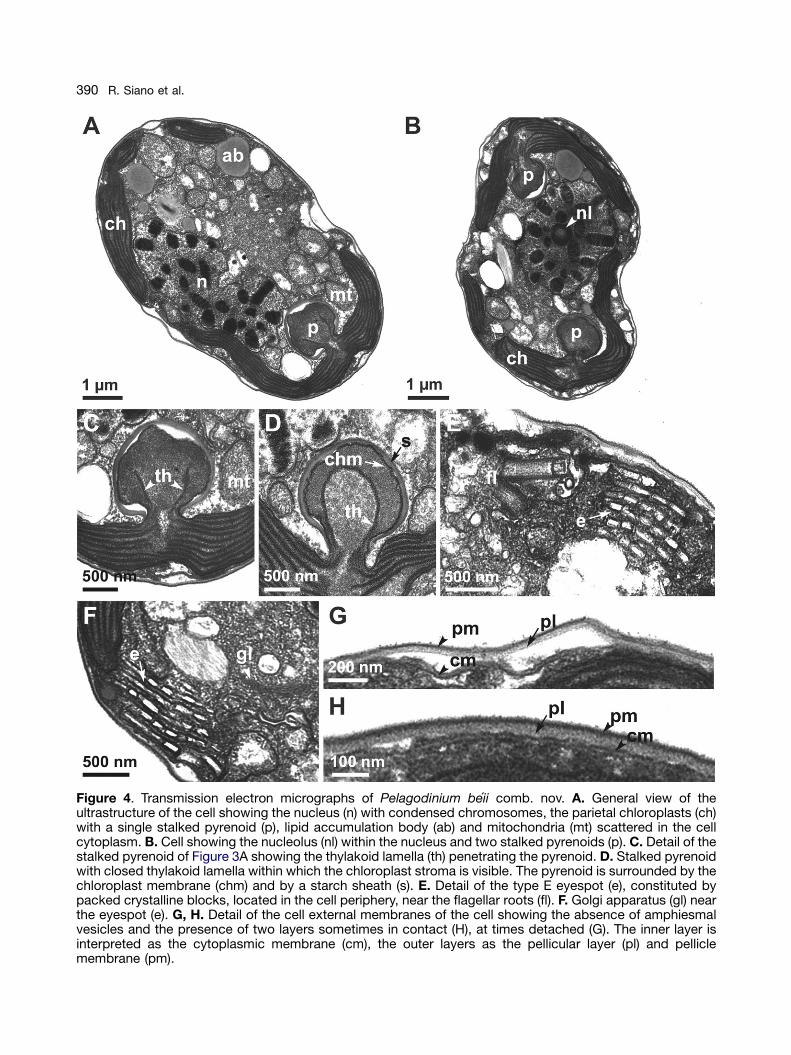
Thin sections of cells observed with TEM revealthe typical ultrastructure of a dinoflagellate, with alarge nucleus (n) with condensed chromosomes(Fig. 4A) within which a small nucleolus (nl) ispresent (Fig. 4B). Chloroplasts (ch) are peripheral,with 1 (Fig. 4A) or 2 (Fig. 4B) stalked pyrenoids (p).Pyrenoids are penetrated by thylakoid lamellae(th), which can appear open (Fig. 4C) or closed(Fig. 4D) depending on the angle of the section.The chloroplast stroma is present within thethylakoid lamellae (Fig. 4D). Pyrenoids areenclosed in a starch sheath (s) (Fig. 4C, D). Thenumber of membranes surrounding chloroplastsand pyrenoids is not clearly detectable from ourthin sections. The eyespot (e) is a multi-vesiculatebody containing packed crystalline blocks. It islocated at the cell periphery, outside the chloro-plasts, near the flagellar roots (fl) (Fig. 4E, F). TheGolgi apparatus (gl) is located near the eyespotand comprises many dictyosomes (Fig. 4F). Lipidaccumulation bodies (ab) and mitochondria (mt),sometimes rather large, are scattered in thecytoplasm (Fig. 4A, C). Trichocysts are absent.The fixation protocol employed most probablyinduced ecdysis because amphiesmal vesicleswere not observed surrounding the cell. The cellappears to be surrounded by two membranelayers, which at times are in tight contact (Fig. 4H),at times detached (Fig. 4G). In accordance withHohfeld and Melkonian (1992), we interpret theinner layer as being the cytoplasmic membrane(cm) and the outer layer as being the pellicularlayer (pl) and pellicle membrane (pm).
Phylogenetic Analysis
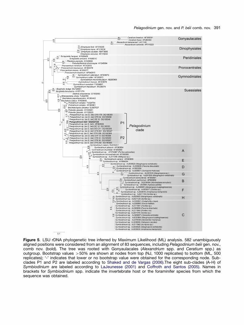
The LSU rDNA sequence of the studied strainclusters within a well-supported clade includingother ‘G. beii’ sequences, here called the Pelago-dinium clade. Two distinct sub-clades, P1 and P2,are recognized at the 99% identity threshold,and the studied sequence belongs to sub-cladeP1 (Fig. 5). The Pelagodinium clade is part ofa larger phylogenetic group including 8 otherdinoflagellate genera (Baldinia Hansen etDaugbjerg, Biecheleria Moestrup, Lindberget Daugbjerg, Biecheleriopsis Moestrup,Lindberg et Daugbjerg, Borghiella Moestrup,Hansen et Daugbjerg, Polarella Montresor,Procaccini et Stoecker, Protodinium Lohmann,Woloszynskia Thompson, and Symbiodinium)which are all part of the order Suessiales. TheSuessiales are clearly distinct from other dino-flagellate orders, the Gymnodiniales, Peridiniales,Prorocentrales, Dinophysiales, and Gonyaulacales
ARTICLE IN PRESS
Figure 4. Transmission electron micrographs of Pelagodinium beii comb. nov. A. General view of theultrastructure of the cell showing the nucleus (n) with condensed chromosomes, the parietal chloroplasts (ch)with a single stalked pyrenoid (p), lipid accumulation body (ab) and mitochondria (mt) scattered in the cellcytoplasm. B. Cell showing the nucleolus (nl) within the nucleus and two stalked pyrenoids (p). C. Detail of thestalked pyrenoid of Figure 3A showing the thylakoid lamella (th) penetrating the pyrenoid. D. Stalked pyrenoidwith closed thylakoid lamella within which the chloroplast stroma is visible. The pyrenoid is surrounded by thechloroplast membrane (chm) and by a starch sheath (s). E. Detail of the type E eyespot (e), constituted bypacked crystalline blocks, located in the cell periphery, near the flagellar roots (fl). F. Golgi apparatus (gl) nearthe eyespot (e). G, H. Detail of the cell external membranes of the cell showing the absence of amphiesmalvesicles and the presence of two layers sometimes in contact (H), at times detached (G). The inner layer isinterpreted as the cytoplasmic membrane (cm), the outer layers as the pellicular layer (pl) and pelliclemembrane (pm).
R. Siano et al.390
ARTICLE IN PRESS
Figure 5. LSU rDNA phylogenetic tree inferred by Maximum Likelihood (ML) analysis. 582 unambiguouslyaligned positions were considered from an alignment of 83 sequences, including Pelagodinium beii gen. nov.,comb nov. (bold). The tree was rooted with Gonyaulacales (Alexandrium spp. and Ceratium spp.) asoutgroup. Bootstrap values 450% are shown at nodes from top (NJ, 1000 replicates) to bottom (ML, 500replicates); ‘-’ indicates that lower or no bootstrap value were obtained for the corresponding node. Sub-clades P1 and P2 are labeled according to Shaked and de Vargas (2006).The eight sub-clades (A-H) ofSymbiodinium are labeled according to LaJeunesse (2001) and Coffroth and Santos (2005). Names inbrackets for Symbiodinium spp. indicate the invertebrate host or the foraminifer species from which thesequence was obtained.
391Pelagodinium gen. nov. and P. beii comb. nov.
ARTICLE IN PRESS
R. Siano et al.392
(Fig. 5). Baldinia and Borghiella form a cluster atthe base of the order Suessiales in both NeighborJoining (NJ) and Maximum Likelihood (ML) topo-logies. The other genera within the Suessialesshow slightly different branching patterns in thetwo topologies. The clade including Biecheleriaand Woloszynskia and the clade withBiecheleriopsis and Protodinium are sisters inNJ, but not in ML. Polarella and Pelagodiniumare sister clades in both NJ and ML, however, verylow bootstrap supports were obtained in bothtopologies. In both NJ and ML trees, the eightSymbiodinium spp. clades (A-G) are wellseparated from the Pelagodinium clade and theother genera within the Suessiales. Thesequences attributed to Gymnodinium linucheaeand G. varians cluster in the Symbiodinium cladesA and E, and thus these species, as alreadypointed out by LaJeunesse (2001), should bereferred to as Symbiodinium linucheae andSymbiodinium varians, respectively.
Discussion
In this study, the morphostructural features andthe phylogenetic position of a symbiotic dino-flagellate isolated from the foraminifer Orbulinauniversa collected offshore Puerto Rico (AtlanticOcean) were analyzed. The external characteris-tics (shape, dimensions, hypoconal flange) andultrastructural features of the cell (stalked pyre-noids penetrated by thylakoid lamellae, absenceof trichocysts), as well as its swimming behaviour,indicated that the dinoflagellate matches themorphological description of Gymnodinium beii(Spero 1987). The new morphological and phylo-genetic information gathered on this speciesprovoke a reassessment of the taxonomy of thedinoflagellate symbiont of planktonic foraminifera.A new genus is erected, Pelagodinium gen. nov.Siano, Montresor, Probert et de Vargas and thespecies Gymnodinium beii Spero is reclassifiedas Pelagodinium beii (Spero) Siano, Montresor,Probert et de Vargas (see taxonomic appendix).
SEM images revealed the presence on the cellsurface of a single and straight elongated apicalvesicle (EAV) ornamented with a row of smallglobular knobs (Fig. 3C, D). This character issufficient to state that ‘G. beii’ does not belong tothe genus Gymnodinium (order Gymnodiniales),which is characterized by possessing a horse-shoe-shaped acrobase running in an anticlock-wise direction on the cell apex (Daugbjerg et al.2000). Phylogenetic analyses inferred from LSU
rDNA sequences support this conclusion, show-ing that Pelagodinium beii is phylogeneticallydistant from the Gymnodiniales, being rather amember of the order Suessiales. According to themorphological criteria proposed by Fensome et al.(1993), the order Suessiales encompassesdinoflagellates with 7 to 10 latitudinal series ofamphiesmal vesicles, although an emendation ofthe order has been suggested to accommodatespecies with more than 10 latitudinal series(Kremp et al. 2005). Being characterized by 8latitudinal series of amphiesmal vesicles, thegenus Pelagodinium fulfils the diagnostic charac-ter of the order Suessiales.
The Order Suessiales
The order Suessiales is presently divided intothree families: Borghiellaceae, Suessiaceae andSymbiodiniaceae (Fensome et al. 1993; Moestrupet al. 2009a). The family Borghiellaceae waserected recently and the diagnosis of the familySuessiaceae emended accordingly (Moestrupet al. 2009a). The two families are distinguishedon the basis of the ultrastructure of the eyespotand the arrangement of the apical furrow. In theBorghelliaceae, the eyespot is ordinary with thecarotenoid globules located within the chloroplast(type B of Moestrup and Daugbjerg 2007) and theapical furrow is constituted by a pair of elongatedvesicles (PEV). In the Suessiaceae, the eyespotcomprises a series of cisternae with brick-likecontent (type E of Moestrup and Daugbjerg 2007)and the apical furrow is a single elongated apicalvesicle (EAV) (Moestrup et al. 2009a) (Table 1).Moreover, the Suessiaceae have clearly identi-fiable latitudinal series of amphiesmal vesicles,ranging in number between 7 and 15, while in theBorghiellaceae the number of latitudinal seriesis not specified in the description of the family(Moestrup et al. 2009a). The Borghelliaceaeincludes the genera Baldinia and Borghiella,although Baldinia does not have the PEV,whereas the Suessiaceae comprises Biecheleria,Biecheleriopsis, Symbiodinium and Polarella(Moestrup et al. 2009a), although Polarella doesnot have an EAV.
The genus Symbiodinium was attributed to thefamily Symbiodiniaceae by Fensome et al. (1993)based mainly on its occurrence as coccoid cells insymbiosis with benthic organisms. However,Moestrup et al. (2009a) transferred the genus tothe family Suessiaceae due to the fact that boththe type species S. microadriaticum Freudenthaland the recently described S. natans Hansen et

ARTIC
LEIN
PRES
S
Table 1. Selected morphological features of genera included in the order Suessiales. The type species of the genus Woloszynszkia (W.reticulata) is included for comparison.
Features Pelagodinium1 Symbiodinium2 Baldinia3 Biecheleria4 Biecheleriopsis5 Borghiella6 Polarella7 Protodinium8 Woloszynskia9
External morphology
Apical furrow EAV EAV absent EAV EAV PEV absent EAV carina runningover the apex,across the whole epicone
Numberof apicalvesiclessurroundingthe furrow
3þX 2þX - ca. 14 2 or 4þX 6 - 2 or 4þX ?
Number oflongitudinalseries
8 7 not defined(4100 vesicles)
not defined(many vesicles)
8-10 16 9 8 9-10
Number ofcingular series
1 2 1 3-4 2 2 2 2 1?
Postcingularseries of smallvesicles
present absent absent Absent(a post cingularrim is present)
present absent absent absent absent
Hypoconalflange
present absent absent absent present absent absent absent absent
Ultrastructure
Eyespot type E E B E E B E ? present,V-shaped,type unknown
Chloroplasts 1 or moreperipheral
1 or moreperipheral
1 central.radiatingfrom thepyrenoid
many,forming aperipheralnetwork
many,peripheral
many,peripheralforming aloosenetwork
1 or many,central
2-4,peripheral
many
Pyrenoids 1-2, stalked,penetratedby thylakoidlamellae
1-2, stalked,not penetratedby thylakoidlamellae
1 central many,stalkedpenetratedby thylakoidlamellae
many, stalkedpenetratedby thylakoid,swollen ends
absent many,central,stalked,penetratedby thylakoidlamellae
? ?
Nuclearconnectoror rhizoplast
absent absent absent absent present absent absent ? ?
Peduncle absent present present present absent absent absent ? ?Trichocysts absent absent absent absent absent absent absent ? ?Pusule absent present (1) present (1) present (1) Present (2) Present (1-2) ? ? ?
Abbreviations: EAV: Elongated apical vesicle; PEV: pair of elongated amphiesmal vesicles. Eyespot types according to Moestrup andDaugbjerg (2007).Literature references: 1Spero (1987), present work; 2Features of the free living stage are considered: Freudenthal (1962), Loeblich III andSherley (1979), Trench and Blank (1987), Hansen and Daugbjerg (2009); 3Hansen et al. (2006); 4Kremp et al. (2005), Moestrup et al. (2009a);5Moestrup et al. (2009b); 6Moestrup et al. (2008); 7Montresor et al. (1999); 8Siano et al. (2009); 9The type species W. reticulata, Thompson(1951).
393
Pela
go
din
ium
gen.
no
v.and
P.
beii
co
mb
.no
v.

ARTICLE IN PRESS
R. Siano et al.394
Daugbjerg have a type E eyespot and an EAV(Hansen and Daugbjerg, 2009; Loeblich III andSherley 1979). A consequence of this new classi-fication is that no genera are presently ascribed tothe Symbiodiniaceae, making this family redun-dant. The genus Protodinium could also belong tothe Suessiaceae since it has an EAV (Siano et al.2009), but no information on the eyespot ultra-structure is presently available.
The affiliation of the genus Woloszynskia tothe Suessiales is uncertain. The morphology of thetype species, W. reticulata Thompson, is clearlydifferent from that of other Suessiales species(Table 1). In contrast to all Suessiales speciesdescribed to date, W. reticulata has thin amphies-mal plates on the episome and notably thick oneson the hypocone, and a large crest or ‘carina’extending across the apical end along the wholeepisome (Thompson 1951). Unfortunately, noultrastructural or molecular data are available forthe type material. Many species previously identi-fied as Woloszynskia have been reclassified in thegenera Tovellia Moestrup, Lindberg et Daugbjerg,Jadwigia Moestrup, Lindberg et Daugbjerg(Lindberg et al. 2005; Moestrup et al. 2006) andBiecheleria (Moestrup et al. 2009a). W. cinctaSiano, Montresor et Zingone, recently describedfrom the Mediterranean Sea (Siano et al. 2009),should most probably also be transferred to thegenus Biecheleria based on the presence of anEAV and its phylogenetic position (Fig. 5). Ultra-structural information, especially on the type ofeyespot, is however needed to confirm thisrecombination.
Distinctive Features of the GenusPelagodinium
Pelagodinium beii fulfils the recently designatedmorphological criteria of the family Suessiaceae,having a type E eyespot, an EAV, and 8 latitu-dinal series of amphiesmal vesicles. The molecu-lar phylogenetic relationship of P. beii withBiecheleria, Biecheleriopsis, Symbiodinium andPolarella corroborates this affiliation.
Pelagodinium beii shares some morphologicalcharacters with the other genera assigned to theSuessiales, while other features make this genusunique within the order (Table 1). The EAV of P. beiiresembles those described for Biecheleria baltica(Elbrachter et Kremp) Moestrup, Lindberg etDaugbjerg (Moestrup et al. 2009a) (=Woloszynskiahalophila Elbrachter et Kremp (Kremp et al.2005), B. pseudopalustris (Schiller) Moestrup,
Lindberg et Daugbjerg (Moestrup et al. 2009a)Biecheleriopsis adriatica Moestrup, Lindberg etDaugbjerg (Moestrup et al. 2009b), Protodiniumsimplex Lohmann (Siano et al. 2009), S. natansHansen et Daugbjerg (Hansen and Daugbjerg,2009) and W. cincta (Siano et al. 2009). The EAV ofP. beii is surrounded, however, by 3 elongatedvesicles and a small X vesicle (Fig. 2D), distin-guishing it from that of Biecheleria (ca. 14 vesicles),Biecheleriopsis (2 or 4þX) and Protodinium (2 or4þX). The presence of 3þX vesicles around theEAV in Pelagodinium is a unique feature within theSuessiales (Table 1). Pelagodinium beii is charac-terized by a single series of vesicles within thecingulum and the arrangement of a series of smallfour- or five-sided vesicles below the cingulum is apeculiar feature within the Suessiales. BothBiecheleria pseudopalustris (Moestrup et al.2009a) and Biecheleriopsis adriatica (Moestrupet al. 2009b) have a post-cingular rim of very smallvesicles, but these are much smaller than those ofPelagodinium and are located on the posterior rimof the cingulum and not in the hypocone.
Like Symbiodinium, Pelagodinium is an endo-symbiotic dinoflagellate. Pelagodinium beii and atleast some Symbiodinium species (S. microadria-ticum and S. natans) can, however, live as motilestages and these somewhat resemble each otherwhen observed in light microscopy. There arenevertheless clear differences between P. beii andthe free-living stages of Symbiodinium species(Table 1). As observed by Spero (1987), P. beii hasa hypoconal flange, a cingulum displaced by onceits width, no peduncle, and pyrenoids penetrated bythylakoid lamellae, whereas in S. microadriaticumno hypoconal flange is present, the cingulum isdisplaced by less than once its width, a peduncleis present, and the pyrenoids are not penetratedby thylakoid lamellae (Freudenthal 1962; LoeblichIII and Sherley 1979 as Zooxanthella microadria-tica; Trench and Blank 1987). Our new SEM andTEM images of P. beii show important newcharacters useful for distinguishing the symbioticgenera. The number of latitudinal series of vesiclesdiffers between P. beii and S. microadriaticum, theformer having 8, the latter 7. This difference is dueto the number of series of vesicles in the epicone: 4in P. beii and 3 in S. microadriaticum. Moreover,S. microadriaticum is described as having two seriesof vesicles within the cingulum, whereas P. beii hasonly one series within the cingulum as well as theseries of small four- or five-sided vesicles in thehypocone, immediately below the cingulum. Thesedifferences between the genera are confirmed bythe recent description of the free-living stage of a

ARTICLE IN PRESS
395Pelagodinium gen. nov. and P. beii comb. nov.
new Symbiodinium species, S. natans, which ischaracterized by having 7 series of latitudinal seriesof vesicles and two cingular series, and by theabsence of the hypoconal flange (Hansenand Daugbjerg 2009). P. beii is characterized bythe presence of an eyespot with a peculiarvesiculate ultrastructure classifiable as type E ofMoestrup and Daugbjerg (2007); this organelle wasnot shown at the time of the first description of thisdinoflagellate (Spero 1987). The eyespot is notclearly described in the original description of S.microadriaticum (Freudenthal 1962), but in the thinsections provided by Loeblich III and Sherley (1979)it is clearly visible. Overall, the morphologicaldifferences between Pelagodinium and Symbiodi-nium, together with their genetic differences, fullysupport the fact that the studied strain is not aSymbiodinium, confirming the first intuition ofSpero (1987).
Phylogenetic Position of Pelagodinium beii
The LSU rDNA sequence of the studied straingroups in clade P1, sister of clade P2, sensuShaked and de Vargas (2006). Both clades includedinoflagellates recorded only as endosymbionts invarious planktonic foraminifera sampled world-wide, and named ‘Gymnodinium beii’ (Shakedand de Vargas 2006). These two clades forma well-supported, independent group, distinctfrom sequences of the genus Gymnodinium(Gymnodiniales). This result clearly indicates that‘G. beii’ was wrongly classified in the genusGymnodinium, corroborating the conclusionobtained from the comparison of morphologicalfeatures. Given the significant genetic diversityobserved both between and within clades P1 andP2 (Shaked and de Vargas 2006), only LSU rDNAsequences identical to our type sequence shouldbe attributed to P. beii. All other sequences of‘G. beii’ belonging to either clades P1 or P2 shouldbe attributed to Pelagodinium sp. awaiting furthermorphological, genetic, and ecological studies toverify their actual taxonomic status.
Symbiotic Relationships
Shaked and de Vargas (2006) demonstrated thata high flexibility characterizes the photosymbioticrelationship between foraminifers and dinoflagel-lates in open oceanic plankton. The fourPelagodinium subgroups detected (containing intotal 21 unique phylotypes) were found withoutany specificity in association with the four differentforaminifera morphospecies (Globigerinoides
ruber, G. sacculifer, G. conglobatus, and O.universa), each of which also harbours crypticdiversity (de Vargas et al. 1999; Morard et al.2009). Unfortunately, we do not have informationon the host genotype from which our strain ofP. beii was isolated, but combined symbiontculture isolation and recovery of DNA from thecrushed host cell should be possible in the future,allowing testing of whether different Pelagodiniumphylotypes show any host specificity.
Symbiotic dinoflagellates of pelagic organismshave previously been studied from either themorphological (Banaszak et al. 1993; Lee 1980;Spero 1987; Trench 1993; Trench and Thinh 1995),or the molecular (Gast and Caron 1996; Shakedand de Vargas 2006) point of view. The matchingmorpho-genetic information gathered here for thefirst time on a pelagic dinoflagellate endosymbiontrevealed clear differentiation from dinoflagellatesymbionts of benthic organisms. Thus, within theorder Suessiales, two different and ancientlineages are involved in photosymbiotic associa-tions, the Symbiodinium spp. in coastal benthicecosystems, and the Pelagodinium spp. in openoceanic waters. Previous phylogenetic analysis ofthe Suessiales suggested either independentendosymbiotic transitions from a free-living dino-flagellate lineage (Polarella) into coastal benthic(Symbiodinium) and pelagic (Pelagodinium,named as G. beii) photosymbioses, or a singlesymbiotic event (Symbiodinium, Pelagodinium)involving a free-living lineage (Polarella), followedby a loss of symbiotic behaviour in Protodiniumand Woloszynskia (Shaked and de Vargas 2006).However, LSU rDNA data are currently not robustenough to confirm either of these hypotheses andfurther morpho-genetic data are needed to resolvethe evolutionary paths that led to the emergenceof major photosymbiotic lineages within theSuessiales.
Finally, our study illustrates the utility of estab-lishing clonal cultures to conduct morpho-mole-cular characterization of symbiotic microalgae.This would be useful, for example, to resolvecases where dinoflagellate symbionts hosted byphylogenetically distant organisms (hydrozoansand radiolarians) have been shown to be geneti-cally similar (Gast and Caron 1996), but theirmorphological affinities are still to be demon-strated. It is also relevant in light of the fact that anumber of described dinoflagellate symbiontswarrant morphological reexamination in order toclarify their systematic positions. For example, themorphology of Symbiodinium linucheae (Trenchand Thinh) LaJeunesse, the symbiont of the
ARTICLE IN PRESS
R. Siano et al.396
jellyfish Linuche unguiculata (Trench and Thinh1995), should be reexamined since this specieswas assigned to Symbiodinium only on the basisof molecular data (LaJeunesse 2001), andGymnodinium vertebralis Lee, the symbiont ofthe foraminifer Marginopora vertebralis (Lee 1980;Trench 1993), is probably not a member of thegenus Gymnodinium. A fundamental question willbe to determine whether open oceanic photo-symbiosis involving Pelagodinium spp. hasdeveloped in host organisms other than theforaminifers, like its coastal benthic counterpart,where Symbiodinium has become associatedwith many protistan and metazoan lineages.Symbiotic associations are likely to be a richsource of yet unsuspected biodiversity, andcultured strains of symbiotic dinoflagellates are goodcandidates for analysis of the molecular and physio-logical mechanisms underlying photosymbioticassociations.
Taxonomic Appendix
Pelagodinium Siano, Montresor, Probert, et deVargas gen. nov.
Diagnosis: cellulae photosynteticae ad Dino-phita pertinentes. Cellulae in libera vita octoseriebus vesicularum amphiesmatis contectae.Quattor in epicono, tres in hypocono, una incingulo. Longa recta vesicula linea recta tuberumglobosorum constituta in cellulae apice. Seriesparvarum vesicularum quadriangularum vel pen-tagonarum sub cingulo proxime est et hypoconumcircumdat. Chloroplasti colore flavente cum pyr-enoidis adherentibus. Pyrenoidi thylachoidorumlamellis invasi. Stigma extra plastidium ad typumE pertinens. Trichocisti absunt.
Photosynthetic dinoflagellate. Free-living cellscovered by eight series of amphiesmal vesicles:four in the epicone, three in the hypocone, andone in the cingulum. A straight single elongatedapical vesicle constituted of a single row ofglobular knobs is present on the cell apex. Aseries of small quadrangular or pentagonal vesi-cles is present immediately below the cingulumand encircles the hypocone. Chloroplasts golden-yellow in colour, with stalked pyrenoids. Pyrenoidspenetrated by thylakoid lamellae. Extraplastidialeyespot present belonging to type E. Trichocystsabsent.
Type species: Pelagodinium beii (Spero) Siano,Montresor, Probert et de Vargas comb. nov.
Etymology: the genus name derives from thelife strategy of this dinoflagellate: dinoflagellate(=dinos) symbiont of pelagic (=pelagos) protists.
Pelagodinium beii (Spero) Siano, Montresor,Probert et de Vargas comb. nov. (Fig. 1 A-D;Fig. 2 A-G, Fig. 3 A-C)
Basionym: Gymnodinium beii Spero in Spero(1987): 316, fig. 7 (holotype), Fig. 3a-d (isotypes,designated herein)
Diagnosis: cells are small: 10.070.8 mm inlength, 6.670.4 mm in width, with a round toelliptical epicone and a slightly asymmetricalhypocone of almost the same dimensions.A flange is present on the left side of the epicone,projecting over the sulcus, it can be shortand rounded to more pointed and elongated.Cingulum wide and shallow, descending anddisplaced one cingulum width. Sulcus deep andnarrow, enlarging only at cell antapex. Flagellaemerging from the sulcal region, no peduncle isevident. When cells are observed in SEM, amphies-mal vesicles are visible on the cell surface,arranged in 8 longitudinal series. A single elongatedapical vesicle (EAV) ornamented with a row ofglobular knobs is present on the cell surface,surrounded by a series of 3 quadrangular vesiclesand a small squared vesicle (X vesicle). Another 3series of vesicles are present in the epiconeconstituted respectively of 7, 2-3 (intercalary), and8 vesicles. Cingulum with one series of vesicles.Hypocone with a series of 16-20 small vesicles,anterior to another series of 8 vesicles and 3-4antapicals. One or two peripheral golden-yellowchloroplasts, with one or two stalked pyrenoids.Pyrenoids are penetrated by thylakoid lamellae.An extraplastidial eyespot of type E is present nearthe flagellar roots. Trichocysts absent.
Taxonomic Note: Gymnodinium beii was sug-gested to resemble the free-living dinoflagellateAureodinium pigmentosum Dodge (Anderson andBe 1976; Hemleben and Spindler 1983; Spindlerand Hemleben 1980). This latter species wasdescribed based on LM and TEM observations asbeing 10 mm in length and 7 mm in width, with anirregular hypoconal outline, peripheral chloro-plasts with two stalked pyrenoids penetrated bythylakoid lamellae, without trichocysts, and with atheca composed of thin polygonal plates (Dodge1967). Believing that the ‘theca’ described forA. pigmentosum was not typical of a thecatedinoflagellate, Loeblich III (1969) transferred thespecies to Gymnodinium, that, at that time,encompassed all species with an entirelymembranous amphiesma, and he recombinedthe species as G. pigmentosum (Dodge) LoeblichIII. In light of the redescription of the genusGymnodinium (Daugbjerg et al. 2000) this assign-ment appears questionable, and the actual

ARTICLE IN PRESS
397Pelagodinium gen. nov. and P. beii comb. nov.
taxonomic position of G. pigmentosum is unclear.Gymnodinium pigmentosum was not obtainedfrom a symbiotic organism, but was isolated ina free-living stage directly from a seawatersample (Dodge 1967). We cannot rule out thehypothesis that G. pigmentosum might representthe free-living stage of an endosymbionticdinoflagellate, but this species might also be afree-living dinoflagellate of the genera Biecheleria,Biecheleriopsis, Protodinium or Woloszynskia. Wetherefore did not consider the possibility of usingthe name Aureodinium for the endosymbionticdinoflagellates of the foraminifer Orbulinauniversa.
Methods
Culture origin and maintenance: The Orbulina universaspecimen from which the algal culture originated was isolatedfrom a sample collected off the coast of Puerto Rico,Caribbean Sea (Atlantic Ocean; 141490N 671030W) in Novem-ber 2005. The foraminiferal specimen was identified under abinocular microscope at � 100 magnification, cleaned bysuccessive transfers into sterile seawater in Petri dishes,before being crushed with a fine needle under the binocularmicroscope. The dinoflagellate culture was obtained bymicropipette isolation of a single cell released from thecrushed specimen. The resulting monoclonal culture wasmaintained in filter-sterilized seawater with K/2(-Tris, -Si)medium supplements (Keller et al. 1987) at 21 1C with anirradiance of 70–80 mmol photons m�2 s�1 in a 12:12 light:darkregime. The culture was deposited in the Roscoff culturecollection (Roscoff Culture Collection, http://www.sb-ros-coff.fr/Phyto/rcc) as RCC1491.
Microscopy preparations and observations: Live cellswere observed and measured with a Nikon Eclipse TS100inverted light microscope (Nikon, New York, USA). Lightmicrographs were taken with a Zeiss Axiophot light micro-scope (Carl Zeiss, Oberkochen, Germany) equipped with aZeiss AxioCam digital camera system (Carl Zeiss, Oberko-chen, Germany).
For scanning electron microscopy (SEM), cells were fixed in1% (v:v) OsO4 for 5–10 min at room temperature. Sampleswere gently filtered onto 3 mm pore-size Nucleopore poly-carbonate filters (Pleasanton, CA, USA), washed with distilledwater, dehydrated in an ethanol series (25%, 50%, 75%, 95%,100%), and critical point dried. The filters were mounted onstubs, sputter coated with gold, and examined with a JEOLJSM-6500F SEM (JEOL-USA Inc., Peabody, MA, USA). Thestub of the analyzed sample is deposited in the museum of theStazione Zoologica Anton Dohrn in Naples and it is availableon request.
For observations of thin sections with transmission electronmicroscopy (TEM), cells were concentrated by gentle cen-trifugation (800 r.p.m. for 7 min), fixed with cold 1% (v:v)gluteraldehyde for 1 h on ice, rinsed with filtered seawater(FSW), and post-fixed with 1% (v:v) osmium tetroxide for30 min on ice. After two rinses with FSW, the sample wasdehydrated in an ethanol series (25%, 50%, 75%, 95%,100%), transferred to propylene oxide, and embedded inEpon resin (v:v, 1:1). After polymerization at 70 1C for 24 h, thinsections were cut using a Reichert Ultracut ultramicrotome
(Depew, NY, USA), stained with uranyl acetate and leadcitrate, and examined with a LEO 912AB EF-TEM (LEO, CarlZeiss, Oberkochen, Germany).
DNA extraction and phylogenetic analysis: DNA wasextracted from an exponentially growing culture of thePelagodinium beii strain using the method described in deVargas et al. (2002). The D1-D2 part of the nuclear largesubunit ribosomal DNA (LSU rDNA) was PCR amplified andsequenced using the methods described in Shaked and deVargas (2006). The partial LSU rDNA sequence of the analyzedstrain is deposited in Genbank (http://www.ncbi.nlm.nih.gov)with the accession number GQ422124.
The sequence generated from the studied strain wasaligned with other LSU rDNA sequences downloaded fromGenBank and attributed to ‘G. beii’, with sequences ofspecies of the order Suessiales and with sequences undoubt-edly attributable to other main dinoflagellate orders, theGonyaulacales, Peridiniales, Prorocentrales, Dinophysiales,Gymnodiniales. An alignment of 83 sequences was generatedusing MAFFT (Katoh et al. 2002;) and manually editedin Bioedit v.7.0.9.0 (Hall 1999). The 582 positions used inphylogenetic analyses were determined using the Gblocksmethod (Castresana 2000) for selecting conserved blocks(minimum block length=5; allowed gap positions=with half).
Phylogenetic analyses were conducted with NeighborJoining (NJ) and Maximum Likelihood (ML) methods. The NJphylogenetic analysis was inferred using pair wise p-distancein MEGA (v. 4.1, Tamura et al. 2007) and bootstrap valueswere calculated from 1000 replicates. The ML analysis wascarried out using PhyML v. 3.0 aLRT (Guindon and Gascuel2003), performed on the web portal Phylogeny.fr (Dereeperet al. 2008). The General Time Reversible (GTR) model ofnucleotide substitution and the number of substitution ratecategories, the shape parameter (a) of the Gamma (G)distribution and the proportion of invariable sites (I) wereestimated from the dataset using default options in Phylo-geny.fr. Bootstrap supports for the tree were obtained after500 replicates. The tree was visualized and edited in MEGA(v. 4.1, Tamura et al. 2007).
Acknowledgements
R.S. was financed by a post-doctoral fellowship(Contract No. 09036) from the Universite Pierreet Marie Curie (UMPC, France). The ElectronMicroscopy Service of the Stazione ZoologicaAnton Dohrn is acknowledged for technicalassistance and Prof. Marta Maria Giannone isthanked for Latin translation of the genus diag-nosis. The authors thank the anonymousreviewers for providing constructive observationsfor the improvement of the manuscript. This workis part of the pluridisciplinary projects BOOM(Biodiversity of Open Ocean Microcalcifiers, IFB,ANR grant 05-BDIV-004), BioMarKs (Biodiversityof Marine euKaryotes), SYMFORAD (SYMbiosisin FOraminifera and RADiolarians) from theRegion Bretagne (France), and ASSEMBLE (EC227799).

ARTICLE IN PRESS
R. Siano et al.398
References
Anderson R, Be AWH (1976) The ultrastructure of aplanktonic foraminifer, Globigerinoides sacculifer (Brady),and its symbiotic dinoflagellates. J Foram Res 6:1–21
Apprill AM, Gates RD (2007) Recognizing diversity in coralsymbiotic dinoflagellate communities. Mol Ecol 16:1127–1134
Arnold AJ, Parker WC (1999) Biogeography of PlanktonicForaminifera. In Gupt BKS (ed) Modern Foraminifera. KluwerAcademic Publishers, London, pp 103–122
Banaszak AT, Iglesias-Prieto R, Trench RK (1993) Scripp-siella velellae sp. nov. (Peridiniales) and Gloeodinium viscumsp. nov (Phytodiniales), dinoflagellate symbionts of 2 hydro-zoans (Cnidaria). J Phycol 29:517–528
Caron D (2000) Symbiosis and Mixotrophy among PelagicMicroorganisms. In Kirchman DL (ed) Microbial Ecology of theOceans. Wiley-Liss, Inc., New York, pp 495–523
Castresana J (2000) Selection of conserved blocks frommultiple alignments for their use in phylogenetic analysis. MolBiol Evol 17:540–552
Coffroth MA, Santos SR (2005) Genetic diversity of symbioticdinoflagellates in the genus Symbiodinium. Protist 156:19–34
Daugbjerg N, Hansen G, Larsen J, Moestrup Ø (2000)Phylogeny of some of the major genera of dinoflagellatesbased on ultrastructure and partial LSU rDNA sequence data,including the erection of three new genera of unarmoureddinoflagellates. Phycologia 39:302–317
de Vargas C, Norris R, Zaninetti L, Gibbs SW, Pawlowski J(1999) Molecular evidence of cryptic speciation in planktonicforaminifers and their relation to oceanic provinces. Proc NatlAcad Sci USA 96:2864–2868
de Vargas C, Bonzon M, Rees NW, Pawlowski J,Zaninetti L (2002) A molecular approach to biodiversity andbiogeography in the planktonic foraminifer Globigerinellasiphonifera (d’Orbigny). Mar Micropaleontol 45:101–116
Dereeper A, Guignon V, Blanc G, Audic S, Buffet S,Chevenet F, Dufayard JF, Guindon S, Lefort V, Lescot M,Claverie JM, Gascuel O (2008) Phylogeny.fr: robust phylo-genetic analysis for the non-specialist. Nucleic Acids Res36:465–469
Dodge JD (1967) Fine structure of the dinoflagellateAureodinium pigmentosum gen. et sp. nov. Br Phycol Bull3:327–336
Farmer MA, Fitt WK, Trench RK (2001) Morphology of thesymbiosis between Corculum cardissa (Mollusca: Bivalvia)and Symbiodinium corculorum (Dinophyceae). Biol Bull200:336–343
Fensome RA, Taylor FJR, Norris G, Sarjeant WAS,Wharton DI, Williams GL eds (1993) A Classification ofFossil and Living Dinoflagellates. Micropaleontology PressSpecial Paper, no.7
Freudenthal HD (1962) Symbiodinium gen. nov. and Symbio-dinium microadriaticum sp. nov; a Zooxanthella: taxonomy,life cycle and morphology. J Protozool 9:45–52
Gast RJ, Caron DA (1996) Molecular phylogeny of symbioticdinoflagellates from planktonic foraminifera and radiolaria.Mol Biol Evol 13:1192–1197
Gast RJ, Caron DA (2001) Photosymbiotic associations inplanktonic foraminifera and radiolaria. Hydrobiologia 461:1–7
Guindon S, Gascuel O (2003) A simple, fast, and accuratealgorithm to estimate large phylogenies by maximum like-lihood. Syst Biol 52:696–704
Hackett JD, Anderson DM, Erdner DL, Bhattacharya D(2004) Dinoflagellates: a remarkable evolutionary experiment.Am J Bot 91:1523–1534
Hall TA (1999) BioEdit: a user-friendly biological sequencealignment editor and analysis program for Windows 95/98/NT.Nucleic Acids Symposium Series 41:95–98
Hansen G, Daugbjerg N (2009) Symbiodinium natans sp nov.:a free-living dinoflagellate from Tenerife (northeast-AtlanticOcean). J Phycol 45:251–263
Hansen G, Daugbjerg N, Henriksen P (2006) Baldiniaanauniensis gen. et sp. nov.: a ‘new’ dinoflagellate from LakeTovel, N. Italy. Phycologia 46:86–108
Hemleben C, Spindler M (1983) Recent advances in researchon living planktonic foraminifera. Utrecht Micropaleontol Bull30:141–170
Hoegh-Guldberg O, Mumby PJ, Hooten AJ, Steneck RS,Greenfield P, Gomez E, Harvell CD, Sale PF, Edwards AJ,Caldeira K, Knowlton N, Eakin CM, Iglesias-Prieto R,Muthiga N, Bradbury RH, Dubi A, Hatziolos ME (2007) Coralreefs under rapid climate change and ocean acidification.Science 318:1737–1742
Hohfeld I, Melkonian M (1992) Amphiesmal ultrastructure ofdinoflagellates: a revaluation of pellicle formation. J Phycol28:82–89
Hunter RL, LaJeunesse TC, Santos SR (2007) Structure andevolution of the rDNA internal transcribed spacer (ITS) region2 in the symbiotic dinoflagellates (Symbiodinium, Dinophyta).J Phycol 43:120–128
Katoh K, Misawa K, Kuma K, Miyata T (2002) MAFFT: anovel method for rapid multiple sequence alignment based onfast Fourier transform. Nucleic Acids Res 30:3059–3066
Keller MD, Selvin RC, Claus W, Guillard RRL (1987) Mediafor the culture of oceanic ultraphytoplankton. J Phycol23:633–638
Kremp A, Elbrachter M, Schweikert M, Wolny JL,Gottschling M (2005) Woloszynskia halophyla (Biecheler)comb. nov.: a bloom-forming cold-water dinoflagellate co-occurring with Scrippsiella hangoei (Dinophyceae) in the BalticSea. J Phycol 41:629–643
LaJeunesse TC (2001) Investigating the biodiversity, ecology,and phylogeny of endosymbiotic dinoflagellates in the genusSymbiodinium using the ITS region: in search of a specieslevel marker. J Phycol 37:866–880
LaJeunesse TC, Lambert G, Andersen RA, Coffroth MA,Galbraith DW (2005) Symbiodinium (Pyrrhophyta) genomesizes (DNA content) are smallest among dinoflagellates.J Phycol 41:880–886
ARTICLE IN PRESS
399Pelagodinium gen. nov. and P. beii comb. nov.
Lee JJ (1980) Nutrition and Physiology of the Foraminifera. InLevandowsky M, Hunter SH (eds) Biochemistry and Physiologyof Protozoa. Academic Press, Inc., London, New York, pp 43–66
Leggat W, Marendy EM, Baillie B, Whitney SM, Ludwig M,Badger MR, Yellowlees D (2002) Dinoflagellate symbioses:strategies and adaptations for the acquisition and fixation ofinorganic carbon. Funct Plant Biol 29:309–322
Lindberg K, Moestrup Ø, Daugbjerg N (2005) Studies onwoloszynskioid dinoflagellates I: Woloszynskia coronatare-examined using light and electron microscopy and partialLSU rDNA sequences, with description of Tovellia gen. nov.and Jadwigia gen. nov. (Tovelliaceae fam. nov.). Phycologia44:416–440
Loeblich III AR (1969) The amphiesma or dinoflagellate cellcovering. Proc North American Paleontol Convention II:867–929
Loeblich III AR, Sherley JL (1979) Observations on the thecaof the motile phase of free-living and symbiotic isolates ofZooxanthella microadriatica (Freudenthal) comb. nov. J MarBiol Assoc UK 59:195–205
Lopes RM, Silveira M (1994) Symbiosis between a pelagicflatworm and a dinoflagellate from a tropical area-structuralobservations. Hydrobiologia 287:277–284
Manning MM, Gates RD (2008) Diversity in populations offree-living Symbiodinium from a Caribbean and Pacific reef.Limnol Oceanogr 53:1853–1861
Moestrup Ø, Daugbjerg N (2007) On Dinoflagellate Phylo-geny and Classification. In Brodie J, Lewis J (eds) Unravellingthe Algae, the Past, Present and Future of Algal Systematics.CRC Press, Taylor and Francis Group, Boca Raton, FL, pp215–230
Moestrup Ø, Hansen G, Daugbjerg N (2008) Studies onwoloszynskioid dinoflagellates III: on the ultrastructure andphylogeny of Borghiella dodgei gen. et sp. nov., a cold-waterspecies from Lake Tovel, N. Italy, and on B. tenuissima comb.Nov. (syn. Woloszynskia tenuissima). Phycologia 47:54–78
Moestrup Ø, Lindberg K, Daugbjerg N (2009a) Studies onwoloszynskioid dinoflagellates IV: The genus Biecheleria gen.nov. Phycol Res 57:203–220
Moestrup Ø, Lindberg K, Daugbjerg N (2009b) Studies onwoloszynskioid dinoflagellates V. Ultrastructure of Biecheler-iopsis gen. nov., with description of Biecheleriopsis adriaticasp. nov. Phycol Res 57:221–237
Moestrup Ø, Hansen G, Daugbjerg N, Flaim G, D’Andrea M(2006) Studies on woloszynskioid dinoflagellates II: On Tovelliasanguinea sp. nov., the dinoflagellate responsible for thereddening of Lake Tovel. N. Italy. Eur J Phycol 41:47–65
Montresor M, Procaccini G, Stoecker DK (1999) Polarellaglacialis gen. nov., sp. nov. (Dinophyceae): Suessiaceae arestill alive!. J Phycol 35:186–197
Morard R, Quillevere F, Escarguel G, Ujiie Y, de Garidel-Thoron T, Norris RD, de Vargas C (2009) Morphological
recognition of cryptic species in the planktonic foraminiferOrbulina universa. Mar Micropaleontol 71:148–165
Schonberg CHL, Loh WKW (2005) Molecular identity of theunique symbiotic dinoflagellates found in the bioerodingdemosponge Cliona orientalis. Mar Ecol Prog Ser 299:157–166
Shaked Y, de Vargas C (2006) Pelagic photosymbiosis: rDNAassessment of diversity and evolution of dinoflagellatesymbionts and planktonic foraminiferal hosts. Mar Ecol ProgSer 325:59–71
Siano R, WHCF Kooistra, Montresor M, Zingone A (2009)Unarmoured and thin-walled dinoflagellates from the Gulf ofNaples, with the description of Woloszynskia cincta sp. nov.(Dinophyceae, Suessiales). Phycologia 48:44–65
Spero HJ (1987) Symbiosis in the planktonic foraminifer,Orbulina universa, and the isolation of its symbiotic dino-flagellate Gymnodinium beii sp. nov. J Phycol 23:307–317
Spindler M, Hemleben CIE (1980) Symbionts in PlanktonicForaminifera (Protozoa).. In Schwemmler W, Schenk HEA(eds) Endosymbiosis and Cell Biology. Walter de Gruyter &Co., Berlin-New York, pp 133–140
Stat M, Morris E, Gates RD (2008) Functional diversity incoral–dinoflagellate symbiosis. Proc Natl Acad Sci USA105:9256–9261
Stoecker DK, Johnson MD, de Vargas C, Not F (2009)Acquired phototrophy in aquatic protists. Aquat Microb Ecol57:279–310
Tamura K, Dudley J, Nei M, Kumar S (2007) MEGA4:Molecular evolutionary genetics analysis (MEGA) softwareversion 4.0. Mol Biol Evol 24:1596–1599
Tchernov D, Gorbunov MY, de Vargas C, Narayan Yadav S,Milligan AJ, Haggblom M, Falkowski PG (2004) Membranelipids of symbiotic algae are diagnostic of sensitivity tothermal bleaching in corals. Proc Natl Acad Sci USA101:13531–13535
Thompson RH (1951) A new genus and new records of fresh-water Pyrrophyta in the Desmokontae and Dinophyceae.Lloydia 13:277–299
Trench RK (1993) Microalgal-invertebrate symbioses – Areview. Endocyt Cell Research 9:135–175
Trench RK, Blank RJ (1987) Symbiodinium microadriaticumFreudenthal; S. goreauii sp. nov; S. kawagutii sp. nov. andS. pilosum sp. nov.: gymnodinioid dinoflagellate symbionts ofmarine invertebrates. J Phycol 23:469–481
Trench RK, Thinh LV (1995) Gymnodinium linucheae sp. nov.:the dinoflagellate symbiont of the jellyfish Linuche unguicu-lata. Eur J Phycol 30:149–154
van Oppen MJH (2007) Perspective: hidden diversity in coralendosymbionts unveiled. Mol Ecol 16:1125–1126