Pediatrics & Therapeutics · • Type III: Subluxation hip. ... creeping reflex can be released in...
7
Early Use of Vojta Therapy in Children with Postural Asymmetry, at Risk of Hip Dysplasia Artur Edward Polczyk * Department of Physiotherapy, Wrocław Medical University, Poland * Corresponding author: Artur Edward Polczyk, Department of Physiotherapy, Wrocław Medical University, Aleja Matejki 26/13 street, Wrocław 50-333, Poland, Tel: 0048793019099; E-mail: [email protected] Received date: Dec 18, 2017; Accepted date: Jan 10, 2018; Published date: Jan 12, 2018 Copyright: © 2018 Polczyk AE. This is an open-access article distributed under the terms of the Creative Commons Attribution License, which permits unrestricted use, distribution and reproduction in any medium, provided the original author and source are credited. Abstract Hip dislocation occurs in 1.5% of new-born babies. Symptoms of dysplastic hip are often connected with infant’s asymmetrical body posture, psychomotor delay and muscular tone dysfunction. Orthopedic supplies improve the positioning of the femur head in acetabulum but do not solve or even make worse the asymmetry, support functions and muscle tonus problems. This study presents 3 different cases of patients with such symptoms which has the Vaclav Vojta neurophysiological stimulation as based or supplemental treatment. The results are supported by ultrasonographic studies and evaluation of the spontaneous motility of the child in subsequent weeks of treatment. The use of the Vojta method is a helpful complement to the treatment of dislocated, unstable and dysplastic hips. Keywords: Vojta method; Pediatric physiotherapy; Hip dysplasia; Postural asymmetry Introduction Preventive hip examination in the first weeks of new-born’s life, now a standard, allows to detect abnormalities of the hip joint and to provide an early therapy. Usual symptoms of dysplasia occur in about 4% of new-borns, hip dislocation occurs in 1.5% of new-born babies. Dysplastic joint problems are more common among girls than boys. Statistically, the leſt hip is twice more likely to be dysplasia than the right [1]. In addition to a physical examination, Barlow test (a test which provokes dislocation of the femoral head from the acetabulum) and Ortolani test (which reposition the head of the femur head to the acetabulum), examining range of motion of lower limb abduction and external rotation, the symmetry of the gluteal and inguinal folds, and the difference in limb length, ultrasound examination is important. Based on Graf Method maturity of hips can be evaluated [2-4]. Integrity joints disorders [5] oſten are not noticed in the preliminary physical examination at the neonatal unit. e first symptoms of abnormal postural control, not only in the lower limbs, excessive tension of adductor muscles and internal rotation of the femoral head in the acetabulum, but also frequently associated asymmetrically expressed reflexes and neonatal automatism, limited mobility in the neck spine with constant asymmetry in the trunk, are recognized in the early physiotherapeutic evaluation. e more accurate assessment of spontaneous motorical activity, seven postural Vojta’s reactions, reflexes and neonatal automatisms examination is done, the easier is to find the exact location of therapeutic problem and the right choice of therapeutic target [1,6,7]. Mainly factors which affect abnormal development of the hip joint are gluteal fetal positioning, oligohydramnios, high fetal mass, mother's hormonal disturbances which relax the joint capsule and genetic factors. Dysplastic hip joint may occur in the youngest patients with postural and functional asymmetry or psychomotor delay. e treatment of choice for hip dysplasia is the use of various types of orthosis supplies, mainly Frejka's cushions and Pavlik’s suspenders [8-11]. Good results are obtained with the Tübingen’s rails, giving the opportunity to more closely match for the patient’s dimensions and condition [12]. e use of the aforementioned supply is not without influence on the qualitative and quantitative motor development of a child. Most of the patients during and aſter treatment show significant deviations in the support function and antigravity capabilities in both the back and abdomen positions [13]. e aim of the study is to present cases of patients with abnormalities in the hip joint coexisted with features of asymmetry, psychomotor delay and muscular tone dysfunction. e treatment was either based on or supplemented by the Vaclav Vojta neurophysiological stimulation. Graf’s Scale Depending on the alpha and beta angle values obtained in the ultrasound test, the baby's hip joints are evaluated as follows: • Type I (Ia, Ib): Healthy joints. • Type IIa: Described as "doubtful", can sometimes lead to dysplasia (Found up to 3 months). • Type IIb: Dysplastic type. • Type IIc: Joint with large acetabular dysplasia, while the femoral head is properly centered. • Type III: Subluxation hip. • Type IV: Joint is dislocated [2,3]. Vojta erapy and its Influence on the Formation of Hip Joints Two large movement complexes are used in Vojta therapy-rolling and creeping reflex. e rolling reflex is divided into four phases, P e d i a t r i c s & T h e r a p e u t i c s ISSN: 2161-0665 Pediatrics & Therapeutics Polczyk, Pediatr Ther 2018, 8:1 DOI: 10.4172/2161-0665.1000339 Case Report Open Access Pediatr er, an open access journal ISSN: 2161-0665 Volume 8 • Issue 1 • 1000339
Transcript of Pediatrics & Therapeutics · • Type III: Subluxation hip. ... creeping reflex can be released in...
Early Use of Vojta Therapy in Children with Postural Asymmetry, at Riskof Hip DysplasiaArtur Edward Polczyk*
Department of Physiotherapy, Wrocław Medical University, Poland*Corresponding author: Artur Edward Polczyk, Department of Physiotherapy, Wrocław Medical University, Aleja Matejki 26/13 street, Wrocław 50-333, Poland, Tel:0048793019099; E-mail: [email protected]
Received date: Dec 18, 2017; Accepted date: Jan 10, 2018; Published date: Jan 12, 2018
Copyright: © 2018 Polczyk AE. This is an open-access article distributed under the terms of the Creative Commons Attribution License, which permits unrestricted use,distribution and reproduction in any medium, provided the original author and source are credited.
Abstract
Hip dislocation occurs in 1.5% of new-born babies. Symptoms of dysplastic hip are often connected with infant’sasymmetrical body posture, psychomotor delay and muscular tone dysfunction. Orthopedic supplies improve thepositioning of the femur head in acetabulum but do not solve or even make worse the asymmetry, support functionsand muscle tonus problems. This study presents 3 different cases of patients with such symptoms which has theVaclav Vojta neurophysiological stimulation as based or supplemental treatment. The results are supported byultrasonographic studies and evaluation of the spontaneous motility of the child in subsequent weeks of treatment.The use of the Vojta method is a helpful complement to the treatment of dislocated, unstable and dysplastic hips.
Keywords: Vojta method; Pediatric physiotherapy; Hip dysplasia;Postural asymmetry
IntroductionPreventive hip examination in the first weeks of new-born’s life, now
a standard, allows to detect abnormalities of the hip joint and toprovide an early therapy.
Usual symptoms of dysplasia occur in about 4% of new-borns, hipdislocation occurs in 1.5% of new-born babies. Dysplastic jointproblems are more common among girls than boys. Statistically, theleft hip is twice more likely to be dysplasia than the right [1].
In addition to a physical examination, Barlow test (a test whichprovokes dislocation of the femoral head from the acetabulum) andOrtolani test (which reposition the head of the femur head to theacetabulum), examining range of motion of lower limb abduction andexternal rotation, the symmetry of the gluteal and inguinal folds, andthe difference in limb length, ultrasound examination is important.Based on Graf Method maturity of hips can be evaluated [2-4].
Integrity joints disorders [5] often are not noticed in the preliminaryphysical examination at the neonatal unit. The first symptoms ofabnormal postural control, not only in the lower limbs, excessivetension of adductor muscles and internal rotation of the femoral headin the acetabulum, but also frequently associated asymmetricallyexpressed reflexes and neonatal automatism, limited mobility in theneck spine with constant asymmetry in the trunk, are recognized inthe early physiotherapeutic evaluation. The more accurate assessmentof spontaneous motorical activity, seven postural Vojta’s reactions,reflexes and neonatal automatisms examination is done, the easier is tofind the exact location of therapeutic problem and the right choice oftherapeutic target [1,6,7].
Mainly factors which affect abnormal development of the hip jointare gluteal fetal positioning, oligohydramnios, high fetal mass,mother's hormonal disturbances which relax the joint capsule andgenetic factors. Dysplastic hip joint may occur in the youngest patients
with postural and functional asymmetry or psychomotor delay. Thetreatment of choice for hip dysplasia is the use of various types oforthosis supplies, mainly Frejka's cushions and Pavlik’s suspenders[8-11]. Good results are obtained with the Tübingen’s rails, giving theopportunity to more closely match for the patient’s dimensions andcondition [12].
The use of the aforementioned supply is not without influence onthe qualitative and quantitative motor development of a child. Most ofthe patients during and after treatment show significant deviations inthe support function and antigravity capabilities in both the back andabdomen positions [13].
The aim of the study is to present cases of patients withabnormalities in the hip joint coexisted with features of asymmetry,psychomotor delay and muscular tone dysfunction. The treatment waseither based on or supplemented by the Vaclav Vojtaneurophysiological stimulation.
Graf ’s ScaleDepending on the alpha and beta angle values obtained in the
ultrasound test, the baby's hip joints are evaluated as follows:
• Type I (Ia, Ib): Healthy joints.• Type IIa: Described as "doubtful", can sometimes lead to dysplasia
(Found up to 3 months).• Type IIb: Dysplastic type.• Type IIc: Joint with large acetabular dysplasia, while the femoral
head is properly centered.• Type III: Subluxation hip.• Type IV: Joint is dislocated [2,3].
Vojta Therapy and its Influence on the Formation ofHip Joints
Two large movement complexes are used in Vojta therapy-rollingand creeping reflex. The rolling reflex is divided into four phases,
Pedi
atrics & Therapeutics
ISSN: 2161-0665 Pediatrics & TherapeuticsPolczyk, Pediatr Ther 2018, 8:1
DOI: 10.4172/2161-0665.1000339
Case Report Open Access
Pediatr Ther, an open access journalISSN: 2161-0665
Volume 8 • Issue 1 • 1000339
creeping reflex can be released in different variations, depending onthe selected main therapeutic problem or the patient's responses forthe stimulation.
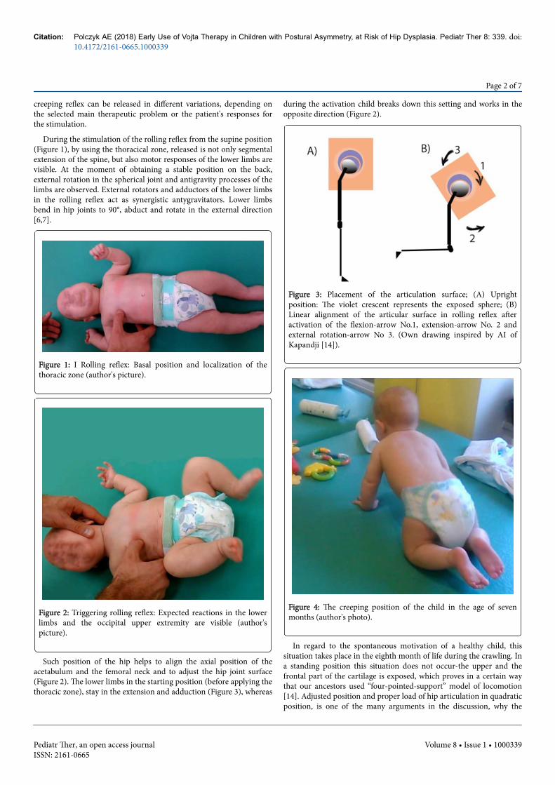
During the stimulation of the rolling reflex from the supine position(Figure 1), by using the thoracical zone, released is not only segmentalextension of the spine, but also motor responses of the lower limbs arevisible. At the moment of obtaining a stable position on the back,external rotation in the spherical joint and antigravity processes of thelimbs are observed. External rotators and adductors of the lower limbsin the rolling reflex act as synergistic antygravitators. Lower limbsbend in hip joints to 90°, abduct and rotate in the external direction[6,7].
Figure 1: I Rolling reflex: Basal position and localization of thethoracic zone (author's picture).
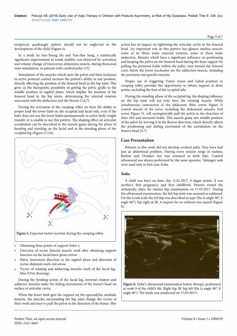
Figure 2: Triggering rolling reflex: Expected reactions in the lowerlimbs and the occipital upper extremity are visible (author'spicture).
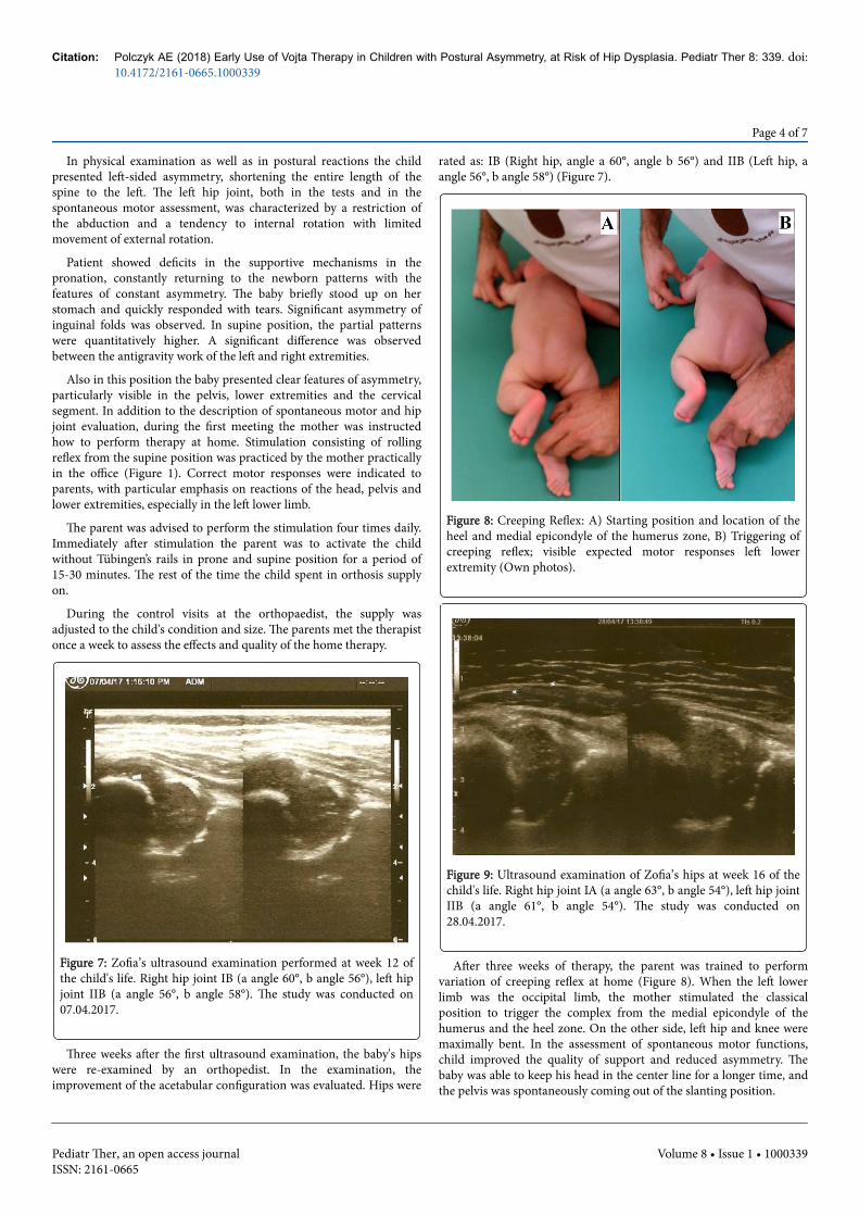
Such position of the hip helps to align the axial position of theacetabulum and the femoral neck and to adjust the hip joint surface(Figure 2). The lower limbs in the starting position (before applying thethoracic zone), stay in the extension and adduction (Figure 3), whereas
during the activation child breaks down this setting and works in theopposite direction (Figure 2).
Figure 3: Placement of the articulation surface; (A) Uprightposition: The violet crescent represents the exposed sphere; (B)Linear alignment of the articular surface in rolling reflex afteractivation of the flexion-arrow No.1, extension-arrow No. 2 andexternal rotation-arrow No 3. (Own drawing inspired by AI ofKapandji [14]).
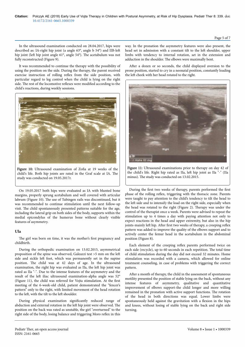
Figure 4: The creeping position of the child in the age of sevenmonths (author's photo).
In regard to the spontaneous motivation of a healthy child, thissituation takes place in the eighth month of life during the crawling. Ina standing position this situation does not occur-the upper and thefrontal part of the cartilage is exposed, which proves in a certain waythat our ancestors used “four-pointed-support” model of locomotion[14]. Adjusted position and proper load of hip articulation in quadraticposition, is one of the many arguments in the discussion, why the
Citation: Polczyk AE (2018) Early Use of Vojta Therapy in Children with Postural Asymmetry, at Risk of Hip Dysplasia. Pediatr Ther 8: 339. doi:10.4172/2161-0665.1000339
Page 2 of 7
Pediatr Ther, an open access journalISSN: 2161-0665
Volume 8 • Issue 1 • 1000339
reciprocal, quadrangle pattern should not be neglected in thedevelopment of the child (Figure 4).
In a study by Sun-Young Ha and Yun-Hee Sung, a statisticallysignificant improvement in trunk stability was observed by activationand volume change of transversus abdominis muscle, during thoracicalzone stimulation, in patients with cerebral palsy [15].
Stimulation of the muscles which steer the pelvis and their inclusionin active postural control increase the patient's ability in any position,directly affecting the position of the femoral head in the hip joint. Thisgives us the therapeutic possibility of getting the pelvic girdle to themiddle position in sagittal plane, which implies the position of thefemoral head in the hip joints, determining the external rotationassociated with the abduction and the flexion [1,6,7].
During the activation of the creeping reflex we have the ability toproper load the lower limb on the occipital and facial side, even if thebaby does not use the lower limbs spontaneously to active body weighttransfer or is unable to use this pattern. The shaping effect on articularacetabulum can be described in the muscle game during the phase ofbending and standing on the facial and in the standing phase of theoccipital leg (Figure 5) [16].
Figure 5: Expected motor reaction during the creeping reflex.
• Obtaining three points of support-letter x.• Direction of rectus femoris muscle work after obtaining support
function on the facial knee-green arrow.• Pelvic movement direction in the sagittal plane and direction of
rectus abdomin work-red arrow.• Vector of rotating and adducting muscles work of the facial leg-
blue (Own drawing).
During the bending action of the facial leg, external rotators andadductor muscles make the sliding movements of the femur’s head onsurface of articular cavity.
When the lower limb gets the support on the epicondylus medialisfemoris, the muscles surrounding the hip joint change the vector oftheir work and start to pull the pelvis in the direction of the femur. This
action has an impact on tightening the articular cavity to the femoralhead. An important role in this pattern has gluteus medius muscle;some of its fibers make external rotation, some of them makeabduction. Muscles which have a significant influence on positioningand keeping the pelvis on the femoral head during the knee support bypulling the proximal holds within the pubic crest toward the femoralcrest, below the lower trochanter are the adductors muscle, includingthe pectineus and gracilis muscles.
Proper use of triggering Vojta’s zones and initial position increeping reflex provides the opportunity to obtain support at threepoints, including the foot of the occipital side.
During the standing phase of the occipital leg, the shaping influenceon the hip joint will not only have the rotating muscle. Whilesimultaneous contraction of the adductors (blue arrow Figure 5)central muscle of the torso, including the abdominal muscles (redarrow Figure 5), will synergistically pull the pelvis in the direction oftheir rib’s and sternum’s holds. This muscle game sets middle positionof the pelvis by moving it in the flexion direction, which directly affectsthe positioning and sliding movement of the acetabulum on thefemur’s head [6,7].
Case PresentationPatients in this work did not develop cerebral palsy. They have had
just an abdominal problem. During every session range of motion,Barlow and Ortolani test was exmined in both hips. Controlultrasound was always performed by the same operator. Tubingen railswere used only in first case-Zofia.
ZofiaA child was born on time, day 11.01.2017, 9 Apgar points. It was
mother’s first pregnancy and first childbirth. Parents visited theorthopedic clinic for routine hip examination on 17.03.2017. Duringthe ultrasound examination, the left hip joint was assessed as subluxed.On the Grafa scale the left hip was described as type IIIa (a angle 40°, bangle 66°), hip right at IB. A request for an orthosis was issued (Figure6).
Figure 6: Zofia’s ultrasound examination before therapy, performedat week 9 of the child's life. Right hip IB, hip left IIIa (a angle 40°, bangle 66°). The study was conducted on 17.03.2017r.
Citation: Polczyk AE (2018) Early Use of Vojta Therapy in Children with Postural Asymmetry, at Risk of Hip Dysplasia. Pediatr Ther 8: 339. doi:10.4172/2161-0665.1000339
Page 3 of 7
Pediatr Ther, an open access journalISSN: 2161-0665
Volume 8 • Issue 1 • 1000339
In physical examination as well as in postural reactions the childpresented left-sided asymmetry, shortening the entire length of thespine to the left. The left hip joint, both in the tests and in thespontaneous motor assessment, was characterized by a restriction ofthe abduction and a tendency to internal rotation with limitedmovement of external rotation.
Patient showed deficits in the supportive mechanisms in thepronation, constantly returning to the newborn patterns with thefeatures of constant asymmetry. The baby briefly stood up on herstomach and quickly responded with tears. Significant asymmetry ofinguinal folds was observed. In supine position, the partial patternswere quantitatively higher. A significant difference was observedbetween the antigravity work of the left and right extremities.
Also in this position the baby presented clear features of asymmetry,particularly visible in the pelvis, lower extremities and the cervicalsegment. In addition to the description of spontaneous motor and hipjoint evaluation, during the first meeting the mother was instructedhow to perform therapy at home. Stimulation consisting of rollingreflex from the supine position was practiced by the mother practicallyin the office (Figure 1). Correct motor responses were indicated toparents, with particular emphasis on reactions of the head, pelvis andlower extremities, especially in the left lower limb.
The parent was advised to perform the stimulation four times daily.Immediately after stimulation the parent was to activate the childwithout Tübingen’s rails in prone and supine position for a period of15-30 minutes. The rest of the time the child spent in orthosis supplyon.
During the control visits at the orthopaedist, the supply wasadjusted to the child's condition and size. The parents met the therapistonce a week to assess the effects and quality of the home therapy.
Figure 7: Zofia’s ultrasound examination performed at week 12 ofthe child's life. Right hip joint IB (a angle 60°, b angle 56°), left hipjoint IIB (a angle 56°, b angle 58°). The study was conducted on07.04.2017.
Three weeks after the first ultrasound examination, the baby's hipswere re-examined by an orthopedist. In the examination, theimprovement of the acetabular configuration was evaluated. Hips were
rated as: IB (Right hip, angle a 60°, angle b 56°) and IIB (Left hip, aangle 56°, b angle 58°) (Figure 7).
Figure 8: Creeping Reflex: A) Starting position and location of theheel and medial epicondyle of the humerus zone, B) Triggering ofcreeping reflex; visible expected motor responses left lowerextremity (Own photos).
Figure 9: Ultrasound examination of Zofia’s hips at week 16 of thechild's life. Right hip joint IA (a angle 63°, b angle 54°), left hip jointIIB (a angle 61°, b angle 54°). The study was conducted on28.04.2017.
After three weeks of therapy, the parent was trained to performvariation of creeping reflex at home (Figure 8). When the left lowerlimb was the occipital limb, the mother stimulated the classicalposition to trigger the complex from the medial epicondyle of thehumerus and the heel zone. On the other side, left hip and knee weremaximally bent. In the assessment of spontaneous motor functions,child improved the quality of support and reduced asymmetry. Thebaby was able to keep his head in the center line for a longer time, andthe pelvis was spontaneously coming out of the slanting position.
Citation: Polczyk AE (2018) Early Use of Vojta Therapy in Children with Postural Asymmetry, at Risk of Hip Dysplasia. Pediatr Ther 8: 339. doi:10.4172/2161-0665.1000339
Page 4 of 7
Pediatr Ther, an open access journalISSN: 2161-0665
Volume 8 • Issue 1 • 1000339
In the ultrasound examination conducted on 28.04.2017, hips weredescribed as: IA-right hip joint (a angle 63°, angle b 54°) and IIB-lefthip joint (left hip joint angle 61°, angle 54°). The acetabulum was notfully reconstructed (Figure 9).
It was recommended to continue the therapy with the possibility ofusing the position on the side. During the therapy, the parent receivedexercise instruction of rolling reflex from the side position, withparticular regard to leg control when the child is lying on the rightside. The rest of the locomotive reflexes were modified according to thechild's reactions, during weekly sessions.
Figure 10: Ultrasound examination of Zofia at 19 weeks of thechild's life. Both hip joints are rated in the Graf scale at IA. Thestudy was conducted on 19.05.2017r.
On 19.05.2017 both hips were evaluated as IA with blunted bonemargins, properly sprung acetabulum and well covered with articularlabrum (Figure 10). The use of Tubingen rails was discontinued, but itwas recommended to continue stimulation until the next follow-upvisit. The child spontaneously presented patterns suitable for the age,including the lateral grip on both sides of the body, supports within themedial epicondylus of the humerus bone without clearly visiblefeatures of asymmetry.
UlaThe girl was born on time, it was the mother’s first pregnancy and
childbirth.
During the orthopedic examination on 13.02.2015, asymmetricalproposition of the spine was observed, Galeazzi test >5 mm on the leftside and sickle left foot, which was permanently set in the supineposition. The child was at 42 days of age. In the ultrasoundexamination, the right hip was evaluated as IIa, the left hip joint wasrated as IIa "-". Due to the intense features of the asymmetry and theresult of the left iliac ultrasound examination-alpha angle was 52°(Figure 11), the child was referred for Vojta stimulation. At the firstmeeting of the 6-week-old child, patient demonstrated the “fencer’spattern” only to the right, with limited movement of the head rotationto the left, with the tilt to the left shoulder.
During physical examination significantly reduced range ofabduction and external rotation in the left hip joint were observed. Theposition on the back was rated as unstable, the girl "overturned" to theright side of the body, losing balance and triggering Moro reflex in this
way. In the pronation the asymmetry features were also present, thehead set in admission with a constant tilt to the left shoulder, upperlimbs with tendency to internal rotation, set in the extension andadduction in the shoulder. The elbows were maximally bent.
After a dozen or so seconds, the child displayed aversion to theprone position, started to cry in a neonatal position, constantly loadingthe left cheek with her head rotated to the right.
Figure 11: Ultrasound examinations prior to therapy on day 42 ofthe child's life. Right hip rated as IIa, left hip joint as IIa "-" (IIaminus). The study was conducted on 13.02.2015.
During the first two weeks of therapy, parents performed the firstphase of the rolling reflex, triggering with the thoracic zone. Parentswere taught to pay attention to the child’s tendency to tilt the head tothe left side and to intensify the load on the right side, especially whenthe head was rotated to the right (Figure 2). Therapy was under thecontrol of the therapist once a week. Parents were advised to repeat thestimulation up to 4 times a day with paying attention not only toexpect reactions in the head and upper extremity, but also in the hipjoints-mainly left hip. After first two weeks of therapy, a creeping reflexpattern was added to improve the quality of the elbows support and toactively center the femur head in the acetabulum in the abdominalposition (Figure 8).
Each element of the creeping reflex parents performed twice oneach side (recycle), up to 60 seconds in each repetition. The total timeof child stimulation during the day did not exceed 32 minutes. Homestimulation was recorded with a camera, which allowed for onlinetreatment counseling, in case of problems with triggering the correctpattern.
After a month of therapy, the child in the assessment of spontaneousmotility presented the position of stable lying on the back, without anyintense features of asymmetry, qualitative and quantitativeimprovement of elbows support-the child longer and more willingremained in the pronation with active support functions. The rotationof the head in both directions was equal. Lower limbs werespontaneously held against the gravitation with a flexion in the hipsand knees, without losing of stable lying on the back and right sideturning.
Citation: Polczyk AE (2018) Early Use of Vojta Therapy in Children with Postural Asymmetry, at Risk of Hip Dysplasia. Pediatr Ther 8: 339. doi:10.4172/2161-0665.1000339
Page 5 of 7
Pediatr Ther, an open access journalISSN: 2161-0665
Volume 8 • Issue 1 • 1000339
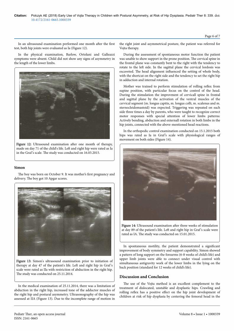
In an ultrasound examination performed one month after the firsttest, both hip joints were evaluated as Ia (Figure 12).
In the physical examination, Barlow, Ortolani and Galleazzisymptoms were absent. Child did not show any signs of asymmetry inthe length of the lower limbs.
Figure 12: Ultrasound examination after one month of therapy,made on day 71 of the child's life. Left and right hip were rated as Iain the Graf ’s scale. The study was conducted on 16.03.2015.
SimonThe boy was born on October 9. It was mother’s first pregnancy and
delivery. The boy got 10 Apgar scores.
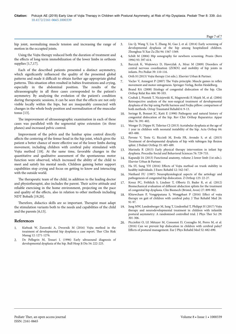
Figure 13: Simon's ultrasound examination prior to initiation oftherapy at day 47 of the patient's life. Left and right hip in Graf 'sscale were rated as IIa with restriction of abduction in the right hip.The study was conducted on 25.11.2014.
In the medical examination of 25.11.2014, there was a limitation ofabduction in the right hip, increased tone of the adductor muscles ofthe right hip and postural asymmetry. Ultrasonography of the hip wasassessed at IIA (Figure 13). Due to the incomplete range of motion in
the right joint and asymmetrical posture, the patient was referred forVojta therapy.
During the assessment of spontaneous motor function the patientwas unable to show support in the prone position. The cervical spine inthe frontal plane was constantly bent to the right with the tendency torotate to the left side. In the sagittal plane the cervical lordosis wasexcaveted. The head alignment influenced the setting of whole body,with the shortcut on the right side and the tendency to set the right hipin adduction and internal rotation.
Mother was trained to perform stimulation of rolling reflex fromsupine position, with particular focus on the control of the head.During the stimulation the improvment of cervicall spine in frontaland sagittal plane by the activation of the ventral muscles of thecervical segment (m. longus capitis, m. longus colli, m. scalenus and m.sternocleidomastoid) was expected. Triggering was repeated on eachside three times a day by parents, who were taught to recognize correctmotor responses with special attention of lower limbs patterns:Actively bending, abduction and externall rotation in both limbs in thehip joints, connected with the above-mentioned head reactions.
In the orthopedic control examination conducted on 15.1.2015 bothhips was rated as Ia in Graf ’s scale with physiological ranges ofmovement on both sides (Figure 14).
Figure 14: Ultrasound examination after three weeks of stimulationat day 89 of the patient's life. Left and right hip in Graf 's scale wererated as IA. The study was conducted on 15.01.2015.
In spontaneous motility, the patient demonstrated a significantimprovement of body symmetry and support capability. Simon showeda pattern of long support on the forearms (6-8 weeks of child’s life) andupper limb joints were able to connect under visual control withsimultaneous antigravity work of the lower limbs in the lying on theback position (standard for 12 weeks of child’s life).
Discussion and ConclusionThe use of the Vojta method is an excellent complement to the
treatment of dislocated, unstable and dysplastic hips. Crawling androlling reflex has a positive effect on the hip joint development ofchildren at risk of hip dysplasia by centering the femoral head in the
Citation: Polczyk AE (2018) Early Use of Vojta Therapy in Children with Postural Asymmetry, at Risk of Hip Dysplasia. Pediatr Ther 8: 339. doi:10.4172/2161-0665.1000339
Page 6 of 7
Pediatr Ther, an open access journalISSN: 2161-0665
Volume 8 • Issue 1 • 1000339
hip joint, normalizing muscle tension and increasing the range ofmotion in the occupied joints.
Using the Vojta therapy reduced both the duration of treatment andthe effects of long-term immobilization of the lower limbs in orthosissupplies [1,7,17].
Each of the described patients presented a distinct asymmetry,which significantly influenced the quality of the presented globalpatterns and made it difficult to obtain further age-appropriate globalpatterns. This situation often resulted in babies frustrations and crying,especially in the abdominal position. The results of theultrasonography in all three cases corresponded to the patient'sasymmetry. By analyzing the evolution of the patient's conditionduring therapeutic sessions, it can be seen that the effects are not onlyvisible locally within the hips, but are inseparably connected withchanges in the whole body position and normalization of the musculartonus [13].
The improvement of ultrasonographic examination in each of thesecases was paralleled with the segmental spine extension (in threeplanes) and increased pelvic control.
Improvment of the pelvis and the lumbar spine control directlyaffects the centering of the femur head in the hip joint, which gives thepatient a better chance of more effective use of the lower limbs duringmovement, including children with cerebral palsy stimulated withVojta method [18]. At the same time, favorable changes in thequantitative and qualitative assessment of the spontaneous motorfunction were observed, which increased the ability of the child tomeet and satisfy his mental needs. Children gaining better supportcapabilities stop crying and focus on getting to know and interactingwith the outside world.
The therapeutic team of the child, in addition to the leading doctorand physiotherapist, also includes the parent. Their active attitude andreliable exercising in the home environment, projecting on the paceand quality of the effects, also in relation to other methods includingNDT Bobath [19,20].
Therefore, didactics skills are so important. Therapist must adaptthe stimulation variants both to the needs and capabilities of the childand the parents [6,13].
References1. Kiebzak W, Żurawski A, Dwornik M (2016) Vojta method in the
treatment of developmental hip dysplasia-a case report. Ther Clin RiskManag 12: 1271-1276.
2. De Pellegrin M, Tessari L (1996) Early ultrasound diagnosis ofdevelopmental dysplasia of the hip. Bull Hosp Jt Dis 54: 222-225.
3. Liu Q, Wang Y, Liu Y, Zhang M, Gao J, et al. (2014) Early screening ofdevelopmental dysplasia of the hip among hospitalized children.Zhonghua Yi Xue Za Zhi 94: 1567-1569.
4. Schilt M (2004) Hip sonography for newborn screening. Praxis (Bern1994) 93: 597-614.
5. Barczyk K, Wojtowicz D, Hawrylak A, Mraz M (2009) Disorders ofcentral nervous coordination (ZOKN) and mobility of hip joints ininfants. Prz Pediat 39: 110-116.
6. Orth H (2013) Vojta therapy (1st edn.). Elsevier Urban & Partner.7. Vaclav V, Annegret P (2007) The Vojta principle: Muscle games in reflex
movement and motor ontogenesis. Springer-Verlag, Berlin Heidelberg.8. Brand RA (2008) Etiology of congenital dislocation of the hip. Clin
Orthop Relat Res 466: 90-103.9. Czubak J, Piontek T, Niciejewski K, Magnowski P, Majek M, et al. (2004)
Retrospective analysis of the non-surgical treatment of developmentaldysplasia of the hip using Pavlik harness and Frejka pillow: comparison ofboth methods. Ortop Traumatol Rehabil 6: 9-13.
10. Seringe R, Bonnet JC, Katti E (1990) Pathogeny and natural history ofcongenital dislocation of the hip. Rev Chir Orthop Reparatrice ApparMot 76: 391-402.
11. Wenger D, Düppe H, Tiderius CJ (2013) Acetabular dysplasia at the age of1 year in children with neonatal instability of the hip. Acta Orthop 84:483-488.
12. Pavone V, Testa G, Riccioli M, Evola FR, Avondo S, et al. (2015)Treatment of developmental dysplasia of hip with tubingen hip flexionsplint. J Pediatr Orthop 35: 485-489.
13. Marinela R (2013) Early physical therapy intervention in infant hipdysplasia. Procedia-Social and Behavioral Sciences 76: 729-733.
14. Kapandji IA (2013) Functional anatomy, volume 2-lower limb (1st edn.).Elsevier Urban & Partner.
15. Ha SY, Sung YH (2016) Effects of Vojta method on trunk stability inhealthy individuals. J Exerc Rehabil 12: 542-547.
16. Niethard FU (1987) Neurophysiological aspects of the aetiology andpathogenesis of congenital hip dislocation. Z Orthop 125: 22-27.
17. Kreuz PC, Fröhlich S, Lindner T, Olbertz D, Bader R, et al. (2012)Biomechanical evaluation of different abduction splints for the treatmentof congenital hip dysplasia. Clin Biomech (Bristol, Avon) 27: 899-902.
18. Khiewcham P, Vongpipatana S, Wongphaet P (2016) Effect of vojtatherapy on gait of children with cerebral palsy. J Thai Rehabil Med 26:91-97.
19. Jung MW, Landenberger M, Jung T, Lindenthal T, Philippi H (2017) Vojtatherapy and neurodevelopmental treatment in children with infantilepostural asymmetry: A randomised controlled trial. J Phys Ther Sci 29:301-306.
20. Picciolini O, LE Métayer M, Consonni D, Cozzaglio M, Porro M, et al.(2016) Can we prevent hip dislocation in children with cerebral palsy?Effects of postural management. Eur J Phys Rehabil Med 52: 682-690.
Citation: Polczyk AE (2018) Early Use of Vojta Therapy in Children with Postural Asymmetry, at Risk of Hip Dysplasia. Pediatr Ther 8: 339. doi:10.4172/2161-0665.1000339
Page 7 of 7
Pediatr Ther, an open access journalISSN: 2161-0665
Volume 8 • Issue 1 • 1000339