PEDIATRIC SURGERY HANDBOOK 2008surgery.uthscsa.edu/pediatric/training/handbook.pdf · PEDIATRIC...
34
PEDIATRIC SURGERY HANDBOOK 2008 San Antonio Pediatric Surgery Associates 4499 Medical Drive, Suite 347 San Antonio, Texas 78229 (210) 615-8757 Barry R. Cofer, M.D., F.A.C.S. John J. Doski, M.D., F.A.C.S. Frank M. Robertson, M.D., F.A.C.S. Joseph N. Kidd, Jr., M.D., F.A.C.S. Robert P. Thomas, M.D., F.A.C.S.
Transcript of PEDIATRIC SURGERY HANDBOOK 2008surgery.uthscsa.edu/pediatric/training/handbook.pdf · PEDIATRIC...
PEDIATRIC SURGERY HANDBOOK
2008
San Antonio Pediatric Surgery Associates 4499 Medical Drive, Suite 347
San Antonio, Texas 78229 (210) 615-8757
Barry R. Cofer, M.D., F.A.C.S. John J. Doski, M.D., F.A.C.S.
Frank M. Robertson, M.D., F.A.C.S. Joseph N. Kidd, Jr., M.D., F.A.C.S. Robert P. Thomas, M.D., F.A.C.S.
SAPSA Handbook 2006
2
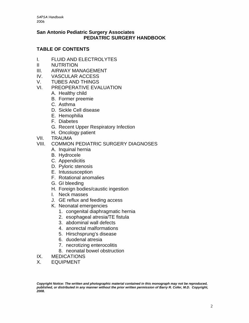
San Antonio Pediatric Surgery Associates PEDIATRIC SURGERY HANDBOOK
TABLE OF CONTENTS I. FLUID AND ELECTROLYTES II NUTRITION III. AIRWAY MANAGEMENT IV. VASCULAR ACCESS V. TUBES AND THINGS VI. PREOPERATIVE EVALUATION
A. Healthy child B. Former preemie C. Asthma D. Sickle Cell disease E. Hemophilia F. Diabetes G. Recent Upper Respiratory Infection H. Oncology patient
VII. TRAUMA VIII. COMMON PEDIATRIC SURGERY DIAGNOSES
A. Inquinal hernia B. Hydrocele C. Appendicitis D. Pyloric stenosis E. Intussusception F. Rotational anomalies G. GI bleeding H. Foreign bodies/caustic ingestion I. Neck masses J. GE reflux and feeding access K. Neonatal emergencies
1. congenital diaphragmatic hernia 2. esophageal atresia/TE fistula 3. abdominal wall defects 4. anorectal malformations 5. Hirschsprung’s disease 6. duodenal atresia 7. necrotizing enterocolitis 8. neonatal bowel obstruction
IX. MEDICATIONS X. EQUIPMENT Copyright Notice: The written and photographic material contained in this monograph may not be reproduced, published, or distributed in any manner without the prior written permission of Barry R. Cofer, M.D. Copyright, 2008.
SAPSA Handbook 2006
3
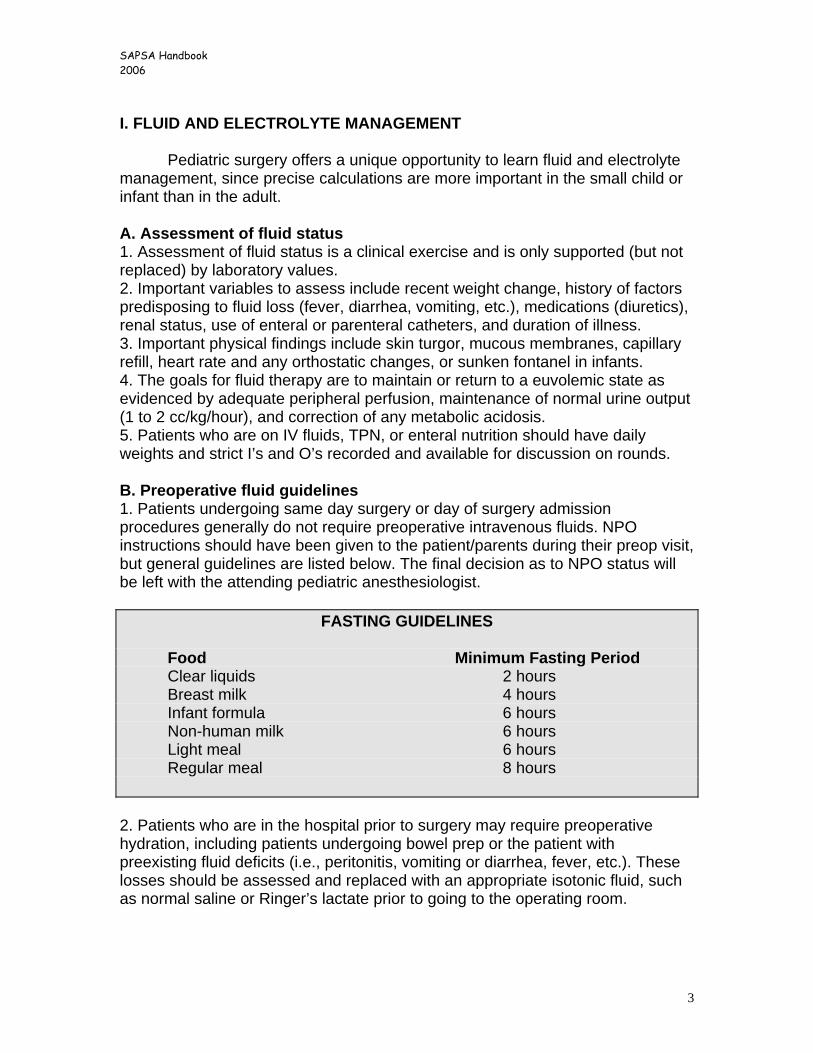
I. FLUID AND ELECTROLYTE MANAGEMENT Pediatric surgery offers a unique opportunity to learn fluid and electrolyte management, since precise calculations are more important in the small child or infant than in the adult. A. Assessment of fluid status 1. Assessment of fluid status is a clinical exercise and is only supported (but not replaced) by laboratory values. 2. Important variables to assess include recent weight change, history of factors predisposing to fluid loss (fever, diarrhea, vomiting, etc.), medications (diuretics), renal status, use of enteral or parenteral catheters, and duration of illness. 3. Important physical findings include skin turgor, mucous membranes, capillary refill, heart rate and any orthostatic changes, or sunken fontanel in infants. 4. The goals for fluid therapy are to maintain or return to a euvolemic state as evidenced by adequate peripheral perfusion, maintenance of normal urine output (1 to 2 cc/kg/hour), and correction of any metabolic acidosis. 5. Patients who are on IV fluids, TPN, or enteral nutrition should have daily weights and strict I’s and O’s recorded and available for discussion on rounds. B. Preoperative fluid guidelines 1. Patients undergoing same day surgery or day of surgery admission procedures generally do not require preoperative intravenous fluids. NPO instructions should have been given to the patient/parents during their preop visit, but general guidelines are listed below. The final decision as to NPO status will be left with the attending pediatric anesthesiologist.
FASTING GUIDELINES
Food Minimum Fasting Period Clear liquids 2 hours Breast milk 4 hours Infant formula 6 hours Non-human milk 6 hours Light meal 6 hours Regular meal 8 hours
2. Patients who are in the hospital prior to surgery may require preoperative hydration, including patients undergoing bowel prep or the patient with preexisting fluid deficits (i.e., peritonitis, vomiting or diarrhea, fever, etc.). These losses should be assessed and replaced with an appropriate isotonic fluid, such as normal saline or Ringer’s lactate prior to going to the operating room.
SAPSA Handbook 2006
4
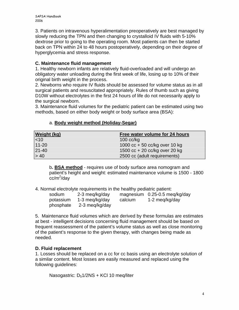
3. Patients on intravenous hyperalimentation preoperatively are best managed by slowly reducing the TPN and then changing to crystalloid IV fluids with 5-10% dextrose prior to going to the operating room. Most patients can then be started back on TPN within 24 to 48 hours postoperatively, depending on their degree of hyperglycemia and stress response. C. Maintenance fluid management 1. Healthy newborn infants are relatively fluid-overloaded and will undergo an obligatory water unloading during the first week of life, losing up to 10% of their original birth weight in the process. 2. Newborns who require IV fluids should be assessed for volume status as in all surgical patients and resuscitated appropriately. Rules of thumb such as giving D10W without electrolytes in the first 24 hours of life do not necessarily apply to the surgical newborn. 3. Maintenance fluid volumes for the pediatric patient can be estimated using two methods, based on either body weight or body surface area (BSA):
a. Body weight method (Holiday-Segar) Weight (kg) Free water volume for 24 hours <10 100 cc/kg 11-20 1000 cc + 50 cc/kg over 10 kg 21-40 1500 cc + 20 cc/kg over 20 kg > 40 2500 cc (adult requirements)
b. BSA method - requires use of body surface area nomogram and patient’s height and weight: estimated maintenance volume is 1500 - 1800 cc/m2/day
4. Normal electrolyte requirements in the healthy pediatric patient: sodium 2-3 meq/kg/day magnesium 0.25-0.5 meq/kg/day potassium 1-3 meq/kg/day calcium 1-2 meq/kg/day phosphate 2-3 meq/kg/day 5. Maintenance fluid volumes which are derived by these formulas are estimates at best - intelligent decisions concerning fluid management should be based on frequent reassessment of the patient’s volume status as well as close monitoring of the patient’s response to the given therapy, with changes being made as needed. D. Fluid replacement 1. Losses should be replaced on a cc for cc basis using an electrolyte solution of a similar content. Most losses are easily measured and replaced using the following guidelines: Nasogastric: D51/2NS + KCl 10 meq/liter

SAPSA Handbook 2006
5
Bile and pancreatic: D5 LR + added sodium bicarbonate (as needed) Ileostomy: D51/2NS + KCl 20 meq/liter + bicarbonate (as needed)
For relatively minor losses in patients with normal kidneys, simple volume replacement with an isotonic fluid will suffice as long as the patient’s electrolytes remain normal. 2. Massive ongoing losses will require analysis of the electrolyte content of the fluid lost, with replacement of a similar fluid on a volume for volume basis. 3. Replacement of intraoperative fluids should consider the amount of measured losses as well as unmeasured losses. For example, unmeasured (insensible) volume loss during laparotomy can be estimated at 10-15 cc/kg/hour of operative time, and should have been included in the intraoperative fluids given. 4. The most important aspect of fluid and electrolyte replacement and fluid resuscitation is continuous clinical reassessment and adjustment of replacement fluids as needed. II. NUTRITIONAL MANAGEMENT OF THE PEDIATRIC SURGICAL PATIENT Fortunately, most pediatric surgical patients are otherwise healthy patients in good nutritional condition with non-emergent disorders. These patients rarely require parenteral or enteral nutrition support. However, a number of conditions exist where total or partial enteral and/or parenteral support is necessary. A. Nutritional requirements 1. Calories - varies with age and physical condition. Requirements are proportionally higher than adult requirements due to the simultaneous needs of weight maintenance and growth as well as higher rates of heat loss. Age (years) Caloric needs 0-1 90-120 kcal/kg/day 1-7 75-90 kcal/kg/day 7-12 60-75 kcal/kg/day 12-18 30-60 kcal/kg/day > 18 25-35kcal/kg/day 2. Protein requirements (should not be more than 15% of total caloric intake, but ordinarily not used in calculating overall caloric needs) Age (years) Protein needs 0-1 2.5-3.5 gm/kg/day 1-7 2-2.5 gm/kg/day 7-12 2 gm/kg/day 12-18 1.5 gm/kg/day > 18 1 gm/kg/day
SAPSA Handbook 2006
6
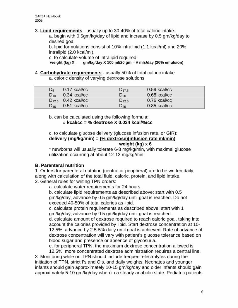
3. Lipid requirements - usually up to 30-40% of total caloric intake. a. begin with 0.5gm/kg/day of lipid and increase by 0.5 gm/kg/day to desired goal b. lipid formulations consist of 10% intralipid (1.1 kcal/ml) and 20% intralipid (2.0 kcal/ml). c. to calculate volume of intralipid required: weight (kg) X ___ gm/kg/day X 100 ml/20 gm = # mls/day (20% emulsion)
4. Carbohydrate requirements - usually 50% of total caloric intake a. caloric density of varying dextrose solutions D5 0.17 kcal/cc D17.5 0.59 kcal/cc D10 0.34 kcal/cc D20 0.68 kcal/cc D12.5 0.42 kcal/cc D22.5 0.76 kcal/cc D15 0.51 kcal/cc D25 0.85 kcal/cc b. can be calculated using the following formula:
# kcal/cc = % dextrose X 0.034 kcal/%/cc c. to calculate glucose delivery (glucose infusion rate, or GIR): delivery (mg/kg/min) = (% dextrose)(infusion rate ml/min) weight (kg) x 6
* newborns will usually tolerate 6-8 mg/kg/min, with maximal glucose utilization occurring at about 12-13 mg/kg/min.
B. Parenteral nutrition 1. Orders for parenteral nutrition (central or peripheral) are to be written daily, along with calculation of the total fluid, caloric, protein, and lipid intake. 2. General rules for writing TPN orders:
a. calculate water requirements for 24 hours. b. calculate lipid requirements as described above; start with 0.5 gm/kg/day, advance by 0.5 gm/kg/day until goal is reached. Do not exceeed 40-50% of total calories as lipid. c. calculate protein requirements as described above; start with 1 gm/kg/day, advance by 0.5 gm/kg/day until goal is reached. d. calculate amount of dextrose required to reach caloric goal, taking into account the calories provided by lipid. Start dextrose concentration at 10-12.5%, advance by 2.5-5% daily until goal is achieved. Rate of advance of dextrose concentration will vary with patient’s glucose tolerance based on blood sugar and presence or absence of glycosuria. e. for peripheral TPN, the maximum dextrose concentration allowed is 12.5%; more concentrated dextrose administration requires a central line.
3. Monitoring while on TPN should include frequent electrolytes during the initiation of TPN, strict I’s and O’s, and daily weights. Neonates and younger infants should gain approximately 10-15 gm/kg/day and older infants should gain approximately 5-10 gm/kg/day when in a steady anabolic state. Pediatric patients
SAPSA Handbook 2006
7
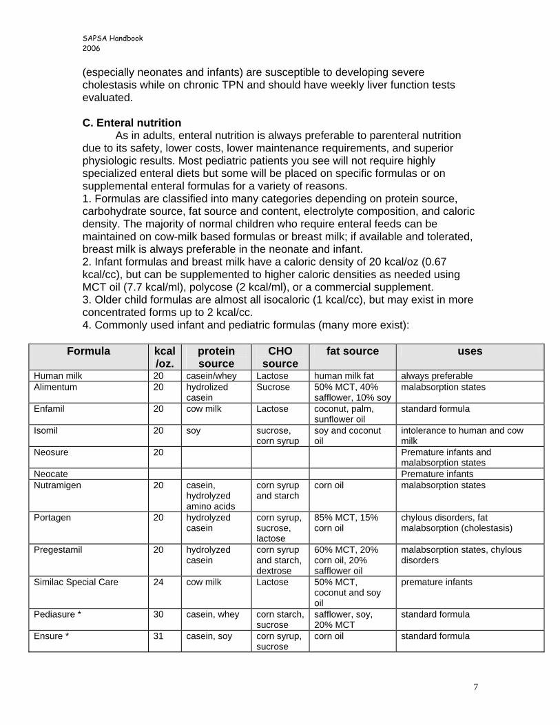
(especially neonates and infants) are susceptible to developing severe cholestasis while on chronic TPN and should have weekly liver function tests evaluated. C. Enteral nutrition As in adults, enteral nutrition is always preferable to parenteral nutrition due to its safety, lower costs, lower maintenance requirements, and superior physiologic results. Most pediatric patients you see will not require highly specialized enteral diets but some will be placed on specific formulas or on supplemental enteral formulas for a variety of reasons. 1. Formulas are classified into many categories depending on protein source, carbohydrate source, fat source and content, electrolyte composition, and caloric density. The majority of normal children who require enteral feeds can be maintained on cow-milk based formulas or breast milk; if available and tolerated, breast milk is always preferable in the neonate and infant. 2. Infant formulas and breast milk have a caloric density of 20 kcal/oz (0.67 kcal/cc), but can be supplemented to higher caloric densities as needed using MCT oil (7.7 kcal/ml), polycose (2 kcal/ml), or a commercial supplement. 3. Older child formulas are almost all isocaloric (1 kcal/cc), but may exist in more concentrated forms up to 2 kcal/cc. 4. Commonly used infant and pediatric formulas (many more exist):
Formula kcal/oz.
protein source
CHO source
fat source uses
Human milk 20 casein/whey Lactose human milk fat always preferable Alimentum 20 hydrolized
casein Sucrose 50% MCT, 40%
safflower, 10% soymalabsorption states
Enfamil 20 cow milk Lactose coconut, palm, sunflower oil
standard formula
Isomil 20 soy sucrose, corn syrup
soy and coconut oil
intolerance to human and cow milk
Neosure
20 Premature infants and malabsorption states
Neocate Premature infants Nutramigen 20 casein,
hydrolyzed amino acids
corn syrup and starch
corn oil malabsorption states
Portagen 20 hydrolyzed casein
corn syrup, sucrose, lactose
85% MCT, 15% corn oil
chylous disorders, fat malabsorption (cholestasis)
Pregestamil 20 hydrolyzed casein
corn syrup and starch, dextrose
60% MCT, 20% corn oil, 20% safflower oil
malabsorption states, chylous disorders
Similac Special Care 24 cow milk Lactose 50% MCT, coconut and soy oil
premature infants
Pediasure * 30 casein, whey corn starch, sucrose
safflower, soy, 20% MCT
standard formula
Ensure * 31 casein, soy corn syrup, sucrose
corn oil standard formula

SAPSA Handbook 2006
8
Isocal * 32 casein, soy Malt soy oil, MCT standard formula
* non-infant formulas
SAPSA Handbook 2006
9
III. AIRWAY MANAGEMENT
A. Loss of airway is the most common cause of cardiac arrest in the child. B. Most airway problems can be managed by positioning, suctioning, and
use of humidified oxygen. C. Orotracheal intubation should be considered for the child with
significant respiratory distress, severe closed head injury, or severe multisystem trauma.
D. Intubation similar as for adults with a few exceptions: 1. a Miller (straight) blade should be used on most children. 2. ET tubes for children < 6 years of age are generally uncuffed. 3. A guestimate for tube size is (16 + age)/4.
E. Caveats 1. it is very easy to intubate the right mainstem bronchus. 2. If you can’t intubate the child, you can almost always bag-mask
ventilate them until help arrives (remember to decompress the stomach).
3. breath sounds can be deceptive; always get a chest xray. 4. a right mainstem intubation may result in collapse of the left lung
and mimic a left hemothorax or a right pneumothorax; always check a chest xray for proper tube position prior to placing a chest tube in this setting.
5. a distended stomach will compress the diaphragm and impede ventilation; always decompress the stomach with a gastric tube.
6. very prone to tension pneumothorax or hemothorax – aggressively decompress the thorax where confirmed to be needed.
7. cricothyroidotomy to be avoided in children < 12 years old; use emergent tracheostomy instead.
F. Useful drugs 1. Atropine – 0.01 – 0.02 mg/kg (min. 0.1 mg, max. 1 mg.). 2. Lidocaine – 1-2 mg/kg. 3. Midazolam – 0.05-0.1 mg/kg. 4. Fentanyl – 1-5 mcg/kg. 5. Vecuronium – 0.1 mg/kg. 6. Thiopental 1-2 mg/kg
IV. VASCULAR ACCESS
A. Indications for vascular access: 1. trauma 2. hyperalimentation 3. longterm antibiotics 4. chemotherapy 5. frequent blood draws or infusions (sickle cell disease, hemophilia).
B. Equipment (see equipment charts) 1. peripheral IV’s- hands, feet, external jugular vein, scalp vein
SAPSA Handbook 2006
10
2. temporary central lines – subclavian, internal jugular, femoral. Usually use Cook polyurethrane catheters, available in the OR anesthesia workroom, PICU.
3. permanent tunneled catheters or ports 4. intraosseous needles – available in the ER, anesthesia carts. Can
also use Jamshidi bone marrow needle if commercial needle not available.
C. Placement 1. placement of tunneled catheters or ports requires general
anesthesia in the majority of pediatric patients. 2. removal of ports requires general anesthesia; removal of tunneled
catheters can be done under conscious sedation. 3. placement of temporary lines can be done under conscious
sedation in the ICU. 4. placement techniques essentially the same as for larger patients;
just remember that all distances are shorter (distance from skin to vein, from entrance site to heart, etc.)
D. Emergency access – it is best to have a well-thought out plan before facing this scenario! 1. intraosseus catheter – placed in uninjured tibia; can draw blood,
administer all medications and blood products, bolus fluids. Should always seek additional access sites when used.
2. venous cutdown – best sites are the saphenous vein at the ankle (older kids), saphenofemoral junction (younger kids and infants), medial antecubital veins, external jugular vein. Usually place large bore angiocath initially, can be converted to more permanent line at later time.
3. percutaneous access – best place is in the groin; emergency subclavian placement associated with increased complications.
E. Broviac catheters – tunneled catheter of choice for children requiring long term venous access. 1. sizes: single lumen – 3.0, 4.2, 5.0, 6.6, 9.6 French
double lumen – 5.0, 7.0, 9.0 French triple lumen (bone marrow txp) – 8.0, 12.0 French
2. placed by cutdown or percutaneous insertion 3. heparinized with 100u/cc solution (don’t overbolus) 4. clotted catheters and ports
a. check position on xray b. attempt to aspirate and flush yourself (don’t take anyone’s word
for it). c. ports should be reaccessed by yourself with a new Huber
needle assembly. d. if clotted, infuse streptokinase per protocol (available from
pharmacy). e. If not successful, attempt second round of streptokinase with
dwell time of 2 to 3 hours.
SAPSA Handbook 2006
11
5. broken catheters a. repair kits are available for all broviac sizes > 4.2 Fr. b. repair is a sterile procedure; seek help if you have never
performed a repair before (written instructions come with the kit).
c. should not be used for 24 hours after repair. 6. infected catheters or ports
a. suspect in any child with fevers and existing catheter. b. confirmed by positive blood cultures through catheter. c. can be successfully treated with antibiotics through catheter (gm
pos ~ 90%; gm neg ~ 50%; fungi ~ 10%). d. catheter should be removed if:
- persistant positive blood cultures despite adequate antibiotics
- tunnel infection - fungal infection present - child presents with septic shock
e. new catheter should not be placed until documented to have negative blood cultures.
f. exit site infections (tunnel infections) usually require removal of the catheter.
V. TUBES AND THINGS
A. Nasogastric tubes 1. used for gastric decompression; routine use after laparotomy or
bowel resection is becoming less common. 2. sizes: use common sense. For small infants (< 5 kg), use 8Fr or
10Fr Replogle sump tube. 3. premature/very small infants- use orogastric route to prevent airway
complications, since babies this age are obligate nose breathers. 4. sump tubes are meant to be placed to continuous, not intermittent,
suction. B. Gastrostomy tubes
1. used for feeding or gastric decompression (or both). 2. can be surgically placed (Stamm) or endoscopically placed (PEG). 3. primary Gtubes usually consist of a MIC-gastrostomy tube, which is
a balloon-type catheter. Others include a DePezzar or Malecot catheter, which have a more solid but deformable end.
4. gastrostomy buttons are frequently used in children; most are replacement devices for existing tubes, although primary buttons will be placed occasionally. a. conversion to a button can usually be performed approximately
6 weeks after the gastrostomy was created. b. We use one type of gastrostomy button – Mic-Key (available
from central supply).
SAPSA Handbook 2006
12
5. displaced gtubes must be replaced quickly or the gastrostomy tract will close. a. if the tract has not contracted, replace with a foley catheter, MIC
tube, or button of the same size. b. if the tract has contracted, gently dilate the tract with
successively larger red rubber catheters until the appropriate size is reached, then replace with a foley or button device. Do not use metal dilators to enlarge the tract!
c. a water-soluble contrast study should be obtained if there is any question about the position of the catheter, or if this is the first time a new gastrostomy has been changed (mandatory).
C. Urethral catheters 1. not routinely used in children, including trauma patients. 2. size will vary with the age and gender of the child 3. for premature infants, should place 5 Fr feeding tube and tape to
the thigh. 4. for term infants, the smallest balloon type catheter available is 6 Fr. 5. always confirm good urine flow prior to inflating the balloon.
D. Chest tubes 1. same indications for placement as in adults. 2. size will vary upon indications and size of child.
< 3 kg 10-12 Fr 3-10 kg 12-16 Fr 11-15 kg 16-20 Fr 16-25 kg 20-24 Fr 26-40 kg 24-28 Fr
3. simple pneumothoraces can be treated with an 8.5 Fr
pleural/pericardial drain (uses the Seldinger technique). 4. technique for placement similar to adults; thoracic cavity smaller,
easier to injury intrathoracic organs; trocar chest tubes are never to be used in pediatric patients.
5. always use sufficient sedation. VI. PREOPERATIVE EVALUATION
A. The healthy child – most children undergoing elective surgery do not need routine laboratory or radiologic evaluations unless specific for their disease state.
B. Former preemie – anticipate problems with airway, temperature instability, venous access. Infants less than 45 to 50 weeks gestation who undergo general anesthesia will require admission overnight with continuous apnea/bradycardia monitoring (requires PICU or PIMC bed status).
C. Asthma – children with asthma are at increased risk of bronchospasm and respiratory infections after general anesthesia. Children who are actively wheezing should have their procedure cancelled if possible until they have
SAPSA Handbook 2006
13
cleared their wheezing for several weeks. In general, children who must be operated upon should have their routine treatment accelerated around the time of surgery: 1. on prn inhalers: use inhalers 3 to 4 times/day for 3 days preoperatively
and postoperatively. 2. on scheduled inhalers: perioperative oral prednisone and scheduled
inhalers; should have pulmonologist involved. 3. on oral steroids: admit to hospital, place on IV solumedrol for 24 hours
before and after surgery, continue on prednisone taper and scheduled inhalers afterwards; should have pulmonologist involved.
D. Sickle Cell Anemia – are at very high risk for perioperative sickling crises. Many patients will require transfusion or exchange therapy prior to surgery to achieve a total Hgb. > 10gm/dl. Other important perioperative tasks: 1. vigorous hydration (admit night before surgery for elective cases).
Many “sicklers” have isosthenuria (inability to concentrate urine due to tubular injury); an adequate urine output may not necessarily indicate an adequate intravascular volume.
2. keep warm – heat OR, use warming blankets, warmed fluids. 3. keep well oxygenated. 4. provide adequate pain control postoperatively. 5. vigorous pulmonary toilet and early mobilization. Complications occur in 20-30% of patients in some series, with acute
chest syndrome being the most serious and potentially fatal. It is vitally important to be very compulsive in the care of these patients perioperatively. E. Hemophilia – Hemophilia A and B (loss of factors 8 and 9) are the most
common coagulation disorders in children. These children will generally have very low levels of factors (< 1%) which need to be replaced to approximately 100% activity preoperatively and continued postoperatively. These patients are managed very carefully by the pediatric hematology service.
F. Type I diabetes mellitus- management of these patients in the perioperative period may require an alteration in their usual insulin dose or the use of a continuous insulin drip. When dealing with insulin-dependant diabetics, the goal is to prevent ketosis as well as hyperglycemia; these two conditions do not have to coexist, so blood sugars and serum or urine ketones should be followed closely. Do not try to achieve euglycemia- all perioperative patients are stressed and insulin resistant, and tight control of their diabetes is dangerous. 1. minor procedures, no NPO period postoperatively: patients should take
½ of their usual regular insulin dose on the morning of surgery; the anesthesiologist will provide dextrose in the IV fluids and monitor the blood sugar intraoperatively. Resume normal insulin regimen postoperatively.
2. major procedures, anticipated NPO period postoperatively: take ½ of insulin prior to surgery, intraoperative monitoring by anesthesia. Postoperatively, begin on regular insulin drip at 1 u/kg/hr, adjust using
SAPSA Handbook 2006
14
q1-2 hour blood sugar checks and frequent serum/urine ketone checks. Attempt to achieve blood sugar of 200-250 and no ketosis. Begin regular insulin sliding scale once diet starts.
G. Recent upper respiratory infection – children with a current or recent URI are at an increased risk for bronchospasm or respiratory infection after general anesthesia and should have their procedure canceled until resolved. If a child has a clear rhinorhea without purulunce, fever, or symptoms of a lower respiratory infection, then anesthesia is probably acceptable. If a child has had tracheobronchitis (croup) or RSV (respiratory syncitial virus) infection, then surgery and anesthesia should be cancelled for four to six weeks if feasible. Children with these conditions who require emergency surgery should be carefully monitored for respiratory complications postoperatively, and treated with aggressive pulmonary toilet and bronchodilators.
H. Oncology patient – many chemotherapeutic agents used in children will have adverse systemic effects, such as bone marrow suppression (multiple agents), cardiotoxicity (adriamycin), renal tubular disorders (carboplatin), and pulmonary toxicity (bleomycin). These children also have had multiple central lines, with the potential for venous thrombosis and obstruction, leading to difficulties with central venous access. Children who have had adriamycin will require a relatively recent echocardiogram to assess myocardial function prior to general anesthesia.
VII. TRAUMA
Trauma is the leading cause of childhood morbidity and mortality, accounting for more deaths than the next 9 causes of death combined. The majority of pediatric trauma in this country is treated by non-pediatric specialists, so it is vitally important that the general surgeon be comfortable with the initial management and resuscitation of the injured child. This section will discuss the basic principles but not specifics of trauma care in the pediatric population.
A. Characteristics of the Pediatric Trauma Victim 1. Disproportionate head size; 90% of adult intracranial volume
reached by 8 years of age. Center of gravity higher, making cranial injuries more likely.
2. Smaller, more anterior airway, leading to difficulties with airway management. More narrow airway leading to higher incidence of airway obstruction. Always suspect airway obstruction in the child with cardiac arrest!
3. Compliant thoracic cage, less protective of lungs, mediastinum, liver, spleen, kidneys. Less likely to have rib fractures with significant intrathoracic injury.
4. Intraabdominal and intrathoracic injuries more likely to involve multiple organs due to distribution of blunt force over relatively large surface area compared to small musculoskeletal mass.
5. Propensity for heat loss and early hypothermia.
SAPSA Handbook 2006
15
6. Response to blood loss by increase in heart rate and systemic vascular resistance; hypotension is late finding, not seen until > 30-40% blood volume lost. More reliable physical sign of blood volume loss is tachycardia and decreased peripheral perfusion.
B. Resuscitation – should follow basic tenets of ATLS system. 1. Airway: smaller and more difficult to intubate. Less than 6 yrs of
age use uncuffed tube. Surgical cricothyroidotomy contraindicated; better to use needle cricothyroidotomy or emergent tracheostomy. Estimate of ET tube size: (16 + age)/4.
2. Breathing: younger the child, faster the normal breathing rate. More susceptible to mediastinal shift and tension pneumothorax. Ventilation easily impaired by gastric distension, treated with OG tube.
3. Circulation: best estimate of hemodynamic status is peripheral perfusion (cap refill, pedal pulses, heart rate). Vascular access can be difficult; need to have well thought out algorithm before facing this problem. Cutdowns at ankle (older children) or at groin (infants/younger children) may be useful, as is use of intraosseus needle in uninjured tibia. Blood volume decreases with age: 80-90cc/kg for infants, to 70cc/kg for older children and adults.
4. Disability (neuro): be aware of age specific changes in verbal component of Glasgow Coma Scale.
5. Exposure: hypothermia common; important to use warmed fluids, warm resuscitation room, heating lamps, etc.
C. Management of systems injuries 1. Head: present in 50% of trauma, responsible for 70% of trauma
deaths. Most common finding is diffuse axonal injury; less commonly see mass lesions or hematoma. Frequently compounded by anoxia or hypotension.
2. C-spine: site of injury more cephalad than in adults, usually upper 3 segments. Be aware of high incidence of pseudosubluxation of upper two cervical segments. Higher incidence of SCIWORA (spinal cord injury without radiographic abnormalities), and injury cannot be ruled out in lieu of normal exam.
3. Thoracic: if present, associated with extrathoracic injuries in > 80% of cases. Most common injury is pulmonary contusion. Rib fractures rare. More prone to tension physiology due to mobile mediastinum. Indications for thoracotomy include initial hemothorax of >20cc/kg or ongoing blood loss of > 2cc/kg/hr.
4. Abdominal: present in over 5-10% of all children presenting to level I trauma center. Abdominal injury and persistent hemodynamic instability requires laparotomy, not DPL or radiographic scanning. Trauma ultrasound not believed to be useful by most pediatric traumatologists.
SAPSA Handbook 2006
16
i. Spleen: most commonly injured organ. Nonoperative therapy successful in > 95% of cases. Angiographic embolization is rarely indicated.
ii. Liver: suspected with increased transaminases and appropriate mechanism. Management almost always nonoperative.
iii. Pancreas: common injury due to relative lack of protective retroperitoneal tissue. Frequently missed on initial exam, suggested by persistent abdominal pain, rising amylase/lipase, “unexplained” intraperitoneal fluid on CT scan. Treatment is by spleen-preserving distal pancreatectomy.
iv. Bowel: more commonly seen than in adults. “Lap-belt” complex consists of seatbelt bruise, bowel perforation, and hyperflexion (“Chance”) fracture of lumbar vertebra. Frequently delayed in diagnosis.
v. GU: kidney is most commonly injured GU organ in children. Usually consists of parenchymal injuries +/- urinary extravasation, usually responds to conservative management. Intraperitoneal bladder rupture more common than extraperitoneal rupture. Urethral injuries can be seen as in adult. Bladder volume estimated as (cc’s) [2+age] X 30.
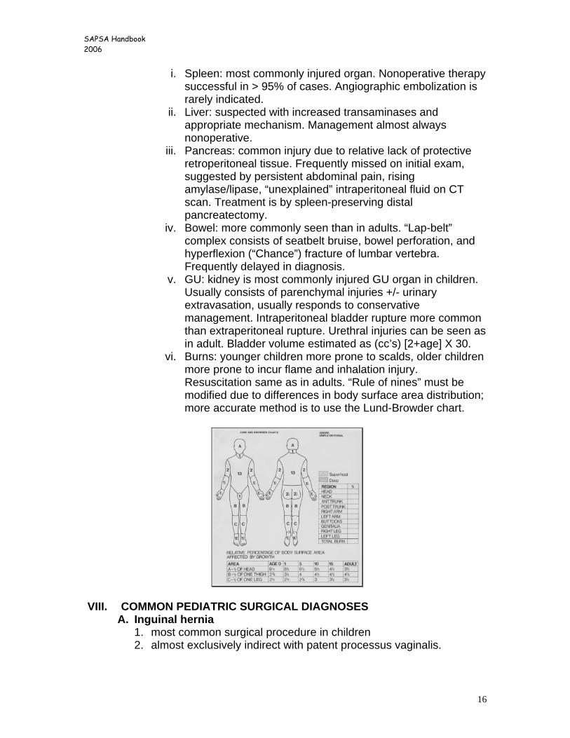
vi. Burns: younger children more prone to scalds, older children more prone to incur flame and inhalation injury. Resuscitation same as in adults. “Rule of nines” must be modified due to differences in body surface area distribution; more accurate method is to use the Lund-Browder chart.
VIII. COMMON PEDIATRIC SURGICAL DIAGNOSES
A. Inguinal hernia 1. most common surgical procedure in children 2. almost exclusively indirect with patent processus vaginalis.
SAPSA Handbook 2006
17
3. more frequent in males, infants, former preemies, children with abdominal wall defects or VP shunts.
4. most cause asymptomatic mass; more likely to become incarcerated in infants and younger children.
5. almost always repaired soon after diagnosis, usually by high ligation of sac; prosthetic repair rarely needed.
6. incarcerated hernias which can be reduced should be scheduled for surgery on the next day.
B. Hydrocele 1. hydroceles are very common in newborn males; most do not
require surgery. 2. majority of noncomplicated hydroceles will resolve by 18 months of
age and should be observed. 3. if hydrocele fluctuates in size, suggests “communicating” type
which is associated with a patent processus vaginalis; these should be treated as hernias and repaired when diagnosed.
4. an occasional “acute” hydrocele in an infant can be confused with an incarcerated inguinal hernia. If in doubt, treat as an incarcerated hernia (ultrasound may help to differentiate hydrocele from hernia).
5. acute hydroceles in the older child should be examined carefully, as 25% of testicular tumors will present with a new hydrocele. Other possibilities include missed hernia, infection, or trauma. These should be examined by scrotal ultrasound.
C. Appendicitis 1. extremely common pediatric disorder 2. usually presents with vague abdominal pain, nausea, low grade
fevers (early); advances to focal or generalized severe abdominal pain, peritonitis (later). Patients with advanced appendicitis may be very toxic.
3. Usually perforated or gangrenous in younger patients. 4. Must consider “nonsurgical” causes of acute abdomen in differential
diagnosis: basilar pneumonia, gastroenteritis, mesenteric adenitis, diabetic ketoacidosis, Henoch-Schoenlien purpura, etc.
5. Evaluation: sine qua non is acute, focal, right lower quadrant pain; may be more difficult to diagnosis with retrocecal appendicitis, younger patients, obese patients. Many patients complain of pain with movement or coughing. May have diarrhea, especially if advanced.
6. Appendicitis is still a clinical diagnosis in the majority of patients, and most cases can be diagnosed by a good physical examination!
7. Imaging: CT scan is most accurate study for those cases where exam is difficult or inconclusive. Findings may include calcified appendicolith, enlarged appendix, pericecal fat stranding or inflammatory changes.
8. Laboratory: always need to have CBC with differential, urinalysis, and B-HCG in females. WBC usually mildly elevated, but may be
SAPSA Handbook 2006
18
totally normal. Markedly elevated WBC uncommon unless advanced disease.
9. Treatment: a. preoperative fluid resuscitation and fever reduction b. operative appendectomy (open or laparoscopic),drainage of any
loculated fluid collections, peritoneal irrigation. Drains usually not used unless established abscess cavity encountered. Routine cultures not necessary or helpful.
c. Antibiotics 1. uncomplicated: ampicillin/sulbactam (Unasyn) or
ticarcillin/tazobactam (Zosyn) preop and 1-2 doses postop. 2. Complicated: ampicillin (50mg/kg/dose every 6hrs);
clindamycin (10mg/kg/dose every 6 hrs); and gentamycin (2.5mg/kg/dose every 8 hrs, adjust to levels) for minimum of 5 days postoperatively. At POD #5, patient may go home if recovered, if no fevers for > 24 hours, and if WBC with manual differential count is normal.
d. no need for routine NG tube, central line, foley catheter, or PICU admission after removal of ruptured or gangrenous appendix; base instead on clinical condition.
D. Pyloric stenosis
1. occurs in approximately 1 in 300 newborn infants. 2. classically occurs in otherwise healthy infant from 4 to 8 weeks of
age. 3. presents with insidious onset of non-bilious emesis, usually
consists of undigested formula. Emesis becomes more frequent and forceful with time (“projectile vomiting”).
4. many patients will present with weight loss, dehydration, and electrolyte disturbances (classic contraction metabolic alkalosis).
5. diagnosis made by suspicious history; palpation of pyloric “olive” in midepigastrium to right upper quadrant.
6. if suspicious history and pylorus cannot be palpated, proceed to pyloric ultrasound. If low suspicion but symptoms persist, consider barium upper GI series.
7. treatment: a. if normal hydration/electrolyte status:
i. place IV, make NPO. ii. begin fluids: D5W1/4NS + KCl 10 meq/l @ 150%
maintenance rate (if certain patient has voided); proceed to surgery.
b. if dehydrated/alkalotic: i. bolus with 10-20 cc/kg normal saline ii. begin fluids: D5W1/2NS at 150% maintenance rate; add
KCl 10-20 meq/l only after urine output established.
SAPSA Handbook 2006
19
iii. may proceed to surgery once child is hydrated and alkalosis resolved (serum Cl > 90 and/or serum HCO3 < 28, adequate urine output).
8. surgical treatment of choice is Fredet-Ramstedt pyloromyotomy via umbilical incision.
9. postoperative care: a. NO NARCOTICS; use acetaminophen for pain. b. IV fluids at maintenance rate, apnea/bradycardia monitor. c. NG tube not needed in most patients. d. begin po fluids 2 hours postop as follows:
Formula ½ ounce po every 3 hours X 2, then Formula 1 ounce po every 3 hours X 2, then Formula 1.5 ounces po every 3 hours X 2, then Formula 2 ounces po every 3 hours
e. heplock IV when tolerating 1.5 ounces. f. most infants can be discharged to home when tolerating 2
ounces of formula. g. expect children to have some emesis perioperatively, but if
maintaining hydration can proceed with above protocol. If doesn’t tolerate an increase, go back to last successful volume. Most children can go home within 24 hours.
E. Intussusception
1. “idiopathic” in majority of children, most commonly seen in 6 month to 2 year age range.
2. usually present with acute, cramping abdominal pain which alternates with normal or “sedate” periods. Frequently have progressive emesis, sometimes low grade fever.
3. physical findings included abdominal distension; occasionally find right upper quadrant mass and bloody (“currant jelly”) stools due to mucosal bleeding.
4. diagnosis: must have high degree of suspicion! Any child who has any suggestion of intussusception should be evaluated immediately.
5. Radiographic studies: the most common radiographic finding is a nonspecific abdominal film; “normal” KUB does not exclude the presence of an intussusception!
6. if intussusception suspected, the diagnostic and therapeutic test of choice is a controlled air- or barium-contrast enema. This is still the “gold standard” for diagnosis, and in over 85-90% of patients the intussusception can be reduced radiographically, negating the need for surgery.
7. treatment:
SAPSA Handbook 2006
20
a. Prior to diagnosis: hydrate, proceed to contrast enema if no peritoneal findings; if peritonitis present, proceed to exploratory laparotomy.
b. Successful radiographic reduction: admit, keep NPO overnight, and begin diet when abdomen soft and child appears recovered.
c. Unsuccessful radiographic reduction: proceed to laparotomy. Small right lower quadrant incision and delivery of bowel. Most of the time can be manually reduced, but if bowel necrotic or if irreducible manually, will generally require resection with primary anastomosis.
F. Rotational anomalies-“malrotation”
1. produces narrow mesenteric base; propensity for midgut volvulus (270o clockwise twist) with duodenal obstruction and mesenteric vascular occlusion.
2. ~ 50% become symptomatic in first six months of life. 3. infants present with bilious emesis due to duodenal obstruction.
BILIOUS EMESIS IN THE INFANT OR YOUNG CHILD IS A SURGICAL
EMERGENCY UNTIL PROVEN OTHERWISE!
4. older children may present with recurrent emesis, chronic abdominal pain, malabsorption, or failure to thrive.
5. diagnosis is made by upper GI series demonstrating a normal duodenal loop and the ligament of Treitz in the appropriate position (left of the vertebral column, same level as the duodenal bulb); lack of these is diagnostic of malrotation. May be suggested, but not confirmed, by an abnormally placed cecum on BE study.
6. the child who presents with acute abdomen, bloody stool, peritonitis, or cardiovascular collapse and who is suspected of having malrotation needs an emergent operation, not a contrast study.
7. treatment is by performing Ladd’s procedure (straightening of duodenum, lysis of Ladd’s bands, broadening of mesentery with placement of cecum in LUQ, and appendectomy).
8. children with “asymptomatic” malrotation should undergo surgery prophylactically.
9. 10% incidence of recurrent symptoms (usually from adhesive small bowel obstruction; recurrent volvulus rare).
G. GI bleeding 1. rarely exanguinating or life-threatening in children 2. may be grouped according to age at presentation:
a. newborns i. swallowed maternal blood (evaluated by the Apt test).
SAPSA Handbook 2006
21
ii. hemorrhagic disease of the newborn (treated with vitamin K) iii. anal fissure iv. necrotizing enterocolitis v. malrotation with midgut volvulus
b. infants i. anal fissure ii. intussusception iii. midgut volvulus iv. gastrointestinal duplications v. gastroenteritis or formula allergies vi. gastroesophageal reflux
c. toddlers and preschoolers i. anal fissure ii. rectal prolapse iii. gastoenteritis iv. Meckel’s diverticulum- evaluated by technitium-99 scan to
look for ectopic gastric mucosa v. Juvenile polyps or lymphoid nodular hyperplasia vi. gastroesophageal reflux vii. Henoch-Schoenlein purpura
d. older children i. gastrointestinal polyposis syndromes ii. inflammatory bowel disease iii. Meckel’s diverticulum iv. peptic ulcer disease v. Henoch-Schoenlein purpura
H. Foreign bodies/caustic ingestion 1. Tracheobronchial F.B. – most commonly found in right mainstem
bronchus; < 10% found above carina. a. requires high degree of suspicion; may have early episode of
coughing/wheezing, then become asymptomatic. b. should always obtain PA/Lat cxray, with expiratory films.
However, xray frequently normal and does not r/o aspiration. c. diagnosis and therapy by rigid bronchoscopy under general
anesthesia. d. after removal of FB, most patients kept in hospital overnight, +/-
steroids for airway edema. 2. Gastrointestinal F.B.
a. almost always asymptomatic unless lodged in the esophagus; most common location in normal esophagus is just proximal to cricopharyngeus – can frequently be seen with laryngoscope.
b. more likely to lodge in esophagus when underlying esophageal abnormality present (esophageal stricture, etc)
c. F.B. lodged in esophagus must be removed when diagnosed; have high incidence of esophageal erosion.
d. gastric FB almost always passed in the stool (95%).
SAPSA Handbook 2006
22
e. sharp FB which pass into the stomach may be observed carefully; should undergo laparotomy if symptoms develop or FB does not progress in reasonable time.
f. all swallowed batteries should be removed due to their propensity to produce bowel erosion and necrosis.
3. Caustic ingestion a. alkaline ingestion much more injurious than acid ingestions
(produces liquefaction necrosis). b. always pay attention to ABC’s first (airway edema and
hypovolemic shock not uncommon). c. do not induce vomiting! d. admit, NPO, IV antibiotics, chest xray; plan for flexible
esophagoscopy within 24 hours. e. no defined role for IV steroids. f. significant esophageal injury will require placement of
gastrostomy for feeding and access for esophageal dilations. g. longterm complications of stricture and malignant degeneration
may require esophageal replacement. h. rarely, massive ingestion produces esophageal/gastric necrosis,
requiring emergent resection. I. Neck masses
1. branchial remnants (cysts and sinuses) a. most commonly seen are 2nd branchial arch derivatives. b. cysts more common in older kids and adults; sinuses/fistulas more
common in younger children. c. usually located along anterior border of SCM muscle. d. If infected, will require I&D, antibiotics. e. definitive treatment by excision under general anesthesia as
outpatient. 2. thyroglossal duct remnants
a. occur due to persistent thyroid tissue along route of normal thyroid descent; may have dysplastic or malignant tissue.
b. duct may communicate with base of tongue (foramen cecum). c. appears as midline mass just below hyoid; may present as infected
cyst; rarely has external opening. d. treatment by excision, including body of hyoid bone (Sistrunk
procedure), as outpatient. Infected cysts should be drained and removed after inflammation resolves.
3. cystic hygroma a. abnormal collection of lymphatic vessels, usually in posterior triangle of
neck but may extend into mediastinum or originate in axilla. b. may enlarge after local infection or URI; occasionally large enough to
cause newborn airway obstruction requiring tracheostomy. c. treated by excision under general anesthesia.
4. cervical adenitis a. usually occurs secondary to bacterial pharyngitis.
SAPSA Handbook 2006
23
b. may produce abscess, requiring incision/drainage. c. cover with appropriate antibiotics (usual organism Staph or Strep spp.).
J. GE Reflux 1. very commonly seen in young infants and in children with devastating
neurologic conditions. 2. majority of neurologically normal children “outgrow” GER, do not require
therapy or surgery. 3. diagnosis may be suggested by UGI; definitive diagnosis by 24 hour pH
probe and/or by esophagoscopy showing inflammation. 4. medical management: avoid overfeeding; upright positioning; thickening
food (oatmeal, rice cereal); H2 blockers or PPI’s (ranitidine, omeprezole); promotility agents (metoclopromide).
5. surgical therapy indicated for: a. failure of medical management (usually > 6 weeks trial). b. persistant failure to thrive. c. apnea/bradycardia spells or acute life threatening events (ALTEs)
corresponding to reflux episodes. d. recurrent pneumonia or reactive airways disease. e. severe esophagitis and/or hemorrhage. f. recurrent esophageal anastomotic stricture.
6. procedure of choice is Nissen fundoplication +/- concomitant gastrostomy. Almost all patients are candidates for a laparoscopic approach.
K. Neonatal surgical emergencies
1. Congenital diaphragmatic hernia (Bochdalek) a. > 90% located on left side; frequently associated with chromosomal or
cardiac malformations. b. usually diagnosed prenatally by ultrasound. c. most present with acute respiratory distress at birth, or progressive
respiratory distress within 6 to 12 hours of birth; uncommonly are asymptomatic.
d. underlying pathophysiology involves pulmonary hypoplasia and pulmonary hypertension with right → left shunt.
e. treatment for symptomatic infants: 1. immediate intubation/ventilation, sedation/paralysis, NG
decompression, IV antibiotics. 2. ventilation best achieved with high-frequency ventilator at lowest
possible airway pressures. 3. pulmonary vasodilators (such as nitric oxide) used but not
shown to change outcome in randomized studies. 4. infants who stabilize with adequate blood gases and resolution
of shunting undergo repair in 36 to 48 hours. 5. infants who do not stabilize may be candidates for ECMO
(venovenous or venoarterial).
SAPSA Handbook 2006
24
6. ECMO criteria: a. A-a DO2 > 610 for 8 to 12 hours.
where A-a DO2 = { FiO2 [760-47] – R/CO2 } – PaO2 b. Oxygenation index (OI) > 40 for 8 to 12 hours.
where OI = Mean Airway Pressure X FiO2 / best PaO2 c. Impending cardiopulmonary collapse.
f. surgical repair – performed only after infant demonstrates minimal or no shunting and is off of ECMO.
1. usually performed in the NICU. 2. performed through subcostal incision; stable patients may be
(selectively) repaired laparoscopically. 3. viscera reduced through defect, then defect primarily repaired if
possible; if large defect, may require Gore-tex patch (1mm) 4. abdominal wall may require Gore-tex patch or skin-only closure to
prevent excessive abdominal pressure.
2. esophageal atresia/tracheoesophageal fistula a. occurs due to abnormal tracheoesophageal separation during early
embryogenesis. b. may be part of the VACTERL association; always rule out other
anomalies (especially cardiac, GI, and GU systems). c. may be diagnosed prenatally by ultrasound. d. infants present with respiratory distress, inability to feed, excessive
salivation; may develop progressive pneumonitis due to aspiration. e. diagnosis by inability to pass orogastric tube. f. Gross-Vogt classification: g. treatment:
1. elevate head of bed to 30o; place Replogle tube into proximal esophageal pouch to continuous suction; IV antibiotics.
2. cardiac echo, renal u/s, chest xray; confirm position of aortic arch. 3. if stable and no severe co-morbid disease, proceed to thoracotomy
and primary repair. 4. if mild pneumonitis, treat with antibiotics and pulmonary toilet, repair
once pneumonitis resolves. 5. severe pneumonitis and/or co-morbid disease may require emergent
ligation of fistula with gastrostomy placement, with delayed esophageal repair.
6. "long gap" atresia and atresia without TEF not amenable to early esophageal repair; best treated by gastrostomy and Replogle tube for 2 to 3 months, allowing esophageal segments to grow; approx. 75% can eventually be primarily repaired; others will require esophageal substitute.
7. postoperative care: a. NPO, IV antibiotics, chest drainage, TPN.
SAPSA Handbook 2006
25
b. Most infants have transanastomotic feeding tube placed at time of procedure, can start enteral feeds soon after surgery.
c. water-soluble esophogram 7-10 days postop; if no leak, remove chest tube and start PO diet.
8. complications: a. anastomotic leak (~ 15%): usually treated by prolonged mediastinal
drainage; majority will heal spontaneously. b. anastomotic stricture (~25%): due to technical problems or
GE reflux. Treated by dilations, antireflux meds; 25-30% will require fundoplication.
3. Abdominal wall defects
a. Gastroschisis - characterized by abdominal wall defect to right of umbilical stalk; uncovered, thickened, inflamed bowel. "Late" gestational defect with few associated anomalies; 10-15% have GI atresia or volvulus.
b. Omphalocele - characterized by defect at umbilical ring with covered and usually normal bowel. "Early" gestational defect, frequently with other anomalies (chromosomal, cardiac, metabolic). Atresias uncommon.
c. both usually diagnosed prenatally; usually allowed to come to term. d. treatment:
1. immediate inspection of bowel, reduction of volvulus if present, coverage of bowel with moist dressings and "bowel bag" to reduce heat loss.
2. OG decompression, IV antibiotics and fluid resuscitation; attention to hypoglycemia and hypocalcemia (especially in omphalocele).
3. cardiac echo, renal u/s (for omphalocele) 4. for omphalocele, most infants undergo operative closure after
evaluation for associated anomalies. 5. for gastroschisis, most infants undergo immediate bedside silo
placement using sedation; rarely will infants need to go to OR on first day of life.
e. silo management: 1. keep covered with moist dressings in NICU. 2. IV antibiotics continued until silo removed. 3. begin attempts at gentle reduction 24 hours after placement, then daily
if possible, using IV fentanyl sedation. 4. no need to keep infant intubated just for silo; extubate based on
infant’s ventilation status. 5. once silo "flat" (usually 5-7 days), schedule for operative closure in OR. 6. usually requires several weeks prior to beginning feeds; up to 25%
may develop some degree of necrotizing enterocolitis. 7. unstable infants with intact omphalocele may be treated by dressings
to the membrane; will eventually epithelialize, leaving ventral hernia.
4. Anorectal malformations
SAPSA Handbook 2006
26
a. seen in ~ 1:5000 live births. Frequently part of the VACTERL association. b. wide spectrum of anomalies, from “low” lesions with ectopic perineal
opening to “high” lesions requiring newborn colostomy and complex, staged repair.
c. males more commonly require colostomy. d. usually obvious on exam, but may present with failure to pass meconium
or with lower GI obstruction. e. Management:
1. NPO, IV antibiotics, OG tube if distended. 2. cardiac ECHO, renal U/S, chest xray. 3. all children require eventual spinal U/S and VCUG to r/o associated
anomalies. 4. perineal examination: any GI opening on the perineum at any location
implies a “low” lesion. 5. absence of perineal opening suggests “high” lesion – can be supported
by performing Rice-Wagensteen invertogram. 6. presence of meconium in urine, meconium above the hymen, or air in
the bladder proves “high” lesion, requires colostomy. f. treatment
1. low lesions – treated by limited posterior sagittal anoplasty (Pena) soon after birth. IV antibiotics continued for 2-3 days. Begun on anal dilations using Hegar dilators 2 weeks postoperatively.
2. high lesions a. newborn colostomy. b. “colostogram” prior to planned repair to define anatomy. c. posterior sagittal anorectoplasty at 2 to 3 months of age, unless
other severe comorbid disease. d. begin dilations at 2 weeks postrepair, continue until predetermined
size achieved. e. colostomy closure once appropriate dilations reached; continue
dilations postoperatively until tapered course achieved. 3. cloacal anomalies
a. challenging lesions seen in females (~15% of lesions in females). b. may present with emergent obstructive urological anomalies c. treated with colostomy at birth, definitive repair at 6-12 months of
life.
5. Hirschsprung’s disease a. majority present in neonatal period as abdominal distension and failure to
pass meconium (> 95% of normal infants pass meconium within first 24 hours of life; most Hirschsprung’s infants do not pass meconium in first 24 hours).
b. results from failure of migration and development of ganglion cells in affected portion of bowel.
c. ~ 80% of patients have “transition zone” located at or distal to the rectosigmoid.
SAPSA Handbook 2006
27
d. diagnosis should be suspected in newborn with abdominal distension, evidence of distal bowel obstruction, and delayed passage of meconium; confirmed by demonstration of aganglionic bowel.
1. most biopsies performed at bedside using suction rectal biopsy techniques; usually obtain 3 to 4 specimens for H&E stains and acetylcholinesterase stains.
2. full-thickness transanal biopsy performed in OR under general anesthesia in those cases where suction biopsy indeterminate or in older children.
3. contrast enema may demonstrate location of transition zone, although not easily determined in neonates.
e. treatment 1. if child is ill, with significant enterocolitis, then requires IV antibiotics,
resuscitation, OG decompression, and urgent leveling colostomy. 2. children who are well may be managed initially by IV antibiotics, OG
decompression, and rectal irrigations (using indwelling rectal tube, 10cc/kg normal saline enemas TID-QID). If successful, may continue irrigations for days to weeks in preparation for surgery.
3. if no significant enterocolitis, procedure of choice is one-stage, transanal neonatal Soave endorectal pullthrough, without colostomy.
4. children who have had an initial leveling colostomy usually can undergo delayed pullthrough at 3-6 months of age, without protective colostomy.
5. enterocolitis – infants with Hirschsprung’s disease are at risk for developing significant enterocoliitis, even after successful pullthrough. a. present with decreased stool frequency, foul smelling stools,
abdominal distension, fever, dehydration. b. treated by NPO, IV hydration and broad-spectrum antibiotics, rectal
irrigations; always check stool cultures (prone to C. difficile and rotavirus enterocolitis).
6. Duodenal atresia
a. most common intestinal atresia. b. early gestational event; commonly seen with trisomy 21 (30%), cardiac
anomalies, rotational anomalies, etc. c. frequently diagnosed prenatally; otherwise presents with bilious emesis in
first day of life. d. xray shows classic “double bubble” sign on plain film, with no distal gas. e. treatment
1. OG decompression, IV antibiotics 2. careful physical exam to r/o associated anomalies, cardiac ECHO 3. side-to-side duodenoduodenostomy; must r/o associated distal
atresias (10% of patients). 4. occasionally will have “megaduodenum”, requiring concomitant
tapering duodenoplasty. f. duodenal stenosis and/or web
SAPSA Handbook 2006
28
1. presents with incomplete upper GI obstruction; usually not detected for months to years.
2. frequently seen with annular pancreas 3. treatment by duodenoplasty or duodenoduodenostomy (if associated
annular pancreas); excision of web if present (always look for and identify ampulla of Vater).
g. most patients will have transanastomotic feeding placed at time of surgery; can begin formula drip beginning early postop.
7. Necrotizing enterocolitis
a. the most common surgical disorder of premature infants. b. 80% of patients < 2500 gms, < 32 weeks gestation. c. more commonly seen in unstable preemies, coexisting cardiac disease,
and in infants who have been fed. d. diagnosis
1. primarily made on clinical grounds; no pathognomonic test. 2. usually see infants of susceptible gestation and size who become ill;
manifest as feeding intolerance, temperature instability, apnea/bradycardia, hyperthermia or hypothermia, or hemodynamic instability.
3. the most common physical finding is abdominal distension, followed by rectal bleeding; advanced cases may have edema or erythema of the abdominal wall.
4. may see thrombocytopenia, leukopenia with bandemia, metabolic acidosis, hypoglycemia or hyperglycemia.
5. Xrays usually show nonspecific ileus pattern; advanced cases show pneumatosis, portal venous gas, ascites, or free air.
e. classification (Bell-Ternberg) 1. Stage I (suspected NEC): abdominal distension, feeding intolerance,
hemodynamically stable, guaiac pos stools; xrays show ileus. 2. Stage II (definate NEC): same as stage I, with marked abdominal
distension, persistant occult or gross blood in stool, pneumatosis or portal vein gas.
3. Stage III (advanced NEC): same as II, with deterioration of hemodynamic status, septic shock, marked hemorrhage, pneumoperitoneum.
f. initial medical management 1. correction of hypovolemia and electrolyte derangements. 2. NPO, gastric decompression. 3. broad-spectrum antibiotics (gentamycin, clindamycin, ampicillin). 4. serial CBC (follow WBC and platelets), ABG’s (follow metabolic
acidosis), abdominal xrays. 5. if stabilizes, then continue nonoperative management for 10 to 14
days. g. operative management
SAPSA Handbook 2006
29
1. indications for surgery include progressive clinical deterioration, free air, persistant fixed bowel loop, portal venous gas (+/-).
2. surgical principles include: a. resection of small, isolated areas of affected bowel. b. resect only obviously necrotic areas, especially if majority of bowel
affected; attempt bowel preservation at all costs. c. attempt to preserve ileocecal valve if at all possible. d. diverting stoma proximal to involved bowel.
3. for critically ill infants, peritoneal drainage at the bedside using a Penrose drain may be the only safe therapy.
4. stoma closure usually 2 to 3 months after surgery, if patient doing well; requires contrast study of defunctionalized bowel prior to closure to rule out strictures (~15%).
8. Neonatal bowel obstruction
a. frequently present with abdominal distension and emesis, +/- failure to pass meconium; may be otherwise asymptomatic.
b. initial evaluation should determine: 1. general health of the infant (sick? septic? peritonitis?) 2. pertinent family history (Hirschsprung’s disease, cystic fibrosis);
pertinent perinatal history (polyhydramnios) 3. general location of obstruction (proximal vs. distal) – based on
abdominal film findings. 4. obvious physical findings: imperforate anus, incarcerated hernia, etc.
c. differential diagnosis: 1. proximal:
a. duodenal atresia/stenosis/web b. malrotation with midgut volvulus c. jejunoileal atresia/stenosis
2. distal a. jejunoileal atresia b. meconium ileus c. Hirschsprung’s disease d. colonic atresia e. small left colon syndrome and meconium plug syndrome – similar
clinical presentation i. premature or otherwise “ill” infant, presents with abdominal distension and failure to pass meconium. ii. may have history of maternal diabetes or preeclampsia with use of magnesium salts. iii. may have family history of Hirschsprung’s disease. iv. diagnosis and treatment usually by water-soluble contrast enema with passage of meconium plug. v. increased incidence of Hirschsprung’s disease and/or cystic fibrosis (~10%) – require rectal biopsy and sweat test to fully evaluate.
SAPSA Handbook 2006
30
f. imperforate anus d. evaluation
1. KUB – evaluate amount of bowel gas and presence/absence of calcifications.
2. paucity of bowel gas = proximal obstruction: almost always requires laparotomy; contrast study usually not required.
3. calcifications with probable bowel obstruction: implies antenatal bowel perforation with complicated meconium peritonitis; usually requires laparotomy.
4. large amount of bowel gas = distal obstruction: many cases treated by contrast study with evacuation of meconium; may require laparotomy at later time.
e. treatment 1. proximal obstruction – urgent laparotomy and primary repair. 2. distal obstruction
a. contrast study – water soluble agents may allow evacuation of meconium plugs in infants with meconium ileus, meconium plug syndrome, or small left colon syndrome.
b. if unable to reflux contrast into dilated bowel (i.e., above the point of obstruction), then proceed to laparotomy.
c. if contrast enema successful, will usually require sweat test and suction rectal biopsy to r/o associated disorders.
SAPSA Handbook 2006
31
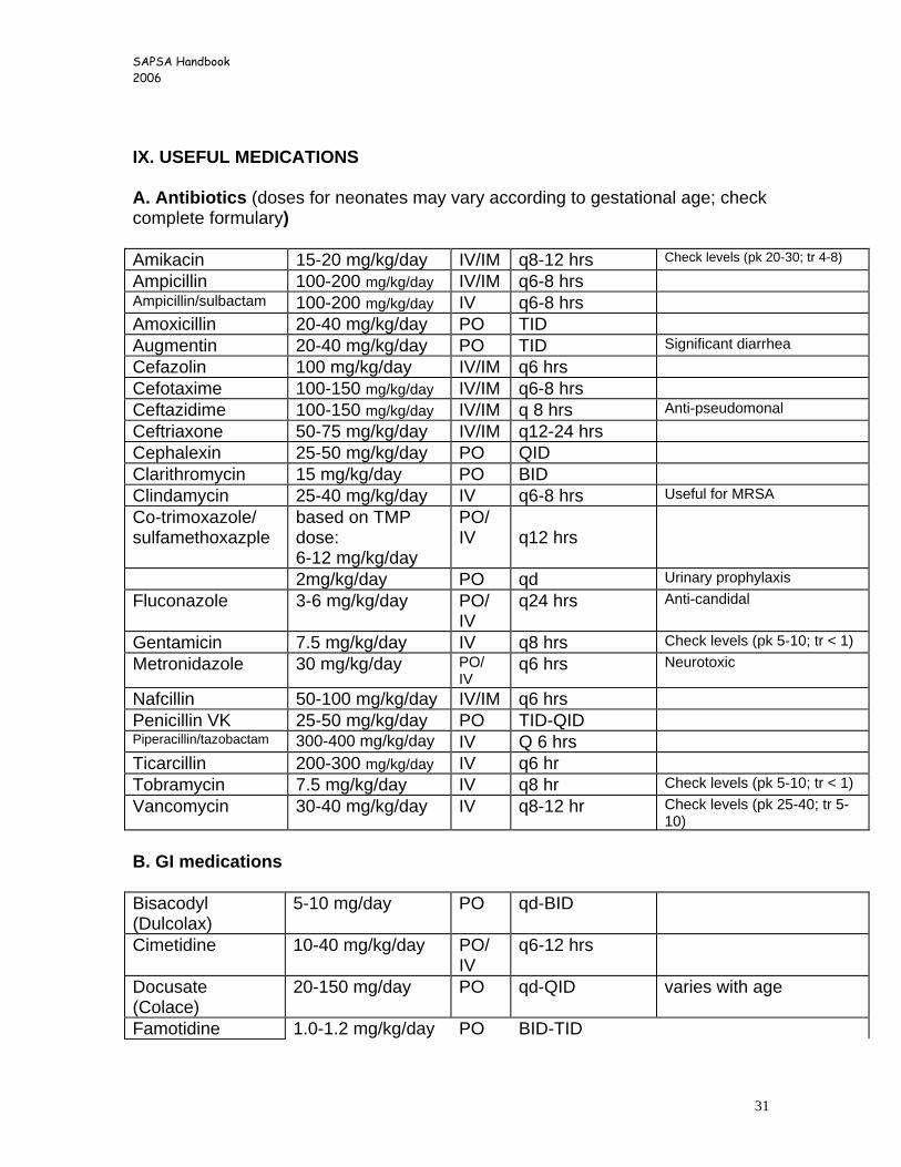
IX. USEFUL MEDICATIONS A. Antibiotics (doses for neonates may vary according to gestational age; check complete formulary) Amikacin 15-20 mg/kg/day IV/IM q8-12 hrs Check levels (pk 20-30; tr 4-8)
Ampicillin 100-200 mg/kg/day IV/IM q6-8 hrs Ampicillin/sulbactam 100-200 mg/kg/day IV q6-8 hrs Amoxicillin 20-40 mg/kg/day PO TID Augmentin 20-40 mg/kg/day PO TID Significant diarrhea Cefazolin 100 mg/kg/day IV/IM q6 hrs Cefotaxime 100-150 mg/kg/day IV/IM q6-8 hrs Ceftazidime 100-150 mg/kg/day IV/IM q 8 hrs Anti-pseudomonal Ceftriaxone 50-75 mg/kg/day IV/IM q12-24 hrs Cephalexin 25-50 mg/kg/day PO QID Clarithromycin 15 mg/kg/day PO BID Clindamycin 25-40 mg/kg/day IV q6-8 hrs Useful for MRSA Co-trimoxazole/ sulfamethoxazple
based on TMP dose: 6-12 mg/kg/day
PO/ IV
q12 hrs
2mg/kg/day PO qd Urinary prophylaxis Fluconazole 3-6 mg/kg/day PO/
IV q24 hrs Anti-candidal
Gentamicin 7.5 mg/kg/day IV q8 hrs Check levels (pk 5-10; tr < 1) Metronidazole 30 mg/kg/day PO/
IV q6 hrs Neurotoxic
Nafcillin 50-100 mg/kg/day IV/IM q6 hrs Penicillin VK 25-50 mg/kg/day PO TID-QID Piperacillin/tazobactam 300-400 mg/kg/day IV Q 6 hrs Ticarcillin 200-300 mg/kg/day IV q6 hr Tobramycin 7.5 mg/kg/day IV q8 hr Check levels (pk 5-10; tr < 1) Vancomycin 30-40 mg/kg/day IV q8-12 hr Check levels (pk 25-40; tr 5-
10) B. GI medications Bisacodyl (Dulcolax)
5-10 mg/day PO qd-BID
Cimetidine 10-40 mg/kg/day PO/ IV
q6-12 hrs
Docusate (Colace)
20-150 mg/day PO qd-QID varies with age
Famotidine 1.0-1.2 mg/kg/day PO BID-TID
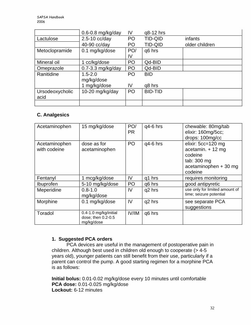
SAPSA Handbook 2006
32
0.6-0.8 mg/kg/day IV q8-12 hrs Lactulose 2.5-10 cc/day PO TID-QID infants 40-90 cc/day PO TID-QID older children Metoclopramide 0.1 mg/kg/dose PO/
IV q6 hrs
Mineral oil 1 cc/kg/dose PO Qd-BID Omeprazole 0.7-3.3 mg/kg/day PO Qd-BID Ranitidine 1.5-2.0
mg/kg/dose PO BID
1 mg/kg/dose IV q8 hrs Ursodeoxycholic acid
10-20 mg/kg/day PO BID-TID
C. Analgesics Acetaminophen 15 mg/kg/dose PO/
PR q4-6 hrs chewable: 80mg/tab
elixir: 160mg/5cc; drops: 100mg/cc
Acetaminophen with codeine
dose as for acetaminophen
PO q4-6 hrs elixir: 5cc=120 mg acetamin. + 12 mg codeine tab: 300 mg acetaminophen + 30 mg codeine
Fentanyl 1 mcg/kg/dose IV q1 hrs requires monitoring Ibuprofen 5-10 mg/kg/dose PO q6 hrs good antipyretic Meperidine 0.8-1.0
mg/kg/dose IV q2 hrs use only for limited amount of
time; seizure potential
Morphine 0.1 mg/kg/dose IV q2 hrs see separate PCA suggestions
Toradol 0.4-1.0 mg/kg/initial dose; then 0.2-0.5 mg/kg/dose
IV/IM q6 hrs
1. Suggested PCA orders PCA devices are useful in the management of postoperative pain in
children. Although best used in children old enough to cooperate (> 4-5 years old), younger patients can still benefit from their use, particularly if a parent can control the pump. A good starting regimen for a morphine PCA is as follows:
Initial bolus: 0.01-0.02 mg/kg/dose every 10 minutes until comfortable PCA dose: 0.01-0.025 mg/kg/dose Lockout: 6-12 minutes
SAPSA Handbook 2006
33
or Continuous hourly infusion of 0.05-0.1 mg/kg/hr; may add small PCA bolus to this rate as needed
Children who are using a PCA device should have a bedside respiratory and heart rate monitor as well as periodic oxygen saturation checks.
2. Epidural pain catheters
Continuous epidural analgesia is encouraged in all pediatric surgery patients who are to undergo major abdominal surgery. These children will require a bedside respiratory and heart rate monitor as well as periodic oxygen saturation checks when on the floor; this should be performed in accordance with the joint anesthesia/nursing protocol for patients with epidural catheters. Male children will generally require a foley catheter while the epidural is in place, although females usually do not. The presence of an epidural catheter is not an excuse to not be ambulatory, and these patients should be encouraged to be out of bed when possible. While the epidural is in place, all narcotics and sedatives are to be managed only with the input of the anesthesiologist responsible for the catheter.
3. Caudal anesthesia
Children who are undergoing lower abdominal/inguinal procedures are candidates for supplementary caudal anesthesia, which allows for good postoperative pain control for about 6 to 8 hours. This is usually performed by the anesthesiologist at the beginning of the case.
4. Postop analgesia for outpatients
Patients who are eligible should have received a caudal injection; for those who do not, then the wound should be liberally infiltrated with 0.25% or 0.5% Marcaine (bupivacaine) at a maximal dose of 3 mg/kg, or approximately 1cc/kg for 0.25% and 0.5cc/kg for 0.5% bupivicaine. The majority of young children can be managed with round-the-clock, alternating doses of acetaminophen and ibuprofen, supplemented with benadryl (1mg/kg/dose q6 hrs) as a sedative. Codeine is not routinely given to infants, although older children and teenagers will benefit from its use for a short duration.
SAPSA Handbook 2006
34
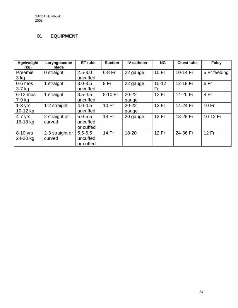
IX. EQUIPMENT
Age/weight (kg)
Laryngoscope blade
ET tube Suction IV catheter NG Chest tube Foley
Preemie 3 kg
0 straight 2.5-3.0 uncuffed
6-8 Fr 22 gauge 10 Fr 10-14 Fr 5 Fr feeding
0-6 mos 3-7 kg
1 straight 3.0-3.5 uncuffed
8 Fr 22 gauge 10-12 Fr
12-18 Fr 6 Fr
6-12 mos 7-9 kg
1 straight 3.5-4.5 uncuffed
8-10 Fr 20-22 gauge
12 Fr 14-20 Fr 8 Fr
1-3 yrs 10-12 kg
1-2 straight 4.0-4.5 uncuffed
10 Fr 20-22 gauge
12 Fr 14-24 Fr 10 Fr
4-7 yrs 16-18 kg
2 straight or curved
5.0-5.5 uncuffed or cuffed
14 Fr 20 gauge 12 Fr 18-28 Fr 10-12 Fr
8-10 yrs 24-30 kg
2-3 straight or curved
5.5-6.5 uncuffed or cuffed
14 Fr 18-20 12 Fr 24-36 Fr 12 Fr