Pediatric Neurological emergencies & stabilization AG
74
Neurological Emergencies & Stabilization By Dr. Akshay
-
Upload
akshay-golwalkar -
Category
Healthcare
-
view
128 -
download
0
Transcript of Pediatric Neurological emergencies & stabilization AG
Neurological Emergencies &
Stabilization
By Dr. Akshay
Objectives
General pediatric neurological assessment
Nonsurgical neurological emergencies
Neurosurgical nontraumatic emerencies
Neurosurgical traumatic injuries
Initial stabilization and emergency management
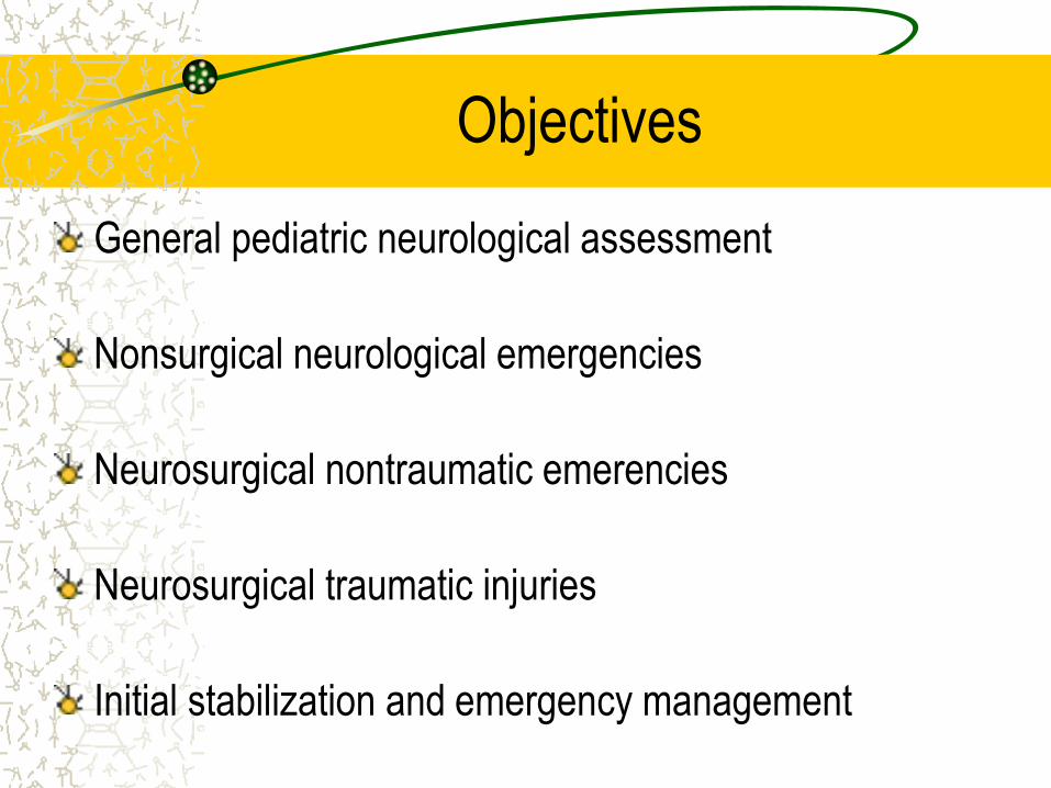
Symptoms of presentation
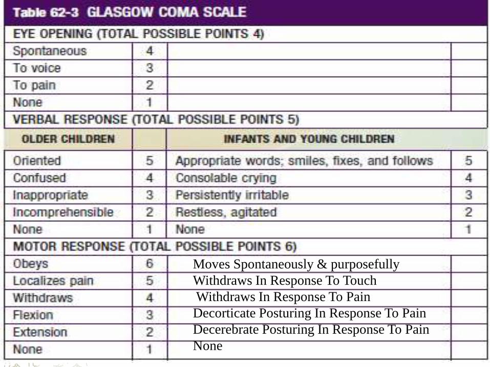
Moves Spontaneously & purposefully
Withdraws In Response To Touch
Withdraws In Response To Pain
Decorticate Posturing In Response To Pain
Decerebrate Posturing In Response To Pain
None
Seizures
– Febrile seizures
– Afebrile Seizures (Childhood epilepsy syndromes)
CNS Infections
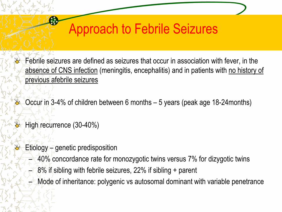
Approach to Febrile Seizures
Febrile seizures are defined as seizures that occur in association with fever, in the
absence of CNS infection (meningitis, encephalitis) and in patients with no history of
previous afebrile seizures
Occur in 3-4% of children between 6 months – 5 years (peak age 18-24months)
High recurrence (30-40%)
Etiology – genetic predisposition
– 40% concordance rate for monozygotic twins versus 7% for dizygotic twins
– 8% if sibling with febrile seizures, 22% if sibling + parent
– Mode of inheritance: polygenic vs autosomal dominant with variable penetrance
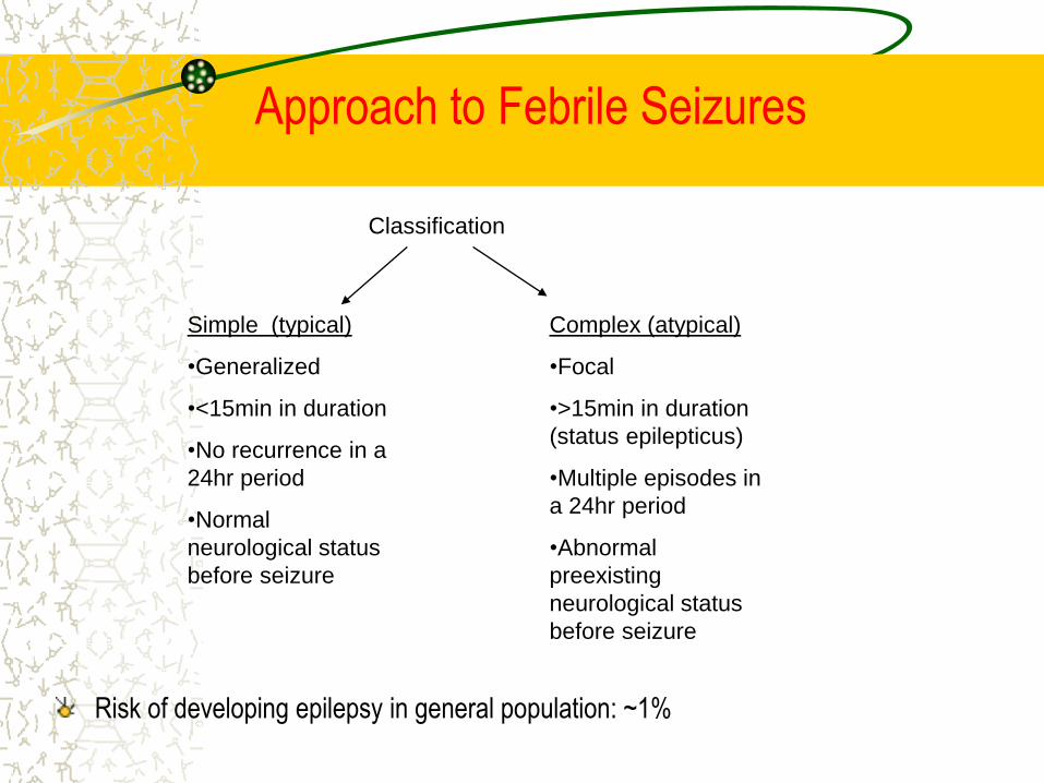
Risk of developing epilepsy in general population: ~1%
Simple (typical)
•Generalized
•<15min in duration
•No recurrence in a
24hr period
•Normal
neurological status
before seizure
Complex (atypical)
•Focal
•>15min in duration
(status epilepticus)
•Multiple episodes in
a 24hr period
•Abnormal
preexisting
neurological status
before seizure
Classification
Approach to Febrile Seizures

Treatment of febrile seizures and prevention of recurrences does not alter risk of later
possible epilepsy - routine use of AEDs not recommended.
Parents can be advised to use anti-pyretics for comfort care, but there is no evidence
that it prevents recurrence of febrile seizures
Intermittent prophylaxis can be used when recurrence is expected; excessive parental
anxiety
– Clobzam (0.5 to 1 mg/kg/day) (Minimum 3days or until fever subsides)
What investigations are necessary:
– Simple febrile seizures: nothing
– Complex febrile seizures: EEG ± neuroimaging
Approach to Febrile Seizures
What do parents want to know:
o Is this harmful
oWill it happen again
o Can I prevent it?
oWill my child develop epilepsy
oWill it go away?
Approach to Febrile Seizures

Intermittent prophalyxis
Clobzam (0.5 to 1 mg/kg/day)(Minimum 3days or until fever subsides)
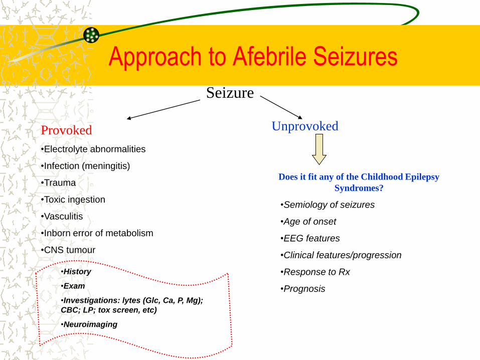
Approach to Afebrile Seizures
Seizure
Provoked
•Electrolyte abnormalities
•Infection (meningitis)
•Trauma
•Toxic ingestion
•Vasculitis
•Inborn error of metabolism
•CNS tumour
Unprovoked
•History
•Exam
•Investigations: lytes (Glc, Ca, P, Mg);
CBC; LP; tox screen, etc)
•Neuroimaging
Does it fit any of the Childhood Epilepsy
Syndromes?
•Semiology of seizures
•Age of onset
•EEG features
•Clinical features/progression
•Response to Rx
•Prognosis

History
•Antenatal History
•Birth history
•Developmental history
•Family history
•Head trauma
•Seizure description (aura,
trigger, eyewitness
description)
Serologies/TORCH
Preeclampsia/GDM/Infections
Substance abuse/meds
Antenatal USG
Fetal distress
Need for postnatal resuscitation
Normal vs delayed vs regressed
Consanguinity, hx of febrile
seizures, epilepsy, developmental
delay, recurrent miscarriages, IEM
Approach to Afebrile Seizures
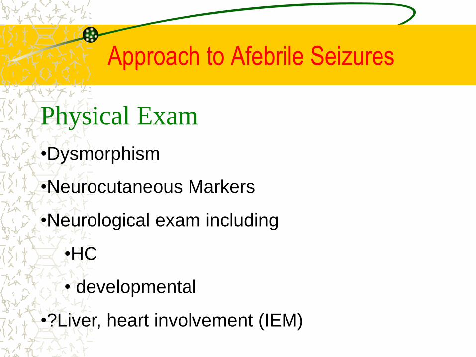
Physical Exam
•Dysmorphism
•Neurocutaneous Markers
•Neurological exam including
•HC
• developmental
•?Liver, heart involvement (IEM)
Approach to Afebrile Seizures

Physical Exam
•Dysmorphism
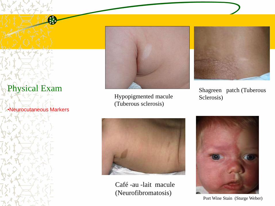
Physical Exam
•Neurocutaneous Markers
Hypopigmented macule
(Tuberous sclerosis)
Shagreen patch (Tuberous
Sclerosis)
Café -au -lait macule
(Neurofibromatosis)Port Wine Stain (Sturge Weber)
Feinichel
Neonatal Period (<28 days)
Seizures in newborns are often difficult to distinguish from normal activity
Most commonly occur within the first week of life
– 2/3 of neonatal seizures are due to Hypoxic-ischemic encephalopathy (HIE)
– Other causes: infection, electrolyte abnormalities, inborn errors of metabolism,
structural
The clinical and electroencephalographic features of neonatal seizures differ
considerably from those in older children and adults.
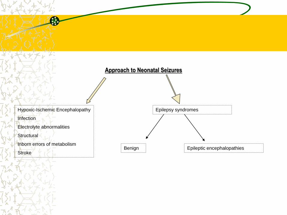
Approach to Neonatal Seizures
Hypoxic-Ischemic Encephalopathy
Infection
Electrolyte abnormalities
Structural
Inborn errors of metabolism
Stroke
Epilepsy syndromes
Benign Epileptic encephalopathies
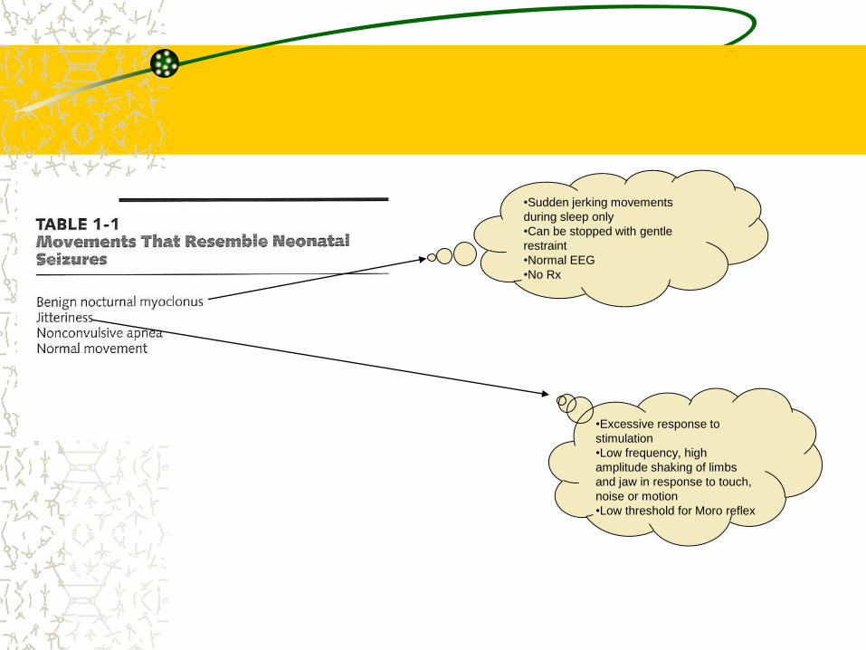
•Sudden jerking movements
during sleep only
•Can be stopped with gentle
restraint
•Normal EEG
•No Rx
•Excessive response to
stimulation
•Low frequency, high
amplitude shaking of limbs
and jaw in response to touch,
noise or motion
•Low threshold for Moro reflex
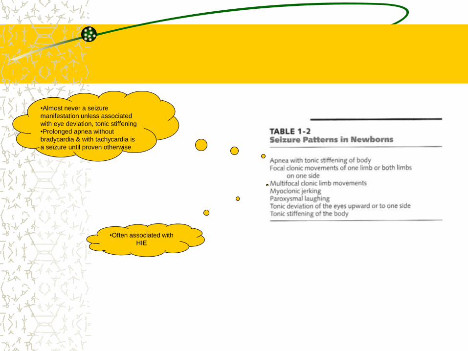
•Almost never a seizure
manifestation unless associated
with eye deviation, tonic stiffening
•Prolonged apnea without
bradycardia & with tachycardia is
a seizure until proven otherwise
•Often associated with
HIE
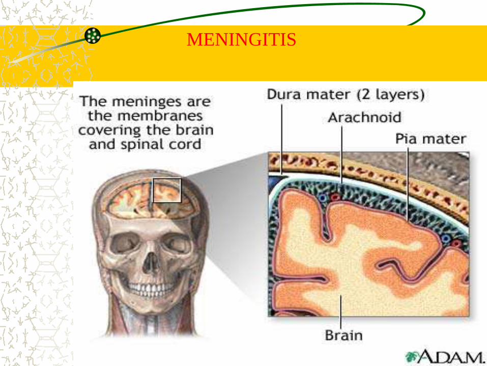
MENINGITIS
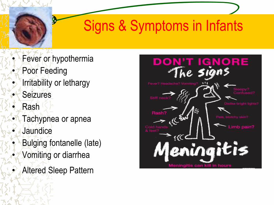
Signs & Symptoms in Infants
• Fever or hypothermia
• Poor Feeding
• Irritability or lethargy
• Seizures
• Rash
• Tachypnea or apnea
• Jaundice
• Bulging fontanelle (late)
• Vomiting or diarrhea
• Altered Sleep Pattern
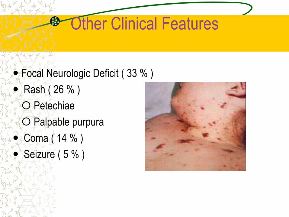
Other Clinical Features
Focal Neurologic Deficit ( 33 % )
Rash ( 26 % )
Petechiae
Palpable purpura
Coma ( 14 % )
Seizure ( 5 % )
Differential Diagnosis
• Encephalitis• Brain Inflammation
• HSV – 1 Most common cause
• Aseptic Meningitis• Viral: Enterovirus, HIV, Mumps, HSV - 2
• Fungal: Coccidioidomycosis, Cryptococcus
• Tuberculosis
• Parasites
• Neoplasm of the leptomeninges
• Drug - Induced: NSAIDs, Allopurinol etc.
• Intracranial Abscess
Treatment
Inpatient ( preferably in ICU )
Appropriate Antibiotic Therapy
Supportive Care(hemo-dynamic ,respiratory, renal &
electrolytes, myocardial support)
Treat coexisting conditions(seizures ,Raised ICT)
Prevent hypothermia and dehydration
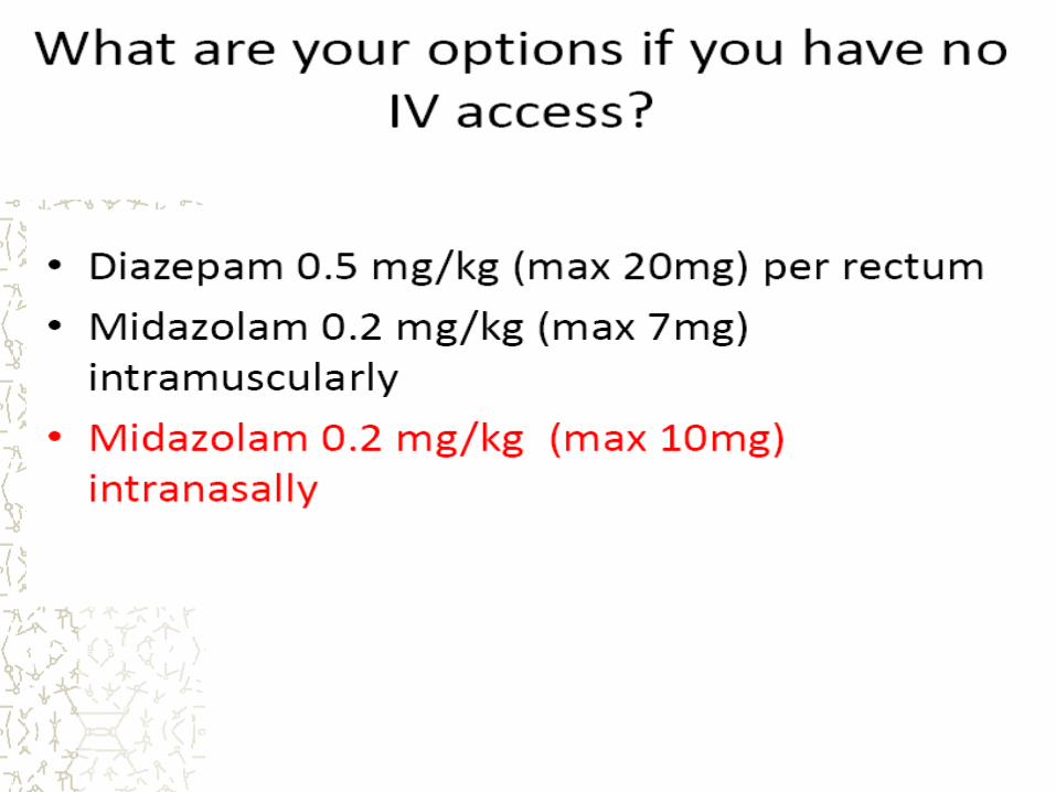
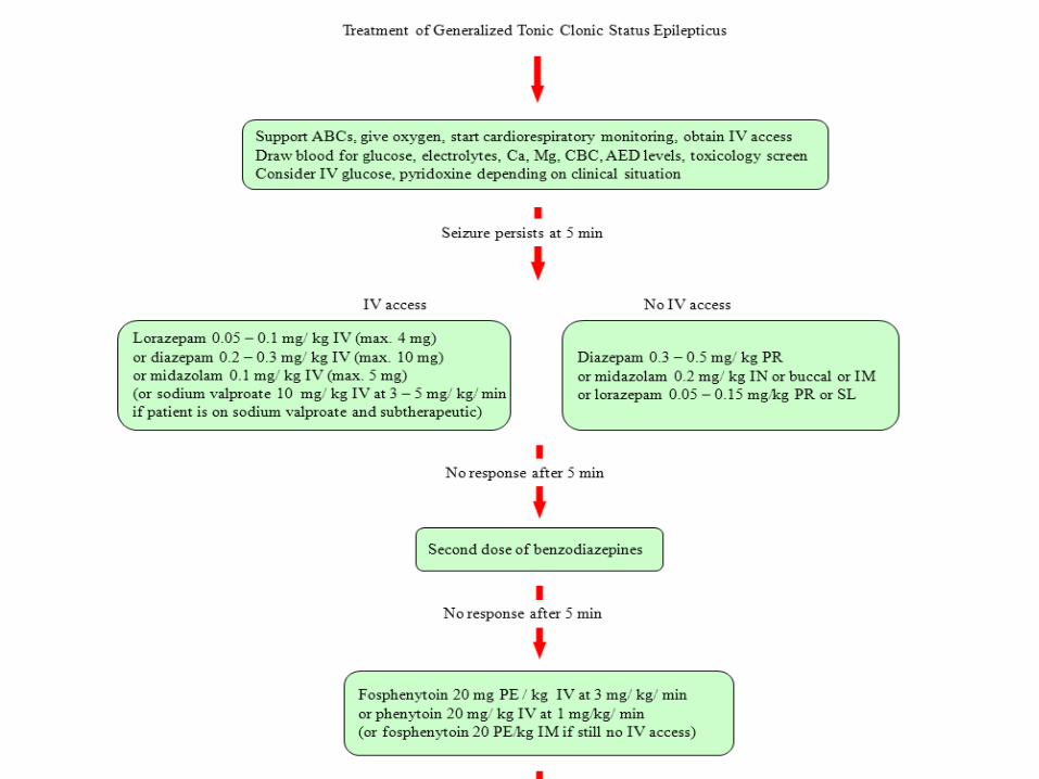
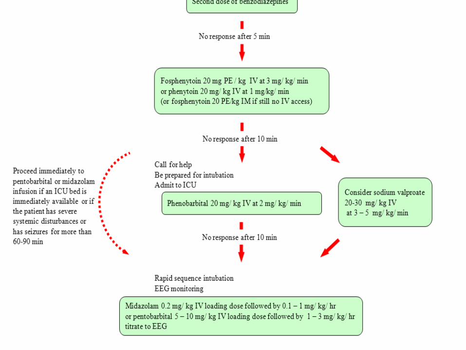
Status Epilipticus
status epilepticus is defined as a generalized
convulsion lasting 30 minutes or longer or when
successive convulsions occur that the patient does
not recover consciousness between.
However, it is usual practice to start anti-
convulsive treatment when the episode has lasted
5 or more minutes
Stroke
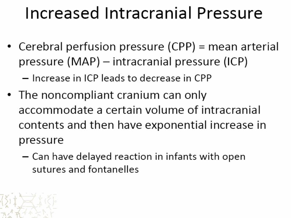
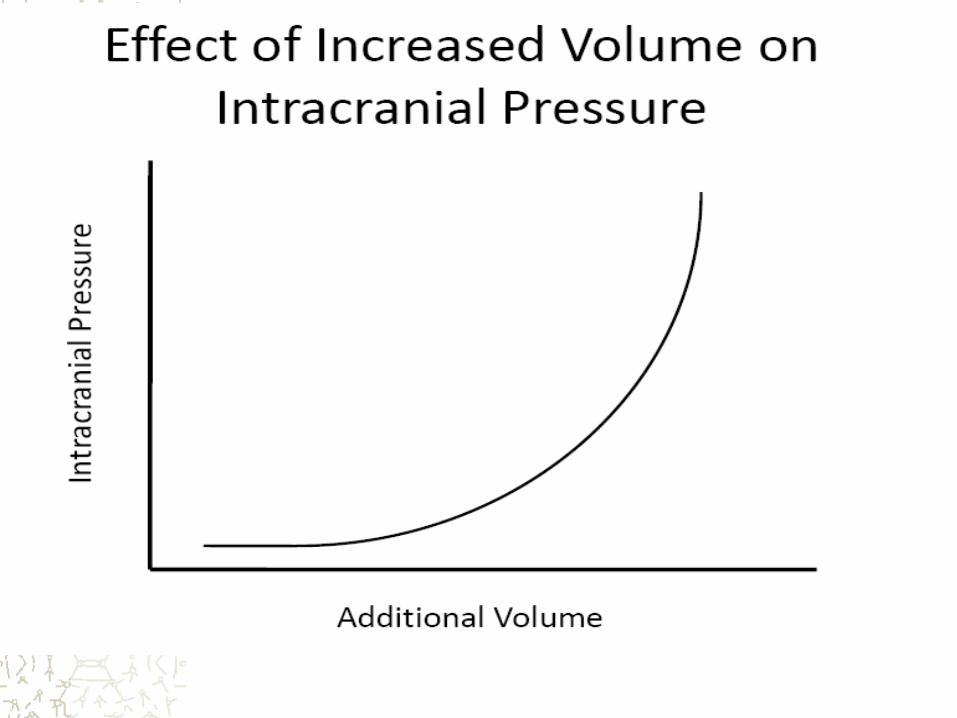
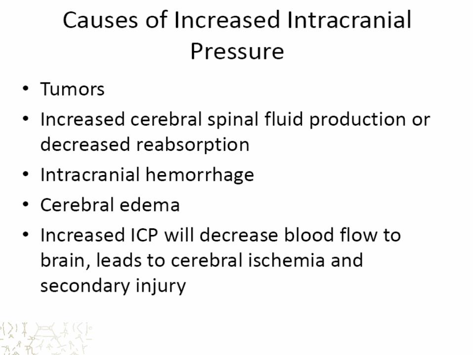

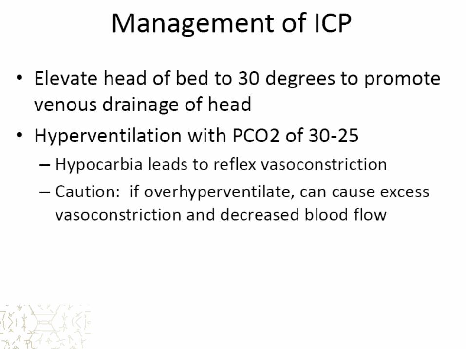

Specific Causes of raised ICP
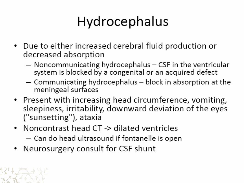

“Sunsetting Eyes”: clinical sign of
increased intracranial pressure
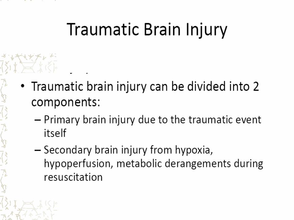
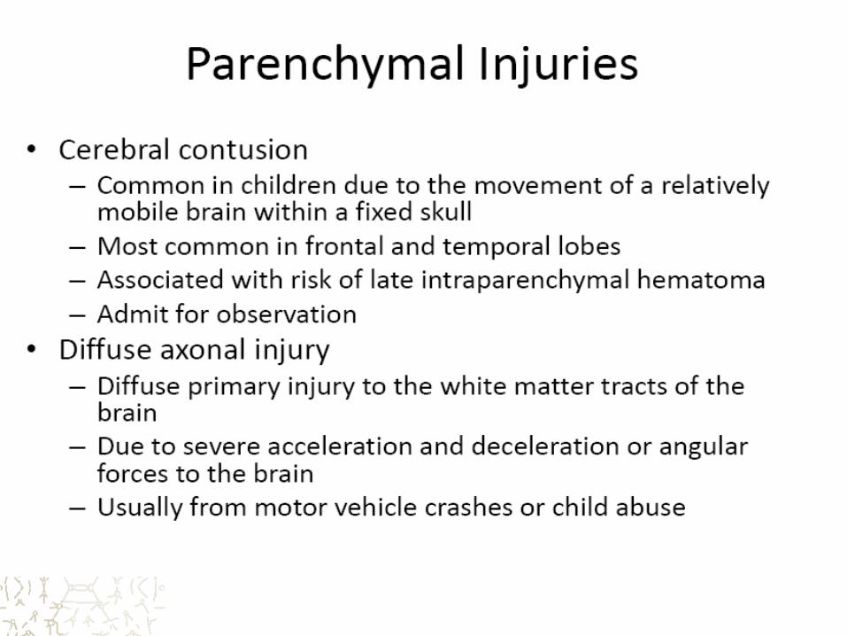
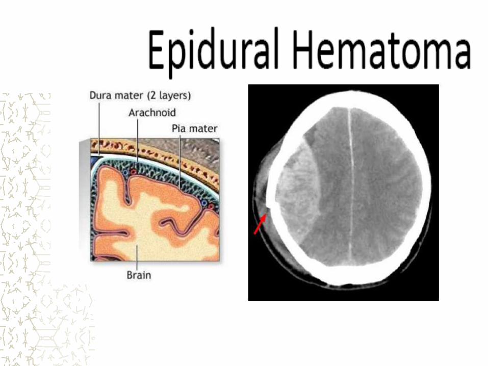
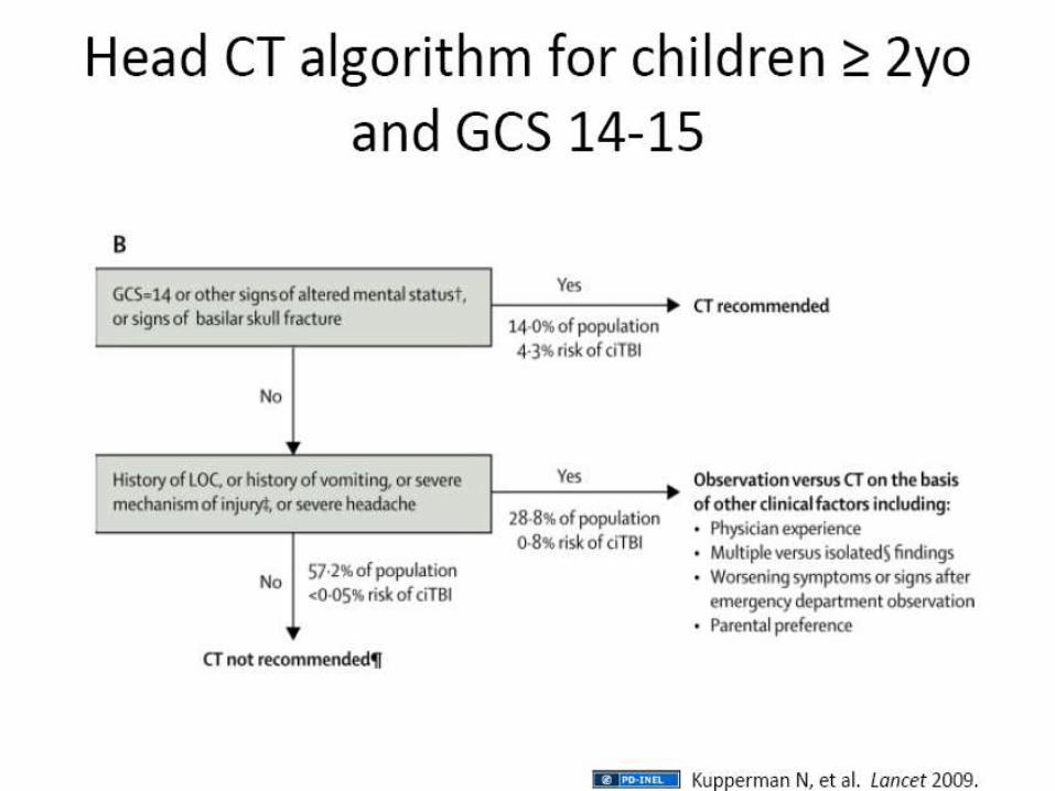
Traumatic Brain Injuries

Disorders of motor function or
weakness
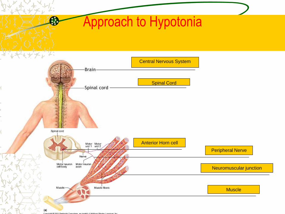
Approach to Hypotonia
Central Nervous System
Spinal Cord
Anterior Horn cell
Peripheral Nerve
Neuromuscular junction
Muscle
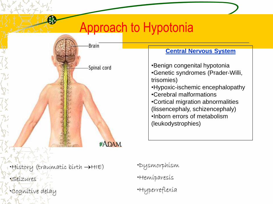
Central Nervous System
•Benign congenital hypotonia
•Genetic syndromes (Prader-Willi,
trisomies)
•Hypoxic-ischemic encephalopathy
•Cerebral malformations
•Cortical migration abnormalities
(lissencephaly, schizencephaly)
•Inborn errors of metabolism
(leukodystrophies)
•History (traumatic birth HIE)
•Seizures
•Cognitive delay
Approach to Hypotonia
•Dysmorphism
•Hemiparesis
•Hyperreflexia
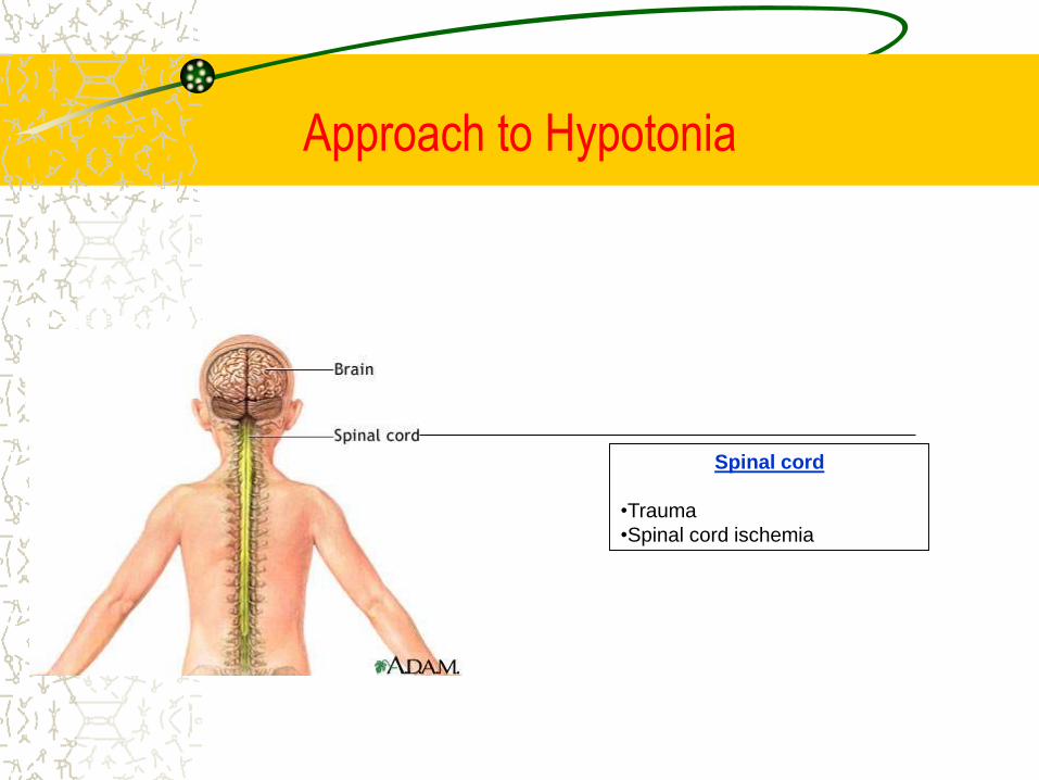
Spinal cord
•Trauma
•Spinal cord ischemia
Approach to Hypotonia
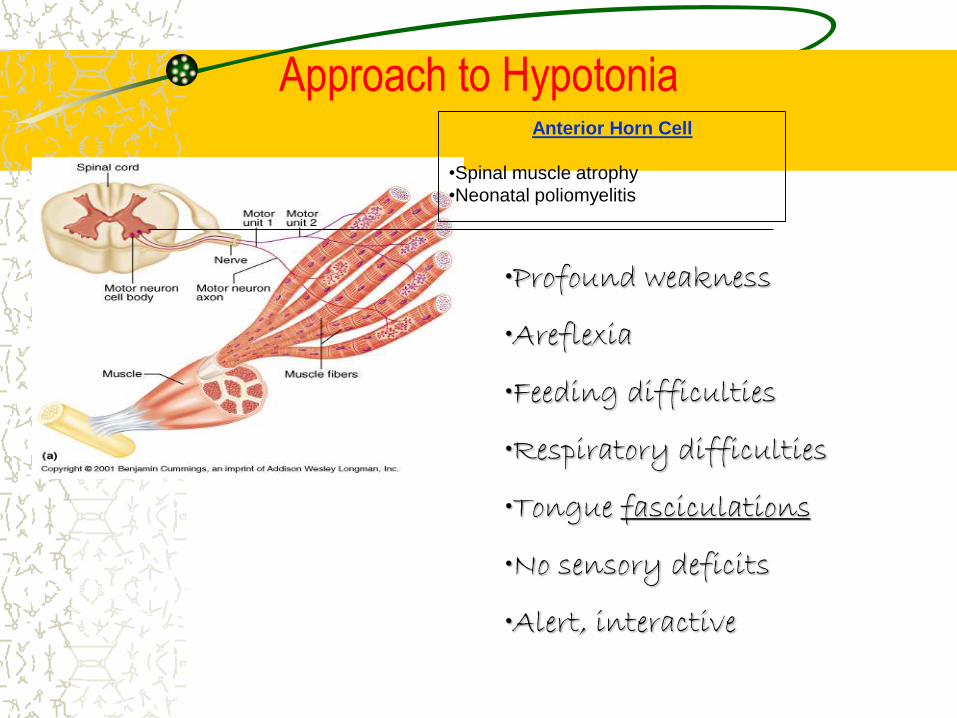
Anterior Horn Cell
•Spinal muscle atrophy
•Neonatal poliomyelitis
•Profound weakness
•Areflexia
•Feeding difficulties
•Respiratory difficulties
•Tongue fasciculations
•No sensory deficits
•Alert, interactive
Approach to Hypotonia
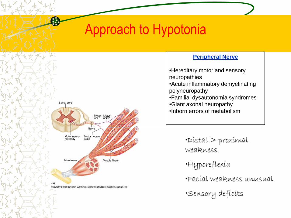
Peripheral Nerve
•Hereditary motor and sensory
neuropathies
•Acute inflammatory demyelinating
polyneuropathy
•Familial dysautonomia syndromes
•Giant axonal neuropathy
•Inborn errors of metabolism
•Distal > proximal weakness
•Hyporeflexia
•Facial weakness unusual
•Sensory deficits
Approach to Hypotonia
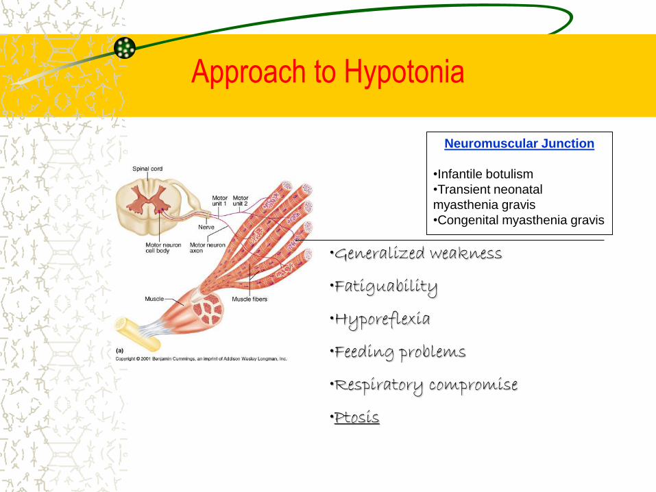
Neuromuscular Junction
•Infantile botulism
•Transient neonatal
myasthenia gravis
•Congenital myasthenia gravis
•Generalized weakness
•Fatiguability
•Hyporeflexia
•Feeding problems
•Respiratory compromise
•Ptosis
Approach to Hypotonia
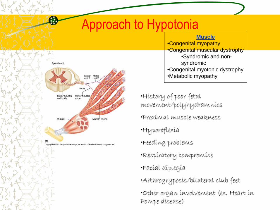
Muscle
•Congenital myopathy
•Congenital muscular dystrophy
•Syndromic and non-
syndromic
•Congenital myotonic dystrophy
•Metabolic myopathy
•History of poor fetal movement/polyhydramnios
•Proximal muscle weakness
•Hyporeflexia
•Feeding problems
•Respiratory compromise
•Facial diplegia
•Arthrogryposis/bilateral club feet
•Other organ involvement (ex. Heart in Pompe disease)
Approach to Hypotonia
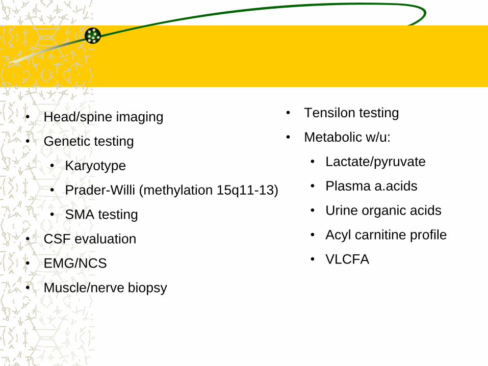
• Head/spine imaging
• Genetic testing
• Karyotype
• Prader-Willi (methylation 15q11-13)
• SMA testing
• CSF evaluation
• EMG/NCS
• Muscle/nerve biopsy
• Tensilon testing
• Metabolic w/u:
• Lactate/pyruvate
• Plasma a.acids
• Urine organic acids
• Acyl carnitine profile
• VLCFA