
Pediatric Emergency It’s not Just A Clinic - · PDF filePediatric Emergency It’s...
47
Pediatric Emergency It’s Not Just A Clinic Stacey Forbes, FNP-BC, MSN
Transcript of Pediatric Emergency It’s not Just A Clinic - · PDF filePediatric Emergency It’s...

Pediatric Emergency It’s Not Just A Clinic
Stacey Forbes, FNP-BC, MSN

Pediatric Emergencies ∗ Comprise 1/3 ED visits
yearly ∗ Management varies
by anatomy, physically, and developmentally
∗ Arrive to unfamiliar environment
∗ Children are frightened and increases stress to family
*Illnesses can be minor and/or major *HCP must be patient and concise *Establish provider/ patient bond *Care must be family centered *Provide care and resources that are needed

Presenting Complaints * Respiratory symptoms -Viral URI, Pneumonia, Congenital * GI and GU-vomiting, thrush, diarrhea, constipation, vaginal discharge * Normal concerns -crying, feeding, sleep, periodic breathing * Fever -rule out sepsis- respiratory, urinary or CNS * Skin issues -rash, birth marks, jaundice * Cardiac -tachyarrhythmia, murmurs, congenital disorders * Eye- discharge, clogged duct *Musculoskeletal- injuries, hair tourniquets *Hematology-blood loss

∗ Leading cause of morbidity and mortality in US and worldwide
∗ 100,00 children presented to CED with severe sepsis
∗ Top cause of death in children ages 0-14 ∗ In past ten years major advancements have
been made ∗ Protocol directed therapy and early
administration of antibiotics ∗ Mortality has improved by 25% over last five
years and is lower in adults
Pediatric Sepsis

∗ More than one change in vital signs( Temp, HR) ∗ Prolonged capillary refill >2 seconds or
prolonged perfusion ∗ Lethargy, confusion, limp ∗ Clinical Def- T>100.4, HR>90, RR>20 ∗ Obtaining H&P(illnesses, wounds, vaccinations) ∗ Sepsis recognized and managed by pediatric
age and weight ???
Identifying Sepsis

∗ Neonate- Birth to 28 days old ∗ Infant- 29 days to 1 year old ∗ Child > 1 year
Pediatric Age Definition


Case Study #1 3 year old male
Mother relates he was not waking up from a nap. Hot to touch, fever 102.5 at home. Runny nose and congestion 1-2 days. Older brother influenza+ two days ago. T-105 rectal, HR-220, RR-24, BP-114/80, capillary refill >2 secs, he is lethargic.

∗ WBC-23.6, Bands-6, ESR-12, CRP-7 ∗ NA 138, GLU-100, K-3.8, BUN-12, Creat-.6 ∗ RSV- neg, Influenza-+, Rapid Strept neg ∗ U/A Leuk+, WBC 68 ∗ Blood, urine and throat cultures pending ∗ Chest x-ray-no infiltrate, no mass ∗ EKG-Sinus Tachycardia ∗ NS 20mg/kg bolus x2, Tylenol suppos, Tamiflu,
Ceftriaxone 75mg/kg
Case Study 3 yr old male

∗ Respiratory- Pneumonia, RSV, Bronchiolitis ∗ GI- Appendicitis, Cholecystitis ∗ GU- UTI
Common Causes of Sepsis


Case Study #1 Outcome Alert and at baseline Drinking, voiding Discharged home with pediatrician follow up next day Admitted next day Blood culture and urine culture +
Suggested Guidelines for the Evaluation and Management
of Neonates, Infants, and Children with Fever
Age Group Evaluation Treatment

Neonate, 0–28 d* of age, ≥38°C (100.4°F) CBC and blood culture. Admit. Incidence of ill appearing: 13%–21%; if not ill appearing: <5% Urinalysis and urine culture. Parenteral antibiotic therapy with ampicillin, 50 milligrams/ kg, and cefotaxime, 50 milligrams/kg, or gentamicin, 2.5 milligrams/kg. CSF cell count, Gram stain, and culture. Chest x-ray is optional, if no respiratory symptoms. Stool testing if diarrhea is present.

Infant 29–56 d* of age, ≥38.2°C (100.8°F) (Philadelphia Protocol) Same as for neonates. Discharge if: WBC ≤15,000/mm3 and ≥5000/mm3 and <20% band forms. SBI incidence of ill appearing: 13%–21%; if not ill appearing: <5% Urinalysis negative. CSF WBC <10 cells/mm 3. Negative chest x-ray or fecal leukocytes if applicable. Admit if any of above criteria are not met and treat with parenteral ceftriaxone, 50 milligrams/kg with normal CSF, 100 milligrams/kg with signs of meningitis.

Infants 57 d* to 6 mo* of age, ≥38°C (100.4°F) Urinalysis and urine culture alone. Discharge if negative. Treat for UTI with cefixime, 8 milligrams/kg/d daily or divided twice a day, or cefpodoxime, 10 milligrams/kg/d divided twice a day, or cefdinir, 14 milligrams/kg/d divided every 12–24 h for 7–10 d as outpatient. Non-UTI SBI incidence is estimated to be negligible For conservative management, treat infants 57–90 d using Philadelphia Protocol above. UTI is 3%–8% Admit and treat with the parenteral ceftriaxone if fails conservative criteria for discharge.

Infants/children 6–36 mo of age Urinalysis and urine culture. Discharge if negative. Non-UTI SBI incidence is <0.4% Girls 6–24 mo. Treat for UTI as above as outpatient. UTI in girls ≤8% Boys 6–12 mo. UTI in boys (<12 mo) ≤2% Uncircumcised boys 12–24 mo. Uncircumcised boys (1–2 y) remains 2% Children >36 mo and older No further workup is routinely necessary. Discharge and treat with acetaminophen, 15 milligrams/kg PO/PR every 4 h, or ibuprofen, 10 milligrams/kg PO every 6h

Pediatric Stroke

Stroke
∗ Brain attack ∗ Any interruption of
blood flow to the brain ∗ Loss of oxygen and
glucose ∗ Causes edema, and
mass effect and exacerbate previous injury
∗ Several types Ischemic (87%),hemorrhagic(10%) and nontraumatic subarachnoid hemorrhage (3%)
∗ 10-25% children die ∗ 25% have a
reoccurrence ∗ 66% neuro deficits,
seizures, learning or development disorders
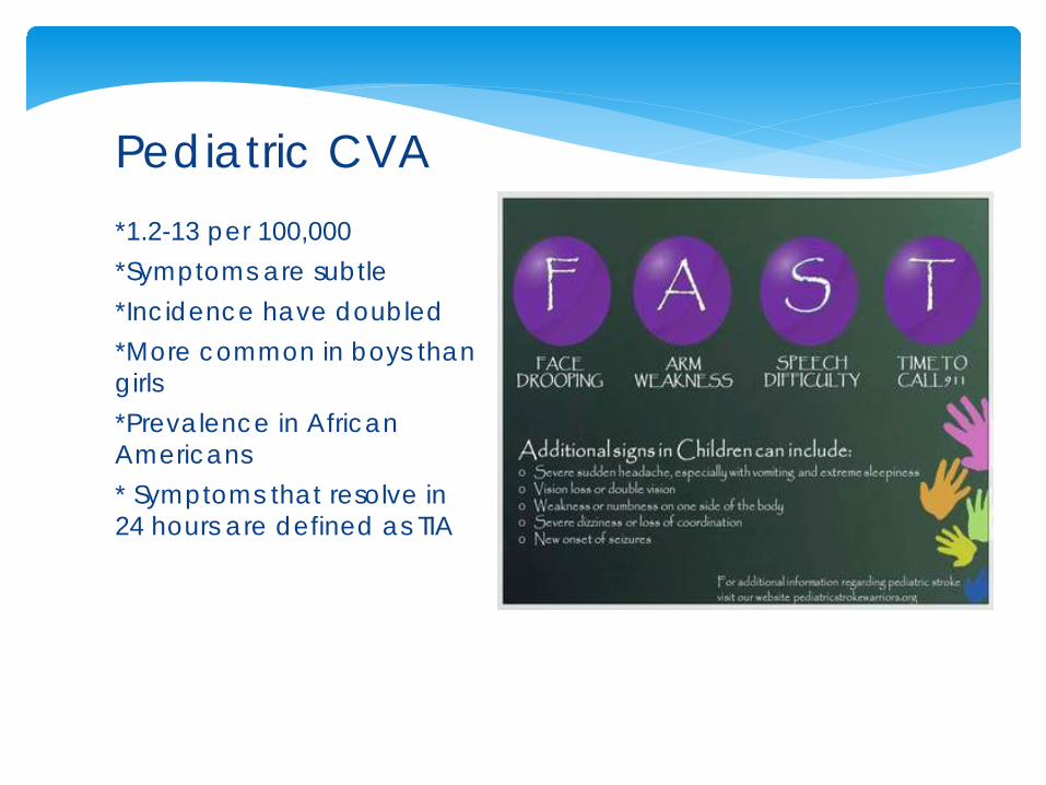
*1.2-13 per 100,000 *Symptoms are subtle *Incidence have doubled *More common in boys than girls *Prevalence in African Americans * Symptoms that resolve in 24 hours are defined as TIA
Pediatric CVA

∗ Early recognition ∗ Rapid neuro consultation ∗ Imaging ∗ Treatment ∗ Improved outcomes
CED Objective

∗ Newborn-focal seizures or lethargy ∗ Lethargy, apnea spells or hypotonia infants ∗ Toddlers show deterioration-crying, irritable,
poor feeding, vomiting, sepsis like symptoms with cold extremities
∗ Older child-Hemiparesis, language/speech difficulties, visual deficits
How do they present?

Risk Factors and Causes ∗ Cardiac most
common-cardiac cath repair occurs in 72 hours
∗ Cardiomyopathies, Rheumatic Heart Disease, prosthetic valves, PFO-patent foramen ovale
∗ Hematologic-Sickle Cell Disease-also common. Most after 5 years of age, as early as 18 months
∗ Prothrombotic disorders-protein C & S, Factor VII & VIII deficiency, iron deficiency anemia- without other etiology

Risk Factors Continued
∗ Infection Varicella, HIV-induced vasculitis, vasculopathy
∗ Mycoplasma, Flu A coxsackie, Rocky Mountain spotted fever, parvovirus19, chlamydia, enterovirus
∗ Bacterial & TB meningitis, viral
∗ Vascular- AVM, moyamoya associated with Down syndrome, neurofibromatosis, and sickle cell
Risk Factors Continued
∗ Syndromic and Metabolic disorders- Marfans, tuberous sclerosis, homocysteinuria, folic acid and B12 deficiencies
∗ Oncologic-Leukemia or Lymphoma, radiation therapy optic area (vasculopathies)
∗ Vasculitis-children>14-Idiopathic, Kawasaki disease, Henoch-Schonlein Purpura (HSP), polyarteritis nodosa, Takayasu’s arteritis, juvenile RA, Systemic lupus, IBS, sarcoidosis, Sjogren syndrome, consider Behcet’s disease

Risk Factors Continued
∗ Trauma- head and neck. Symptoms can be delayed 24 hours.
∗ Intraoral trauma- pencil to mouth, tonsillectomy
∗ Chiropractor manipulation
∗ Drugs- illicit and prescribed. Such as amphetamines, PCP ecstasy, cocaine, glue sniffing, stimulants and heroine
∗ Oral contraceptives ∗ Overuse of ergot
alkaloids treating migraines

∗ CT noncontrast ∗ MRI/MRV ∗ Transcranial Doppler ∗ Lumbar puncture ∗ Blood glucose, CBC, pregnancy, LFTs, ESR,
ANA
Imaging and Labs

∗ Depends on cause ∗ Prevent rebleed. Correction anticoagulation ∗ Factor VIIa ∗ Low molecular weight heparin ∗ Transfer to tertiary facility ∗ Surgical management of hemorrhagic stroke,
early evacuation, stereotactic radiosurgery, microsurgical or endovascular techniques
∗ Thrombolytic therapy ∗ Contact 1-800-NOCLOTS
Treatment
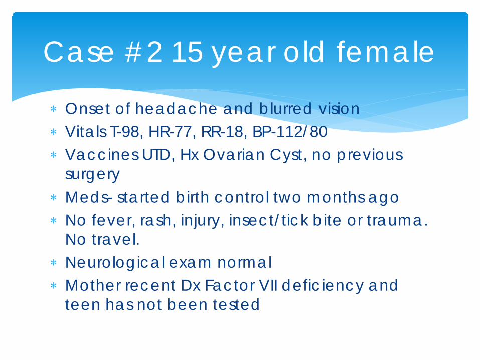
∗ Onset of headache and blurred vision ∗ Vitals T-98, HR-77, RR-18, BP-112/80 ∗ Vaccines UTD, Hx Ovarian Cyst, no previous
surgery ∗ Meds- started birth control two months ago ∗ No fever, rash, injury, insect/tick bite or trauma.
No travel. ∗ Neurological exam normal ∗ Mother recent Dx Factor VII deficiency and
teen has not been tested
Case #2 15 year old female
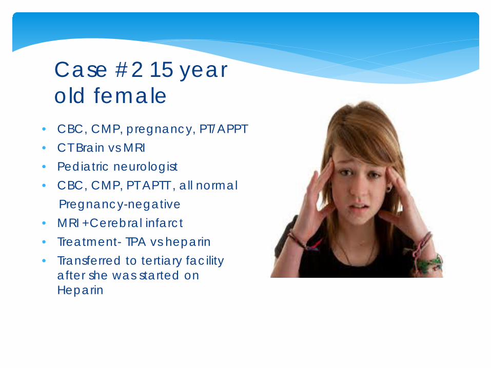
• CBC, CMP, pregnancy, PT/APPT • CT Brain vs MRI • Pediatric neurologist • CBC, CMP, PT APTT , all normal Pregnancy-negative • MRI +Cerebral infarct • Treatment- TPA vs heparin • Transferred to tertiary facility
after she was started on Heparin
Case #2 15 year old female
Heart Failure
The Pediatric Patient

∗ Result of ventricular pump dysfunction ∗ Loss of myocardium, reduced contractility ∗ Leads to inadequate perfusion, shock, and
pulmonary edema ∗ More common in adults ∗ 90% occur < 1 year old
What is Heart Failure ?

∗ Feeding-lower intake, longer time . Sluggish weight gain
∗ Less active, more irritable ∗ Higher resting heart rate ∗ Tachypnea ∗ No rales in infants or children ∗ Enlarged liver ∗ S3 gallop ∗ Diminished peripheral pulse ∗ Arrhythmias can occur and cause sudden
death
Sign/Symptoms

Causes of Heart Failure Neonatal Sepsis- prolonged rupture of membranes, maternal infection Hematology- anemia, cancer Metabolic- hypoglycemia, hypocalcemia Congenital defects Arrhythmias- SVT, bradycardia
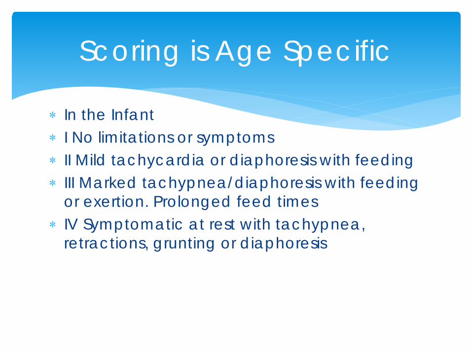
∗ In the Infant ∗ I No limitations or symptoms ∗ II Mild tachycardia or diaphoresis with feeding ∗ III Marked tachypnea/diaphoresis with feeding
or exertion. Prolonged feed times ∗ IV Symptomatic at rest with tachypnea,
retractions, grunting or diaphoresis
Scoring is Age Specific
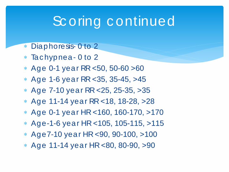
∗ Diaphoresis- 0 to 2 ∗ Tachypnea- 0 to 2 ∗ Age 0-1 year RR <50, 50-60 >60 ∗ Age 1-6 year RR <35, 35-45, >45 ∗ Age 7-10 year RR <25, 25-35, >35 ∗ Age 11-14 year RR <18, 18-28, >28 ∗ Age 0-1 year HR <160, 160-170, >170 ∗ Age-1-6 year HR <105, 105-115, >115 ∗ Age7-10 year HR <90, 90-100, >100 ∗ Age 11-14 year HR <80, 80-90, >90
Scoring continued

∗ A Risk of developing HF, normal cardiac function and no cardiac overload
∗ B Abnormal cardiac function, no symptoms of HF
∗ C Underlying structural or functional heart disease with HF symptoms
∗ D End Stage NF treated with continuous inotropic drugs, circulatory support, heart transplant or hospice
Staging for Infants and Children
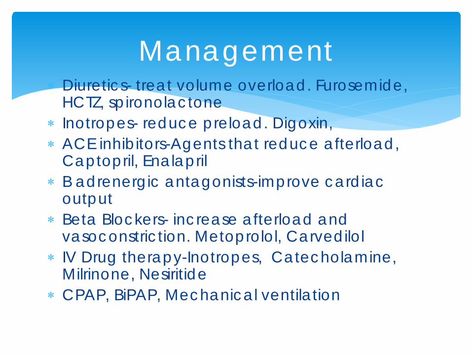
∗ Diuretics- treat volume overload. Furosemide, HCTZ, spironolactone
∗ Inotropes- reduce preload. Digoxin, ∗ ACE inhibitors-Agents that reduce afterload,
Captopril, Enalapril ∗ B adrenergic antagonists-improve cardiac
output ∗ Beta Blockers- increase afterload and
vasoconstriction. Metoprolol, Carvedilol ∗ IV Drug therapy-Inotropes, Catecholamine,
Milrinone, Nesiritide ∗ CPAP, BiPAP, Mechanical ventilation
Management

∗ Nutrition- calorie intake 120 kcal/kg via nasogastric or gastrostomy tube. Breast feeding less stressful than bottle.
∗ Exercise Rehab-data limited further study needed
∗ Surgery-correct congenital problem, AICD. Risk after surgery is CHF.
Management continued

12 Year old male arrives with mother. Has had abdominal pain and fatigued. T-97, HR-122, RR-22, BP-138/80 Sat98 % RA Ht- 62 inches, Wt-160 lbs. Hx vaccines UTD, premature, HF, HTN. Surgery-Repaired PFO. No medications
Case Study #3

Case Study #3
∗ Labs-CBC, CMP, PT/APTT, BNP, CK Troponin
∗ EKG ∗ Chest x-ray
∗ CBC, CMP, PT/APTT- normal
∗ BNP-155, Troponin <.04 ∗ Chest x-ray + HF ∗ EKG- Sinus
Tachycardia


∗ Started on IV Furosemide 20mg, nasal cannula oxygen 2 L
∗ Maintained cardiac monitoring for arrhythmias ∗ Transferred to tertiary facility- PICU
Case Study #3
∗ Bhatnagar, S. Naware, S, & Thind, SS (2013) Pediatric Stroke:
Neurological Sequelae in Uncorrected Tetralogy of Fallot. Annuals Medical Health Sciences Research, Nov.3 , S27-S30. Retrieved from http://www-ncbi-nlm-nih-gov.hlspremote.sennylrc.org/pmc/articles/PMC3853601/?report=pr
∗ Denny, C. J. & Schull, M.J. (2012) 7th Ed. Tintanalli’s Emergency Medicine Manual, p.1122-1135
∗ Dudley, N, MD, Ackerman, A, MD, Brown, K.M, MD & Snow, S.K. (2015) Patient and Family Centered Care of Children in the Emergency Department. Pediatrics, 135 (1) pp. 256-272 • Goldman, R.D., Meckler, G.D. (2012) 7th Ed. Tintanalli’s
Emergency Medicine Manual, p. 731-755
References

∗ Krishnamurthi, R.V., et al. (2015) Neuroepidemiology. Stroke Prevalence. Mortality and Disability-Adjusted Life Years in Children and Youth Aged 0-19Years: Data from Global and Regional Burden of Stroke 2013, 45 pp.177-189
∗ Ladner, T.R., et al. (2015) Stroke. Pediatric Acute Stroke Protocol Activation in a Children’s Hospital Emergency pp.2328-2331
∗ Mittal, S.O., ThatiGanfanna, S., Kuhns, B., Strian, D. & Sundararajan, S. ( 2015) Stroke. Acute Stroke in Pediatric Patients, 46 pp.32-34
∗ Rafay, M.F., et al. (2009) Stroke. Delay to Diagnosis in Acute Pediatric Arterial Ischemic Stroke, 40 pp.58-64
∗ Peacock, W.F. 2012) 7th Ed. Tintanalli’s Emergency Medicine Manual, p. 405-415
∗ Satou, G.M., (2015) Pediatric Congestive Heart Failure. Retrieved from http://emedicine.medscape.comarticle/2069746-overview
References continued

∗ Singh, R.K, Singh, TP (2015) Management of Heart Failure in Infants and Children. Retrieved from http//www.uptodate.com/contents/management-of-heart-failure-in-infants-and-children?topic
∗ Sheellhaas, R.A., Smith, S. E., O’Toole, E., Licht, D.J., Ichord, R. N. (2006) Pediatrics. Mimics od Childhood Stroke: Characteristics of a Prospective Cohort, 118 (2). pp.704-709
∗ Tsze, D.S, Valente, J.H. (2011) Emergency International Medicine. Pediatric Stroke: A Review Retrieved from http://www.ncbi.nlm.nih.gov/pmc/articlesPMC3255104/
References continued


