Pediatric Cap Guideline Final - OHSU
18
DATE: July 2020 © Office of Clinical Integration and EBP, 2016 OHSU Health System, Updated February 2018 1 OHSU HEALTH SYSTEM OFFICE OF CLINICAL INTEGRATION AND EVIDENCE-BASED PRACTICE GUIDELINE FOR PEDIATRIC COMMUNITY-ACQUIRED PNEUMONIA Background Pediatric pneumonia is an acute infection typically associated with respiratory symptoms and clinical and/or radiological evidence of parenchymal involvement. Evaluation and management of pediatric community- acquired pneumonia (CAP) is based largely on consensus and expert guidelines; diagnostic and therapeutic algorithms vary across health systems and medical associations, and rigorous studies are limited by issues such as lack of a universally accepted reference standard. Preventive, diagnostic, and treatment options have changed significantly since the 2011 publication of the Pediatric Infectious Diseases Society and the Infectious Diseases Society of America’s guidelines, as have considerations for antibiotic stewardship in the context of evolving causal pathogens. The Pediatric Community- acquired Pneumonia Guideline provides evidence-based recommendations on the diagnosis, management and follow-up for children with suspected CAP. Prevalence The introduction of vaccines against Haemophilus influenzae type b and Streptococcus pneumonia has significantly reduced the incidence of clinical and radiologic childhood pneumonia and subsequent morbidity and mortality. However, vaccination coverage is suboptimal in certain populations, and etiology is evolving. Viral pathogens (including respiratory syncytial virus and human influenza) and sequential or concurrent infections by multiple pathogens are increasingly responsible for pediatric pneumonia, and atypical bacteria (including Mycoplasma pneumoniae and Chlamydophila pneumoniae) and multidrug-resistant pathogens have been detected. Data on incidence of pediatric CAP in the U.S. are limited, but the most recent estimates for annual incidence is approximately 2 million outpatient visits 1 and 16-22 cases per 10,000 children hospitalized 2 (highest in children younger than 2 years). Risks CAP is a significant cause of respiratory morbidity and mortality in children. 3 Worldwide, CAP is the leading cause of death in children younger than five years old. 4 Factors that increase the incidence and severity of pneumonia in children include prematurity, malnutrition, low socioeconomic status, exposure to tobacco smoke, and child care attendance. 5 Definitions Community-Acquired Pneumonia (CAP): Clinical signs and symptoms of an acute infection of the pulmonary parenchyma in a previously healthy child caused by an infection that has been acquired outside of the hospital. Hospital-Acquired Pneumonia (HAP): Pneumonia not incubating at the time of hospital admission and occurring 48 hours or more after admission. Complicated Pneumonia: Pneumonia plus presence of significant effusion, empyema, severe or impending respiratory failure, and/or signs and symptoms of sepsis or shock. Atypical pneumonia: Pneumonia caused by atypical bacteria (such as Mycoplamsa or Chlamydophila) rather than viruses or typical bacteria (such as Streptococcus pneumoniae, Haemophilus influenzae, or Moraxella catarrhalis). Guideline Eligibility Criteria Patients between the ages of 60 days and 18 years. Guideline Exclusion Criteria Children < 60 days old, and patients with: Hospital-acquired pneumonia COVID-19 Cystic fibrosis and other chronic lung diseases Tracheostomy
Transcript of Pediatric Cap Guideline Final - OHSU
DATE: July 2020
© Office of Clinical Integration and EBP, 2016 OHSU Health System, Updated February 2018
1
OHSU HEALTH SYSTEM
OFFICE OF CLINICAL INTEGRATION AND EVIDENCE-BASED PRACTICE
GUIDELINE FOR PEDIATRIC COMMUNITY-ACQUIRED PNEUMONIA
Background Pediatric pneumonia is an acute infection typically associated with respiratory symptoms and clinical and/or radiological evidence of parenchymal involvement. Evaluation and management of pediatric community-acquired pneumonia (CAP) is based largely on consensus and expert guidelines; diagnostic and therapeutic algorithms vary across health systems and medical associations, and rigorous studies are limited by issues such as lack of a universally accepted reference standard. Preventive, diagnostic, and treatment options have changed significantly since the 2011 publication of the Pediatric Infectious Diseases Society and the Infectious Diseases Society of America’s guidelines, as have considerations for antibiotic stewardship in the context of evolving causal pathogens. The Pediatric Community-acquired Pneumonia Guideline provides evidence-based recommendations on the diagnosis, management and follow-up for children with suspected CAP. Prevalence The introduction of vaccines against Haemophilus influenzae type b and Streptococcus pneumonia has significantly reduced the incidence of clinical and radiologic childhood pneumonia and subsequent morbidity and mortality. However, vaccination coverage is suboptimal in certain populations, and etiology is evolving. Viral pathogens (including respiratory syncytial virus and human influenza) and sequential or concurrent infections by multiple pathogens are increasingly responsible for pediatric pneumonia, and atypical bacteria (including Mycoplasma pneumoniae and Chlamydophila pneumoniae) and multidrug-resistant pathogens have been detected. Data on incidence of pediatric CAP in the U.S. are limited, but the most recent estimates for annual incidence is approximately 2 million outpatient visits1 and 16-22 cases per 10,000 children hospitalized2 (highest in children younger than 2 years).
Risks CAP is a significant cause of respiratory morbidity and mortality in children. 3 Worldwide, CAP is the leading cause of death in children younger than five years old. 4
Factors that increase the incidence and severity of pneumonia in children include prematurity, malnutrition, low socioeconomic status, exposure to tobacco smoke, and child care attendance. 5 Definitions Community-Acquired Pneumonia (CAP): Clinical signs
and symptoms of an acute infection of the pulmonary parenchyma in a previously healthy child caused by an infection that has been acquired outside of the hospital.
Hospital-Acquired Pneumonia (HAP): Pneumonia not incubating at the time of hospital admission and occurring 48 hours or more after admission.
Complicated Pneumonia: Pneumonia plus presence of significant effusion, empyema, severe or impending respiratory failure, and/or signs and symptoms of sepsis or shock.
Atypical pneumonia: Pneumonia caused by atypical bacteria (such as Mycoplamsa or Chlamydophila) rather than viruses or typical bacteria (such as Streptococcus pneumoniae, Haemophilus influenzae, or Moraxella catarrhalis).
Guideline Eligibility Criteria Patients between the ages of 60 days and 18 years. Guideline Exclusion Criteria Children < 60 days old, and patients with:
Hospital-acquired pneumonia COVID-19 Cystic fibrosis and other chronic lung diseases Tracheostomy
DATE: July 2020
© Office of Clinical Integration and EBP, 2016 OHSU Health System, Updated February 2018
2
At risk for aspiration pneumonia Sickle cell disease Pre-existing and/or congenital neurologic,
hematologic, renal, metabolic, and cardiac conditions
Immunodeficiency or immunosuppressive therapy
DATE: July 2020
© Office of Clinical Integration and EBP, 2016 OHSU Health System, Updated February 2018
3
Clinical Practice Recommendations
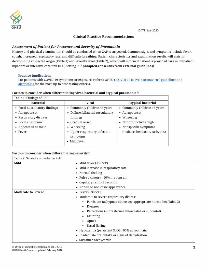
Assessment of Patient for Presence and Severity of Pneumonia History and physical examination should be conducted when CAP is suspected. Common signs and symptoms include fever, cough, increased respiratory rate, and difficulty breathing. Patient characteristics and examination results will assist in determining suspected origin (Table 1) and severity level (Table 2), which will inform if patient is provided care in outpatient, inpatient or intensive care unit (ICU) setting. 6,7,8 (Adapted-consensus from external guidelines)
Practice Implications For patients with COVID-19 symptoms or exposure, refer to OHSU’s COVID-19 (Novel Coronavirus) guidelines and algorithms for the most up-to-date testing criteria.
Factors to consider when differentiating viral, bacterial and atypical pneumonia9:
Table 1: Etiology of CAP Bacterial Viral Atypical bacterial
Focal auscultatory findings Abrupt onset Respiratory distress Local chest pain Appears ill or toxic Fever
Commonly children <5 years Diffuse, bilateral auscultatory
findings Gradual onset Wheezing Upper respiratory infection
symptoms Mild fever
Commonly children >5 years Abrupt onset Wheezing Nonproductive cough Nonspecific symptoms
(malaise, headache, rash, etc.)
Factors to consider when differentiating severity9:
Table 2. Severity of Pediatric CAP Mild Mild fever (<38.5°C)
Mild increase in respiratory rate Normal feeding Pulse oximetry >90% in room air Capillary refill <2 seconds Non-ill or non-toxic appearance
Moderate to Severe Fever (≥38.5°C) Moderate to severe respiratory distress
Persistent tachypnea above age-appropriate norms (see Table 3) Dyspnea Retractions (suprasternal, intercostal, or subcostal) Grunting Apnea Nasal flaring
Hypoxemia (persistent SpO2 <90% at room air) Inadequate oral intake or signs of dehydration Sustained tachycardia
DATE: July 2020
© Office of Clinical Integration and EBP, 2016 OHSU Health System, Updated February 2018
4
Capillary refill ≥2 seconds Failure of outpatient therapy (worsening symptoms or no response >48 hours
after initial outpatient therapy) Severe Severe respiratory distress
Remains hypoxic on >50% FiO2 Concern for impending respiratory failure Inadequate perfusion (altered mental status, hypotension, sustained
tachycardia) Need for mechanical ventilator support with artificial airway New or increased CPAP or BiPap support
Tachypnea Criteria10:
Table 3. Tachypnea age-adjusted respiratory rates Age (year) Respiratory Rate (breaths/minute)
2 months–1 year* 24–38 1–3 years 22–30 4–6 years 20–24 7–9 years 18–24 10–14 years 16–22 14–18 years 14–20
Clinical setting determined by severity Mild - Outpatient Management: Patients with mild CAP (as defined in table 2), adequate observation and follow-up care and ability to adhere to therapy, including adequate PO can be managed in the outpatient setting.9 (Strong Recommendation; Moderate Quality Evidence) Moderate – Inpatient Management Patients who have moderate to severe CAP (as defined in table 2), including significant respiratory distress and hypoxemia, or inability to tolerate PO (vomiting), should be hospitalized.9 (Strong Recommendation; Moderate Quality Evidence) Threshold for admission should be lower for infants 2-6 months, as infants may need additional monitoring and supportive care to prevent clinical deterioration. (Consensus) Additional Considerations Favoring Hospitalization:
• Suspected complicated CAP (pleural effusion/empyema, abscess) • Children who cannot be adequately cared for at home • Unable to comply with therapy, including inadequate PO • Unable to follow up with appointments
Severe – ICU Management Decision to treat severe patients (as defined in table 2) in an ICU unit should include signs of clinical deterioration such as sustained tachycardia, hypotension, altered mental status, or other signs of shock/impaired perfusion.9 (Adapted-consensus from external guidelines)

DATE: July 2020
© Office of Clinical Integration and EBP, 2016 OHSU Health System, Updated February 2018
5
Additional Consideration Favoring ICU Admission: • Patient does not respond to initial resuscitation and is clinically deteriorating
Diagnostic Evaluation To establish diagnosis of CAP, consider severity of disease factors (Table 2). History and physical assessment have demonstrated similar sensitivity and specificity to additional testing in predicting the etiologic agent of CAP and are generally sufficient to confirm diagnosis in cases of strongly suspected CAP. (Consensus) For patients with suspected viral pneumonia, consider viruses such as respiratory syncytial virus (RSV), influenza, COVID-19 and/or seasonal appropriateness of additional studies. 9 (Strong Recommendation; High Quality Evidence) Most laboratory tests (such as complete blood count or blood cultures) are not routinely recommended, as there is risk of potential contamination by other colonizing pathogens or multiple sources of infection, limited sensitivity and/or specificity for pathogens, difficulty in differentiating viral and bacterial pneumonia, and limited utility in informing clinical management. However, recommended testing will depend on severity and type of pneumonia, and requires clinical judgement based on patient assessment. Patients with signs and symptoms of moderate to severe disease and those with suspected bacterial CAP are more likely to develop complications and may therefore benefit from the use of chest radiograph or other imaging modalities. 9,11 (Strong Recommendation; High Quality Evidence) Imaging Mild No diagnostic testing is indicated for mild cases, unless patient meets criteria for hospitalization. Many studies use chest radiography as the preferred diagnostic modality, but positive findings have not been shown to improve clinical outcomes or significantly change treatment. Chest imaging is most useful when the diagnosis is uncertain or when the findings from the history and physical examination are inconclusive. 9,12-14 (Strong Recommendations; High Quality Evidence) Moderate and Severe For patients with equivocal clinical findings, chest radiograph (CXR) may be helpful when considering possible causes of respiratory distress. Bacterial pneumonia may be suspected based on radiographic findings; however, these findings are not highly specific. Pleural effusion is the most significant predictor of bacterial pneumonia. Alveolar consolidation is more suggestive of bacterial than viral infection, especially if the consolidation is lobar. Interstitial infiltrates can occur in viral or bacterial infections. Positive radiographic findings may be absent in patients with early bacterial pneumonia. 9,12-17 (Strong Recommendation; Moderate Quality Evidence)
Obtain both anterior-posterior (AP) or posterior-anterior (PA) and lateral views o AP in children <4 years o PA in children >4 years to minimize cardiac shadow
Follow-up chest radiograph not indicated, unless progressive symptoms or clinical deterioration after 48 to 72 hours post-therapy initiation or as recommended by a radiologist.
Point of care lung ultrasound is a potential alternative diagnostic modality to radiography, if obtained by proficient provider according to OHSU standards. If proficient provider is unavailable, consider formal ultrasound or chest radiography.
For suspected complications associated with CAP: Pleural effusion: consider point of care chest ultrasound if obtained by proficient provider. If proficient provider is
unavailable, consider formal ultrasound. Necrotizing pneumonia (prolonged fever, septic appearance): consider computed tomography (CT) with contrast or
CXR
DATE: July 2020
© Office of Clinical Integration and EBP, 2016 OHSU Health System, Updated February 2018
6
Lung abscess: consider CT with contrast or chest radiographs Microbiologic Testing Moderate Blood cultures are not routinely recommended in children requiring hospitalization for presumed uncomplicated bacterial CAP that is moderate in severity.9, 25, 26 (Strong Recommendation; Moderate Quality Evidence) Severe Clinicians should obtain blood cultures in cases of complicated or severe pneumonia and for those who are under- or unimmunized, particularly those with complicated pneumonia. 9, 26 (Strong Recommendations; Moderate Quality Evidence) A complete blood cell count (CBC) should be obtained only for patients with severe pneumonia, to be interpreted in the context of the clinical examination and other laboratory and imaging studies. 8,9 (Conditional Recommendation; Low Quality Evidence) Initial Treatment Consideration When initiating treatment, the clinician should consider setting, immunization status, β-lactam allergy, and suspected etiology. Immunization status should factor into threshold for initiating antibiotics, as under- or unimmunized patients are at high risk for bacterial CAP. See Table 5 for empiric selection of antibiotic therapy and Table 6 for Alternative therapy for beta-lactam allergy.9,12 (Consensus adapted from external guidelines) In children less than 5 years of age, etiology is more likely to be viral and routine use of antibiotics is not recommended.9 (Strong Recommendation; High Quality Evidence) For patients with suspected typical bacterial CAP,
In both fully and partially immunized children, amoxicillin is considered acceptable first line therapy for outpatient management.9,12 (Strong Recommendation; Moderate Quality Evidence)
o In children who are penicillin-allergic, consider a 3rd generation cephalosporin or clindamycin.
For patients with suspected atypical bacterial CAP, If >/= 5 years old, consider monotherapy with a macrolide or can be added to beta-lactam therapy if uncertainty of
diagnosis.9,12 (Conditional Recommendation; Moderate Quality Evidence) o Azithromycin is an acceptable first line therapy, o Doxycycline and/or levofloxacin are acceptable second line therapies.
For patients with suspected viral CAP,
Consider not initiating antibiotic therapy unless concerns for co-bacterial infection. If treatment is necessary for influenza, oseltamivir is considered acceptable first line therapy, and inhaled zanamivir is considered acceptable second line therapy if patient is older than 7 years old.9
Patients receiving intravenous therapy may be switched to oral treatment once they are afebrile and improving clinically, can tolerate oral intake, and have no complications (table 7).18
DATE: July 2020
© Office of Clinical Integration and EBP, 2016 OHSU Health System, Updated February 2018
7
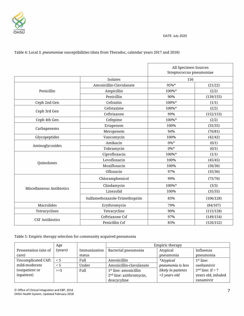
Table 4: Local S. pneumoniae susceptibilities (data from Theradoc, calendar years 2017 and 2018)
All Specimen Sources Streptococcus pneumoniae
Isolates 156
Penicillin Amoxicillin-Clavulanate 95%* (21/22)
Ampicillin 100%* (2/2) Penicillin 90% (139/155)
Ceph 2nd Gen Cefoxitin 100%* (1/1)
Ceph 3rd Gen Cefotaxime 100%* (2/2) Ceftriaxone 99% (152/153)
Ceph 4th Gen Cefepime 100%* (2/2)
Carbapenems Ertapenem 100% (35/35)
Meropenem 94% (76/81) Glycopeptides Vancomycin 100% (42/42)
Aminoglycosides Amikacin 0%* (0/1)
Tobramycin 0%* (0/1)
Quinolones
Ciprofloxacin 100%* (1/1) Levofloxacin 100% (45/45) Moxifloxacin 100% (36/36)
Ofloxacin 97% (35/36)
Miscellaneous Antibiotics
Chloramphenicol 99% (75/76)
Clindamycin 100%* (3/3) Linezolid 100% (35/35)
Sulfamethoxazole-Trimethoprim 83% (106/128)
Macrolides Erythromycin 79% (84/107) Tetracyclines Tetracycline 90% (115/128)
CSF Antibiotics Ceftriaxone Csf 97% (149/154)
Penicillin Csf 83% (126/152)
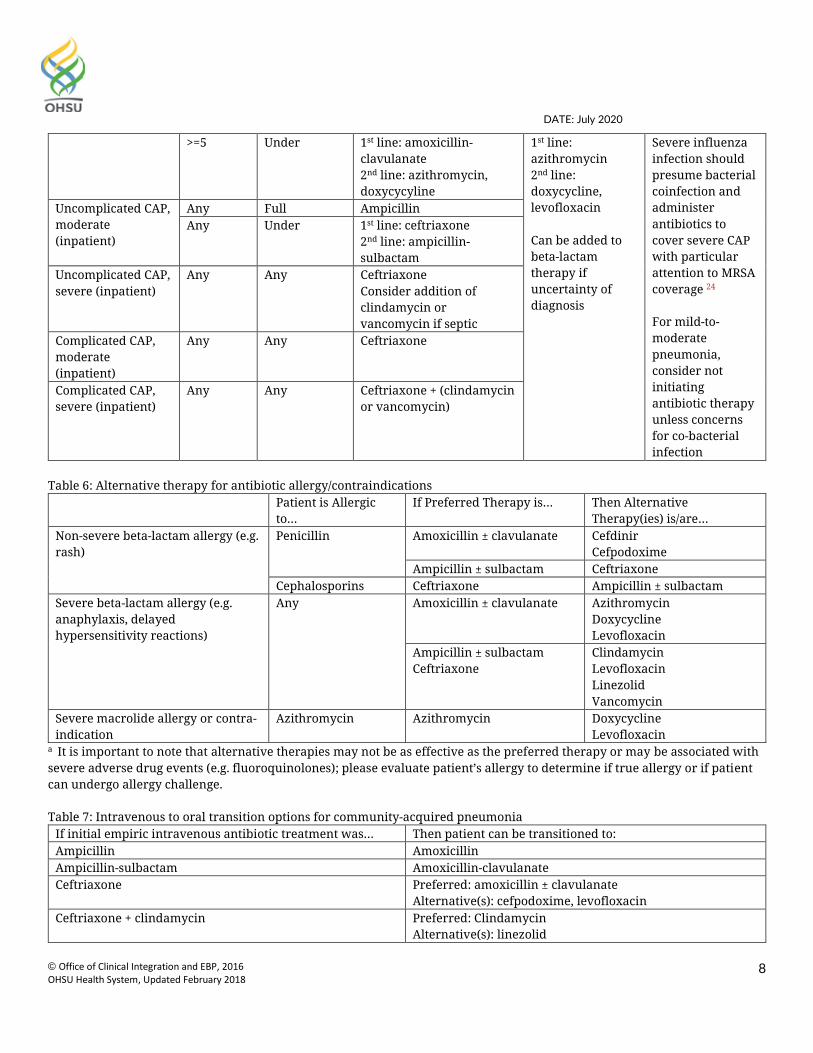
Table 5: Empiric therapy selection for community acquired pneumonia
Presentation (site of care)
Age (years) Immunization
status
Empiric therapy Bacterial pneumonia Atypical
pneumonia Influenza pneumonia
Uncomplicated CAP; mild-moderate (outpatient or inpatient)
< 5 Full Amoxicillin *Atypical pneumonia is less likely in patients <5 years old
1st line: oseltamivir 2nd line: if > 7 years old, inhaled zanamivir
< 5 Under Amoxicillin-clavulanate >=5 Full 1st line: amoxicillin
2nd line: azithromycin, doxcycyline
DATE: July 2020
© Office of Clinical Integration and EBP, 2016 OHSU Health System, Updated February 2018
8
>=5 Under 1st line: amoxicillin-clavulanate 2nd line: azithromycin, doxycycyline
1st line: azithromycin 2nd line: doxycycline, levofloxacin Can be added to beta-lactam therapy if uncertainty of diagnosis
Severe influenza infection should presume bacterial coinfection and administer antibiotics to cover severe CAP with particular attention to MRSA coverage 24
For mild-to-moderate pneumonia, consider not initiating antibiotic therapy unless concerns for co-bacterial infection
Uncomplicated CAP, moderate (inpatient)
Any Full Ampicillin Any Under 1st line: ceftriaxone
2nd line: ampicillin-sulbactam
Uncomplicated CAP, severe (inpatient)
Any Any Ceftriaxone Consider addition of clindamycin or vancomycin if septic
Complicated CAP, moderate (inpatient)
Any Any Ceftriaxone
Complicated CAP, severe (inpatient)
Any Any Ceftriaxone + (clindamycin or vancomycin)
Table 6: Alternative therapy for antibiotic allergy/contraindications
Patient is Allergic to…
If Preferred Therapy is… Then Alternative Therapy(ies) is/are…
Non-severe beta-lactam allergy (e.g. rash)
Penicillin Amoxicillin ± clavulanate Cefdinir Cefpodoxime
Ampicillin ± sulbactam Ceftriaxone Cephalosporins Ceftriaxone Ampicillin ± sulbactam
Severe beta-lactam allergy (e.g. anaphylaxis, delayed hypersensitivity reactions)
Any Amoxicillin ± clavulanate
Azithromycin Doxycycline Levofloxacin
Ampicillin ± sulbactam Ceftriaxone
Clindamycin Levofloxacin
Linezolid
Vancomycin Severe macrolide allergy or contra-indication
Azithromycin Azithromycin Doxycycline Levofloxacin
a It is important to note that alternative therapies may not be as effective as the preferred therapy or may be associated with severe adverse drug events (e.g. fluoroquinolones); please evaluate patient’s allergy to determine if true allergy or if patient can undergo allergy challenge. Table 7: Intravenous to oral transition options for community-acquired pneumonia
If initial empiric intravenous antibiotic treatment was… Then patient can be transitioned to: Ampicillin Amoxicillin Ampicillin-sulbactam Amoxicillin-clavulanate Ceftriaxone Preferred: amoxicillin ± clavulanate
Alternative(s): cefpodoxime, levofloxacin Ceftriaxone + clindamycin Preferred: Clindamycin
Alternative(s): linezolid
DATE: July 2020
© Office of Clinical Integration and EBP, 2016 OHSU Health System, Updated February 2018
9
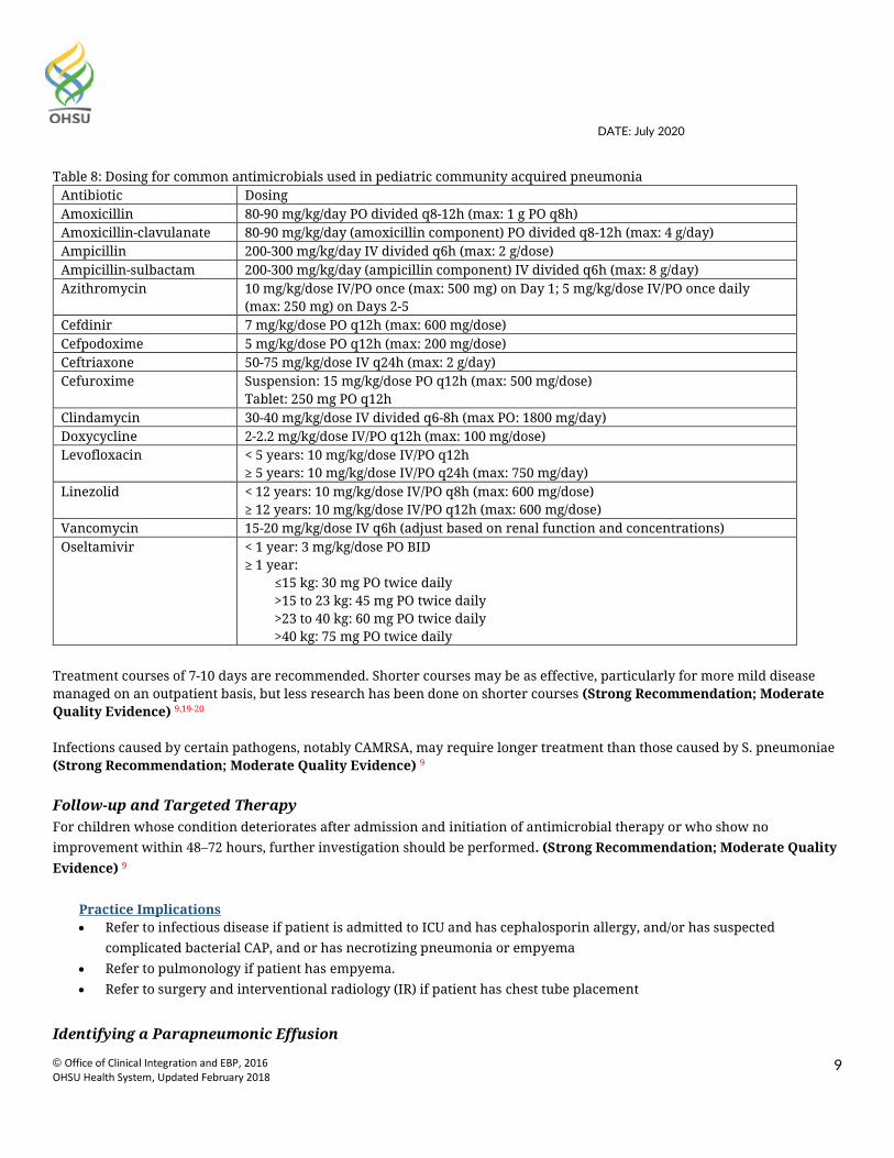
Table 8: Dosing for common antimicrobials used in pediatric community acquired pneumonia
Antibiotic Dosing Amoxicillin 80-90 mg/kg/day PO divided q8-12h (max: 1 g PO q8h) Amoxicillin-clavulanate 80-90 mg/kg/day (amoxicillin component) PO divided q8-12h (max: 4 g/day) Ampicillin 200-300 mg/kg/day IV divided q6h (max: 2 g/dose) Ampicillin-sulbactam 200-300 mg/kg/day (ampicillin component) IV divided q6h (max: 8 g/day) Azithromycin 10 mg/kg/dose IV/PO once (max: 500 mg) on Day 1; 5 mg/kg/dose IV/PO once daily
(max: 250 mg) on Days 2-5 Cefdinir 7 mg/kg/dose PO q12h (max: 600 mg/dose) Cefpodoxime 5 mg/kg/dose PO q12h (max: 200 mg/dose) Ceftriaxone 50-75 mg/kg/dose IV q24h (max: 2 g/day) Cefuroxime Suspension: 15 mg/kg/dose PO q12h (max: 500 mg/dose)
Tablet: 250 mg PO q12h Clindamycin 30-40 mg/kg/dose IV divided q6-8h (max PO: 1800 mg/day) Doxycycline 2-2.2 mg/kg/dose IV/PO q12h (max: 100 mg/dose) Levofloxacin < 5 years: 10 mg/kg/dose IV/PO q12h
≥ 5 years: 10 mg/kg/dose IV/PO q24h (max: 750 mg/day) Linezolid < 12 years: 10 mg/kg/dose IV/PO q8h (max: 600 mg/dose)
≥ 12 years: 10 mg/kg/dose IV/PO q12h (max: 600 mg/dose) Vancomycin 15-20 mg/kg/dose IV q6h (adjust based on renal function and concentrations) Oseltamivir < 1 year: 3 mg/kg/dose PO BID
≥ 1 year: ≤15 kg: 30 mg PO twice daily >15 to 23 kg: 45 mg PO twice daily >23 to 40 kg: 60 mg PO twice daily >40 kg: 75 mg PO twice daily
Treatment courses of 7-10 days are recommended. Shorter courses may be as effective, particularly for more mild disease managed on an outpatient basis, but less research has been done on shorter courses (Strong Recommendation; Moderate Quality Evidence) 9,19-20 Infections caused by certain pathogens, notably CAMRSA, may require longer treatment than those caused by S. pneumoniae (Strong Recommendation; Moderate Quality Evidence) 9 Follow-up and Targeted Therapy For children whose condition deteriorates after admission and initiation of antimicrobial therapy or who show no improvement within 48–72 hours, further investigation should be performed. (Strong Recommendation; Moderate Quality Evidence) 9
Practice Implications Refer to infectious disease if patient is admitted to ICU and has cephalosporin allergy, and/or has suspected
complicated bacterial CAP, and or has necrotizing pneumonia or empyema Refer to pulmonology if patient has empyema. Refer to surgery and interventional radiology (IR) if patient has chest tube placement
Identifying a Parapneumonic Effusion
DATE: July 2020
© Office of Clinical Integration and EBP, 2016 OHSU Health System, Updated February 2018
10
History and physical examination may be suggestive of parapneumonic effusion in children suspected of having CAP, but presence of pleural fluid should be confirmed with chest radiography (CXR) or chest ultrasound. If CXR identifies effusion greater than 10 mm, then further imaging with chest ultrasound is recommended to determine whether fluid is loculated or free-flowing. Contrast-enhanced CT plays a limited role in evaluation of pleural fluid and should only be obtained for this purpose at the request of an attending physician. (Strong recommendation; Moderate Quality Evidence) 7-9, 11, 13
The child’s degree of respiratory compromise is an important factor that determines management of parapneumonic effusions. (Strong Recommendation; Moderate Quality Evidence) 21
The size of the effusion and whether free-flowing or loculated are important factors that determines management. (Strong Recommendation; Moderate Quality Evidence) 7
Laboratory Testing Gram stain and bacterial culture of pleural fluid should be performed whenever a pleural fluid specimen is obtained. (Strong Recommendation; Moderate Quality Evidence) 9
Nucleic acid amplification through PCR increases the detection of pathogens in pleural fluid and may be useful for management of Streptococcus pneumoniae. (Strong Recommendation; Moderate Quality Evidence) 9 Analysis of pleural fluid parameters such as pH and levels of glucose, protein, and lactate dehydrogenase, rarely change patient management and are not recommended. (Conditional Recommendation; Very Low Quality Evidence) 9 Analysis of the pleural fluid white blood cell (WBC) count, with cell differential analysis, is recommended primarily to help differentiate bacterial from mycobacterial etiologies and from malignancy. (Conditional Recommendation; Moderate Quality Evidence) 9
Drainage Small, uncomplicated parapneumonic effusions should not routinely be drained and can be treated with antibiotic therapy alone. (Strong Recommendation; Moderate Quality Evidence) 7,9,13, 21 Moderate parapneumonic effusions associated with respiratory distress, large parapneumonic effusions, or documented purulent effusions should be drained. (Strong Recommendation; Moderate Quality Evidence) 7,9,13,21
Both chest thoracostomy tube drainage with the addition of fibrinolytic agents (tPA) and VATS have been demonstrated to be effective methods of treatment. The choice of drainage procedure depends on local expertise. Both of these methods are associated with decreased morbidity compared with chest tube drainage alone. However, in patients with moderate-to-large effusions that are free flowing (no loculations), placement of a chest tube without fibrinolytic agents is a reasonable first option. (Strong Recommendation; High Quality Evidence) 9
Video-assisted Thoracoscopic Surgery (VATS) VATS should be considered when there is persistence of moderate to large effusions and ongoing respiratory compromise after failure of management with maximal chest tube and fibrinolytic therapy. (Strong Recommendation; Low Quality Evidence) 6-7,9,13
DATE: July 2020
© Office of Clinical Integration and EBP, 2016 OHSU Health System, Updated February 2018
11
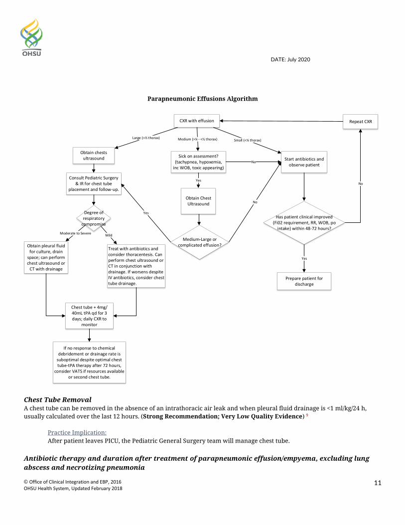
Parapneumonic Effusions Algorithm
CXR with effusion
Small (<¼ thorax)
Start antibiotics and observe patient
Medium (>¼ - <½ thorax)Large (>½ thorax)
Obtain chests ultrasound
Has patient clinical improved (Fi02 requirement, RR, WOB, po
intake) within 48-72 hours?
No
Repeat CXR
Yes
Prepare patient for discharge
Sick on assessment? (tachypnea, hypoxemia,
inc WOB, toxic appearing)
Yes
Obtain Chest Ultrasound
Medium-Large or complicated effusion?
Consult Pediatric Surgery & IR for chest tube
placement and follow-up.
No
No
YesDegree of respiratory
compromise
Moderate to Severe
Obtain pleural fluid for culture, drain
space; can perform chest ultrasound or
CT with drainage
Mild
Treat with antibiotics and consider thoracentesis. Can perform chest ultrasound or CT in conjunction with drainage. If worsens despite IV antibiotics, consider chest tube drainage.
Chest tube + 4mg/40mL tPA qd for 3 days; daily CXR to
monitor
If no response to chemical debridement or drainage rate is
suboptimal despite optimal chest tube-tPA therapy after 72 hours,
consider VATS if resources available or second chest tube.
Chest Tube Removal A chest tube can be removed in the absence of an intrathoracic air leak and when pleural fluid drainage is <1 ml/kg/24 h, usually calculated over the last 12 hours. (Strong Recommendation; Very Low Quality Evidence) 9
Practice Implication: After patient leaves PICU, the Pediatric General Surgery team will manage chest tube.
Antibiotic therapy and duration after treatment of parapneumonic effusion/empyema, excluding lung abscess and necrotizing pneumonia
DATE: July 2020
© Office of Clinical Integration and EBP, 2016 OHSU Health System, Updated February 2018
12
When blood or pleural fluid bacterial culture identifies a pathogenic isolate, antibiotic susceptibility should be used to determine the antibiotic regimen. (Strong Recommendation; High Quality Evidence) 9
Practice Implication: Consult with Pediatric Infectious Disease.
Empiric therapy selection as described in Table 5 for ‘Complicated CAP – Severe (Inpatient) o Antibiotic therapy should be pathogen-directed, based on results of bacterial culture o If culture negative, or treatment started prior to obtaining fluid, therapy selection should be guided
by regional epidemiology De-escalate and continue therapy for additional 7 days, once all criteria below are met
o Chest tube removed o Afebrile o If patient had a positive blood culture, at least 1 negative blood culture
Appropriate management if patient is not responding to treatment Children who are not responding to initial therapy after 48-72 hours should be managed by one or more of the following:
Clinical and laboratory assessment of the current severity of illness and anticipated progression in order to determine whether higher levels of care or support are required. (Strong Recommendation; Low Quality Evidence) 9 Practice Implication:
o Obtain sputum using flexible bronchoscopy with bronchoalveolar lavage to identify causative pathogens, if possible
o For additional clarification post-bronchoscopy, may consider CT with contrast
Further investigation to identify whether the original pathogen persists, the original pathogen was developed resistance to the agent used, or there is a new secondary infecting agent. (Conditional Recommendation; Low Quality Evidence) 9
Practice Implication: Treatment
o Expand coverage for MRSA and common local pathogens (e.g., ceftaroline, vancomycin, linezolid) Discharge Consider discharge if patient demonstrates overall clinical improvement including: (Adapted-consensus based on external guidelines) 7-9, 13-14, 22-23
Improved work of breathing Increased activity Decreased respiratory rate Decreasing fever curve Pulse oximetry >90% on room air for at least 12 – 24 hours Able to take medications orally Follow-up care coordinated:
o Scheduled an appointment with primary care provider within 72 hours For patients with complicated pneumonia, consider additional appointments with o Scheduling appointment with infectious disease provider o Scheduling appointment with pulmonary provider in 6 to 8 weeks.
Prevention
DATE: July 2020
© Office of Clinical Integration and EBP, 2016 OHSU Health System, Updated February 2018
13
Screen all patients for influenza, pneumococcal, Hib, and/or pertussis immunizations at admission. (Adapted-consensus based on external guideline) 22
Provide influenza antiviral therapy for all children hospitalized with flu. (Adapted-consensus based on external guideline) 22
Children should be immunized with vaccines for bacterial pathogens, including S. pneumoniae, Haemophilus influenzae type b, and pertussis to prevent CAP. (Strong Recommendation; High Quality Evidence) 9
All infants >6 months of age and all children and adolescents should be immunized annually for influenza virus to prevent CAP. (Strong Recommendation; High Quality Evidence) 9
Parents and caretakers of infants <6 months of age, including pregnant adolescents, should be immunized with vaccines for influenza virus and pertussis to protect the infants from exposure. (Strong Recommendation; Low Quality Evidence) 9
Pneumococcal CAP after influenza virus infection is decreased by immunization against influenza virus. (Strong Recommendation; Low Quality Evidence) 9
Improved hand hygiene is an important prevention strategy. (Adapted-consensus based on external guideline) 23
Practice Implication: For patients requiring isolation, please refer to OHSU Isolation Orders
DATE: July 2020
© Office of Clinical Integration and EBP, 2016 OHSU Health System, Updated February 2018
14
Quality Measures: Process:
Percentage of patients receiving appropriate antibiotics Total duration of antibiotics by type (complicated vs. uncomplicated) Number of tissue plasminogen activator (tPA) treatments Duration of chest tube therapy (earlier tPA, efficacy of chest tube management) Diagnostic approach (# of CTs, blood culture, point of care ultrasound, full ultrasound)
Outcomes:
Length of stay Readmissions % of patients receiving immunizations
Implementation Needs:
Point of Care Ultrasound (POCUS) Education Delegation Protocol (Giving immunizations; involving nutrition & respiration therapy) Order Set (Create different order sets for complicated and uncomplicated)
o Create best practice alert Education
o Dissemination meeting for residents o Patient education on immunizations and hand washing o Launch Get Well Campaign
Dashboard to monitor data Dissemination (Sub-specialties & partner sites)
DATE: July 2020
© Office of Clinical Integration and EBP, 2016 OHSU Health System, Updated February 2018
15
References
1. Kronman, M. P., et al. (2011). "Ambulatory Visit Rates and Antibiotic Prescribing for Children With Pneumonia, 1994–2007." Pediatrics 127(3): 411-418.
2. Jain, S., et al. (2015). "Community-acquired pneumonia requiring hospitalization among U.S. children." New England Journal of Medicine 372(9): 835-845.
3. Mulholland, E. K., et al. (1992). "Standardized diagnosis of pneumonia in developing countries." Pediatric Infectious Disease Journal 11(2): 77-81.
4. Black, R. E., et al. (2010). "Global, regional, and national causes of child mortality in 2008: a systematic analysis." Lancet 375(9730): 1969-1987.
5. Jadavji, T., et al. (1997). "A practical guide for the diagnosis and treatment of pediatric pneumonia." CMAJ Canadian Medical Association Journal 156(5): S703-711.
6. New South Wales Health (2018). Infants and Children: Acute Management of Community Acquired Pneumonia. Accessed on October 14, 2019 at https://www1.health.nsw.gov.au/pds/ActivePDSDocuments/GL2018_007.pdf.
7. Children’s Hospital of Philadelphia (2017). Pathway for Evaluation and Treatment of Child with Community-Acquired Pneumonia. Accessed October 14, 2019 at https://www.chop.edu/clinical-pathway/pneumonia-community-acquired-clinical-pathway.
8. Seattle Children’s Hospital (2018). Community-Acquired Pneumonia. Accessed October 14, 2019 at https://www.seattlechildrens.org/pdf/pneumonia-pathway.pdf.
9. Bradley, J. S., et al. (2011). "The management of community-acquired pneumonia in infants and children older than 3 months of age: clinical practice guidelines by the Pediatric Infectious Diseases Society and the Infectious Diseases Society of America." Clinical Infectious Diseases 53(7): e25-76.
10. Gillon, J. Pulmonology. In: The Harriet Lane Handbook: A Manual for Pediatric House Officers. 21st ed. Philadelphia, PA: Elsevier, 2018.
11. American Academy of Pediatrics (2017). AAP Section on Emergency Committee on Quality Transformation Clinical Algorithm for Emergency Department Evaluation and Management of Pediatric Community Acquired Pneumonia. Accessed October 14, 2019 at https://downloads.aap.org/DOCCSA/SOEM%20Pneum.pdf.
12. Stuckey-Schrock, K., et al. (2012). "Community-acquired pneumonia in children." American Family Physician 86(7): 661-667.
13. Messinger, A. I., et al. (2017). "Management of Pediatric Community-acquired Bacterial Pneumonia." Pediatrics in Review 38(9): 394-409.
14. Brown, M. D., et al. (2016). "Clinical Policy for Well-Appearing Infants and Children Younger Than 2 Years of Age Presenting to the Emergency Department With Fever." Annals of Emergency Medicine 67(5): 625-639.e613.
15. Michelow, I. C., et al. (2004). "Epidemiology and clinical characteristics of community-acquired pneumonia in hospitalized children." Pediatrics 113(4): 701-707
16. Virkki, R., et al. (2002). "Differentiation of bacterial and viral pneumonia in children." Thorax 57(5): 438-441. 17. Margolis, P. and A. Gadomski (1998). "The rational clinical examination. Does this infant have pneumonia?"
JAMA 279(4): 308-313. 18. Harris, M., et al. (2011). "British Thoracic Society guidelines for the management of community acquired
pneumonia in children: update 2011." Thorax 66(Suppl 2): ii1-ii23. 19. Greenberg, D., et al. (2014). "Short-course antibiotic treatment for community-acquired alveolar pneumonia in
ambulatory children: a double-blind, randomized, placebo-controlled trial." Pediatric Infectious Disease Journal 33(2): 136-142.
20. Lopez-Alcalde, J., et al. (2018). "Short-course versus long-course therapy of the same antibiotic for community-acquired pneumonia in adolescent and adult outpatients." Cochrane Database of Systematic Reviews 9: CD009070.
21. Le Saux, N., et al. (2015). "Uncomplicated pneumonia in healthy Canadian children and youth: Practice points for management." Paediatrics & child health 20(8): 441-450.
DATE: July 2020
© Office of Clinical Integration and EBP, 2016 OHSU Health System, Updated February 2018
16
22. Intermountain Health (2013). Pediatric Community-Acquired Pneumonia (CAP) – patients age 3 months and older without bronchiolitis. Accessed October 14, 2019 at https://intermountainhealthcare.org/ckr-ext/Dcmnt?ncid=522578601.
23. Children’s Hospital Colorado (2016). Uncomplicated Community Acquired Pneumonia. Accessed 14, 2019 at https://www.childrenscolorado.org/globalassets/healthcare-professionals/clinical-pathways/community-acquired-pneumonia-uncomplicated.pdf.
24. Randolph, A. G., et al. (2019). "Vancomycin Monotherapy May Be Insufficient to Treat Methicillin-resistant Staphylococcus aureus Coinfection in Children With Influenza-related Critical Illness." Clinical infectious diseases : an official publication of the Infectious Diseases Society of America 68(3): 365-372.
25. Neuman, M. I., et al. (2017). "Utility of Blood Culture Among Children Hospitalized With Community-Acquired Pneumonia." 140(3): e20171013.
26. Fritz, C. Q., et al. (2019). "Prevalence, Risk Factors, and Outcomes of Bacteremic Pneumonia in Children." 144(1): e20183090.

DATE: July 2020
© Office of Clinical Integration and EBP, 2016 OHSU Health System, Updated February 2018
17
Guideline Preparation This guideline was prepared by the Office of Clinical Integration (CI) and Evidence-Based Practice (EBP) in collaboration with content experts at Oregon Health and Science University and Hillsboro Medical Center. Content Expert Team Alex Foster, Pediatric Hospitalist, OHSU Andrew Johnson, Interventional Radiology, OHSU Beech Burns, Emergency Medicine, OHSU Ben Hoffman, Pediatrics, OHSU Bronwyn Baz, Pediatrics, Kaiser Permanente Cat Livingston, Family Medicine, OHSU Cheri Warren, Informatics, OHSU Christina Ramo, Pediatrics, OHSU Cydni Williams, Critical Care, OHSU Dawn Nolt, Infectious Disease, OHSU Diana Yu, Pharmacy, OHSU Deidra Weinert, Acute Care Nursing, OHSU Elizabeth Fialkowski, General Surgery, OHSU George Schwoegler, Respiratory Therapy, OHSU Hayes Bakken, Pediatrics, OHSU Jared Austin, Hospital Medicine, OHSU Katharine Hopkins, Diagnostic Radiology, OHSU Kim Wirth, Family Representative Louise Vaz, Infectious Disease, OHSU Mike Powers, Pulmonology, OHSU Mina Tahai, Pediatrics, OHSU Natalie Wilcox, Pediatrics, OHSU Rachel Castelli, Emergency Medicine, Tuality Richard (Mick) Scanlan, Pathology, OHSU
Clinical Integration and EBP Team Marcy Hager, MA, EBP Program Manager Andrew Hamilton, MS/MLS, Liaison Librarian Stephanie Halvorson, MD, Medical Director, Clinical Integration Rebecca Jungbauer, DrPH, MPH, MA, Research Associate/Project Manager, Evidence-based Practice Center Marian McDonagh, PharmD, Associate Director of the Evidence-based Practice Center (EPC) Development Process This guideline was developed using the process outlined in the CI and EBP Manual (2016). The review summary documents the following steps: 1. Review Preparation
- PICO questions established
- Evidence search confirmed with content experts 2. Review of Existing Internal and External Guidelines
- Literature Review of Relevant Evidence 3. Critically Analyze the Evidence 4. Summarize the Evidence by preparing the guideline, and
order sets - Materials used in the development of the guidelines, review summaries are maintained in ...
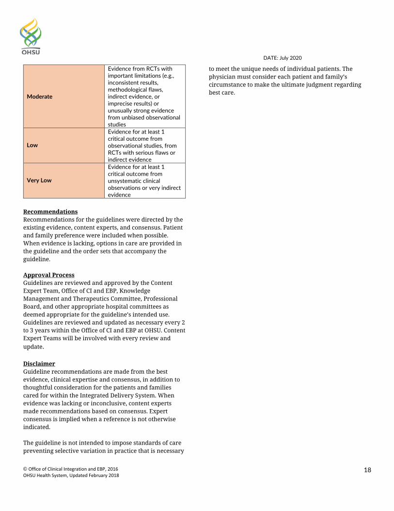
Evaluating the Quality of the Evidence Published clinical guidelines were evaluated for this review using the University of Pennsylvania’s Trustworthy Guideline Rating Scale. The summary of these guidelines are included in the evidence summary. The rating scale is based on the Institute of Medicine’s “Standards for Developing Trustworthy Clinical Practice Guidelines” (IOM), as well as a review of the AGREE Enterprise and Guidelines International Network domains. This scale evaluates a guideline’s transparency, conflict of interest, development group, systematic review, supporting evidence, recommendations, external review and currency and updates. The purpose of this scale is to focus on the weaknesses of a guideline that may reduce the trust a clinical user can have in the guideline, and distinguish weaknesses in documentation (e.g. guideline does not have a documented updating process) from weaknesses in the guidance itself (e.g. recommendations are outdated). The GRADE (Grading of Recommendations, Assessment, Development and Evaluation) criteria were utilized to evaluate the body of evidence used to make clinical recommendations. The table below defines how the quality of the evidence is rated and how a strong versus conditional recommendation is established. The evidence summary reflects the critical points of evidence.
Recommendation
STRONG Desirable effects clearly outweigh undesirable effects or vice versa
CONDITIONAL Desirable effects closely balanced with undesirable effects
Quality Type of Evidence
High
Consistent evidence from well-performed RCTs or exceptionally strong evidence from unbiased observational studies
DATE: July 2020
© Office of Clinical Integration and EBP, 2016 OHSU Health System, Updated February 2018
18
Moderate
Evidence from RCTs with important limitations (e.g., inconsistent results, methodological flaws, indirect evidence, or imprecise results) or unusually strong evidence from unbiased observational studies
Low
Evidence for at least 1 critical outcome from observational studies, from RCTs with serious flaws or indirect evidence
Very Low
Evidence for at least 1 critical outcome from unsystematic clinical observations or very indirect evidence
Recommendations Recommendations for the guidelines were directed by the existing evidence, content experts, and consensus. Patient and family preference were included when possible. When evidence is lacking, options in care are provided in the guideline and the order sets that accompany the guideline. Approval Process Guidelines are reviewed and approved by the Content Expert Team, Office of CI and EBP, Knowledge Management and Therapeutics Committee, Professional Board, and other appropriate hospital committees as deemed appropriate for the guideline’s intended use. Guidelines are reviewed and updated as necessary every 2 to 3 years within the Office of CI and EBP at OHSU. Content Expert Teams will be involved with every review and update.
Disclaimer Guideline recommendations are made from the best evidence, clinical expertise and consensus, in addition to thoughtful consideration for the patients and families cared for within the Integrated Delivery System. When evidence was lacking or inconclusive, content experts made recommendations based on consensus. Expert consensus is implied when a reference is not otherwise indicated. The guideline is not intended to impose standards of care preventing selective variation in practice that is necessary
to meet the unique needs of individual patients. The physician must consider each patient and family’s circumstance to make the ultimate judgment regarding best care.