Pediatric Advanced Life Support Overview - c.ymcdn.comc.ymcdn.com/sites/ · Pediatric Advanced Life...
26
Pediatric Advanced Life Support Overview 2010 American Heart Association Guidelines for CPR and ECC The following is not meant to replace AHA printed materials. It is meant to be used in conjunction with the PALS textbook and other printed materials. Judy Haluka 2/1/2011
Transcript of Pediatric Advanced Life Support Overview - c.ymcdn.comc.ymcdn.com/sites/ · Pediatric Advanced Life...

Pediatric Advanced Life
Support Overview 2010 American Heart Association Guidelines for
CPR and ECC
The following is not meant to replace AHA printed materials.
It is meant to be used in conjunction with the PALS textbook
and other printed materials.
Judy Haluka
2/1/2011
Pediatric Advanced Life Support Overview 2011
2
Prepared by Judy Haluka
Change is about the only constant in medicine. Lucky for us medicine moves forward at
exciting speed allowing us to heal injuries, conquer illness and prolong life more efficiently with
each step forward. For those of us who have been in healthcare for a period of time change
can be frustrating at least. After all, have we really been killing people with the way we’ve been
doing things all these years??? Resistance to change is resistance to forward movement. If
everyone resisted change we would still be riding donkeys as everyone knows it is impossible to
train a horse for anything of any values. Those young kids…..the ones that insist on chopping of
the corners of the stones we are trying to pull up the hill….they say it will be easier if it is
rounded at the edges…..but we’ve always done it this way. Get the idea?
This overview is based on the 2010 American Heart Association Guidelines for
Cardiopulmonary Resuscitation and Emergency Cardiovascular Care – Supplement to
Circulation – Volume 122 – Issue 18 – Supplement 3 – November 2, 2010. It is not meant to
take the place of AHA publications or textbooks. It is meant to be use as an study adjunct to
those publications.
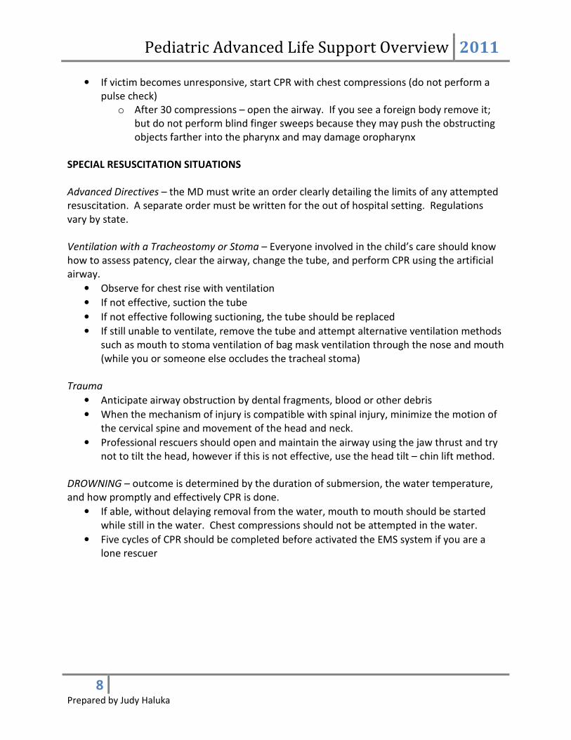
The Pediatric Chain of Survival emphasizes that multiple things must come together to enhance
survival in cardiac arrest. The biggest impact on pediatric survival is had when bystander CPR is
performed quickly and effectively. There have been up to 70% neuro intact survival reported
when everything goes perfectly. However only 1/3 to ½ of infants and children who have
cardiac arrest actually receive bystander CPR. The survival rates for infants and children not
changed substantially in over 20 years.
• Infants – 4% survival in out of hospital arrest
• Children 10% survival in out of hospital arrest
• Adolescents – 13% survival in out of hospital arrest
In hospital survival numbers are better at 27%. Infants and children who are not permitted to
completely arrest (with a pulse but poor perfusion and bradycardia) who required CPR had the
best in hospital survival rate of 64% to discharge. Children are more likely to survive in hospital
cardiac arrest than adults and infants have a higher survival rate than children.

Pediatric Advanced Life Support Overview 2011
3
Prepared by Judy Haluka
The change from ABC to CAB applies to infants and children as well as adults.
• High quality compressions are essential to generate blood flow to vital organs
• Beginning CPR with 30 compressions rather than 2 ventilations leads to shorter delays
to first compression
• All rescuers should be able to deliver compressions almost immediately. Positioning the
head and attaining a seal for ventilation takes time and delays the initiation of CPR
• Hypoxic arrest is more common than VF in infants and children and ventilations are
extremely important in pediatric resuscitation.
• Starting with compressions only delays ventilations by approximately 18 seconds for a
lone rescuer and even shorter for two rescuers
• CAB simplifies training with the hope that more victims will receive bystander CPR
Age Definitions
• Infant = under one year of age
• Child = children approximately 1 year to puberty
o Defined as breast development in girl
o Presence of armpit hair in the boy
• Adult = applies to all beyond puberty
BASIC LIFE SUPPORT FOR LAY RESCUERS
ASSESSMENT – the lay rescuer simply checks for responsiveness and absence of NORMAL
breathing. It is very important to emphasize NORMAL breathing as many mistaken agonal
gasps for breathing and therefore do not intervene. If normal breathing is not present and the
patient is not responsive CPR should be started.
• If the patient is found to be unresponsive but breathing – leave the child and summon
medical assistance immediately
• Children respiratory distress often assume a position in which they can breathe the
easiest. Allow the child to assume this position of comfort.
CHEST COMPRESSIONS – immediately begin chest compressions (30 compressions)
• “Push Hard, Push Fast” at a rate of at least 100/minute
• Depress the chest at least one third the anterior-posterior diameter of the chest
(approx. 1 ½ in infants and 2 inches in children)
o Inadequate chest compression is common both in the lay rescuer and the
healthcare professional
o There is no data to show that one or two hand method produces better
compressions or outcomes
• Chest recoil must be complete. This is the time during which the heart fills with blood
and the coronary arteries are perfused. It is acceptable to come slightly off the chest
wall in between compressions to ensure complete chest recoil. Remember that this can
also be a function of tiring. As the rescuer gets fatigued he/she tends to lean on the

Pediatric Advanced Life Support Overview 2011
4
Prepared by Judy Haluka
chest wall and not allow complete recoil. This is the reason for the recommended
switch of compressors that occurs every 2 minutes of the resuscitation
• Minimize interruptions of chest compressions. Some studies show that compressions
may only be performed half of the time during resuscitations. Given that adequate
compressions are the single most determinant of survival; this is unacceptable. Care
must be taken to minimize interruptions that are not absolutely necessary.
• Avoid hyperventilation – many studies have shown that patients are hyperventilated.
This is bad for a number of reasons; but the most basic is that when the chest cavity is
filled with air, no gradient occurs with chest recoil and the heart or the coronary arteries
do not fill with blood.
AIRWAY AND VENTILATIONS – for the lone rescuer the compression to ventilation ratio is 30:2
for all age groups.
• Open the airway – use the head tilt chin lift maneuver for both injured and non-injured
victims. The jaw thrust method should not be taught or used for lay rescuers.
• Use mouth to mouth and nose technique to give breaths to an infant
• Use mouth to mouth to give breaths to a child
• Each breath should take approximately 1 second and result in chest rise
o If the chest fails to rise, reposition the head, attempt a better seal and attempt
ventilation again. It may be necessary to move the patient’s head to a range o f
positions to provide optimal airway patency and effective breathing
• Mouth to nose or mouth to mouth may also be used of difficulty is encountered
• The lone rescuer should immediately begin compressions following 2 breaths. This cycle
of 30:2 should be continued for approximately 2 minutes before leaving the victim to
activate the Emergency Response System and obtain an automated external defibrillator
(AED) if one is nearby.
ACTIVATE THE EMERGENCY RESPONSE SYSTEM
• If two rescuers, one should start CPR immediately and the other should activate the
EMS system and get the AED if available
• Because most infants and children have a respiratory cause of arrest, not a cardiac one,
2 minutes of CPR are recommended before leaving the child to activate the EMS system
if there is only one rescuer at the scene.
BASIC LIFE SUPPORT FOR THE HEALTHCARE PROVIDER
Healthcare providers most often work in teams, such as in a hospital, or on an ambulance.
Therefore the steps of CPR do not have to be sequential, but can be accomplished
simultaneously by a healthcare team.
Healthcare providers are able to tailor the sequence of resuscitation actions to the most likely
cause of arrest.

Pediatric Advanced Life Support Overview 2011
5
Prepared by Judy Haluka
• If the arrest is sudden in an adolescent or child identified at high risk for arrhythmia or
during an athletic event, the provider may assume the victim has suffered a sudden VF
arrest and proceed accordingly.
ASSESSMENT
• If the victim is unresponsive and not breathing assume the need for CPR and begin
immediately.
• Healthcare providers may take 10 seconds to check for a pulse. If after 10 seconds you
do not feel a pulse or are not sure, begin CPR
o Brachial in an infant
o Carotid or femoral in a child
• If there is a pulse, but no or inadequate breathing, rescue breaths are provided at a rate
of 12-20 breaths per minute (1 every 3-5 seconds)
BRADYCARDIA WITH POOR PERFUSION
• Pulse less than 60 with poor perfusion despite support of oxygenation and ventilation,
begin chest compressions
o Cardiac output in infancy and childhood is dependent upon heart rate
o Cardiac arrest is imminent and beginning CPR prior to full cardiac arrest results in
increased survival
o These patients have the highest survival rate among in hospital patients
CHEST COMPRESSIONS – the only difference between healthcare professionals and lay persons
is in compressions for infants
• The lone rescuer should use the 2 finger chest compression technique for infants
• If not a lone rescuer – the two thumb encircling hands technique is recommended when
CPR is provided by 2 rescuers. Encircle the chest with both hands; spread your fingers
around the thorax, and place your thumbs together over the lower third of the sternum
o Preferred because it provides higher coronary artery perfusion pressures and
more consistently results in appropriate depth or force of compressions
o May generate higher systolic and diastolic pressures.

Pediatric Advanced Life Support Overview 2011
6
Prepared by Judy Haluka
VENTILATIONS
• Ventilation to Compression Ratio 30:2 if single rescuer
o 15:2 if two rescuers
• Airway is opened with head tilt – chin lift unless there is evidence of trauma
o If trauma use the jaw thrust method to open the airway – IF IT IS DIFFICULT TO
MAINTAIN A PATENT AIRWAY WITH THIS METHOD – USE THE HEAD TILT – CHIN
LIFT because airway is of utmost importance in infants and children
• Coordinate ventilations with chest compressions until there is an advanced airway in
place
o After advanced airway placement (intubation, supraglottic airway) cycles of
compressions and ventilations are no longer performed
o Compressions are performed at a rate of at least 100 per minute
o Ventilations are performed at a rate of 8-10 breaths per minute (one every 6-8
seconds)
DEFIBRILLATION USING AUTOMATED EXTERNAL DEFIBRILLATOR (AED)
• AEDs have high specificity in recognizing pediatric shockable rhythms
• Some are equipped to attenuate the delivered energy to make them suitable for infants
and children
• In infants a manual defibrillator is preferred when a shockable rhythm is identified by a
healthcare provider
• Recommended first dose energy is 2 joules/kg
o Second dose is 4 joules per kg
o At least 4 joules/kg for subsequent doses and higher energy levels may be
considered not to exceed 10 joules/kg
• AED equipped with a pediatric attenuator (pediatric pads) is preferred for infants and
children. If it is not available and an AED without a dose attenuator is available it should
be used on both infants and children with shockable rhythms.
• The time between the last compression and the shock delivery should be minimized as
much as possible
HANDS ONLY (COMPRESSION ONLY) CPR – most arrests in infants and children are of a
respiratory nature and rescue breathing is more effective than compressions alone when this is
the case and the arrest was from a non cardiac etiology. In some studies survival when hands
only CPR was used in infants and children were no better than when no CPR was done at all.
Optimal CPR in infants and children includes both compressions and ventilations, but
compressions alone are preferable to no CPR.
BREATHING DEVICES
• Barrier devices have not reduced the low risk of transmissions of infection and some
increase the resistance to flow
• If there is any delay in obtaining a barrier device or ventilation equipment, give mouth
to mouth ventilations or continue chest compressions alone

Pediatric Advanced Life Support Overview 2011
7
Prepared by Judy Haluka
• Bag Valve Mask ventilation is an essential CPR technique for healthcare providers
o Self inflating bag of at least 450-500ml for infants and young children
o 1000ml for children or adolescents
o Not recommended for the lone rescuer – two person technique with one person
sealing the mask and the other depressing the bag is more effective in providing
adequate ventilation
HYPERVENTILATION is harmful because
• Intra-thoracic pressure is increased and venous return to the heart is decreased.
Therefore, cardiac output, cerebral blood flow and coronary perfusion pressure are all
compromised.
• Air is trapped and barotrauma can occur
• Increases the risk of regurgitation and aspirations in patients without an advanced
airway in place
• Ventilation should stop when chest rise is noted.
• Patients with airway obstruction or poor lung compliance may require higher pressures
to ventilate properly. Be sure that your BVM allows the use of high pressures.
OXYGEN DELIVERY – it is reasonable to use 100% oxygen during resuscitation
• Once perfusion returns, monitor systemic oxygen saturation and titrate oxygen to
maintain an oxygen saturation of 94% or greater.
• Since an oxygen saturation of 100% may correspond to a Pa02 anywhere between 80-
500 mmHg, it is appropriate to wean FI02 for a saturation of 100% provided the
oxyhemoglobin saturation can be maintained at 94% or greater.
• Whenever possible oxygen should be humidified.
CPR ADJUNCTS – there is insufficient data in infants and children to recommend for or against
the use of mechanical devices to compress the chest, active compression-decompression CPR,
interposed abdominal compression, the impedance threshold device, or pressure sensor
accelerometers.
FOREIGN BODY AIRWYA OBSTRUCTION – 90% of childhood deaths from foreign body
aspiration occur in children less than 5 years old. Liquids are the most common cause of
chocking in infants. Balloons, small objects and foods (hot dogs, round candies, nuts and
grapes) are the most common causes in children.
Sudden onset of respiratory distress with coughing, gagging, stridor or wheezing;
• If mild, do not interfere. Allow the victim to clear by coughing
• If victim unable to make sound
o Perform subdiaphragmatic abdominal thrusts (Heimlich maneuver) until the
body is expelled in children
o For INFANT deliver repeated cycles of 5 back blows and 5 chest compressions
until the object is expelled or victim becomes unresponsive
Pediatric Advanced Life Support Overview 2011
8
Prepared by Judy Haluka
• If victim becomes unresponsive, start CPR with chest compressions (do not perform a
pulse check)
o After 30 compressions – open the airway. If you see a foreign body remove it;
but do not perform blind finger sweeps because they may push the obstructing
objects farther into the pharynx and may damage oropharynx
SPECIAL RESUSCITATION SITUATIONS
Advanced Directives – the MD must write an order clearly detailing the limits of any attempted
resuscitation. A separate order must be written for the out of hospital setting. Regulations
vary by state.
Ventilation with a Tracheostomy or Stoma – Everyone involved in the child’s care should know
how to assess patency, clear the airway, change the tube, and perform CPR using the artificial
airway.
• Observe for chest rise with ventilation
• If not effective, suction the tube
• If not effective following suctioning, the tube should be replaced
• If still unable to ventilate, remove the tube and attempt alternative ventilation methods
such as mouth to stoma ventilation of bag mask ventilation through the nose and mouth
(while you or someone else occludes the tracheal stoma)
Trauma
• Anticipate airway obstruction by dental fragments, blood or other debris
• When the mechanism of injury is compatible with spinal injury, minimize the motion of
the cervical spine and movement of the head and neck.
• Professional rescuers should open and maintain the airway using the jaw thrust and try
not to tilt the head, however if this is not effective, use the head tilt – chin lift method.
DROWNING – outcome is determined by the duration of submersion, the water temperature,
and how promptly and effectively CPR is done.
• If able, without delaying removal from the water, mouth to mouth should be started
while still in the water. Chest compressions should not be attempted in the water.
• Five cycles of CPR should be completed before activated the EMS system if you are a
lone rescuer

Pediatric Advanced Life Support Overview 2011
9
Prepared by Judy Haluka
Pediatric Advanced Life Support
2010 American Heart Association Guidelines for Cardiopulmonary
Resuscitation and Emergency Cardiovascular Care
Adults most often experience cardiac arrest via ventricular fibrillation caused by either an
ischemic event or sudden arrhythmic death. Children on the other hand more often experience
cardiac arrest as a result of respiratory failure or shock. Instead of a sudden event as in adults,
cardiac arrest in infants and children is usually a progressive failure that ends in cardiac arrest.
Thus we have the opportunity to catch and reverse the progression prior to cardiac arrest.
Ventricular Tachycardia and Ventricular Fibrillation is the initial rhythm in only 5 to 15% of
cardiac arrests in infants and children. However, ventricular fibrillation is present at some time
during the resuscitation of in patients in 27% of cases so it is imperative that healthcare
providers become proficient at its treatment.
Despite the fact that in the 1980s survival from in hospital arrest in pediatrics was 9% and has
since increased to 27%, out of hospital cardiac arrest survival rates have not changed
substantially in 20 years and remains at about 6%. (3% for infants and 9% for children and
adolescents) Even in those children who survive a large number are incapacitated.
Basic Life Support: the highest success rate will occur when there is an organized response in
an advanced healthcare environment.
• Multiple responders are rapidly mobilized and capable of simultaneous action
• Have access to invasive patient monitoring
The problem is maintaining the efficiency of the team in an organized fashion.
1. Chest compressions should be started immediately.
2. At the SAME TIME ventilations should be started with a BVM by another rescuer.
Ventilations are important in the infant and child because most cardiac arrests are the
result of respiratory failure and therefore require proper oxygenation.
a. At least 100/min
b. 1 ½ inches in infants and 2inches in children
c. Avoid hyperventilation
3. Other rescuers should obtain vascular access and calculate and prepare medications for
administration
If available, arterial monitoring should be utilized to adjust compressions to obtain the best
arterial waveform. It is also useful in determining ROSC early in the resuscitation.
The secret to treating pediatrics and preventing cardiac arrest is to recognize and treat
problems before the progress. Respiratory Failure is one of the most common and important
Pediatric Advanced Life Support Overview 2011
10
Prepared by Judy Haluka
ones to recognize. Respiratory failure can be secondary to inadequate ventilation, insufficient
oxygenation or both. Be on the lookout for respiratory failure if any of the following are
present:
• Increased work of breathing
o Rate
o Effort – nasal flaring, retractions, seesaw breathing or grunting
• Inadequate respiratory rate, effort or chest excursion
o Diminished breath sounds or gasping
o Decreased mental status
• Cyanosis with abnormal breathing despite oxygen administration
Shock is the second most common cause of cardiac arrest and healthcare providers must be
prepared to recognize it and intervene as necessary. Shock is defined as inadequate blood flow
and oxygen delivery to meet the metabolic demands of tissue. There are several types of
shock, but the most common in children is hypovolemic.
Shock is a progressive state. In its early stages the body compensates for shock by increasing
heart rate and using vascular resistance to maintain blood pressure. Decompensation occurs
when compensatory mechanisms fail and the patient becomes hypotensive.
The key to early treatment of shock and therefore prevention of progression to cardiac arrest is
to recognize the early signs and symptoms which are related to the body’s attempt to
compensate for inadequate tissue perfusion. They include:
• Tachycardia
• Cool and pale distal extremities caused by the body using vasoconstriction to push blood
from extremities toward central circulation
• Prolonged (>2 seconds) capillary refill (despite warm temperatures)
• Week peripheral pulses compared with central pulses
• Normal systolic blood pressure
The problem is that as adult care providers we are used to many of these conditions being
present at baseline in many of our patients. For example in the elderly it is common to have
diminished distal pulses or delayed capillary refill due to peripheral vascular disease. We have
to change our mind set in infants and children and realize that they are brand new and remain
under warranty so all of their systems work normally. There should be NO abnormalities at
baseline, so when we find them they must be investigated.
As these compensatory mechanisms fail, signs of inadequate tissue perfusion develop. These
can include:
• Depressed mental status
• Decreased urine output
• Metabolic acidosis
• Tachypnea

Pediatric Advanced Life Support Overview 2011
11
Prepared by Judy Haluka
• Weak central pulses
• Deterioration in color such as mottling
• Hypotension
Hypotension
AIRWAY MANAGEMENT
It is important that each and every healthcare provider who is going to assess or treat infants
and/or children be proficient at airway management at whatever level is within the scope of
practice of the individual. Proper airway management can and will save many pediatric lives.
Airways
Sizing of both oropharyngeal and nasopharyngeal airways is of the utmost importance. Oral
airways are used in patients who are deeply unconscious. However one that is too small can
actually obstruct the airway by pushing the base of the tongue farther into the airway. If it is
too large, the airway itself may obstruct.
Nasopharyngeal airways work well in children who have an intact gag reflex. However, an
airway that is too short may not work to maintain the airway and one that is to long may
obstruct it. They may become easily obstructed because of the small diameter and require
frequent suctioning.
Laryngeal Mask Airway (LMA) The LMA is the only supraglottic airway that has been studied in
infants and children. It is acceptable to utilize it when BVM ventilation is not working or
difficult and intubation is not possible. It is associated with a higher incidence of complications
in young children compared with older children and adults.
Oxygen – can be used at 100% during CPR but once ROSC oxygen should be titrated to maintain
94% or greater oxygen saturation. Hyperoxia should be avoided. Adequate oxygen delivery
requires not only arterial oxyhemoglobin saturation but also adequate hemoglobin
concentration and cardiac output.
Age Hypotensive Systolic Blood Pressure
0-28 days <60mmHg in term neonates
1-12 months <70mmHg in Infants
1-10 years <70mmHg + (2x age in years) in children 1-10
Greater than 10 yrs <90mmHg in children older than 10

Pediatric Advanced Life Support Overview 2011
12
Prepared by Judy Haluka
Pulse oximetry must be monitored continually in perfusing patients, however keep in mind that
it may be unreliable in patients with poor perfusion, carbon monoxide poisoning or several
other condition.
Bag Valve Mask Ventilation – can be as effective and may be safer than intubation for short
periods during out of hospital resuscitation.
• Tidal volumes must be limited to the amount needed to cause chest rise
• Hyperventilation should be avoided
• If not intubated a pause should take place after every 30 compressions for ventilation.
• If two rescuers then the patient should be ventilated at a rate of 15:2
• If perfusion rhythm one can use the mnemonic “squeeze-release-release” at a normal
speaking rate to deliver a breath every 3-5 seconds or 12-20 times per minute
• May be more effective to utilize the BVM with two people when there are enough
rescuers available. One to seal the mask and one to push the bag
Gastric inflation can be avoided by:
• Avoiding excessive peak inspiratory pressure by ventilating slowly and limiting tidal
volume.
• Cricoid pressure by a third rescuer. Avoid excessive cricoids pressure so as not to
obstruct the trachea
• Nasogastric or orogastic tube to relieve gastric inflation
INTUBATION
Intubation of infants and children requires special training. The pediatric airway differs from
that of the adult. Complications are directly related to experience and training of the provider.
Rapid Sequence Intubation (RSI) – may be used by skilled, experienced providers to facilitate
emergency intubation and reduce the incidence of complications. If RSI is being used, a
secondary plan must be in place for airway management should intubation attempts fail.
Cuffed vs Uncuffed Tubes – either tube is acceptable. In the operating room, the use of cuffed
tubes is associated with correct tube size selection more often. In intensive care units the
number of complications from intubation were not affected by whether a cuffed or uncuffed
tube was used.

Pediatric Advanced Life Support Overview 2011
13
Prepared by Judy Haluka
Endotracheal Tube Size – the use of resuscitation tapes is more accurate than age based
formulas for children up to 35kg. There should always be a tube 0.5mm larger and 0.5mm
smaller available when intubation is being attempted.
Uncuffed tube size (ID) = 4 + (Age/4)
Cuffed Tube ID = 3.5 + (Age/4)
Verification of Tube Placement – no single method is reliable so both clinical assessment and
confirmatory devices should be used to verify placement. Tube placement should be verified
immediately after intubation, during transport and each time the patient is moved.
• Bilateral chest movement and equal breath sounds
• Listen for gastric insufflations sounds over the stomach
• Check for exhaled C02
• If perfusing rhythm, check saturation
• If uncertain, direct laryngoscopy and visualize the endotracheal tube to confirm that it
lies between the vocal cords
• In hospital perform a chest xray.
Endotracheal tubes in infants and children are very easy to dislodge. If at any time after
intubation, the patient’s condition worsens the intubation should be evaluated based on the
mnemonic DOPE.
D – displacement of the tube
O – obstruction of the tube
P – pneumothorax
E – equipment failure
Exhaled or End Tidal C02 Monitoring – recommended as confirmation of tube position for
neonates, infants and children with perfusing rhythms in all settings. Color change or presence
of capnography waveform confirms tube placement in the airway but does not rule out right
mainstem intubation. The absence of C02 may reflect very low pulmonary flow during cardiac
arrest rather than tube misplacement.
Pediatric Advanced Life Support Overview 2011
14
Prepared by Judy Haluka
Colormetric Detectors may be inaccurate because of:
• Contamination with gastric contents
• IV bolus of Epinephrine which may reduce pulmonary blood flow
• Severe airway obstruction and pulmonary edema may impair C02 elimination below
what can be detected
• Large glottis air leak may reduce exhaled tidal volume through the tube and dilute C02
concentration
Resuscitation of the Newly Born – is different than the resuscitation of infants. The
compression to ventilation rate is 3:1. Newborns who require resuscitation outside of the
hospital setting should receive CPR according to infant guidelines. It is reasonable to
resuscitation newborns with a primary cardiac etiology arrest, regardless of location, according
to infant guidelines with emphasis on chest compressions.
Extracorporeal Life Support (ECLS) – modified form of cardiopulmonary bypass that prolongs
delivery of oxygen to tissues. If it is to be used in resuscitation it must be instituted early in the
resuscitation. Outcome is better for children with underlying cardiac disease.
Monitoring the Cardiac Arrest Patient
Obviously, arrhythmia monitoring should begin at the earliest possible time and continue into
and beyond the post arrest period. There may be limited indications for the use of
echocardiography during arrest, but science does not support it either way.
End Tidal C02 (PETCO2)
If available, can be very helpful in monitoring the efficiency of cardiac compressions. If PETCO2
remains <10-15mmHg, the efficacy of chest compressions must be evaluated and improved. It
is also helpful in noting the return of spontaneous circulation (ROSC), so that a large increase in
PETCO2 is a reliable indicator of ROSC and therefore repetitive interruptions in chest
compressions for pulse checks are not necessary if end tidal C02 is monitored.
Vascular Access – is obviously a very important part of resuscitation. Given the difficulty of
obtaining IV access in pediatrics, especially infants, little time should be wasted attempted IV
access. Intraosseous (IO) access is easy and safe to achieve and should be used immediately if
there is any concern about the ability to achieve vascular access. Following resuscitation the IO
needle can be replaced by IV peripheral or central access. All medications that can be given IV
can be given via the IO route. In many cases blood samples can be drawn for laboratory
analysis by this route.

Pediatric Advanced Life Support Overview 2011
15
Prepared by Judy Haluka
Central venous access is NOT recommended during resuscitation due to the training and time
required to place it. There is no scientific evidence that drugs or fluids given centrally during
arrest are any more efficient than those given via the IO route.
If vascular access is not possible, lipid soluble medications may be given via the endotracheal
tube, although not recommended unless this is the only possible route. Given the ease of
placement of the IO needle, the ET route will rarely if even be necessary.
EMERGENCY MEDICATIONS AND FLUID RESUSCITATION
The child’s weight can be very difficult to approximate and may be unknown particularly in the
out of hospital setting. Tapes such as the Broslow tape is more accurate basing medications on
body weight approximation using body length. When available they should be used rather than
“approximating” the patient’s weight. If the patient’s weight is known, then drug doses should
be calculated based on known weight. It is unknown whether drug doses must be adjusted
when using length base tapes in obsess children.
The following medications are routinely used during infant and pediatric resuscitation and the
healthcare provider should be familiar with all of them.
Adenosine works by blocking the conduction through the AV node. It has an extremely short
half life (seconds) and therefore is a very safe drug. Because of its short half life it must be
administered rapid bolus and followed with a bolus of saline. The result will be a prolonged
pause in the rhythm followed by conversion to sinus rhythm if the rhythm being treated
involves the AV node. It is used in the treated of narrow complex tachycardias.
Dosage: 0.1mg/kg (maximum of 6mg)
Second Dose: 0.2mg/kg (maximum of 12mg)
Amiodarone works on a number of levels within the heart to terminate arrhythmias. It slows
AV nodal conduction, prolongs the QT interval, slows ventricular conduction. Unless absolutely
impossible it is recommended that expert consultation be sought prior to using Amiodarone in
the perfusing pediatric patient.
The patient can become severely hypotension, therefore BP and rhythm must be monitored
closely. The rate must be decreased if the QRS complex widens by 50% or if heart block is
noted. Because it prolongs QT interval, Amiodarone should not be combined with other
medications that may cause QT prolongation such as procainamide.
Dosage: 5mg/kg (may repeat twice) up to 15mg/kg
Maximum dose: 300mg
Pediatric Advanced Life Support Overview 2011
16
Prepared by Judy Haluka
Atropine – accelerates sinus or atrial pacemakers by blocking the influence of the
parasympathetic system. It should be administered rapid IV/IO bolus as slow administration
may result in a slowing of heart rate.
Dosage: 0.02mg/kg Minimum dose of 0.1mg and maximum single dose
of 0.5mg.
Epinephrine – is both alpha and beta and therefore increases diastolic pressure and coronary
perfusion pressure. It is a drug which is critical to the success of resuscitation. If used to
control bradycardia or blood pressure in the perfusing patient, the patient should be monitored
for arrhythmias or signs of ischemia as a common side effect is tachycardia.
Dosage: 0.01mg/kg (0.1ml/kg) 1:10,000 IV/IO Dose may be repeated every 3-5
minutes
Glucose – infants have a high demand for glucose and low store of glucose so glucose levels
must be monitored closely in infants and children who have or are experiencing critical injury or
illness. Hypoglycemia should be treated promptly.
Dosage: Newborn: 5-10ml/kg D10W
Infant and Children: 2-4ml/kg D25W
Adolescents: 1-2ml/kg D50W
Lidocaine – depresses automaticity and ventricular arrhythmias. As such it can have a negative
effect on left ventricular function. It is not as effective as Amiodarone for improving return of
spontaneous circulation or survival to hospital admission. Therefore, Lidocaine is only
recommended if Amiodarone is unavailable for use during cardiac arrest resuscitation.
Dosage: 1mg/kg IV/IO
Magnesium has a limited indication. It is indicated if the patient is documented
hypomagnesemia or if the patient with known prolonged QT interval then goes into
polymorphic ventricular tachycardia (torsades de pointe)
Dosage: 25-50mg/kg over 10-20 minutes (faster if in torsades de pointe)
Sodium Bicarbonate – is not recommended during resuscitation (Class III, potentially harmful)
Blood gases that are drawn during arrest are inaccurate for acid base balance and do not reflect
tissue or venous acidosis. Excessive bicarbonate may impair oxygen delivery, cause
hypokalemia, hypocalcemia, hypernatremia, and hyperosmolality. In addition, it may decrease
the VF threshold and impair cardiac function. Acid base correction should not be done until
perfusion has returned and accurate blood gases can be obtained.

Pediatric Advanced Life Support Overview 2011
17
Prepared by Judy Haluka
Vasopressin – there is insufficient evidence for or against the routine use of vasopressin in
infants and children in cardiac arrest. A large case series suggested that vasopressin is
associated with lower ROSC and trend toward lower 24 hour and discharge survival than
Epinephrine.
Algorithms
The goal of pediatric resuscitation will always be to avoid cardiac arrest at all costs by
recognizing the signs and symptoms of respiratory distress, respiratory failure, compensated
and decompensated shock. When arrest occurs the resuscitation team must work together in
an organized methodical manner to assure that all resuscitation steps are taken and that the
patient is continually re-assessed for change in condition. Assessment is paramount to
successful resuscitation and must occur frequently. The team must be ready to change
interventions based on changes in the condition of the patient.
Pediatric Advanced Life Support Overview 2011
18
Prepared by Judy Haluka
PULSELESS ARREST IN THE PEDIATRIC PATIENT
1. Immediately call for help and send for a defibrillator or AED
2. Start CPR (with oxygen if available)
3. Attach ECG electrodes or AED pads as soon as they become available
4. Emphasis should be placed on high quality CPR. The quality of CPR should be monitored
closely throughout resuscitation and corrected if efficiency is less than optimal
a. Ensure adequate depth and rate of compressions
b. Ensure complete chest recoil
c. Minimize interruptions
d. Eliminate hyperventilation
5. Determine the child’s rhythm to determine if it is “shockable” or not. Although VF is not
common in infant and pediatric arrest it is not unheard of. It is more common the older
the child. However, VF may develop at any time during the resuscitation and if it does,
it should be defibrillated promptly.
NON-SHOCKABLE RHYTHM – Pulseless Electrical Activity or Asystole
1. Continue CPR with as few interruptions as possible
2. Obtain vascular access and administer Epinephrine 0.01mg/kg (0.1ml/kg) of 1:10,000
solution to a maximum of 1mg (10ml) This dose can be repeated every 3-5 minutes
throughout the resuscitation
a. High dose Epinephrine is NOT recommended and may be associated with harm.
It can be considered in the rare case of beta blocker overdose only
3. Once a definitive airway is in place, compressions are delivered at a rate of 100/min
without interruption for ventilations which are delivered every 6-8 seconds.
4. Check the rhythm every 2 minutes for the presence of a shockable rhythm
5. Search for and treat reversible causes
SHOCKABLE RHYTHM – Ventricular Fibrillation or Ventricular Tachycardia
The earlier defibrillation occurs, the greater than chance of survival. There is an overall survival
rate of 17% - 20% in pediatrics. Survival is higher for primary rather than secondary (ventricular
fibrillation that occurs as the result of another injury or illness and is not the primary cardiac
arrest rhythm), for example ventricular fibrillation that develops after resuscitation from PEA.
• Minimizing the time between the last compression and the defibrillation is very
important because coronary perfusion pressures drop to zero very quickly.
Compressions should begin immediately after defibrillation no matter what the result of
the shock.
1. Provide CPR until the defibrillator becomes available.
2. Deliver one shock at 2 joules/kg and immediately resume CPR. Minimize time from end
of compressions to shock and from shock to resumption of compressions.
Pediatric Advanced Life Support Overview 2011
19
Prepared by Judy Haluka
3. Continue CPR for 2 minutes. Obtain vascular access.
4. After 2 minutes recharge the defibrillator and deliver shock at 4 joules/kg
5. If a shockable rhythm exists, give another shock (4 joules/kg or higher) followed by
immediate CPR
6. After two minutes of CPR give Epinephrine .01mg/kg of 1:10,000 to a maximum of 1mg.
This is repeated every 3-5 minutes during the resuscitation
7. After two minutes of CPR, check the rhythm. If the rhythm is shockable, deliver at shock
at 4joules/kg or higher and immediately resume CPR.
8. Administer Amiodarone or Lidocaine if Amiodarone is not available.
• If defibrillation is successful as evidenced by an abrupt rise in PETCO2 or visible
pulsations on an arterial waveform, or palpable pulses, continue with post resuscitation
care.
• If defibrillation is successful, but VF recurs, resume CPR and give another bolus of
Amiodarone before trying to defibrillate with the previously successful shock dose.
• Search for reversible causes of cardiac arrest.
DEFIBRILLATION
The use of paddles and self adhesive pads appear to be equally effective. The largest pad or
paddle should be used that can be placed on the patient’s chest without touching. Usually an
infant of one year of age is large enough to use adult pads or paddles. If pads are used, nothing
need be applied to the chest. If paddles are used and electrode gel must be applied liberally.
Do not use saline soaked pads, bare paddles or alcohol pads.
Follow the package instructions for placement of adhesive pads. Paddles are placed with one
over the right side of the upper chest and the other to the left of the nipple over the left lower
ribs. Firm pressure must be applied so that there is no air between the chest wall and the
paddle when the shock is delivered. There is no advantage to anterior-posterior placement in
the pediatric patient.
The correct energy dose for pediatric defibrillation is unknown. It is reasonable to start with a
dose of 2 joules/kg, doubled to 4 joules/kg for the second shock. For refractory ventricular
fibrillation it is reasonable to increase to higher energy doses to a maximum of 10joules/kg
which is the adult dose. If an AED is being used it is best to utilize the pediatric pads as an
energy attenuator is locating in the line of the pads. However, if pediatric pads or a manual
defibrillator is not available, the adult combination pads from the AED may be used on both
children and infants.

Pediatric Advanced Life Support Overview 2011
20
Prepared by Judy Haluka
SYMPTOMATIC BRADYCARDIA
Treatment of bradycardia is necessary when it results in hemodynamic compromise. Infants
and children do not tolerate bradycardia. It is often a terminal rhythm and is most often caused
by hypoxia in infants and children.
1. Support patient airway, breathing and circulation. Administer oxygen and attach ECG
electrodes for monitoring
2. Reassess the patient to determine if bradycardia persists and is still causing cardiac or
respiratory compromise.
3. If pulses, perfusion and respirations are adequate, no emergency treatment is
necessary.
4. If heart rate is less than 60 with poor perfusion despite oxygenation and ventilation CPR
should be started.
5. After two minutes the patient should be re-evaluated for perfusion status.
6. If bradycardia persists, administer Epinephrine 0.01mg/kg 1:10,000 solution
7. If you are suspicious that the bradycardia is caused by increased vagal tone or primary
AV conduction block give Atropine 0.02mg/kg
8. Emergency Transcutaneous pacing may be lifesaving if the bradycardia is due to
complete heart block or sinus node dysfunction unresponsive to ventilation.
9. Pacing is not useful for Asystole or bradycardia due to post arrest hypoxic ischemic
myocardial insult or respiratory failure.
Pediatric Advanced Life Support Overview 2011
21
Prepared by Judy Haluka
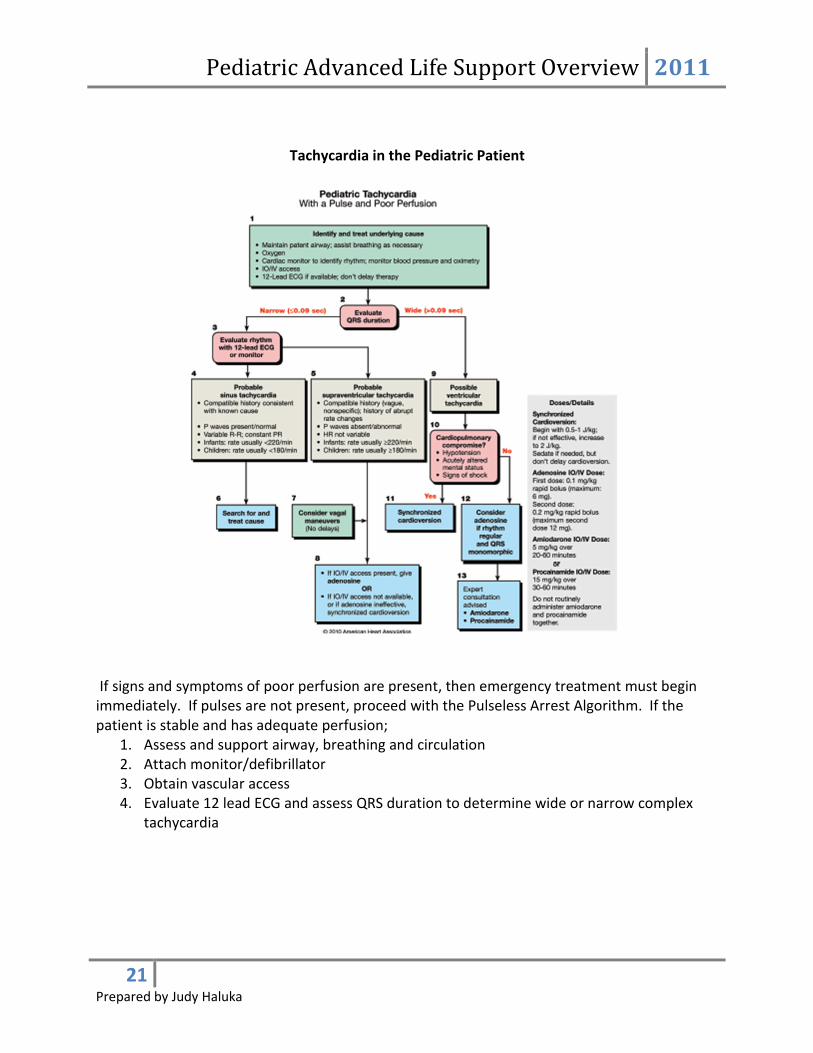
Tachycardia in the Pediatric Patient
If signs and symptoms of poor perfusion are present, then emergency treatment must begin
immediately. If pulses are not present, proceed with the Pulseless Arrest Algorithm. If the
patient is stable and has adequate perfusion;
1. Assess and support airway, breathing and circulation
2. Attach monitor/defibrillator
3. Obtain vascular access
4. Evaluate 12 lead ECG and assess QRS duration to determine wide or narrow complex
tachycardia
Pediatric Advanced Life Support Overview 2011
22
Prepared by Judy Haluka
Narrow Complex Tachycardia (QRS <0.9 seconds) use the history of the patient and an
electrocardiogram to determine sinus tachycardia vs Supraventricular tachycardia. The history
of the patient with sinus tachycardia will include a reason for the child to be compensating with
heart rate. Common presentations of sinus tachycardia include, hypovolemia from trauma or
vomiting and diarrhea, sepsis, high fever, etc. If the rhythm is determined to be sinus
tachycardia then it is treated by finding the cause and fixing it.
Supraventricular Tachycardia
1. Monitor for the effect of intervention. Treatment is determined by the stability of the
patient.
2. Attempt vagal stimulation first. In infants and small children ice can be applied to the
face. In the older child having them blow bubbles through a straw and reaching over
and kinking off the straw will usually cause a vagal. In older children carotid sinus
massage is safe.
3. Pharmacological intervention includes Adenosine which is the first drug of choice. It is
safe as it has a very short half life and is very efficient in rhythms that involve the AV
node for conduction. 0.1mg/kg is administered as fast as possible and followed
immediately with a bolus of saline.
4. Verapamil 0.1 to 0.3mg can be effective in older children but should not be used in
infants because of potential myocardial depression, hypotension and cardiac arrest.
5. If the patient is or becomes hemodynamically unstable he/she must be electrically
cardioverted. Start with a dose of 0.5 joules/kg to 1 joule/kg. The dose can be
increased to 2joules/kg if necessary. If a second shock is unsuccessful or if the rhythm
quickly recurs, a dose of Amiodarone 5mg/kg should be given prior to a third shock.
Amiodarone must be infused slowly over 20-60 minutes depending on the urgency of
the patient. Rhythm and blood pressure must be monitored carefully during
administration.
Wide Complex Tachycardia
Not all wide complex tachycardia originates in the ventricle. Many are Supraventricular in
origin. All therapies for arrhythmias have the potential for serious side effects. For this reason
if possible expert consultation should take place before they are administered to infants or
children.
1. Adenosine may be given to differentiate SVT from VT and converting wide complex
tachycardia that is Supraventricular in origin. It should be given only if the rhythm is
regular and the QRS is monomorphic. Do not use Adenosine in WPW.
2. Consider cardioversion after sedation with a starting dose of 0.5joules/kg – 1.0 joules/kg
with an increase to 2 joules/kg if the first is unsuccessful.
3. Consider pharmacologic conversion with Amiodarone over 20-60 minutes (5mg/kg) or
Procainamide 15mg/kg over 30-60 minutes.

Pediatric Advanced Life Support Overview 2011
23
Prepared by Judy Haluka
SPECIAL RESUSCITATION SITUATIONS
SEPTIC SHOCK
• No difference in survival if treated with colloid or isotonic crystalloid solutions
• Monitoring central venous oxygenation saturation (SVC) is useful to titrate therapy in
infants and children. Target Scv02 >70% associated with improved survival in severe
shock
• Early assisted ventilation
• Etomidate has been shown to facilitate endotracheal intubation in infants and children
without effecting hemodynamics
HYPOVOLEMIC SHOCK
• Isotonic crystalloid for resuscitation
• 20ml/kg even if blood pressure is normal.
• Additional boluses of 20ml/kg if systemic perfusion fails to improve
• Insufficient evidence in infants and children to make a recommendation about the best
timing of volume resuscitation for children with hypovolemia following trauma
TRAUMA – improperly performed resuscitation is a major cause of preventable pediatric
deaths
Common errors include failure to provide appropriate fluid resuscitation, manage and
maintain airway and failure to recognize internal bleeding
Involve a qualified surgeon early in the treatment of a child with trauma. When possible
transport the child to a multisystem trauma center with pediatric experience.
1. If mechanism of injury is compatible with cervical spine injury, restrict motion and avoid
traction or movement of the head and neck. Open the airway with a jaw thrust and do
not tilt the head.
2. If airway cannot be open with the jaw thrust, use a head tilt chin lift because you must
establish a patent airway
3. Do not routinely hyperventilate even in the case of head injury.
4. Suspect thoraco-abdominal trauma even in the absence of external injuries.
5. If the patient has maxillofacial trauma or skull fracture, insert an orogastric rather than
Nasogastric tube.
POST RESUSCITATION STABILIZATION
Reassessment is key. The goal is to optimize perfusion to prevent organ injury and to preserve
neurologic function. The cause of arrest must be diagnosed and treated.

Pediatric Advanced Life Support Overview 2011
24
Prepared by Judy Haluka
Respiratory
• Data suggests that hyperoxemia (high Pa02) is associated with organ injury. The goal
following arrest is to decrease the FI02 to the lowest level that will maintain an
oxyhemoglobin saturation of 94% or greater.
• Evaluate acid base balance and treat imbalances.
• Assist ventilation if significant respiratory compromise. If already intubated, verify tube
placement and position. Consider arterial blood gases 10-15 minutes after establishing
ventilator settings.
• Control pain and discomfort with analgesics and sedatives. Neuromuscular blocking
agents with analgesia or sedation may improve oxygenation and ventilation
• Monitor exhaled C02, especially during transport and diagnostic procedures
• Insert a gastric tube to relieve and help prevent gastric inflation
Cardiovascular
• Monitor heart rate and blood pressure. Consider monitoring urine output with
catheter. 12 Lead ECG may be helpful in discovering cause of cardiac arrest
• Remove IO access if used for resuscitation and establish venous catheters
• Monitor electrolytes, glucose and blood gases
• Drugs used to maintain cardiac output
o Epinephrine
o Dopamine
o Dobutamine Hydrochloride
o Sodium Nitroprusside
o Inodilators (inamrinone and milrinone)
Neurologic
• Do not routinely provide excessive ventilation or hyperventilation. Has no benefit and
may impair neurologic outcome by adversely affecting cardiac output and cerebral
perfusion
• Therapeutic hypothermia may be considered for children who remain comatose after
resuscitation from cardiac arrest.
o The ideal method for cooling and re-warming is not known.
• Treat post ischemic seizures aggressively; search for a correctable cause
Inter-hospital Transport should be done by a specially trained team from a pediatric tertiary
care facility. The team should be contacted as early as possible in the resuscitation to decrease
waiting time. The team should be supervised by a pediatric emergency medicine or critical care
specialist.
Pediatric Advanced Life Support Overview 2011
25
Prepared by Judy Haluka
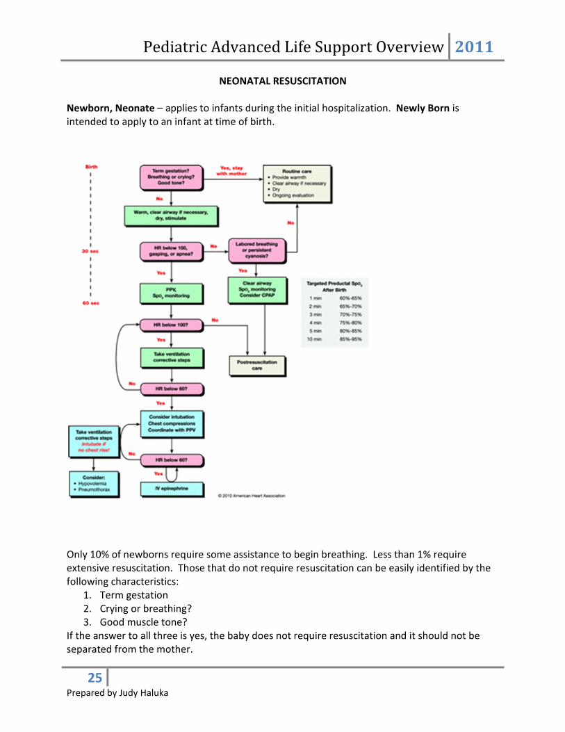
NEONATAL RESUSCITATION
Newborn, Neonate – applies to infants during the initial hospitalization. Newly Born is
intended to apply to an infant at time of birth.
Only 10% of newborns require some assistance to begin breathing. Less than 1% require
extensive resuscitation. Those that do not require resuscitation can be easily identified by the
following characteristics:
1. Term gestation
2. Crying or breathing?
3. Good muscle tone?
If the answer to all three is yes, the baby does not require resuscitation and it should not be
separated from the mother.

Pediatric Advanced Life Support Overview 2011
26
Prepared by Judy Haluka
If the answer to any is no the infant should receive one or more of the following 4 categories of
action in sequence:
1. Initial steps in stabilization (warming, clear airway, if necessary, dry, and stimulate)
2. Ventilation
3. Chest Compressions
4. Administration of epinephrine and/or volume expanders
Approximately 60 seconds are allotted for completely the initial steps, reevaluating and
beginning ventilation if required. The decision to progress beyond the initial steps is
determined by simultaneous assessment of respirations and heart rate (>100)
Assessment of heart rate should utilize the Precordial pulse. If detectable, palpation of the
umbilical pulse can be used.