Pearls, Pitfalls and Advances in Neuro … Causes •Inflammatory •Vascular...
12
1 Pearls, Pitfalls and Advances in Neuro-Ophthalmology Nancy J. Newman, MD Emory University Atlanta, GA Consultant for Gensight Biologics, Santhera Data Safety Monitoring Board for Quark AION Study Medical-legal consultant (IIH and peri-op vision loss) Where ? What? Now What?
Transcript of Pearls, Pitfalls and Advances in Neuro … Causes •Inflammatory •Vascular...

1
Pearls, Pitfalls and Advances in
Neuro-OphthalmologyNancy J. Newman, MD
Emory UniversityAtlanta, GA
Consultant for Gensight Biologics, SantheraData Safety Monitoring Board for Quark AION StudyMedical-legal consultant (IIH and peri-op vision loss)
Where ?
What?
Now What?

2
CRAO
BRAO
Vascular TMVL
Acute retinal ischemiaDifferent visual outcomes
Same systemic implications
Study MRI Results CorrelationBoston 2012
DWI+ in 31/129 (24%)Same vascular territory as visual loss in 28/31Small, multiple infarctions
Neuro sx+Permanent VL > TMVLIdentified causeEmbolic cause
Korea 2014
DWI+ in 8/33 (24.2%)Same vascular territory as visual loss in 8/8Small, multiple infarctions
Neuro sx+CRAO > BRAOIdentified causeEmbolic cause
Germany 2015
DWI+ in 49/213 (23%)Same vascular territory as visual loss in 55%Small, multiple infarctions
Neuro sx+Identified causeEmbolic cause
Am J Ophthalmol 2014
“TIA” + = STROKE

3
(The Neurologist 2012;18:350–355)
Non-mydriatic fundus cameras• Easy for non-ophthalmic
trained individuals to use• No pupillary dilation• Able to take quality
photographs of the posterior pole
• Reveals unrecognized findings in ED (Bruce et al. NEJM 2011; 364:387-9)
Arch Ophthalmol 2012; 130: 939-940
4
Optic NeuropathyClassic Features
•Decreased visual acuity•Abnormal visual field•Relative afferent pupillary defect•Can see through to the nerve•Swollen or pale optic nerve
Optic NeuropathyDisc Alternatives Causes
• Inflammatory•Vascular•Compressive/Infiltrative•Toxic/Nutritional•Hereditary•Traumatic•Elevated intracranial pressure•Elevated intraocular pressure
Optic Neuropathy

5
Causes• Inflammatory•Vascular•Compressive/Infiltrative•Toxic/Nutritional•Hereditary•Traumatic•Elevated intracranial pressure•Elevated intraocular pressure
Optic Neuropathy Optic NeuropathyPapilledema
• Disc swelling from ↑intracranial pressure• Any age• Painless• Bilateral • Spares visual acuity• Constriction of visual field
PapilledemaCauses
• Intracranial mass lesions
•Hydrocephalus•Meningeal processes•Cerebral venous thrombosis
• Idiopathic (pseudotumor cerebri)
Idiopathic intracranial hypertension
•Papilledema•Headaches•No localizing neurologic symptoms/signs except for VIth
•No intracranial process, no venous sinus thrombosis
•Normal CSF contents•CSF opening pressure ≥25cm H2O

6
• Elevated ICP measured in the lateral decubitus position: neonates: >76 mm H2O, age 1–18 years: >280 mm H2O
• Normal CSF composition except in neonates who may have up to 19 WBC/mm3 if 0–28 days and up to 9 WBC/mm3 if between 29 and 56 days old; the protein may be as high as 150 mg/dl
IIH imaging
� Not just a diagnosis of exclusion� New diagnostic criteria� Papilledema� Measure of intracranial pressure� Neuroimaging findings
IIH: Poor visual prognosis
�Patient’s characteristics• Black race. Neurology 2008; 70: 861-7
• Male. Neurology 2009; 72:304-9
• Severe obesity. J Neuro-Ophthalmol 2013; 33: 4-8
• Anemia / sleep apnea syndrome / HTN�Rapid onset (fulminant IIH). Neurology 2007; 68: 229-232

7
� IIH is everywhere there are obese people

8
TS stenosis
After stenting
Clinical course of idiopathic intracranial hypertension with transverse sinus stenosisNeurology 2013;80:289-95
ScientificWorldJournal. 2015; 2015: 140408. Causes• Inflammatory•Vascular•Compressive/Infiltrative•Toxic/Nutritional•Hereditary•Traumatic•Elevated intracranial pressure•Elevated intraocular pressure
Optic Neuropathy

9
Optic NeuropathyTypical Optic Neuritis
• Inflammation of the optic nerve• F:M 3:1• Age: 15-45• Pain on eye movement• Normal or swollen disc• Spontaneous improvement• Associated with multiple sclerosis
ONTT
•No difference in visual acuity between steroid and placebo groups at 6 months.
• I.V. steroids may accelerate recovery by 2 to 3 weeks.
•P.O. steroids doubled the risk of recurrence in either eye.
(NEJM 326:581, 1992)
ONTT: MRI predicts the risk of MS
0
10
20
30
40
50
60
70
Years 1 2 3 4 5 6 7 8 9 10 11 12
Total 50% at 15 yrs
No lesion 25%
≥ 1 lesion 72%

10
Clinical Features of Optic Neuritis with Low Risk of CDMS in Patients with No Brain MRI Lesions
No cases of CDMS have developed when any one of the following clinical features* was present:
• Severe Disc Swelling (21 patients)• Hemorrhage, disk or peripapillary (16 patients)• Macular Exudates (8 patients)• Painless (19 patients)• No Light Perception (7 patients)
OCT: Retinal Nerve Fiber Layer (RNFL) Thickness
• Correlates with axonal loss
• Correlates with visual dysfunction
• Correlates with: – Brain atrophy in MS– Disability– Quality of life
From: Relationships Between Retinal Axonal and Neuronal M easures and Global Central Nervous System Pathology in Multiple Sclerosis
JAMA Neurol. 2013;70(1):34-43. doi:10.1001/jamaneurol.2013.573
-Healthy subject. 3-dimensional macular volume cube generated by Cirrus HD-OCT from the macular region
-The individual layers of the retina are readily discernible, except for GCL and IPL, which are difficult to distinguish.-During the segmentation process (performed in 3-dimension), the segmentation software identifies the outer boundaries of the macular RNFL, IPL, and OPL, as well as the inner boundary of the RPE, which is identified by the conventional Cirrus HD-OCT algorithm. The identification of these boundaries facilitates OCT segmentation, enabling determination of the thicknesses of themacular RNFL, GCL + IPL, the INL + OPL, and the ONL including the inner and outer photoreceptor segments
Cellular composition of the retinal layers :ILM: inner limiting membraneRNFL: retinal nerve fiber layerGCL: ganglion cell layerIPL: inner plexiform layer INL: inner nuclear layer OPL: outer plexiform layer ONL: outer nuclear layer ELM: external limiting membraneIPS: inner photoreceptor segmentsOPS: outer photoreceptor segmentsPR: photoreceptorsRPE: retinal pigment epithelium
11
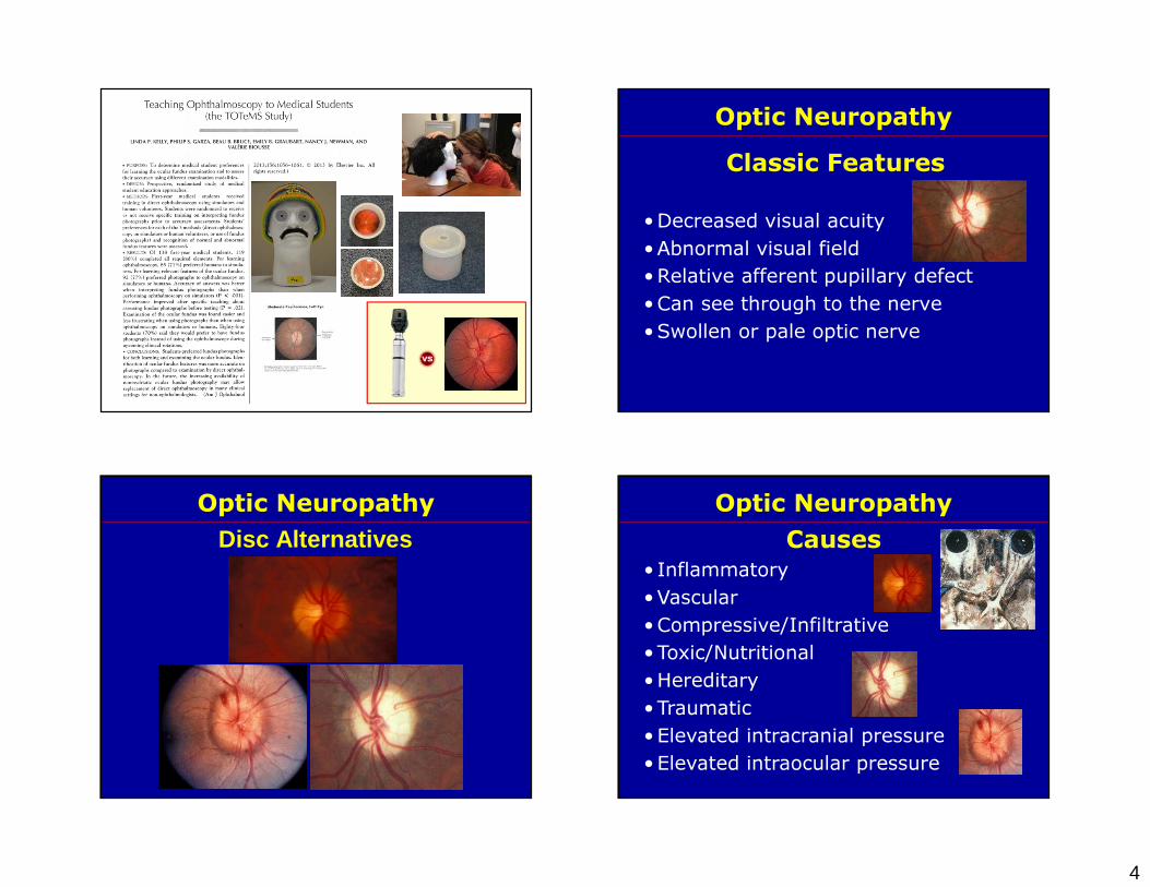
Fingolimod and Macular Edema
1. Incidence of macular edema is low (~1%); (uveitis, DM increase risk). Ophthalmology 2013; 120: 1432-1439
2. Screening evaluation for uveitis, macular or retinal vascular disease prior to starting, or within the first few weeks of starting fingolimod
3. Re-evaluation (complete eye exam +/-macular OCT) at 3-4 months of therapy (most reported cases of macular edema occurred within 3-4 months)
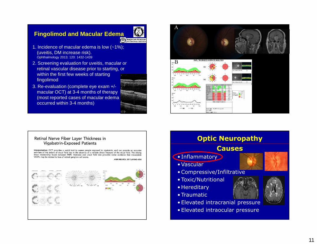
Causes• Inflammatory•Vascular•Compressive/Infiltrative•Toxic/Nutritional•Hereditary•Traumatic•Elevated intracranial pressure•Elevated intraocular pressure
Optic Neuropathy

12
Optic Neuritis and NMO Abs
Optic Neuritis and NMO Abs• Severe
• Bilateral
• Poor recovery• Recurrent
• Bilateral
• Severe
• Poor recovery• Recurrent