Pearls and myths in sle
44
Pearls and Myths in SLE By Ahmed Mohammed Abd El Wahab Lecturer of Internal Medicine (Nephrology)
-
Upload
- -
Category
Health & Medicine
-
view
358 -
download
4
Transcript of Pearls and myths in sle

Pearls and Myths in SLEBy
Ahmed Mohammed Abd El WahabLecturer of Internal Medicine
(Nephrology)
Clinical Features
Myth:The major cause of death in SLE patients is: (a) active lupus; (b)
complications of renal failure; or c) infection.
Reality:The correct answer: none of the above! In the 1970s, Urowitz and
Gladman reported on the bimodal pattern of mortality in SLE. Whereas “early” deaths were caused predominantly by active SLE, “later” deaths were linked primarily to cardiovascular disease (Urowitz et al. 1976).
Contd.
Myth:SLE causes pain. Reality:1. The most frequent cause of a positive ANA and chronic pain is
fibromyalgia! Estimates of the prevalence of fibromyalgia among patients with SLE range from 10 to 30%.
2. Arthralgias are the principal musculoskeletal feature of SLE, but synovitis also occurs. SLE patients also have pain from musculoskeletal damage (e.g., osteonecrosis).
3. Processes unrelated to SLE such as osteoarthritis, mechanical low back pain, bursitis, compression neuropathy.
Contd.
Myth:Fatigue is a sign of active SLE. Reality:1. Acute fatigue often accompanies SLE flares, but this usually resolves
along with the other features of active disease.2. Fatigue is a chronic symptom that is unresponsive to NSAIDs,
glucocorticoids, and immunosuppressive medications. In these SLE patients, chronic fatigue is probably due to fibromyalgia. Thus, treating all chronic fatigue with glucocorticoids exposes many SLE patients to unnecessary toxicity.
3. Every SLE patient with chronic fatigue deserves an evaluation for comorbid processes, including anemia, hypothyroidism, adrenal insuficiency, sleep disorders, fibromyalgia, and depression.
Contd. Myth:
Lymphadenopathy must be investigated aggressively to rule out lymphoma. Reality:
1. Lymphadenopathy occurs in approximately 40% of lupus patients. 2. The involved lymph node chains are found usually in the cervical, axillary, and
inguinal areas.
3. Pathological features include the absence of tenderness in the nodes; enlargement greater than three centimeters; induration; age older than 40, and a location above the clavicle . Additional indicators of concern are splenomegaly and a monoclonal expansion of CD19+/CD22+ B lymphocytes in the peripheral blood.
4. Lupus patients have a slightly increased risk for both Hodgkin’s (3.6%) and non-Hodgkin’s(3.1%) lymphoma.
Contd.
Pearl:Up to one-third of lupus patients have one or more additional
autoimmune diseases. Comment:Other autoimmune disorders often linked to SLE include Sjögren’s
syndrome, aPL, autoimmune thrombocytopenia, hypothyroidism, and polymyositis .

Contd. Myth:
Autoimmune liver disease is common in SLE. Reality:
1. Autoimmune liver disease is relatively uncommon in SLE. It affects only about 2% of patients.
2. Positive ANA assays are the norm in both conditions. However, ASMA and AMA antibodies occur in fewer than 30% of SLE patients and, when present, are usually found in low titers.
3. Elevations of the serum hepatic aminotransferases tend to be lower in lupus-associated hepatitis when compared with autoimmune liver disease.
4. Histopathology may resolve the question. Autoimmune hepatitis is associated with a characteristic periportal hepatitis and piecemeal necrosis.
Contd.
Myth:Autoimmune hemolytic anemia is the most common cause of anemia in
lupus. Reality:1. AIHA is found in approximately 10% of patients with SLE. 2. The most common causes of anemia in SLE are chronic disease
and iron deficiency. 3. For lupus patients with stage 4 CKD, the KDOQI guidelines
recommend maintaining a target hemoglobin of 11–12 mg/dLthrough the use of erythropoietin .
Contd.
Myth:Erosive arthritis does not occur in patients with SLE. Reality:1. As a general rule, this is true. However, up to 5% of patients with
SLE develop an erosive arthropathy.2. Joint involvement in SLE can be categorized as follows: A non-deforming arthropathy A deforming but non-erosive arthropathy (Jaccoud’s arthropathy) An erosive arthritis:Sometimes termed “rhupus” comprises a clinical and serological
overlap syndrome between RA and SLE.
Contd.
Pearl:Lupus is associated with the production of type I
interferon and a peripheral blood interferon “signature.” The fact that type I and type II interferons are powerful inhibitors of osteoclastsis one likely explanation for the fact that only a small subset of lupus patients has bony erosions.
Contd.
Myth: Serum creatinine is a reliable indicator of renal function. Reality: Measurement of the serum creatinine level is a practical but relatively
insensitive indicator of abnormalities in GFR as level is affected by variables with little direct relationship to renal function, including sex, muscle mass, and age.
Pearl:In a pregnant lupus patient, substantial renal dysfunction can be present
despite the finding of a normal serum creatinine level.
Contd.
Pearl: An experienced clinician recognizes the point of return with the
use of immunosuppression for glomerulonephritis. Comment: It’s not worth saving the kidneys if you’ve lost the patient. The
efficacy of renal replacement therapy – both dialysis and transplantation – means that end-stage renal disease no longer marks the end of the line for a patient with SLE.
Contd.
Pearl: Proteinuria can be diminished by 50% without prescribing a single
milligram of prednisone. Comment:1. The reduction of proteinuria through ACE inhibition delays and
prevents renal sclerosis, hyperlipidemia, hypercoagulability.2. The use of ACE inhibitors and ARBs can reduce proteinuria by up
to 50%. Spironolactone also helps to reduce proteinuria (In men, eplerenone should be used, because of its lower risk of gynecomastia.)
Contd.
Pearl: Proteinuria is a better indicator of lupus nephritis than is hematuria. Comment:1. Hematuria is one of the renal manifestations of lupus listed on the
(SLEDAI) . However, sound clinical judgment is essential before ascribing hematuria to active lupus. SLE patients may have hematuria from a variety of causes, including menses, trauma, renal calculi.
2. In the SELENA studies, the investigators amended the SLEDAI to require that hematuria only be used in the presence of proteinuria as a manifestation of lupus .
Contd.
Myth:“Lupus headaches” are part of the disease. Reality:1. Although “lupus headache” is one of the neurologic
manifestations listed on (SLEDAI), headaches are actually rare in lupus. Most women with SLE who have headaches have migraine physiology.
2. An SLE patient with a severe headache should have an evaluation designed to exclude brain hemorrhage or dural sinus thrombosis. Lumbar puncture is also appropriate to help exclude infection, malignancy, and pseudo-tumor cerebri.
Contd.
Pearl:Patients with neuropsychiatric lupus, improvement is likely to
take several months. Patience is required. Comment: In one single-center study, only 10 of 485 patients with SLE
developed major neuropsychiatric disease manifestations (Pego-Reigosa and Isenberg, 2008). In eight of these ten cases, the outcome was ultimately excellent but the time to recovery was long, often up to 12 months or longer for complete symptom resolution.
Contd.
Pearl:It is often difficult to separate central nervous system disease manifestations that are due to inflammation from those that are caused by thrombosis.
Comment: 1. Anticoagulation with warfarin is a cornerstone of treatment for
lupus patients who present with neuropsychiatric events that are characteristic of APS.
2. When the relative contributions of “inflammatory” mechanisms as opposed to “thrombotic” pathways are unclear, the patient may require combination therapy with glucocorticoids, cytotoxic drugs, and warfarin.
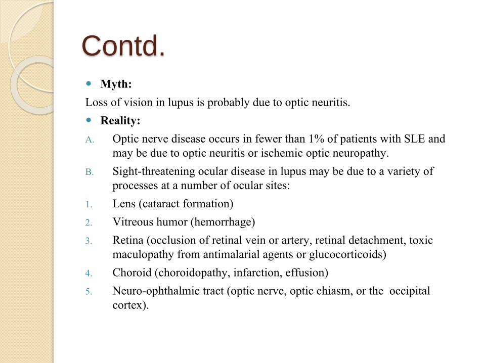
Contd. Myth:
Loss of vision in lupus is probably due to optic neuritis.
Reality:
A. Optic nerve disease occurs in fewer than 1% of patients with SLE and may be due to optic neuritis or ischemic optic neuropathy.
B. Sight-threatening ocular disease in lupus may be due to a variety of processes at a number of ocular sites:
1. Lens (cataract formation)
2. Vitreous humor (hemorrhage)
3. Retina (occlusion of retinal vein or artery, retinal detachment, toxic maculopathy from antimalarial agents or glucocorticoids)
4. Choroid (choroidopathy, infarction, effusion)
5. Neuro-ophthalmic tract (optic nerve, optic chiasm, or the occipital cortex).
Diagnosis
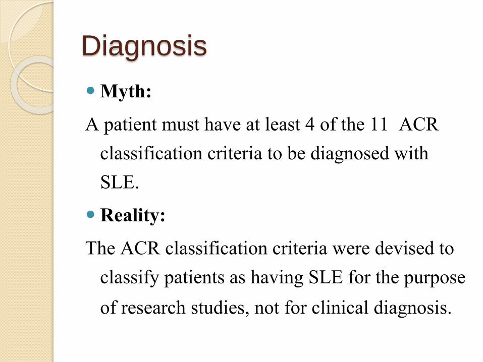
Myth:
A patient must have at least 4 of the 11 ACR classification criteria to be diagnosed with SLE.
Reality:
The ACR classification criteria were devised to classify patients as having SLE for the purpose
of research studies, not for clinical diagnosis.
Contd.
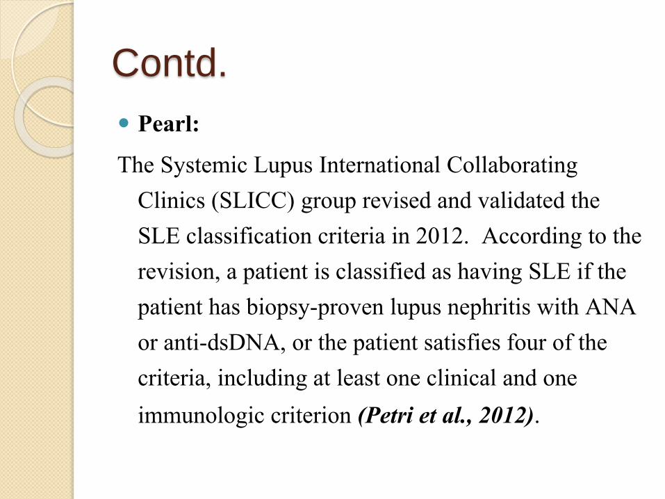
Pearl:
The Systemic Lupus International Collaborating Clinics (SLICC) group revised and validated the SLE classification criteria in 2012. According to the revision, a patient is classified as having SLE if the patient has biopsy-proven lupus nephritis with ANA or anti-dsDNA, or the patient satisfies four of the criteria, including at least one clinical and one
immunologic criterion (Petri et al., 2012).
Contd.
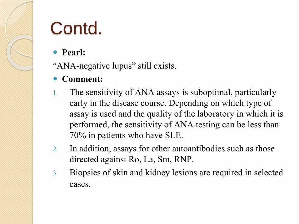
Pearl: “ANA-negative lupus” still exists. Comment: 1. The sensitivity of ANA assays is suboptimal, particularly
early in the disease course. Depending on which type of assay is used and the quality of the laboratory in which it is performed, the sensitivity of ANA testing can be less than 70% in patients who have SLE.
2. In addition, assays for other autoantibodies such as those directed against Ro, La, Sm, RNP.
3. Biopsies of skin and kidney lesions are required in selected cases.
Contd. Pearl:
Not all lupus is diagnosed in patients before the age of 50.
Comment:
1. Up to 20% of patients with SLE develop their disease after the age of 50 . The interval between symptom onset and diagnosis is often longer for older patients, because the clinical manifestations for patients in this subgroup can be insidious and atypical .
2. The overwhelming female predominance is less evident among patients diagnosed later in life.
3. Older patients are less likely than are younger ones to have malarrashes, other cutaneous manifestations, clinically significant renal disease, and hypocomplementemia.
4. Serositis and sicca features are typical of late-onset SLE.
Management
Myth:A high (ESR) indicates active clinical SLE. Reality: 1. Many SLE patients have dramatic elevations in their ESRs
in the absence of apparent clinical disease activity. Similarly, hypocomplementemia and elevated titers of dsDNA antibodies also occur frequently in the absence of clinical disease. Such patients fall into the category of “serologically active but clinically quiescent” SLE .
2. These patients should not be treated for active SLE, but rather be watched carefully: they are more likely to experience disease flares within the next year.
Contd. Myth:
In a febrile lupus patient, both C-reactive protein and procalcitonin levels are useful in excluding infection.
Reality:
1. The oft-quoted myth is that SLE flares do not increase either C-reactive protein or procalcitonin. This statement is wrong often enough to make it dangerous.
2. In a study from the Netherlands, serum CRP levels were normal (less than 6 mg/L) in only 34% of patients with SLE flares (13 of 38 cases), and were also normal in 10% of those with a systemic infection (4 of 36 cases) (terBorg et al. 1990). Thus, even if the CRP level is normal, infections must be excluded in all SLE patients with fever.
3. Arthritis and pleuro-pericarditis, are often associated with elevated CRP levels.
Contd.
Pearl:Lupus patients are immunosuppressed, even without our
help. Comment:1. SLE patients have difficulty with multiple viral
infections, especially herpes zoster, cytomegalovirus, and papillomaviruses. They are also subject to fungal infections, especially oral candidiasis.
2. Perhaps because of splenic dysfunction, SLE patients have difficulty with encapsulated organisms such as the pneumococcus and meningococcus. Don’t forget to vaccinate all SLE patients.
Contd.
Myth: Vaccines cause SLE flares. Reality: 1. Vaccines and lupus are a complicated story. There is
no evidence that inactivated vaccines cause SLE flares. This has been studied rigorously for both the influenza vaccine and the pneumococcal vaccines and the risk posed by the hepatitis B virus vaccination also appears negligible .
2. Because SLE patients may not mount vigorous vaccine responses, the pneumococcal vaccine should be administered every 5 years.
Contd.
Pearl:
The situation with live vaccines is more complex.
Comment:
Vaccines such as Varicella zoster and MMR have been considered contraindicated in patients with SLE and any patient receiving more than 10 mg of prednisone a day or other immunosuppressive agents. It has also been suggested that live vaccines should not be given for 3 months after cessation of
immunosuppressive drugs.
Contd. Pearl: Lupus patients tolerate trimethoprim-sulfamethoxazole medications
poorly. Comment:1. Hypersensitivity to sulfonamides is more common among
patients with SLE when compared with the general population. In addition, some investigations have indicated that sulfonamide use in SLE patients is associated with disease flares. Such reactions are probably type IV (cell-mediated) hypersensitivity responses .
2. Whether to use TMP-SMX as opposed to dapsone, atovaquone, or aerosolized pentamidine for PCP prophylaxis in SLE remains a controversial issue. If the decision is made in favor of TMX-SMX, the medication should be introduced gradually over a 2-week period (Desensitization protocol).
Contd.
Myth: Statins should be used with caution in SLE patients
because they heighten patients’ risk of muscle injury. Reality: 1. Severe myopathy affects only about 0.1% of all
individuals who take statins. Thus, the issue with regard to statin use for many patients with SLE is not whether or not to use these drugs, but rather how to use them wisely.
2. Patients with significant renal insufficiency or hypothyroidism are at increased risk of skeletal muscle toxicity from statin use.
Contd.
Pearl:
Diffuse or focal myocarditis can mimic myocardial infarction.
Comment:
In a lupus patient with chest pain, regardless of the patient’s age, exclude active coronary ischemia. Patients with SLE can and do develop coronary
arteritis, leading to myocardial infarction.
Contd.
Pearl:
50% of SLE patients who have a lupus anticoagulant at diagnosis will suffer a thrombotic event.
Comment:
This is true, and the lupus anticoagulant is a more powerful predictor of thrombotic risk
than is anticardiolipin antibody.
Contd. Pearl:
Hydroxychloroquine should be put in the water supply.
Comment:
1. At least in the water imbibed by lupus patients. Experienced lupus clinicians refer to this medication as “lupus health insurance”.
2. HCQ reduces the frequency of SLE flares, reduces the “spread” of lupus to renal disease, diminishes the likelihood of thrombosis caused by aPL antibodies .
3. The retinal complication of this medication occurs quite rarely (on the order of 1 in 5,000 patients on long-term therapy), develops slowly, and is reversible if the medication is stopped promptly. A safe recommendation is to insist that patients treated with HCQ have an annual ophthalmology examination.
4. The dose of HCQ should be reduced in patients with significant renal dysfunction, in the elderly, and in patients with a low body mass index (e.g., children).
Contd.
Pearl:
Cigarette smoking is associated with an increased risk of cutaneouslupus and decreased responsiveness to antimalarialtherapy.
Contd. Myth:
Ultrapotent topical glucocorticoids should never be used on the face for cutaneous lupus.
Reality:
1. The truth is that dermatologists almost always use ultrapotenttopical glucocorticoids for chronic cutaneous lupus, even on the face. If this strategy is employed early enough, one can clear DLE lesions completely, leaving neither a scar nor a pigment abnormality.
2. Acne and slight hypopigmentation are the most common side effects of topical glucocorticoid therapy.
Contd.
Pearl:
Many SLE flares are due to noncompliance.
Pearl:
The development of end-stage renal disease in SLE is linked to poor medical compliance in many patients.
Contd.
Myth:
Osteonecrosis is caused by doses of prednisone higher than 40–60 mg/day.
Reality:
The threshold dose of prednisone for the risk of osteonecrosis is only 20 mg/day. Other potential risk factors are ethnicity (African–Americans), vasculitis, Raynaud’s phenomenon, and the presence
of a hypercoagulable state such as the aPL.
Contd.
Myth:In proliferative lupus nephritis, a “wait and see” approach can
save the patient from the toxicity of immunosuppressive drugs.
Reality: 1. Some clinicians, arguing that cyclophosphamide saves
kidneys but not lives, delay the use of cytotoxic therapy in patients with proliferative lupus nephritis.
2. Unfortunately, delay in cytotoxic therapy for such patients diminishes the likelihood of remission and increases the risk of subsesquent relapses and end-stage renal disease.
Contd.
Pearl:
In a prospective observational study of 50 consecutive patients with biopsy-proven active lupus nephritis, the application of a protocol that avoided long-term oral steroid therapy resulted in complete remission or partial remission in 45 patients (90%) at a median follow-up of 37 weeks. This protocol involved administration of 2 doses of rituximab 1 g plus methylprednisolone 500 mg on days 1 and 15, followed by maintenance treatment with MMF.
Condon et al. Prospective observational single-centre cohort study to evaluate the effectiveness of treating lupus nephritis with rituximab and mycophenolate mofetil but no oral steroids. Ann Rheum Dis. Aug 2013;72(8):1280-6.
Contd. Pearls:1. HD is preferred to PD; several studies have
documented higher levels of anti (dsDNA), more thrombocytopenia, and higher steroid requirements in patients with SLE and ESRD who are on PD
2. HD also has anti-inflammatory effects with decreased T-helper lymphocyte levels.
3. SLE is generally quiescent in patients on HD, although flares may occur necessitating specific treatment.
Contd.
Myth:
Medications that cause drug-induced lupus cannot be used in idiopathic SLE.
Reality:
The “big three” medications linked to drug-induced lupus are isoniazid, procainamide, and hydralazine. Contrary to popular opinion, these medications can be used in idiopathic SLE. There is no evidence that
any of them exacerbates SLE.

Contd.
Pearl:TNF inhibitors have also been implicated in causing drug-induced
lupus. Although TNF inhibitors can induce ANA, antidsDNA, and anticardiolipin antibodies, these medications may have a role in the treatment of some lupus patients (Aringer et al. 2004).
Great caution is urged when using TNF inhibitors in patients with SLE, because the full implications of elevations in anticardiolipin and antidsDNA antibody titers remain unclear (Fusconi et al. 2007; Jonsdottir et al. 2004).
Contd.
Myth: The management of lupus is empiric and varies widely among
practicing physicians. Reality: 1. Reasonable recommendations exist based on a combination
of evidence and expert opinion . The EULAR recommendations for the management of SLE cover general management as well as selected aspects of lupus such as the aPL, pregnancy, neuropsychiatric lupus, and nephritis.
2. The recommendations provide a general framework for the management of lupus yet allow latitude for physician autonomy and patient preferences.

THANK YOU