Pearl Oyster Health Management
137
Transcript of Pearl Oyster Health Management
7/27/2019 Pearl Oyster Health Management
http://slidepdf.com/reader/full/pearl-oyster-health-management 1/136

7/27/2019 Pearl Oyster Health Management
http://slidepdf.com/reader/full/pearl-oyster-health-management 2/136
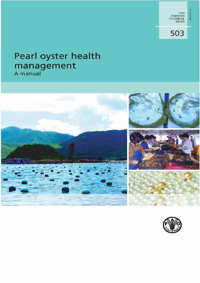
Cover photos:Left : Pearl oyster farm in China (FAO/M.G. Bondad-Reantaso).Right, top to bottom: Pinctada maxima with mudworms (FAO/M.G. Bondad-Reantaso);South Korean women cleaning and sorting mother-of-pearls before grafting (courtesyof F.C.J. Berthe/EFSA); South Sea pearls (FAO/M.G. Bondad-Reantaso).
��

7/27/2019 Pearl Oyster Health Management
http://slidepdf.com/reader/full/pearl-oyster-health-management 3/136
Pearl oyster healthmanagementA manual
byMelba G. Bondad-ReantasoFishery Resources Officer (Aquaculture)Aquaculture Management and Conservation ServiceFisheries and Aquaculture Management DivisionFAO Fisheries and Aquaculture DepartmentRome, Italy
Sharon E. McGladderyAquatic Animal Health DivisionCanadian Food Inspection AgencyOttawa, Canada
and
Franck C.J. BertheAnimal Health and Welfare PanelEuropean Food Safety Authority
Parma, Italy
FOOD AND AGRICULTURE ORGANIZATION OF THE UNITED NATIONS
Rome, 2007
FAOFISHERIES
TECHNICALPAPER
503
7/27/2019 Pearl Oyster Health Management
http://slidepdf.com/reader/full/pearl-oyster-health-management 4/136
The designations employed and the presentation of material in this informationproduct do not imply the expression of any opinion whatsoever on the partof the Food and Agriculture Organization of the United Nations (FAO) concerning thelegal or development status of any country, territory, city or area or of its authorities,or concerning the delimitation of its frontiers or boundaries. The mention of specificcompanies or products of manufacturers, whether or not these have been patented, doesnot imply that these have been endorsed or recommended by FAO in preference toothers of a similar nature that are not mentioned.
The views expressed in this information product are those of the authors anddo not necessarily reflect the views of FAO.
ISBN 978-92-5-105896-1
All rights reserved. Reproduction and dissemination of material in this informationproduct for educational or other non-commercial purposes are authorized withoutany prior written permission from the copyright holders provided the source is fullyacknowledged. Reproduction of material in this information product for resale or othercommercial purposes is prohibited without written permission of the copyright holders.Applications for such permission should be addressed to:Chief Electronic Publishing Policy and Support BranchCommunication DivisionFAOViale delle Terme di Caracalla, 00153 Rome, Italyor by e-mail to:[email protected]
© FAO 2007

7/27/2019 Pearl Oyster Health Management
http://slidepdf.com/reader/full/pearl-oyster-health-management 5/136
iii
Preparation of this document
This document was prepared in an effort to determine what health management optionscan best support development and sustainability of the pearl oyster industry as well ascollate past experiences in dealing with pearl oyster disease outbreaks and other healthproblems. This is the second occasion that FAO is publishing important informationabout pearl oyster. The first and pioneering publication was the Pearl Oyster Farmingand Culture, an output prepared for the Pearl Oyster Farming Training Course (TrainingManual 8) conducted by the Central Marine Fisheries Research Institute at Tuticorin,India and organized by FAO’s Regional Seafarming Development and DemonstrationProject (RAS/90/002).
This paper was prepared under the technical supervision of Dr Melba G. BondadReantaso, Fishery Resources Officer, Aquaculture Management and ConservationService, Fisheries and Aquaculture Management Division, FAO Fisheries andAquaculture Department.
Part 1 consists of two articles: “Why the interest in pearl oyster health?” byDr Sharon E. McGladdery of the Canadian Food Inspection Agency (CFIA) and“Overview of the cultured marine pearl industry” by Prof. Paul Southgate of JamesCook University. Part 2 on Pearl oyster health management was jointly written by DrSharon E. McGladdery of CFIA, Dr Melba G. Bondad-Reantaso of FAO and Dr FranckC.J. Berthe of the European Food Safety Authority. Part 3, consisting of experiencesin dealing with pearl oyster mortalities and other health management options, wascontributed, in alphabetical order by Dr Franck C.J. Berthe (France/Italy), Dr Jeremy
Carson (Australia), Dr Melba G. Bondad-Reantaso (Philippines/Italy), Dr Ben Diggles(New Zealand/Australia), Dr Francis Mike Hine (New Zealand/France), Dr J. Brian
Jones (Australia), Ms Daisy Ladra (Philippines), Dr Sharon E. McGladdery (Canada),Dr Jean Prou (France), Dr Katsuhido Wada (Japan) and Dr Wang Chongming (China).

7/27/2019 Pearl Oyster Health Management
http://slidepdf.com/reader/full/pearl-oyster-health-management 6/136

7/27/2019 Pearl Oyster Health Management
http://slidepdf.com/reader/full/pearl-oyster-health-management 7/136
v
Contents
Preparation of this document iiiAbstract ivContributors viiAcknowledgements viiiAbbreviations and acronyms ixGlossary x
PART 1 – PEARL OYSTER HEALTH AND INDUSTRY 1
1.1 Why the interest in pearl oyster health? 3 S E. MG
1.2 Overview of the cultured marine pearl industry 7 P C. S
Introduction 7
Silver-lip/gold-lip pearl oyster, Pinctada maxima 8
Black-lip pearl oyster, Pinctada margaritifera 9
Akoya pearl oyster, Pinctada fucata 11
Winged pearl oyster, Pteria spp. 13
Summary 15
Acknowledgements 15
References 15
PART 2 – PEARL OYSTER HEALTH MANAGEMENT 19
S E. MG, M G. B-R F C.J. B
2.1 Introduction 21
2.1.1 Purpose, approach and target audience 21
2.2 General 21
2.2.1 Husbandry and handling 21
2.2.2 Hatchery production 24
2.2.3 Introduction and transfers 27
2.3 Disease diagnostic protocols 29
2.3.1 Field collection of samples 29
2.3.2 Gross external observations 32
2.3.3 Gross internal observations 33
2.3.4 Laboratory protocols 33
2.4 Health zonation 43
2.5 Disease outbreak investigation procedure 44
2.6 National strategies on aquatic animal health 47
2.7 References 48
Annexes 53
PART 3 – EXPERIENCES IN DEALING WITH PEARL OYSTER MORTALITIES 59
3.1 Review of pearl oyster mortalities and disease problems 61
J. B J
Abstract 61Introduction 61
Infectious diseases and parasites 61

7/27/2019 Pearl Oyster Health Management
http://slidepdf.com/reader/full/pearl-oyster-health-management 8/136
vi
Diseases with non-infectious aetiology 65
Management options 66
Conclusions 66
Acknowledgements 67
References 673.2 The Cook Islands experience: pearl oyster health investigations 71
B D, P. M H J C
Abstract 71
Introduction 72
Pearl oyster health investigations 72
Results 74
Discussion 80
Acknowledgements 83
References 83
3.3 The Australian experience: pearl oyster mortalities and disease problems 87
J. B J
Abstract 87
History of the industry 87
Disease issues 88
Western Australian pearl oyster health management 90
Conclusion 91
Acknowledgements 92
References 92
3.4 The Japanese experience: pearl oyster mortalities and constraints 95
K T. W
Abstract 95
History of fishery of pearls in Japan 95
Parasites and pathogens 96Fouling organisms 97
Predators 97
Red tide 97
Mass mortality 98
Conclusion 99
Acknowledgements 99
References 99
3.5 The French Polynesian experience 103
F C.J. B J P
Abstract 103
Introduction 103
Outbreaks of mass mortality 104
Main pathogens recorded in French Polynesia 104
Health management 106
Conclusion 107
References 107
3.6 Pearl oyster health: experiences from the Philippines, China,the Persian Gulf and the Red Sea 111
M G. B-R, S E. MG, D L W C
Abstract 111
The Philippines experience 111
The Chinese experience 118
The Persian Gulf experience 119The Red Sea experience 120
References 120

7/27/2019 Pearl Oyster Health Management
http://slidepdf.com/reader/full/pearl-oyster-health-management 9/136
vii
Contributors
Franck C.J. BertheEuropean Food Safety AuthorityParma, Italy
Melba G. Bondad-ReantasoFAO Fisheries and Aquaculture DepartmentRome, Italy
Jeremy Carson
Department of Primary Industries, Water and the EnvironmentTasmania, Australia
Ben DigglesDigsFish Services Pty LtdBribie Island, Queensland, Australia
P. Mike Hine73 rue de la Fée au Bois17450, Fouras, France
J. Brian Jones
Department of Fisheries, Government of Western AustraliaWestern Australia, Australia
Daisy LadraBureau of Fisheries and Aquatic ResourcesQuezon City, Metro Manila, Philippines
Sharon E. McGladderyCanadian Food Inspection AgencyOttawa, Ontario, Canada
Jean Prou
IFREMERLa Tremblade, France
Paul C. Southgate James Cook UniversityTownsville, Queensland, Australia
Katsuhiko WadaNational Research Institute of Fisheries ScienceYokohama, Japan
Wang ChongmingYellow Sea Fisheries Research Institute, Chinese Academy of Fishery SciencesQingdao, China
7/27/2019 Pearl Oyster Health Management
http://slidepdf.com/reader/full/pearl-oyster-health-management 10/136
viii
Acknowledgements
This publication is an outcome of the contribution from many individuals and theyare all sincerely acknowledged. We also thank the numerous pearl farmers and pearlfarm operators in the many countries mentioned in this report for their assistance,during the course of visits and conduct of field surveys and studies, to the authors of the different papers. The main authors also wish to thank T. Farmer and F. Schatto of the FAO Fisheries and Aquaculture Department, A. Fontelera and J.L. Castilla forvarious types of assistance towards the final production of this document. J. Jiansanand R. Subasinghe both of the Aquaculture and Management Service, FAO Fisheriesand Aquaculture Department are gratefully acknowledged for support, guidance and
encouragement. All images contained in this technical paper were contributed bythe authors and other photo contributors (J. Taylor, J. Lucas, B. Tioti, A. Wang andA. Teitelbaum) and they are also acknowledged.

7/27/2019 Pearl Oyster Health Management
http://slidepdf.com/reader/full/pearl-oyster-health-management 11/136
ix
Abbreviations and acronyms
ASBUMI Indonesian Pearl Culturer’s AssociationASE accelerated solvent extractionBFAR Bureau of Fisheries and Aquatic Resources (Philippines)BHIA brain heart infusion agarBOD biological oxygen demandBRD Brown Ring DiseaseCMFRI Central Marine Fisheries Research InstituteDO dissolved oxygenEDTA ethylenediaminetetraacetic acid
EM electron microscopyFAO Food and Agriculture Organization of the United NationsFDA Food and Drug AdministrationGC-FID gas chromatography/flame ionization detectionICES International Council for the Exploration of the SeasICP-MS inductively coupled plasma massLD50 lethal dose 50MA marine agarMMR Ministry of Marine Resources (Cook Islands)MOP mother-of-pearlNT Northern TerritoryOIE World Organisation for Animal Health
PCR polymerase chain reactionPPTA phosphotungstic acidQLD Queensland, AustraliaRFTM Ray’s Fluid Thioglycollate MediumSOD sediment oxygen demandSOPAC the Pacific Islands Applied Geoscience CommissionTCBS thiosulphate citrate bile saltTEM transmission electron microscopyTSA tryptone soya agarWA Western Australia

7/27/2019 Pearl Oyster Health Management
http://slidepdf.com/reader/full/pearl-oyster-health-management 12/136
x
Glossary
Abcess an aggregation of haemocytes (blood cells) which contains necrotic(decaying) host cells
Akoya pearl saltwater pearls cultivated from Pinctada fucata; the mainstay of the Japanese and Chinese cultured pearl industries
Bivalve mollusc a mollusc, such as an oyster or a clam, that has a shell consisting of two hinged valves. Bivalves are members of the phylum Mollusca,class Bivalvia.
Blister pearls a natural pearl, usually irregular in shape, which occurs when aparasite (or an irritant) enters a mollusc through its outer shellcausing the mollusc to secrete nacre over the irritant, cementing it tothe shell
Ceroid non-staining metabolic by-product found in many bivalves.Abnormally high concentrations indicate possible environmental orpathogen-induced physiological stress
Conchiolin nitrogenous albuminoid substance, dark brown in color, that formsthe organic base of molluscan shells
Concretions non-staining inclusions in the tubule and kidney cells of pearloysters, produced during the digestive cycle. Similar inclusions arealso found in the epithelia of other bivalves
Cultured pearl pearls which are produced by the reaction of an oyster or molluscsto insertion of a foreign object (called a nucleus or a bead) into itstissue; this induces secretions to cover the nucleus. Culture pearls areformed when a pearl oyster secretes nacre over the nucleus
Fouling accumulation and deposition of living organisms and certainnon-living material on hard surfaces, most often in an aquaticenvironment
Gold-lip oyster one of two varieties of Pinctada maxima; the other is the silver-lipped oyster. The names relate to the colour of the mother-of-pearllining the shell
Gonad the reproductive organ that produces either the sperm or the eggs.gonads in males are called testes; gonads in females are calledovaries
Grafting also known as “seeding”, “nucleus implantation” or “nucleation” is a
surgical procedure where a nucleus and a small piece of mantle tissue(from another oyster) are inserted into the gonad for cultured pearlformation

7/27/2019 Pearl Oyster Health Management
http://slidepdf.com/reader/full/pearl-oyster-health-management 13/136
xi
Mabé pearl a pearl which is formed when a flat-sided nucleus is glued to theinside of a pearl oyster shell. Also known as “half-pearls” and“blister-pearls” they can be made in a variety of shapes determinedby nucleus shape
Mantle the part of a pearl oyster’s soft tissue that lines the inside of the shells
and secretes nacre
Mantle retraction/ during periods of no growth in molluscs, the mantle retracts awayrecession from the edge of the shell. Prolonged mantle retraction leaves the
inner shell edge open to erosion and fouling
Mikimoto pearls a leading brand of pearls founded by the Japanese Kokichi Mikimoto,the Japanese credited for creating the cultured pearls
Mother-of-Pearl the substance which is secreted by pearl oysters to line the inside of their shells. It is also called “nacre” and is the same substance whichforms pearls. Mother-of-pearl is now used extensively as the nucleusin pearl cultivation. The shell of a mussel is cut into squares and thenrun through a process which rounds the pieces into beads. Thesebeads are then implanted into the oysters which then secrete nacreupon the mother-of-pearl beads to form the cultured pearl
Nacre also known as mother-of-pearl is the basic substance which issecreted by oysters and molluscs after a foreign substance (e.g. agrain of sand, a piece of rock or even a parasite) has entered theshell and caused irritation. Nacre is composed of layers of calcium
carbonate (in a crystalline form) and conchiolin (an organic proteinsubstance which provides bonding)
Natural pearl pearls which are formed in nature, following the actions of a parasiteor foreign body lodging itself in the gonad or mantle tissues of a hostoyster
Nucleus a bead or implant onto which nacre is secreted to form culturedpearls. They may be round (round pearl production) or flat sided(mabé production). Round nuclei are generally made of molluscshell that has been cut, rounded and polished
Pearl a hard, round object produced by certain animals (primarilymolluscs) such as pearl oysters particularly valued as a gemstone andis cultivated or harvested for jewellery
Pearl formation when a small irritant or parasite penetrates the shell and irritatesthe mantle tissue a pearl may be formed when nacre is secreted asa response. As nacre builds up in layers, it surrounds the irritantforming a pearl. Pearls that form within tissues generally do so whenmantle epithelial cells are dislodged into the tissue
Pearl oyster bivalve molluscs of the Family Pteridae (genera Pinctada and Pteria)
all members of the Family share the physiological properties thatlead to the production of large pearls of commercial value

7/27/2019 Pearl Oyster Health Management
http://slidepdf.com/reader/full/pearl-oyster-health-management 14/136
xii
Pinctada fucata Akoya pearl oyster producing cultured Akoya pearls
Pinctada the black-lip pearl oyster producing the “black” South Sea pearlsmargaritifera
Pinctada maxima the gold-lip or silver-lip pearl oyster producing “white” South Seapearls
Pteria penguin species of pearl oyster also known as the “winged pearl oyster”,rainbow pearl oyster” or “penguin shell” used to primarily toproduce mabé
Pteria sterna species of pearl oyster from Central America also known as “conchanácar” or “rainbow lip pearl oyster” used to produce mabé andcultured round pearls
South Sea Pearls pearls produced by both Pinctada maxima and P. margaritifera which are differentiated on the basis of their colour
Spat young juvenile pearl oyster or other bivalve mollusc
7/27/2019 Pearl Oyster Health Management
http://slidepdf.com/reader/full/pearl-oyster-health-management 15/136
1
PART 1
PEARL OYSTER HEALTH AND INDUSTRY
1.1 Why the interest in pearl oyster health?Sharon E. McGladdery
1.2 Overview of the cultured marine pearl industry
Paul C. Southgate

7/27/2019 Pearl Oyster Health Management
http://slidepdf.com/reader/full/pearl-oyster-health-management 16/136
7/27/2019 Pearl Oyster Health Management
http://slidepdf.com/reader/full/pearl-oyster-health-management 17/136

7/27/2019 Pearl Oyster Health Management
http://slidepdf.com/reader/full/pearl-oyster-health-management 18/136
Pearl oyster health management: a manual 4
pearl surgery and sub-optimal growing conditions (Dybdahl, Harders and Nicholson,1990; Sims, 1990; Rio-Portilla, Re-Araujo and Voltolina , 1992; Buestel et al., 1995).The lack of contagious disease problems, although an unquestionable blessing, hasalso left the industry with relatively minimal pathology support or a good referenceof information documenting normal versus abnormal parasites, pest and diseases forthe various species cultured (Sims, 1990; Joll, 1992). Since increased development of the industry will, inevitably lead to pressure to select oysters from more and moreremote sources (Wada, 1993, 1996; Benzie, 1994; Fassler, 1994, 1998; Sims and Sarver,1994; Numaguchi, 1995) and sub-optimal growing areas (Gervis and Sims, 1992), thisincreases the risk of accidental disease introduction or induction. Both remote sourcesand mixed stocks enhance the chance of introducing a pathogen to a naïve or vulnerable
(stressed) population (Sindermann, 1986; ICES, 1995) and the best defense againstsuch an unwanted event is a solid knowledge of the health profiles of the animals on aculture site, as well as those from source sites.
PLATE 1.1.1Species of pearl oyster which produce pearls of gem quality
C O U R T E S Y O F S . E . M C G L A D D E R Y
Gold-lip or silver-lip pearl oyster (Pinctada maxima) producing the “white”
South Sea cultured pearls
Black-lip pearl oyster (Pinctada margaritifera) producing the “black” South Sea cultured pearls
Nucleus implantation in Akoia oysters (Pinctada fucata)
C O U R T E S Y O F S . E . M C G L A D D E R Y
C O U R T E S Y O F M . G . B O N D A D - R E A N T A S O
C O U R T E S Y O F M . G . B O N D A D - R E A N T A S O
C O U R T E S Y O F F . C . J . B E R T H E
C O U R T E S Y O F F . C . J . B E R T H E

7/27/2019 Pearl Oyster Health Management
http://slidepdf.com/reader/full/pearl-oyster-health-management 19/136
51.1 Why the interest in pearl oyster health?
Once an epizootic occurs in an aquatic habitat, the chances of eradication andcontrol are limited. In fact, there are no examples, to date, of any molluscan diseaseagent being actively eradicated from an open-water system. This is important toremember when conducting risk-benefit analyses for new species, stocks, growing
techniques or habitats. It is also an important fact to remember when mortalities areobserved and quick health management action is required.
REFERENCESAwaji, M. & Suzuki, T. 1995. The pattern of cell proliferation during pearl sac formation
in the pearl oyster. Fisheries Science 61: 747-751.Benzie, J.A.H. 1994. Genetics of black-lipped pearl oyster (Pinctada margaritifera). Journal
of Shellfish Research 13: 331 (abstract).Buestel, D., Pouvreau, S., Tiaparii, J., Bougrier, S., Chabirand, J.M., Geairon, P. &
Fougerousse, A. 1995. Ecophysiology of the pearl oyster. Relations between the growthof the oyster Pinctada margaritifera and the environment in Takapoto Atoll. IFREMER,
Taravao Tahiti (Polynesie Française) 1995, 132 pp. (in French).Dybdahl, R., Harders, S. & Nicholson, C. 1990. Developing on-growing techniques anddisease prevention husbandry of pearl oysters in Western Australia (FIRTA Project87/81) and on-growing mariculture techniques for the pearl oyster Pinctada maxima spatin Western Australia (FIRDTF Project 89/60). Final reports. Western Australian MarineResearch Laboratories, Waterman, W.A. 57 pp.
Fassler, R. 1994. Hawaii’s impact on the International Pearl Industry. Journal of ShellfishResearch 13:335 (abstract).
Fassler, C.R. 1998. Opportunities for investing in Pearl Farming. World Aquaculture 29:6-13.
Gervis, M.H. & Sims, N.A. 1992. The biology and culture of pearl oysters (Bivalvia:Pteridae). ICLARM Stud. Rev. ICLARM, Metro Manila, Philippines, No. 21: 49.
ICES. 1995. ICES Code of Practice on the Introductions and Transfers of MarineOrganisms 1994. International Council for the Exploration of the Sea, Copenhagen,Denmark. 12 pp.
Intés, A. 1994. Growth and mortality of Pinctada maragaritifera in French Polynesia. Journal of Shellfish Research 13: 337-338. (abstract).
Joll, L.M. 1992. Stock evaluation and recruitment measurement in the W.A. PearlOyster Fishery. Fisheries Department of Western Australia and Fisheries Resaerch and Development Corporation Project Report No. 92/147 (ISBN 0 7309 1848 3) 63 pp.
Numaguchi, K. 1995. Effects of water temperature on catabolic losses of meat andcondition index of unfed pearl oyster Pinctada fucuta martensii. Fisheries Science 61: 735-738.
Numaguchi, K. 1996. A review on the feeding ecology and food environment of the Japanese pearl oyster, Pinctada fucuta martensii. Bulletin of Natural Research Instituteof Fisheries Science 8: 123-138 (in Japanese, English abstract).
Rio-Portilla, M.A. del, Re-Araujo, A.D. & Voltolina, D. 1992. Growth of the pearl oysterPteria sterna under different thermic and feeding conditions. Marine Ecological ProgressSeries 89: 221-227.
Sims, N.A. 1992a. Abundance and distribution of the black-lip pearl oyster, Pinctadamargaritifera (L.), in the Cook Islands, South Pacific. Australian Journal of Marine
Freshwater Research 43: 1409-1421.Sims, N.A. 1992b. Population dynamics and stock management of the black-lip pearl
oyster, Pinctada margaritifera (L.), in the Cook Islands, South Pacific. Australian Journal of Marine Freshwater Research 43: 1423-1435.
Sims, N.A. & Sarver, D.J. 1994. Hatchery culture of he black-lip pearl oyster inHawaii - stock re-establishment and expansion of commercial pearl culture throughoutthe region. Journal of Shellfish Research 13: 350 (abstract).

7/27/2019 Pearl Oyster Health Management
http://slidepdf.com/reader/full/pearl-oyster-health-management 20/136
Pearl oyster health management: a manual 6
Sindermann, C.J. 1986. Strategies for reducing risks from introductions of aquaticorganisms: a marine perspective. Fisheries 11(2): 1015.
Sparks, A.K. 1985. Synopsis of Invertebrate Pathology Exclusive of Insects. The Netherlands,Elsevier, Amsterdam. 431 pp.
Wada, K.T. 1993. Bivalve broodstock development in Japan. World Aquaculture 24(3):54-57.
Wada, K. 1996. Genetical and physiological control of calcification in pearl cultivation.Bulletin of the Institute of Oceanography of Monaco 14: 183-193.

7/27/2019 Pearl Oyster Health Management
http://slidepdf.com/reader/full/pearl-oyster-health-management 21/136

7/27/2019 Pearl Oyster Health Management
http://slidepdf.com/reader/full/pearl-oyster-health-management 22/136
Pearl oyster health management: a manual 8
around 24 percent. Global marine pearl production in 2004 had an estimated valueof approximately US$475 million of which white South Sea pearls from P. maxima contributed more than 46 percent.
Silver-lip/gold-lip pearl oyster, Pinctada maxima Pinctada maxima is the largest pearl oyster species (Shirai, 1994) and is consequentlyused to produce the largest cultured pearls (approximately 10-20 mm in diameter). It isdistributed within the central Indo-Pacific region, bounded by the Bay of Bengal to thewest, Solomon Islands to the east, the Philippines to the north, and northern Australiato the south.
The terms “South Sea cultured pearl” and “South Sea pearl” are used for pearlsproduced in marine waters south of Japan. These names are associated with largecultured pearls produced from both P. maxima and P. margaritifera (Strack, 2006). Theinternational market recognizes and distinguishes between “white“ and “black" SouthSea cultured pearls, produced by P. maxima and P. margaritifera, respectively.
The major producers of cultured pearls from P. maxima are Indonesia, Australiaand the Philippines with approximately 40 percent, 32 percent and 20 percent of totalproduction, respectively (Table 1.2.2). Total production of pearls from P. maxima in2005 was more than 9.3 tonnes with a total value of US$248 million. Pearl productionfrom P. maxima increased by approximately 260 percent between 1999 and 2005(Henricus-Prematilleke, 2005) to become the leading pearl category.
AustraliaPearl production began in Australia inthe 1950s. Total pearl exports generallyvaried between 200 000 and 600 000pearls per year from 1965 to 1995. There
was a decline from 500 000 to 50 000pearls per year in the mid-late 1980sresulting from high oyster mortalities(Pass, Dybdahl and Mannion, 1987).Exports increased from 200 000 to2 million pearls per year between 1995and 2006, however, the unit value of exported Australian pearls reached a20 year low during 2004-2006. Thisdecline probably reflected increasedproduction, as well as external factorssuch as the Asian economic crisis of thelate 1990s.
Australia enjoys an excellentreputation for the quality of its pearls.
TABLE 1.2.2
Production of cultured white South Sea pearls from Pinctada maxima in 2005
Country Volume (kg) Value (US$ millions)
Indonesia 3 750 85
Australia 3 000 123
Philippines 1 875 25
Myanmar 563 13
Malaysia 75 2
Papua New Guinea 75 unknownTotal 9 338 248 million
Source: Henricus-Prematilleke (2005)
C O U R T E S Y O F J O S E P H T A Y L O R
South Sea pearls produced from Pinctada maxima range from
white/silver through to gold in colour. They are the largest of
the cultured pearls.
7/27/2019 Pearl Oyster Health Management
http://slidepdf.com/reader/full/pearl-oyster-health-management 23/136
91.2 Overview of the cultured marine pearl industry
This is demonstrated by the data inTable 2 showing that Australian pearlsmade up approximately 32 percent of total white South Sea pearl production
in 2005 but accounted for almost 50percent of the total value. However,Australia faces increasing competitionfrom other producers who, one wouldassume, will be seeking to improve pearlquality. The Australian pearl industryis based primarily on adult oysters thatare collected from the wild and useddirectly for pearl production (Wellsand Jernakoff, 2006). The proportionof hatchery produced oysters used
by the industry is therefore small(approximately 20 percent). Given thathatchery production provides the basisfor selective breeding programmes, thisstrategy may, in the long term, favour other producers of white South Sea pearls, suchas Indonesia, that rely on hatchery production.
Indonesia, Philippines and other countriesThe Indonesian cultured pearl industry began in the 1970s when new laws enabled foreigncompanies to invest in Indonesia. The 1990s brought much-needed modernization of pearl farms resulting primarily from investment by foreign companies, which enteredpartnerships in Indonesia. The Indonesian Pearl Culturer’s Association (ASBUMI)
was founded in 1995 to develop marketing strategies. By 1999, Indonesia suppliedmore than a third of the world’s South Sea cultured pearls and by 2005 production hadrisen to more than 3.7 tonnes (Table 2). There are currently around 107 pearl farmsin Indonesia. All commercial pearl production is hatchery-based and the industry issupplied by at least 36 hatcheries.
Production of South Sea pearls from around 30 farms in the Philippines has risenfrom approximately 0.5 tonnes to 2 tonnes a year since 1999. Many of the farms have
Japanese partners and much of the crop is exported to Japan. The pearl farms are centeredto the north of Palawan Island and the adjoining Calamian group, in Samar and CebuIsland around the southern tip of Palawan and in Mindanao Island. Only wild collectedP. maxima were used for pearl production until about 1990; however, hatchery-producedoysters have played an increasingly important role since the end of the 1990s.
Other countries producing significant quantities of cultured South Sea pearls fromP. maxima include Myanmar, Malaysia and Papua New Guinea (Table 2). Small-scalepearl production from P. maxima also occurs in Thailand (Bussarawit, 1995), northernViet Nam and south-western China.
Black-lip pearl oyster, Pinctada margaritifera Pinctada margaritifera has a wide geographical distribution from the Red Sea and eastAfrica to eastern Polynesia. Despite its vast range, this species is used for commercialcultured pearl production almost exclusively within the atoll lagoons of Polynesia, inFrench Polynesia and the Cook Islands. It is the second largest pearl oyster species andgenerally produces cultured pearls in the 9–20 mm size range.
Kokichi Mikimoto established a pearl farm at Ishigaki, Okinawa in 1914 and asecond farm in Palau in 1923 from where he succeeded in producing round pearls fromP. margaritifera (Hisada and Fukuhara, 1999). In 1951, there were nine companies
C O U R T E S Y
O F J O S E P H T A Y L O R
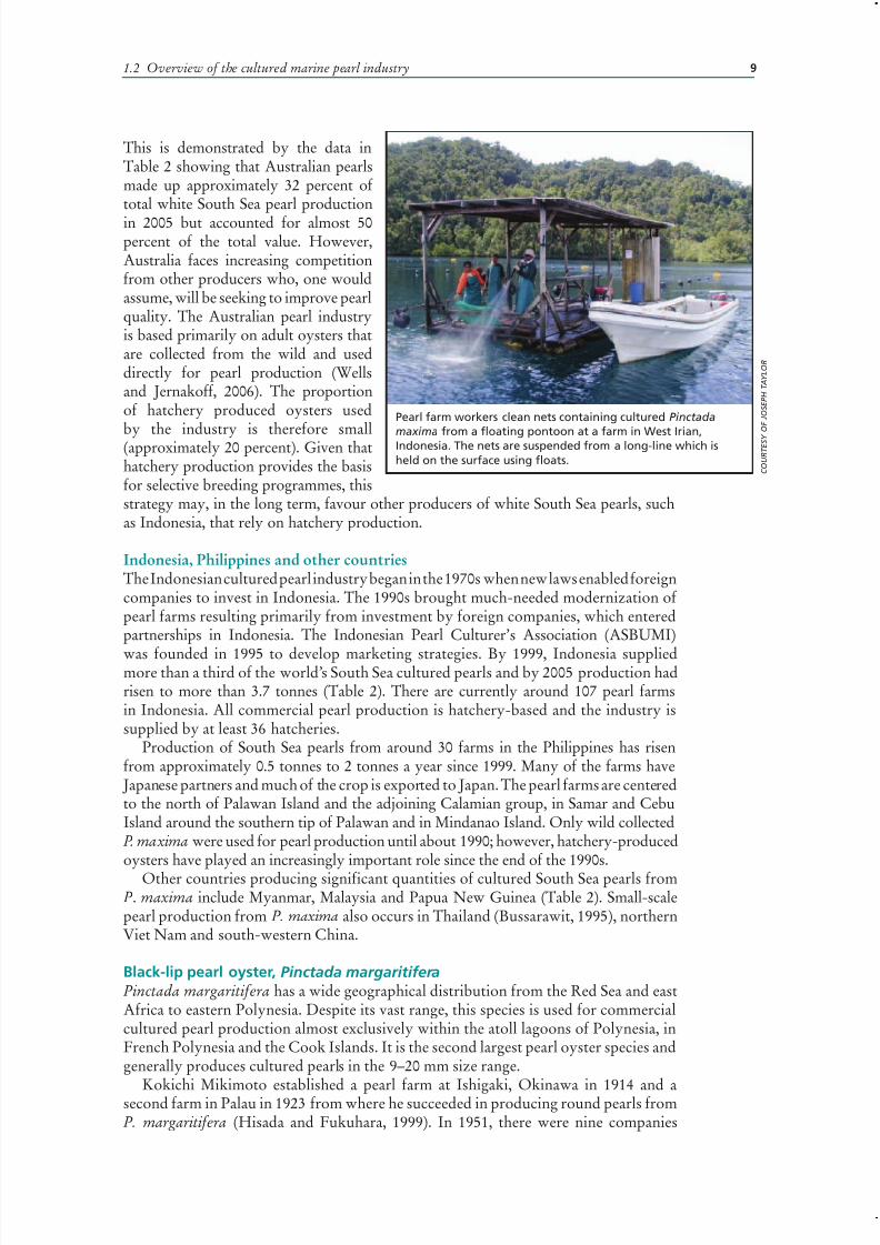
Pearl farm workers clean nets containing cultured Pinctada
maxima from a floating pontoon at a farm in West Irian,Indonesia. The nets are suspended from a long-line which is
held on the surface using floats.

7/27/2019 Pearl Oyster Health Management
http://slidepdf.com/reader/full/pearl-oyster-health-management 24/136
Pearl oyster health management: a manual 10
in Okinawa producing cultured blackpearls. Only one of these survivedand it reports annual production of approximately 2 000–3 000 pearls
annually (Hisada and Fukuhara, 1999).Okinawan pearls provided the basisfor market acceptance of culturedblack pearls in Japan and, when FrenchPolynesia became the dominantproducer of these pearls in the mid-1970s, their product found a readymarket.
French PolynesiaThe cultured pearl industry in French
Polynesia is based on collection of wildpearl oyster spat (juveniles), whichare grown to a size suitable for pearl
production (approximately 100 mm). Spat are easily collected by immersing a suitablesubstrate into lagoon waters when pearl oyster larvae are abundant. The larvae attachto the “spat collectors” and grow into juveniles, which are removed to become culturestock when required. The geomorphology of the atolls of eastern Polynesia, and theirlimited flushing by oceanic water, support abundant aggregations of pearl oyster larvaeand high rates of spat collection. Using natural spat collection, it was easy for islandresidents to develop their own farms throughout the archipelagos of French Polynesiaand there was a rapid increase in the number of authorized leases for pearl farmsthroughout the 1980s and into the late 1990s. By 2001, the number of pearl farms in
French Polynesia had reached more than 2 500.The first 71 cultured round black pearls were harvested in French Polynesia in
1972 and by 1977 the harvest had risen to 28 000 pearls. The rapid increase in thenumber of pearl farms during the 1980s and 1990s supported an exponential rise inpearl production, which peaked at approximately 11 tonnes in 2000 with a value of approximately US$170. However, over- production, declining pearl quality and a floodof lower grade pearls brought prices for black pearls down and market demand declined.Pearl exports from French Polynesia between 2000 and 2005 declined by more than20 percent and their value declined by approximately 40 percent. Total production in2005 was in the range of 8–9 tonnes and currently represents approximately 20 percentof total pearl market value (Table 1). Government regulatory measures now maintain aminimum standard for pearls exported from French Polynesia.
Recent years have seen a decline in the number of pearl farms in French Polynesiato 516 in 2006. They vary from small (approximately <5 ha. in area) to large (>40 ha inarea). Most farms are situated in the Tuamotu and Gambier archipelagos. Pearl cultureis French Polynesia’s second largest economic resource after tourism and the first interms of exports. The industry generates employment for thousands of families spreadover 30 islands in French Polynesia and is an essential part of the social and economiclife of the country.
Cook IslandsCook Islanders generated income from the collection and sale of P. margaritifera MOPuntil the early 1970s (Strack, 2006). Round pearl culture from P. margaritifera, using
the technique developed in French Polynesia, began in 1972 and in 1991 the CookIslands Pearl Farmer’s Association offered 30 000 pearls for sale at its first auction.The industry peaked in 2000 with export revenue of US$18 million, accounting for
A technician inserts a nucleus and a piece of mantle tissue
from a donor oyster into the gonad of a host oyster (Pinctada
margaritifera) for cultured pearl production. This process is
called “seeding”, “grafting” or “nucleation”. C O
U R T E S Y O F J O H N L U C A S

7/27/2019 Pearl Oyster Health Management
http://slidepdf.com/reader/full/pearl-oyster-health-management 25/136
111.2 Overview of the cultured marine pearl industry
20 percent of the country’s gross domestic product. However, poor farming practices,particularly overstocking, meant that the oysters were susceptible to disease. Theindustry was virtually decimated by a disease outbreak towards the end of 2000 whena rise in water temperature resulting from limited flushing of the Manihiki lagoon,
combined with a mass spawning of oysters, triggered a rapid rise in the levels of pathogenic bacteria (Heffernan, 2006). To help ensure the long-term sustainability of the Cook Islands pearl industry and avoid further problems with disease, on-goingmonitoring of water quality and a greater understanding of the bathymetry andhydrodynamics in Manihiki lagoon have been critical in developing a Pearl FarmingManagement Plan for Manihiki (Heffernan, 2006).
There were 205 pearl farms in the Cook Islands in 2003 with an estimated 1 millioncultured adult oysters. However, as a result of increasing pearl production in FrenchPolynesia, low international pearl prices and the continuing impacts of the year 2000disease outbreak, pearl export revenue from the Cook Islands declined to aboutUS$2 million in 2005. Currently, 78 percent of the Cook Islands black pearl farms are
within the lagoon of Manihiki Atoll where 90 farms nucleate approximately 900 000pearl oysters annually to produce approximately 300 000 saleable pearls. The remaining20 percent of pearl culture occurs on Penrhyn Atoll where pearl culture began in 1994.Pearl production in the Cook Islands amounts to approximately 5 percent of worldproduction of black South Sea cultured pearls.
Other countriesCultured pearl production from P. margaritifera has received considerable researchattention in other parts of the Pacific and some has resulted in commercial production.In 2000, a pearl farm was established in the island of Vanua Levu in Fiji. The farmis situated in a deep bay on a high island, and subject to nutrient-rich upwelling – asituation that differs greatly from that of pearl farms in the oligotrophic atoll lagoons
of eastern Polynesia. Approximately 80 percent of the farmed oysters are obtainedfrom spat collectors. Local communities are engaged in spat collection, which providesthe much-needed income to communities close to the farm. The first auction of “Fijipearls” in Japan in 2007 offered 30 000 pearls (Anon., 2007).
Cultured round pearls from P. margaritifera have been produced from a numberof research and pilot projects in other Pacific nations including Solomon Islands,Kiribati and Micronesia (Fassler, 2002; Ito, Jackson and Singeo , 2004; Southgate, 2004).P. margaritifera has also been used for trial mabé pearl production in Tanzania in aproject to determine the potential of small-scale pearl production to generate incomefor coastal communities in support of marineconservation efforts (Southgate et al., 2006).
Akoya pearl oyster, Pinctada fucata
There is considerable taxonomic confusionabout the Akoya pearl oyster which at thisstage is probably best considered as anunresolved species complex encompassingPinctada fucata, P. imbricata, P. martensii andP. radiata. Members of this complex have awide distribution from the MediterraneanSea, through the Red Sea and Indian Ocean,including the Persian Gulf, into the Pacific
Ocean and throughout southeast Asia andnorthern Australia. It also occurs in theCaribbean Sea. C
O U R T E S Y
O F B E E R O T I O T I
Cultured “black” pearls produced from Pinctada
margaritifera in Kiribati, central Pacific. The pearls are
held in a P. margaritifera shell.

7/27/2019 Pearl Oyster Health Management
http://slidepdf.com/reader/full/pearl-oyster-health-management 26/136
Pearl oyster health management: a manual 12
JapanThe technique for culturing roundpearls from pearl oysters wasdeveloped in Japan using the Akoya
pearl oyster. Regular mass productionof cultured pearls using this methodhas occurred in Japan since 1916. By1926, there were 33 pearl farms in
Japan and by 1938, this number hadincreased to 360, which producedmore than 10 million pearls. Harvestsof cultured pearls in Japan increasedrapidly from the 1950s. In 1952,production was almost 10 tonnes;this increased to 52 tonnes in 1960
and reached a peak of 230 tonnesin 1966 produced from 4 700 farms(Strack, 2006).
Pollution of pearl farming sites became an increasing problem and in 1976, only2 000 pearl farms remained. This number had declined further to approximately 1 000farms producing about 35 tonnes of pearls by 1977. In the 1980s, production could notmeet demand for high quality pearls and large quantities of low quality pearls floodedthe market. By this time, there was also strong competition to the Akoya pearl marketfrom China’s increasing production of freshwater pearls. Following greater emphasison larger and better quality pearls in the early 1990s, which saw prices increase, in1996 an epidemic claimed vast numbers of pearl oysters in Japan and was a catastrophefor the industry. It is estimated that the epidemic caused the loss of approximately
75 percent of the oysters in Japanese pearl farms. By 1999, annual pearl production haddeclined to < 20 tonnes with a value of approximately US$130 million, compared to anannual value of US$550–600 million in the early 1990s. Annual production levels havesince remained at about 20–25 tonnes.
Mie Prefecture today produces about 33 percent of the total Akoya pearl harvest in Japan (Strack, 2006), with Ehime and Kochi Prefectures also contributing significantlyto the total. Kyushu Island has produced slightly greater volumes of pearls than Mieand Ehime Prefectures since 1996, with about 40 perecent of total production comingfrom Nagasaki Prefecture (Strack, 2006). Constraints affecting Akoya pearl productionin Japan include: (1) the impacts of parasites such as Polydora spp., boring spongesand trematodes (e.g. Mizumoto, 1975); (2) periodic abnormal blooming of toxicdinoflagellate algae or “red tide” (e.g. Honjo, 1994); (3) seasonal changes in seawatertemperature and reduced food availability (e.g. Tomaru et al ., 2002); and (4) massmortalities associated with pollution, over-crowding and viral infection (e.g. Miyazakiet al ., 1999). Pearl farm management practices that reduce the risk of mass mortalitiesof oysters have been recommended to pearl farmers, and genetic programs to breedresistant strains of oysters have been initiated (Uchimura et al ., 2005).
ChinaMarine pearl oyster cultivation began in China in 1961 and pearl production increasedrapidly during the 1980s when private farms became established (O’Connor and Wang,2001). Annual Akoya pearl production was estimated to be greater than 20 tonnes atthe start of the new millennium (Wang et al ., 2007). The major culture areas are in
the southern provinces of Guangxi, Guangdong and Hainan with Guangxi Provinceproducing about 8-9 tonnes of pearls annually. There are over 1 000 pearl farmsalong the coast of Leizhou in Guangdong Province which, together with farms in
C O U
R T E S Y O F A I M I N W A N G
Akoya pearls being sorted in a factory in Guangdong Province,
China.

7/27/2019 Pearl Oyster Health Management
http://slidepdf.com/reader/full/pearl-oyster-health-management 27/136
131.2 Overview of the cultured marine pearl industry
Xuwen, harvest approximately 9-10tonnes of pearls annually; Akoya pearlproduction from Hainan Province isless than one tonne (A. Wang, pers.
comm., 2007).China produced 5-6 tonnes of
marketable cultured marine pearlsin 1993 and this stimulated Japaneseinvestment in Chinese pearl farmsand pearl factories. Pearl processingis done either in Japan or in Japanese-supported pearl factories in China. Themajority of the higher quality ChineseAkoya pearls are exported to Japan.Additionally, MOP from pearl shells is
used in handicrafts and as an ingredientin cosmetics, while oyster meat is soldat local markets.
India and other countriesIndia began Akoya pearl culture research at theCentral Marine Fisheries Research Institute (CMFRI)at Tuticorin in 1972 and the first experimental roundpearl production occurred in 1973. Although a numberof farms have been established, particularly along thesoutheastern coast, commercial pearl farming has notbecome established on a large scale (Upare, 2001).
Akoya pearls from India generally have a diameter of less than 5-6 mm (Mohamed et al ., 2006; Kripa et al .,2007).
Halong Bay in the Gulf of Tonking in Viet Namhas been famous for its natural pearls for manycenturies (Strack, 2006). Since 1990, more than twentycompanies have established Akoya pearl farms in VietNam and production exceeded 1 000 kg in 2001.
Akoya pearl culture has also been investigated onthe Atlantic coast of South America (Urban, 2000;Lodeiros et al ., 2002), in Australia (O’Connor etal ., 2003), Korea (Choi and Chang, 2003) and in theArabian Gulf (Behzadi, Parivak and Roustaian, 1997).However, information on commercial productionof cultured pearls from these regions is not yetavailable.
Winged pearl oysters, Pteria spp.The common name “winged pearl oyster” relates to the elongated hinge of Pteria spp.There are numerous species of Pteria but only two, Pteria penguin and Pteria sterna,are used for commercial scale pearl culture. Pteria penguin is cultured throughoutSoutheast Asia, in Australia and in some Pacific island nations (Beer and Southgate,2000) and P. sterna is commercially cultured in the Gulf of California, Mexico (Kiefert
et al ., 2004; Ruiz-Rubio et al ., 2006). Pteria spp. are generally used for mabé pearl (alsocalled half pearl or blister pearl) culture and less commonly for round pearl culture. Itis generally acknowledged that this is more difficult to achieve with Pteria spp. than
Pearl farm workers clean and sort nets used for pearl oyster
culture on a floating pontoon in Li’an Bay, Hainan Island, China.
Young women clean pearl oysters and cultureequipment from boats in Li’an Bay, Hainan
Island, China.

7/27/2019 Pearl Oyster Health Management
http://slidepdf.com/reader/full/pearl-oyster-health-management 28/136
Pearl oyster health management: a manual 14
Pinctada spp. as a result of morphological differencesbetween genera. Only in recent years has successfulproduction of round pearls from Pteria spp. beenreported (Farell et al ., 1998; Yu and Wang, 2004).
Pteria penguinPteria penguin is the most widespread culturedwinged pearl oyster. It is readily collected using spatcollectors although hatchery production has beendescribed (Beer, 1999; Yu and Wang, 2004).
In the 1950s, Japanese companies began usingP. penguin (called “mabé gai” in Japanese) on theRyukyu Islands for production of mabé pearls.There are currently three or four companies inRyukyu producing approximately 200 000 pearls per
annum (Hisada and Fukuhara, 1999) from hatcheryproduced oysters.Pteria penguin is widely distributed along the
southern coast of China where it is used for hatchery-based pearl culture (Yu and Wang, 2004). Threecompanies have been established at Hainan Islandand Leizhou Peninsula for cultivation of mabé pearlsfrom P. penguin (Yu and Wang, 2004) and round
pearls have also been produced from this species at Hainan Island.The two major pearl farms at Phuket Island in Thailand were reported to hold
30 000 P. penguin for mabé production in addition to a number of smaller family farmsthat also produce mabé from P. penguin (Bussarawit, 1995). Pteria penguin collected as
natural spat are used for production of mabé in Vava’u islands, Tonga (Finau, 2005).
Pteria sterna There has been regular production of pearls, both mabé and round pearls, from P. sterna in Mexico since 1993. Development of a seeding technique for round pearl productionfrom P. sterna was a breakthrough for the cultured pearl industry in Mexico (Nava etal ., 2000) and research has also been carried out to determine factors that influence thequality of mabé pearls from this species (Ruiz-Rubio et al ., 2006). The mabé are in therange of 12-15 mm and round pearls are generally sized between 6.5-8.5 mm, but mayreach up to 14 mm. Current production is approximately 4 000 round pearls and 8 000
mabé annually.Natural spatfall of P. sterna can
supply the oysters currently requiredby commercial pearl farms in Mexico.However, hatchery production of thisspecies has been described (McAnally-Salas and Valenzuela-Espinoza, 1990;Araya-Nuñez, Ganning and Bueckle-Ramirez, 1995) as well as factorsinfluencing nursery culture (e.g.Monteforte and Garcia-Gasca, 1994).
Other Pteria species
Pteria colymbus has recently beenthe subject of research in Venezuelaand Colombia (Marquez et al ., 2000;
Shell of Pteria penguin with mabé pearls.
The pearls will be drilled from the shell for
processing.
Mabé pearls produced from Pinctada magraritifera in Tanzania,
east Africa.

7/27/2019 Pearl Oyster Health Management
http://slidepdf.com/reader/full/pearl-oyster-health-management 29/136
151.2 Overview of the cultured marine pearl industry
Lodeiros et al ., 1999), where it could beused to produce cultured round pearlsof a size similar to Akoya.
SummaryThe global cultured pearl industry isdiverse in its methods, technologicallevels and products. In FrenchPolynesia, oysters can be easily collectedusing spat collectors. This providedthe opportunity for pearl culture toexpand through the establishment of small- scale or family-based farms. Anindividual, or family, can enter theindustry at a number of levels. They
may simply collect spat for sale to alarger pearl farm, grow pearl oysters fortheir MOP, or produce mabé or roundpearls. Furthermore, the pearl industry provides opportunity for the involvement of women and provides the raw materials for local handicraft manufacture, which mayinclude lower grade pearls or pearl shell. In general, the pearling industry providessignificant socio-economic benefits for coastal communities where it occurs (Tisdelland Poirine, 2000).
In contrast to family-based ventures, the dominant companies within the industryare large, wealthy and highly mechanized, and many have active research programmes.Hatchery cultivation of pearl oysters offers opportunity for selective breeding andstock enhancement, yet this area of research has been slowly embraced by the pearling
industry compared to other aquaculture industries. Indeed, the two largest culturedpearl industries, in Australia and French Polynesia, are based on oysters collected fromthe wild. Furthermore, we still have limited understanding of the respective influences of genetics and environment on pearl quality. The next step in the evolution of the culturedpearl industry will probably be based on development of appropriate selective breedingprogrammes and improved knowledge of the factors influencing pearl quality.
ACKNOWLEDGEMENTSThe author acknowledges the following people who provided information used in thisarticle: Elisabeth Strack, Anthony Hart, Hector Acosta-Salmon, Joseph Taylor andAimin Wang.
REFERENCESAnon. 2006. “Golay’s global view” Pearl World: The International Pearling Journal
15(1):7-8.Anon. 2007. Justin Hunter and Pearls Fiji. Pearl World: The International Pearling Journal
16(4):10-15.Araya-Nunez, O., Ganning, B., & Bueckle-Ramirez, F. 1995. Embryonic development,
larval culture, and settling of American pearl-oyster (Pteria sterna, Gould) spat.California Fish Game 81:10-21.
Beer, A. 1999. Larval culture, spat collection and juvenile growth of the winged pearloyster, Pteria penguin. World Aquaculture ’99. The Annual International Conferenceand Exposition of the World Aquaculture Society 26th April – 2nd May 1999, Sydney,
Australia. Book of Abstracts, 63 pp.Beer, A.C. & Southgate, P.C. 2000. Collection of pearl oyster (family Pteriidae) spat atOrpheus Island Great Barrier Reef (Australia). Journal of Shellfish Research 19: 821-826.
C O U
R T E S Y O F A N T O I N E T E I T E L B A U M
Handicrafts made from lower grade pearls and pearl shell
offer opportunities for income generation in coastal and island
communities. The photograph shows participants in a pearl
handicraft training workshop held in Kiribati, central Pacific.

7/27/2019 Pearl Oyster Health Management
http://slidepdf.com/reader/full/pearl-oyster-health-management 30/136
Pearl oyster health management: a manual 16
Behzadi, S., Parivak, K. & Roustaian, P. 1997. Gonadal cycle of pearl oyster, Pinctada fucata(Gould) in Northeast Percian Gulf, Iran. Journal of Shellfish Research 16: 129-135.
Bussawarit, S. 1995. Marine pearl farms at Phuket Island. Phuket Marine Biology Centre,Special Publication 15: 41-42.
Choi, Y.H. & Chang, Y.J. 2003. Gametogenic cycle of the transplanted-cultured pearl oyster,Pinctada fucata martensii (Bivalvia: Pteriidae) in Korea. Aquaculture 220: 781-790.
Farell, S., Arizmendi, E., McLaurin, D. & Nava, M. 1998. “Perlas de Guaymas”: An updateon the first commercial marine pearl farm on the American continent, ‘Aquaculture ‘98’Book of Abstracts. World Aquaculture Society. 171 p.
Fassler, R. 2002. Recent developments in selected Pacific and Indian Ocean pearl projects.‘Aquaculture 2002’ Book of Abstracts. World Aquaculture Society. 218 p.
Finau, M.W. 2005. Tonga country report. SPC sub-regional technical meeting on pearlculture. Nadi, Fiji. 30 November- 2 December 2005. Noumea, Secretariat of the PacificCommunity (SPC).
Heffernan, O. 2006. Pearls of Wisdom. The Marine Scientist 16: 20-23.
Henricus-Prematilleke, J. 2005. South Sea pearl production seen exceeding 2,000 kan. Jewel. News Asia, December 2005, 75-80.Hisada, Y. & Fukuhara, T. 1999. Pearl marketing trends with emphasis on black pearl
market. FAO Field Document No. 13, FAO, Rome. 31 pp.Honjo, T. 1994. The biology and prediction of representative red tides associated with fish
kills in Japan. Review of Fishery Science 2: 225-253.Ito, M., Jackson, R. & Singeo, S. 2004. Development of pearl aquaculture and expertise
in Micronesia. ‘Aquaculture 2004’ Book of Abstracts. World Aquaculture Society.279 p.
Kiefert, L., McLaurin-Moreno, D., Arizmendi, E., Hanni, H.A. & Elen, S. 2004. Culturedpearls from the Gulf of California, Mexico. Gems Gemmology 40, 26-38.
Kripa, V., Mohamed, K.S., Appukuttan, K.K. & Velayudhan, T.S. 2007. Production of
Akoya pearls from the southwest coast of India. Aquaculture 262: 347-354.Lodeiros, C.J., Rengel, J.J. & Himmelman, J.H. 1999. Growth of Pteria colymbus
(Roding, 1798) in suspended culture in Golfo de Cariaco, Venezuela. Journal of ShellfishResearch 18: 155-158.
Lodeiros, C., Pico, D., Prieto, A., Narvaez, N. & Guerra, A . 2002. Growth and survivalof the pearl oyster Pinctada imbricata (Roding 1758) in suspended and bottom culture inthe Golfo de Cariaco, Venezuela. Aquaculture Research 10: 327-338.
Marquez, B., Lodeiros, C., Jimenez, M. & Himmelman, J.H. 2000. Disponibilidad de juveniles por captacion natural de la ostra Pteria colymbus (Bivalvia: Pteriidae) en elGolfo de Cariaco, Venezuela. Review Biological Tropics 48 (Suppl. 1): 151-158.
McAnally-Salas, L. &Valenzuela-Espinoza, E. 1990. Growth and survival of larvae of thepearl oyster Pteria sterna under laboratory conditions. Cienc. Mar . 16: 29-41.
Miyazaki T., Goto K., Kobayashi T., Kageyama T. & Miyata M. 1999. Mass mortalitiesassociated with a virus disease in Japanese pearl oysters Pinctada fucata martensii.Diseases of Aquatic Organisms 37: 112.
Mizumoto S. 1975. Parasites affecting the pearl industry in Japan. Proc. 3rd US JapanMeeting Aquaculture, Tokyo, Oct 1516, 1974. Special Publication of Fishery Agencyof Jap. Government). 19971999. Fish Pathology 34:207216 (in Japanese with Englishabstract).
Mohamed, K.S., Kripa, V., Velayudhan, T.S. & Appukuttan, K.K. 2006. Growth andbiometric relationships of the pearl oyster, Pinctada fucata (Gould) on transplantingfrom the Gulf of Mannar to the Arabian Sea. Aquaculture Research 37: 725-741.
Monteforte, M. & García-Gasca, A. 1994. Spat collection studies of pearl oysters
Pinctada mazatlanica and Pteria sterna (Bivalvia, Pteriidae) in Bay of La Paz, South BajaCalifornia, México. Hydrobiologia 291: 21-34.
7/27/2019 Pearl Oyster Health Management
http://slidepdf.com/reader/full/pearl-oyster-health-management 31/136
17
Nava, M., Arizmendi, E., Farell, S. & McLaurin, D. 2000. Evaluation of success inthe seeding of round nuclei in Pteria sterna (Gould, 1851), a new species in pearlculture. Noumea, New Caledonia. Secretariat of the Pacific Community. Pearl Oyster Information Bulletin 14: 12-16.
O’Connor, W.A. & Wang, A. 2001. Akoya pearl culture in China. World AquacultureSociety Magazine, September 18-20, 2001.
O’Connor, W., Lawler, N.F. & Heasman, M.P. 2003. Trial farming the Akoya pearl oyster,Pinctada imbricata in Port Stevens, NSW. New South Wales Fisheries, New South WalesGovernment, Sydney. 170 pp.
Pass, D.A., Dybdahl, R. & Mannion, M.M. 1987. Investigations into the causes of mortalityof the pearl oyster, Pinctada maxima (Jameson), in Western Australia. Aquaculture 65:149-169.
Ruiz-Rubio, H., Acosta-Salmón, H., Olivera, A., Southgate, P. C. & Rangel-Dávalos, C. 2006. The influence of culture method and culture period on quality of half-pearls (‘mabé’)from the winged pearl oyster Pteria sterna, Gould, 1851. Aquaculture 254: 269-274.
Shirai, S. 1994. Pearls and Pearl Oysters of the World. Marine Planning Co. Japan. 95 pp.(in Japanese and English).Southgate, P.C. 2004. Progress towards development of a cultured pearl industry in
Kiribati, central Pacific. ‘Aquaculture 2004’ Book of Abstracts, World AquacultureSociety. 556 p.
Southgate, P., Rubens, J., Kipanga, M. & Msumi, G. 2006. Pearls from AfriapproximatelyNoumea, New Caledonia. Secretariat of the Pacific Community.Pearl Oyster InformationBulletin 17: 16-17.
Strack, E. 2006. Pearls. Ruhle-Diebener-Verlag GmbH & Co., Stuttgart. 707 pp.Tisdell, C.A. & Poirine, B. 2000. Socio-economics of pearl culture: Industry changes and
comparisons focussing on Australia and French Polynesia. World Aquaculture 11; 30-37,58-61.
Tomaru, Y., Ebisuzaki, S., Kawabata, Z.I. & Nakano, S. 2002. Respiration rates of the Japanese pearl oyster, Pinctada fucata martensii, feeding on Pavlova lutheri andChaetoceros gracilis. Aquaculture Research 33: 33-36.
Uchimura, Y., Nishikawa, S., Hamada, K., Hyodou, K., Hirose, T., Ishikawa, K.,Sugimoto, K. & Nakajima, N. 2005. Production of strains of the Japanese pearloysters Pinctada fucata martensii with low mortality through the less damages causedby infectious disease. Fish Genet. Breed. Sci. 34, 91-97 ( in Japanese with Englishsummary).
Upare, M.A. 2001. New horizon of fisheries development - mariculture through credit.In: Menon, N.G., Pillai, P.P. (eds.). Perspectives in Mariculture. The Marine BiologicalAssociation of India, Cochin, India. pp. 421-428.
Urban, H.-J. 2000. Culture potential of the pearl oyster (Pinctada imbricata) from theCaribbean. II. Spat collection, and growth and mortality in culture systems. Aquaculture 189: 375-388.
Wang, A., Shi, Y., Wang, Y. & Gu, Z. 2007. Present status and prospect of Chinese pearloyster culturing. Aquaculture 2007, February 26 - 2 March 2007. San Antonio, Texas.World Aquaculture Society.
Wells, F. E. & Jernakoff, P. 2006. An assessment of the environmental impact of wildharvested pearl aquaculture (Pinctada maxima) in Western Australia. Journal of ShellfishResearch 25: 141-150.
Yu, X. Wang, M. 2004. The farming of and pearl cultivating from wing oyster Pteria penguin in southern China. ‘Aquaculture 2004’ Book of Abstracts. World AquacultureSociety. 665 p.
1.2 Overview of the cultured marine pearl industry

7/27/2019 Pearl Oyster Health Management
http://slidepdf.com/reader/full/pearl-oyster-health-management 32/136

7/27/2019 Pearl Oyster Health Management
http://slidepdf.com/reader/full/pearl-oyster-health-management 33/136
19
PART 2
PEARL OYSTER HEALTH MANAGEMENTSharon E. McGladdery, Melba G. Bondad-Reantaso and Franck C.J. Berthe
2.1 Introduction
2.2 General
2.3 Disease diagnostic protocols
2.4 Health zonation
2.5 Disease outbreak investigation procedure
2.6 National strategies on aquatic animal health
2.7 References

7/27/2019 Pearl Oyster Health Management
http://slidepdf.com/reader/full/pearl-oyster-health-management 34/136

7/27/2019 Pearl Oyster Health Management
http://slidepdf.com/reader/full/pearl-oyster-health-management 35/136
21
2.1 Introduction
The pearl production industry has evolved significantly since its first developmentin Japan at the turn of the century. Expansion volume and species cultured for pearlproduction, principally throughout the Asia-Pacific region, has increased attentionto health management, since pearl production relies entirely upon the health of theoyster. The pearl is a product of a strong immune response to soft-tissue irritation.The stronger the immune system, the better the pearl quality. However, in order toproduce cultured pearls, the mother-of-pearl oyster (MOPs) receives regular handling,including tissue surgery to introduce the irritant (“nucleus”) for the cultured pearl.Although pearl oyster aquaculture has not faced the types of disease epizootics which
have impacted edible molluscs elsewhere in the world, the ongoing development of the industry necessitates movements of oysters, equipment and people that warrantsincreased attention to the risk of disease introduction and spread, and awareness of health management measures that can reduce or prevent such risks.
2.1.1 Purpose, approach and target audienceThe purpose of this manual is to provide technical guidance in managing the healthof pearl oysters, based on a review of the literature of South Sea pearl oysters. It is,however, hoped that the procedures outlined in this manual will be equally useful forhealth management of other pearl oyster species.
The first section deals with general information related to husbandry and handling,hatchery production; the second concerns introduction and transfers and risk
assessment; the third provides detailed protocols for disease diagnostics; the fourthdeals with disease zoning; the fifth deals with disease outbreak scenarios; the sixthdescribes the development of national strategies on aquatic animal health; and the lastsection provides useful references.
This manual is intended for people at national and state agencies or institutes andprivate sector individuals involved in pearl oyster health management both at farm andhatchery production levels.
2.2 GENERALAll commercially important species belong to the bivalve Family Pteriidae (Gray,1847), a sister Family to the true oysters, the Ostreidae (Rafinesque, 1815). All speciesdiscussed in this manual fall in the genera Pinctada (Röding, 1798), the pearly oysters;or Pteria (Scopoli, 1777), the winged oysters.
2.2.1 Husbandry and handlingIntroductionTropical and sub-tropical sub-tidal bivalve species, such as Pinctada andPteria, do not adaptas readily as inter-tidal or temperate species to rapid changes in temperature, salinity, turbidityand water pressure. Thus, rapid environmental changes can induce significant physiologicalstress. Such stress can reduce resistance to disease and infection by opportunisticpathogens (Snieszko, 1974), thus, this factor is a key consideration for all the husbandryand handling techniques recommended below for pearl oyster health management.
CollectionMovement of adult pearl oysters from deep-water sources, for transfer to holdingtanks/nets/cages in shallow water, should take into account changes in water pressure
7/27/2019 Pearl Oyster Health Management
http://slidepdf.com/reader/full/pearl-oyster-health-management 36/136
Pearl oyster health management: a manual 22
and temperature, where possible. Any extreme environmental changes should befollowed by a period of convalescence with minimal/no handling, prior to furthertransportation (Dybdhal and Pass, 1985; Pass, Dybdahl and Mannion, 1987; Dybdahl,Harders and Nicholson, 1990). The period required will vary depending on the degree
of environmental change, pearl oyster size, species, and level of shell fouling (epibionts).Where such information is not known, it is recommended that sub-samples of oystersfrom different species or size-groups be held in hanging baskets at the collection site forvarying periods prior to transfer to the farm site. This will provide collection site-specificinformation required to determine the optimum convalescence period needed to reducemortalities. Convalesence periods may range from 24 hours to 1week.
Spat collection also requires care, although depth and temperature considerationsare less important as larval oysters tend to frequent the upper water column prior tosettlement (Monteforte, Kappelamn-Pina and Lopez-Espinosa, 1995). Once collectedin spat collector bags, the first health management measure is to minimise unwantedhitch-hikers, such as spat of other bivalve species, predators and fouling organisms.
This is necessary to reduce food competition and/or asphyxiation. Such stresses duringearly development may compromise the quality of the shell and oyster health later inlife. Removal of spat from the collector bags also requires care. Air exposure gives gooddetachment results but is particularly stressful to this stage of development of sub-tidalspecies and was found to have inferior post-detachment results compared with trialsusing hypersaline water (40–45 ppt) or sub-ambient salinities (25–30 ppt) (Taylor,Rose and Southgate, 1997a). Re-attachment and survival was found to be 100 percent24 hours post-detachment using saline treatments.
HandlingHandling for monitoring, sorting, defouling or transfer purposes should be minimisedas much as possible and undertaken under shaded conditions or where the pearl oysters
can easily be immersed or kept wet with ambient seawater. Transportation requiresspecialized equipment to ensure adequate water exchange, maintenance of ambientwater temperatures and to avoid overcrowding and particulate contamination (Pass,Dybdahl and Mannion, 1987; Dybdahl, Harders and Nicholson , 1990; Joll, 1994;Norton, 1994). No handling is recommended during convalescence periods or duringseasons when water temperatures favour proliferation of infectious microbes or toxicalgae. Handling stress, in addition to defence against opportunistic infections, is likelyto accelerate pearl oyster health problems.
DefoulingFouling organisms, also known as epibionts, affect pearl oysters, and other bivalvesthey use as substrate, in a number of different ways. Encrusting coralline coloniesand sponges can spread over the hinge or shell margins inhibiting normal openingand closure for feeding and respiration. Heavy fouling may also increase the amountof mechanical energy required for shell opening. Widman and Rhodes (1991) noted apossible correlation between broken ligaments and barnacle colonization of the shellsof bay scallops, Argopecten irradians during a growth study. Shell edge encroachmentcan stimulate mantle retraction and this, in turn, can cause permanent shell deformities(“double-back” Taylor, Southgate and Rose , 1997). Excessive colonization alsosignificantly increases the weight of suspended cages or lines, to the extent that theline may sink in the water column or the oysters get stripped-off. If suspended overan unfavourable bottom, this can further reduce the oysters chances of survival. Otherdirect impacts of fouling can be competition for particulate nutrients (Lesser et al.,
1992), e.g. with filter-feeding organisms such as spionid polychaetes, barnacles, spongesand corals; and mechnical blockage of water circulation through holding cage mesh(Parsons and Dadswell, 1992). Interestingly, Lodeiros and Himmelman (1996) found

7/27/2019 Pearl Oyster Health Management
http://slidepdf.com/reader/full/pearl-oyster-health-management 37/136
Pearl oyster health management 23
that growth of the tropical scallop, Euvola ziczac , was more severely inhibited byfouling of the pearl nets than by fouling directly on the shells, although, heavily fouledshells demonstrated higher mortalities than those with little surface colonisation. Thus,pearl oysters grown on long-lines, which are known to be heavily fouled, may not
require as much cleaning as pearl shells held in suspension cages.Fouling is usually controlled by manual removal (machete, blunt chisel, high
pressure water hose) with frequency of cleaning varying with the nature of thefouling community, grow-out technology, holding depths and season of proliferation.Inevitably this increases the amount of handling required (see Section 2.2.1) and careis required to minimize the subsequent stress on the oyster. Ideally, methods whichminimize removal from the water will reduce the stress of handling, e.g. underwaterdefouling by divers or cleaning of cages or individual oysters in tanks with flow-through seawater. Unfortunately, this stripping activity also means that the foulingorganisms remain immersed and this increases their chances of survival, proliferationand re-attachment. This can be circumvented by moving the cages to a remote
defouling station for either immersion or demersion cleaning. Interestingly, P. maxima appears tolerant of defouling, with maximum growth being demonstrated in oysterscleaned most frequently (every 2 or 4 weeks) (Taylor, Southgate and Rose, 1997). Thisis in contrast to other species, where defouling is correlated to reduced growth rates(Parsons and Dadswell, 1992).
As with other suspension-grown bivalves, holding depth may affect the degree andrate of fouling, with reduced fouling at greater depths (MacDonald and Bourne, 1989;Côté et al., 1993 ; Claereboudt et al., 1994; Lodeiros and Himmelman, 1996). Studiesof growth of the winged oyster P. penguin, found mortalities of uncleaned oystersdecreasing with increasing depth from 40 percent at the surface, to 33.3 percent at 4 mto 6.7 percent at 8 m in 10 m deep water (Smitasiri, Kajiwiwat and Tantichodok ,1994).Chlorophyll a concentrations did not appear to vary with depth. In deeper water
grow-out sites, however, the effect of surface fouling and cleaning has to be weighedagainst the decrease in food availability with increasing depth (MacDonald and Bourne,1989; Côté et al., 1993 ; Claereboudt et al., 1994; Lodeiros and Himmelman, 1996).
The use of antifouling paint (Lee, 1992) or antifouling wax (Dybdahl, Hardersand Nicholson, 1990) has proven useful in reducing the concentration and rate of colonization of both holding cages and shell by fouling organisms. However, thecomposition of any anti-fouling agent must be carefully assessed, since many aredesigned to combat mollusc settlement and may be toxic to the pearl oyster (especiallylarval and seed stages). The effect of any chemical on the ecology and water qualityof the grow-out site also needs careful assessment. A rich fouling community usuallyindicates a healthy aquatic environment. An advantage of non-toxic anti-foulingagents, however, is the reduced amount of handling required.
SurgeryThe most obvious disease concern with respect to surgery is the opening of the softtissue which forms the first physical defence against tissue infection. In addition, thenucleus stimulates a defence response that is energetically costly to the pearl oyster. If not in optimum health, this tissue trauma and defence response may weaken the oysterto the extent that it may cease feeding and die. The haemocyte-mediated response tothe artificial nucleus can also divert defence resources away from other irritants orinfections, rendering the oyster more susceptible to opportunistic infections. Thus,a post-surgery convalescence period is recommended (as with post-collection andpost-transportation).
Physiological stress induced by prying open the shell, holding in open air, etc., can bereduced by using relaxants (Norton, 1994). In addition, pre-operation treatments, suchas varying seawater flow or feed to inhibit or stimulate gonad development, increase

7/27/2019 Pearl Oyster Health Management
http://slidepdf.com/reader/full/pearl-oyster-health-management 38/136
Pearl oyster health management: a manual 24
physiological stress. Although high survival rates (mean of 75.3 percent) are reported inP. martensii (Deng et al., 1995), special care is required to prevent exposure to additionalstresses. Much lower survival rates and nucleus-retention are reported in P. nigra,depending on the development stage of the gonad. The highest survival (62 percent)
and nucleus retention (61.5 percent) occurred as the gonads were enlarging (Meng etal., 1994). This period coincides with the use of energy resources for gametogenesis, aprocess which is curtailed in conditions of energy deficit (MacDonald and Thompson,1986; MacDonald, Thompson and Bayne, 1987; MacDonald, Thompson and Bourne,1991; Thompson and MacDonald, 1990). This means that energy is available torepair tissue damage due to surgery as well as additional physiological challenges.Post-spawning oysters (“shrinking” and “transparent” stages) would have the leastenergy reserves and the resting stage of gametogenesis usually coincides with somaticgrowth, which may or may not reflect energy availability for use in tissue defence.
The different types of graft tissue used may also play a role in pearl oyster healthand pearl formation (Tun, 1994; Wada, 1996; Wada and Komaru, 1996). Autograft
methodology, using tissues from the individual being seeded, is the least likely to provokean extreme foreign body response and is, thus, least energetically costly. Homografts,using tissues from other individuals of the same species, are likely to invoke a greatertissue response, but may be required for smaller pearl oyster species, which have lesstissue available for the autograft technique. Heterografts, using tissue from othermollusc species, invoke the greatest tissue response. Tissue from incompatible speciesresult in a massive haemocyte infiltration response, abscess formation, tissue ruptureand “rejection” of the nucleus. This response is especially costly for the pearl oysterand may render it more susceptible to additional physiological challenges (disease,environmental changes, etc.).
Intervals between seeding with artificial nuceli and mabé (half-shell pearls)production, should take into account the fitness of the pearl oyster (e.g. assessed
by demonstration of somatic/shell growth) and optimum energy surfeit periods, toenhance the success of repeat surgery.
2.2.2 Hatchery productionIntroductionWith decreasing wild sources of pearl oysters and increasing interest in developmentof stocks of consistent, superior quality, more pearl oyster producers are usinghatchery-production of seed for grow out (Fisheries Western Australia, 1997). Advantagesof hatchery production are reduced pressure on wild populations of pearl oysters, abilityto select individuals that have optimal characteristics for pearl productivity, and reducedneed for transfer of oysters from remote sites with the associated risk of introductionof new pests or diseases. Disadvantages associated with hatchery production are anincreased need to handle the early, more delicate, developmental stages and the needfor specialized expertise and technology for spawning and successful rearing of thelarvae to metamorphosis and grow-out size. Hatchery production is also equally, if notmore, susceptible to opportunistic disease problems than wild populations, but carefulmanagement and biosecurity measures can reduce this susceptibility.
BroodstockThere are no reports of health problems in pearl oysters held and spawnedas broodstock, although the same problems associated with gonad manipulationfor surgical implantation procedures could be expected to apply to spawningmanipulations for spat production (see Section 2.2.1). Of particular significance to
pearl oyster broodstock development is the need to monitor the gonadal developmentto determine the optimum time to induce spawning. Opening of the oyster throughmechanical wedging can damage the adductor muscle and mantle margins (Mills,

7/27/2019 Pearl Oyster Health Management
http://slidepdf.com/reader/full/pearl-oyster-health-management 39/136
Pearl oyster health management 25
Tlili and Norton, 1997), as well as the hinge. The anaesthetic, propylene phenoxetol,appears to circumvent the need to physically pry the shell open and has been reportedto cause minimal mortalities, even when used on a large scale (Mills, Tlili and Norton,1997). These authors note, however, that optimal results are only obtained for oysters
which are relaxed prior to immersion in the anaesthetic solution and which have beencleaned of fouling organisms at least 24 hr prior to anaesthesia. Handling stress reducesthe gape achieved using anaesthesia and fouling organisms reduce the anaestheticconcentration. Recovery is most rapid in oysters given the minimum exposure requiredto provide an adequate gape for examination purposes.
In other bivalve species, repeat spawnings and prolonged holding within hatcherieshave frequently been associated with outbreaks of disease. Typically, the infectiousagents involved are present in the bivalves in the open-water environment, but inclosed-circulation facilities, can proliferate to abnormally pathogenic levels of infection(Whyte, Cawthorn and McGladdery, 1994a, b). Control of such infections usuallyinvolves modifying husbandry practices to reduce physiological stress and prevent
a build up of potential pathogens on tank surfaces and in pipelines (Elston, 1984).Chemotherapeutants can be applied, but the expense of repeated applications againstubiquitous marine organisms, the risk of development of drug-resistant pathogens,and potential adverse environmental effects (see papers on Chemotherapy in Shariff,Subasinghe and Arthur, 1992), usually make alternative strategies more attractive.Examples include moving animals to disinfected tanks, reducing stocking densities,food concentrations, and temperatures, and increasing monitoring and removal of mortalities.
Although infectious agents usually build-up within the holding system, anothersource of potential contamination is the algal food supply. Most hatcheries are suppliedby filtered seawater, in order to minimize contamination and clogging of the system bymacroplankters. This means that cultured algae is necessary to provide or supplement
the food required for the animals under production. Careful control of the microbialload within the algal supply and delivery system is necessary to prevent the build up of opportunistic pathogens from this source (Elston, 1984).
Another health risk associated with hatcheries is the potential for introductionof infectious agents into the hatchery system via the gut and mantle contents of thebroodstock. Although these infectious agents may not affect the adult oysters, thelarval offspring may be susceptible to infection. Minimizing the period of exposureof the spawning adults to their spawning products is usually effective in reducingcontamination of the larvae (Elston, 1984).
SeedstockSuccessful seedstock production is a challenge for any hatchery operation, sinceyoung bivalves are extremely vulnerable to energy deficits induced by competition fornutrients, toxic by-products from contaminants and microbes, as well as rapid changesin environmental conditions (temperature, salinity, pH). The concentrated somaticgrowth effort at this stage of development leaves little energy “buffer” for other energydemands and requires special care in provision of adequate and uncontaminated food(Krishnan and Alagarswami, 1993a, b). If anything upsets the balance between feed andgrowth, mortality rates occur much more rapidly than in juveniles and adult stages.Seedstock mortalities can reach 100 percent within 12-24 hr, leaving little time forremedial action. Such heavy mortalities, along with undigested food particles, producea nutrient base for proliferation of saprobionts and secondary infectious organisms.Monitoring is, thus, of paramount importance at this stage of hatchery production
(Elston, 1984). This can take the form of direct observation of the larvae themselves(velar activity), or monitoring of tank sediment/flow-through effluent particulatematter for undigested algae (indicating reduced feeding).

7/27/2019 Pearl Oyster Health Management
http://slidepdf.com/reader/full/pearl-oyster-health-management 40/136
Pearl oyster health management: a manual 26
Hatchery techniques, aimed at minimizing the stress of handling on youngerdevelopmental stages of pearl oysters, are being developed (Rose and Baker, 1994). Amonitoring study using histological cassettes to hold individual spat reduced directcontact of the fragile and convoluted edge of the developing shell and was found to
have a negligible effect on larval growth (Mills, 1997). Food consumption by the larvaein cassettes was less than by free larvae, however, conversion efficiency was greater– possibly due to less handling stress. Comparisons with growth rates from parallelstudies showed that P. maxima appear to be slower growing than P. margaritifera andP. fucata and that spat held in farm-based nurseries grow faster than those maintainedin the hatchery.
Stocking density is also important for the health of the early developmental stagesof bivalve molluscs. Southgate and Beer (1997) studied the effect of stocking density onblack-lip pearl oysters, P. margaritifera grown on plastic mesh trays and in pearl netsover a 19 week growth trial. Larger spat (>10 mm) were placed in plastic mesh trays(55 x 30 x 10 cm) and, interestingly, showed greatest growth at the highest density
(100 spat/tray). Spat measuring 5-10 mm were placed 7 mm mesh pearl nets and showedgreatest growth at the lowest stocking densities (20 and 50 per net), which is moreconsistent with results from studies with P. maxima (Taylor et al., 1997). The reason forthe difference in density effect between size-groups of black-lipped pearl oysters is unclear.It is known that pearl oyster spat are gregarious and clump together (Crossland 1957;Gervis and Sims 1992; Taylor et al., 1997), which can result in smothering of the internalindividuals and shell deformities. The initial high growth rates observed by Southgateand Beer (1997) may reflect a growth spurt following transfer from the hatchery to open-water, and precede a slowing down when the clumps begin to fill the available space inthe trays. Stocking densities which induce clumping are well documented as causing shelldeformities (Taylor et al ., 1997) and require more handling to break the clumps apartwhich, in itself, may cause shell damage. This would be expected to reduce the health,
quality and survival potential for the oysters as they grow-out to commercial size.Open-water spat holding techniques with good water exchange and nutrient provision
may encourage a healthy start to spat growth (Taylor et al., 1997; Mills, 1997), althoughRose and Baker (1994) found lower mortalities in spat reared for 5 months post-settlementin downweller facilities in a hatchery (1-2 percent mortality) compared to those in open-water plastic cages (9-12 percent). The open environment is subject to wave action andpredation which, affect spat growth and health. Although suspension culture reduceslosses to benthic predators (such as, crabs, starfish, gastropods), Southgate and Beer(1997) noted the presence of the fish Paramonacanthus japonicus (“leatherjacket”) in thepearl nets and trays used in their experiment. These fish graze on the soft margins andgrowth processes of the shell and may also attack mantle margins. Fish, as well as otherpredators, have also been reported from pearl producing areas in the Red Sea, SolomonIslands and Western Australia (Crossland, 1957; Sims, 1994; Friedman and Bell, 1996).Since spat are more vulnerable to predation than older pearl oysters, regular monitoringis necessary to prevent predators from building up, or getting trapped inside, pearl netsand cages. The health risks imposed by open-water challenges need to be assessed againstthe capital cost and expertise required for long-term holding within a hatchery.
Where open-water culture is used for spat grow-out, suspension culture plays asignificant role in enhancing successful growth of P. maxima spat (Taylor, Rose andSouthgate, 1997b). Surface waters contain a greater biomass and diversity of planktonicand particulate nutrients than deeper or bottom waters (Taylor, Rose and Southgate,1997b).
Selective breedingWith the production of seed, the opportunity to improve the genetics of cultured stocksby selection of favoured traits, such as shell colour, productivity and disease-resistance,
7/27/2019 Pearl Oyster Health Management
http://slidepdf.com/reader/full/pearl-oyster-health-management 41/136
Pearl oyster health management 27
presents itself (Wada, 1987). Ideally, animals selected for breeding should havedemonstrable traits for survival at the sites for intended growth. Oysters from remotesites should be bred in facilities which keep them separate from local-local crosses,to minimise the chance of contamination of local stocks - both genetically or with
potential pathogens (see Section 2.2.3). This also preserves the gene pool of the naturalpopulation for back-crosses if required to reverse the development of unwanted traitsor reverse in-breeding (Wada, 1993; Wada and Komaru, 1994). Dauphin and Cuif (1995) describe a change in the colour of the black pearls from P. margaritifera whichthey attribute to increased mixing of different (genetically distinct) populations of black-lip pearl oysters. Selection for white shell in the Japanese pearl oyster P. fucatamartensii was found to have an adverse effect on growth and survival, when comparedwith brown-white shell hybrids (Wada and Komaru, 1994). However, the inferiorgrowth performance of the white shelled oysters was, subsequently, found to be usefulin enhancing white pearl production when used as the donor tissue for implantation inhybrid recipient oysters (Wada and Komaru, 1996).
Although inbreeding problems usually take many generations to develop whenstarting from a “wild” broodstock, care should be taken to use a large number of parentstocks, the crosses from which should be carefully separated and followed. This isnecessary to trace both good and bad traits which may develop. Many selective breedingprograms - especially for fast growth - produce unexpected “side-effects”, which mayor may not have an adverse effect on health. Knowing the genetic line of each generationwill help with the management of any such problems, should they arise.
Triploidy – DiploidyTriploids are usually produced to ensure sterility and enhance somatic growth. Inaddition, diversion of energy reserves from gametogenesis may also enhance reservesavailable for tissue defence. There is some evidence that triploid American oysters
(Crassostrea virginica) are faster growers and demonstrate more resistance to infec-tious agents than diploid individuals (Matthiessen and Davis, 1992), however, otheroyster species show negligible differences in resistance (Nell et al., 1994). Reversion todiploidy and mosaic triploids (Allen et al., 1996) may complicate the interpretation of disease resistance and triploidy correlations. This may also result in some individualswhich can reproduce, although the resultant embryos may not be viable (He, Lin and
Jiang, 1996). Triploids should, therefore, be used in areas where accidental liberationcan be avoided and any escapees easily controlled.
Although there is little information available on pearl oyster triploids and diseaseresistance, there is evidence that growth and pearl production is enhanced in triploidP. martensii (Jiang et al., 1993). This indicates that triploid sterility may enhancesoft-tissue defence mechanisms, at least in this pearl oyster species. It should be noted,however, that triploidy itself may enhance mortality (since triploidy is not a naturalstate). Lin, He and Jiang (1996) found significantly higher mortalities in triploidsduring the straight-hinge to juvenile stage of development, compared with diploids.Interestingly, embryos and adults showed no difference in rates of mortality.
2.2.3 Introductions and transfersIntroductionDisease risks associated with uncontrolled introductions and transfers arewell-recognized (Sindermann, 1986, 1991; Brock, 1992; DeVoe, 1992; ICES, 1995; OIE,2006, 2007), especially with the increase over the last 20-30 years of hatcherybasedseed production, remote setting and use of nonindigenous species for aquaculture.
Disease risk assessment for any introduction or transfer of aquatic organisms requiresan accurate knowledge of the health status of both the shellfish being moved and theshellfish in the receiving waters.
7/27/2019 Pearl Oyster Health Management
http://slidepdf.com/reader/full/pearl-oyster-health-management 42/136
Pearl oyster health management: a manual 28
Drawing upon the primary literature and other information that is often not readilyavailable (i.e., research laboratory reports, government technical reports and personalcommunications with colleagues), a comprehensive and worldwide synopsis onshellfish diseases of commercially important molluscs, echinoderms and crustaceans
(Bower, McGladdery and Price, 1994; Bower and McGladdery 1997) and an Asiadisease diagnostic guide for important pathogens of finfish, mollusc and crustaceans(Bondad-Reantaso et al., 2001) were developed. A significant gap in the knowledgecontained in these documents, however, is data for south sea pearl oyster infectiousagents. This makes assessment of the disease risks associated with the movement of these molluscs particularly difficult. Historic movements of pearl oysters have, forthe most part, been local in nature, however, increased pressure to supply seed andadult pearl oysters as local populations dwindle, is focusing attention on more remotesources and even international transfers (Benzie, 1994; Fassler, 1994; Sims and Sarver,1994, 1996).
Local movementsMovements of pearl oysters within a region probably present minimal health riskconcerns, especially if the stocks used have been moved traditionally from the sourcesite to grow-out site, with no disease problems. The main precaution required forthis established practice is that the source stock does not change its health statusby becoming mixed with stocks from other areas or become depleted, resulting incollection of sub-optimal oysters.
Movements between remote areas within a country represent a greater health risk,since one population or stock may be susceptible to infection by an organism whichis benign in the other stock. It is also harder logistically to monitor the health statusof remote oyster populations, in which case a health check of the source stock, beforetransfer to the farm site, as a minimum precaution is highly recommended.
International movementsNo international movements of live aquatic organisms should be undertaken withouta detailed evaluation of the health status of the stock being introduced. The reasonsgiven in Section 2.2.3 also apply to international transfers but, in addition, compatibilityof the source habitat with the import habitat may also need to be evaluated. If these differ significantly (temperature, salinity, turbidity, fouling organisms, etc.)the source stock may be subject to severe physiological stress with the transfer,making them more susceptible to health problems following introduction. Althoughthere is debate over the emphasis given such risks (Sims and Sarver, 1996), thesequestions can be answered by precautionary introductions of trial numbers of animalsmaintained in quarantine with an ambient water supply (e.g. Wang, 1994). Guidelinesfor quarantine assessments are given by ICES (1995) and OIE (2006). Additionalconcerns, such as the introduction/spread of toxic algal cysts can also be addressedby quarantine introductions (Dijkema, 1995). The standard protocol for minimisingthe risk of adverse effects from international aquatic animal transfers is introductionof a broodstock into quarantine for spawning. Once spawned and the spat generationhave reached a size where the chance of survival is stable, the broodstock should all(100 percent) be examined for infectious agents of disease concern to the importing waters.If the broodstock are free of such agents, the spat generation can be checked. If they arealso free of infectious organisms of concern, they can be released from quarantine.
Disease risk assessment
An assessment requires a detailed knowledge of what is present in both the sourcestock and the stocks present in the receiving site. In addition to disease, genetic andecological impacts associated with movement of live aquatic animals should also be
7/27/2019 Pearl Oyster Health Management
http://slidepdf.com/reader/full/pearl-oyster-health-management 43/136
Pearl oyster health management 29
assessed (ICES, 1995). The ICES (1995) Code of Practice is particularly useful for pearloyster movements, since it aims at identification of unknown risks, unlike the OIECode (2007) which concentrates on known disease agents. Currently, no pearl oysterdiseases are listed in the OIE Code.
Ideally, a health profile (see Section 2.3) should be available for the source stock,however, such information may be rare for pearl oysters. Thus, one or more samples of the source stock should be examined for all pests and diseases before any movementsto the pearl culture farm take place. Once the profile is obtained, the pearl farmercan determine whether there are any infectious agents or pests present which do notoccur at the farm site. If so, the farmer is faced with the choice of finding another,more compatible, source or taking the risk of exposing the farm to potentially harmfuladditions. The methodologies used for health examinations are outlined in Sections 2.2and 2.3.
2.3 DISEASE DIAGNOSTIC PROTOCOLS
2.3.1 Field collection of samples
Background information
Shellfish Health Questionnaire (Annex 2.1)
Field Data Sheet (Annex 2.2), note the following:• hinge-lip length• wet weight• surface appearance (shell and soft-tissues)• any damage to soft-tissues during opening of the shell.
Number of specimens collected (see Annex 2.3).Check the number required with thepathology laboratory and ensure each specimen is intact, i.e., no empty or mud-filledshells.
Shell surface, note the following:• the presence of fouling organisms (barnacles, slipper limpets, sponges, polychaete
worms, bivalve settlement, etc)• shell deformities (shape, holes in the surface)• obvious shell-fragility• any abnormal colouration/smell• any shell breakage or repair.
Inner shell, following removal of the soft-tissues note these observations:• the presence of fouling organisms on the inner surface• shell deformities (shape, holes in the surface, mud or water blisters)• obvious shell-fragility• abnormal colouration/smell• pearls attached to the inner surface (cultured or wild).
Soft-tissues, note the following• presence of abscess-like lesions, pustules or other tissue discolouration• oedema (water blisters)• overall transparency or wateriness
• any abnormal smell• pearls (cultured or wild).
7/27/2019 Pearl Oyster Health Management
http://slidepdf.com/reader/full/pearl-oyster-health-management 44/136
Pearl oyster health management: a manual 30
If fixing the tissue section on site, note the following:• any worms or other organisms (small crabs, copepods, sponges) in the gut, mantle
cavity or on the gills. Estimate numbers present (e.g. <50, >100, >1 000, etc.) andrecord.
• avoid fixation of tissues that contain pearls or sand or grit, where possible.
Sample size and conditionSamples collected due to abnormal mortalities should consist of live individualswherever possible, along with samples of unaffected shellfish, as available. Freshlydead shellfish may be collected if the soft tissues are intact and show no obvious signsof necrosis. Where in vitro culture is being used as the primary diagnostic tool forspecific parasites (such as Perkinsus sp.), moribund shellfish with tissues in early stagesof necrosis, may still be used.
It is important to emphasize that many disease agents of shellfish have carrier stateswhich are impossible to detect using routine diagnostic techniques. Where there is
the possibility of such a disease agent, sampling should be designed to maximize theopportunity for detecting the pathogen. This may involve:• repeat sampling over one or more seasons• transportation of samples to a diagnostic facility for live-holding and temperature
stressing prior to analysis• duplicate sampling for use of multiple detection techniques• selection of appropriate specimen size/age.The number of specimens collected for a disease outbreak may vary according to the
extent of the problem (different genetic lines, sites, ages, etc.) and performance of tests tobe applied. Samples for disease-screening of healthy populations, however, are usuallybased on a sampling regime designed to provide 95 percent confidence of detection of asingle infected individual in a given population size at 5 percent prevalence of infection
(see Annex 2.3, shaded column) (Ossiander and Wedermeyer, 1973).
Sample preservationFixatives should be prepared and used with adequate ventilation to reduce inhalationof noxious gases.
1G4F (1 percent glutaraldehyde/4 percent formaldehyde)1G4F is a fixative which can be used for light and electron microscopy examinations.Tissue samples should not exceed 2-3 mm in thickness. Following fixation, the tissueshould be rinsed well in seawater before embedding or post-fixation for electronmicroscopy. Tissues can be stored in 1G4F at room temperature until ready forembedding or post-fixation and will tolerate long-term storage (months) in this fixative.
If glutaraldehyde is not readily available, or larger tissue samples have to bepreserved, Davidson’s fixative or 10 percent formalin (below) should be used. Thesefixatives are not suitable for electron microscopy or direct long-term storage. Bothfixatives should be changed, either to 70 percent alcohol (ethanol or isopropanol) orfresh 10 percent formalin for long-term storage.
Stock solution: formalin (37–40 percent formaldehyde solution) 5 galNa2HPO4 (disodium phosphate) 284 gphenol red (pH indicator) 0.5 gNaOH (sodium hydroxide) 1.2 g
Working solution: 37–40 percent buffered formalin stock 120 ml
50 percent glutaraldehyde 20 mltap water 360 mlfiltered natural or artificial seawater 500 ml
7/27/2019 Pearl Oyster Health Management
http://slidepdf.com/reader/full/pearl-oyster-health-management 45/136
Pearl oyster health management 31
The working solution should be prepared immediately prior to use. Seawater willcause flocculence and /or precipitate but this does not adversely affect fixation.
Carson’s fixative
Similar to 1G4F except for the substitution of paraformaldehyde for formaldehyde.
Davidson’s fixativeTissue up to 10 mm in thickness can be fixed. Prior to embedding, tissues can betransferred either to 50 percent ethanol for 2 hrs (minimum) and then to 70 percentethanol for an additional 2 hours (minimum), or directly to 70 percent isopropanol. Bestresults are obtained if the fixative is made up in the following order of ingredients.
Stock solution: glycerin 400 mlformalin (37-40 percent formaldehyde) 800 ml95 percent ethanol (or 99 percent isopropanol) 1200 ml
filtered natural or artificial seawater 1200 mlWorking Solution: dilute 9 parts stock with 1 part glacial acetic acid
10 percent formalinTissue up to 10 mm in thickness can be fixed using this solution. Wash in a bufferedsolution of ambient salinity for 30 min to 4 hrs prior to paraffin embedding.
Working Solution: formalin (37-40 percent formaldehyde) 10 mlfiltered natural or artificial seawater 90 ml
Note: The same flocculence and precipitate may occur as noted for the working solutionof 1G4F. This does not adversely affect fixation of the tissue.
Label requirements
Storage jars• Collection date (+ date of fixation, if different);• Geographic location (as exact as possible);• Scientific name of the shellfish or initials of genus and species;• Number of specimens in each jar and number of jars in the sample;• Name of fixative;• Name and telephone number of sampler.
Tissue cassettes• Date of collection (day: month: year);• Initials of genus and species (as for the storage jars);• Specimen No.;• Code for geographic origin of the sample.
Specimen No. and Code for geographic origin should cross-reference to informationon the questionnaire (Annex 2.1) and field data forms (Annex 2.2).
Shipping box or cooler• The complete address of pathology laboratory doing the diagnostic testing;• The complete address of the sender;
• An inventory of the contents (e.g., 10 jars containing American oyster tissuesamples in 1G4F fixative, 2 data sheets, 1 completed questionnaire form).

7/27/2019 Pearl Oyster Health Management
http://slidepdf.com/reader/full/pearl-oyster-health-management 46/136
Pearl oyster health management: a manual 32
Mailing information Contact the laboratory before collecting the sample
• to ensure the lab is ready to process the samples and that they are scheduled for
examination;• to verify mailing address, contact names and telephone numbers.• to ensure that someone will be available to accept and properly store the parcel if
arriving after normal working hours.• to check number of specimens required by the pathology laboratory and determine
whether or not live shellfish are necessary for tissue culture, bacteriology or otherlive specimen processing.
Ensure that fixative solutions and shipping containers are labelled for compliancewith any chemical transportation requirements
All air shipments must meet International Air Transportation Association (IATA)regulations. Details can be obtained from the courier or airline company.
Shipping instructions for live shellfishPack shellfish in seawater soaked burlap, newspaper or paper towels with gelatine coldpacks (ice packs are not allowed by airlines and freshwater leaks can affect the shellfishtissues) in a sealed watertight container. Label:
“LIVE SPECIMENS, REFRIGERATE BUT DO NOT FREEZE”
If being shipped by air also indicate:
“HOLD AT AIRPORT AND CALL FOR PICK-UP”
For all shipments:• clearly indicate the name and telephone number of the contact person responsible
for picking up the package at the airport or receiving it at the lab.• ship early in the week to avoid arrival during the weekend with possible loss of
samples due to improper storage.• inform the contact person as soon as the shipment has been sent and, where
appropriate, give them the name of the carrier and waybill number.
2.3.2 Gross external observations• All gross external clinical signs or abnormalities, which may indicate a disease
problem should be noted (new growth, damage, fouling, hinge-ligament rupture).Note: Pea crabs (Pinnotheres spp.) are commonly found in the mantle cavity of P. maxima. Although fairly large, they do not appear to harm the pearl oyster(Fisheries Western Australia, 1997).
• If external shell material is to be tested, it should be collected following processingof the soft tissues which may be damaged by shell sampling.
• If organisms in the soft tissues require culture (e.g. bacteriology), samples shouldbe removed prior to tissue collection for histology.
• Larval suspensions should be examined under a dissecting or light microscope,before being shipped to the diagnostic laboratory. Any abnormalities shouldbe noted and submitted with the sample (e.g., reduced velar activity, bacterial
“swarms”, fouling by stalked ciliates).

7/27/2019 Pearl Oyster Health Management
http://slidepdf.com/reader/full/pearl-oyster-health-management 47/136
Pearl oyster health management 33
2.3.3 Gross internal observationsSoft-tissue surfaces (abscess, oedema, mantle retraction, pearls)Features which should be noted in the soft-tissues to ensure accurate interpretation of subsequent histology material include:
• abscess lesions – yellow/green spots in the mantle;• water blisters or systemic oedema;• mud blisters – formed due to perforation of the shell by boring sponges (Cliona
spp.) or polychaetes (Polydora spp.) and susbequent invasion of the tunnels bymud or other irritants which come into contact with the soft-tissues;
• mantle retraction – acute retraction will show new shell growth and no innersurface fouling;
• chronic retraction may be associated with no new shell growth and fouling of theinner periphery of the shell;
• pearls – within the tissues or attached to the inner surface of the shell;• gill deformities – convoluted edges, cysts, filament fusion;
• mantle-dwellers – nematodes; flatworms; pea crabs, etc.Smears and tissue squashes
• blood samples can be taken either by heart imprint or by haemolymph suspensionsin seawater. These can be air-dried, fixed and stained by a number of differentcommercial rapid stain kits or techniques. Blood samples should be examined forsystemic infections by bacteria and protistans, or for neoplastic changes in cellmorphology.
• gut contents can be smeared to check for internal parasites (protistans, helminths,copepods) as well as toxic algae. These can be examined fresh or fixed andstained.
• abscess lesion contents can be examined by preparing a smear or tissue squash,
fixing and staining. Routine histological stains can be used, or Gram stain kits,depending on the suspected aetiology.
2.3.4 Laboratory protocolsIf the shellfish are delivered live to the diagnostic laboratory, then the informationgiven for field collections and gross observations apply to the first step of laboratoryexamination (Sections 2.2.1 and 2.2.2).
BiosecurityAll samples received for diagnostic examination should be treated as if they arepositive for infection. Thus, the diagnostic laboratory is responsible for disposing of sample waste and materials in a manner which will ensure that nothing infectious isspread from the laboratory to the surrounding enviornment. All materials, includingtransport containers and water, shellfish remains, parasites, microbial cultures,contaminated equipment and instruments should be autoclaved, incinerated, orchemically decontaminated.
Storage of materials and clothing used for diagnostic examinations should be keptseparate from all other laboratory areas and activities. All tissues and shells that remainafter tissue samples have been collected should be labelled “biohazard” during thedisinfection and disposal procedures. Laboratory effluent or liquid waste should alsobe disinfected before release (i.e. no direct flushing of chemicals or liquid waste into themunicipal waste collection system).
All samples received for diagnostic testing should be clearly labelled on receipt by
the laboratory and be tracked throughout the examination process (from necropsy todiagnostic report).

7/27/2019 Pearl Oyster Health Management
http://slidepdf.com/reader/full/pearl-oyster-health-management 48/136
Pearl oyster health management: a manual 34
Light microscopyIn general, bivalve molluscs are screened for diseases using histology, with individualspecimens fixed separately. Duplicate tissue samples are stored in fixative to ensuretissue availability for additional sectioning, staining, electron microscopy, or molecular
testing if required. Additional samples may be required if a disease agent is detectedwhich requires live culture for specific identification.
FixativesThe same fixatives described under Section 2.2.1 for field preservation of samples arerecommended for use in the laboratory. Frozen tissues are unsuitable for fixation andlight microscopy.
Tissue collectionFouling organisms should be cleaned from the shell to prevent contamination of thesoft tissues. Soft tissues should be examined for discolouration and deformity and any
abnormal tissues preserved for histological examination, or collected for culture if bacteria or fungi are suspected.Transverse body sections should contain samples of as many tissues as possible.
Large specimens will require several sections to be cut from different areas of the body.This should be performed as quickly as possible and at cool temperatures to minimiseprocessing-induced histology changes.
• Spat (< 20 mm in length) can be preserved whole in individual histologycassettes. This may require placing spat 1-3 mm in length inside a biopsybag (or other commercially available cassette liner) to prevent the specimensslipping though the holes in the cassette. Once fixed and infiltrated withparaffin, the spat can be scraped out of the bag and embedded in a cluster in theparaffin block. Multiple specimens on a single slide provide sections through
most planes of the body.• Juvenile pearl oysters (< 20-40 mm hinge to lip length) can have a single cross
section removed and placed within a single cassette. This is usually through thedigestive gland behind the hinge, out towards the gills and mantle margin. Theoptimum orientation will include sections of the cardiac cavity, gonad, digestivegland, gills and mantle.
• Adult pearl oysters (> 40 mm) require multiple tissue sections to be removed forhistological examination. Depending on the size of the oyster, these may fit withinone or more cassettes, or each need an individual cassette.
Stressed pearl oysters can produce large quantities of mucous which adversely affecttissue collection and preservation. Tissues removed from oysters in this conditionshould be placed in the histology cassette and then rinsed quickly (seconds) in a dish of ambient temperature, clean, seawater to remove the surface coating of mucous beforefixation.
Oysters in spawning condition also pose a problem for fresh tissue sampling andfixation. Fragile gonadal tissue and gamete release are difficult to keep intact forparaffin mounting on the microscope slide. Such oysters should either be removedfrom their shells or kept attached to the half-shell (cleaned of superficial foulingorganisms) and placed in 10% buffered formalin or Davidson’s fixative for 30 minsto 1 hour (depending on size) to allow fixation of the surface tissues. Following thistreatment, tissue sections should be removed, as described above and fixed using thesame fixative solution.
To achieve good preservation of tissues fixed within the shell, oysters should be
anaesthetized prior to fixation. This can be achieved by adding propylene phenoxetol(1-3 ml/l) (Mills, Tlili and Norton, 1997) or magnesium chloride (14 gm/l) (Culloty andMulcahy, 1992) to seawater containing actively swimming larvae or juveniles with their
7/27/2019 Pearl Oyster Health Management
http://slidepdf.com/reader/full/pearl-oyster-health-management 49/136
Pearl oyster health management 35
valves agape1. Food may be provided for the oysters prior to adding the anaesthetic toencourage them to open their shells. Sufficient exposure to the anaesthetic is achievedwhen they do not close their shells on being disturbed (several minutes to several hoursdepending on species, size and water temperature). After fixation, the shells should be
decalcified prior to embedding.
DecalcificationPearl oysters fixed within the shell (juvenile or spat), or which have tissues whichcontain calcified material, should be decalcified prior to being embedded.
• Fix tissue in fixative of choice.• Rinse well in running water.• Decalcify, using any commercially-available chelating agent, e.g. 5 percent
trichloroacetic acid (TCA). If EDTA is used (10 g of ethylenediaminetetraacetic acid (99.5 percent powder) in 100 ml distilled water), use the followingprocedure:
− Place tissue in a solution of EDTA.− Change solution every 2-3 days to ensure optimum decalcification.− Retain tissues in EDTA solution until decalcification is complete (tissue no
longer producing gas bubbles and shells are rubbery to the touch).− Specimens may be left in EDTA solution for up to 14 days without affecting the
staining qualities of subsequent histological sections.− Rinse well in running water (30 min–1 hour, depending on the size of tissue).− Store in 70 percent ethanol until ready to process.
Tissue storageDuplicate tissue sections from all specimens should be kept in storage, either on-site,or at the diagnostic laboratory. The duration of storage depends on the purpose of the
health examination, but should exceed the period required to do the initial diagnosis.Tissues kept in fixative should be stored away from points of combustion – 1G4F and70 percent ethanol are combustible and should be stored with due caution. Tissuesstored for electron microscopy should also be treated as toxic. No tissues or theirfixatives should have access to open water or drainage points into open water, due totheir toxicity. Consult the manufacturer’s Material Safety Data Sheets for safe disposalof all chemicals used for fixatives and fixed tissues.
Paraffin blocks should be stored in a relatively cool place to prevent meltdown.Ideally, temperatures should not exceed 20 °C for paraffin block storage and theduration of storage depends on the purpose of the examination, as for tissues in wetstorage.
Electron microscopy (EM)Many intracellular and microscopic lesions cannot be identified directly usinglight microscopy, therefore, the increased magnification and resolution of electronmicroscopy is required. This is specialised technique, using toxic chemicals andhighly sensitive equipment. Electron microscopes are not available in all diagnosticlaboratories and analysis using this technique may, therefore, take longer than standardlight microscopy. Electron microscopy can also be use as a confirmatory back-up tolight microscope observations.
1 N.B. These anaethetics have been used for adult pearl oysters and other oyster species - optimal
anaethesia should not be fatal, which could potentially affect the histopathology. Conduct trials on larval juvenile recovery to determine optimum concentrations for the pearl oyster size, water temperatures,etc.

7/27/2019 Pearl Oyster Health Management
http://slidepdf.com/reader/full/pearl-oyster-health-management 50/136
Pearl oyster health management: a manual 36
Fixation for transmission electron microscopy (TEM) Note: all fixatives are toxic and should be handled in a fume cupboard/hood. The post- fixatives used for TEM tissue preparation are particularly dangerous and should only beused by laboratory personnel who have received training in their use.
• The standard electron microscope fixative is 2 percent gluteraldehyde in ambientseawater. 1G4F may be used if 2 percent gluteraldehyde is unavailable. Formalinand ethanol fixatives are unsuitable, and frozen tissues cannot be fixed for electronmicroscopy.
• Post-fix tissue in 4 percent osmium tetroxide and embed in a resin matrix suitablefor ultramicrotome sectioning.
• Stain with lead citrate and uranyl acetate or an equivalent EM stain.
Negative stainTissues showing lesions, abscesses, granulomas, etc., but no obvious causative agent,can be processed for direct EM using negative-stain preparation2.
Fresh tissues are ground down to a homogenous suspension in a buffer which isisosmotic with ambient seawater and supplemented with 2 percent gluteraldehyde.Some fragile viruses may be destroyed by this process, but if present in concentrationshigh enough to cause tissue pathology, virions should still be distinguishable.
• Drops from the tissue suspension are placed on clean Parafilm® and a carbon-coated 400 mesh copper grid is placed coated side down on top of the drop for10 min.
• The grid is rinsed through two drops of phosphotungstic acid (PPTA) beforebeing placed on a fresh drop of PPTA for a further 10 min.
• The grid is then air-dried and ready for examination.
Bacteriology
For specimens which require bacterial examination, external tissues should bedisinfected prior to collecting samples. Such disinfection should be noted for correlationto subsequent histology sections, which may show evidence of surface tissue irritationby the disinfection procedure. Examples of procedures used for bacterial infections inbivalves are given below:
Smear preparations
Bacterial abscess lesionsSuccessful isolation of any single etiologic agent from such lesions has not beenachieved to date. Gram-positive bacteria (Micrococcus sp.) and Gram-negative species(Vibrio, Pseudomonas and Aeromonas) have been cultured from abscess lesions inbivalves from Canada and the USA.
• Surface-sterilize tissues by wiping with 70 percent ethanol.• Make a smear of abscess contents, air dry and stain with a commercial Gram stain
kit, as per manufacturer's instructions.• If bacteria are indicated by the Gram stain, attempt to culture the bacteria.Non-fastidious marine culture media, such as marine agar (MA) or trypticase soy
agar (TSA) and brain heart infusion agar (BHIA) have been used to culture bacteriafrom these abscesses, however, care is required to avoid contamination by surface orsecondary bacteria.
2 Note that the examination for viral particles in negative-stain preparations should be done by personnelwith experience or training, because artifacts can easily be mistaken for viral particles, especially inbivalve molluscs.

7/27/2019 Pearl Oyster Health Management
http://slidepdf.com/reader/full/pearl-oyster-health-management 51/136
Pearl oyster health management 37
Nocardiosis of oystersAlthough not reported from pearl oysters, this bacterium is found in other oyster speciesand can be cultured, although cultures are not required for identification. Becauseinfections involve Gram-positive bacteria – an unusual feature for the majority of marine
bacteria – infections can be confirmed by routine Gram stains on histological sections orsmears of suspected tissues to show the Gram-positive branching bacterial colonies.
Media culture
Hinge ligament disease of juvenile bivalve molluscsCytophaga sp., the causative agent of hinge ligament disease of juvenile bivalvemolluscs, belongs to a group of bacteria characterized by the following features:
• gliding motility resulting in colonies having a rhizoidal appearance on agar cultureplate surfaces
• no flagellar appendages;
• long and variable cell lengths ranging from 2.5 microns to several hundred microns;• flexible cell walls of typical Gram-negative structure; and• ability to metabolize biomacromolecules.Cytophaga sp. is isolated from infected hinges of most bivalves and grown on
seawater Cytophaga agar, with a low nutrient concentration:
50 percent agar-tryptone 0.5 g 60 percent (enriched) agar -tryptone 2.0 gyeast extract 0.5 g yeast extract 0.5 gbeef extract 0.2 g sodium acetate 0.2 gsodium acetate 0.2 g artificial seawater 600 mlartificial seawater 500 ml distilled water 400 mldistilled water 500 ml agar 18.0 g
agar 11.0 g
Boil to dissolve solutes. Adjust pH to 7.2. Autoclave. Pour into Petri plates.
Procedure• Remove hinge ligaments from up to ten spat per sample, keeping the dorsal
surface uppermost to prevent contamination with dissection products, use apointed scalpel to sever the adductor muscle.
• Remove the soft tissues, separate the valves, and excise the hinge ligament intochilled sterile saline (1.5 percent w/v NaCl).
• Place ligaments in a chilled tissue grinder with 1.0 mL of ambient saline andhomogenize.
• Ten-fold dilutions of homogenate may be prepared.• Dispense 0.1 mL portions onto 50 percent Cytophaga agar3 and spread with a
glass rod.• Incubate agar plates at 15 to 20 oC for 5-10 days.
Examination• Examine agar surface for rhizoidal colonies.• For further identification, rhizoidal colonies can be removed from agar surface,
suspended in saline and subcultured onto enriched cytophaga medium orcommercially-available marine agar (Difco).
3 Note: Cytophaga bacteria are slow-growing and are quicky overwhelmed by other bacteria. Thus, initialisolation is made on the low nutrient medium, which reduces growth of other bacteria, and promotesgliding motility, enhancing recognition by behaviour and colony morphology.

7/27/2019 Pearl Oyster Health Management
http://slidepdf.com/reader/full/pearl-oyster-health-management 52/136
Pearl oyster health management: a manual 38
Vibrio tapetis (Brown-ring disease) of Manila clams, Tapes philippinarumVibrio tapetis can be cultured on commercially-available marine agar or thiosulphatecitrate bile salts sucrose agar (TCBS) supplemented to contain 2-3 percent NaCl.
Culture media:• commercially-available marine agar (Difco)• thiosulphate citrate bile salts sucrose agar (TCBS) (Difco) supplemented with
1 percent NaCl
Confirmatory tests:• O/129® vibriostatic compound sensitivity disks (Oxoid), 10 and 150 mg• Pathotec® cytochrome oxidase test strips• Gram stain kit• oxidative-fermentative medium, consisting of:
phenol red broth medium 16.0 g
glucose 10.0 gyeast extract 3.0 gsodium chloride 25.0 gagar 3.0 gdistilled water 1 000 ml
Dissolve ingredients, adjust pH to 7.6, add 3 g of agar and boil. Dispense 10 ml into
each test tube and autoclave. To conduct the oxidation-fermentation test, inoculatemedium by making a stab in the medium using an inoculating loop or wire probe coatedwith bacteria from the colony. In a positive test, the lower pH of the acid formed fromglucose fermentation changes the medium colour from orange to yellow.
Procedure• Scrape material from the suspected site of infection using a sterile loop and smear
onto marine agar or TCBS agaror
• Excise the suspected site aseptically and homogenize the tissue in about 1 mL of sterile seawateror
• If the animals are too small to be dissected, take a sample of packed larvae witha 1 ml syringe. Let larvae settle to the delivery end of the syringe and dispense0.5 ml of larvae into 2 ml of sterile seawater or 1.5 percent (w/v) NaCl solution.Grind the sample using a tissue grinder. Allow the large particulate matter (suchas large pieces of shell) to settle for 10 min. Using a sterile loop, smear a sampleof the suspended material onto prepared Petri plates containing marine agar orTCBS agar.
Examination• Incubate Petri plates at 10 to 20 oC. Check daily.• If using TCBS media look for yellow colonies or a colour change in the medium
from green to yellow within 48 hr.• Characterize colonies which resemble the colony morphology and physiological
characteristics described for Vibrio spp. in Bergey’s Manual of SystematicBacteriology (Holt and Krieg, 1984) (i.e. Gram-negative, cytochrome oxidasepositive, motile, possessing the ability to ferment glucose (oxidative-fermentation
test), and sensitivity to O/129®
(Oxoid).Note that Vibrio spp. are ubiquitous in the marine environment and most of thespecies associated with shellfish are thought to be facultative pathogens and, thus,
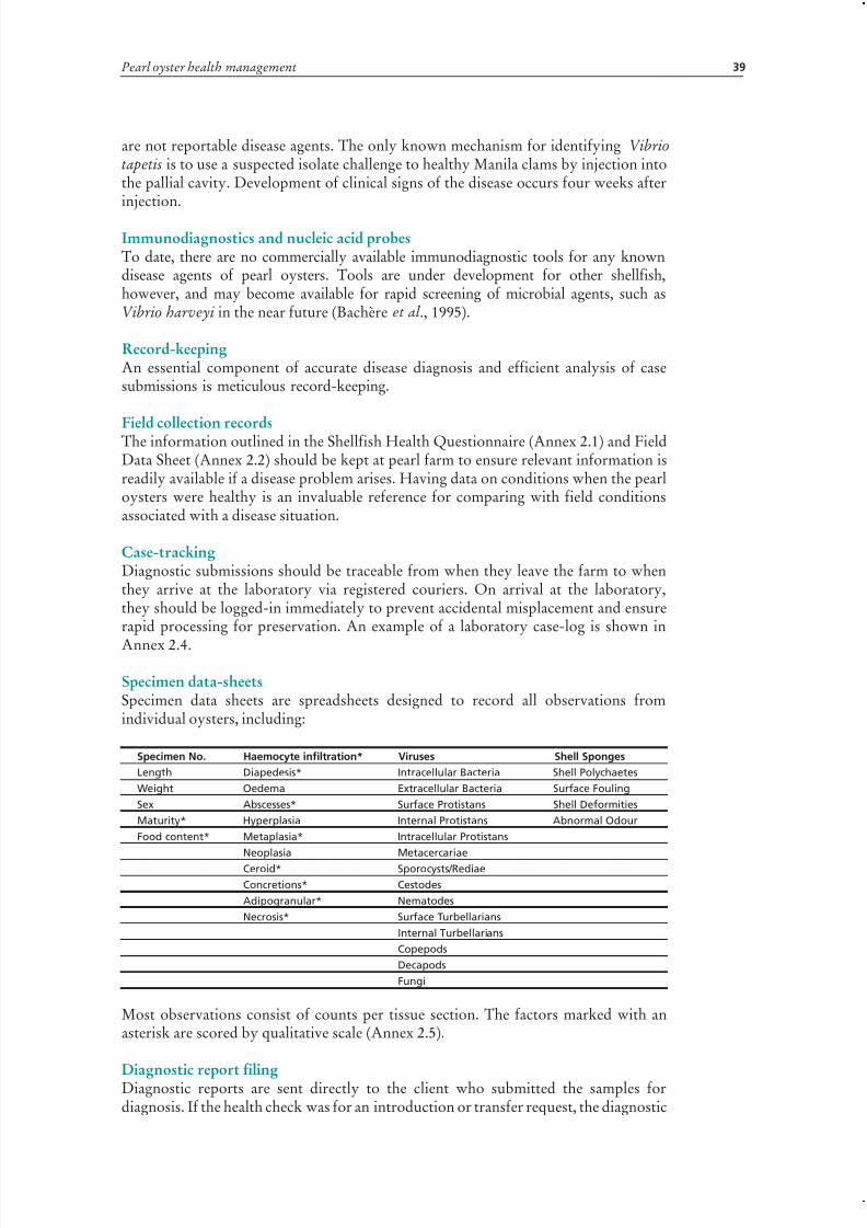
7/27/2019 Pearl Oyster Health Management
http://slidepdf.com/reader/full/pearl-oyster-health-management 53/136
Pearl oyster health management 39
are not reportable disease agents. The only known mechanism for identifying Vibriotapetis is to use a suspected isolate challenge to healthy Manila clams by injection intothe pallial cavity. Development of clinical signs of the disease occurs four weeks afterinjection.
Immunodiagnostics and nucleic acid probesTo date, there are no commercially available immunodiagnostic tools for any knowndisease agents of pearl oysters. Tools are under development for other shellfish,however, and may become available for rapid screening of microbial agents, such asVibrio harveyi in the near future (Bachère et al ., 1995).
Record-keepingAn essential component of accurate disease diagnosis and efficient analysis of casesubmissions is meticulous record-keeping.
Field collection recordsThe information outlined in the Shellfish Health Questionnaire (Annex 2.1) and FieldData Sheet (Annex 2.2) should be kept at pearl farm to ensure relevant information isreadily available if a disease problem arises. Having data on conditions when the pearloysters were healthy is an invaluable reference for comparing with field conditionsassociated with a disease situation.
Case-trackingDiagnostic submissions should be traceable from when they leave the farm to whenthey arrive at the laboratory via registered couriers. On arrival at the laboratory,they should be logged-in immediately to prevent accidental misplacement and ensurerapid processing for preservation. An example of a laboratory case-log is shown in
Annex 2.4.
Specimen data-sheetsSpecimen data sheets are spreadsheets designed to record all observations fromindividual oysters, including:
Specimen No. Haemocyte infiltration* Viruses Shell Sponges
Length Diapedesis* Intracellular Bacteria Shell Polychaetes
Weight Oedema Extracellular Bacteria Surface Fouling
Sex Abscesses* Surface Protistans Shell Deformities
Maturity* Hyperplasia Internal Protistans Abnormal Odour
Food content* Metaplasia* Intracellular Protistans
Neoplasia Metacercariae
Ceroid* Sporocysts/Rediae
Concretions* Cestodes
Adipogranular* Nematodes
Necrosis* Surface Turbellarians
Internal Turbellarians
Copepods
Decapods
Fungi
Most observations consist of counts per tissue section. The factors marked with anasterisk are scored by qualitative scale (Annex 2.5).
Diagnostic report filingDiagnostic reports are sent directly to the client who submitted the samples fordiagnosis. If the health check was for an introduction or transfer request, the diagnostic

7/27/2019 Pearl Oyster Health Management
http://slidepdf.com/reader/full/pearl-oyster-health-management 54/136
Pearl oyster health management: a manual 40
PLATE 2.1.1Examples of histopathological observations (concretions, abcess-like lesions, granuloma,
neoplasia) on molluscan species
C O U R T E S Y O F F . C . J . B E R T
H E
Low magnification of concretions (arrows) inthe digestive gland of a scallop (Placopecten
magellanicus)
High magnification
Low magnification of abcess-like lesion
(arrows) in the mantle of a scallop
(Placopecten magellanicus)
High magnification of a granuloma (arrows)
High magnification of disseminated neoplasia
(neoplasia) in the connective tissue of the
digestive gland of a mussel (Mytilus trossulus)
High magnification

7/27/2019 Pearl Oyster Health Management
http://slidepdf.com/reader/full/pearl-oyster-health-management 55/136
Pearl oyster health management 41
PLATE 2.1.2Examples of histopathological observations (haemocytic infiltration, congestion) on
molluscan species
C O U R T E S Y O F F . C . J . B E R T H E
Low magnification of haemocytic infiltration
(arrows) of the digestive gland connective
tissue of a flat oyster (Ostrea edulis)
High magnification
High magnification showing infiltration
(arrows) of the gill filaments
Haemocyte infiltration associated with
disseminated neoplasia (arrows) in the
connective tissue of the digestive gland of a
mussel (Mytilus galloprovincialis)
Congestion (arrow) of a vessel in the gill of an
Eastern oyster (Crassostrea virginica)
High magnification of the connective tissue
between digestive gland and gonad (arrow)

7/27/2019 Pearl Oyster Health Management
http://slidepdf.com/reader/full/pearl-oyster-health-management 56/136
Pearl oyster health management: a manual 42
Pearl implantation
PLATE 2.1.3Examples of histopathological observations (Cliona lesion, haemocytic infiltration,
Vibrio sp. infection, oedema-type lesion, starving oyster) on molluscan species
C O U R T E S Y O F F . C . J . B E R T H E
a) Low magnification of Cliona sp. lesionof the shell. Note haemocytic infiltration
(arrows) of the adjacent muscular tissue
(Eastern oyster, Crassostrea virginica)
b) High magnification shows the breach ofshell integrity (arrow)
c) Vibrio sp. infection (arrow) of the foot of an
abalone (Haliotis asinina)
d) Gram stain of Vibrio sp. infection (arrow)
e) Low magnification of oedema type
lesion (arrows) in the mantle of
Eastern oyster (Crassostrea virginica)
f) Low magnification of the digestive gland
of a starving oyster (Crassostrea virginica)
(arrows)
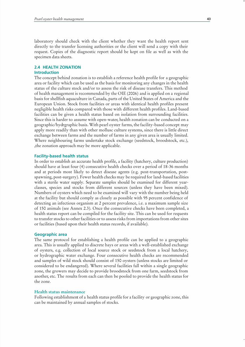
7/27/2019 Pearl Oyster Health Management
http://slidepdf.com/reader/full/pearl-oyster-health-management 57/136
Pearl oyster health management 43
laboratory should check with the client whether they want the health report sentdirectly to the transfer licensing authorities or the client will send a copy with theirrequest. Copies of the diagnostic report should be kept on file as well as with thespecimen data sheets.
2.4 HEALTH ZONATION IntroductionThe concept behind zonation is to establish a reference health profile for a geographicarea or facility which can be used as the basis for monitoring any changes in the healthstatus of the culture stock and/or to assess the risk of disease transfers. This methodof health management is recommended by the OIE (2006) and is applied on a regionalbasis for shellfish aquaculture in Canada, parts of the United States of America and theEuropean Union. Stock from facilities or areas with identical health profiles presentnegligible health risks compared with those with different health profiles. Land-basedfacilities can be given a health status based on isolation from surrounding facilities.
Since this is harder to assume with open-water, health zonation can be conducted on ageographic/hydrgraphic basis. With pearl oyster farms, the facility-based concept mayapply more readily than with other mollusc culture systems, since there is little directexchange between farms and the number of farms in any given area is usually limited.Where neighbouring farms undertake stock exchange (seedstock, broodstock, etc.),,the zonation approach may be more applicable.
Facility-based health statusIn order to establish an accurate health profile, a facility (hatchery, culture production)should have at least four (4) consecutive health checks over a period of 18-36 monthsand at periods most likely to detect disease agents (e.g. post-transportation, post-spawning, post-surgery). Fewer health checks may be required for land-based facilities
with a sterile water supply. Separate samples should be examined for different year-classes, species and stocks from different sources (unless they have been mixed).Numbers of oysters which need to be examined will vary with the number being heldat the facility but should comply as closely as possible with 95 percent confidence of detecting an infectious organism at 2 percent prevalence, i.e. a maximum sample sizeof 150 animals (see Annex 2.3). Once the consecutive checks have been completed, ahealth status report can be compiled for the facility site. This can be used for requeststo transfer stocks to other facilities or to assess risks from importations from other sitesor facilities (based upon their health status records, if available).
Geographic areaThe same protocol for establishing a health profile can be applied to a geographicarea. This is usually applied to discrete bays or areas with a well-established exchangeof oysters, e.g. collection of local source stock or seedstock from a local hatchery,or hydrographic water exchange. Four consecutive health checks are recommendedand samples of wild stock should consist of 150 oysters (unless stocks are limited orconsidered to be endangered). Where several facilities fall within a single geographiczone, the growers may decide to provide broodstock from one farm, seedstock fromanother, etc. The results from each can then be pooled to provide the health status forthe zone.
Health status maintenanceFollowing establishment of a health status profile for a facility or geographic zone, this
can be maintained by annual samples of stocks.

7/27/2019 Pearl Oyster Health Management
http://slidepdf.com/reader/full/pearl-oyster-health-management 58/136
Pearl oyster health management: a manual 44
2.5 DISEASE OUTBREAK INVESTIGATION PROCEDUREIntroductionA systematic procedure to help identify the cause or causes of disease events andprevent the further transmission of the pathogen is required to control an ongoing
and to prevent future disease events. Any disease outbreak investigation will stronglybenefit from using an epidemiological approach which assesses the pattern of infectionwithin the affected population (Lilley et al ., 1998).
Lilley et al. (1998) outlined the nine basic steps to a disease outbreak investigationdeveloped for investigating outbreak of epizootic ulcerative syndrome (EUS) of freshand brackishwater finfish in Asia. This approach is also applicable to pearl oysterhealth disease investigations. The nine steps include: (1) establishing a diagnosis,(2) establishing a “case definition”, (3) confirming that an outbreak is actuallyoccurring, (4) characterising the outbreak in terms on time, affected/unaffected species,and place, (5) analysing the data, (6) developing a working hypothesis, (7) makingintensive follow-up with different laboratory testing and further epidemiological
analysis, (8) establishing control and prevention measures and (9) reporting. These ninesteps may not necessarily be included in every investigation, nor will an investigationalways follow the same sequence; they may be undertaken simultaneously.
One of the most important criteria to establish, as quickly as possible, is whether ornot a disease outbreak is due to an infectious agent or due to stress and/or secondaryopportunistic infection. This can be achieved by sending samples of affected andunaffected oysters to a diagnostic laboratory as soon as abnormal mortalities aredetected. Since some tests may take weeks, some interim control measures will berequired pending diagnostic results.
A careful record of the pattern of mortalities may assist in determining the natureof the problem (although not the cause). Generally-speaking, environmental stress-related mortalities may affect animals throughout the stock. Stocking density effects
will tend to cause mortalities at the centre of the cultured population, with peripherallines/cages showing relatively little effect. Toxins or pollutants may cause the samepattern of mortality or manifest themselves as acute mortalities throughout the stock.Infectious disease agents will show a progressive pattern of mortality spreadingbetween neighbouring lines or nets.
Diagnostic submissionAs soon as abnormal rates of mortality are detected, samples of moribund (not dead)and unaffected (if available) oysters should either be preserved on site (see Section2.2.1) or submitted live to a diagnostic laboratory with molluscan shellfish healthexpertise. If there is evidence of a microbial (bacterial or fungal) infection, fresh ratherthan preserved samples should be sent to the laboratory. Phone/Fax the laboratory inadvance to let them know the nature of the problem, determine the number of samplesthey need and ensure that they are prepared for the diagnostic case. The laboratorymay need to prepare specialized media for tissue culture and fixatives.
Quarantine (isolation)Quarantine is a holding method which prevents the escape or release of shellfish and anyof their disease agents. Different periods of quarantine may be required depending onthe disease. Quarantine is also applied to shellfish being introduced to a site from an areawhere there is a disease agent of concern or no health history information (see Section2.2.1). Quarantine, by definition, is therefore difficult to manage in open-water.
Isolation is used to prevent infection by organisms from the surrounding environment.
This may occur within a site where there is a disease outbreak and unaffected stocks areplaced under isolation (while the diseased stock fall within quarantine). Isolation canbe conducted under two different conditions:

7/27/2019 Pearl Oyster Health Management
http://slidepdf.com/reader/full/pearl-oyster-health-management 59/136
Pearl oyster health management 45
• Land-based isolation prevents access to the shellfish by other shellfish or associateddisease agents from surrounding waters. Although shellfish may not be introducedto an isolation facility or site, they can be released into the surrounding waters.
• Geographic isolation is the spatial separation of shellfish from neighbouring
shellfish beds or sites.
DisinfectionDisinfection involves the application of chemical treatments at sufficient concentrations,and for sufficient periods of time, to kill or inactivate harmful organisms. Since theinherent toxicity of disinfectants negates safe use in open-water, or flow-throughsystems, disinfection can only be applied with reasonable control within hatcheries,tanks or land-based holding facilities. Disinfectants must be neutralised before releaseinto the surrounding environment, especially seawater treatments, which produceresidual oxidants which are particularly toxic to bivalves (OIE, 2007).
If a disease agent is detected, disinfection of tanks, nets and equipment is
recommended. Stock infected by an opportunistic pathogen may be destroyed, ortransferred following surface disinfection to clean holding tanks at lower stockingdensities, water temperatures, and with new food sources.
Where stocking densities are high, shellfish should be rotated between disinfectedtanks as frequently as practical. Each new batch of shellfish introduced to a facilityshould be placed in pre-disinfected tanks. Because the presence of organic matterwill reduce the disinfection capacity of most disinfectants, filtering influent wateris recommended. In addition, all surfaces should be thoroughly cleaned prior todisinfection. The detergent used for this purpose should be compatible with thedisinfectant and both should be compatible with the surface being treated. All wastefrom washing should be disinfected before disposal.Regular air- or heat-drying of pipelines (daily), tanks and other equipment (e.g. algal culture carboys), in addition
to disinfection of their surfaces, is also recommended. All chemical treatmentsshould be undertaken in a manner that prevents their release into the surroundingenvironment.
TreatmentsChlorine is usually applied as sodium hypochlorite (Chlorox®, household bleach, etc.).Fill all pipelines with 50 ppm chlorine (= 50 mg/l). Allow an exposure time of at least30 minutes before flushing with clean seawater. This solution is effective against mostmicrobial agents as well as labyrinthulid protozoans. Chlorinated seawater should beneutralised prior to release from the holding facility.
Iodophors are applied as alkaline solutions (Wescodyne®, Betadine®), at an iodine(I2) concentration of 200-250 ppm, for a contact time of at least 10 minutes, however,they are not effective against certain protistans (Bower, 1989) and may need to besupplemented by air- or heat-drying of tank surfaces and pipelines.
Testing treatmentsFollowing a disease outbreak, or the initial set-up of a disinfection system and priorto introducing fresh shellfish stock or flushing effluent from the facility, routinebacteriological culture of disinfected surfaces and water for ubiquitous bacteria (suchas Vibrio spp. and Pseudomonas spp.) should be performed. If present, this indicatesthat the disinfection procedure used was inadequate.
Residual monitoring
Residual or free chlorine (or other halogen) ions are indicative of the degree of bindingto/ oxidation of organic compounds or organisms in the water being treated. If noresidual oxidizing ions remain after a set period of treatment, it is assumed that:
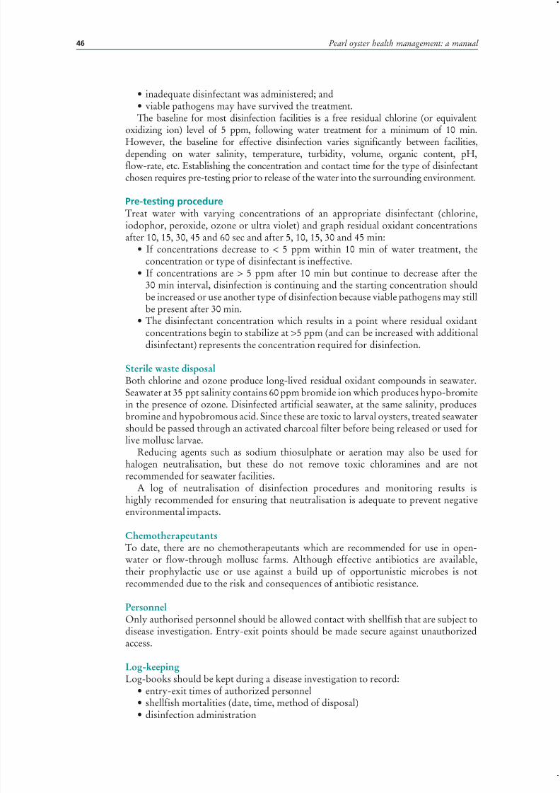
7/27/2019 Pearl Oyster Health Management
http://slidepdf.com/reader/full/pearl-oyster-health-management 60/136
Pearl oyster health management: a manual 46
• inadequate disinfectant was administered; and• viable pathogens may have survived the treatment.The baseline for most disinfection facilities is a free residual chlorine (or equivalent
oxidizing ion) level of 5 ppm, following water treatment for a minimum of 10 min.
However, the baseline for effective disinfection varies significantly between facilities,depending on water salinity, temperature, turbidity, volume, organic content, pH,flow-rate, etc. Establishing the concentration and contact time for the type of disinfectantchosen requires pre-testing prior to release of the water into the surrounding environment.
Pre-testing procedureTreat water with varying concentrations of an appropriate disinfectant (chlorine,iodophor, peroxide, ozone or ultra violet) and graph residual oxidant concentrationsafter 10, 15, 30, 45 and 60 sec and after 5, 10, 15, 30 and 45 min:
• If concentrations decrease to < 5 ppm within 10 min of water treatment, theconcentration or type of disinfectant is ineffective.
• If concentrations are > 5 ppm after 10 min but continue to decrease after the30 min interval, disinfection is continuing and the starting concentration shouldbe increased or use another type of disinfection because viable pathogens may stillbe present after 30 min.
• The disinfectant concentration which results in a point where residual oxidantconcentrations begin to stabilize at >5 ppm (and can be increased with additionaldisinfectant) represents the concentration required for disinfection.
Sterile waste disposalBoth chlorine and ozone produce long-lived residual oxidant compounds in seawater.Seawater at 35 ppt salinity contains 60 ppm bromide ion which produces hypo-bromitein the presence of ozone. Disinfected artificial seawater, at the same salinity, produces
bromine and hypobromous acid. Since these are toxic to larval oysters, treated seawatershould be passed through an activated charcoal filter before being released or used forlive mollusc larvae.
Reducing agents such as sodium thiosulphate or aeration may also be used forhalogen neutralisation, but these do not remove toxic chloramines and are notrecommended for seawater facilities.
A log of neutralisation of disinfection procedures and monitoring results ishighly recommended for ensuring that neutralisation is adequate to prevent negativeenvironmental impacts.
ChemotherapeutantsTo date, there are no chemotherapeutants which are recommended for use in open-water or flow-through mollusc farms. Although effective antibiotics are available,their prophylactic use or use against a build up of opportunistic microbes is notrecommended due to the risk and consequences of antibiotic resistance.
PersonnelOnly authorised personnel should be allowed contact with shellfish that are subject todisease investigation. Entry-exit points should be made secure against unauthorizedaccess.
Log-keepingLog-books should be kept during a disease investigation to record:
• entry-exit times of authorized personnel• shellfish mortalities (date, time, method of disposal)• disinfection administration

7/27/2019 Pearl Oyster Health Management
http://slidepdf.com/reader/full/pearl-oyster-health-management 61/136
Pearl oyster health management 47
• residual levels and neutralisation• samples sent to laboratories for disease diagnosis.
Investigation
Mortalities with no immediately apparent cause may take years to investigate,depending on the epidemiology of infection. This means that disease control measuresmust be balanced in terms of cost and practicality with the continued operation of thefarm. Disease control measures and research plans are most effective where the farmmanagement and personnel actively participate. The latter have the day-to-day contactwith the shellfish which provides the information essential for accurate interpretationof disease observations and experimental results.
2.6 NATIONAL STRATEGIES ON AQUATIC ANIMAL HEALTHIn recent years, countries are developing and implementing “national strategies” onaquatic animal health management (AFFA, 1999; Olivier, 2004; Kanchanakhan and
Chinabut, 2004; Amos, 2004). In Asia, the development of the “national strategy” wasan outcome of an FAO-supported regional technical cooperation project on “healthmanagement for the responsible movement of live aquatic animals” (Bondad-Reantaso,2004). The “National Strategy on Aquatic Animal Health” contains the actions plans of the government at the short-, medium, and long-term to implement the Asia RegionalTechnical Guidelines for the Responsible Movement of Live Aquatic Animals (FAO/NACA, 2000). The essential components (or elements) of the “national strategy onaquatic animal health” include the following: (i) policy, legislation and enforcement,(ii) pathogen list and information system, (iii) diagnostics, (iv) health certification andquarantine, (v) risk analysis for aquatic animal movement, (vi) surveillance, monitoringand reporting, (vii) zoning, (viii) response and contingency plans to disease emergencies,(ix) research, (x) institutional structure, (xii) human resources development and (xiii)
regional and international cooperation.There are varying processes involved in the development of the “national
strategy”. In some countries, the take-off of national strategy development wasimmediate; for some others, it went through a slow process for various reasons.Although not aimed specifically at diseases of pearl oysters, the development of such strategies is a valuable framework for enhancing basic health management andbiosecurity education and awareness for aquatic animal health at both a nationaland regional levels. Experience in Asia provided some essential considerationsfor ensuring success in developing and implementing a “national strategy”. Theseinclude: (i) strong national coordination, (ii) a good driver for the process (throughthe Competent Authority, a commission, a committee, a task force, or a focal person)with clear terms of reference, (iii) needs assessment and prioritization exercise,(iv) active stakeholder consultation, (v) approval from highest authority, (vi) animplementation strategy, (vii) a monitoring and review plan, (viii) development of proposal for addressing the different component (elements) of the national strategyand (ix) resource and funding allocation.
Development and implementation of national strategies on aquatic animal healthwithin the broader aquaculture development and biosecurity framework should becontinuously pursued. The national strategy is comprehensive enough, and using theconcept of “ phased implementation based on national needs”, it could provide a goodand strong entry point for capacity building for many countries regardless of aquaticanimal health infrastructure or economic development.

7/27/2019 Pearl Oyster Health Management
http://slidepdf.com/reader/full/pearl-oyster-health-management 62/136
Pearl oyster health management: a manual 48
2.7 REFERENCESAFFA. 1999. AQUAPLA. Australia’s National Strategic Plan for Aquatic Animal Health
1998-2003. Government of Australia, Canberra. 34 pp.Allen, S.K. Jr., Guo, X., Burreson, E.G. & Mann, R. 1996. Heteroploid mosaics and
reversion among triploid oysters, Crassostrea gigas. Fact or artifact? Journal of ShellfishResearch 15: 514. (Abstract).
Amos, K.H. 2004. National Aquatic Animal Health Plan for the United States of America,pp. 147-150. In J.R. Arthur and M.G. Bondad-Reantaso (eds). Capcity and awarenessbuilding on import risk analysis for aquatic animals, Proceedings of the workshops heldon 1-6 April 2002 in Bangkok, Thailand and 12-17 August 2002 in Mazatlan, Mexico.APEC FWG01/2002. Bangkok, NACA. 203 pp.
Bachère, E., Mialhe, E., Noël, D., Boulo, V., Morvan, A. & Rodriguez, J. 1995. Knowledge andresearch prospects in marine mollusc and crustacean immunology. Aquaculture 132: 17-32.
Benzie, J.A.H. 1994. Genetics of black-lipped pearl oyster (Pinctada margaritifera). Journal of Shellfish Research 13: 331 (abstract)
Bondad-Reantaso, M.G. 2004. Development of national strategy on aquatic animal healthmanagement in Asia, pp. 103-108. In J.R. Arthur and M.G. Bondad-Reantaso (eds).Capcity and awareness building on import risk analysis for aquatic animals, Proceedingsof the workshops held on 1-6 April 2002 in Bangkok, Thailand and 12-17 August 2002in Mazatlan, Mexico. APEC FWG01/2002. Bangkok, NACA. 203 pp.
Bondad-Reantaso, M.G., McGladdery, S.E., East, I., & Subasinghe, R.P. 2001. AsiaDiagnostic Guide to Aquatic Animal Diseases. FAO Fisheries Technical Paper 402/2, FAO and NACA. 237 pp
Bower, S.M. 1989. Disinfectants and therapeutic agents for controlling Labyrinthuloideshaliotidis (Protozoa: Labyrinthomorpha), an abalone pathogen. Aquaculture 78: 207-215.
Bower, S.M. and McGladdery, S.E. 1997. Synopsis of Infectious Diseases and Parasites of Commercially Exploited Shellfish: www.pac.dfo-mpo.gc.ca/sci/shelldis/defin_e.htm
Bower, S.M., McGladdery, S.E. & Price, I.M. 1994. Synopsis of infectious diseases andparasites of commercially exploited shellfish. Annual Review of Fish Disease 4: 1-199.
Brock, J.A. 1992. Procedural requirements for marine species introductions into and outof Hawaii. pp. 5153. In De Voe, R., ed , Proceedings of the Conference & Workshop:Introductions & Transfers of Marine Species Achieving A Balance Between Economic Development and Resource Protection, pp. 51-53. South Carolina Sea Grant Consortium,October 30 November 2, 1991. Hilton Head Island, South Carolina.
Claereboudt, M.R., Bureau, D., Côté, J. & Himmelman, J.H. 1994. Fouling developmentand its effect on the growth of juvenile giant scallops (Placopecten magellanicus) insuspended culture. Aquaculture 121: 327-342.
Côté, J., Himmelman, J.H., Claereboudt, M.R. & Bonardelli, J. 1993. Influence of densityand depth on the growth of juvenile giant scallops (Placopecten magellanicus, Gmelin1791) in suspended culture in the Baie des Chaleurs. Canadian Journal of Fisheries and
Aquatic Science 50: 1857-1869.Crossland, C. 1957. The cultivation of the mother-of-pearl oyster in the Red Sea. Australian
Journal of Marine and Freshwater Research 8: 111-130.Culloty, S.C. & Mulcahy, M.F. 1992. An evaluation of anaesthetics for Ostrea edulis (L.).
Aquaculture 107: 249-252.Dauphin, Y. & Cuif, J.-P. 1995. Trichromatic characterization of the “black pearls” from
aquaculture centres of French Polynesia. Aquaculture 133: 113-121.Deng, C., Lin, Y., Du, T., Fu, S. & Luo, J. 1995. Experiment on pre-operation treatment of
Pinctada martensii. J. Zhanjiang Fish. 15: 6-9 (In Chinese. Abstract read only)De Voe, R. 1992. (ed.) Proceedings of the Conference & Workshop: Introductions &
Transfers of Marine Species Achieving A Balance Between Economic Development and Resource Protection. South Carolina Sea Grant Consortium, October 30 November 2,1991. Hilton Head Island, South Carolina.
7/27/2019 Pearl Oyster Health Management
http://slidepdf.com/reader/full/pearl-oyster-health-management 63/136
Pearl oyster health management 49
Dijkema, R. 1995. Large-scale recirculation systems for storage of imported bivalves as ameans to counteract introduction of cysts of toxic dinoflagellates in the coastal watersof The Netherlands. In Poggi, R. & LeGall, J.Y. eds, Shellfish Depuration, pp. 355-367. IFREMER, Plouzane, France.
Dybdahl, R. & Pass, D.A. 1985. An investigation of mortality of the pearl oyster, Pinctadamaxima, in Western Australia. Fisheries Department of Western Australia. Report No.71: 1-78.
Dybdahl, R., Harders, S. & Nicholson, C. 1990. Developing on-growing techniques anddisease prevention husbandry of pearl oysters in Western Australia (FIRTA Project87/81) and on-growing mariculture techniques for the pearl oyster Pinctada maxima spatin Western Australia (FIRDTF Project 89/60). Final reports. Western Australian MarineResearch Laboratories, Waterman, W.A. 57pp.
Elston, R.A. 1984. Prevention and management of infectious diseases in intensive molluschusbandry. Journal of the World Mariculture Society 14: 284-300.
FAO/NACA. 2000. Asia Regional Technical Guidelines on Health Management for
the Responsible Movement of Live Aquatic Animals and the Beijing Consensus andImplementation Strategy. FAO Fisheries Technical Paper. No. 402. Rome, FAO. 2000. 53 pp.Fassler, R. 1994. Hawaii’s impact on the International Pearl Industry. Journal of Shellfish
Research 13: 335 (abstract)Fisheries Western Australia. 1997. Pearling in W.A. - Pearl Creation. (available at URL:
http: //www.wa.gov.au/westfish/aqua/broc/pearl/pearlcreation.html)Friedman, K.J. & Bell, J.D. 1996. Effects of different substrata and protective mesh bags on
collection of spat of pearl oysters, Pinctada margaritifera (Linnaeus, 1758) and Pinctadamaculata (Gould, 1950). Journal of Shellfish Research 15: 535-541.
Gervis, M.H. & Sims, N.A. 1992. The biology and culture of pearl oysters (Bivalvia:Pteridae). ICLARM Stud. Rev. ICLARM, Metro Manila, Philippines, No. 21: 49 pp.
He, M., Lin, Y. & Jiang, W. 1996. Studies on the sterility of triploid pearl oyster, Pinctada
martensii (D.). Trop. Oceanol . 15: 17-21. (In Chinese. Abstract read only)Holt, J.G. & Kreig, N.R. 1984. (eds.) Bergey’s Manual of Systemic Bacteriology, Vol. 1,
Williams and Wilkins, Baltimore, MD, USA, 1092 pp.ICES. 1995. ICES Code of Practice on the Introductions and Transfers of Marine Organisms
1994. International Council for the Exploration of the Sea, Copenhagen, Denmark. 12 pp. Jiang, W., Li, G., Xu, G., Lin, Y. & Qing, N. 1993. Growth of the induced triploid pearl
oyster, Pinctada martensii (D.). Genetics in Aquaculture IV. Elsevier, Amsterdam, TheNetherlands, pp. 245-253.
Joll, L.M. 1994. Shell handling and mortality - the Western Autralian Experience. In Golden,S., Turnbull, C. & Coles, R., eds, Developing the Torres Strait and Queensland East CoastPearl Industry. Conf. Workshop Ser. Dep. Primary Ind. (Queensl.) pp. 35-37 Brisbane.
Kanchanakhan, S. & Chinabut, S. 2004. Strategies for aquatic animal health managementin Thailand, pp. 139-141. In J.R. Arthur and M.G. Bondad-Reantaso (eds). Capacityand awareness building on import risk analysis for aquatic animals, Proceedings of theworkshops held on 1-6 April 2002 in Bangkok, Thailand and 12-17 August 2002 inMazatlan, Mexico. APEC FWG01/2002. Bangkok, NACA. 203 pp.
Krishnan, A. & Alagarswami, K. 1993a. Effect of larval density and algal cell concentrationon hatchery rearing and production of the Indian pearl oyster Pinctada fucata (Gould).In Natarajan, P. & Jayaprakas, V., eds. Proceedings of the National Seminar on
Aquaculture Development in India - Problems and Prospects, pp.123-130. Kerala Univ.,Thiruvananthapuram, India.
Krishnan, A. & Alagarswami, K. 1993b. Studies on larval nutrition in the pearl oysterPinctada fucata (Gould). CMFRI Special Publication 56: 29-34.
Lee, D.Y. 1992. The effect of antifouling paint against the fouling organisms in the pearl-oyster, Pinctada fucata, farming. Bulletin of the. National Fisheries Research Development Agency (Korea) 46: 129-143.
7/27/2019 Pearl Oyster Health Management
http://slidepdf.com/reader/full/pearl-oyster-health-management 64/136
Pearl oyster health management: a manual 50
Lesser, M.P., Shumway, S.E., Cucci, T. & Smith, J. 1992. Impact of fouling organismson mussel rope culture: Interspecific competition for food among suspension-feedinginvertebrates. Journal of Experimental Marine Biology and Ecology 165: 91-102.
Lilley, J.H., Callinan, R.B., Chinabut, S., Kanchanakhan, S., MacRae, I.H. & Phillips,
M.J. 1998. Epizootic ulcerative syndrome (EUS) technical handbook. Bangkok, TheAquatic Animal Health Research Institute. 88 pp.
Lin, Y., He, M. & Jiang, W. 1996. An observation on the mortality of triploids in Pinctadamartensii (D.). Trop. Oceanol./ Redai Haiyang 15: 80-84. (In Chinese. Abstract read only)
Lodeiros, C.J.M. & Himmelman, J.H. 1996. Influence of fouling on the growth andsurvival of the tropical scallop, Euvola (Pecten) ziczac (L. 1758) in suspended culture.
Aquaculture Research 27: 749-756.MacDonald, B.A. & Bourne, N.F. 1989. Growth of the purple-hinge rock scallop,
Crassodoma gigantea Gray, 1825 under natural conditions and those associated withsuspended culture. Journal of Shellfish Research 7: 146-147.
MacDonald, B.A. & Thompson, R.J. 1986. Influence of temperature and food availability
on the ecological energetics of the giant scallop Placopecten magellanicus. 3. Physiologicalecology, the gametogenic cycle and scope for growth. Marine Biology 93: 37-48.MacDonald, B.A., Thompson, R.G. & Bayne, B.L. 1987. Influence of temperature and
food availability on the ecological energetics of the giant scallop Placopecten magellanicus.4. Reproductive effort, value and cost. Oecologia 72: 550-556.
MacDonald, B.A., Thompson, R.G. & Bourne, N.F. 1991. Growth and reproductiveenergetics of three scallop species from British Columbia (Chlamys hastata, Chlamysrubida and Crassadoma gigantea). Canadian Journal of Fisheries and Aquatic Science 48:215-221.
Mattheissen, G.C. & Davis, J.P. 1992. Observations on growth rate and resistance toMSX (Haplosporidium nelsoni) among diploid and triploid eastern oysters (Crassostreavirginica (Gmelin)) in New England. Journal of Shellfish Research 11: 449-454.
Meng, Z., Zhang, Q., Li, Y., & Xing, K. 1994. A study on antomy of sites of nucleus-insertion and pearl culture in Pinctada nigra (Gould). J. Fish. Sci. China/ ZhongguoShuichan Kexue 1: 25-30 (In Chinese, abstract read only).
Mills, D. 1997. Evaluation of histological cassettes as holding containers for individual spat,and a weekly handling protocol to assess growth of the silver-lip pearl oyster, Pinctadamaxima (Jameson). Journal of Shellfish Research 16: 555-559.
Mills, D., Tlili, A. & Norton, J. 1997. Large-scale anaethesia of the silver-lip pearl oyster, Pinctada maxima Jameson. Journal of Shellfish Research 16: 573-574.
Monteforte, M., Kappelman-Pina, E. & Lopez-Espinosa, B. 1995. Spatfall of pearloyster, Pteria sterna (Gould), on experimental collectors at Bahia de La Paz, South BajaCalifornia, Mexico. Aquaculture Research 26: 497-511.
Nell, J.A., Cox, E., Smith, I.R. & Maguire, G.B. 1994. Studies on triploid oystersin Australia. 1. The farming potential of triploid Sydney rock oysters Saccostreacommercialis (Iredale and Roughley). Aquaculture 126: 243-255.
Norton, J.H. 1994. Pearl oyster transport and handling tecniques. In Golden, S., Turnbull,C. & Coles, R. (eds). Developing the Torres Strait and Queensland East Coast Pearl Industry. Conf. Workshop Ser. Dep. Primary Ind. (Queensl.). Brisbane. pp. 27-34.
OIE. 2006. OIE Diagnostic Manual for Aquatic Animal Diseases. 4th Ed. OfficeInternational des Epizooties, Paris, France.
OIE. 2007. OIE International Aquatic Animal Health Code 6th Ed. Office Internationaldes Epizooties, Paris, France.
Olivier, G. 2004. Canada’s National Aquatic Animal Health Program, pp. 115-117. In J.R.Arthur and M.G. Bondad-Reantaso (eds). Capcity and awareness building on import
risk analysis for aquatic animals, Proceedings of the workshops held on 1-6 April 2002in Bangkok, Thailand and 12-17 August 2002 in Mazatlan, Mexico. APEC FWG01/2002.Bangkok, NACA. 203 pp.

7/27/2019 Pearl Oyster Health Management
http://slidepdf.com/reader/full/pearl-oyster-health-management 65/136
Pearl oyster health management 51
Ossiander, F.J. & Wedermeyer, G. 1973. Computor program for sample sizes required todetermine disease incidence in fish populations. Journal of the Fisheries Research Board of Canada 30: 1383-1384.
Parsons, G.J. & Dadswell, M.J. 1992. Effect of stocking density on growth, production and
survival of the giant scallop, Placopecten magellanicus, held in internediate suspensionculture in Passamaquoddy Bay, New Brunswick. Aquaculture 103: 291-309.
Pass, D.A., Dybdahl, R. & Mannion, M.M. 1987. Investigations into the causes of mortalityof the pearl oyster, Pinctada maxima (Jamson), in Western Australia. Aquaculture 65:149-169.
Rose, R.A. & Baker, S.B. 1994. Larval and spat culture of the Western Australian silver-or gold-lip pearl oyster, Pinctada maxima (Jameson) (Mollusca: Pteriidae). Aquaculture 126: 33-50.
Shariff, M., Subasinghe, R.P. & Arthur, J.R. 1992. (eds.) Diseases in Asian Aquaculture I .Fish Health Section, Asian Fisheries Society, Manila, Philippines, pp. 493-566.
Sims, N.A. 1994. Growth of wild and cultured black-lip pearl oysters, Pinctada
margaritifera (L.) (Pteriidae; Bivlavia), in the Cook Islands. Aquaculture 122: 181-191.Sims, N.A. & Sarver, D.J. 1994. Hatchery culture of he black-lip pearl oyster in Hawaii -Stock re-establishment and expansion of commercial pearl culture throughout the region.
Journal of Shellfish Research 13: 350 (Abstract only).Sims, N.A. & Sarver, D.J. 1996. The potential for growth of pearl culture in the Pacific
Islands. Proceedings of the Pacon Conference on Sustainable Aquaculture ‘95. pp. 318-326.
Sindermann, C.J. 1986. Strategies for reducing risks from introductions of aquaticorganisms: a marine perspective. Fisheries 11(2): 1015.
Sindermann, C.J. 1991. Case histories of effects of transfers and introductions on marineresources: Introduction. Journal de Conseil d’Exploration de la Mer (ICES) 47: 377378.
Smitasiri, R., Kajiwiwat, J. & Tantichodok, P. 1994. Growth of a winged pearl oyster,
Pteria penguin, suspended at different depths. Special Publication of the. Phuket MarineBiol. Center 13: 213-216.
Snieszko, S.F. 1974. The effects of environmental stress on outbreaks of infectious diseasesof fishes. Journal of Fish Biology 6: 197-208.
Southgate, P.C. & Beer, A.C. 1997. Hatchery and early nursery culture of the blacklippearl oyster (Pinctada margaritifera L.). Journal of Shellfish Research 16: 561-567.
Taylor, J.J., Rose, R.A. & Southgate, P.C. 1997a. Inducing detatchment of silver-lip pearloyster (Pinctada maxima, Jameson) spat from collectors. Aquaculture 159: 11-17.
Taylor, J.J., Rose, R.A. & Southgate, P.C. 1997b. Effects of stocking density on the growthand survival of juvenile silver-lip pearl oysters (Pinctada maxima, Jameson) in suspendedand bottom culture. Journal of Shellfish Research 16(2): 569-572.
Taylor, J.J., Rose, R.A., Southgate, P.C. & Taylor, C.E. 1997. Effects of stocking densityon growth and survival of early juvenile silver-lip pearl oysters, Pinctada maxima (Jameson), held in suspended nursery culture. Aquaculture 153: 41-49.
Taylor, J.J., Southgate, P.C. & Rose, R.A. 1997. Fouling animals and their effect ont hegrowth of silver-lip pearl oysters, Pinctada maxima (Jameson) in suspended culture.
Aquaculture 153: 31-40.Thompson, R.G & MacDonald, B.A. 1990. The role of environmental conditions in the
seasonal synthesis and utilisation of biochemical energy reserves in the giant scallop,Placopecten magellanicus. Canadian Journal of Fisheries and Aquatic Science 68: 750-756.
Tun, T. 1994. A view on seeding. Journal of Shellfish Research 13: 352-353.Wada, K.T. 1987. Selective breeding and intraspecific hybridization - molluscs. In: Tiews,
K. (ed.) Selection, Hybridization and Genetic Engineering in Aquaculture. Vol. 1, Schr.Bundesforschungsanst. Fisch. Hamburg. pp. 313-322.Wada, K.T. 1993. Bivalve broodstock development in Japan. World Aquaculture 24(3): 54-57.
7/27/2019 Pearl Oyster Health Management
http://slidepdf.com/reader/full/pearl-oyster-health-management 66/136
Pearl oyster health management: a manual 52
Wada, K.T. 1996. Genetical and physiological control of calcification in pearl cultivation.Bull. Inst. Oceanogr. Monaco 14: 183-193.
Wada, K.T. & Komaru, A. 1994. Effect of selection for shell coloration on growth rateand mortality in the Japanese pearl oyster Pinctada fucata martensii. Aquaculture 126:
59-65.Wada, K.T. & Komaru, A. 1996. Color and weight of pearls produced by grafting the
mantle tissue from a selected population for white shell color of the Japanese pearl oysterPinctada fucata martensii (Dunker). Aquaculture 142: 25-32.
Wang, J.-K. 1994. Laboratory growth rates of Pinctada margaritifera - a preliminaryreport. Journal of Shellfish Research 13: 353. (Abstract only).
Whyte, S.K., Cawthorn, R.J. & McGladdery, S.E. 1994a. Co-infection of bay scallops, Argopecten irradians, with Perkinsus karlssoni (Apicomplexa, Perkinsea) and anunidentified coccidian parasite. Diseases of Aquatic Organisms 18: 53-62.
Whyte, S.K., Cawthorn, R.J. & McGladdery, S.E. 1994b. QPX (Quahaug Parasite X), apathogen of northern quahaug Mercenaria mercenaria from the Gulf of St. Lawrence,
Canada. Diseases of Aquatic Organisms 19: 129-136.Widman, J.C. & Rhodes, E.W. 1991. Nursery culture of the bay scallop, Argopectenirradians irradians in suspended mesh nets. Aquaculture 99: 257-267.

7/27/2019 Pearl Oyster Health Management
http://slidepdf.com/reader/full/pearl-oyster-health-management 67/136
Pearl oyster health management 53
ANNEX 2.1
Shellfish Health Questionnaire
Name and address of client
Contact telephone number Fax E-mail
Reason for submission:
Stock transfer (indicate from where to where)
Abnormal mortalities (give approx. percentage of losses)Other
Growing conditions:
Growing Method
Water temperature Salinity Turbidity
Other environmental observations (e.g. predators, high rainfall, wave action, pollution,etc.)
General observations on the stock being submitted:
Appearance (gaping, shell damage, fouling, etc.)
Behaviour (feeding, growth, maturation, movements, etc.)
Recent handling history of the stock or neighbouring stocks
Comments

7/27/2019 Pearl Oyster Health Management
http://slidepdf.com/reader/full/pearl-oyster-health-management 68/136
Pearl oyster health management: a manual 54
A N N E X 2 . 2
F i e l d D a t a S h e e t
S p e c i e
s
L o c a t i o n
D a t e
N a m e
& p
h o n e n u m b e r o f s a m p l e r
S p e c i m e n
N o .
L e n g t h
( m m )
W e i g h t
( g )
B o r i n g
s p o n g e
B o r i n g
w o r m s
S u r f a c e
g i l l - w o r m s
( T u r b s )
C l a m -
w o r m
F u n
g u s
O e d e m a
A b s c e s s
S h e l l b l i s t e r s ,
b u m p s ,
r e p a i r s , e t c .
S o f t - t i s s u e /
g i l l d e f o r m -
a t i e s , e t c .
F o u l o d o u r .
l i q u e f a c t i o n ,
e t c .
N o t e s
P a g e___
o f
____
1 2 3 4 5 6 7 8 9 1 0
1 1
1 2
1 3
1 4
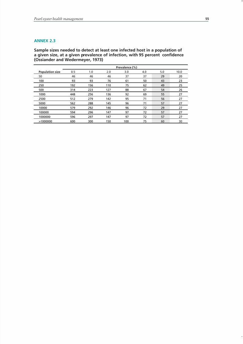
7/27/2019 Pearl Oyster Health Management
http://slidepdf.com/reader/full/pearl-oyster-health-management 69/136
55
ANNEX 2.3
Sample sizes needed to detect at least one infected host in a population ofa given size, at a given prevalence of infection, with 95 percent confidence
(Ossiander and Wedermeyer, 1973)Prevalence (%)
Population size 0.5 1.0 2.0 3.0 4.0 5.0 10.0
50 46 46 46 37 37 29 20
100 93 93 76 61 50 43 23
250 192 156 110 75 62 49 25
500 314 223 127 88 67 54 26
1000 448 256 136 92 69 55 27
2500 512 279 142 95 71 56 27
5000 562 288 145 96 71 57 27
10000 579 292 146 96 72 29 27
100000 594 296 147 97 72 57 27
1000000 596 297 147 97 72 57 27
>1000000 600 300 150 100 75 60 30
Pearl oyster health management
7/27/2019 Pearl Oyster Health Management
http://slidepdf.com/reader/full/pearl-oyster-health-management 70/136
Pearl oyster health management: a manual 56
A N N E X 2 . 4
E x a m p l e o f a l a b o r a t o r y c a s e - l o g
C a s e N o .
D a t e
( o u t o f w a t e r )
L o c a t i o n
s p e c i e s
N o .
D a t e
f i x e d
D a t e
i n f i l t r a t e d /
e m b e d d e d
D a t e
s e c t i o n e d
D a t e
s t a i n e d
C u l t u r e
S l i d e s r e a d
( d a t e
+ p e r s o n )
R e p o r t s e n t
( d a t e + p e r s o n )
R e m a r k s
C o m p u t e r
f i l e N o .

7/27/2019 Pearl Oyster Health Management
http://slidepdf.com/reader/full/pearl-oyster-health-management 71/136
Pearl oyster health management 57
ANNEX 2.5
Histology qualitative scales
Gonad maturity
0 – no gametes (resting or immature)1 – developing gametes attached to gonoduct epithelium (ripening)2 – gametes filling gonoducts (mature)3 – evidence of loss of mature gametes (spawning)4 – residual mature gametes (post-spawning)5 – gamete resorbtion by haemocyte infiltration (recovery)6 – prespawning resporbtion (spawning failure)
Gut content
0 – none1 – small amounts of food in one or other intestinal look2 – food in both intestinal loops or one intestinal loop plus partial stomach3 – food in both intestinal loops and stomach
Infiltration
0 – no haemocytes present in the connective tissue (rare)1 – haemocytes 1–2 cells deep around the intestine and stomach epithelia2 – thick layer of haemocytes around the stomach and intestine, +/– small numbers
of focal aggregations in the connective tissue
3 – systemic infiltration of haemocytes throughout the connective tissue
Diapedesis
0 – none1 – minor diapedesis across the odd duct or intestine wall2 – diapedesis across several epithelial borders3 – extensive diapedesis across stoamch, intestine and duct epithelia
Metaplasia
0 – none
1 – < half the tubules with flattened epithelia2 – > half the tubules with flattened epithelia3 – almost all tubules with flattened epithelia4 – chronically flattened epithelia
Adipogranular storage tissue
0 – none1 – small deposits in mantle only2 – large amounts throughout the mantle3 – mantle and peripheral digestive gland connective tissue4 – mantle and throughout the diegstive gland5 – throughout the connective tissue of the whole tissue section.
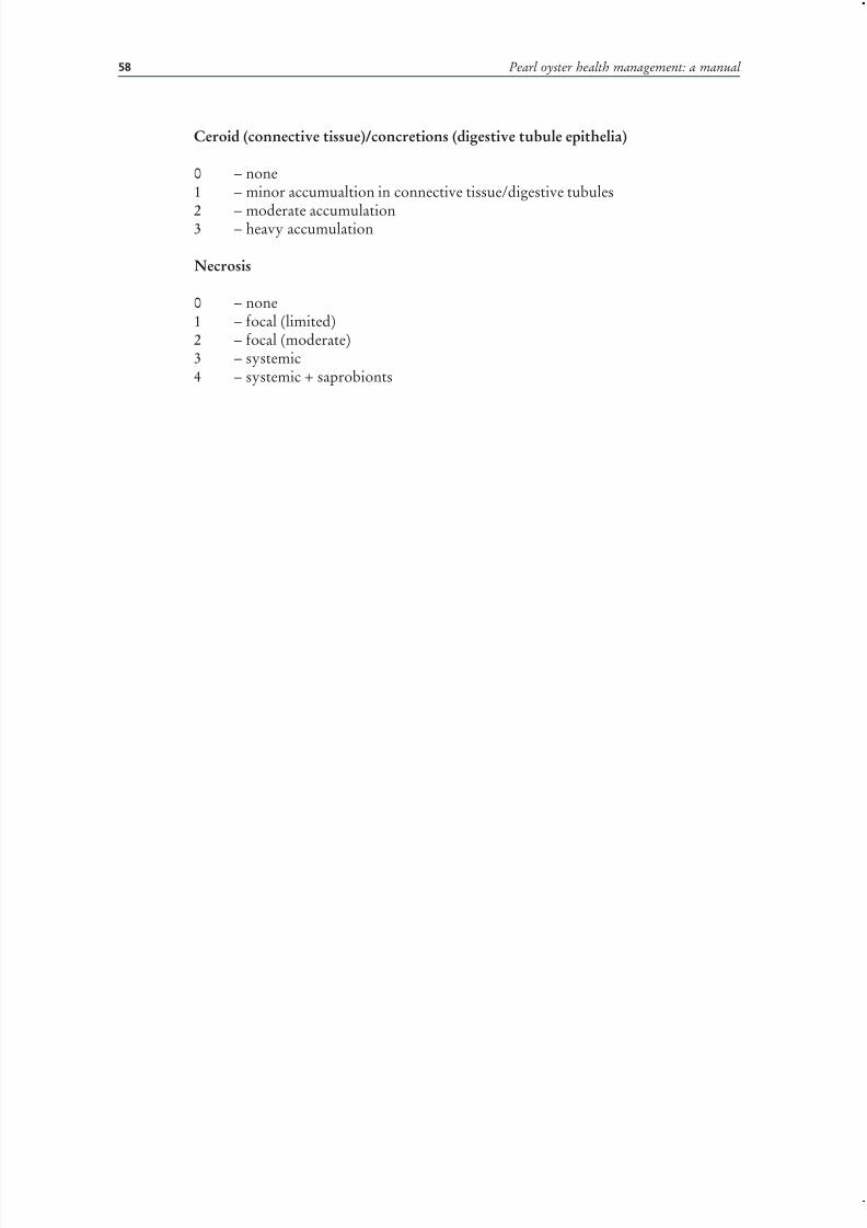
7/27/2019 Pearl Oyster Health Management
http://slidepdf.com/reader/full/pearl-oyster-health-management 72/136
Pearl oyster health management: a manual 58
Ceroid (connective tissue)/concretions (digestive tubule epithelia)
0 – none1 – minor accumualtion in connective tissue/digestive tubules
2 – moderate accumulation3 – heavy accumulation
Necrosis
0 – none1 – focal (limited)2 – focal (moderate)3 – systemic4 – systemic + saprobionts

7/27/2019 Pearl Oyster Health Management
http://slidepdf.com/reader/full/pearl-oyster-health-management 73/136

7/27/2019 Pearl Oyster Health Management
http://slidepdf.com/reader/full/pearl-oyster-health-management 74/136
7/27/2019 Pearl Oyster Health Management
http://slidepdf.com/reader/full/pearl-oyster-health-management 75/136

7/27/2019 Pearl Oyster Health Management
http://slidepdf.com/reader/full/pearl-oyster-health-management 76/136
Pearl oyster health management: a manual 62
histopathology. Though molecular techniques such as Polymerase Chain Reaction(PCR) are useful for identifying specific pathogens, histology is still preferable as itis capable of detecting multiple infections and other physiological problems such asstarvation or loss of condition following spawning. In pearl oysters, the most common
response to an insult is an inflammatory reaction. Cellular elements that participatein the inflammatory response and wound repair include agranular amoebocytes,basophilic semi-granulocytes and eosinophilic granulocytes. A typical response of aggregation, phagocytosis, hyperplasia and encapsulation has been described (Feng,1988; Suzuki, 1992). The haemocyte accumulations, infiltrations and granulomaformations seen in the absence of aetiological agents therefore represent a spectrumof past or existing inflammatory response to a variety of antigens, including solubleantigens, not visible histologically.
VirusesPass, Perkins and Dybdahl (1988) described intranuclear viral inclusion bodies presentin the digestive gland epithelium of oysters (Pinctada maxima) from Australia. Theseinclusion bodies have been demonstrated to consist of large, icosahedral, envelopedvirus particles (Humphrey et al., 1998).
Norton, Shepherd and Prior (1993a) reported that papova-like viral inclusionsoccurred commonly in the epithelium of the palp in both wild caught and farmedoysters (P. maxima) in Queensland, Australia. Mild to moderate digestive glandhyperplasia and degeneration are associated with heavy infections suggesting thatthe causative agents may potentially be pathogenic (Humphrey et al., 1998) and as aresult movement of live infected oysters between Queensland Northern Territory andWestern Australia is currently prohibited.
Suzuki, Kamakura and Kusuda (1998) and Kitamura et al . (2000) reported thepresence of a birnavirus in the haemocytes and digestive gland tubules of Pinctada fucata
martensii in Japan. The pathological significance of the birnavirus is uncertain. It shouldbe noted that pearl oysters, as with other oyster species, probably sequester a widevariety of viruses from the environment - not all of which will be pathogenic to oystersand some, such as hepatitis and Norwalk virus, would be pathogenic to humans.
In 1995, there was a mass mortality of Pinctada margaritifera in pearl farms inseveral Atolls of the Gambier Archipelago, French Polynesia (Comps, Herbaut andFougerous, 1999; 2001). On-going research has identified a 40 nm virus associatedwith necrosis of the adductor muscle, the histopathology of which Comps, Herbautand Fougerous (2001) suggested was similar to that described in Akoya oysters byMiyazaki et al. (1999). Severe mortalities of P. maxima in Exmouth Gulf, WesternAustralia during 2006 are also believed to have a viral aetiology, though no infectiousagent has yet been identified.
“Akoya virus”Mass mortalities of Japanese oyster (P. fucata martensii) were first noticed in YusuBay and Uchiumi Bay, in Ehime Prefecture, Japan during 1994 and have subsequentlyoccurred annually both at these sites and other bays in western Japan. Mortalitiesinclude juvenile, adult and seeded oysters. In 1996 and 1997, losses were reported to be50 percent of all oysters in production in western Japan (Miyazaki et al., 1998; 1999)though local losses of about 80 percent have been recorded (Tomaru, Kawabata andNakano, 2001). The mortalities have been the subject of intensive study within Japanincluding a workshop in September 1999 to overview research on the Japanese pearloyster mortalities (Muroga, Inui and Matsusato, 1999).
Gross pathology associated with the syndrome includes poor growth of shell onvalves, sluggish closing of valves, atrophy and a distinctive yellowish to red-browncolour of adductor muscle and watery appearance due to lack of nutrient storage
7/27/2019 Pearl Oyster Health Management
http://slidepdf.com/reader/full/pearl-oyster-health-management 77/136
63
(Miyazaki et al., 1998). The histopathology includes necrosis, haemocyte infiltration,atrophy, swelling and vacuolization of adductor and foot muscle fibers. Digestivediverticulae with large lumen and greatly reduced vacuolation and lysosome activityby epithelial cells are consistent with starvation (Shinomiya et al ., 1997; Miyazaki et
al ., 1998). Kurokawa et al. (1999) showed that the mortalities were associated with aninfectious agent causing the distinctive colour change as well as the histopathologicalchanges to the digestive gland. Miyazaki et al. (1999) independently described asmall (25–33 nm) virus (Akoya virus) which affected all smooth muscle fibers, not
just adductor muscle, affecting feeding, respiration and cardiac function. Miyazakiet al. (2000) showed that the effects of the virus could be reduced by injections of arecombinant feline interferon-w.
The Japanese mortalities were associated with illegal imports of Chinese pearloysters (Wada, 1997) and subsequently large numbers of hybrid akoya/Chinese pearloysters have been produced in Japan because the hybrid is resistant to the disease,although producing pearls of low quality (Miyazaki et al., 2000). The disease maykill other molluscs, including Chlamys nobilis and Crassostrea gigas (Miyazaki et al.,1998).
Tomaru et al. (2001) suggested, based on sampling in Uchiumi and Yusu Bays, thatthere was a relationship between water temperature and mortality in autumn (Septemberto November) and that these mortalities were not due to toxic algae. They hypothesizedthat, when Nitzschia spp. (which is inedible to oysters) dominated the culture area, thehealth of the oyster deteriorated due to food limitation and high water temperatureand that subsequently, the infective agent affected the digestive glands causing deathby starvation. This hypothesis may explain the results obtained by Hirano, Kanaiand Yoshikoshi (2002) who demonstrated, using contact infection trials with diseasedoysters in pocket-type cages in the sea, that infection (as expressed by mortality) wasnot transmitted. Further analysis of these mass mortality events by Hirano, Sugishita
and Mobin (2005) concluded that the disease is not due to viral, bacterial, fungal orparasitic organisms but might be due to organic pollution effects from neighbouringfish farms, a view challenged by Nagai et al. (2000) who again emphasized the infectivenature of the disease and its dependence on high water temperature.
RickettsialesPolymorphic rickettsiales have been described from P. maxima from the South ChinaSea (Wu and Pan, 1999) and from P. margaritifera in the Pacific (Comps, Fougerouseand Buestel, 1998). These appear, by light microscopy, to be identical to the rickettsialesalso seen in Western Australia in P. maxima. Rickettsiales have also been reportedfrom P. maxima in Queensland and the Northern Territory in Australia (Humphreyet al ., 1998). Worldwide, rickettsiales are recognized as asymptomatic infections in a
range of molluscs including Crassostrea gigas, C. virginica and Mya arenaria and havebeen associated with mass mortalities of the scallop Placopecten magellanicus (Sparks,1985).
BacteriaBacterial culture under tropical conditions, where water temperatures are around29 °C to 33 °C, frequently results in the isolation of large numbers of commensalor contaminant organisms, which cannot be readily distinguished from potentialpathogens. However, a number of studies have identified bacteria, particularly speciesof the genus Vibrio, as pathogens of oysters, often associated with stress inducedby poor management practices or environmental perturbations. Bacterial infections
usually incite haemocytic inflammatory lesions (Pass, Dybdahl and Mannion, 1987;Suzuki, 1995). The formation of conspicuous deposits of brown-black conchiolinon the inner nacreous surface of the shell of P. maxima, described by Perkins (1996),
3.1 Review of pearl oyster mortalities and disease problems

7/27/2019 Pearl Oyster Health Management
http://slidepdf.com/reader/full/pearl-oyster-health-management 78/136
Pearl oyster health management: a manual 64
was observed in 12–14 percent of shell by Humphrey et al. (1998) and has also beenreported in P. margaratifera in French Polynesia (Marin and Dauphin, 1991; Cuif and Dauphin, 1996). Though common in many bivalves and attributed to bacterialirritation (Paillard, Maes and Oubella, 1994), the aetiology is still uncertain.
HaplosporidiumAn unnamed species of Haplosporidium was found in a batch of P. maxima spat fromthe Carnarvon hatchery, Western Australia, in 1993 (Hine and Thorne, 1998). The samehaplosporidian was subsequently found in P. maxima spat from a marine farm site atKing Sound in the Kimberley region of Western Australia in 1995 and again in spatat a marine farm site in Broome in 2000 (Jones and Creeper, 2006). The 1995 incidentinvolved spat that had been free of infection on leaving the hatchery and 6 weeks laterwere found to have a prevalence of infection of 4.6 percent. On re-testing 15 dayslater, the prevalence had increased to just over 10 percent, at which point the spat weredestroyed. Electron microscope examination confirmed that the parasite was the sameparasite as that reported by Hine and Thorne (1998). The relationship between thishaplosporidian and one occurring in Saccostrea cucculata in the same area is currentlyunder investigation (Bearham et al., 2007).
PerkinsusPerkinsus sp. is routinely diagnosed by thioglycollate culture rather than histology. Ithas been commonly isolated by culture from tropical Australian molluscs, includingPinctada spp. in the absence of any pathology (Goggin and Lester, 1987, 1995).Perkinsus-like protozoa were described by Norton et al . (1993b) in focal granulomatouslesions in adult P. maxima from a population undergoing high mortality in TorresStrait, Australia, though a causative relationship could not be established. Park et. al .(2001) were unable to demonstrate the presence of Perkinsus in P. fucata martensii from
Korea either by thioglycollate culture or histology.
Other agentsA number of infectious organisms do not appear to cause significant tissuedamage or inflammatory response from their host. These include turbellarians andAncistracomid-like ciliates. While turbellarians caused no apparent damage or hostresponse in the study by Humphrey et al . (1998), northern hemisphere studies on theturbellarian Urastoma cyprinae have shown that it is attracted to oyster gill mucous(Brun, Boghen and Allard, 1999) and causes biochemical changes to gill mucous of infected Crassostrea virginica (Brun, Ross and Boghen, 2000). Therefore, absence of histopathological change does not mean that there is no effect on pearl oysters. In2001, an intracellular rhynchodid-like ciliate was found in oysters (P. maxima) from
the Exmouth Gulf and Montibello Islands in Western Australia. The ciliate appearssimilar to those described from mussels (Mytilus spp.) in Europe (McGladdery andBower 2002; Jones and Creeper, 2006). Sampling has shown that the ciliate occurs inhigh prevalence and abundance in juvenile oysters, disappearing from oysters overabout 90 mm diameter.
Gregarines occur in P. maxima in Australia and appear not to cause significantdamage (Humphrey et al., 1998). However, an unidentified intracellular gregarineparasite, described from the gut of P. margaritifera from atolls in the TuamotuArchipelago, French Polynesia, from the Fiji Islands and from the Red Sea (Gulf of Aqaba), causes local or complete destruction of rectal epithelial cells and may beassociated with mortalities in French Polynesia (Chagot et al., 1993).
Metacercariae of a bucephalid trematode occur in the gonad of P. radiata in thePersian Gulf (Khamdan, 1998). Third and fourth stage larvae of the ascariid nematodehas been reported to occur in the adductor muscle, digestive gland and gonads of

7/27/2019 Pearl Oyster Health Management
http://slidepdf.com/reader/full/pearl-oyster-health-management 79/136
65
Pinctada spp. The adult nematode occurs in loggerhead turtles, Caretta caretta (Berryand Cannon, 1981).
Larval cestodes of the family Lecanicaphalidae which include the generaTylocephalum and Polypocephalus are commonly associated with discrete focal or
multifocal granuloma in interstitial tissues. Larval Tylocephalum sp. have been reportedin most bivalves examined in northern Australia (Wolf, 1976, 1977; Hine and Thorne,2000). Larval Tylocephalum sp. are not considered host specific and may occur at highprevalence and intensity in oysters where they may reduce the condition of their host(Sindermann, 1990).
The copepod Anthessius pinctadae was described from P. mararitifera from theTorres Strait (Humes, 1973). The copepod also occurs in P. maxima throughoutnorthern Australia (Humphrey et al., 1998). In moderate numbers, this copepod causeserosion of the oesophageal epithelium and entrapment in the digestive gland resulted inencapsulation and a haemocytic response. Thus, under certain conditions, the parasitemay be potentially pathogenic or predispose the oyster to infection.
Fouling organismsBoring molluscs Lithophaga spp. are common in Australia and produce large holesof 1-2 cm sometimes disrupting or breaching the nacreous layer. Polychaete wormsinvading the shell nacre are also common, resulting in “mud blisters” (Humphrey etal ., 1998). Boring sponges, family Clionidae, including the bright red coloured Cliona sp., are a major problem for the industry in Australia, leading to severe erosion of theshell matrix and premature removal of shells from pearl production (Vblayudhan,1983; Taylor, Southgate and Rose , 1997). Treatment and prevention involves regularscrubbing of shell, either manually or by high-pressure hose. There has also been somesuccess with freshwater baths for 30–60 min, while trials are underway testing anti-fouling paints.
Commensal animalsPea crabs and shrimps are common, occurring in up to 85 percent and 72 percent of Australian P. maxima oyster populations respectively (Humphrey et al., 1998). Apartfrom local oedema in the mantle and depressions in both the mantle and gill tissuescaused by the pea crabs, no pathology is associated with these commensal organisms(Dix, 1973).
DISEASES WITH NON-INFECTIOUS AETIOLOGYTemperatureThere is little published information on the histopathology associated with temperature,but it has a marked effect on the oysters. Tomaru et al. (2002) showed that growth in
height, length and thickness of the shells of P. fucata martensii was limited by watertemperatures less that 20 °C. Pouvreau and Prasil (2001) showed that temperaturesover 30 °C had a negative impact on growth of P. margaratifera. Likewise, Mills (2002)showed that survival of P. maxima spat was greatest between 23 °C and 32 °C, with35 °C resulting in high mortalities.
Toxic algaeDuring a red tide event in Ago Bay, Japan in 1992, the maximum cell density of Heterocapsa circularisquama reached over 85 000 cells/ml and there was a concurrentmass mortality of pearl oysters. Subsequent trials (Nagai et al., 1996; Nagai et al .,2000) showed that mortality of two-month-old pearl oysters (P. fucata) was closely
correlated with the cell density of H. circularisquama and that, on exposure, oystersrapidly contracted their mantles and closed their shell valves. Negri et al. (2003) showedthat Trichodesmium blooms also caused poor condition in pearl oysters.
3.1 Review of pearl oyster mortalities and disease problems

7/27/2019 Pearl Oyster Health Management
http://slidepdf.com/reader/full/pearl-oyster-health-management 80/136
Pearl oyster health management: a manual 66
Coral spawningMortalities caused by low oxygen levels associated with coral spawning are notuncommon, though not reported in the literature.
StarvationDegenerative changes, not associated with causative agents, are relatively commonand include oedema, increased pigmentation in macrophages in interstitial tissuesand kidney and mineralisation. Oedema is believed by Humphrey et al. (1998) tobe a degenerative response in a physiologically compromised oyster and is also seencommonly in oysters removed from the water column and held at high ambient airtemperatures. The significance of increased brown pigmentation in cells (so-calledbrown cells) is unclear but is believed to be associated with prior cellular breakdownand detoxification (Zaroogian and Yevich, 1994) though it should be noted that brownpigmentation of the heart and epithelium of the mantle tissue is normal. Lamellarmineralization is associated with pearl formation, however, mineralization may occurwithout nacre formation (Comps, Herbaut and Fougerouse , 2000).
MiscellaneousTearing of the adductor muscle through the practice of wedging open oysters forseeding operations also results in a recognizable wound healing response in the affectedmuscle (Norton, 2000). A non-specific inflammatory haemocytic infiltration in theadductor muscle is typical of this change (Humphrey et al ., 1998).
Slightly refractile ovoid brown pigmented “protistan parasites” have been describedin cytoplasm of digestive gland epithelium of P. maxima in Australia (Wolf andSprague, 1978) and in P. margaritifera from the Red Sea (Nasr, 1982). These bodiesoccur commonly in both healthy and diseased oysters. They are “residual bodies”,storage or secretory products which are ultimately released from the cell (Pass and
Perkins, 1985) and are not of pathological significance.Though neoplasia are rare, neurofibromatous tumours in Australian P. maxima were recorded by Humphrey et al . (1998). Two polypous mesenchymal tumours in P.margaritifera have also been described from Australia (Dix, 1972).
MANAGEMENT OPTIONSThere are two factors that together make pearl farming less susceptible to disease-induced mass mortalities than other molluscs. The first is the panel based culturesystem, which apart from providing a degree of predator protection, makes monitoringof individual shells possible. The second is the requirement to lift and clean biofoulingoff the panels every four weeks or so, which means that shell is regularly inspected formortality.
The majority of the internationally notifiable diseases of molluscs have been spreadby human activities, usually associated with aquaculture. Movement controls toprevent the indiscriminate movement of shell between areas and between countriesis thus the first step in disease management. Within Australia, regional differencesin distribution of several agents including Papovavirus-like inclusion bodies wereidentified by Humphrey et al. (1998) and form the basis for regional control measuresincluding batch testing of animals for disease status prior to authorizing movements.In Western Australia, the use of quarantine sites and the provision of a mandatory fivenautical mile exclusion zone for pearl oyster farming activities around farms serves, inpart, as a barrier to prevent the spread of disease from farm to farm.
CONCLUSIONSPearl oysters are relatively free of serious pathogens – so far. However, there has beenlittle study of causes of mortalities of pearl oysters in regional areas where farming is
7/27/2019 Pearl Oyster Health Management
http://slidepdf.com/reader/full/pearl-oyster-health-management 81/136
67
now spreading. For that reason, both government agencies and industry members needto be careful about the source of their stocks and the movement of oysters betweenregions if the disasters that have befallen other shellfish industries are to be avoided.
ACKNOWLEDGEMENTSA wide range of people have assisted and contributed to my understanding of pearloysters in Australia and I am grateful for the help and advice from Dr John Humphrey(Northern Territory). Members of the pearling industry and company staff have alsogone out of their way to provide helpful material and logistic support.
REFERENCESBearham, D., Spiers, Z., Raidal, S., Jones, J.B. & Nicholls, P.K. 2007. Molecular
characterisation of a Haplosporidian parasite infecting rock oysters Saccostrea cuccullata in north Western Australia. Journal of Invertebrate Pathology 95: 33-40.
Berry G. N. & Cannon, L. R. G. 1981. The life history of Sulcascaris sulcata (Nematoda:Ascaridoidea), a parasite of marine molluscs and turtles. International Journal for Parasitology 11: 43-54.
Brun, N. T., Boghen, A.D. & Allard, J. 1999. Attraction of Urastoma cyprinae (Turbellaria:Urastomidae) to the eastern oyster Crossostrea virginica. Diseases of Aquatic Organisms 37: 139-144.
Brun, N.T., Ross, N. W. & Boghen, A.D. 2000. Changes in the electrophoretic profiles of gill mucus proteases of the eastern oyster Crassostrea virginica in response to infectionby the turbellarian Urastoma cyprinae. Canadian Journal of Fisheries and Aquatic Science 75: 163-170.
Chagot, D., Fougerouse, A., Weppe, M., Marques, A. & Bouix, G. 1993. Presenced’une gregarine Protozoa Sporozoa parasite de l’huitre perlier a levres noires Pinctadamargaratifera L. 1758. Mollusca Bivalvia en Polynesie Francaise. Comptes rendus de
l’Academie Science de Paris 316 Serie iii: 239-244.Comps, M., Fougerouse, A. & Buestel, D. 1998. Note: A procaryote infecting the black-lipped pearl oyster Pinctada margaritifera. Journal of Invertebrate Pathology 72: 87-89.
Comps, M., Herbaut, C. & Fougerouse, A. 1999. Virus-like particles in pearl oysterPinctada margaritifera. Bulletin of the European Association of Fish Pathologists 13:85 ¯ 88.
Comps, M., Herbaut, C. & Fougerouse, A. 2000. Abnormal periostracum secretion duringthe mineralization process of the pearl in the blacklip pearl oyster Pinctada margaritifera.
Aquatic Living Resources 13: 49-55.Comps, M., Herbaut, C., Fougerouse, A. & Laporte, F. 2001. Progress in pathological
characterization of Syndrome 85 in the black-lip pearl oyster Pinctada margaritifera. Aquatic Living Resources 14: 195-202.
Cuif, J-P. & Dauphin, Y. 1996. Occurrence of mineralization disturbances in nacreouslayers of cultivated pearls produced by Pinctada margaritifera var. cumingi from FrenchPolynesia. Comparison with reported shell alterations. Aquatic Living Resources 9: 187-193.
Dix, T.G. 1972. Two mesenchymal tumours in a pearl oyster Pinctada margaritifera. Journal of Invertebrate Pathology 20: 317-320.
Dix, T.G. 1973. Mantle changes in the pearl oyster Pinctada maxima induced by the peacrab Pinnotheres villosus. Veliger 15: 330-331.
Feng, S.Y. 1988. Cellular defense mechanisms of oysters and mussels. In Fisher, W.S.(Ed.) Disease processes in marine bivalve molluscs. American Fisheries Society Special Publication 18: 153-168.
Goggin, C.L. & Lester, R.J.G. 1987. Occurrence of Perkinsus
species (Protozoa,Apicomplexa) in bivalves from the Great Barrier Reef. Diseases of Aquatic Organisms 3: 113-117.
3.1 Review of pearl oyster mortalities and disease problems
7/27/2019 Pearl Oyster Health Management
http://slidepdf.com/reader/full/pearl-oyster-health-management 82/136
Pearl oyster health management: a manual 68
Goggin, C.L. & Lester, R.J.G. 1995. Perkinsus, a protozoan parasite of abalone inAustralia: A review. Marine and Freshwater Research 46: 639-646.
Grizel, H., Bachère, E., Mialhe, E. & Tige, G. 1986. Solving parasite related problemsin cultured molluscs, pp. 301-308. In Howell, M.J. (ed.) Parasitology – Quo Vadit:
Proceedings of the 6th International Congress of Parasitology, Brisbane. Canberra,Australian Academy of Science.
Hine, P.M. & Thorne, T. 1998. Haplosporidium sp. (Haplosporidia) in hatchery reared pearloysters Pinctada maxima (Jameson, 1901) in Western Australia. Journal of InvertebratePathology 71: 48-52.
Hine, P.M. & Thorne, T. 2000. A survey of some parasites and diseases of several species of bivalve mollusc in northern Western Australia. Diseases of Aquatic Organisms 40: 67-78.
Hirano, M., Kanai, K. & Yoshikoshi, K. 2002. Contact infection trials failed to reproducethe disease condition of mass mortality in cultured pearl oyster Pinctada fucata martensii.
Fisheries Science 68: 700-702.Hirano, M., Sugishita, Y. & Mobin, S.M.A. 2005. An investigation of the pathology
associated with mass mortality events in the cultured Japanese pearl oyster Pinctada fucata martensii at four farms in western Japan. Journal of Aquatic Animal Health 17:323-337.
Humes, A.G. 1973. Cyclopoid copepods associated with marine bivalve molluscs in NewCaledonia. Cahiers O.R.S.T.O.M. Séries Océanographie 11: 3-25.
Humphrey, J.D., Norton, J.H., Jones, J.B., Barton, M.A., Connell, M.T., Shelley, C.C.& Creeper, J.H. 1998. Pearl oyster (Pinctada maxima) aquaculture: Health survey of Northern Territory, Western Australia and Queensland pearl oyster beds and farms.
Fisheries Research Development Corporation Final Report 94/079. 108 p. Jones, J.B. & Creeper, J. 2006. Diseases of pearl oysters and other molluscs: A Western
Australian perspective. Journal of Shellfish Research 25: 233-238.Khamdan, S.A.A. 1998. Occurrence of Bucephalus sp. trematode in the gonad of the pearl
oyster, Pinctada radiata. Environment International 24: 117-120.Kitamura, S.I., Jung, S.J. & Suzuki, S. 2000. Seasonal change of infective state of marinebirnavirus in Japanese pearl oyster Pinctada fucata. Archives of Virology 145: 2003-2014.
Kurokawa, H., Suzuki, T., Okauchi, M., Miwa, S., Nagai, S., Nakamura, K., Honjo, T.,Nakajima, K., Ashida, K. & Hunakoshi, S. 1999. Experimental infections of a diseasecausing mass mortalities of Japanese pearl oyster Pinctada fucata martensii by tissuetransplantation and cohabitation Nippon Suisan Gakkaishi 65: 241-251 (In Japanese,English abstract).
Marin, F. & Dauphin, Y. 1991. Diversité des altérations dans la composition en acidesaminés de la phase organique de la nacre des huîtres perlières de Polynésie française(Pinctada maragaritifera) atteintes par l’épizootie. Comptes Rendue Acad. Sci. Paris 312:
483-488.McGladdery, S.E. & Bower, S.M. 2002. Synopsis of infectious diseases and parasites of
commercially exploited shellfish. Intracellular ciliates of mussels. URL:http//www-sci.pac.dfo-mpo.gc.ca/sealane/aquac/pages/icmu.htm
Mills, D. 2002. Combined effects of temperature and algal concentration on survival,growth and feeding physiology of Pinctada maxima (Jameson) spat. Journal of ShellfishResearch 19: 159-166.
Miyazaki, T., Nozawa, N. & Kobayashi, T. 2000. Clinical trial results on the use of a recombinant feline interferon-w to protect Japanese pearl oysters Pinctada fucatamartensii from akoya-virus infection. Diseases of Aquatic Organisms 43: 15-26.
Miyazaki, T., Goto, K., Kobayashi, T. & Miyata, M. 1998. An emergent virus disease
associated with mass mortalities in Japanese pearl oystersPinctada fukata martensii,
pp.154-159. In VII International Colloquium on Invertebrate Pathology and MicrobialControl. Sapporo, August 23-28, 1998.
7/27/2019 Pearl Oyster Health Management
http://slidepdf.com/reader/full/pearl-oyster-health-management 83/136
69
Miyazaki, T., Goto, K., Kobayashi, T., Kageyama, T. & Miyata, M. 1999. Mass mortalitiesassociated with a virus disease in Japanese pearl oysters Pinctada fucata martensii.Diseases of Aquatic Organisms 37: 1-12.
Muroga, K., Inui, Y. & Matsusato, T. 1999. Workshop on “Emerging diseases of cultured
marine molluscs in Japan”. Fish Pathology 34: 219-220 (In Japanese, English abstract).Nagai, K., Go, J., Segawa, S. & Honjo, T. 2007. A measure to prevent relapse of reddening
adductor disease in pearl oysters (Pinctada fucata martensii) by.low-water-temperatureculture management in wintering fisheries. Aquaculture 262: 192-201.
Nagai, K., Matsuyama, Y., Uchida, T., Akamatsu, S. & Honjo, T. 2000. Effect of a naturalpopulation of the harmful dinoflagellate Heterocapsa circularisquama on the survival of the pearl oyster Pinctada fucata. Fisheries Science 66: 995-997.
Nagai, K., Matsuyama, Y., Uchida, T., Yamaguchi, M., Ishimura, M., Nishimura, A.,Akamatsu, S. & Honjo, T. 1996. Toxicity and LD50 levels of the red tide dinoflagellateHeterocapsa circularisquama on juvenile pearl oysters. Aquaculture 144: 149-154.
Nasr, D.H. 1982. Observations on the mortality of the pearl oyster, Pinctada margaritifera in Dongonab Bay, Red Sea. Aquaculture 28: 271-281.
Negri, A.P., Bunter, O., Jones, B. & Llewellyn, L. 2004. Effects of the bloom-forming algaeTrichodesmium erythraeum on the pearl oyster Pinctada maxima. Aquaculture 232: 91-102.
Norton, J.H. 2000. Approaches to improve cultured pearl formation in Pinctadamargaritifera through use of relaxation, antiseptic application and incision closureduring bead insertion Aquaculture 184: 1-17.
Norton, J.H., Shepherd, M.A. & Prior, H.C. 1993a. Papovavirus-like infection of thegolden-lipped pearl oyster, Pinctada maxima, from the Torres Strait, Australia. Journal of Invertebrate Pathology 62: 198-200.
Norton, J.H., Shepherd, M.A., Perkins, F.O. & Prior, H.C. 1993b. Perkinsus-like infectionin farmed golden-lipped pearl oyster, Pinctada maxima, from the Torres Strait, Australia.
Journal of Invertebrate Pathology 62: 105-106.
Paillard, C., Maes, P. & Oubella, R. 1994. Brown ring disease in clams. Annual Review of Fish Diseases 4: 219-240.
Park, K.I., Choi, K.S. & Jeong, W.G. 2001. An examination for the protozoan parasite,Perkinsus sp. in the pearl oyster, Pinctada fucata martensii (Dunker) from the southerncoast of Korea. Bulletin of the European Association of Fish Pathologists 21: 30-32.
Pass, D.A. & Perkins, F.O. 1985. “Protistan parasites” or residual bodies in Pinctadamaxima. Journal of Invertebrate Pathology 46: 200-201.
Pass, D.A., Dybdahl, R. & Mannion, M.M. 1987. Investigations into the causes of mortality of the pearl oyster, Pinctada maxima (Jameson) in Western Australia. Aquaculture 65: 149-169.
Pass, D.A., Perkins, F.O. & Dybdahl, R. 1988. Virus-like particles in the digestive gland of the pearl oyster Pinctada maxima. Journal of Invertebrate Pathology 51: 166-167.
Perkins, F.O. 1996. Shell disease in the gold lip pearl oyster, Pinctada maxima and the
European oyster, Crassostrea virginica. Aquatic Living Resources 9: 159-168.Pouvreau, S. & Prasil, V. 2001. Growth of the black-lip pearl oyster,Pinctada margaritifera,
at nine culture sites of French Polynesia: synthesis of several sampling designs conductedbetween 1994 and 1999. Aquatic Living Resources 14: 155-163.
Shinomiya, Y., Iwanaga, S., Yamaguchi, T., Kouno, K. & Uchimura, Y. 1997. Relationshipsbetween autumn mortality, glycogen content and carbohydrate metabolism enzymeactivity in Japanese pearl oyster Pinctada fucata martensii. Suisan Zoshoku 45: 47-53 (In
Japanese, English abstract)Sindermann, C.J. 1990. Principal diseases of marine fish and shellfish, Volume 2. Diseases
of marine shellfish. New York, Academic Press.Sparks, A.K. 1985. Synopsis of Invertebrate Pathology Exclusive of Insects. Amsterdam,
Elsevier Science Publishers B.V.Suzuki, S., Kamakura, M. & Kusuda, R. 1998. Isolation of birnavirus from Japanese pearloyster Pinctada fucata. Fisheries Science 64: 342-343.
3.1 Review of pearl oyster mortalities and disease problems

7/27/2019 Pearl Oyster Health Management
http://slidepdf.com/reader/full/pearl-oyster-health-management 84/136
Pearl oyster health management: a manual 70
Suzuki, T. 1992. Functions of hemocytes during the wound healing process in the pearloyster. NOAA Technical Report NMFS 111: 109-112.
Suzuki, T. 1995. Accumulation of injected proteins in the auricles of the pearl oysterPinctada fucata. Bulletin of the National Research Institute for Aquaculture Japan
Yoshokukenho 24: 23-32.Taylor, J.L., Southgate, P.C. & Rose, R.A. 1997. Fouling animals and their effect on
the growth of silverlip peal oysters Pinctada maxima (Jameson) in suspended culture. Aquaculture 153: 31-40.
Tomaru, Y., Kawabata, Z. & Nakano, S. 2001. Mass-mortality of the Japanese pearloyster Pinctada fucata martensii in relation to water temperature, chlorophyll a andphytoplankton composition. Diseases of Aquatic Organisms 44: 61-68.
Tomaru, Y., Kumatabara, Y., Kawabata, Z. & Nakano, S. 2002. Effect of watertemperature and chlorophyll abundance on shell growth of the Japanese pearl oyster,Pinctada fucata martensii, in suspended culture at different depths and sites. AquacultureResearch 33: 109-116.
Vblayudhan, T.S. 1983. On the occurrence of shell boring polychaetes and sponges on pearloyster Pinctada fucata and control of boring organisms. Proceedings of the Symposiumon Coastal Aquaculture 2: 614-618.
Wada, K. 1997. Causative factors and characteristics of mass mortalities that have occurredin cultured pearl oysters since 1994. Shinju-Kenkyu 6: 2-23 (In Japanese).
Wolf, P.H. 1976. Occurrence of larval stages of Tylocephalum Cestoda: Lecanicephaloidaein two oyster species from Northern Australia. Journal of Invertebrate Pathology 27:129-131.
Wolf, P.H. 1977. An unidentified protistan parasite in the ova of the blacklipped oysterCrassostrea echinata from Northern Australia. Journal of Invertebrate Pathology 29:244-246.
Wolf, P.H. & Sprague, V. 1978.An unidentified protistan parasite of the pearl oyster,
Pinctada maxima, in tropical Australia. Journal of Invertebrate Pathology 31: 262-263.Wu, X. & Pan, J. 1999. Studies on rickettsia-like organism disease of the tropical marinepearl oyster I: the fine structure and morphogenesis of Pinctada maxima pathogenrickettsia-like organism. Journal of Invertebrate Pathology 73: 162-172.
Zaroogian, G. & Yevich, P. 1994. The nature and function of the brown cell in Crassostreavirginica. Marine Environmental Research 37: 355-373.

7/27/2019 Pearl Oyster Health Management
http://slidepdf.com/reader/full/pearl-oyster-health-management 85/136
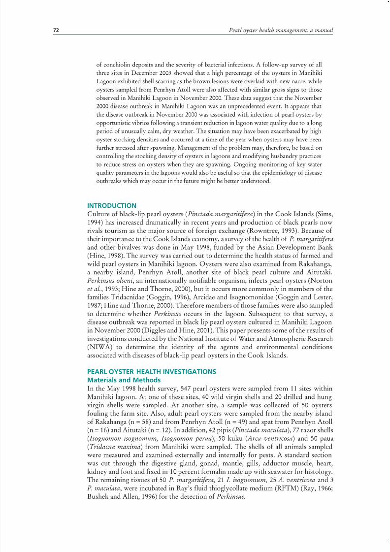
7/27/2019 Pearl Oyster Health Management
http://slidepdf.com/reader/full/pearl-oyster-health-management 86/136
Pearl oyster health management: a manual 72
of conchiolin deposits and the severity of bacterial infections. A follow-up survey of allthree sites in December 2003 showed that a high percentage of the oysters in ManihikiLagoon exhibited shell scarring as the brown lesions were overlaid with new nacre, whileoysters sampled from Penrhyn Atoll were also affected with similar gross signs to those
observed in Manihiki Lagoon in November 2000. These data suggest that the November2000 disease outbreak in Manihiki Lagoon was an unprecedented event. It appears thatthe disease outbreak in November 2000 was associated with infection of pearl oysters byopportunistic vibrios following a transient reduction in lagoon water quality due to a longperiod of unusually calm, dry weather. The situation may have been exacerbated by highoyster stocking densities and occurred at a time of the year when oysters may have beenfurther stressed after spawning. Management of the problem may, therefore, be based oncontrolling the stocking density of oysters in lagoons and modifying husbandry practicesto reduce stress on oysters when they are spawning. Ongoing monitoring of key waterquality parameters in the lagoons would also be useful so that the epidemiology of diseaseoutbreaks which may occur in the future might be better understood.
INTRODUCTIONCulture of black-lip pearl oysters (Pinctada margaritifera) in the Cook Islands (Sims,1994) has increased dramatically in recent years and production of black pearls nowrivals tourism as the major source of foreign exchange (Rowntree, 1993). Because of their importance to the Cook Islands economy, a survey of the health of P. margaritifera and other bivalves was done in May 1998, funded by the Asian Development Bank(Hine, 1998). The survey was carried out to determine the health status of farmed andwild pearl oysters in Manihiki lagoon. Oysters were also examined from Rakahanga,a nearby island, Penrhyn Atoll, another site of black pearl culture and Aitutaki.Perkinsus olseni, an internationally notifiable organism, infects pearl oysters (Norton
et al., 1993; Hine and Thorne, 2000), but it occurs more commonly in members of thefamilies Tridacnidae (Goggin, 1996), Arcidae and Isognomonidae (Goggin and Lester,1987; Hine and Thorne, 2000). Therefore members of those families were also sampledto determine whether Perkinsus occurs in the lagoon. Subsequent to that survey, adisease outbreak was reported in black lip pearl oysters cultured in Manihiki Lagoonin November 2000 (Diggles and Hine, 2001). This paper presents some of the results of investigations conducted by the National Institute of Water and Atmospheric Research(NIWA) to determine the identity of the agents and environmental conditionsassociated with diseases of black-lip pearl oysters in the Cook Islands.
PEARL OYSTER HEALTH INVESTIGATIONSMaterials and Methods
In the May 1998 health survey, 547 pearl oysters were sampled from 11 sites withinManihiki lagoon. At one of these sites, 40 wild virgin shells and 20 drilled and hungvirgin shells were sampled. At another site, a sample was collected of 50 oystersfouling the farm site. Also, adult pearl oysters were sampled from the nearby islandof Rakahanga (n = 58) and from Penrhyn Atoll (n = 49) and spat from Penrhyn Atoll(n = 16) and Aitutaki (n = 12). In addition, 42 pipis (Pinctada maculata), 77 razor shells(Isognomon isognomum, Isognomon perua), 50 kuku ( Arca ventricosa) and 50 paua(Tridacna maxima) from Manihiki were sampled. The shells of all animals sampledwere measured and examined externally and internally for pests. A standard sectionwas cut through the digestive gland, gonad, mantle, gills, adductor muscle, heart,kidney and foot and fixed in 10 percent formalin made up with seawater for histology.
The remaining tissues of 50 P. margaritifera, 21 I. isognomum, 25 A. ventricosa and 3P. maculata, were incubated in Ray’s fluid thioglycollate medium (RFTM) (Ray, 1966;Bushek and Allen, 1996) for the detection of Perkinsus.

7/27/2019 Pearl Oyster Health Management
http://slidepdf.com/reader/full/pearl-oyster-health-management 87/136
3.2 The Cook Islands experience: pearl oyster health investigations 73
During the disease outbreak in November 2000, black lip pearl oysters (n = 300)and water were sampled from 10 sites within Manahiki lagoon for histopathology,bacteriology and toxicology. Examination began by noting the presence of grosslyevident infections with metazoan parasites and fouling organisms (mudworms, fungi,
boring sponges, etc.). The dorso-ventral measurement (DVM) of each oyster wasrecorded to the nearest mm before the oyster was opened by severing the adductormuscle. The nacre of each shell valve was examined for abnormal conchiolin depositsand the severity of conchiolin lesions was assessed using a six-grade qualitative scoringsystem (Table 3.2.1).
The first 10 oysters examined from each site were subjected to bacteriologicalanalysis prior to being processed for histopathology and RFTM incubation. Samplesof 0.1 ml of hemolymph were obtained from the pericardium with a sterile 1 mlsyringe and 25 G hypodermic needle and inoculated onto one half of plates containingthiosulphate citrate bile salt sucrose agar (TCBS) and for oysters from sites 1 to 6,Tryptone Soya Agar (TSA) with 2 percent NaCl added (TSA+2). The other half of each plate was streaked with a flamed wire loop which had swabbed the surface of theadductor muscle which had been cut with a sterile scalpel to provide an uncontaminatedsurface. Sub-samples of 0.1 ml of seawater samples obtained for plankton analysis fromthe top and the bottom of the water column from each site were also examined forwaterborne bacteria in a similar manner. The bacteriology plates were incubated atroom temperature (30 °C) and examined for bacteria after 18 hrs and again after 36 hrs.Samples of different colony types observed were sub-cultured to ensure purity beforebeing stored in long term preservation medium (Beuchat, 1974) for up to 2 weeks priorto identification.
Biochemical characterization of selected isolates was undertaken by subjecting themto 52 phenotypic tests using methods described by Baumann, Baumann and Mandel(1971), Furniss, Lee and Donovan (1978) and West and Colwell (1984). Phenotypic
data were compared to a probabilistic data matrix for Vibrio species (Bryant and Smith,1986) using a regularly updated version of the Bacterial Identifier program (Bryant andSmith, 1991). An acceptable identification was reached when the identification scoreequaled or exceeded 0.98.
The first 10 oysters examined from each site were also examined for the presenceof Perkinsus sp. by placing excised samples of mantle, digestive gland, gills, foot andkidney into RFTM. Tissues were incubated in RFTM in the dark between 7 and 14days then removed, blotted to remove excess RFTM and placed in dilute Lugol’s iodinebefore examination under a dissecting microscope at 40x magnification for stainedenlarged hypnospores (Ray, 1966; Bushek and Allen, 1996).
All 30 oysters from each site were sampled for histopathology. An oblique transversesection, approximately 5-7 mm thick, was cut from each oyster using a scalpel. The
section was oriented to include mantle, gonad, digestive gland, gills, foot and sometimeskidney. The tissues were fixed in 10 percent formalin in filtered seawater for at least96 hrs before being embedded in paraffin on the cut surface. One section 6 µm thickwas cut and stained with hematoxylin and eosin (H&E) using standard histologicaltechniques before being examined under a compound microscope.
TABLE 3.2.1
The qualitative scoring system used to grade the severity of abnormal conchiolin deposits in
oysters from Manihiki Lagoon
Grade Abnormal conchiolin lesion severity
0 apparently healthy, no lesions evident
1 1 or 2 focal lesions
2 <25% of shell valve perimeter affected
3 25 to 50% of shell valve perimeter affected4 50 to 75% of shell valve perimeter affected
5 >75% of shell valve perimeter affected
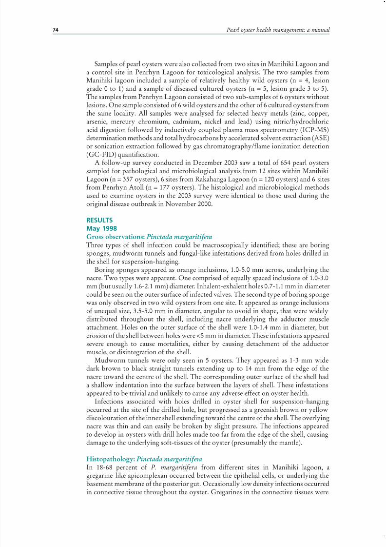
7/27/2019 Pearl Oyster Health Management
http://slidepdf.com/reader/full/pearl-oyster-health-management 88/136
Pearl oyster health management: a manual 74
Samples of pearl oysters were also collected from two sites in Manihiki Lagoon anda control site in Penrhyn Lagoon for toxicological analysis. The two samples fromManihiki lagoon included a sample of relatively healthy wild oysters (n = 4, lesiongrade 0 to 1) and a sample of diseased cultured oysters (n = 5, lesion grade 3 to 5).
The samples from Penrhyn Lagoon consisted of two sub-samples of 6 oysters withoutlesions. One sample consisted of 6 wild oysters and the other of 6 cultured oysters fromthe same locality. All samples were analysed for selected heavy metals (zinc, copper,arsenic, mercury chromium, cadmium, nickel and lead) using nitric/hydrochloricacid digestion followed by inductively coupled plasma mass spectrometry (ICP-MS)determination methods and total hydrocarbons by accelerated solvent extraction (ASE)or sonication extraction followed by gas chromatography/flame ionization detection(GC-FID) quantification.
A follow-up survey conducted in December 2003 saw a total of 654 pearl oysterssampled for pathological and microbiological analysis from 12 sites within ManihikiLagoon (n = 357 oysters), 6 sites from Rakahanga Lagoon (n = 120 oysters) and 6 sitesfrom Penrhyn Atoll (n = 177 oysters). The histological and microbiological methodsused to examine oysters in the 2003 survey were identical to those used during theoriginal disease outbreak in November 2000.
RESULTSMay 1998Gross observations: Pinctada margaritiferaThree types of shell infection could be macroscopically identified; these are boringsponges, mudworm tunnels and fungal-like infestations derived from holes drilled inthe shell for suspension-hanging.
Boring sponges appeared as orange inclusions, 1.0-5.0 mm across, underlying thenacre. Two types were apparent. One comprised of equally spaced inclusions of 1.0-3.0
mm (but usually 1.6-2.1 mm) diameter. Inhalent-exhalent holes 0.7-1.1 mm in diametercould be seen on the outer surface of infected valves. The second type of boring spongewas only observed in two wild oysters from one site. It appeared as orange inclusionsof unequal size, 3.5-5.0 mm in diameter, angular to ovoid in shape, that were widelydistributed throughout the shell, including nacre underlying the adductor muscleattachment. Holes on the outer surface of the shell were 1.0-1.4 mm in diameter, buterosion of the shell between holes were <5 mm in diameter. These infestations appearedsevere enough to cause mortalities, either by causing detachment of the adductormuscle, or disintegration of the shell.
Mudworm tunnels were only seen in 5 oysters. They appeared as 1-3 mm widedark brown to black straight tunnels extending up to 14 mm from the edge of thenacre toward the centre of the shell. The corresponding outer surface of the shell had
a shallow indentation into the surface between the layers of shell. These infestationsappeared to be trivial and unlikely to cause any adverse effect on oyster health.
Infections associated with holes drilled in oyster shell for suspension-hangingoccurred at the site of the drilled hole, but progressed as a greenish brown or yellowdiscolouration of the inner shell extending toward the centre of the shell. The overlyingnacre was thin and can easily be broken by slight pressure. The infections appearedto develop in oysters with drill holes made too far from the edge of the shell, causingdamage to the underlying soft-tissues of the oyster (presumably the mantle).
Histopathology: Pinctada margaritiferaIn 18-68 percent of P. margaritifera from different sites in Manihiki lagoon, a
gregarine-like apicomplexan occurred between the epithelial cells, or underlying thebasement membrane of the posterior gut. Occasionally low density infections occurredin connective tissue throughout the oyster. Gregarines in the connective tissues were
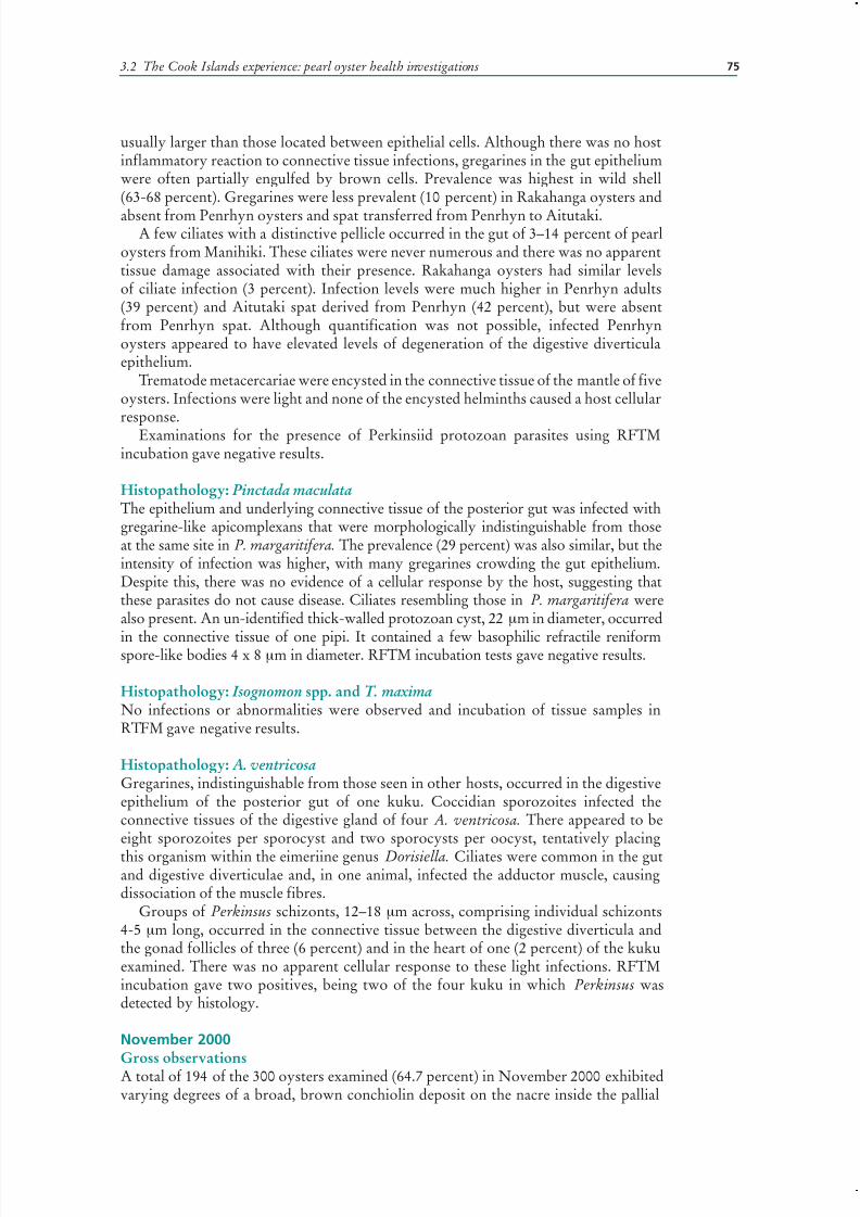
7/27/2019 Pearl Oyster Health Management
http://slidepdf.com/reader/full/pearl-oyster-health-management 89/136
3.2 The Cook Islands experience: pearl oyster health investigations 75
usually larger than those located between epithelial cells. Although there was no hostinflammatory reaction to connective tissue infections, gregarines in the gut epitheliumwere often partially engulfed by brown cells. Prevalence was highest in wild shell(63-68 percent). Gregarines were less prevalent (10 percent) in Rakahanga oysters and
absent from Penrhyn oysters and spat transferred from Penrhyn to Aitutaki.A few ciliates with a distinctive pellicle occurred in the gut of 3–14 percent of pearl
oysters from Manihiki. These ciliates were never numerous and there was no apparenttissue damage associated with their presence. Rakahanga oysters had similar levelsof ciliate infection (3 percent). Infection levels were much higher in Penrhyn adults(39 percent) and Aitutaki spat derived from Penrhyn (42 percent), but were absentfrom Penrhyn spat. Although quantification was not possible, infected Penrhynoysters appeared to have elevated levels of degeneration of the digestive diverticulaepithelium.
Trematode metacercariae were encysted in the connective tissue of the mantle of fiveoysters. Infections were light and none of the encysted helminths caused a host cellularresponse.
Examinations for the presence of Perkinsiid protozoan parasites using RFTMincubation gave negative results.
Histopathology: Pinctada maculataThe epithelium and underlying connective tissue of the posterior gut was infected withgregarine-like apicomplexans that were morphologically indistinguishable from thoseat the same site in P. margaritifera. The prevalence (29 percent) was also similar, but theintensity of infection was higher, with many gregarines crowding the gut epithelium.Despite this, there was no evidence of a cellular response by the host, suggesting thatthese parasites do not cause disease. Ciliates resembling those in P. margaritifera werealso present. An un-identified thick-walled protozoan cyst, 22 μm in diameter, occurred
in the connective tissue of one pipi. It contained a few basophilic refractile reniformspore-like bodies 4 x 8 μm in diameter. RFTM incubation tests gave negative results.
Histopathology: Isognomon spp. and T. maximaNo infections or abnormalities were observed and incubation of tissue samples inRTFM gave negative results.
Histopathology: A. ventricosaGregarines, indistinguishable from those seen in other hosts, occurred in the digestiveepithelium of the posterior gut of one kuku. Coccidian sporozoites infected theconnective tissues of the digestive gland of four A. ventricosa. There appeared to beeight sporozoites per sporocyst and two sporocysts per oocyst, tentatively placing
this organism within the eimeriine genus Dorisiella. Ciliates were common in the gutand digestive diverticulae and, in one animal, infected the adductor muscle, causingdissociation of the muscle fibres.
Groups of Perkinsus schizonts, 12–18 μm across, comprising individual schizonts4-5 μm long, occurred in the connective tissue between the digestive diverticula andthe gonad follicles of three (6 percent) and in the heart of one (2 percent) of the kukuexamined. There was no apparent cellular response to these light infections. RFTMincubation gave two positives, being two of the four kuku in which Perkinsus wasdetected by histology.
November 2000
Gross observationsA total of 194 of the 300 oysters examined (64.7 percent) in November 2000 exhibitedvarying degrees of a broad, brown conchiolin deposit on the nacre inside the pallial

7/27/2019 Pearl Oyster Health Management
http://slidepdf.com/reader/full/pearl-oyster-health-management 90/136

7/27/2019 Pearl Oyster Health Management
http://slidepdf.com/reader/full/pearl-oyster-health-management 91/136
3.2 The Cook Islands experience: pearl oyster health investigations 77
some oysters without obvious conchiolin deposits had bacterial infections (Table 3.2.3).There was no significant correlation between the severity of conchiolin deposits andthe severity of bacterial infections. Only light to moderate numbers of bacteria wereisolated from water samples, with more bacteria being isolated from bottom samples.The identifications for 17 representative isolates of bacteria obtained from pearl oystersand from water collected during field sampling are summarised in Table 3.2.4. Isolates of
Vibrio harveyi predominated in samples from adductor muscle, hemolymph and water.All isolates of V. harveyi examined had different biochemical phenotypes. Undescribedspecies of Vibrio were also common in water, hemolymph and from the surface of conchiolin deposits. Less common isolates included V. tubiashii from hemolymph, V.mediterranei from hemolymph and V. pelagius biovar II from adductor muscle.
HistopathologyMost bacteria occurred in the mantle epithelium and were associated with necrosis andsloughing of epithelial and sub-epithelial cells (Figure 3.2.2). Other sites of infectionincluded the gonad and digestive glandtubule epithelium. Oysters with higherconchiolin scores (2 or more) tended
to have depleted or resorbing gonads(Figure 3.2.3). Infections with gregarineprotozoans were noted principally inthe epithelium of the mid and hindgut (Figure 3.2.4), but also in the sub-epithelial connective tissue surroundingthe gut. There was no significantcorrelation between the intensity of gregarine infection and conchiolinlesion severity.
Other notable pathological lesions in
the oysters examined by histopathologyincluded breakdown, sloughing and focalnecrosis of the digestive gland epithelium
TABLE 3.2.4
Bacteriology results for selected isolates obtained from pearl oysters and water during field
sampling
Isolatenumber
Sample source Identification
1 Site 1, bottom water, yellow on TCBS Vibrio sp. not identifiable2 Site 1, oyster D hemolymph, yellow on TCBS Vibrio tubiashii
3 Site 2, oyster A adductor muscle, yellow on TCBS Vibrio harveyi
4 Site 2, oyster F, hemolymph, yellow on TCBS Vibrio harveyi
5 Site 5, oyster E adductor muscle, yellow on TCBS Vibrio pelagius biovar II
6 Site 1, oyster F adductor muscle, green on TCBS Vibrio harveyi
7 Site 2, oyster D adductor muscle, green on TCBS Vibrio harveyi
8 Site 5, oyster H small green colonies on TCBS Staphylococcus-Micrococcus-like sp.
9 Site 2, bottom water, cream on TSA+2 Vibrio sp. not identifiable
10 Site 2, bottom water, clear on TSA+2 Not recovered on subculture
11 Site 2, oyster F, hemolymph, clear on TSA+2 Vibrio mediterranei
12 Site 1, oyster C, hemolymph, cream on TSA+2 Vibrio tubiashii
13 Site 1, bottom water, spreading on TSA+2 Vibrio sp. not identifiable
14 Site 2, bottom water, spreading on TSA+2 Vibrio harveyi
15a Site 2, oyster F, hemolymph, spreading on TSA+2 Vibrio sp. not identifiable
15b Site 2, oyster F, hemolymph, spreading on TSA+2 Vibrio sp. not identifiable
16 Site 1, oyster P, from brown stain on nacre Vibrio sp. not identifiable
17 Site 4, oyster F, adductor muscle, very small colonies Acinetobacter -like
FIGURE 3.2.2
A necrotic lesion in the mantle associated with bacterial
infection
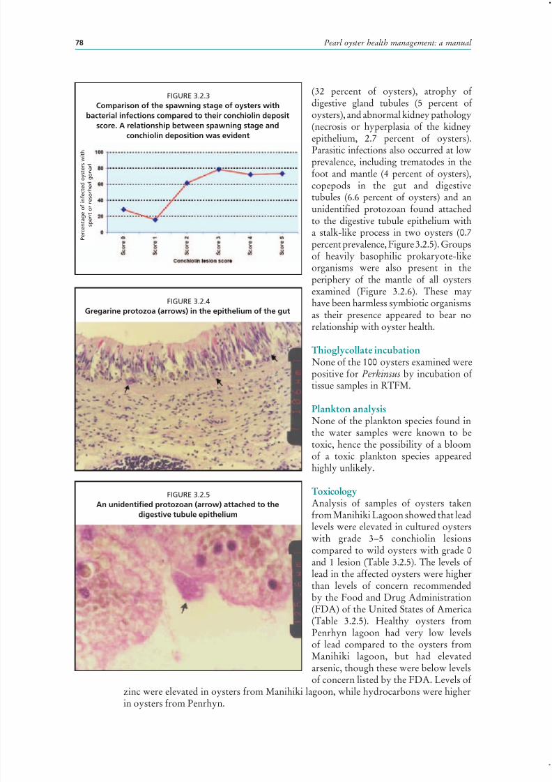
7/27/2019 Pearl Oyster Health Management
http://slidepdf.com/reader/full/pearl-oyster-health-management 92/136
Pearl oyster health management: a manual 78
(32 percent of oysters), atrophy of digestive gland tubules (5 percent of oysters), and abnormal kidney pathology(necrosis or hyperplasia of the kidney
epithelium, 2.7 percent of oysters).Parasitic infections also occurred at lowprevalence, including trematodes in thefoot and mantle (4 percent of oysters),copepods in the gut and digestivetubules (6.6 percent of oysters) and anunidentified protozoan found attachedto the digestive tubule epithelium witha stalk-like process in two oysters (0.7percent prevalence, Figure 3.2.5). Groupsof heavily basophilic prokaryote-likeorganisms were also present in theperiphery of the mantle of all oystersexamined (Figure 3.2.6). These mayhave been harmless symbiotic organismsas their presence appeared to bear norelationship with oyster health.
Thioglycollate incubationNone of the 100 oysters examined werepositive for Perkinsus by incubation of tissue samples in RTFM.
Plankton analysisNone of the plankton species found inthe water samples were known to betoxic, hence the possibility of a bloomof a toxic plankton species appearedhighly unlikely.
ToxicologyAnalysis of samples of oysters takenfrom Manihiki Lagoon showed that leadlevels were elevated in cultured oysterswith grade 3–5 conchiolin lesions
compared to wild oysters with grade 0and 1 lesion (Table 3.2.5). The levels of lead in the affected oysters were higherthan levels of concern recommendedby the Food and Drug Administration(FDA) of the United States of America(Table 3.2.5). Healthy oysters fromPenrhyn lagoon had very low levelsof lead compared to the oysters fromManihiki lagoon, but had elevatedarsenic, though these were below levels
of concern listed by the FDA. Levels of zinc were elevated in oysters from Manihiki lagoon, while hydrocarbons were higherin oysters from Penrhyn.
FIGURE 3.2.4
Gregarine protozoa (arrows) in the epithelium of the gut
FIGURE 3.2.5
An unidentified protozoan (arrow) attached to the
digestive tubule epithelium
FIGURE 3.2.3
Comparison of the spawning stage of oysters with
bacterial infections compared to their conchiolin deposit
score. A relationship between spawning stage and
conchiolin deposition was evident
P e r c e n t a g e o f i n f e c t e d o y s t e r s w i t h
s p e n t o r r e s o r b e d g o n a d

7/27/2019 Pearl Oyster Health Management
http://slidepdf.com/reader/full/pearl-oyster-health-management 93/136
3.2 The Cook Islands experience: pearl oyster health investigations 79
December 2003A significant proportion of both wildand cultured oysters from Manihikiand Penrhyn lagoons displayed brown
coloured conchiolin deposits in the nacreof one or both shell valves (Diggles andMaas, 2004). The oysters from Penrhynwere the worst affected, while this lesionwas absent from oysters sampled fromRakahanga Lagoon. The prevalence andintensity of this lesion in oysters fromManihiki Lagoon was much lower thanrecorded in November 2000, but itspersistence suggests that the oystersin Manihiki remained affected byenvironmental stress in at least someparts of the lagoon. The emergence of the conchiolin lesions in oysters from Penrhyn lagoon may be related to stress frompoor water quality due to low levels of dissolved oxygen recorded by water qualitymonitoring buoys in that lagoon.
Signs of recovery from the previous outbreak of bacterial disease in November2000 were observed in both wild and cultured oysters from Manihiki Lagoon. Inthese oysters the conchiolin lesions remained visible but had been overlaid by newlydeposited nacre (Figure 3.2.7). This permanent scarring of the nacre of the shell wasalso associated with development of a prominent check around the periphery of theshell (Figure 3.2.8), indicating that shell growth ceased for a significant period of time subsequent to November 2000. Recovering oysters were not observed in any of
the earlier samples of oysters from Manihiki Lagoon from May 1998 or November2000, suggesting that the November 2000 disease episode was unprecedented and hadsignificant long term effects on the health of surviving oysters in Manihiki Lagoon.
FIGURE 3.2.6
Groups of deeply basophilic prokaryote-like organisms
(arrows) in the mantle
TABLE 3.2.5
Comparison of toxicology tests for oysters from Manihiki and Penrhyn lagoons. Results are for
4 to 6 pooled oysters from each site. Notable differences between lagoons are in bold
Sample nameManihiki
Lagoon, culturedoysters
ManihikiLagoon,
wild oysters
PenrhynLagoon,
cultured oysters
PenrhynLagoon,
wild oysters
USA FDAlevels ofconcern
Range of conchiolin
lesion severity Grade 3 to 5 Grade 0 to 1
Grade 0,
healthy
Grade 0,
healthy
Zinc (mg/kg) 52 80.3 13.6 6.35 n/a
Copper (mg/kg) 0.33 0.53 0.38 0.48 n/a
Arsenic (mg/kg) 7.27 5.79 13.1 10.5 86
Cadmium (mg/kg) 1.02 0.85 1.63 1.61 3.7
Mercury (mg/kg) <0.005 0.008 0.004 0.004 1
Chromium (mg/kg) 0.13 0.16 0.08 0.11 13
Nickel (mg/kg) 0.1 0.19 0.1 0.16 80
Lead (mg/kg) 2.7 0.08 0.007 0.009 1.7
Hydrocarbons
C7-C9 (mg/kg) <10 <10 <4 <4 -
C10-C14 (mg/kg) <20 <20 <8 <7 -
C15-C36 (mg/kg) 50 100 160 183 -
TOTAL <80 100 160 180 -

7/27/2019 Pearl Oyster Health Management
http://slidepdf.com/reader/full/pearl-oyster-health-management 94/136
Pearl oyster health management: a manual 80
DISCUSSIONThe study of May 1998 revealed thepresence of a few trivial infections(e.g. gregarines, ciliates, trematode
metacercariae) and three potentiallypathogenic groups, boring sponges, thefungus from infected drill holes andPerkinsus. The identity of the orangesponges that bore into Manihiki andRakahanga oyster shell is currentlyunclear, as is the taxonomy of clionidsponges. The descriptions of manyspecies differ from the descriptions of the same species by other authors. Themore common orange sponge resemblesCliona celata, but the description byThomas (1979) is too vague to be of useand C. celata has been reported fromcold temperate waters (Baxter, 1984).The other, less common, boring spongeresemblesCliona vastifica, which infectsP. margaritifera in French Polynesia(Mao Che et al., 1996), but C. vastificaranges from dark green to yellowishgrey in colour and the dimensions of the sponge differ from those given byThomas (1979). Cliona vastifica and C.
celata are the most widespread clionidinfestations in pearl shells (Thomas,1979; Mao Che et al., 1996).
The organism associated with theinfection of drilled holes appearedhyphae-like and most closely resemblesthe fungus Ostracoblabe implexa fromP. margaritifera in French Polynesia(Mao Che et al., 1996). Ostracoblabeimplexa is primarily a fungal pathogenin shells of bivalves in temperate waters(Li et al., 1983), however the site and
growth of the organism reported hereappears to be the same as for O implexa.The burrowing fungus appears to gainentrance through drill holes and obtains
its nourishment from the breakdown of the proteinaceous shell matrix. However,when it reaches the inner shell surface it sets up an irritation that changes the cellularstructure of the mantle, resulting in deposition of conchiolin-rich nacre over thefungus. This results in the thin nacre, lacking refringence and lustre that overlays theinfection and which can be readily broken by light pressure. Fortunately it appears thatinfection is rare, except through holes drilled in the shell for suspension hanging andimprovements in technique will overcome the problem.
Perkinsus is potentially the most serious pathogen. The species in Manihiki lagoonis probably P. olseni, as this species is known to occur around northern New Zealand(Hine and Diggles, 2002), Australia (Lester and Davis, 1981; Hine and Thorne, 2000),
FIGURE 3.2.8
Regrowth of nacre over conchiolin deposits immediately
proximal to a prominent check in shell growth (arrows)
in an oyster taken from Manihiki lagoon 3 years after
the November 2000 disease event
FIGURE 3.2.7
This photo of an oyster taken from Manihiki lagoon in
December 2003, around 3 years after the November 2000
disease event shows conspicuous regrowth of nacre
over conchiolin deposits (arrows) laid down during thedisease event

7/27/2019 Pearl Oyster Health Management
http://slidepdf.com/reader/full/pearl-oyster-health-management 95/136
3.2 The Cook Islands experience: pearl oyster health investigations 81
Korea (Choi and Park, 1997; Park, Choi and Choi , 1999; Lee et al., 2001) and Japan(Hamaguchi et al., 1998). This suggests that it is an Indo-Pacific species and that it islikely therefore that the Manihiki Perkinsus is P. olseni. It has been shown that anotherPerkinsus, P. atlanticus, which occurs in clams in Spain, is actually P. olseni (Robledo
et al., 2000), that was probably introduced into Spain in Manila clams (Ruditapesdecussatus) from Southeast Asia.
Perkinsus olseni was originally described in association with mass mortalities amongabalone (Haliotis spp.) on the coast of South Australia (Lester and Davis, 1981). It wassubsequently shown that it infects many families and species of molluscs (Goggin andLester, 1987; Hine and Thorne, 2000) and that isolates from one species can infect manyother species (Goggin et al., 1989). Any one isolate has developmental stages of sizesthat vary from host to host and therefore size does not distinguish species. In the greatmajority of infections, the parasite occurs as spherical clusters of schizonts withouteliciting a host response (Hine and Thorne, 2000), as was observed here. When P. olsenicauses disease, the other histozoic developmental stages (merozoites which develop tomeronts and then to schizonts) are present. What triggers the parasite to change from abenign parasite into a proliferating pathogen is unknown. It does appear, however, thatpearl oysters are less susceptible than many other hosts and therefore perkinsosis maynot be a great threat to pearl farming.
The study in November 2000 suggested that the outbreak of disease associatedwith brown conchiolin deposits on the nacre had a bacterial aetiology. There was noevidence of widespread infection of any viral, protozoan or metazoan pathogens inthe diseased pearl oysters examined. The presence of the gregarine protozoans in thegut epithelium was not correlated with the severity of the conchiolin lesions. The 1998survey found that these protozoans also occur in apparently healthy pearl oysters, asalso found by Humphrey et al. (1998). We consider the association of gregarines withdiseased P. margaritifera both here and previously in French Polynesia (Chagot et
al., 1993) as likely to be incidental and they are considered unlikely to cause diseaseunder normal circumstances (Humphrey et al., 1998). Their presence in P. maculataand A. ventricosa indicate that both other pteriid species and other bivalve families areinfected by these apparently benign parasites. The basophilic prokaryote infections of the mantle seen in the November 2000 survey do not appear to have previously beenreported, although similar inclusions have been reported from the digestive epithelia of P. margaritifera from French Polynesia (Comps et al., 1998).
The predominance of the bacterium V. harveyi and other vibrios in cultures takenfrom affected oysters, together with the occurrence of pathological lesions consistentwith bacterial infection in many affected oysters, indicates that infection by V. harveyi and other opportunistic vibrios, was associated with this disease syndrome. There wasno evidence to suggest that the disease syndrome was primarily caused by a single
pathogenic strain of V. harveyi. This is a fundamental difference between the diseasesyndrome of P. margaritifera in Manihiki Lagoon and that of Brown Ring Disease(BRD). BRD is also characterised by abnormal brown conchiolin deposits adheringbetween the pallial line and the edge of the shell (Paillard and Maes, 1994). However,BRD is caused only by strains of a pathogenic bacterium, Vibrio tapetis (formerlyknown as Vibrio P1) (Borrego et al., 1992; Paillard, Maes and Oubella, 1994; Novoa etal., 1998) and the disease can be reproduced by experimental exposure to these strains.
The anomalous conchiolin deposition in BRD occurs as a definite thin brown ringon the nacre around the perimeter of the shell (Paillard and Maes, 1994). In contrast,most of the oysters sampled from Manihiki Lagoon displayed a broad conchiolindeposition resulting from retraction of the mantle, with the deposits lying outside the
edge of the mantle. This condition was virtually identical to that described for Pinctadamaxima from Western Australia associated with the presence of Vibrio harveyi (seeDybdahl and Pass, 1985; Pass, Dybdahl and Mannion , 1987; Perkins, 1996).

7/27/2019 Pearl Oyster Health Management
http://slidepdf.com/reader/full/pearl-oyster-health-management 96/136
Pearl oyster health management: a manual 82
Bacterial diseases in aquaculture are often associated with opportunistic bacteriawhich invade hosts which are stressed due to unfavorable conditions such asovercrowding, abnormally high or low water temperatures and/or poor water quality.Pass, Dybdahl and Mannion (1987) found that V. harveyi infection in P. maxima
was associated with poor water quality conditions, low water temperatures andovercrowding during transport of oysters to lease sites. Figueras et al. (1996) found thehighest prevalence of BRD in storage areas where clams were kept at high populationdensities for at least 1 month. In Manihiki Lagoon, one potential stressor which mayhave been related to the onset of mortalities was the high stocking density. Prior to thedisease outbreak the number of oysters cultured in Manihiki Lagoon was reportedly atan all time high (B. Ponia, MMR, pers. comm.).
There is also evidence to suggest that a decline in water quality preceded the diseaseoutbreak. Water exchange in the semi-enclosed Manihiki lagoon is poor, at around onewater exchange every 2 months (Anderson, 1998). A long period of unusually calm,dry weather was reported prior to the disease outbreak. At the time oysters were firstsampled in late November 2000, water temperatures in Manihiki Lagoon were normal(29 °C), but dissolved oxygen (DO) levels were low, ranging between 1 and 3 mg/l (S.Sharma and G. Frost, SOPAC, pers. comm.). However, during the one week periodin which field sampling was conducted, the mean DO of the lagoon increased to over6 mg/l, probably due to high wind conditions which would have helped with aerationand water exchange in the lagoon (S. Sharma and G. Frost, SOPAC, pers. comm.).These water quality data, albeit limited, suggest that the disease outbreak followed atransient period of poor water quality in Manihiki lagoon.
High stocking densities reduce the food supply available to each oyster (Anderson,1998). Starvation may have been indicated in some of the diseased oysters bypathologies such as atrophy of digestive gland tubules. Reduced food supply canalso be an important stressor in bivalve molluscs (Tomaru et al., 2001) and can cause
mortality (Numaguchi, 1995). Spawning is another potential stressor which may havepredisposed oysters to disease. We found that oysters with bacteria present in sectionsand with high lesion scores (2 or more) tended to have depleted or resorbing gonads.
Anecdotal evidence supplied by oyster farmers suggest the performance of pearloysters in Manihiki Lagoon in the years following the November 2000 disease outbreakhas been reduced compared to pre-outbreak times. Certainly many of the oysterswhich survived were still showing signs from the disease outbreak in a follow upsurvey of oyster health done 3 years later in December 2003 (Diggles and Maas, 2004).These signs included permanent scarring of the nacre of the shell and development of aprominent check around the periphery of the shell, indicating that shell growth ceasedfor a significant period of time subsequent to November 2000. These signs of diseasehad never been previously seen by pearl oyster farmers throughout the production
history of the lagoon, indicating that the November 2000 disease outbreak in ManihikiLagoon was unprecedented and had significant long term deleterious effects on oysterhealth in that lagoon.
In conclusion, our data suggest that an unprecedented disease outbreak in P.margaritifera in Manihiki lagoon in November 2000 was associated with vibriosiscaused by V. harveyi and other opportunistic vibrios. The disease outbreak appearedto follow a transient reduction in lagoon water quality associated with a period of calmweather and was probably exacerbated by high oyster stocking densities. Furthermore,the mortalities occurred at a time of the year when oysters may have been furtherstressed after spawning. Management of the disease could, therefore, be based oncontrolling the stocking density of oysters in the lagoon, and modifying husbandry
practices to reduce stress on oysters when they are spawning. Ongoing monitoring of key water quality parameters such as water temperature, DO, chlorophyll α, and bothsediment oxygen demand (SOD) (Anderson, 1998) and biological oxygen demand

7/27/2019 Pearl Oyster Health Management
http://slidepdf.com/reader/full/pearl-oyster-health-management 97/136
3.2 The Cook Islands experience: pearl oyster health investigations 83
(BOD) may also be useful so that the epidemiology of disease outbreaks which mayoccur in the future might be better understood.
ACKNOWLEDGEMENTS
The authors wish to thank Ian Bertram, Tangi Napara, Des Piho, Ben Ponia andall other MMR staff on Manihiki Atoll, as well as Graeme Frost from SOPAC forassistance with field sampling and Mark Ellis, Ron Powell, Bob Tarau, John Lyonsand all other pearl farmers who helped with field sampling or supplied oystersfor examination. The authors also thank Navy Epati of MMR for assistance withorganizing transport and accommodation on Rarotonga and Manihiki. We are alsograteful to Joan Nichol of the Wellington Hospital Medical School for the promptprocessing of samples. Dr H. Chang performed the plankton analysis and Brian Bullassisted with data analysis.
REFERENCESAnderson, M. 1998. The ecological sustainability of pearl farming in Manihiki lagoon,
Northern Cook Islands. In Pearl oyster information bulletin No. 11, May 1998 (ed. byN.A. Sims), pp 7 - 11. Secretariat of the Pacific Community, New Caledonia.
Baumann, P., Baumann, L. & Mandel, M. 1971. Taxonomy of marine bacteria: the genusBeneckea. Journal of Bacteriology 1971: 268-294.
Baxter, J.M. 1984. The incidence of Polydora ciliata and Cliona celata boring the shell of Patella vulgata in Orkney. Journal of the Marine Biological Association 64: 728-729.
Beuchat, L.R. 1974. Combined effects of water activity, solute, and temperature on thegrowth of Vibrio parahaemolyticus. Applied Microbiology 27: 1075-1080.
Borrego, J.J., Castro, D., Luque, A., Paillard, C., Maes, P., Garcia, M.T. & Ventosa,A. 1992. Vibrio tapetis sp.nov., the causative agent of the brown ring disease affectingcultured clams. International Journal of Systematic Bacteriology 46: 480-484.
Bryant, T.N., Lee, J.V., West. P.A. & Colwell, R.R.1986. A probability matrix for theidentification of species of Vibrio and related genera. Journal of Applied Bacteriology 61:469-480.
Bryant, T.N. & Smith, K.R. 1991. Bacterial Identifier, Version M 1.0. Oxford,Blackwells.
Bushek, D. & Allen, S.K. 1996. Host-parasite interactions among broadly distributedpopulations of the eastern oyster Crassostrea virginica and the protozoan Perkinsusmarinus. Marine Ecology Progress Series 139: 127-141.
Bushek, D., Ford, S.E. & Allen, S.K. 1994. Evaluation of methods using Ray’s fluidthioglycollate medium for diagnosis of Perkinsus marinus infection in the eastern oyster,Crassostrea virginica. Annual Review of Fish Diseases 4: 201-217.
Chagot, D., Fougerouse, A. Weppe, M., Marques, A. & Bouix, G. 1993. A gregarine
(Protozoa Sporozoa) parasite of black lipped pearl oysters Pinctada margaritifera (L.,1758) (Mollusca Bivalvia) from French Polynesia. C. R. Acad. Sci. Paris 316: 239-244.
Choi, K.S. & Park, K.I. 1997. Report on the occurrence of Perkinsus sp. in the Manilaclams, Ruditapes philippinarum in Korea. Journal of Aquaculture 10: 227-237.
Comps, M., Fougerouse, A. & Buestel, D. 1998. A prokaryote infecting the black-lippedpearl oyster Pinctada margaritifera. Journal of Invertebrate Pathology 72: 87-89.
Diggles, B.K & Hine P.M. 2001. Mortality of black-lip pearl oysters (Pinctada margaritifera)in Manihiki Lagoon. NIWA client report no. WLG 01/5 prepared for The Ministry of Marine Resources, Government of the Cook Islands. 36 pp.
Diggles B.K & Maas E. 2004. Cook Islands Pearl Oyster and Lagoon Health MonitoringProgramme, December 2003. NIWA Client Report WLG2004-24. April 2004, 48 pp.
Dybdahl, R. & Pass, D.A. 1985. An investigation of mortality of the pearl oyster,Pinctada
maxima, in Western Australia. Western Australian Fisheries Department Report No. 71.78 pp.

7/27/2019 Pearl Oyster Health Management
http://slidepdf.com/reader/full/pearl-oyster-health-management 98/136
Pearl oyster health management: a manual 84
Figueras, A., Robledo, J.A.F. & Novoa, B. 1996. Brown ring disease and parasites in clams(Ruditapes decussatus and R. philippinarum) from Spain and Portugal. Journal of ShellfishResearch 15: 363-368.
Food and Drug Administration. 1993. Guidance document for lead in shellfish. Centre
for Food Safety and Applied Nutrition, US FDA, Washington. (http://vm.cfsan.fda.gov/~frf/guid-sf.html)
Furniss, A.L., Lee, J.V. & Donovan, T.J. 1978. The Vibrios. Monograph Series 11, PublicHealth Laboratory Service. London, Her Majesty’s Stationery Office.
Goggin, C.L. 1996. Effect of Perkinsus olseni (Protozoa, Apicomplexa) on the weight of Tridacna crocea (Mollusca, Bivalvia) from Lizard Island, Great Barrier Reef. Aquaculture 141: 25-30.
Goggin, C.L. & Lester, R.J.G. 1987. Occurrence of Perkinsus species (Protozoa, Apicomplexa)in bivalves from the Great Barrier reef. Diseases of Aquatic Organisms 3: 113-117.
Goggin, C.L., Sewell, K.B. & Lester, R.J.G. 1989. Cross-infection experiments withAustralian Perkinsus species. Diseases of Aquatic Organisms 7: 55-59.
Hamaguchi, M., Suzuki, N., Usuki, H. & Ishioka., H. 1998. Perkinsus protozoan infectionin short-necked clam Tapes (=Ruditapes) philippinarum in Japan. Fish Pathology 33: 473-480.
Hine, P.M. 1998. A survey of the health of black-lip pearl oysters (Pinctada margaritifera)and other bivalves in the Cook Islands. NIWA report to the Ministry of MarineResources of the Government of the Cook Islands (funded by the Asian DevelopmentBank, August 1998).
Hine, P.M. & Diggles, B.K. 2002. The distribution of Perkinsus olseni in New Zealandbivalve molluscs. Surveillance 29: 8-11.
Hine, P.M. & Thorne, T. 2000. A survey of some parasites and diseases of several species of bivalve mollusc in northern Western Australia. Diseases of Aquatic Organisms 40: 67-78.
Humphrey, J.D., Norton, J.H., Jones, J.B., Barton, M.A., Connell, M.T., Shelley, C.C.
& Creeper, J.H. 1998. Pearl oyster (Pinctada maxima) aquaculture: Health survey of Northern Territory, Western Australia and Queensland pearl oyster beds and farms.Fisheries Research and Development Corporation (FRDC) Project No. 94/079, FinalReport. 108 pp.
Lee, M-K., Cho, B-Y., Lee, S-J., Kang, J-Y., Jeong, H.S., Huh, S.H. & Huh, M-D. 2001.Histopathological lesions of Manila clams, Tapes philippinarum, from Hadong andNamhae coastal areas of Korea. Aquaculture 201: 199-209.
Lester, R.J.G. & Davis G.H.G. 1981. A new Perkinsus species (Apicomplexa, Perkinsea)from the abalone Haliotis ruber . Journal of Invertebrate Pathology 37: 181-187.
Li, M.F., Drinnan, R.E., Drebot, M. & Newkirk, G. 1983. Studies on shell disease of theEuropean flat oyster Ostrea edulis Linné in Nova Scotia. Journal of Shellfish Research
3: 135-140.
Mao Che, L., Le Campio-Alsumard, T., Boury-Esnault, N., Payri, C., Golubric, S. &Bezac, C. 1996. Biodegradation of shells of the black pearl oyster, Pinctada margaritiferavar. cumingii, by microborers and sponges of French Polynesia. Marine Biology 126:509-519.
Norton, J.H., Shepherd, M.A. Perkins, F.P. & Prior, H.C. 1993. Perkinsus-like infectionin farmed golden-lipped pearl oyster Pinctada maxima from the Torres Strait, Australia.
Journal of Invertebrate Pathology 62: 105-106.Novoa, B., Luque, A. Castro, D., Borrego, J.J. & Figueras, A. 1998. Characterization
and infectivity of four bacterial strains isolated from brown ring disease affected clams. Journal of Invertebrate Pathology 71: 34-41.
Numaguchi, K. 1995. Influences of unfed condition on the mortality of pearl oysterPinctada fucata martensii
. Fisheries Science
61: 739-742.Paillard, C. & Maes, P. 1994. Brown ring disease in the Manila clam Ruditapes philippinarum:establishment of a classification system. Diseases of Aquatic Organisms 19: 137-146.
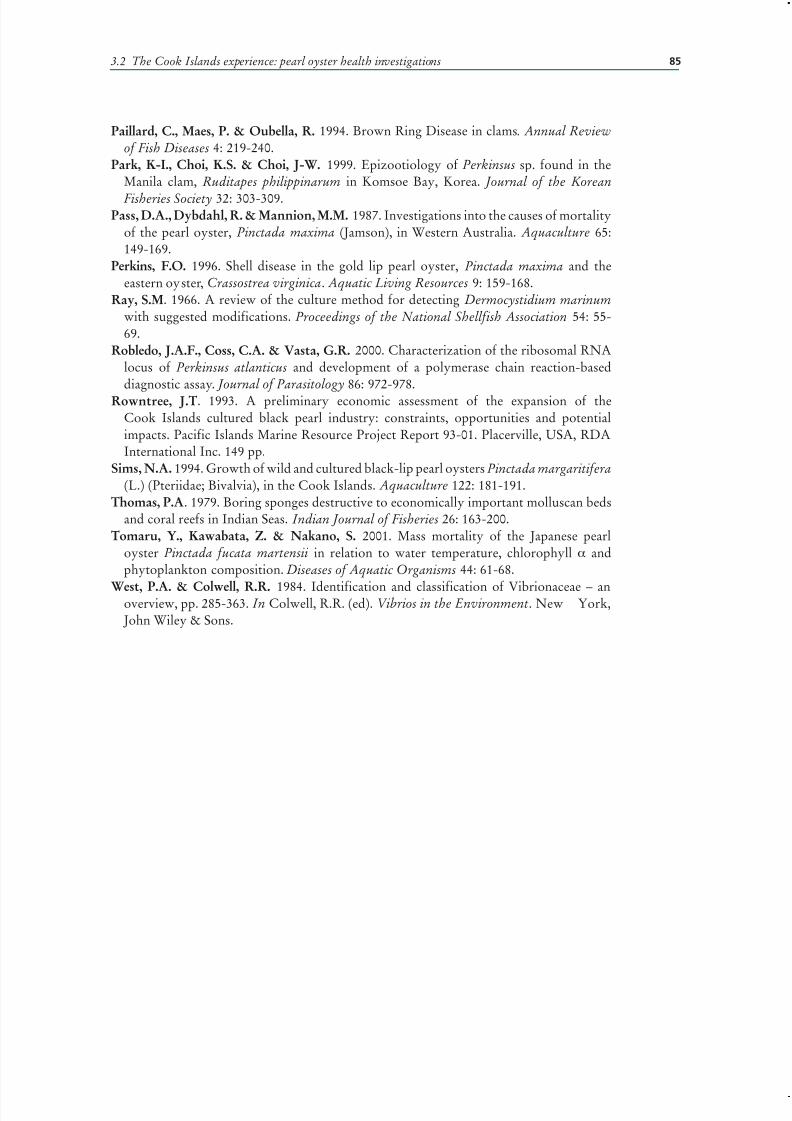
7/27/2019 Pearl Oyster Health Management
http://slidepdf.com/reader/full/pearl-oyster-health-management 99/136
3.2 The Cook Islands experience: pearl oyster health investigations 85
Paillard, C., Maes, P. & Oubella, R. 1994. Brown Ring Disease in clams. Annual Reviewof Fish Diseases 4: 219-240.
Park, K-I., Choi, K.S. & Choi, J-W. 1999. Epizootiology of Perkinsus sp. found in theManila clam, Ruditapes philippinarum in Komsoe Bay, Korea. Journal of the Korean
Fisheries Society 32: 303-309.Pass, D.A., Dybdahl, R. & Mannion, M.M. 1987. Investigations into the causes of mortality
of the pearl oyster, Pinctada maxima (Jamson), in Western Australia. Aquaculture 65:149-169.
Perkins, F.O. 1996. Shell disease in the gold lip pearl oyster, Pinctada maxima and theeastern oyster, Crassostrea virginica. Aquatic Living Resources 9: 159-168.
Ray, S.M. 1966. A review of the culture method for detecting Dermocystidium marinum with suggested modifications. Proceedings of the National Shellfish Association 54: 55-69.
Robledo, J.A.F., Coss, C.A. & Vasta, G.R. 2000. Characterization of the ribosomal RNAlocus of Perkinsus atlanticus and development of a polymerase chain reaction-baseddiagnostic assay. Journal of Parasitology 86: 972-978.
Rowntree, J.T. 1993. A preliminary economic assessment of the expansion of theCook Islands cultured black pearl industry: constraints, opportunities and potentialimpacts. Pacific Islands Marine Resource Project Report 93-01. Placerville, USA, RDAInternational Inc. 149 pp.
Sims, N.A. 1994. Growth of wild and cultured black-lip pearl oysters Pinctada margaritifera (L.) (Pteriidae; Bivalvia), in the Cook Islands. Aquaculture 122: 181-191.
Thomas, P.A. 1979. Boring sponges destructive to economically important molluscan bedsand coral reefs in Indian Seas. Indian Journal of Fisheries 26: 163-200.
Tomaru, Y., Kawabata, Z. & Nakano, S. 2001. Mass mortality of the Japanese pearloyster Pinctada fucata martensii in relation to water temperature, chlorophyll α andphytoplankton composition. Diseases of Aquatic Organisms 44: 61-68.
West, P.A. & Colwell, R.R. 1984. Identification and classification of Vibrionaceae – anoverview, pp. 285-363. In Colwell, R.R. (ed). Vibrios in the Environment. New York, John Wiley & Sons.

7/27/2019 Pearl Oyster Health Management
http://slidepdf.com/reader/full/pearl-oyster-health-management 100/136

7/27/2019 Pearl Oyster Health Management
http://slidepdf.com/reader/full/pearl-oyster-health-management 101/136

7/27/2019 Pearl Oyster Health Management
http://slidepdf.com/reader/full/pearl-oyster-health-management 102/136
Pearl oyster health management: a manual 88
and 3 500 people. However, the collapse in market price for MOP after World War I(WW1) was equally dramatic. By 1939, the number of luggers had reduced to 73 andafter WWII, only 15 boats resumed pearling (Malone, Hancock and Jeffries, 1988;O’Sullivan, 1995).
In 1956, Pearls Proprietary Ltd. introduced pearl culture at Kuri Bay (see Plate 1) inassociation with Japanese and other overseas investors and under agreement with the
Japanese Government. By 1973, Kuri Bay was said to produce around 60 percent of the world’s finest round pearls and by 1987 there were 11 companies farming pearls inthe Broome region (Malone, Hancock and Jeffries, 1988). In 2001, there were five pearlhatcheries and 16 companies operating farms in Western Australia from North EastCape to the Northern Territory border though recent rationalization in the industrysaw the number of hatcheries reduced to three by 2006. The annual quota that can beseeded in Western Australia consists of 572 000 wild stock shell and 350 000 hatcheryproduced pearl oysters. At present, leases in Western Australia cover 150 squarenautical miles and that area is predicted to increase to 280 square nautical miles by2010. Availability of suitable sites has been identified as a critical constraint to industryexpansion. Annual production is now in excess of AUS$190 million with productionof pearls from P. maxima being supplemented with “black” pearls farmed in Pinctadamargaritifera in the Shark Bay area.
The Northern Territory has a history of over exploitation of natural pearl oysterbeds, which began in Darwin Harbour, when beds were depleted early last century.The western grounds, near Bathurst Island, were discovered around 1929-1930 andwere fished out by 1939. The eastern grounds, off Boucat Bay, were discovered in1936 and by the 1950s were no longer commercially viable. Presently, pearl cultureoperations in the Northern Territory are centered on Bynoe Harbour, CoburgPeninsula, and the English Company Islands. Pearl cultivation also occurs in Darwinand Bynoe Harbours. An annual quota of 420 000 P. maxima oysters can be seeded that
is split up between 7 pearl licensees. There are currently two hatcheries (J. Humphrey,Darwin DPIE, pers. comm.)In Queensland, the pearling beds collapsed from overfishing in the 1930s. In 1999-
2000, there were approximately 20 licensed areas for pearl cultivation in Queensland,but not all were in use and about 16 000 shells were seeded in that reporting period.In 2003-2004, the value of the pearling industry was reported as AU$338 000 andthere has been little growth since then. There is some minor production from molluschatcheries. The main species farmed is P. maxima, with some production from P.margaritifera, P. radiata and Pteria penguin (T. Hawkesford, Queensland DPIE, pers.comm.; Lobegeiger, 2001).
In New South Wales, a small P. imbricata farm has met with considerable public andpolitical opposition, however, the experience gained has been used to develop other P. imbricata farms in Queensland and Western Australia.
DISEASE ISSUESUnexplained mortalities of oysters on lease sites in the Kuri Bay region commencedin the 1970s and occurred subsequently in leases in the Broome area and at Darwin.Deaths of more than 50 percent of the oysters following transfer from the “80 MileBeach” fishing ground to the farms were recorded, but not among wild stocks oroysters harvested then “dumped” near the grounds. The stock situation becameso serious in the early 1980s that the Western Australian Government was forcedto impose a moratorium, until December 1987, on the number of companieslicensed to fish for and farm shell. Results published by Dybdahl and Pass (1985)
and Pass, Dybdahl and Mannion (1987) attributed the losses of the oysters to theconditions during transport that allowed build up of the pathogenic bacterium Vibrioharveyi. Their report made recommendations to modify management practices. These

7/27/2019 Pearl Oyster Health Management
http://slidepdf.com/reader/full/pearl-oyster-health-management 103/136
89
modifications were successful in reducing the mortalities but the problem also madethe companies acutely aware of disease issues and the potential for transfer of diseasebetween areas. Pass, Perkins and Dybdahl (1988) described a virus from the digestivegland of P. maxima. The inclusion bodies were visible in apparently healthy oysters
and no pathological significance could be ascribed to them (see Plate 2). In 1993, apapovavirus-like infection and a Perkinsus-like infection (Norton, Shepherd and Prior,1993) were described from P. maxima collected from the Torres Strait region. In 1994,industry approved funding by the Fisheries Research Development Corporation(FRDC)of a survey to determine the disease status of the oyster industry across allthree affected states. The survey was undertaken from 1995 to 1998 (Humphrey etal., 1998) (Table 1). Shell disease was independently described from P. maxima in 1996(Perkins, 1996) and a haplosporidian, found in 1993 from the same oyster species wasfinally described in 1998 (Hine and Thorne, 1998). The emergence of an intracellularciliate in the digestive gland of oysters in the Exmouth Gulf region was reported by
Jones and Creeper (2006). A severe mortality with an infectious aetiology occurred inPinctada maxima, but not other Pinctada species growing in Exmouth
Gulf in 2006. The cause is still under investigation. The pathogens and diseases of Pinctada maxima in Australia are listed in Table 3.3.1.
TABLE 3.3.1
Parasites and diseases previously recorded in pearl oysters Pinctada maxima in northern
Australia. QLD = Queensland, NT= Northern Territory, WA= Western Australia
Aetiological agent Disease/Pathology Geographic location Reference
Viruses
Papova-like virus Epithelial hypertrophyof palp
QLD Norton et al., 1993b;Humphrey et al., 1998
Intranuclear viral inclusions None WA, NT, QLD Pass et al., 1988;Humphrey et al., 1998
Bacteria
Rickettsiales : Large form None WA, NT, QLD Humphrey et al., 1998
Rickettsiales : Small form None WA Humphrey et al., 1998
V. alginolyticus WA Humphrey et al., 1998
V. anguillarum WA Humphrey et al., 1998
V. harveyi Mortalities WA Dybdahl and Pass, 1985;Pass et al., 1987
V. mediterranei WA Humphrey et al., 1998
V. parahaemolyticus WA Humphrey et al., 1998
V. pelagius WA Humphrey et al., 1998
Vibrio sp. Mortalities WA Dybdahl and Pass, 1985
V. splendidus II WA Humphrey et al., 1998
Corynebacterium sp. WA Humphrey et al., 1998
Erwinia hebicola WA Humphrey et al., 1998
Photobacterium sp. WA Humphrey et al., 1998Pseudomonas putrefaciens WA Humphrey et al., 1998
Protozoa
Intracellular cilliate None WA Jones and Creeper, 2006
Gregarines None
Haplosporidium sp. None WA Hine and Thorne, 1998
Perkinsus sp. Mortalities QLD Norton et al., 1993a
Apicomplexan Hine and Thorne, 2000
Cryptosporidia-like NT Humphrey et al., 1998
Metazoa; Crustacea
Conchodytes maculatus None Bruce, 1989; Chace andBruce, 1993
Pinnotheres villosus Dix, 1973
Anthessius pinctadae NT, WA Humphrey et al., 1998
Metazoa: Platyhelminthes
Larval lecanicephalidcestodes
WA, NT, QLD Humphrey et al., 1998
3.3 The Australian experience: pearl oyster mortalities and disease problems
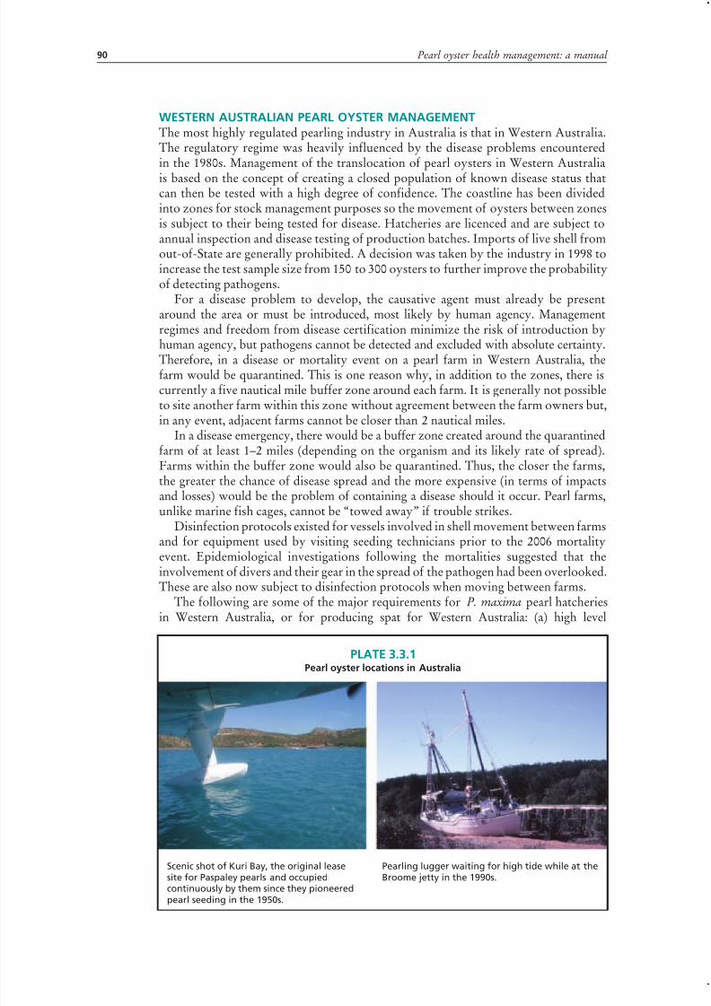
7/27/2019 Pearl Oyster Health Management
http://slidepdf.com/reader/full/pearl-oyster-health-management 104/136
Pearl oyster health management: a manual 90
WESTERN AUSTRALIAN PEARL OYSTER MANAGEMENTThe most highly regulated pearling industry in Australia is that in Western Australia.The regulatory regime was heavily influenced by the disease problems encounteredin the 1980s. Management of the translocation of pearl oysters in Western Australia
is based on the concept of creating a closed population of known disease status thatcan then be tested with a high degree of confidence. The coastline has been dividedinto zones for stock management purposes so the movement of oysters between zonesis subject to their being tested for disease. Hatcheries are licenced and are subject toannual inspection and disease testing of production batches. Imports of live shell fromout-of-State are generally prohibited. A decision was taken by the industry in 1998 toincrease the test sample size from 150 to 300 oysters to further improve the probabilityof detecting pathogens.
For a disease problem to develop, the causative agent must already be presentaround the area or must be introduced, most likely by human agency. Managementregimes and freedom from disease certification minimize the risk of introduction byhuman agency, but pathogens cannot be detected and excluded with absolute certainty.Therefore, in a disease or mortality event on a pearl farm in Western Australia, thefarm would be quarantined. This is one reason why, in addition to the zones, there iscurrently a five nautical mile buffer zone around each farm. It is generally not possibleto site another farm within this zone without agreement between the farm owners but,in any event, adjacent farms cannot be closer than 2 nautical miles.
In a disease emergency, there would be a buffer zone created around the quarantinedfarm of at least 1–2 miles (depending on the organism and its likely rate of spread).Farms within the buffer zone would also be quarantined. Thus, the closer the farms,the greater the chance of disease spread and the more expensive (in terms of impactsand losses) would be the problem of containing a disease should it occur. Pearl farms,unlike marine fish cages, cannot be “towed away” if trouble strikes.
Disinfection protocols existed for vessels involved in shell movement between farmsand for equipment used by visiting seeding technicians prior to the 2006 mortalityevent. Epidemiological investigations following the mortalities suggested that theinvolvement of divers and their gear in the spread of the pathogen had been overlooked.These are also now subject to disinfection protocols when moving between farms.
The following are some of the major requirements for P. maxima pearl hatcheriesin Western Australia, or for producing spat for Western Australia: (a) high level
PLATE 3.3.1Pearl oyster locations in Australia
Scenic shot of Kuri Bay, the original lease
site for Paspaley pearls and occupiedcontinuously by them since they pioneeredpearl seeding in the 1950s.
Pearling lugger waiting for high tide while at the
Broome jetty in the 1990s.

7/27/2019 Pearl Oyster Health Management
http://slidepdf.com/reader/full/pearl-oyster-health-management 105/136
91
of filtration of incoming water to remove otherlarvae and most bacteria; (b) use of axenic algalcultures where possible to reduce the build-up of bacteria in the larval tanks; (c) testing of all outgoing
spat batches and (d) filtration of all effluent waterfrom the hatchery to prevent disease spread from ahatchery to the environment.
Filtration of hatchery inflow water andbacterial control through hygiene is not uniqueto the pearling industry. Anyone who has seenthe results of a barnacle settlement or a sabellidtubeworm settlement in the pipes will appreciatethe economic value of filtration. The problem,however, is not confined to bacteria free in the watercolumn. Bacteria on biofilms can produce exotoxins,including proteinases and ciliostatic toxins that arepathogenic to larvae (Tubiash, 1975; Nottage andBirkbeck, 1987; Nottage, Sinclair and Birkbeck,1989; Riquelme et al., 1996).
The control of bacterial growth in molluschatcheries is often associated with the routine useof antibiotics (Jeffries, 1982; Prieur, 1990; Mooreet al . , 1993; Riquelme et al., 1996). This practice isdiscouraged in Western Australia, in part because nodrugs are registered for such use and also becauseof the disease resistance problems which can occur(Kerry et al ., 1994; Riquelme et al., 1996; pers. obs.).
Sorgeloos (1995), in discussing hatcheriesgenerally, commented that the reduction of bacterialloads, particularly in the feed and culture water,should be considered to be important and that stricthygiene measures should be taken including regulardisinfection and dry-out of the complete culturecircuit (including piping) between production cycles.This is common practice in pearl hatcheries.
The principle behind the requirement that effluentbe filtered is recognition that hatcheries can bea source of infection for the surrounding wildfishery. In particular, it would be possible to breed a
virulent strain of pathogen, possibly drug resistant,which would be released in large numbers into theenvironment (McVicar, 1997). Studies in BritishColumbia (Canada) have associated an increasedrisk of Aeromonas salmonicida in sea cages whichare within 10 km of infected cages and, even whererelease of pathogens can not be clearly demonstrated,public perception of a “disease risk” from a hatcherymay be enough to force closure.
CONCLUSION
A number of disease issues have arisen in theindustry during the last 5 years, including vibriosis,Haplosporidium sp., a rickettsia in the digestive
PLATE 3.3.2Diseases of pearl oysters in Australia
Small intranuclear virus-inclusion in digestivegland of Pinctada maxima (H & E, x1000).
Haplosporidian spores in digestive gland ofPinctada maxima (H & E, x1000).
Larval cestodes associated with discrete focalgranuloma in interstitial tissues of Pinctadamaxima (H & E, x 200).
3.3 The Australian experience: pearl oyster mortalities and disease problems
7/27/2019 Pearl Oyster Health Management
http://slidepdf.com/reader/full/pearl-oyster-health-management 106/136
Pearl oyster health management: a manual 92
gland and an intracellular ciliate. These incidents have demonstrated the value of themanagement regimes in place and the close links that have developed between theindustry and the government fish health laboratories. These links proved to be of greatvalue in generating early reporting of mortalities and in cooperation between companies
and government to contain and mitigate the impact of the mortality event in 2006.
ACKNOWLEDGEMENTSI wish to acknowledge the advice of Dr John Humphrey (Northern Territory) andDr Tina Hawksford (Queensland) in compiling this paper.
REFERENCESBruce, A.J. 1989. Notes on some Indo-Pacific Pontoniinae. 45 Conchodytes maculatus
sp. nov., a new bivalve associate from the Australian northwest shelf. Crustaceana 56:182-192.
Chace, F.A. & Bruce, A.J. 1993. The Caridean shrimps (Crustacea: Decapoda) of theAlbatross Philippine Expedition, 1907-1910, Part 6: Superfamily Palaemonoidea.Smithsonian Contributions to Zoology 39: 337-339.
Dix, T.G. 1973. Mantle changes in the pearl oyster Pinctada maxima induced by the peacrab Pinnotheres villosus. Veliger 15: 330-331.
Dybdahl, R. & Pass, D.A.. 1985. An investigation of mortality of the pearl oyster Pinctadamaxima, in Western Australia. Western Australia Department of Fishery. Report. 71: 1-78.
Hine, P.M. & Thorne, T. 1998. Haplosporidium sp. (Haplosporidia) in hatchery reared pearloysters Pinctada maxima (Jameson, 1901) in Western Australia. Journal of InvertebratePathology 71: 48-52.
Humphrey, J.D., Norton, J.H., Jones, J.B., Barton, M.A., Connell, M.T., Shelley, C.C.& Creeper, J.H. 1998. Pearl oyster (Pinctada maxima) aquaculture: health survey of Northern Territory, Western Australia and Queensland pearl oyster beds and farms.
Fisheries Research and Development Corporation Final Report 94/079: 108 pp. Jeffries, V. 1982. Three Vibrio strains pathogenic to larvae of Crassostrea gigas and Ostreaedulis. Aquaculture 29: 201-226.
Jones, J. B. & Creeper, J. 2006. Diseases of pearl oysters and other molluscs: A WesternAustralian perspective. Journal of Shellfish Research 25: 233-238.
Kerry, J., Hidney, M., Coyne, R., Cazabon, D., NicGbhainn, S. & Smith, P. 1994.Frequency and distribution of resistance to oxytetracycline in microorganismsisolated from marine fish farm sediments following therapeutic use of oxytetracycline.
Aquaculture 123: 43-54.Lobegeiger, R. 2001. Report to farmers: Queensland Aquaculture Production Survey 1999-
2000. Department of Primary Industries, Queensland, 24 pp.Malone, F.J., Hancock, D.A. & Jeffries, B. 1988. Final report of the Pearling Industry
Review Committee. Western Australian Fisheries Management Paper 17: 1-216.McVicar, A.H. 1997. Interaction of pathogens in aquaculture with wild fish populations.
Bull. Eur. Assn. Fish Pathol . 17: 197-200.Moore, W., Anderson, G. & Manzi, J.J. 1993. Effects of the antibiotic oxytetracycline
hydrochloride on Vibrio spp bacteria in the larval and early post-test culture of the hardclam Mercenaria mercenaria. World Aquaculture 24: 75-78.
Norton, J.H., Shepherd, M.A., Perkins F.O. & Prior, H.C. 1993. Perkinsus-like infectionin farmed golden-lipped Pearl oyster, Pinctada maxima, from the Torres Strait, Australia.
Journal of Invertebrate Pathology 62 :105-106.Norton, J.H., Shepherd, M.A. & Prior, H.C. 1993. Papovavirus-like infection of the
golden-lipped Pearl oyster, Pinctada maxima, from the Torres Strait, Australia. Journal of Invertebrate Pathology
62 :198-200.Nottage, A.S. & Birkbeck, T.H. 1987. The role of toxins in Vibrio infections of bivalvemolluscs. Aquaculture 67: 244-246.
7/27/2019 Pearl Oyster Health Management
http://slidepdf.com/reader/full/pearl-oyster-health-management 107/136
93
Nottage, A.S., Sinclair, P.D. & Birkbeck, T.H. 1989. Role of low molecular weightciliostatic toxins in vibriosis of bivalve molluscs. Journal of Aquatic Animal Health 1:180-186.
O’Sullivan, D. 1995. Pearls of Australia: an overview of pearl production techniques in
Australia. Australian Gemmologist 19: 155-161.Pass, D.A., Dybdahl, R. & Mannion, M.M.. 1987. Investigations into the causes of mortality
of the Pearl oyster, Pinctada maxima (Jamson) in Western Australia. Aquaculture 65:149-169.
Pass, D.A., Perkins, F. O. & Dybdahl, R. 1988. Virus-like particles in the digestive gland of the pearl oyster (Pinctada maxima). Journal of Invertebrate Pathology 51:166-167.
Perkins, F.O. 1996. Shell disease in the gold lip pearl oyster, Pinctada maxima and theeastern oyster, Crassostrea virginica. Aquatic Living Resources 9 :159-168.
Prieur, D., Mervel, G., Nicolas, J.L., Plusquellec, A. & Vigneulle, M. 1990. Interactionsbetween larval molluscs and bacteria in the marine environment . Oceanographic and Marine Biological Annual Review 28: 277-352.
Riquelme, C., Toranzo, A.E., Barja J.L., Vergara, N. & Araya, R. 1996. Associationof Aeromonas hydrophila and Vibrio alginolyticus with larval mortalities of scallop( Argopecten purpuratus). Journal of Invertebrate Pathology 67: 213-218.
Sorgeloos, P. 1995. Bioengineering of hatcheries for marine fish and shellfish. Journal of Marine Biotechnology 3: 42-45.
Tubiash, H.S. 1975. Bacterial pathogens associated with cultured bivalve mollusc larvae,pp. 61-71. In Smith W.L. & Chanley M.H. (eds). Culture of marine invertebrate animals.New York, Plenum Press. 388 pp.
3.3 The Australian experience: pearl oyster mortalities and disease problems

7/27/2019 Pearl Oyster Health Management
http://slidepdf.com/reader/full/pearl-oyster-health-management 108/136

7/27/2019 Pearl Oyster Health Management
http://slidepdf.com/reader/full/pearl-oyster-health-management 109/136
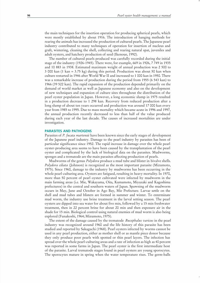
7/27/2019 Pearl Oyster Health Management
http://slidepdf.com/reader/full/pearl-oyster-health-management 110/136
Pearl oyster health management: a manual 96
the main techniques for the insertion operation for producing spherical pearls, whichwere mostly established by about 1916. The introduction of hanging methods forrearing the animals has increased the production of cultured pearls. The Japanese pearlindustry contributed to many techniques of operation for insertion of nucleus and
graft, wintering, cleaning the shell, collecting and rearing natural spat, juveniles andadult oysters, and hatchery production of seed (Ikenoue, 1992).
The number of cultured pearls produced was carefully recorded during the initialstage of the industry (1926-1945). There were, for example, 669 in 1926, 7 749 in 1935and 10 883 in 1938. Estimated maximum weight of annual production was 2 500 to3 000 kan (1 kan = 3.75 kg) during this period. Production was about 50 kan whenculture restarted in 1946 after World War II and increased to 1 000 kan in 1950. Therewas a remarkable increase of production during the period from 1955 (6 543 kan) to1966 (39 522 kan). The rapid expansion of the production depended primarily on thedemand of world market as well as Japanese economy and also on the developmentof new techniques and expansion of culture sites throughout the distribution of thepearl oyster population in Japan. However, a long economic slump in 1975 resultedin a production decrease to 1 298 kan. Recovery from reduced production after along slump of about ten years occurred and production was around 17 000 kan everyyear from 1985 to 1995. Due to mass mortality which became acute in 1996 and 1997,the annual production recently decreased to less than half of the value producedduring each year of the last decade. The causes of increased mortalities are underinvestigation.
PARASITES AND PATHOGENSParasites of P. fucata martensii have been known since the early stages of developmentof the Japanese pearl industry. Damage to the pearl industry by parasites has been of particular significance since 1952. The rapid increase in damage over the whole pearl
oyster-producing area seems to have been caused by the transplantation of the pearloyster and complicated by the lack of biological data on the parasites. Mudworms,sponges and a trematode are the main parasites affecting production of pearls.
Mudworms of the genus Polydora produce a mud tube and blister in bivalve shells.Polydora ciliata (Johnson) is recognized as the most important parasite (Mizumoto,1975). Since 1960, damage to the industry by mudworms has been occurring in thewhole pearl-culturing area. Oysters are fatigued, resulting in heavy mortality. In 1970,more than 50 percent of pearl oyster cultivated were infected by mudworm in themain farming areas (i.e. Mie, Wakayama, Oita, Kumamoto, Miyazaki and Kagoshimaprefectures) in the central and southern waters of Japan. Spawning of the mudwormoccurs in May, June and October in Ago Bay, Mie Prefecture. Larvae settle on theshell and mud tubes and blisters are formed in summer and winter. To exterminate
mud worm, the industry use brine treatment in the larval setting season. The pearloysters are dipped into sea water for about five min, followed by a 15 min freshwatertreatment, then in 22 percent brine for about 20 min and then exposure air in theshade for 15 min. Biological control using natural enemies of mud worm is also beingexplored (Funakoshi, 1964; Mizumoto, 1975).
The extent of the damage caused by the trematode Bucephalus varicus in the pearlindustry was recognized around 1960 and the life history of this parasite has beenstudied and reported by Sakaguchi (1968). Pearl oysters infected by worms cannot beused in any pearl production, either as mother shell or as mantle piece donor becausethey only produce poor pearls with spotted or thin pearl layers. The infection hasspread over the whole pearl-culturing areas and a rate of infection as high as 40 percent
was reported in some farms in Japan. The pearl oyster is the first intermediate hostof the parasite. Larval trematode stages found in pearl oysters are young sporocytes.The sporocytes mature in spring when the water temperature rises. The germ-balls

7/27/2019 Pearl Oyster Health Management
http://slidepdf.com/reader/full/pearl-oyster-health-management 111/136
97
in mature sporocytes develop into cercariae, which are motile and are released fromthe oyster tissues through summer and autumn. The second intermediate hosts aresmall fish, such as Atherina bleekeri, Spratelloides japonicus and Engraulis japonica,which are abundant in the waters near the pearl farms. The B. varicus cercariae invade
the muscular tissue of the fish and encyst as metacercariae. Fish species, such asCaranx sexfasciatus, C. equula and C. ignobilis are the final hosts, which ingest fishinfected with the metacercariae. Once ingested, the metacercariae emerge from theircysts and mature into adults in the digestive system of the fish. Another trematodespecies Proctoeces ostrea has also been found in the internal organs of pearl oystersand is common throughout the pearl farms, but damage to the industry is not severe(Sakaguchi, Kamakura and Kusuda, 1970). A proposed practical method to controlthese parasites is to avoid culture of pearl oysters in September when these trematodesundertake their peak period of transmission. It is impossible to remove the host fishfrom the coastal areas (Sakaguchi, 1968; Mizumoto, 1975).
There are fewer reports of microbial pathogens (bacteria and virus) causingmortalities of Japanese pearl oysters than those in the tropical species. Kotake andMiyawaki (1954, 1955) isolated two strains of unidentified bacteria from Japanesepearl oysters during an episode of mortality. Miyazaki et al . (1999) reported isolatingan unidentified virus as a cause of the mass mortality that has emerged recently. Theseresults were not confirmed, either as to the identity of the pathogen or its pathogenicityto pearl oysters (Nakajima, 1999; Hirano, Kanai and Yoshikoshi, 2002). Suzuki,Kamakura and Kusuda (1998), Suzuki (1999) and Kitamura, Jung and Suzuki (2000)also isolated a birnavirus from Japanese pearl oyster.
FOULING ORGANISMSMany species of invertebrates and seaweeds inhibit growth and injure pearl oysters byattaching to the shell, particularly at the hinge, often interfering with the opening of
the shell, which may cause mortality. Fouling organisms include barnacles, sponges,worms, edible oysters, and seaweeds. The dominant species of barnacles in the Japanesepearl farms are Balanaus variegatus tesselatus and B. amphitrite. The polychaete wormsHydroides norvegica and Dexiospira forminosus, as well as the bryozoan, Dakariasubovoidea, are also fouling organisms in Japan. Periodic physical cleaning of theshell or dipping the oysters in fresh water are the practical defense measures takenagainst these organisms. Shell cleaning is carried out during the period from April toNovember. Freshwater treatment is effective particularly for removing larvae or youngstages of fouling animals on the shell.
PREDATORSThe predators of cultured pearl oysters include the eel, Anguilla japonica, the black
porgy, Sparus sp. and globe fish, Sphoreoides sp., as well as the octopus, Octopus sp.They especially attack the pearl oysters just after the nucleus operation.
RED TIDEOne of the important constraints has been the effect of red tide on marine culturedorganisms. Gymnodinium mikimotoi is a well-known dinoflagellate red tide whichwas named after the late Kokichi Mikimoto, one of the inventors of pearl culture inMie Prefecture, where they found this species. Since then many records have describedthe damage to the pearl oyster by this dinoflagellate in Japan. The other red tidespecies reported are Heterocapsa circularsquama, Gonyalaux sp. and Cocurodium sp.Abnormal blooms of these toxic dinoflagellates have been suspected as the cause of
mass mortalities in pearl farms in localized areas. Most of the Japanese pearl farmsare located in semi-closed coastal estuaries or small bays, sites that favour abnormalblooming of red tides in Japan.
3.4 The Japanese experience: pearl oyster mortalities and constraints

7/27/2019 Pearl Oyster Health Management
http://slidepdf.com/reader/full/pearl-oyster-health-management 112/136
Pearl oyster health management: a manual 98
A large-scale bloom of Heterocapsa circularsquama occurred in Ago Bay in 1992causing a new type of red tide mass mortality that had not seen before with red tidesof Gymnodinium mikimotoi or other species (Matsuyama et al ., 1995). They reportedthat in the summer of 1992 in Ago Bay the maximum cell density of Heterocapsa sp.
was 87 420 cells/ml and it caused mass mortality of pearl oysters, although mortalitiesof cultured and feral finfish were not observed. That was the first report of this speciesof red tide causing mass mortality of pearl oysters in Japan. Many studies have beenreported in Japan on the factors that stimulate phytoplankton blooms, physiologicaldamage and mortality of pearl oyster, prediction of blooms on the basis of the fieldobservation and experimental exposure of animals to red tide dinoflagellates (e.g.Honjo, 1994; Nagai et al ., 1996; Iwata et al ., 1997). Nagai et al . (1996) used laboratoryexperiments to determine the mechanism by which H. circularsquama caused mortalityof juvenile pearl oyster. The lethal dose 50 (LD50) was approximately 20 000 cells/mlafter 24 hr of exposure to H. circularsquama. They suspected that the cause of deathwas the direct action of cells. Immediately after exposure to algal cells, animals rapidlycontracted their mantles, closed their shell valves, then contracted gills and heart beatbecame irregular until it stopped. Transporting the pearl nets that hold pearl oystersfrom the bloom area to a non-blooming site is a usual procedure to avoid mortalitiesin the case of red tides in Japan.
MASS MORTALITYIn pearl farms located at the end of semi-closed estuaries or bays, mass mortalitiesoccurred in the summer season in Mie Prefecture between the 1950s and 1970s. Basedon the analysis of seawater data, high temperature was considered to be the primarycause of the mortalities, which ranged from 4 to 70 percent. At the time of summermortalities, water temperatures rose over 29 °C, the oxygen content was low andhydrogen sulphate concentration was high, particularly near the bottom, all of which
are suspected to have contributed to the mass mortalities. Physiological condition of the pearl oysters is variable and is dependent on the phytoplankton availability in pearlfarms before and during periods of high water temperature. Mortalities are stronglyinfluenced by physiological condition. These experiences motivated the farmers andscientists to recognise the importance of monitoring the condition of seawater in pearlfarms. Co-occurrence of high water temperatures and low levels of planktonic food aresometimes the main cause of mass mortalities. Food availability and temperature areimportant environmental factors for physiological conditioning of bivalves in culture.Elucidation of the influences of food deprivation on the mortality of pearl oysters wasstudied in laboratory rearing experiments (Numaguchi, 1995a, b). Two-year-old pearloysters were held in tanks of seawater filtered through a series of filters (10, 5, 1 µm)for 115 days during June to September at the natural temperature of 23 °C to 29 °C.
The mortality of unfed oysters increased remarkably when the condition index (drymeat weight/dry shell weight) dropped below 4 (initial value 13.7), dry meat lossesincreased more than 70 percent and the weight of the crystalline style decreased below10 mg (initial weight: 30 mg). A level of 28 °C is a critical temperature indicator forsome physiological effects in pearl oysters based on experiments on filtering rate, foodintake and oxygen consumption (Uemoto, 1968; Numaguchi, 1995a, b). Mass moralityobserved in western Japan from summer to autumn may, therefore, be caused by thesefactors in association with the effect of oceanic seawater which is warmer and includesless food plankton than coastal seawater.
Since 1994, mass mortalities have been occurring which are causing significanteconomic losses to the pearl culture industry in western Japan (Sorimachi, 2000). This
disease occurs from summer to autumn and the affected oysters exhibit a yellowish-red coloration of the adductor muscle. Histopathological changes commonly appearin the loose connective tissue of the mantle and adductor muscle in the affected pearl

7/27/2019 Pearl Oyster Health Management
http://slidepdf.com/reader/full/pearl-oyster-health-management 113/136
99
oyster. Various factors such as toxic dinoflagellate blooms, infectious diseases causedby a filterable virus-like agent and environmental factors were suspected as causes of the mortality. To clarify the cause of this epizootic, experimental transplantation of the mantle piece of the affected oyster into healthy oysters, cohabitation of affected
and healthy oysters and inoculation of filtrate of affected oyster hemolymph intohealthy oysters were performed. After 2 to 3 months, healthy oysters displayedthe signs similar to those of spontaneously affected oysters and mortality occurred.These results indicated that the mass mortality of the cultured pearl oysters is causedby an infectious filterable agent. However, the agent of the disease has not beenconclusively identified. Mortality decreased when water temperature declined inOctober-November indicating the disease is highly dependent on water temperature.Low water temperatures in the previous winter appeared to suppress the occurrenceof the disease during the following growing season (Kurokawa et al ., 1999; Miyazakiet al ., 1999; Muroga, Inui and Matsumoto, 1999; Morizane, 1999; Takami, 1999;Yoshikoshi, 1999; Nakajima, 1999; Sorimachi, 2000; Maeno et al ., 2001; Morizane etal ., 2001; Uchimura et al ., 2001; Tomaru, Kawabata and Nakano, 2001). Managementpractices have been recommended for the pearl farmers to reduce mortalities. In someregions, prohibition on transplanting live pearl oyster shells from the area where theinfection was suspected has been proposed. Imports of live oysters from out of Japanare generally prohibited.
Funakoshi (1999) discussed the history and current problems caused by massmortality to pearl culture in Japan. He commented that after the 1970s many pearlfarmers have tended to produce fast-growing and large-size mother-of-pearl (MOP)shell for oyster production. These appear more resistant to mass mortalities and canproduce larger pearls. This has resulted in most MOP shells being produced fromnatural or artificial (hatchery) setting spat at the southwestern region of Japan. Thenew infectious disease emerged in part of these regions around 1995 and then spread
over to pearl farms in other regions after 1997. It was suspected that the unknownpathogen was introduced by the transplantation of seeds or MOP. Also there have beenintroductions of live shells from out of country for genetic selection or crossing, andthis is suspected, without evidence, as the cause of invasion of the pathogen.
CONCLUSIONWhen live shells are introduced from other prefectures or areas, or when pearl oysterseed are introduced, it would be desirable to carefully evaluate their susceptibilityor tolerance to disease. These concepts are fundamental to the prevention of massmortality caused by infectious pathogens and must be understood by farmers,scientists and policy makers. Routine monitoring of the physiological condition of cultured animals and oceanographic aspects of water quality by farmers, cooperatives
or local governments at every culture site is also necessary.
ACKNOWLEDGEMENTSI wish to acknowledge Drs Kiyoshi Inoue and Minoru Sorimachii who gave me achance to compile this paper. Thanks are extended to Drs Tetsuji Masaoka and ToruSuzuki for their reviewing the draft and to Dr Brian Jones, Western Australian MarineResearch Laboratory for his English correction.
REFERENCESFunakoshi, S. 1964. Studies on the methods using saturated salt solution to kill PolydoraI.
On killing an inhabiting mother oyster shells. Bulletin of Natural Pearl ResearchLaboratory
9:1156-1160 (in Japanese).Funakoshi, S. 1999. History and current problems in pearl culture in Japan. Fish Pathology 34: 219-231 (abstract in Japanese).
3.4 The Japanese experience: pearl oyster mortalities and constraints

7/27/2019 Pearl Oyster Health Management
http://slidepdf.com/reader/full/pearl-oyster-health-management 114/136
Pearl oyster health management: a manual 100
Gervis, M.H & Sims, A. 1992. The biology of pearl oysters (Bivalvia: Pteriidae). ICLARMStudies and Review 21. London, UK, Overseas Development Administration. 49 pp.
Hirano, M., Kanai, K. & Yoshikoshi, K. 2002. Contact infection trials failed to reproducethe disease condition of mass mortality in cultured pearl oyster Pinctada fucata. Fishery
Science 68: 700-702.Honjo, T. 1994. The biology and prediction of representative red tides associated with fish
kills in Japan. Review of Fishery Science 2: 225-253.Ikenoue, H. 1992. Pearl oyster (Pinctada fucata), pp.195-205. In Ikenoue, H. and T.
Kafuku (eds). Modern methods of aquaculture in Japan (2nd ed). Amsterdam, ElsevierScience Publishers V.B. 272 pp.
Iwata, Y., Sugawara, I. Kimura, T., Silapajan, K., Sano, M., Mizuguchi, T., Nishimura,A., Inoue, M. & Takeuchi, T. 1997. Growth potential of Gymnodinium mikimotoi inGokasho Bay. Nippon Suisan Gakkaishi 63: 578-584.
Kitamura, S., Jung, S. & Suzuki, S. 2000. Seasonal changes of infective state of marinebirnavirus in Japanese pearl oyster Pinctada fucata. Arch. Virol . 145: 2003-2014.
Kotake, N. & Miyawaki, Y. 1954. Experimental studies on the propagation of the pearlOyster. II. On the unusual high mortality of pearl oyster caused by bacteria . Bulletin of the Japanese Society of Fisheries 9: 952-956.(in Japanese with English abstract).
Kotake, N. & Miyawaki, Y. 1955. Experimental studies on the propagation of the pearlOyster. III. On the unusual high mortality of pearl oyster caused by bacteria (2).Bulletin of the Japanese Society of Fisheries 20: 979- 981 (in Japanese with Englishabstract).
Kurokawa, T., Suzuki, T., Okauchi, M., Miwa, S., Nagai, K., Nakamura, K., Honjo, T.,Nakajima, K., Ashida, K. & Hunakoshi, S. 1999. Experimental infection of a diseasecausing mass mortalities of Japanese pearl oyster Pinctada fucata martensii by tissuetransplantation and cohabitation. Nippon Suisan Gakkaishi 65: 241-251.
Maeno, Y., Ito, T., Kamaishi, T., Morizane, T. & Nakajima, K. 2001. Morphological
changes of hemocytoes in hemolymph smear preparations of diseased Japanese pearloyster Pinctada fucata martensii with mass mortality. Fish Pathology 36: 225-230 (in Japanese with English abstract).
Matsuyama, Y., Nagai, K., Mizuguchi, T., Fujiwara, M., Ishimura, M., Yamaguchi, M.,Uchida, T. & Honjo, T. 1995. Ecological features and mass mortality of pearl oystersduring red tide of Heterocapsa sp. in Ago Bay in 1992. Nippon Suisan Gakkaishi 61: 35-41 (in Japanese with English summary).
Miyazaki, T., Goto, K., Kobayashi, T., Kageyama, T. & Miyata, M. 1999. Mass mortalitiesassociated with a virus disease in Japanese pearl oysters Pinctada fucata martensii.Diseases of Aquatic Organisms 37:1-12.
Miyazaki, T. 1999. Histopathology of Japanese pearl oysters with an Akoya-virus infection. Fish Pathology 34: 229 (in Japanese with English abstract).
Mizumoto, S. 1964. Studies on disease of the shells of the pearl oyster (Pinctada martensii).I. On the species of parasitic polychaetes in shells, the condition of the damages and theextirpation of disease caused by Polydora ciliata. Studies ondisease of the shells of thepearl oyster (Pinctada martensii). Bulletin of Natural Pearl Research Laboratory 9: 1143-1155 (in Japanese).
Mizumoto, S. 1968. Studies on disease of the shells of the pearl oyster (Pinctada martensii).II. On the seasonal variation in occurrence of Polydora ciliata in shells of pearl oyster.Bulletin of Natural Pearl Research Laboratory 11: 1368-1377 (in Japanese).
Mizumoto, S. 1968. Studies on disease of the shells of the pearl oyster (Pinctada martensii).III. On the seasonal process of the disease caused by Polydora ciliata. Bulletin of Natural Pearl Research Laboratory 13: 1624-1634 (in Japanese).
Mizumoto, S. 1975. Parasites affecting the pearl industry in Japan. Proc. 3rd US- JapanMeeting Aquaculture, Tokyo, Oct 15-16, 1974. Special Publication of Fishery Agency of Japanese Government.
7/27/2019 Pearl Oyster Health Management
http://slidepdf.com/reader/full/pearl-oyster-health-management 115/136
101
Morizane. Y. 1999. The mass mortality of pearl oysters in Uwa Sea, Ehime. Fish Pathology34: 223-2224 (in Japanese with English abstract).
Morizane, Y., Takimoto, S., Nishikawa, S., Matsuyama, N., Thono, K., Uemura, S.,Fujita, Y., Yamashita, H., Kawakami, H., Koizumi, Y., Uchimura, Y. & Ichikawa, M.
2001. Mass mortalities of Japanese pearl oyster in Uwa-Sea, Ehime in 1997-1999. FishPatholology 34: 207-216 (in Japanese with English abstract).
Muroga, K., Inui, Y. & Matsusato, Y.1999. Workshop “Emerging disease of cultured marinemolluscs in Japan. Fish Patholology 34: 219-231 (in Japanese with English abstract).
Nagai, K., Matsuyama, Y., Uchida, T., Yamaguchi, M., Ishimura, M., Nishimura, A.,Akamatsu, S. & Honjo, T. 1996. Toxicity and LD50 levels of the red tide dinoflagellateHeterocapsa circularisquama on juvenile pearl oysters. Aquaculture 144:149-154.
Nagai, K., Matsuyama, Y., Uchida, T., Akamatsu, S. & Honjo, T. 2000. Effect of a naturalpopulation of the harmful dinoflagellate Heterocapsa circularisquama onsurvival of thepearl oyster Pinctada fucata. Fishery Science 66: 995-997.
Nakajima, K. 1999. Mass mortality outbreaks associated with an infectious pathogenamong pearl oyster Pinctada fucata martensii in Japan. Fish Pathology. 4:227 (Englishabstract).
Numaguchi, K. 1995a. Effects of water temperature on catabolic losses of meat andcondition index of unfed pearl oyster Pinctada fucata martensii. Fishery Science 61: 735-738.
Numaguchi, K. 1995b. Influence of unfed condition on the mortality of pearl oysterPinctada fucata martensii. Fishery Science 61: 739-742.
Ogushi, J. 1938. Studies on pearls. Ito Bunshodo, Tokyo, Japan (in Japanese). 218 pp.Sakaguchi, S. 1968. Studies on the life-history of the trematode parasitic in pearl oyster,
Pinctada martensii, and on the hindrance for pearl culture. Bulletin of Natural Pearl Research Laboratory 13:1635 - 1688 (in Japanese with English summary).
Sakaguchi, S., Hoshina, T. & Minami, T.1970. Studies on a trematode genus Proctoces in
pearl oyster, Pinctada martensii - On the morphology, parasitic status and the distributionof pearl oyster infected with the parasite. Bulletin of Natural Pearl Research Laboratory15: 1931 -1938 (in Japanese with English summary).
Sorimachi, M. 2000. Mass mortalities of Japanese pearl oyster Pinctada fucata martensii. Aquabiology 126: 39-44 (in Japanese with English abstract).
Suzuki, S., Kamakura, M. & Kusuda, R. 1998. Isolation of birnavirus from Japanese pearloyster Pincatada fucata. Fishery Science 64: 342-343.
Suzuki, S. 1999. Infective state and seasonal occurrence of marine birnavirus in Japanesepearloyster. Fish Pathology 34: 228 (in Japanese with English abstract).
Takami, I. 1999. Mass mortality of pearl oyster at Tsushima in Nagasaki Prefecture. FishPathol. 34: 224- 225 (in Japanese with English abstract).
Tomaru, Y., Kawabata, Z. & Nakano, S. 2001. Mass mortality of the Japanese pearl
oyster Pinctada fucata martensii in relation to water temperature, chrolophyll a andphytoplankton composition. Diseases of Aquatic Organism 44: 61-68.
Uchimura, Y., Morizumi, T., Fujita, Y., Hyoudou, K. & Hirata, T. 2001. Response of haemocytes to infection by red coloring of soft tissue disease in Japanese pearl oysterPinctada fucata martensii. Bulletin of Ehime Prefecture Fishery Experimental Station 9:5-12. (in Japanese with English abstract).
Uemoto. H. 1968. Relationship between oxygen consumption by the pearl oyster and itsenvironmental temperature. Bulletin of Natural Pearl Research Laboratory 13:1617-1623(in Japanese).
Wada, K.T. 1991. The Japanese pearl oyster, Pinctada fucata (Gould) (Family Pteridae),pp.245-260. In Estuary and Marine Bivalve Mollusks Culture. Menzel, W. (ed). Boca
Raton, FL. U.S.A , CRC Press,.Yoshikoshi, K. 1999. Epidemiologic studies on the mass mortalities of cultured Japanesepearl oyster, Pinctada fucata martensii. Fish Pathology 34:226 –22.
3.4 The Japanese experience: pearl oyster mortalities and constraints

7/27/2019 Pearl Oyster Health Management
http://slidepdf.com/reader/full/pearl-oyster-health-management 116/136
7/27/2019 Pearl Oyster Health Management
http://slidepdf.com/reader/full/pearl-oyster-health-management 117/136

7/27/2019 Pearl Oyster Health Management
http://slidepdf.com/reader/full/pearl-oyster-health-management 118/136
Pearl oyster health management: a manual 104
for about 7 000 people. While pearl farming in French Polynesia meets an undeniablesuccess, the industry has not evenly grown. In the mid-1980s, mass mortality of oystershas occurred in several atolls and challenged the development of pearl farming activity.The government of French Polynesia has introduced stringent quality control system
in order to maintain high quality of the pearls and sustain market demand. Here wereview this experience and lessons learnt from the past.
OUTBREAKS OF MASS MORTALITYIn 1985, the pearl oyster industry has faced series of mass mortality outbreaks in severalislands of the Tuamotu Archipelago. During these outbreaks, recorded mortality rateswere over 60 percent in some locations based on enumeration of tagged individuals inwild populations (Cabral, 1990). The mortality persisted up to 1986 and also affectedcultivated stocks (Intés, 1994). Mortality of pearl oysters was accompanied by shelldisorders, mantle lesions and necrosis of the adductor muscle (Grizel et al., 1986). Thesesigns led to the description of complex syndrome currently known as “syndrome 85”(Comps, Herbaut and Fougerouse, 2000). While no abnormal mortality was reported,these signs have been recurrently observed although at low prevalence in some islandsuntil 1996-1997.
Despite many investigations and attempts to give the 1985 outbreaks a cause, noinfectious aetiology was demonstrated. The presence of a gregarine in affected pearloysters was reported (Chagot et al., 1993). Gregarines, when present in low numbers,are usually regarded harmless to pearl oysters and the role of this parasite was notclearly assessed later (Fougerouse et al., 1994; Comps et al., 2001). It is accepted thata deleterious combination of biotic and a-biotic environmental factors, along withstress caused by culture handling, crowding and grafting, contributed to cause massmortality of pearl oysters (Grizel, 1986; Cabral, 1990; Dauphin and Cuif, 1990; Cuif and Dauphin, 1996; Dauphin and Denis, 1987).
Between 1982 and 1983, six hurricanes wiped out shallow bottom beds in certainlagoons. Takapoto lagoon revealed oligotrophic conditions characterized by nitrogenand phosphorus deficits and low concentrations in bacteria and phytoplankton.Although the pearl oysters contributed to the suspended particulate matter used bymicro-organisms, this appeared to have a negative balance against the rate of oystergrazing and indicated a limited carrying capacity for the lagoon (Vacelet, Arnoux andThomassin , 1996). Such conditions may have also exacerbated recovery of the pearloyster lagoons which were restocked, post-typhoon.
It is likely that such poor conditions associated with local high densities of oysterson culture rafts also may have exacerbated detrimental effect of pathogens. This paperreviews the pathogens reported from P. margaritifera in French Polynesia.
MAIN PATHOGENS RECORDED IN FRENCH POLYNESIAThere is very little known about diseases of pearl oysters in French Polynesia beforeany development of the industry. Initial studies focused on parasites having potentialto induce pearl formation (Seurat, 1906; Dubois, 1907) and it was only during the1985 mass mortality that light has been shed on pathogens of pearl oysters in FrenchPolynesia.
Shell disorder, or disease, is part of the “syndrome 85” (Grizel et al., 1986; Compset al., 2001). This disorder is characterised by brown organic matter deposits onthe inner shell associated with inflammation in the mantle displaying yellowishcoloration and swelling. Bio-mineralization disorders had previously been interpretedas consequences of mechanical trauma or chemical stress (Grizel, 1986; Dauphin
and Cuif, 1990; Fougerouse et al., 1994). Although the disorder has persistentlybeen recorded by farmers in numerous areas from 1996 to 1997, the prevalencewas low in comparison with the prevalence observed in 1985 (Comps et al., 2001).

7/27/2019 Pearl Oyster Health Management
http://slidepdf.com/reader/full/pearl-oyster-health-management 119/136
105
Microstructure and composition of the affected shells have shown malformations inthe nacreous layer associated with biochemical abnormalities occurring during thebiomineralisation process (Marin and Dauphin, 1991, 1992; Cuif and Dauphin, 1996).Electron-dense particles were observed in the granulomatous tissue contiguous to
similar organic deposits and interpreted as possible virus-like particles (Combs et al.,2001). This condition has also been compared with brown ring disease of clams, Tapes
philippinarum and T. decussatus, caused by Vibrio tapetis (Paillard, Maes and Oubella,1994; Borrego et al., 1996) and a contagious aetiology was suggested (Cabral, 1994 citedin Cuif and Dauphin, 1996), although evidence of a pathogen has yet to be found (Cuif and Dauphin, 1996). In the course of brown ring disease of clam, infection with V.tapetis provokes disorganization of the periostracal lamina and a brown periostracumdeposit in the inner surface of the shell (Paillard and Maes, 1990, 1995) very similarto those reported in P. margaritifera (Comps et al., 2001). According to the authors,this might also result from physiological changes caused by the environmental, culturehandling and grafting conditions considering that such organic deposits in the shellof marine bivalves are a frequent sign of reaction stimulated by wounds, parasites ordebris (Aldermann and Gareth-Jones, 1971; Perkins, 1996).
Abscess-like lesions were reported in the adductor muscle of weak oysters displayingalso abnormal mucus secretion. These lesions are characterised in histology by focalnecrosis of the muscular tissue and haemocytic infiltration (Comps et al., 2001). Thestudy shows that prevalence of these symptoms vary; with highest prevalence beingobserved in Raiatea, Manihi and Takapoto lagoons. Virus-like particles – the verynature and role of which is still unknown – were reported from similar granulomatoustissues (Comps, Herbaut and Fougerouse, 1999). Similar lesions of necrosis, atrophy,swelling and vacuolization of muscle fibres were associated with virus particles inP. fucata martensii during a mass mortality which occurred in western regions of
Japan in 1996 and 1997 (Miyazaki et al., 1999). The size of the particles is apparently
slightly different with virus-like particles in P. margaritifera of 40 nm while akoya-virus particles measure 33 nm (Comps et al., 2001). A papova-type viral infectionis also reported from the gold-lip pearl oyster P. maxima associated with nucleushypertrophy of the epithelial cells of the labial palps (Norton, Shepherd and Prior,1993).
Abscesses in the pearl bag are reported in the course of grafting and certain caseswere possibly associated with the presence of intracellular procaryotes (Comps etal., 2001). The secretory epithelium of the pearl sac may be highly damaged withaccumulation of haemocytes and cellular debris surrounded by strong inflammatoryreaction. Micro-organisms may be introduced in the pearl bag with mantle tissueintroduction during the course of grafting and become a cause of abscess formation.In response to mechanical wound or to the accidental introduction of a foreign body
during grafting, local abnormal secretion of periostracum by the epithelium of the pearlsac sometimes induces formation of whitish paraspherical bodies (Comps, Herbautand Fougerouse, 2000).
Micro-colonies of bacteria were found in the epithelial cells of the digestive tubules(Comps, Fougerouse and Buestel, 1998; Comps et al ., 2001). The bacteria exhibitcharacteristics of members of Rickettsiales. All of these were reported during routineexaminations and were not associated with significant disorder of pearl oysters, exceptin certain instances where fibrous layer surrounds some of these micro-colonies of bacteria.
The gregarine described by Chagot et al. (1993) is commonly reported from severallagoons (Gambier, Raiatea and Manihi) in the digestive tract of P. margaritifera with no
particular impact on the host (Fougerouse et al., 1994; Comps et al., 2001).Although scarcely observed, stages of a cestode, possibly related to the genusTylocephalum, were recently reported from Raiatea and Gambier islands (Combs et al.,
3.5 The French Polynesian experience

7/27/2019 Pearl Oyster Health Management
http://slidepdf.com/reader/full/pearl-oyster-health-management 120/136
Pearl oyster health management: a manual 106
2001). Such observations confirm previously reported occurrence of helminths fromthe same islands (Seurat, 1906).
Shell damages in the black-lip pearl of French Polynesia may be caused by variousboring organisms (Mao Che et al., 1996). These include cyanobacteria, green algae,
the marine phycomycete fungus Ostracoblabe implexa and clionid sponges, Clionamargaritifera and C. vastifica. Apparently, Ostracoblabe implexa would be moredamaging to the nacreous layers of the shell compared to clionid sponges limited toouter prismatic region of the shell (Cuif and Dauphin, 1996, Comps, Herbaut andFougerouse, 2000). However, clionid sponges have been shown to produce perforationthroughout the shell three layers and even induce haemocytic response in adjacentmuscular tissue of edible oysters (Groman and Berthe, unpublished data).
HEALTH MANAGEMENTThese different studies have shown a reasonably good health condition of P. margaritifera in French Polynesia despite persistent manifestation of the syndrome85. The most serious concern probably comes from described virus-like particles andintracellular prokaryotes. The mass mortality outbreaks encountered by the pearlindustry in Japan strongly contributed to reinforce this concern (Miyazaki et al.,1999).
Transfers of pathogens via movements of live molluscs are generally recognizedas a major cause of disease outbreaks and epizootics. In French Polynesia, hatcheryproduction is still in development and despite the economic importance of thisindustry, the pearl oyster culture is strongly dependent on natural spat collection.This collection occurs in atolls where natural stocks are abundant and where spatproduction is significant. Closed lagoons, such as Takapoto or Hikueru, weretraditionally exploited for this specific purpose. Collected spat are then spread tonon-collecting atolls where there is usually limited or no natural stock. The spatio-
temporal variability in production of wild spat as well as lack of regulation regardingtransfers of spat from collection to farming areas are underlying causes of numeroustransfers between islands (Cabral, Mizuno and Tauru , 1985; Prou, Benett and Tiapari,1999).
The exponential development of the pearl oyster industry has been accompaniedby an increasing number of farms and farmed atolls. A direct consequence of this hasbeen a rise in the frequency and volume of oyster transfers from collecting to non-collecting atolls. Animal exchanges increase mixing of oysters populations from thedifferent atolls among Polynesian archipelagos (Society Islands, Marquesas Islandsand Tuamotu–Gambier) as underlined by the significant genetic homogenization trendbetween populations previously distinct (Arnaud-Haond, 2003). The oyster transfersalso generate anemone dissemination and this is becoming an important ecological
problem in some islands with economic consequences as a result of the increasedfrequency needed for cleaning livestock and rearing structures (LeMoullac et al., 2003).The great demand for spat also had, as a consequence, high densities of reared pearloysters in the collecting atolls, such as, for example, Takaroa.
In order to prevent and control spreading of any emerging disease in the FrenchPolynesian pearl farming sector, surveys were undertaken and a basic surveillanceprogramme was proposed for implementation (Combs et al., 2001; Thébault, 1999).This program was initiated in 2003 as a network based on five islands of the FrenchPolynesian archipelagos. The programme is based on monitoring of pearl oyster farmsand producing areas by routine collection of samples and investigation of abnormalmortality outbreaks. The strong collaboration between actors of the scientific
community and delegates of pearl oyster administration in atolls (through meetings,coordination, training courses) enabled increased awareness of farmers for the benefitof the industry.
7/27/2019 Pearl Oyster Health Management
http://slidepdf.com/reader/full/pearl-oyster-health-management 121/136
107
CONCLUSIONThe pearl farming in French Polynesia has been extremely successful. Involvementof the government and cooperation of research and private sectors are two keycomponents of this model. French Polynesia has developed a strong proactive policy
in support of the pearl industry with efforts to develop and implement a nationalstrategy for pearl oyster health management. This policy is still under review withnew management measures being considered. Among those, quotas of pearl farms arediscussed to reduce the risk of diseases of pearl oysters and avoid problems of poorquality of the pearls.
REFERENCESAldermann, D.J. & Gareth-Jones, E.B. 1971. Shell disease of oyster. Fisheries Investigation
26:1-19.Anon. 2006. http://www.ifremer.fr/aquaculture/en/molluscs/pearl_oysters.htm (accessed
on October 2007).Anon. 2007. Service du commerce extérieur de la polynésie française. http://www.tahiti-
export.pf (accessed on October 2007).Arnaud-Haond, S., Vonau, V., Bonhomme, F., Boudry, P., Prou, J., Seaman, T., Veyret,
M. & Goyard, E. 2003. Spat collection of the pearl oyster (Pinctada margaritiferacumingii) in French Polynesia: an evaluation of the potential impact on genetic variabilityof wild and farmed populations after 20 years of commercial exploitation. Aquaculture 219(1-4), 181-192.
Borrego, J.J., Castro, D., Luque, A., Paillard, C., Maes, P., Garcia, M.T. & Ventosa, A.1996. Vibrio tapetis sp. nov., the causative agent of the brown ring disease affecting clams.International Journal of Systematic Bacteriology 46: 480–484.
Cabral, P. 1990. Some aspects of the abnormal mortalities of the pearl oysters, Pinctadamargaritifera L. in the Tuamotu Archipelago (French Polynesia). Advances in Tropical
Aquaculture, Aquacop Ifremer, Actes Coll., 9. Ifremer, Plouzané. pp. 217–226.Cabral, P., Mizuno, K. & Tauru, A. 1985. Preliminary data on the spat collection of mother of pearl (Pinctada margaritifera, Bivalve, Mollusc) in French Polynesia, pp.177-182. In B. Delesalle, R. Galzin and B. Salvat (eds.). Proc. Vth Int. Coral Reef Cong.French Polynesian Coral Reefs Vol. 1. Moorea, French Polynesia, Antenne Museum-EPHE.
Chagot, D., Fougerouse, A., Weppe, M., Marquès, A. & Bouix, B. 1993. Présence d’unegrégarine (Protozoa Sporozoa) parasite de l’huître perlière à lèvres noires Pinctadamargaritifera (L., 1758) (Mollusca Bivalvia) en Polynésie française. C.R. Acad. Sci. ParisIII 316, pp. 239–244.
Claereboudt, M.R., Bureau, D., Côté, J & Himmelman, J.H. 1994. Fouling developmentand its effect on the growth of juvenile giant scallops (Placopecten magellanicus) in
suspended culture. Aquaculture 121: 327-342.Comps, M., Fougerouse, A. & Buestel, D. 1998. A procaryote infecting the black-lipped
pearl oyster Pinctada margaritifera. Journal of Invertebrate Pathology 72(1): 87-89.Comps, M., Herbaut, C. & Fougerouse, A. 1999. Virus-like particles in pearl oyster
Pinctada margaritifera. Bulletin of the European Association of Fish Pathologists 19:85–88.
Comps, M., Herbaut, C. & Fougerouse, A. 2000. Abnormal periostracum secretion duringthe mineralization process of the pearl in the blacklip pearl oyster Pinctada margaritifera.
Aquatic Living Resources 13(1): 49-55.Comps, M., Herbaut, C., Fougerouse, A. & Laporte, F. 2001. Progress in pathological
characterization of Syndrome 85 in the black-lip pearl oyster Pinctada margaritifera. Aquatic Living Resources
14(3): 195-202.Comps, M. & Raimbault, R. 1978. Infection rickettsienne de la glande digestive de Donaxtrunculus Linné. Sci. Pêche. Bull. Inst. Pêches Marit. 281 : 11–12.
3.5 The French Polynesian experience
7/27/2019 Pearl Oyster Health Management
http://slidepdf.com/reader/full/pearl-oyster-health-management 122/136
Pearl oyster health management: a manual 108
Cuif, J.P. & Dauphin, Y. 1996. Occurrence of mineralization disturbances in nacreouslayers of cultivated pearls produced by Pinctada margaritifera var. cumingii from FrenchPolynesia. Comparison with reported shell alterations. Aquatic Living Resources 9:187–193.
Dauphin, Y. 2000. Comparison of the soluble organic matrices of healthy and diseasedshells of Pinctada margaritifera (L.) and Pecten maximus L. (Mollusca, Bivalvia). Journal of Invertebrate Pathology 76(1): 49-55.
Dauphin, Y. & Cuif, J.P. 1990. Epizootic disease in the “black-lip” pearl oyster Pinctadamargaritifera in French Polynesia: ultrastructural alterations of the nacreous layer,pp. 167-171. In Suga, S. and Nakahara, H. (eds.) Mechanisms and Phylogeny of Mineralization in Biological Systems. Springer-Verlag.
Dauphin, Y. & Cuif, J.P. 1995. Trichromatic characterization of the “black pearls” fromaquaculture centres of French Polynesia. Aquaculture 133: 113-121.
Dauphin, Y. & Denis, A. 1987. Altérations microstructurales de la nacre des huîtresperlières (Pinctada margaritifera) atteintes par l’épizootie en Polynésie française. C.R.
Acad. Sci. Paris III 306: 649–654.Doubilet, D. 1997. Black pearls of French Polynesia. National Geographic 191: 30-37.Dubois, R. 1907. Sur un sporozoaire parasite de l’huître perlière, Margaritifera vulgaris
Jam. Son rôle dans la formation des perles fines. C.R. Scéance Soc. Biol. 62: 310–311.Fougerouse, A., Herbaut, C., Weppe, M. & Buestel, D. 1994. Mise en évidence
d’une pathologie d’origine infectieuse. Rapport définitif de la Fiche-Programme n°1.Convention particulière n° 09/94 EVAAM-Ifremer.
Grizel, H. 1986. Étude des mortalités de l’huître perlière Pinctada margaritiferadans l’archipel des Tuamotu (Polynésie française). Contrat étude EVAAM-Ifremern°85/1210177.
Intés, A. 1994. Growth and mortality of Pinctada maragaritifera in French Polynesia. Journal of Shellfish Research 13: 337-338. (abstract)
Kitamura, S., Tomaru, I., Kawabata, Z. & Suzuki, S. 2002. Detection of marine birnavirusin the Japanese pearl oyster Pinctada fucata and seawater from different depths. Diseaseof Aquatic Organisms 50(3): 211-217.
Le Moullac, G., Goyard, E., Saulnier, D., Haffner, P., Thouard, E., Nedelec, G.,Goguenheim, J., Rouxel, C. & Cuzon, G. 2003. Recent improvements in broodstockmanagement and larviculture in marine species in Polynesia and New Caledonia: geneticand health approaches. Aquaculture 227(1-4): 89-106.
Mao Che, L., Le Campion-Alsumard, T., Boury-Esnault, N., Payri, C., Golubic, S. &Bezac. C. 1996. Biodegradation of shells of the black pearl oyster, Pinctada margaritifera var. cumingii, by microborers and sponges of French Polynesia. Marine Biology 126:509-519.
Marin, F. & Dauphin, Y. 1991. Diversité des altérations dans la composition en acides
aminés de la phase organique de la nacre des huîtres perlières de Polynésie française(Pinctada margaritifera) atteintes par l’épizootie. C.R. Acad Sci. III 312 : 483–488.
Marin, F., & Dauphin, Y. 1992. Malformations de la couche nacrée de l’huître perlièrePinctada margaritifera (L) de la Polynésie française : rapport entre altérationsmicrostructurales et composition en acides aminés. Ann. Sci. Nat. Zool Paris 13: 157–168.
Miyazaki, T., Goto, K., Kobayashi, T., Kageyama, T. & Miyata, M. 1999. Mass mortalitiesassociated with a virus disease in Japanese pearl oysters Pinctada fucata martensii. Diseaseof Aquatic Organisms 37(1): 1-12.
Norton, J.H., Shepherd, M.A. & Prior, H.C. 1993. Papovavirus-like infection of thegolden-lipped pearl oyster, Pinctada maxima, from the Torres Strait, Australia. Journal of Invertebrate Pathology
62(2): 198-200.Paillard, C. & Maes, P. 1990. Etiologie de la maladie de l’anneau brun chez Tapes philippinarum : pathogénicité d’un Vibrio sp. C.R. Acad. Sci. Paris 310: 15–20.
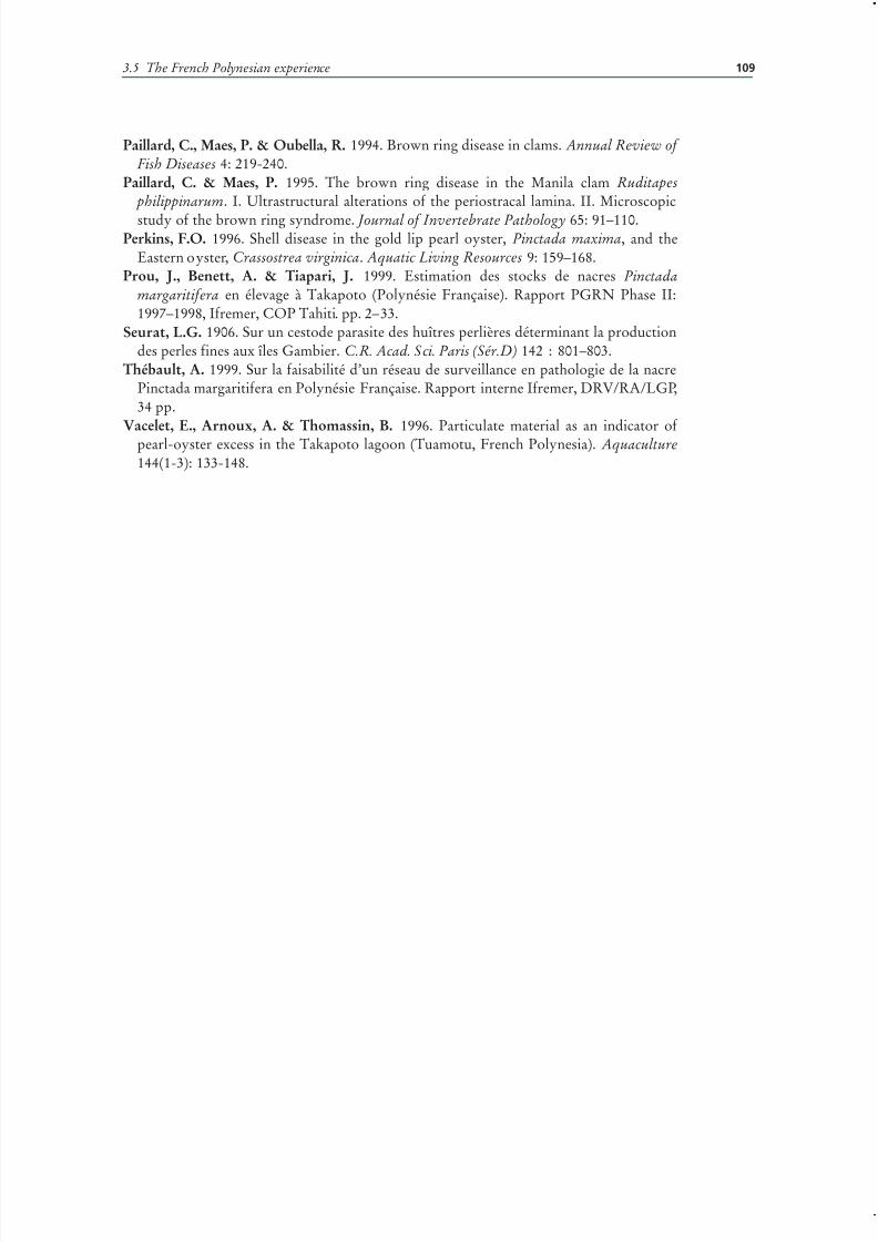
7/27/2019 Pearl Oyster Health Management
http://slidepdf.com/reader/full/pearl-oyster-health-management 123/136
109
Paillard, C., Maes, P. & Oubella, R. 1994. Brown ring disease in clams. Annual Review of Fish Diseases 4: 219-240.
Paillard, C. & Maes, P. 1995. The brown ring disease in the Manila clam Ruditapes philippinarum. I. Ultrastructural alterations of the periostracal lamina. II. Microscopic
study of the brown ring syndrome. Journal of Invertebrate Pathology 65: 91–110.Perkins, F.O. 1996. Shell disease in the gold lip pearl oyster, Pinctada maxima, and the
Eastern oyster, Crassostrea virginica. Aquatic Living Resources 9: 159–168.Prou, J., Benett, A. & Tiapari, J. 1999. Estimation des stocks de nacres Pinctada
margaritifera en élevage à Takapoto (Polynésie Française). Rapport PGRN Phase II:1997–1998, Ifremer, COP Tahiti. pp. 2–33.
Seurat, L.G. 1906. Sur un cestode parasite des huîtres perlières déterminant la productiondes perles fines aux îles Gambier. C.R. Acad. Sci. Paris (Sér.D) 142 : 801–803.
Thébault, A. 1999. Sur la faisabilité d’un réseau de surveillance en pathologie de la nacrePinctada margaritifera en Polynésie Française. Rapport interne Ifremer, DRV/RA/LGP,34 pp.
Vacelet, E., Arnoux, A. & Thomassin, B. 1996. Particulate material as an indicator of pearl-oyster excess in the Takapoto lagoon (Tuamotu, French Polynesia). Aquaculture144(1-3): 133-148.
3.5 The French Polynesian experience

7/27/2019 Pearl Oyster Health Management
http://slidepdf.com/reader/full/pearl-oyster-health-management 124/136

7/27/2019 Pearl Oyster Health Management
http://slidepdf.com/reader/full/pearl-oyster-health-management 125/136
111
3.6 Pearl oyster health: experiencesfrom the Philippines, China, thePersian Gulf and the Red Sea
Melba G. Bondad-Reantaso Aquaculture Management and Conservation ServiceDepartment of Fisheries and Aquaculture, Food and Agriculture Organization of theUnited Nations, Viale delle Terme di Caracalla, Rome 00153, [email protected]
Sharon E. McGladdery Aquatic Animal Health Division/Santé des animaux aquatiquesCanadian Food Inspection Agency1341 Baseline Road, T7, Room/Salle 5121, Ottawa, ON K1A [email protected] LadraBureau of Fisheries and Aquatic Resources
3rd Floor, PCA Building, Elliptical Road, Quezon City 1100, [email protected] ChongmingMaricultural Organism Diseases Control & Molecular Pathology LaboratoryYellow Sea Fisheries Research Institute, Chinese Academy of Fishery Sciences
106 Nanjing Road, Qingdao, SD 266071 PR [email protected]
Bondad-Reantaso, M.G., McGladdery, S.E., Ladra, D. and Chongming, W. 2007.Pearl oyster health: experiences from the Philippines, China, the Persian Gulf and theRed Sea, pp. 111–120. In: M.G. Bondad-Reantaso, S.E. McGladdery and F.C.J. Berthe.Pearl oyster health management: a manual. FAO Fisheries Technical Paper. No. 503.Rome, FAO. 120p.
ABSTRACTAvailable information, based on field and literature survey, on pearl oyster health in the
Philippines, China, the Persian Gulf and the Red Sea, are compiled in this paper. While the
case studies presented here are limited, the collected information, nevertheless, represent
the currently available knowledge. There are some similarities in the abnormalities
experienced by the countries particularly shell damage due to heavy fouling, clionid
sponge and Polydora infection as well as presence of unidentified parasites, inclusion
bodies and rickettsia-like organisms. Some farm management practices are discussed.
THE PHILIPPINE EXPERIENCE
The Philippine Pearl Oyster Industry is based primarily on the gold- or silver-lippearl oyster, Pinctada maxima, along with the winged pearl oyster, Pteria penguin,for round, half-round and three-quarter shells and other shell products (Ladra, 1994;

7/27/2019 Pearl Oyster Health Management
http://slidepdf.com/reader/full/pearl-oyster-health-management 126/136
Pearl oyster health management: a manual 112
Ladra, 1997). Pearl farming used to be a minor mariculture activity in the country hasnow grown to be the eighth dollar earner for the fishery sector.
There are at least 30 registered marine pearl farms in the Philippines, with areascovering 1–242 hectares/licensee and occupying a water area if more than 2 000 ha
(BFAR, unpublished data). The farms are located in the provinces of Masbate, Sulu,Zamboanga, Tawi-tawi, Guimaras, Davao, Palawan, Quezon, Pangasinan and Samar.The best culture sites can be found in the southern part of the country in Suu, Tawi-tawi and Basilan.
The industry is involved in collection, production and trade. In Bulacan Province,for example, more than 2 000 workers are involved in the jewelry trade; in CebuProvince, there are about 49 mother-of-pearl (MOP) costume jewelry operators andshell-craft processors. Revenue generated from pearl oyster products has increasedtremendously from US$9 million in 1991 to US$12 million in 1994 (Table 3.6.1) and toUS$15 billion in 2005 (Table 3.6.2).
During the early 1990s, the Pearl Oyster Industry experienced growing mortalitylosses, as well as shell deformity problems, to the extent that some formerly productivesites have become unworkable.
With the support of the South Sea Pearl Industries of the Philippines1, the BFARand collaborating agencies2, took an initiative to undertake a preliminary assessment of the pearl oyster health problems was conducted in 1996. The assessment involved farmvisits, interview with pearl farmers and examination of pearl oyster specimens (grossexamination of shell abnormalities and histological examination).
The case-histories presented by the various farms visited under the Pearl OysterHealth Initiative revealed patterns of disease and mortality which matched severalof the scenarios of pearl oyster health problems in other countries. Notably, manyfarmers were reporting high losses shortly after arrival of wild MOP shell stock at thefarm site. Plates 1-3 show some characteristic abnormalities observed from Philippine
pearl oysters.Wild oysters collected from deep water (> 60 feet) and raised directly to the surfaceundergo pressure changes that are assumed to have some impact on physiologicalfunctions. In addition, rapid changes in ambient temperature result from transferfrom deep to surface waters. Problems with heavy fouling (e.g. encrusting and sessileinvertebrates) of oysters collected from shallower waters are also common. Thesefouling organisms can overgrow the complete shell, including the lips and hinge,impeding feeding and growth. Cleaning of heavily fouled oysters requires removalfrom the holding system and air exposure which, if not conducted under shaded/conditions can also be physiologically taxing. Transportation to off-site areas for de-
1 “Coron Development Corporation, Guian South Sea Pearl Farm, Hikari South Sea Pearl Corporation,Sommaco South Sea Pearl Corporation, Sea Queen, Tawi-Tawi Pearl Farm.
2 Philippine Council for Aquatic and Marine Resources Development (PCAMRD), Technology
Application and Promotions Institute (TAPI) both of the Department of Science and Technology of thePhilippines; Canada’s Department of Fisheries and Oceans (DFO-Canada), Thailand’s Aquatic AnimalHealth Research Institute (AAHRI) and Canadian Executive Service Organization (CESO).
TABLE 3.6.1
Revenue from pearl oyster products (in US$, 1991–1994)Commodity 1994 1993 1992 1991
Raw shells 359 030 422 506 3 014 745 898 456
Jewelry 3 601 226 1 775 983 1 656 835 1 997 930
Shell blanks 2 970 000 1 788 258 1 250 497 1 103 037
Processed shells 172 429 176 914 731 954 95 060
Pearls 5 585 906 3 278 042 1 473 726 3 780 000
Shell button – 2 929 646 9 453 210 9 103 837
Total 12 689 590 10 371 349 9 453 210 9 103 837

7/27/2019 Pearl Oyster Health Management
http://slidepdf.com/reader/full/pearl-oyster-health-management 127/136
3.6 Pearl oyster health: experiences from the Philippines, China, the Persian Gulf and the Red Sea 113
fouling would be the ideal situation for farm management, but involve more exposureof the pearl oysters to air/shallow holding as well as time and transportation expense.
The problem of heavy fouling experienced in the Philippines and its effect on shellquality and survival is a situation similar to that described from the Persian Gulf as well
as elsewhere. This problem shows no host-specificity and applies to both P. maxima and P. penguin.
Biomineralization problems similar to those described from French Polynesia werealso reported at certain Philippine pearl farms, both in P. maxima and P. penguin,however, the extreme examples demonstrated by P. margaritifera were not observed.
In addition to the above information, collected through farmer interviews, grossexamination of shells provided the following observations:
a) heavy fouling: mollusc encrustment as well as multi-taxa fouling, particularly inP. penguin;
b) shell damage due to clionid sponge;c) blisters at the adductor muscle attachment of P. penguin;
d) Polydora-related tunnel damage to inner shell;e) mantle recession in P. maxima andf) inner shell discolouration.
TABLE 3.6.2
Philippine export data (cultured pearl and mother-of-pearl and products) for 2005
Product Exporting countryWeight
(kg)FOB value
(Philippine pesos)FOB value (US$)
Cultured pearlUnworked
Hong Kong 100 19 211 428 371 320
United States of America 27 9 471 247 183 060Switzerland 13 12 727 597 246 000
Japan 8 9 833 828 190 069
Total 148 51 240 100 990 450
Cultured pearlworked
Hong Kong 744 255 731 281 4 865 482
Japan 332 127 437 447 2 463 121
Australia 275 159 615 969 3 055 070
United States of America 119 110 051 239 2 127 079
Switzerland 34 29 751 172 575 033
Others 53 28 514 732 551 135
Total 1 557 707 101 840 13 666 920
Shell buttons China PR 36 000 1 955 704 37 800
Japan 6 886 5 975 589 115 458
Hong Kong 674 615 685 11 900Korea 525 336 298 6 500
Germany 350 783 679 15 147
Others 261 903 365 17 499
Total 44 096 10 570 320 204 304
Mother of pearlunworked
Hong Kong 316 731 8 114 619 156 840
China 54 619 6 818 370 131 876
Korea 52 524 8 105 513 156 664
Japan 19 300 2 672 973 52 050
Italy 10 100 827 811 16 000
Others 39 507 843 622 15 919
Total 27 382 908 529 259
Mother of pearl
worked
Hong Kong 15 010 1 823 254 35 240
Thailand 13 200 134 623 2 602
Germany 2 836 3 404 891 65 810
United States of America 2 604 1 936 302 37 425
Spain 1 453 591 988 11 442
Others 3 060 4 110 549 79 449
Total 38 163 12 001 607 231 068
Grand total 15 622 601
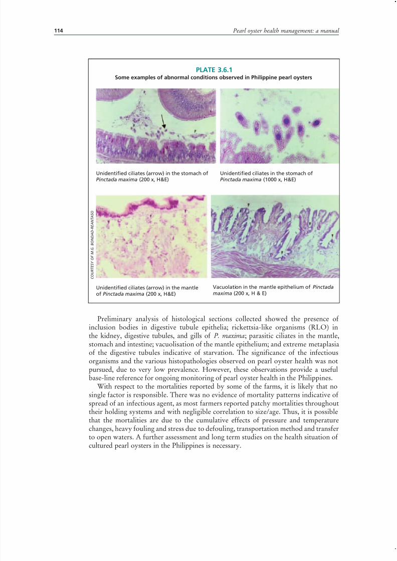
7/27/2019 Pearl Oyster Health Management
http://slidepdf.com/reader/full/pearl-oyster-health-management 128/136
Pearl oyster health management: a manual 114
Preliminary analysis of histological sections collected showed the presence of inclusion bodies in digestive tubule epithelia; rickettsia-like organisms (RLO) inthe kidney, digestive tubules, and gills of P. maxima; parasitic ciliates in the mantle,stomach and intestine; vacuolisation of the mantle epithelium; and extreme metaplasiaof the digestive tubules indicative of starvation. The significance of the infectiousorganisms and the various histopathologies observed on pearl oyster health was notpursued, due to very low prevalence. However, these observations provide a usefulbase-line reference for ongoing monitoring of pearl oyster health in the Philippines.
With respect to the mortalities reported by some of the farms, it is likely that no
single factor is responsible. There was no evidence of mortality patterns indicative of spread of an infectious agent, as most farmers reported patchy mortalities throughouttheir holding systems and with negligible correlation to size/age. Thus, it is possiblethat the mortalities are due to the cumulative effects of pressure and temperaturechanges, heavy fouling and stress due to defouling, transportation method and transferto open waters. A further assessment and long term studies on the health situation of cultured pearl oysters in the Philippines is necessary.
PLATE 3.6.1Some examples of abnormal conditions observed in Philippine pearl oysters
Unidentified ciliates (arrow) in the stomach ofPinctada maxima (200 x, H&E)
Unidentified ciliates in the stomach ofPinctada maxima (1000 x, H&E)
Unidentified ciliates (arrow) in the mantle
of Pinctada maxima (200 x, H&E)
Vacuolation in the mantle epithelium of Pinctada
maxima (200 x, H & E)
C O U R T E S Y O F M . G . B O N D A D - R E A N T A S O
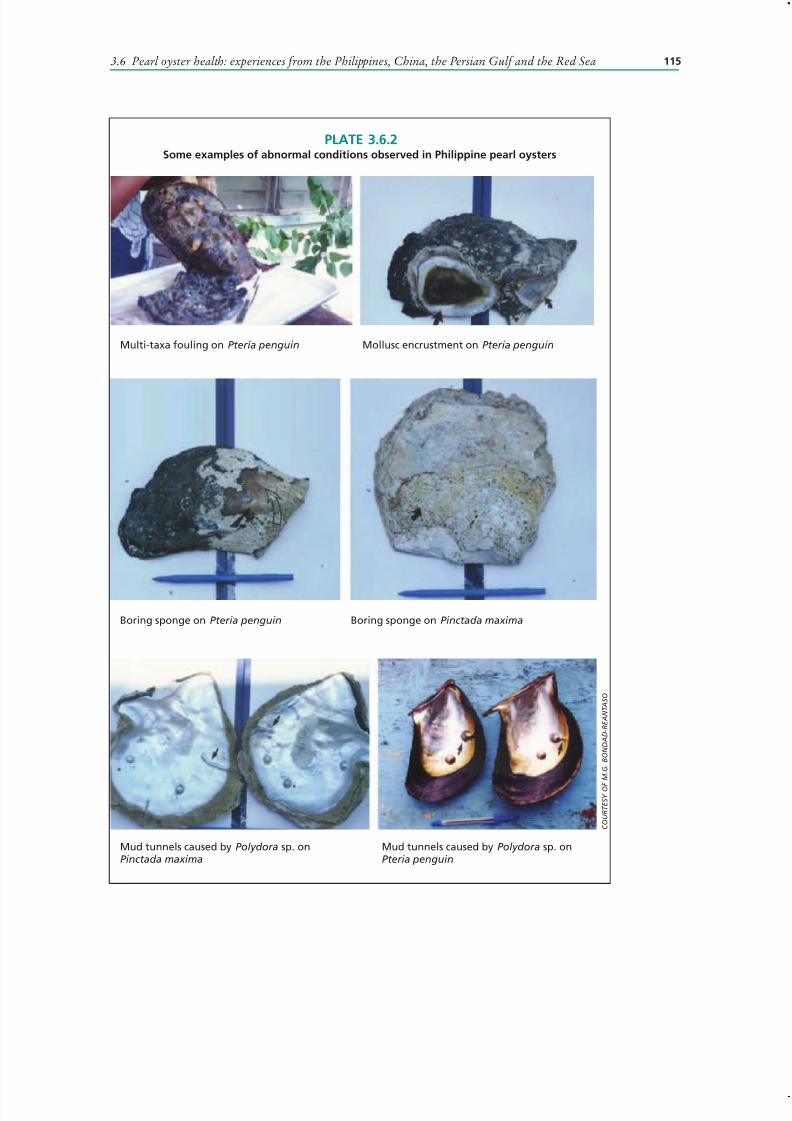
7/27/2019 Pearl Oyster Health Management
http://slidepdf.com/reader/full/pearl-oyster-health-management 129/136
3.6 Pearl oyster health: experiences from the Philippines, China, the Persian Gulf and the Red Sea 115
PLATE 3.6.2Some examples of abnormal conditions observed in Philippine pearl oysters
Multi-taxa fouling on Pteria penguin Mollusc encrustment on Pteria penguin
Boring sponge on Pteria penguin Boring sponge on Pinctada maxima
Mud tunnels caused by Polydora sp. onPinctada maxima
Mud tunnels caused by Polydora sp. onPteria penguin
C O U R T E S Y O F
M . G . B O N D A D - R E A N T A S O
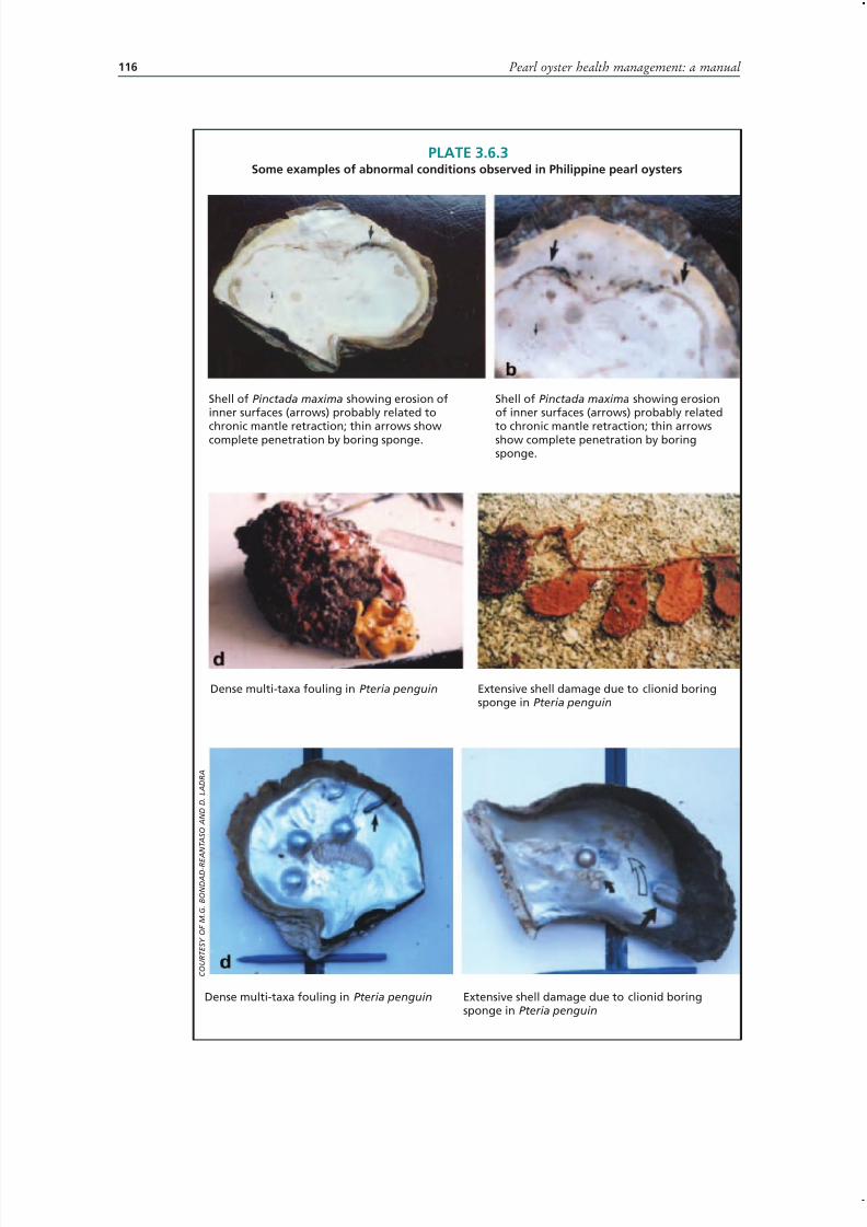
7/27/2019 Pearl Oyster Health Management
http://slidepdf.com/reader/full/pearl-oyster-health-management 130/136
Pearl oyster health management: a manual 116
PLATE 3.6.3Some examples of abnormal conditions observed in Philippine pearl oysters
Shell of Pinctada maxima showing erosion ofinner surfaces (arrows) probably related to
chronic mantle retraction; thin arrows showcomplete penetration by boring sponge.
Shell of Pinctada maxima showing erosionof inner surfaces (arrows) probably related
to chronic mantle retraction; thin arrowsshow complete penetration by boringsponge.
Dense multi-taxa fouling in Pteria penguin Extensive shell damage due to clionid boringsponge in Pteria penguin
Dense multi-taxa fouling in Pteria penguin Extensive shell damage due to clionid boringsponge in Pteria penguin
C O U R T E S Y O F M . G . B O N D A D - R E A N T A S O A N D D . L A D R A

7/27/2019 Pearl Oyster Health Management
http://slidepdf.com/reader/full/pearl-oyster-health-management 131/136
3.6 Pearl oyster health: experiences from the Philippines, China, the Persian Gulf and the Red Sea 117
PLATE 3.6.4Some examples of abnormal conditions observed in Philippine pearl oysters
Rickettsia-like organisms (RLO, arrow) in thegills of Pinctada maxima (200 x, H & E)
Rickettsia-like organisms (RLO) in the gills ofPinctada maxima (1000 x, H & E)
Rickettsia-like organisms (RLO, arrow) in thekidney of Pinctada maxima (200 x, H & E)
Rickettsia-like organisms (RLO) in the digestivetubules of Pinctada maxima (1000 x, H & E)
Inclusion bodies in the digestive tubuleepithelia of Pinctada maxima (arrows,800 x, H & E)
Inclusion bodies in the gills of Pinctada maxima (arrows, 800 x, H & E)
C O U R T E S Y O F M . G . B O N D A D - R E A N T A S O

7/27/2019 Pearl Oyster Health Management
http://slidepdf.com/reader/full/pearl-oyster-health-management 132/136

7/27/2019 Pearl Oyster Health Management
http://slidepdf.com/reader/full/pearl-oyster-health-management 133/136

7/27/2019 Pearl Oyster Health Management
http://slidepdf.com/reader/full/pearl-oyster-health-management 134/136

7/27/2019 Pearl Oyster Health Management
http://slidepdf.com/reader/full/pearl-oyster-health-management 135/136

7/27/2019 Pearl Oyster Health Management
http://slidepdf.com/reader/full/pearl-oyster-health-management 136/136