Small diameter blood vessel prostheses from blends of … diameter blood vessel prostheses from...
7
Small diameter blood vessel prostheses from blends of pol@ih$lene oxide andpomropylene oxide J.G.F. Bots,L. van der Doesand A. Bantjes Department of Chemical Technology, Biomaterials Section, Twenfe University of Technology, PO Box 2 17, 7500 AE Enschede, The Netherlands (Received 5 September 1985; accepted 7 November 1985) Studies on the relationship between blood platelet adhesion and type and amount of polyether segments in copolyetherurethanes report a reduced platelet adhesion with increasing polyether content. We therefore assumed that combinations of polyethylene oxide (PEO) and polypropylene oxide (PPOX) might give materials with a good blood compatibility. Water-soluble PEO wes attached to PPOX by u.v.-initiated crosslinking. Films were tested for hydrophilicity, mechanical properties, protein adsorption and blood compatibility. The hydrophilicity was determined by swelling experiments. A compromise between hydro- philicity (PEO) and mechanical strength (PPOX) was met at a swelling of 0.5 (PPOX/PEO ratio: go/l 0). In protein adsorption studies only small amounts of adsorbed proteins were found. Three blood material interaction in vitro tests gave good results: a low platelet adhesion and kallikrein generation and a high APTT value. Porous tubings (inner diameter 1.3 mm) were fabricated, by spinning from solution, for implantation in the abdominal aorta of rats. Stress-strain diagrams were comparable to those reported for natural blood vessels. Keywords: Prostheses, polvethers, blood vessels. in vitro evaluation, mechanical propeflies, polyethylene oxide, polypropylene A special type of block copolymer, consisting of alternating hard and soft segments, is widely known and two represen- tatives, i.e. Biomer@’ and Avcothanbe2 are used as biomaterials in several blood contacting devices such as catheters, artificial blood vessels and heart valves. Kim eta/.3 and Lyman et al.4 reported that a segmented copoly- etherurethane-urea (PEUU) compared to silicone rubber (SR) and a fluorinated ethylene-propylene copolymer (FEP) showed an increased albumin adsorption together with a low y-globulin and fibrinogen adsorption and a low platelet adhesion. This behaviour is generally accepted as being indicative of a blood material interaction without severe reactions like thrombus formation. Indications were obtained by Lyman4 that protein is more strongly adsorbed onto the hard polyurethane-urea segments than on the soft polypropylene oxide blocks. He concluded from ESCA measurements that the air side of a cast film was richer in polyether than the glass mould side of the film. Interesting observations have also been reported by Lelah et al. 5, who found that extruded BiomeP had a higher content of soft polytetramethylene oxide segments on the surface than cast Biomep, resulting in a lower fibrinogen adsorption and platelet adhesionfortheformer. Results from these and other studie&’ indicate that the polyether segments in copolyurethanes are probably responsible for the preferred adsorption of albumin and the reduced platelet adhesion. Apart from this, the ratio of hydrophilic to hydrophobic areas on the polymer surface is known to have a marked influence on the blood compatibility. For instance, a different behaviour in protein adsorption’ and platelet adhesiong, lo was found in contact with hydrophilic or hydrophobic surfaces. The importance of the morphology and texture of the blood contacting surface has also been reported by Yui et al. ’ ’ and Hess et al.“. Yui synthesized polypropylene oxide segmented polyamides with various polyamide segment lengths and found the platelet adhesion to be strongly dependent on the size of the crystalline and amorphous domains. The platelet adhesion was significantly miminized on a copolymer, in which the average diameters of the crystalline and amorphous domains were 6.42 and 5.18 nm, respectively. It was concluded from in viva experiments12 that the neo-intima formation in small diameter (1.5 mm) prosthetic blood vessels was strongly stimulated by a porous fibrillar inner structure. Sa da Costal extensively studied the relationship between platelet adhesion and type and amount of polyether segments in copolyetherurethanes and she found a reduced platelet adhesion with increasing polyether content. Poly- ethylene oxide (PEO) was reported to be most active in 0 1986 Eutterworth 8 Co (Publishers) Ltd. 0142-9612/86/050393-07$03.00 Biomaterials 1986, Vol 7 September 393
Transcript of Small diameter blood vessel prostheses from blends of … diameter blood vessel prostheses from...
Small diameter blood vessel prostheses from blends of pol@ih$lene oxide andpomropylene oxide
J.G.F. Bots, L. van der Does and A. Bantjes Department of Chemical Technology, Biomaterials Section, Twenfe University of Technology, PO Box 2 17, 7500 AE Enschede, The Netherlands (Received 5 September 1985; accepted 7 November 1985)
Studies on the relationship between blood platelet adhesion and type and amount of polyether segments in copolyetherurethanes report a reduced platelet adhesion with increasing polyether content. We therefore assumed that combinations of polyethylene oxide (PEO) and polypropylene oxide (PPOX) might give materials with a good blood compatibility. Water-soluble PEO wes attached to PPOX by u.v.-initiated crosslinking. Films were tested for hydrophilicity, mechanical properties, protein adsorption and blood compatibility. The hydrophilicity was determined by swelling experiments. A compromise between hydro- philicity (PEO) and mechanical strength (PPOX) was met at a swelling of 0.5 (PPOX/PEO ratio: go/l 0). In protein adsorption studies only small amounts of adsorbed proteins were found. Three blood material interaction in vitro tests gave good results: a low platelet adhesion and kallikrein generation and a high APTT value. Porous tubings (inner diameter 1.3 mm) were fabricated, by spinning from solution, for implantation in the abdominal aorta of rats. Stress-strain diagrams were comparable to those reported for natural blood vessels.
Keywords: Prostheses, polvethers, blood vessels. in vitro evaluation, mechanical propeflies, polyethylene oxide, polypropylene
A special type of block copolymer, consisting of alternating hard and soft segments, is widely known and two represen- tatives, i.e. Biomer@’ and Avcothanbe2 are used as biomaterials in several blood contacting devices such as catheters, artificial blood vessels and heart valves. Kim eta/.3
and Lyman et al.4 reported that a segmented copoly- etherurethane-urea (PEUU) compared to silicone rubber (SR) and a fluorinated ethylene-propylene copolymer (FEP) showed an increased albumin adsorption together with a low y-globulin and fibrinogen adsorption and a low platelet adhesion. This behaviour is generally accepted as being indicative of a blood material interaction without severe reactions like thrombus formation.
Indications were obtained by Lyman4 that protein is more strongly adsorbed onto the hard polyurethane-urea segments than on the soft polypropylene oxide blocks. He concluded from ESCA measurements that the air side of a cast film was richer in polyether than the glass mould side of the film.
Interesting observations have also been reported by Lelah et al. 5, who found that extruded BiomeP had a higher content of soft polytetramethylene oxide segments on the surface than cast Biomep, resulting in a lower fibrinogen adsorption and platelet adhesionfortheformer. Results from these and other studie&’ indicate that the polyether segments in copolyurethanes are probably responsible for
the preferred adsorption of albumin and the reduced platelet adhesion.
Apart from this, the ratio of hydrophilic to hydrophobic areas on the polymer surface is known to have a marked influence on the blood compatibility. For instance, a different behaviour in protein adsorption’ and platelet adhesiong, lo was found in contact with hydrophilic or hydrophobic surfaces.
The importance of the morphology and texture of the blood contacting surface has also been reported by Yui et al. ’ ’ and Hess et al.“. Yui synthesized polypropylene oxide segmented polyamides with various polyamide segment lengths and found the platelet adhesion to be strongly dependent on the size of the crystalline and amorphous domains. The platelet adhesion was significantly miminized on a copolymer, in which the average diameters of the crystalline and amorphous domains were 6.42 and 5.18 nm, respectively. It was concluded from in viva experiments12 that the neo-intima formation in small diameter (1.5 mm) prosthetic blood vessels was strongly stimulated by a porous fibrillar inner structure.
Sa da Costal extensively studied the relationship between platelet adhesion and type and amount of polyether segments in copolyetherurethanes and she found a reduced platelet adhesion with increasing polyether content. Poly- ethylene oxide (PEO) was reported to be most active in
0 1986 Eutterworth 8 Co (Publishers) Ltd. 0142-9612/86/050393-07$03.00
Biomaterials 1986, Vol 7 September 393

Polyether vascular grafts: J.G.F. Bots er al.
suppressing platelet adhesion compared to polypropylene oxide (PPOX) and polytetramethylene oxide (PTMO).
From these results we derived the hypothesis that combining PEO with PPOX or PTMO might lead to materials with a good blood compatibility: PEO as the blood compatible, hydrophilic component and PPOX or PTMO as the more hydrophobic and mechanically stronger one.
PEO was attached to high molecular weight atactic PPOX by u.v.-initiated crosslinking in the presence of dicumyiperoxide (DCP). Swelling experiments were performed with the resulting films in order to determine the equilibrium water content. Blood material interactions were studied with three tests: (i) kallikrein generation (Factor XII activation), (ii) activated partial thromboplastin time, and (iii) blood platelet adhesion. Adsorption of albumin, fibrinogen, fibronectin and y-globulin was investigated using an ELISA test’. For in viva experiments blood vessel prostheses were spun from solution and implanted in the abdominal aorta of rats. The mechanical properties were compared to those of natural blood vessels.
EXPERIMENTAL
Materials
High molecular weight PEO, (M,., = 1, 3 and 6 X 1 05) was purchased from Polysciences Inc. (Warrington, USA). The crystalline and water-soluble polymers showed a broad molecular weight distribution: MJM, = 6.1 (GPC-LALLS).
Atactic PPOX (d.s.c.; 95% atactic) was synthesized14 from propylene oxide (Merck) with a catalyst prepared from diethylzinc [Schering AG, Berlin; 20% (w/w) in toluene] and triphenyltin hydroxide (M &T,Vlissingen,The Netherlands). M, varied between 0.8 and 2.5 X 1 05, depending on the monomer/catalyst ratio and for all PPOX batches MJM, < 1.3. Semicrystalline PPOX was prepared15 with FeCls as a catalyst and the crystalline fraction (d.s.c.; 95% crystallinity) was separated from the amorphous one by recrystallization (twice) from methanol, which dissolved the atactic component.
Semicrystalline PTMO (d.s.c.; 70% crystallinity) was synthesized from tetrahydrofuran (Merck) with SbCls as a catalyst . ” Molecular w ‘g er hts of 2 to 4 X 1 O4 (m,.,) were obtained: MdMi, = 1.2 (GPC-LALLS). For crosslinking experiments DCP (Schuchardt, Germany), recrystallized twice from methanol, was used. The solvents, used for the catalyst preparation for the polymerization of propylene oxide [(xylene, toluene and hexane) (Merck)] were distilled from sodium. Tetrahydrofuran and propylene oxide were purified by distillation from lithium aluminium hydride (Merck) and calcium chloride (Merck), respectively. All purifications were performed in a nitrogen atmosphere.
Methods
Film preparation and swelling experiments. Mixtures of PEO, PPOX and DCP were dissolved in dichloromethane or chloroform and films were cast from viscous 5-l 0% (w/w) solutions. After slow evaporation of the solvent the films were dried in vacuum (0.2-0.5 mmHg) at room temperature and subsequently u.v.-irradiation (Hanau lamp TQ 81, wavelengths 254 and 366 nm) proceeded in a nitrogen atmosphere (the temperature during irradiation was kept constant). The irradiated films were washed with water for 24 h to remove non-crosslinked PEO and with acetone (24 h) for removal of non-crosslinked PPOX as well as DCP and decomposition products like acetophenone and 2-phenyl-
isopropanol. Weight losses were determined and the equilibrium water content after 24 h of swelling was measured. Swelling was calculated using the relationship:
s = weq/wdry- 1.
Films of PEO, PPOX and mixtures containing 4.8% DCP (w/w) for measurements of mechanical properties and protein adsorption were cast from CH-j&, irradiated for 1 h at 50°C and water washed (no acetone washing).
Samples for thermal analysis (d.s.c.) were cast from CH&I, without adding DCP. The coatings of PPDX/PEO, PPOX, PTMO and BiomeP (Ethicon Inc., Sommerville NJ, USA) on the inside of glass tubes for the kallikrein generation and APlT test and on glass slides for the blood platelet adhesion tests were obtained by covering the glass surfaces with a thin layer of a slightly viscous solution of BiomeP (DMF), PTMO (THF) or PPOX, PPOX/PEO (CH,CIz) and allowing the solvent to evaporate. The PPDX containing coatings [with an initial DCP content of 4.8% (w/w)] were irradiated for 1 h at 50°C and subsequently washed with water (no acetone washing since this loosens the coating).
Blood vessel prostheses. From a 4% (w/w) solution of PPOWPEO/DCP: 85.8/9.5/4.7 (%, w/w) in dichloro- methane/dichloroethane: 70/30 (v/v) fibres with a diameter ranging from 5 to 50 pm were spun and by winding on a rotating axis porous ‘woven-like’ tubings were fabricated (and subsequently u.v.-irradiated during 60 min). Inner and outer diameter could be varied by changing the axis diameter (0.6-2.0 mm) and the spinning time (30 min-2 h).The pore size (5-200 urn) as well as the fibre diameter could be varied depending on the flow rate of the polymer solution.
Mechanical properties. Stress-strain measurements were made with porous tubings (length 40 mm; inner diam. 1.3 mm; outer diam. 1.7 mm) and dumbbell shaped test strips (6 X 40 mm) using a tensile testing device (Instron floor model ‘IT-CM, High Wycombe, UK) with drawing rates of 5 mm/min.
Differentialscanning calorimetry. In the temperature range from 20-100°C thermograms were recorded using a Du Pont DSC 990 at a heating rate of 1 O”C/min. lndium was used to calibrate the temperature scale and to determine the heat of fusion of PEO, atactic and crystalline PPOX and FTMO. Transition temperatures were determined for PEO, PPOX and PTMO with samples weighing 10 k 2 mg.
Protein adsorption. Crosslinked films of PPOX/PEO: 90/l 0 were washed with PBS buffer and screened on adsorption of albumin(BSA),fibronectin,fibrinogenandy-globulinwithan ELISA test’. Equilibrium protein adsorption was measured after 100 min incubation with human plasma. Tissue culture polystyrene (Costar, Cambridge, Mass., USA) was taken as reference material and BiomeB, cast from DM F [ 10% (w/w), air side] was used for comparison.
Kallikrein generation . l7 This could be detected using a chromogenic substrate (AB KABI Diagnostics, type 52302). The release ofp-nitroaniline was measured by spectrophoto- metric analysis at 405 nm. Aliquots of 1 ml 10% titrated human plasma [diluted to 10 ml with Tris buffer, 50 mM, pH 7.4 (Merck)] were incubated at 37°C in polymer coated glass tubes (see Film preparation). Within a period of 15min,attime= 1,2,3.5,5,7.5, 10, 12.5and 15min, samples of 100 ul were taken and added to the chromogenic
394 Biomaterials 1986, Vol 7 September

substrate (300 ).rI Tris buffer and 100 $ S2302 stock solution, 37°C). After addition of 200 ul 50% (v/v) acetic acid solution the enzymatic reaction was stopped and the releasedp-nitroaniline was measured. The reference materials were silica borate glass and SR’* (RTV adhesive sealant, General Electric, Bergen op Zoom, The Netherlands), the latter as a coating on glass tubes [from a 5% (w/w) solution in THF: see Film preparation].
AP77test’g. Citrated human plasma (0.5 ml) was incubated at 37°C in polymer coated glass tubes. After 1 min 50 ~1 cefalin solution [Sigma, RBC, l/3 stock suspension, 2/3 0.85% (w/w) NaCl solution] was added, followed after another 1 min by 50 ~1 0.2 M CaCI, solution (Merck). The interval between CaCI, addition and the beginning of coagulation was determined (references: silica borate glass and SR).
Blood platelet adhesion”. A cell consisting of two glass slides, separated by a PVC ring, (inner diameter 10 mm) was filled without air bubbles with titrated human plasma containing + 5 X IO4 platelets/$. One of the glass slides was coated with the polymer to be tested (see Film preparation), the other with SR as a reference. The cell was centrifuged twice, the first time in a centrifugal force towards the polymer coated test surface and afterwards to the reference by turning the cell 180". The adhered platelets on the polymer surfaces were counted using a microscope (magnifi- cation X 400).
RESULTS AND DISCUSSION
In order to study the crosslinking process, films were prepared from PEO (M, = 6 X 1 05) differing in initial DCP content and u.v.-irradiation time. Figure 1 shows a decrease of swelling (S) with increasing DCP concentrations and
15.
IO-
F .- - a, 3
Q-
I
OJ 30 90 150
u.v. - irradiation (min) O/o PEO (w/w) Figure 1 Swelling of PEO/DCP irradiated (50°C) films. % .DCP (w/w): Figure 2 Swelling of PPOX/PEO irradiated (50°C) films; DCP: 9% (w/w). A, 4.8; 0, 10.5; 0, 13.0; A, 16.7. u.v.-irradiation (min): A, 30; l , 90; o, 150.
Polyether vascular grafts: J.G.F. Bats at al.
Table 1 Weight losses of PEO/DCP films in water and acetone
u.v.-Irradiation (min) DCP (%) Weight losses (%)
Water Acetone
30 4.8 55 4
90 4.8 30 3
150 4.8 20 2
30 16.7 23 14
90 16.7 13 9
150 16.7 1 6
longer irradiation time. The increasing crosslink density lowers S and the amount of noncrosslinked PEO, which could be determined by washing with water after irradiation (Table 7). After washing with acetone in order to remove DCP and decomposition products it could be concluded from n.m.r. measurements of the acetone fraction that not all DCP had been used, - even after 150 min irradiation - , and that a certain amount of peroxy radicals was probably incorporated in the polymer network.
The temperature dependence of the curing process was investigated by irradiating 10% DCP (w/w) containing films at 20”, 35”, 50”, 75” and 90°C for 90 min with U.V. At 75” and 90°C the polymer films deformed and melted (d.s.c.: t, = 66°C). The 50°C sample gave the highest crosslink density: S = 6.8, whereas at 35” and 20°C S values of 7.8 and 10 were found respectively, illustrating the influence of the temperature on the crosslinking process. The resulting films had no mechanical strength due to the high water content.
Attempts were made to reduce the swelling and to improve the mechanical properties by preparing films of PEO and PPOX in the presence of DCP. A linear relationship between the PEO content and S was found for irradiation times of 30,90 and 150 min at 50°C and it is obvious from the data in Figure 2 that the presence of PPOX had resulted
Biomaterials 1986, Vol 7 September 395
Poqtpther yascoier grafts: J.G.F. Buts et al.
in a substantial decrease of the swelling. Weight losses of 15-2556 after washing with acetone, which dissolves PPDX, DCP and decomposition products, were observed but no relationship between weight loss and irradiation time could be determined. Extraction of the films with water to remove noncrosslinked PEO gave O-5% decrease in weight, indicating that nearly ail of the PEO had been crosslinked.
Unlike the PEO/DCP samples the PPOX containing films showed elastomeric properties. However, for the preparation of these films a rather high DCP content and U.V. radiation times of 30-I 50 min were used. For possible application as biomaterials the concentration of the toxic DCP should be as low as possible and long U.V. radiation times should be avoided to prevent polymer degradation and incorporation of peroxy radicals in the polymer network. By lowering the DCP content the sameS values can be obtained by longer u.v.- irradiation. Films were made from PPOWPEO having a low DCP content [4.8% (w/w)]. Although a short u.v.-irradiation (30 min) was applied, films were obtained with not too high S values.
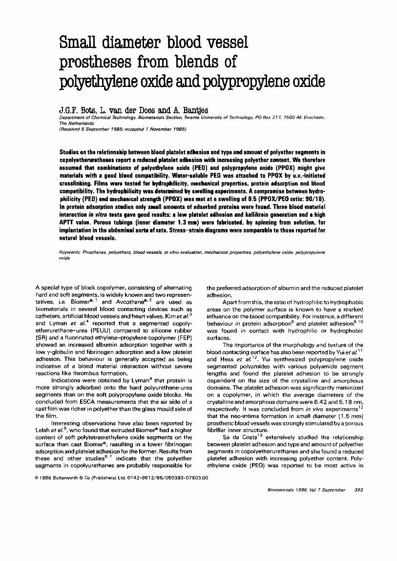
Stress-strain diagrams (~~~u~e 3) of these films having a PPDX/PEO ratio of 100/O, 90/l 0 or 80/20 (irradiated at 50°C) show that the stress for a particular strain increases with an increase in PEO content. Figure 4 shows the stress-strain curves of PPDX and PPDWPEO (90/l 0) films before and after u.v.-irradiation. The behaviour of the irradiated films differs from the classical theory, which predicts an increase in stress at any particular strain due to crosslinking.
For the interpretation of the results of the blood material interaction tests information about the morphology of the polymeric films is necessary. It has been reported2’ that the miscibility of PPOX and PEO is confined to the very low molecular weight polymers i.e. PPOX: a, = 425 and PEO: a,,, = 300. Mixtures of higher molecular weight polymers are incompatible and phase separation occurs. In the compatible mixtures Rastogi and St. Pierre” observed
Strain
Figure 3 Stress- strain diagram of irred&?d and w~er~es~d PPOWPEO films. 0, PPOX A; PPOX/PEOC (9WlO); 0. PPOX/PEO (80/20].
t 0.5
Strain AL/L
I
1
Figure 4 Stress-strain diagram of PPOX/PEO films; influence of the ~r~jation and water~ash~ng. Open symbols = before ~rmd~a~jon~ closed symbols = afterirrediation: 0, PPOX; A. PPOX/PEO (9WlO); a, PPOX/u.v.: A, PPOX/PEO (90/ 10) UK
surface activity and for binary blends of PPDX and PEO this results in an increased surface concentration of PPOX. In order to get more information about the morphology of our high molecular PPOX/PEO blends, d.s.c. and ESCA studies are in progress.
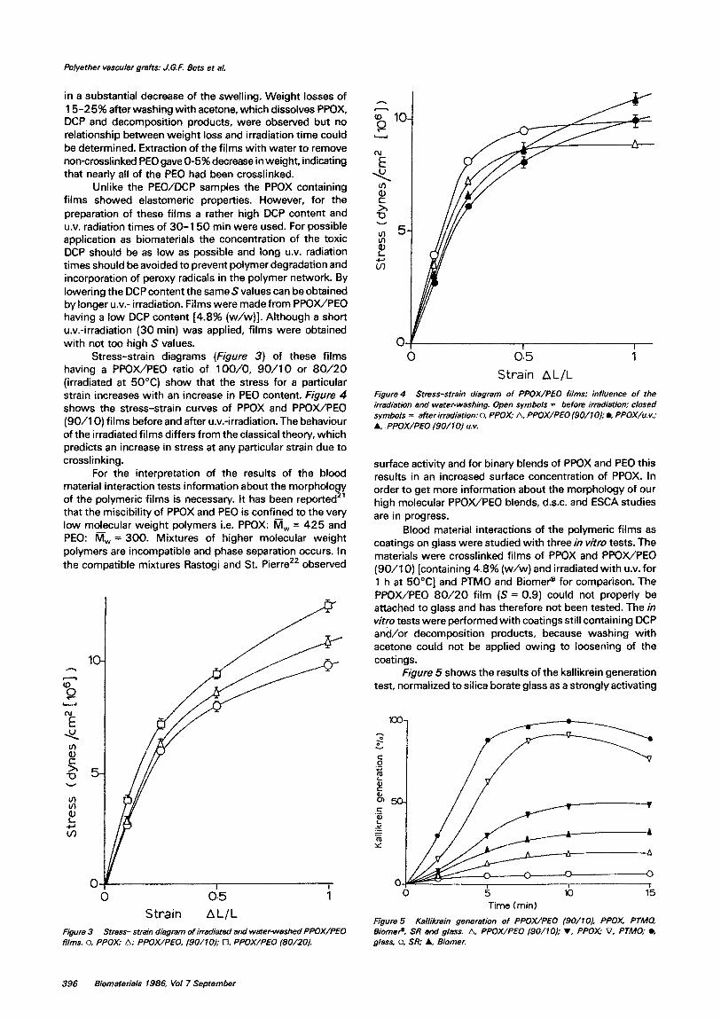
Blood material interactions of the polymeric films as coatings on glass were studied with three in vitro tests. The materials were crosslinked films of PPOX and PPDX/PEO (90/l 0) [containing 4.8% (w/w) and irradiated with U.V. for 1 h at SO”C] and PTMO and Biomer’@ for comparison. The PPOWPEO 80/20 film (S = 0.9) could not properly be attached to glass and has therefore not been tested. The in Virgo tests were performed with coatings still containing DCP and/or decomposition products, because washing with acetone could not be applied owing to loosening of the coatings.
Figure 5 shows the results of the kallikrein generation test, normalized to silica borate glass as a strongly activating
Time (mid
Figun? 5 Kallikrein generation of PPOX/PEO (90/l& PPOX, PTMO, Biomer? SR and gi8ss A. PPOX/PEO f9WlO); 7, PPOX; 7, PTMO: l . giess, 0. SR; A. Biomer.
396 Biomaterals 1986, VW 7 September

Polyather vascular grafts: J.G.F. Bats et al.
surface and compared to silicone rubber, which is non- activating. Kallikrein generation reflects the activation of Factor XII, which generally has been accepted to occur at negatively-charged surfaces. Although no net charge is present on the polyether surfaces, a generation of kallikrein was observed, possibly resulting from a certain degree of polarity in the carbon-oxygen bonds. In discussing kallikrein generation by the polyethers it should also be mentioned that PPOX coatings with and without DCP gave about the same results, indicating only a small effect of remaining DCP and/or decomposition products.
The polyethers studied differ in hydrophilicity and crystallinity, which might result in different Factor XII activation. The polymers PPDX and PTMO are both hydro- phobic with S values of 0.07 and 0.04, respectively (Sa da Costa found 0.05 for PTMO). From d.s.c. measurements it was concluded that PPOX was highly atactic, whereas PTMO had a crystallinity of about 70%. Because of the
comparable, slightly hydrophilic character, the difference in Factor XII activation may be attributed to the crystallinity of PTMO.
If crystallinity is assumed to stimulate Factor XII activation, the presence of crystalline PEO in PPOX/PEO (90/l 0) would have been expected to result in an increase of kallikrein generation. Merrill et a/.23 have reported, however, that equilibration of a copolyetherurethane with water at 25°C depressed the crystalline-amorphous transition of the soft PEO segments to temperatures below 0°C. thus creating an ‘amorphous’ hydrophilic PEO phase.
The presence of PEO in PPDX/PEO (90/l 0), probably as an amorphous phase, had a marked effect on the hydrophilicity, with S values of 0.3 to 0.5 depending on the irradiation time. The reduction of the kallikrein generation, compared to PPOX and PTMO, therefore seems to indicate a relationship between hydrophilicity and Factor XII activation.
The results of the APlT test (Table 2). displaying the activation of both the intrinsic and extrinsic coagulation system, show the same trend as the kallikrein generation, giving the highest coagulation time for PPOWPEO (90/l 0). Blood platelet adhesion tests (Table 3) confirmed the findings of Lyman et al4 and Sa da Costa eta/.687, giving the lowest platelet adhesion for PPOX/PEO (90/l 0).
From the results of the kallikrein generation test, the APlT determination and the platelet adhesion experiments it can be concluded that the in vitro blood compatibility of the polyethers, in particular of the PPOWPEO (90/O), is as good as or even better than of Biomer@.
Table 2 APTT values of polyathers. Biomef? SR and glass
Polymer APlT (s)
PTMO 245 + 40 Biomer@ 425 + 20 PPOX 490 + 25 PPOWPEO (90/l 0) 580 k 30 SR 650 k 30
Glass 150120
Table 3 Blood platelet adhesion on polyethers, BiomeP and SR
Polymer Platelets/4 X 1 O4 pm2
PTMO 8iomeP PPOX PPOWPEO (90/l 0) SR
90 f 20 120+30
12k4 5k2
600 k 70
Table 4 Protein adsorption’ on polyethers, Biomef? and tissue culture
polystyrene surfaces
Polymer Albumin Fibrinogen Fibronectin yglobulin
PPOX 12f 10 15f 12 10+2 19+ 15
PPOWPEO (90/l 0) 8f6 6irl 13* 1 9k2
PPOWPEO (80/20) 9+7 6kl 14f 10 9+7 Biomep 450 i- 10 480 + 50 lo* 10 ilk 10 TCPS 1900 + 400
a A450 x 10-3
Figure 6 Lumen (a) and outside (6) of a prosthesis (X 10).
In order to elucidate the difference in behaviour between the polyethers and Biomep, in contact with blood or plasma, protein adsorption was studied using an ELISA test, in which absorbance is measured at 450 nm’. Fibrinogen deposition (Table 4) was not significant on the polyether surfaces (PPOX/PEO: 1001’0, 90/l 0 and 80/20) in contrast to the adsorption on Biomep, cast from DMF. Lelah et aL5 found considerably less fibrinogen adsorption on the surface of extruded Biomer@ than on Biomep, cast from DMA. Fibrinogen adsorption on the polyether surfaces is lower than on extruded Biomep, which has been reported by Lelah et a/. to be the most thromboresistant.
The very low protein adsorption on the polyethers suggests a certain inertness towards protein induced
Biomaterials 1986, Vol 7 September 397
Myether vascular grafts: J.G.F. Eats et al.
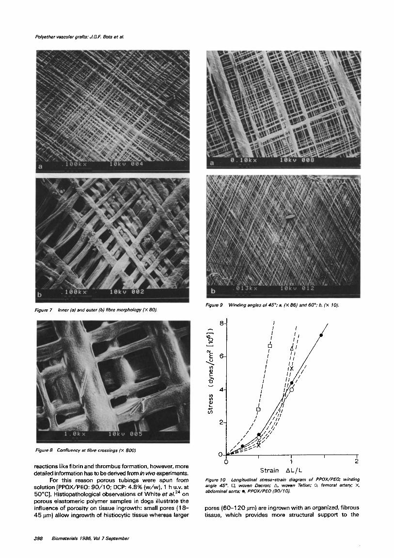
Figure 7 inner (al and outer (b) fibre morphology (X 8Oj.
Figure 8 Confluency at fibre crossings {X 800)
reactions like fibrin and thrombus formation. however, more detailed information has to be derived from in vko experiments.
For this reason porous tubings were spun from solution [PPOX/PEO: 90/l 0; DCP: 4.8% (w/w), 1 h U.V. at 5O”C]. Histiopathological observations of White et a/.24 on porous elastomeric polymer samples in dogs illustrate the influence of porosity on tissue ingrowth: small pores (18- 45 pm) allow ingr~h of histiocytic tissue whereas larger
EQUm 9 Wind&g angles of 45”; a. [X 861 and 60”; b, (X 10).
-k I I I I
0 1 2
Strain AL/L
Figure 10 f ong~odinal stress-strain diagram of PPOX/PEO; WindinQ angle 45”. Cl. woven Dacmn; A, woven Teflon; 0, femoral artery; X, abdominal aorta; l , PPOX/PEO (90/l 0).
pores (60-I 20 urn) are ingrown with an organized, fibrous tissue, which provides more structural support to the
398 Biomaterials 1986, Vof 7 September
implant than the former. Effects of the lack of pores in vascular grafts are reported by Lyman et al. 25, who observed fibrotic reactions in solid wall Biomer@ grafts, also induced by the mismatch in mechanical properties between the prosthesis and the natural vessel.
Hess eta/.12 found a continuous and permanent neo- intima on highly fibrillar surfaces with pore sizes of 20- 50 pm and claimed that this structure provides essential anchoring facilities to the cells invading the prosthesis from the anastomotic areas.
From these experiments we designed a small diameter blood vessel prosthesis (inner diam. 1.3 mm), having pores of 20-40 pm and fibres with diameters of 5-20 Frn on the lumen side, which both gradually increase to pores of 1 OO- 200 urn and fibre diameters of 35-50 j_rrn at the outside of the prosthesis. This concept allows formation of neo-intima beginning at the anastomoses using the anchoring positions of the fibrillar structure as well as ingrowth of tissue through the prosthesis wail to further support the endothelial layer.
Figure 6 shows the lumen and outside of the prosthesis at the same magnification, indicating the difference in pore diameter. A closer look (Figure 7) also illustrates the different fibre morphology. The large fibres are flattened after spinning due to the presence of a high boiling solvent (dichloroethane), which allows some deformation to take place but this is not observed for the smaller fibres, from which the solvent evaporates more easily. The presence of a high boiling solvent in the solvent mixture is necessary to obtain optimal confluency at the fibre crossings, which is essential for good mechanical properties like tear strength (Figure 8).
By changing the winding angle (F&m 9) the mechanical properties can be influenced. Stress-strain diagrams (Figure 70) of PPDWPEO (90/10) (winding angle 45”) indicate the resemblance between the prosthesis and the natural vessels26 like the abdominal aorta and the femoral artery: the same low stress at small elongations, when predominantly changes in fibre orientation take place, is observed. Since for natural blood vessels the longitudinal distensibili~ tends to be less than the circumferential one, a winding angle of 35 40” will result in optimal compliance in both directions, which according to Leidner et AZ7 is advantageous since lower winding angles show less local fibre deformation caused by stress of sutures.
This study shows that by a simple procedure elastomeric products with a reproducible hydrophilicity can be made in combination with the spinning from solution method, which gives every opportunity to vary the pore size and fibre thickness of the prosthesis. This allows us to fabricate ‘tailor- made’ small diameter blood vessel prostheses. The encouraging results from the in vitro tests and the good mechanical properties justify more research into the in vivo behaviour, which is performed at the moment with rats (abdominal aorta). The results of this in viva study and the characterization of the blood contacting polyether surfaces with ESCA and d.s.c. will be published.
ACKNOWLEDGEMENTS
The authors are indebted to Dr Olijslager of KRI-TNO, Delft, for the blood platelet adhesion tests and to Dr Buijs of TNO, Utrecht, for assisting in the PPOX synthesis.
Polyerber vascular grafts: J.G,E Sots et al.
This study was subsidized by the Netherlands Organization for the Advancement of Pure Research (ZWO-SON).
REFERENCES
1
2
3
4
5
6
7
8
9
10
11
12
13 14 15
16
17
18
19
20
21
22
23
24
25
26
27
Ihlenfeld, J.V., Mathis, T.R., Riddle, L.M. and Cooper, S.L., Measure- ments of transient thrombus formation on polymeric materials. Thromb. Res. 1979, 14, 953 Graham, S.W. and Hercules, D.M., Surface spectroscopic studies of Avcothane, J. Biomed. Mater. Res. 1981, 15. 349 Kim, SW., Lee, R.G.. Oster, H., Coleman, D., Andrade, J.D., Len@, D.J. and Olsen, D., Platelet adhesion to polymer surfaces, Trarts. Am. Sot. Artif. int. Org. 1974, 20. 449 Lyman, D.J., Knutson, K., McNeill, B. and Shibatani, K., The effect of
chemical structure and surface properties of synthetic polymers on the coagulation of blood. Trans. Am. Sot. Artif In?. Org. 1975, 21, 49 Lelah, M.D., Lambrecht, L.K., Young, B.R. and Cooper, S.L., Physico- chemical characterisation and in viva blood tolerability of cast and extruded Biomer. J. Biomed. Mater. Res. 1983, 17. 1 Sa Da Costa, V.. Brier-Russell, D., Salzman, E.W. and Merrill, E.W.. ESCA studies of ~lyur~hanes, J. Colt. Interf. Sci. 198 I, 80,445 Sa Da Costa, V., Brier-Russell, D.,Trudel Ill, G., Waugh, D.F., Salzman, E.W. and Merrill, E.W., Polyetherurethane surfaces: thrombin adsorption, platelet adhesion and ESCA scanning, J. Co//. Interf Sci. 1980, 76, 596 Breemhaar, W., Brinkman, E., Ellens, DJ., Beugeiing, T. and Bantjes, A.. Preferential adsorption of high density li~p~tein from blood plasma onto biomaterial surfaces, B&materials 1984, 5, 269 Ykada,Y., Iwata, H., Horii, F., Matsunaga,T.,Taniguchi, M., Suzuki, M., Taki, W., Yamagata, S., Yonekawa. Y. and Handa, H., Blood compatibility of hydrophilic polymers, J. Homed. Mater. Res., 198 1, 15, 697 Coleman, D.L., Gregonis, D.E. and Andrade, J.D., Blood-materials interaction, J Borned. Mater. Res. 1982, 16. 38 1 Yui, N., Tanaka, J., Sanui, K., Ogata, N.. Okano, T. and Sakurai. Y., Characterisation of the microstructure of polypropylene oxide segmented polyamide and its suppression of platelet adhesion, Polym. J. 1984, 16, 1 19 Hess. F., Jerusalem, C.. Grande, P. and Braun, B., Significance of the inner surface structure of small caiiber prosthetic Mood vessels in relation to development, presence and fate of a neo-intima: A morphological evaluation, J. Biomed Mater. Res. 1984, 18, 745 Sa Da Costa, V.. PhD Thesis, Mass. Inst. Techn., USA, 1979 Buijs, H.C.W.M., US Patent No. 3 798 249 (1974) Sorenson, W.R. and Campbell, T.W., PreparattLe Methods of Po/ymer Chemistry, Interscience, New York, 1968 Muetterties. E.L., US Patent Nos 2 748 145 (1956) and 2 856 370 (1958) Hennink, W.E., Albumin-heparin conjugate as a coating for biomaterials. PhD Thesis, Techn. Univ. Twente. The Netherlands, 1985 Gallimore, MJ. and Friberger, P., Simple chromogenic peptide substrateas~ysfordetermining prekallikrein, kallikrein inhibition and kallikrein-like activity in human plasma, lhromb. Res. 1982, 25. 293 Sederel, L.. On the properties of a synthetic heparinoid polyelectroly-te and its biomedical applications, PhD Thesis, Techn. Univ. Twente, The Netherlands, 1982
George, J.M., Direct assessment of platelet adhesion to glass, Bfood 1972,40,862 Krause, S.. Polymer-polymer compatibility, in Polymer Blends, (Eds D.R. Paul and S. Newman), Academic Press, New York, 1978 Rastogi, K. and St. Pierre, L.E.. Interfacial phenomena in macro- molecular systems Ill and V, .I. Co//. lnterf Sci 1969, 31, 168 and 1971,35,16 Merrill, E.W.. Salzman, E.W., Wan, S.. Nahmud, N., Kushner, L., Lindon. J.N. and Curme, J., Platelet compatible hydrophilic segmented polyurethanes from polyethylene glycols and cyclohexane diisccyanate, Trans. Am. Sot. Artif lnt. Org. 1982, 28, 482 White, R.A. Hirose, F.M., Sproat, R.W., Lawrence, R.S. and Nelson. RJ., Histio~thol~ic observations after short term implantation of two porous elastomers in dogs, ~iomaterials. 198 I, 2, 17 1 Lyman. D.J.. Fazzio, F.J., Voorhees, Ii.. Robinson, G. and Albo, D., Compliance as a factor effecting the patency of a copolyurethane vascular graft, J. Biomed. Mater. Res. 1978. 12, 337 Hasegawa, M. and Azuma, T., Mechanical properties of synthetic arterial grafts, .I. Biomechan. 1978, 12, 509 Leidner, J., Wang, E.W.C., MacGregor, DC. and Wilson, G.J., A novel process for the manufacturing of porous grafts, J. Biomed. Mater. Res. 1983, 17, 229
~iomate~;a/s 1986, Vat 7 September 333