Low Resolution Solution Structure of HAMLET and...
12
Low Resolution Solution Structure of HAMLET and the Importance of Its Alpha-Domains in Tumoricidal Activity James Ho CS 1 , Anna Rydstrom 1 , Malathy Sony Subramanian Manimekalai 2 , Catharina Svanborg 1 *, Gerhard Gru ¨ ber 2 * 1 Department of Microbiology, Immunology and Glycobiology (MIG), Institute of Laboratory Medicine, Lund University, Lund, Sweden, 2 School of Biological Sciences, Nanyang Technological University, Singapore, Republic of Singapore Abstract HAMLET ( Human Alpha-lactalbumin Made LEthal to Tumor cells) is the first member in a new family of protein-lipid complexes with broad tumoricidal activity. Elucidating the molecular structure and the domains crucial for HAMLET formation is fundamental for understanding its tumoricidal function. Here we present the low-resolution solution structure of the complex of oleic acid bound HAMLET, derived from small angle X-ray scattering data. HAMLET shows a two-domain conformation with a large globular domain and an extended part of about 2.22 nm in length and 1.29 nm width. The structure has been superimposed into the related crystallographic structure of human a-lactalbumin, revealing that the major part of a-lactalbumin accommodates well in the shape of HAMLET. However, the C-terminal residues from L105 to L123 of the crystal structure of the human a-lactalbumin do not fit well into the HAMLET structure, resulting in an extended conformation in HAMLET, proposed to be required to form the tumoricidal active HAMLET complex with oleic acid. Consistent with this low resolution structure, we identified biologically active peptide epitopes in the globular as well as the extended domains of HAMLET. Peptides covering the alpha1 and alpha2 domains of the protein triggered rapid ion fluxes in the presence of sodium oleate and were internalized by tumor cells, causing rapid and sustained changes in cell morphology. The alpha peptide-oleate bound forms also triggered tumor cell death with comparable efficiency as HAMLET. In addition, shorter peptides corresponding to those domains are biologically active. These findings provide novel insights into the structural prerequisites for the dramatic effects of HAMLET on tumor cells. Citation: Ho CS J, Rydstrom A, Manimekalai MSS, Svanborg C, Gru ¨ ber G (2012) Low Resolution Solution Structure of HAMLET and the Importance of Its Alpha- Domains in Tumoricidal Activity. PLoS ONE 7(12): e53051. doi:10.1371/journal.pone.0053051 Editor: Petri Kursula, University of Oulu, Finland Received July 5, 2012; Accepted November 26, 2012; Published December 27, 2012 Copyright: ß 2012 Ho CS et al. This is an open-access article distributed under the terms of the Creative Commons Attribution License, which permits unrestricted use, distribution, and reproduction in any medium, provided the original author and source are credited. Funding: This research was supported by the Sharon D. Lund foundation grant and the American Cancer Society, the Swedish Cancer Society, the Medical Faculty (Lund University), the So ¨ derberg Foundation, the Segerfalk Foundation, the Anna-Lisa and Sven-Erik Lundgren Foundation for Medical Research, the Knut and Alice Wallenberg Foundation, the Lund City Jubileumsfond, the John and Augusta Persson Foundation for Medical Research, the Maggie Stephens Foundation, the Gunnar Nilsson Cancer Foundation, the Inga-Britt and Arne Lundberg Foundation, the HJ Forssman Foundation for Medical Research and the Royal Physiographic Society to C. Svanborg and by the Ministry of Education (MOE), Singapore (AcRF, Tier 1; RG 45/10) to G. Gru ¨ ber. The funders had no role in study design, data collection and analysis, decision to publish, or preparation of the manuscript. Competing Interests: The authors have declared that no competing interests exist. * E-mail: [email protected] (GG); [email protected] (CS) Introduction HAMLET (Human Alpha-lactalbumin Made LEthal to Tumor cells) kills a wide range of tumor cells in vitro, including carcinoma and lymphoma cells from different species and has shown therapeutic efficacy against glioblastomas, papillomas and bladder cancer in vivo [1–3]. Healthy, differentiated cells are much less sensitive to HAMLET and toxic effects on healthy tissues have not been observed in tumor models. While the molecular basis of this difference in sensitivity is not entirely understood, the sensitivity to HAMLET has been shown to reflect c-myc oncogene expression and the glycolytic state of tumor cells [4]. Tumor cells have also been shown to internalize HAMLET more efficiently than healthy differentiated cells and once the intracellular HAMLET reaches different molecular targets in mitochondria, proteasomes and nuclei, several critical cellular functions are perturbed, leading to cell death [5–9]. HAMLET is formed from a-lactalbumin after partial unfolding of the protein and binding of oleic acid, with a stoichiometry of 4– 8 fatty acid residues per protein molecule [10]. The tumoricidal activity of HAMLET was discovered in the casein fraction obtained after low pH precipitation of human milk. Since then, the HAMLET complex is formed by first purifying a-lactalbumin from human milk, unfolding by release of Ca 2+ with EDTA, addition to an oleic acid-conditioned ion exchange matrix and elution with a step-wise NaCl gradient [6,11]. Other similar protein-lipid complexes have also been reported, including BAMLET, ELOA and b-lactoglobulin-oleate complex [12–14]. It has been proposed that in the HAMLET complex, a- lactalbumin retains a molten globule like tertiary conformation also in the presence of calcium and at physiologic solvent conditions [11]. While a-lactalbumin acts as the glucose specifier for b-1,4-galactosyltransferase [15,16], the native protein does not form tumoricidal complexes with oleic acid, demonstrating that partial unfolding of the protein alters its activity. Based on these findings, we have proposed that proteins may respond to different environments by changing their fold and binding partners and that this process allows a single polypeptide chain to exert vastly different biologic functions in different tissue compartments [11]. PLOS ONE | www.plosone.org 1 December 2012 | Volume 7 | Issue 12 | e53051
Transcript of Low Resolution Solution Structure of HAMLET and...
Low Resolution Solution Structure of HAMLET and theImportance of Its Alpha-Domains in Tumoricidal ActivityJames Ho CS1, Anna Rydstrom1, Malathy Sony Subramanian Manimekalai2, Catharina Svanborg1*,
Gerhard Gruber2*
1 Department of Microbiology, Immunology and Glycobiology (MIG), Institute of Laboratory Medicine, Lund University, Lund, Sweden, 2 School of Biological Sciences,
Nanyang Technological University, Singapore, Republic of Singapore
Abstract
HAMLET (Human Alpha-lactalbumin Made LEthal to Tumor cells) is the first member in a new family of protein-lipidcomplexes with broad tumoricidal activity. Elucidating the molecular structure and the domains crucial for HAMLETformation is fundamental for understanding its tumoricidal function. Here we present the low-resolution solution structureof the complex of oleic acid bound HAMLET, derived from small angle X-ray scattering data. HAMLET shows a two-domainconformation with a large globular domain and an extended part of about 2.22 nm in length and 1.29 nm width. Thestructure has been superimposed into the related crystallographic structure of human a-lactalbumin, revealing that themajor part of a-lactalbumin accommodates well in the shape of HAMLET. However, the C-terminal residues from L105 toL123 of the crystal structure of the human a-lactalbumin do not fit well into the HAMLET structure, resulting in an extendedconformation in HAMLET, proposed to be required to form the tumoricidal active HAMLET complex with oleic acid.Consistent with this low resolution structure, we identified biologically active peptide epitopes in the globular as well as theextended domains of HAMLET. Peptides covering the alpha1 and alpha2 domains of the protein triggered rapid ion fluxes inthe presence of sodium oleate and were internalized by tumor cells, causing rapid and sustained changes in cellmorphology. The alpha peptide-oleate bound forms also triggered tumor cell death with comparable efficiency as HAMLET.In addition, shorter peptides corresponding to those domains are biologically active. These findings provide novel insightsinto the structural prerequisites for the dramatic effects of HAMLET on tumor cells.
Citation: Ho CS J, Rydstrom A, Manimekalai MSS, Svanborg C, Gruber G (2012) Low Resolution Solution Structure of HAMLET and the Importance of Its Alpha-Domains in Tumoricidal Activity. PLoS ONE 7(12): e53051. doi:10.1371/journal.pone.0053051
Editor: Petri Kursula, University of Oulu, Finland
Received July 5, 2012; Accepted November 26, 2012; Published December 27, 2012
Copyright: � 2012 Ho CS et al. This is an open-access article distributed under the terms of the Creative Commons Attribution License, which permitsunrestricted use, distribution, and reproduction in any medium, provided the original author and source are credited.
Funding: This research was supported by the Sharon D. Lund foundation grant and the American Cancer Society, the Swedish Cancer Society, the MedicalFaculty (Lund University), the Soderberg Foundation, the Segerfalk Foundation, the Anna-Lisa and Sven-Erik Lundgren Foundation for Medical Research, the Knutand Alice Wallenberg Foundation, the Lund City Jubileumsfond, the John and Augusta Persson Foundation for Medical Research, the Maggie StephensFoundation, the Gunnar Nilsson Cancer Foundation, the Inga-Britt and Arne Lundberg Foundation, the HJ Forssman Foundation for Medical Research and theRoyal Physiographic Society to C. Svanborg and by the Ministry of Education (MOE), Singapore (AcRF, Tier 1; RG 45/10) to G. Gruber. The funders had no role instudy design, data collection and analysis, decision to publish, or preparation of the manuscript.
Competing Interests: The authors have declared that no competing interests exist.
* E-mail: [email protected] (GG); [email protected] (CS)
Introduction
HAMLET (Human Alpha-lactalbumin Made LEthal to Tumor
cells) kills a wide range of tumor cells in vitro, including carcinoma
and lymphoma cells from different species and has shown
therapeutic efficacy against glioblastomas, papillomas and bladder
cancer in vivo [1–3]. Healthy, differentiated cells are much less
sensitive to HAMLET and toxic effects on healthy tissues have not
been observed in tumor models. While the molecular basis of this
difference in sensitivity is not entirely understood, the sensitivity to
HAMLET has been shown to reflect c-myc oncogene expression
and the glycolytic state of tumor cells [4]. Tumor cells have also
been shown to internalize HAMLET more efficiently than healthy
differentiated cells and once the intracellular HAMLET reaches
different molecular targets in mitochondria, proteasomes and
nuclei, several critical cellular functions are perturbed, leading to
cell death [5–9].
HAMLET is formed from a-lactalbumin after partial unfolding
of the protein and binding of oleic acid, with a stoichiometry of 4–
8 fatty acid residues per protein molecule [10]. The tumoricidal
activity of HAMLET was discovered in the casein fraction
obtained after low pH precipitation of human milk. Since then,
the HAMLET complex is formed by first purifying a-lactalbumin
from human milk, unfolding by release of Ca2+ with EDTA,
addition to an oleic acid-conditioned ion exchange matrix and
elution with a step-wise NaCl gradient [6,11]. Other similar
protein-lipid complexes have also been reported, including
BAMLET, ELOA and b-lactoglobulin-oleate complex [12–14].
It has been proposed that in the HAMLET complex, a-
lactalbumin retains a molten globule like tertiary conformation
also in the presence of calcium and at physiologic solvent
conditions [11]. While a-lactalbumin acts as the glucose specifier
for b-1,4-galactosyltransferase [15,16], the native protein does not
form tumoricidal complexes with oleic acid, demonstrating that
partial unfolding of the protein alters its activity. Based on these
findings, we have proposed that proteins may respond to different
environments by changing their fold and binding partners and that
this process allows a single polypeptide chain to exert vastly
different biologic functions in different tissue compartments [11].
PLOS ONE | www.plosone.org 1 December 2012 | Volume 7 | Issue 12 | e53051
Furthermore, HAMLET exemplifies how protein unfolding may
be beneficial and not just a cause of toxic amyloid formation.
The structural basis for HAMLET’s tumoricidal activity is not
fully understood. Studies of human a-lactalbumin lacking all
disulphide bridges (rHLAall-ALA-OA) [10] show that protein
unfolding alone is not sufficient to kill tumor cells and oleic acid
alone is not toxic in the molecular range, where the complex kills
tumor cells [10,17]. This suggests that partial unfolding alters the
protein structure to facilitate fatty acid binding and molecular
interactions that initiate cell death. Since native a-lactalbumin
lacks these activities, we hypothesized that the partially unfolded
and fatty acid bound form of a-lactalbumin might expose novel
epitopes for tumor cell interaction. In this study, we have solved
the HAMLET solution structure at low resolution and have
mapped exposed epitopes for contributions to the tumoricidal
activity. We have also constructed a functional peptide map in the
presence of sodium oleate, using cellular uptake, holography, ion
channel activation and cell death. These results show that a-helical
domains of HAMLET interact with tumor cells in the presence of
sodium oleate and that they trigger many of the cellular responses
seen in HAMLET treated cells.
Materials and Methods
Production of HAMLETa-Lactalbumin was purified from human breast milk by
ammonium sulphate precipitate and hydrophobic interaction
chromatography. The purified a-lactalbumin was partially un-
folded by EDTA treatment subjected to DEAE Trisacryl M matrix
preconditioned with oleic acid and converted to HAMLET by
removal of calcium and binding to oleic acid as previously
described [11]. Human milk was obtained from individual donors,
after signed informed consent. Each donor was aware that the
samples may be used in scientific research. The samples were de-
identified and steps were taken to protect the participants’
identities. The procedure was approved by the human ethics
committee of the Medical Faculty, Lund University, Lund,
Sweden.
X-ray scattering experiments and data analysis ofHAMLET
Small angle X-ray scattering (SAXS) data for HAMLET were
collected at the German Electron Synchrotron (DESY) of the
EMBL Hamburg using the X33 SAXS camera [18,19] located on
a bending magnet (sector D) on the storage ring DORIS III.
Pilatus 1 M pixel detector (676420 mm2) that operates in single
photon counting mode was used. A sample - detector distance of
2.4 m which covers the range of momentum transfer
0.1,s,4.5 nm21 (s = 4p sin(q)/l, where q is the scattering angle
and l= 0.15 nm is the X-ray wavelength) was utilized. The
calibration of the s-axis was done by the scattering pattern of
Silver-behenate salt (d-spacing 5.84 nm). For each sample
measurement the scattering from the buffer was measured before
and after. The average scattering for the before and after buffer
measured was calculated and it is used for background subtraction
for each sample. In order to assess and remove any concentration-
dependant inter-particle effects for HAMLET, the protein
concentrations 2.5 and 6.5 mg/ml in phosphate-buffered saline
were used for the measurement. Automated sample-changing
robot was used for injecting the protein as well as the buffer
samples at the SAXS station X33 [20]. All the data were processed
using the program package PRIMUS [21] automatically. Guinier
approximation [22] was used to evaluate the forward scattering
I(0) and the radius of gyration Rg which assumes that for a
spherical particles at very small angles (s,1.3/Rg) the intensity is
represented by I(s) = I(0) exp(-(sRg)2/3). The indirect transform
package GNOM [23] was also used for computing these
parameters for the entire scattering patterns as well as the distance
distribution function r(r).
By comparing the forward scattering from the reference solution
of bovine serum albumin (BSA), the molecular mass of HAMLET
was calculated as described in [18]. Ab initio low-resolution models
for HAMLET were built by the program GASBOR [23]. In order
to compare the solution structure of HAMLET with the atomic
structure of human a-lactalbumin, the high resolution model (PDB
entry 1B9O [24]) has been aligned using SUBCOMB [23]. This
program aligns all possible pairs of models and arranges the
smallest average discrepancy among the models. The data
collection and scattering-derived parameters are given in Table 1.
Peptide synthesisThe designed peptides were synthesized by Mimotopes Pty Ltd,
Melbourne, Australia. The peptides were synthesized using the
mild Fmoc chemistry method. For biotinyalated peptides, an
aminohexanoic acid (Ahx) spacer was added to ensure adequate
separation between the biotin and the peptide moiety. The
primary sequence follows the residue numbering in human a-
lactalbumin. The sequences for the three larger peptides are as
such: alpha1: KQFTKAELSQLLKDIDGYGGIALPELIATM-
FHTSGYDTQGWG; beta: IVENNESTEYGLFQISNKL-
WAKSSQVPQSRNIADISADKF alpha2: GWGLDDDITDDI-
MAAKKILDIKGIDYWLAHKALATEKLEQWLAEKL; pep-
tide1: KQFTKAELSQLLKDI; peptide2: LLKDIDGYGGIAL-
PE; peptide3: IALPELIATMFHTSG; peptide4: FHTSGYDT-
QAIVENN; peptide5: IVENNESTEYGLFQI; peptide6:
GLFQISNKLWAKSSQ; peptide7: AKSSQVPQSRNIADI;
Table 1. Data-collection and scattering-derived parameters.
Data-collection parameters
Wavelength (nm) 0.15
s range (nm21) 0.1–4.5
Exposure time (min) 2
Concentration (mg ml21) 2.5 and 6.5
Temperature (uC) 15
Structural parameters
I(0) (cm21) [from P(r)] 13.4660.04
Rg (nm) [from P(r)] 1.7860.05
I(0) (cm21) [from Guinier] 14.6560.01
Rg (nm) [from Guinier] 2.0860.02
Dmax (nm) 5.6960.1
Porod volume estimate (A3) 20 876
Dry volume calculated from sequence (A3) 17 100
Molecular-mass determination
Molecular mass Mr [from I(0)] 15 00062 000
Software employed
Data processing PRIMUS
Ab initio analysis GASBOR
Validation and averaging DAMAVER
Computation of model intensities CRYSOL
Three-dimensional graphic representation PyMOL
doi:10.1371/journal.pone.0053051.t001
HAMLET and Its Tumoricidal Domains
PLOS ONE | www.plosone.org 2 December 2012 | Volume 7 | Issue 12 | e53051
peptide8: NIADISADKFLDDDI; peptide9: LDDDITDDI-
MAAKKI; peptide10: AAKKILDIKGIDYWL; peptide11:
IDYWLAHKALATEKL; peptide12: ATEKLEQWLAEKL.
GWG is a linker added to the construct to facilitate concentration
determination for alpha1. This tripeptide linker has been used
previously [25] in a minimized a domain molecule of a-
lactalbumin (known as MinLeu), whereby all hydrophobic residues
were substituted with leucine, and it had been shown to retain the
secondary structural features of the native a-lactalbumin, thus it
has minimal effect on the structure. For consistency, the linker is
also added to alpha2. The 15-amino-acid peptides were numbered
from the N-terminal. Purified peptides were assessed by analytical
reverse phase HPLC (RP-HPLC) and checked for the correct
identity by electrospray mass spectrometry (ESMS). The peptides
were stored at 220uC upon delivery and stored lyophilized before
use.
Cellular assaysA549 human lung carcinoma cell line and the Jurkat human
acute T-cell leukemia cell line (obtained from ATCC, Manassas,
VA, USA) were cultured in RPMI-1640 with non-essential amino
acids (1:100), 1 mM sodium pyruvate (all from PAA, Pasching,
Austria), 50 mg/ml Gentamicin (Gibco, Paisley, UK) and 5% fetal
calf serum (FCS) at 37uC, 5% CO2. Cells (5*105 cells/ml for A549
cells, 1*106 cells/ml for Jurkat cells) in suspension were incubated
with either HAMLET or peptide-oleate mixtures in serum-free
RPMI-1640 at 37uC, 5% CO2. 5% FCS was added after 1 hour.
Cell death was quantified by ATP level measurement (ATPlite Kit,
PerkinElmer, Waltham, MA, Infinite F200, Tecan Group Ltd.,
Mannedorf, Switzerland) and by using PrestoBlueTM Cell
Viability Reagent (Invitrogen, Carlsbad, CA).
Confocal microscopyFor cellular uptake assays, A549 lung carcinoma cells were
grown overnight on 8-well chamber slides (Nalge Nunc, Roches-
ter, NY). Cells were incubated 1 hour with 35 mM biotinylated
peptide with or without 175 mM sodium oleate (Sigma) in serum
free RPMI media, followed by fixation (4% paraformaldehyde)
and permeabilization (0.25% Triton X-100, 5% FCS in
phosphate-buffered saline). Peptides were visualized by incubation
with streptavidin-Alexa568 (Molecular Probes) and Wheat Germ
Agglutinin (WGA) (Molecular Probes) was used to visualize plasma
and nuclear membranes. Images were captured on a LSM510
META confocal microscope (Carl Zeiss, Jena, Germany) with
pinhole settings corresponding to one airy unit. Quantification of
fluorescence intensities was performed with the ImageJ software.
Phase Holographic ImagingThe HoloMonitorTM M3 digital holographic microscope (Phase
Holographic Imaging AB, Lund, Sweden) records 3D information
of cells using interfering wave fronts induced by the exposure to a
0.8 mW HeNe laser (633 nm), resembling a Mach-Zender
interferometer where the cells, placed in one of the interfering
wave fronts, induce a phase difference between the two beams
[26–28]. The interference pattern (hologram) is recorded on a
digital sensor and is used to reconstruct the amplitude and phase of
the object as described [29,30]. 40 000 A549 cells plated on m-
Slide I coated with ibiTreat (ibidi, Martinsried, Germany) were
treated with HAMLET or the peptide-oleate mixtures and were
incubated at 37uC, 5% CO2. The images were captured with an
imaging time of 2.4 msec, every 15-minute interval.
Ion flux measurementTo quantify the ion fluxes, fluorometry was measured in a plate
reader (TECAN infinite F200, Tecan Group, Switzerland). For
calcium fluxes measurement, the Fluo-4 NW calcium assay kit
(Invitrogen) was used. For K+ fluxes, the FluxORTM potassium ion
channel assay (Invitrogen) was used according to the manufactur-
er’s instructions but component E was excluded. Briefly, cells were
incubated with FluxORTM, which is a Tl+ indicator. An increase
in fluorescence signal corresponds to an influx of Tl+, indicating
opening of potassium channels, which was measured at 535 nm
after excitation at 485 nm in the TECAN infinite plate reader.
Given the driving force for K+ flux across the plasma membrane
this corresponds to K+ efflux. Calcium and potassium fluxes was
measured in 20 000 A549 cells plated in a 96-well plate. Sodium
was measured in 100 000 Jurkat cells using the CoroNa Green
Sodium Indicator (Invitrogen).
Results
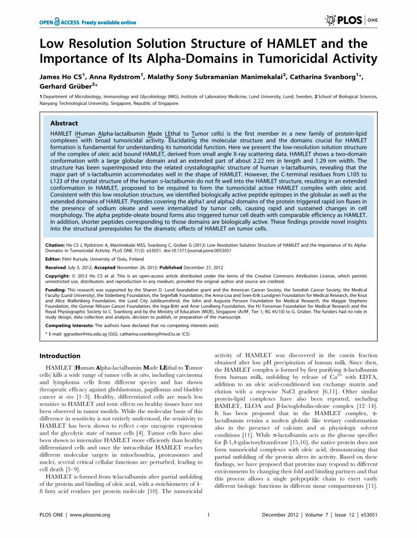
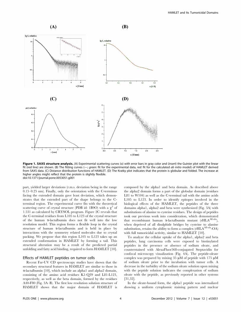
Low resolution solution structure of HAMLETThe high purity of HAMLET enabled us to perform small-angle
X-ray scattering experiments, to verify the proper three dimen-
sional folding and to determine the first low resolution structure of
this protein. SAXS patterns of HAMLET were recorded as
described in Materials and Methods to yield the final composite
scattering curve shown in figure 1A, which indicates a mono-
dispersed protein in solution. Inspection of the low angle of the
Guinier plots reveals a good data quality and no protein
aggregation (inset of Fig. 1A). HAMLET has a radius of gyration
(Rg) of 1.7860.05 nm and a maximum dimension Dmax of
5.6960.1 nm (Fig. 1C). Comparison of the forward scattering of
HAMLET with the values obtained from a reference solution of
bovine serum albumin, (BSA; 66.462 kDa) yields a molecular
mass of 1562 kDa, in agreement with the mass of a-lactalbumin
(24), indicating that HAMLET is monomeric at the concentrations
used. Qualitative analysis of the distance distribution function
suggests that HAMLET consists of a major portion, yielding a
principal maximum in the p(r) around 1.9 nm (Fig. 1C), whereas
the separated protuberance domain giving rise to a shoulder from
3.9 nm to 5.7 nm. The Kratky plot reveals that the protein is
globular and folded (Fig. 1D). The increase in higher angles in the
plot might indicate that the protein is slightly flexible.
In a complementary approach the native molecular mass of
HAMLET was determined via size exclusion chromatography. A
Superdex 75 gel filtration column was calibrated by determining
the Kav values for a set of standard proteins of known MM (Fig.
S1). A calibration curve based on these Kav values is shown in
figure S1B. Comparison of the Kav for HAMLET versus the
standard proteins suggests the native molecular mass (MM) of
approximately 1662 kDa.
The solution structure of HAMLET was restored ab initio from
the scattering patterns, shown in figure 1A. The obtained shape for
the protein yields a good fit to the experimental data in the entire
scattering range. The corresponding fit, shown in figure 1B, has a
discrepancies of x2 = 1.36. The protein appears as a two domain
molecule with a large globular domain and an extended part of
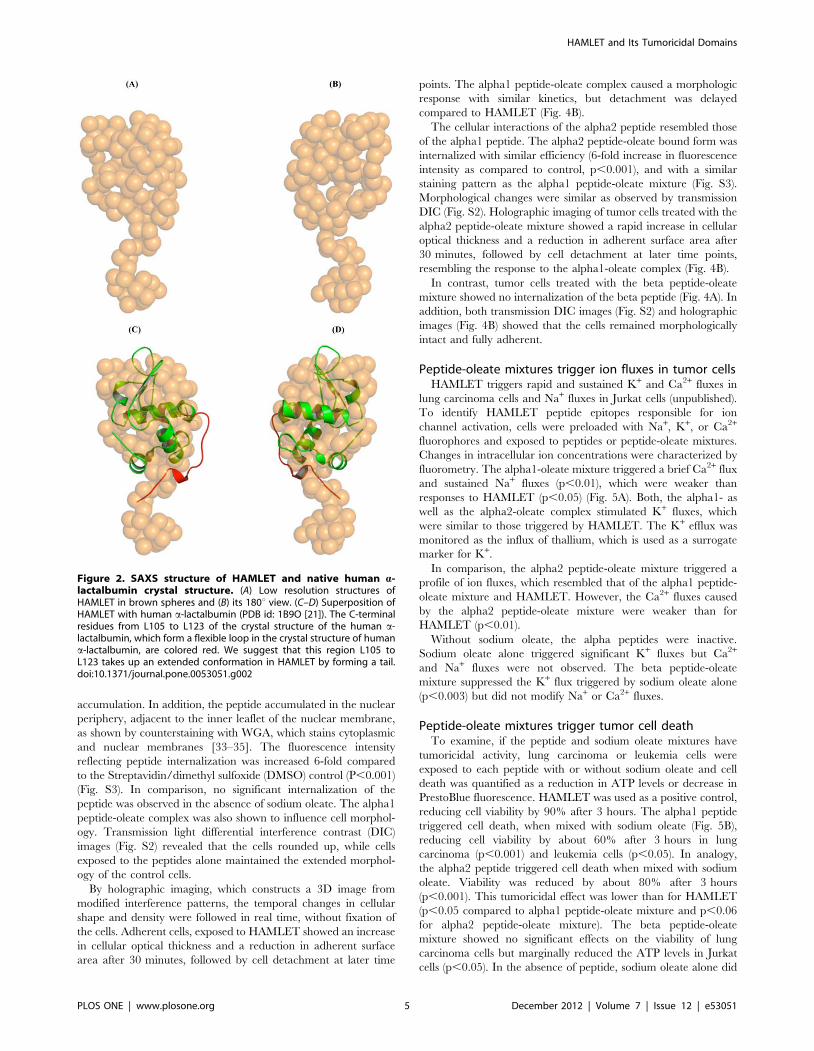
about 2.22 nm in length and 1.29 nm width (Fig. 2A–B). The
major domain has dimensions of about 3.1662.57 nm.
Superimposing the crystallographic structure of human a-
lactalbumin (PDB id: 1B9O) into the low resolution solution
structure of HAMLET shows that the major part of a-lactalbumin
accommodates well in the shape of HAMLET with an r.m.s.
deviation of 0.11 nm (Fig. 2C–D). Superposing other orientations
of the a-lactalbumin, such us the N-terminus near the extended
HAMLET and Its Tumoricidal Domains
PLOS ONE | www.plosone.org 3 December 2012 | Volume 7 | Issue 12 | e53051
part, yielded larger deviations (r.m.s. deviation being in the range
0.15–0.25 nm). Finally, only the orientation with the C-terminus
facing the extended domain gave least deviation, which demon-
strates that the extended part of the shape belongs to the C-
terminal region. The experimental curve fits with the theoretical
scattering curve of crystal structure (PDB id: 1B9O) with a x2 of
1.531 as calculated by CRYSOL program. Figure 2C reveals that
the C-terminal residues from L105 to L123 of the crystal structure
of the human a-lactalbumin does not fit well into the low
resolution model. This region forms a flexible loop in the crystal
structure of human a-lactalbumin and is held in place by
interactions with the symmetry related molecules due to crystal
packing. We propose that this region L105 to L123 takes up an
extended conformation in HAMLET by forming a tail. This
structural alteration may be a result of the predicted partial
unfolding and fatty acid binding, required to form HAMLET [10].
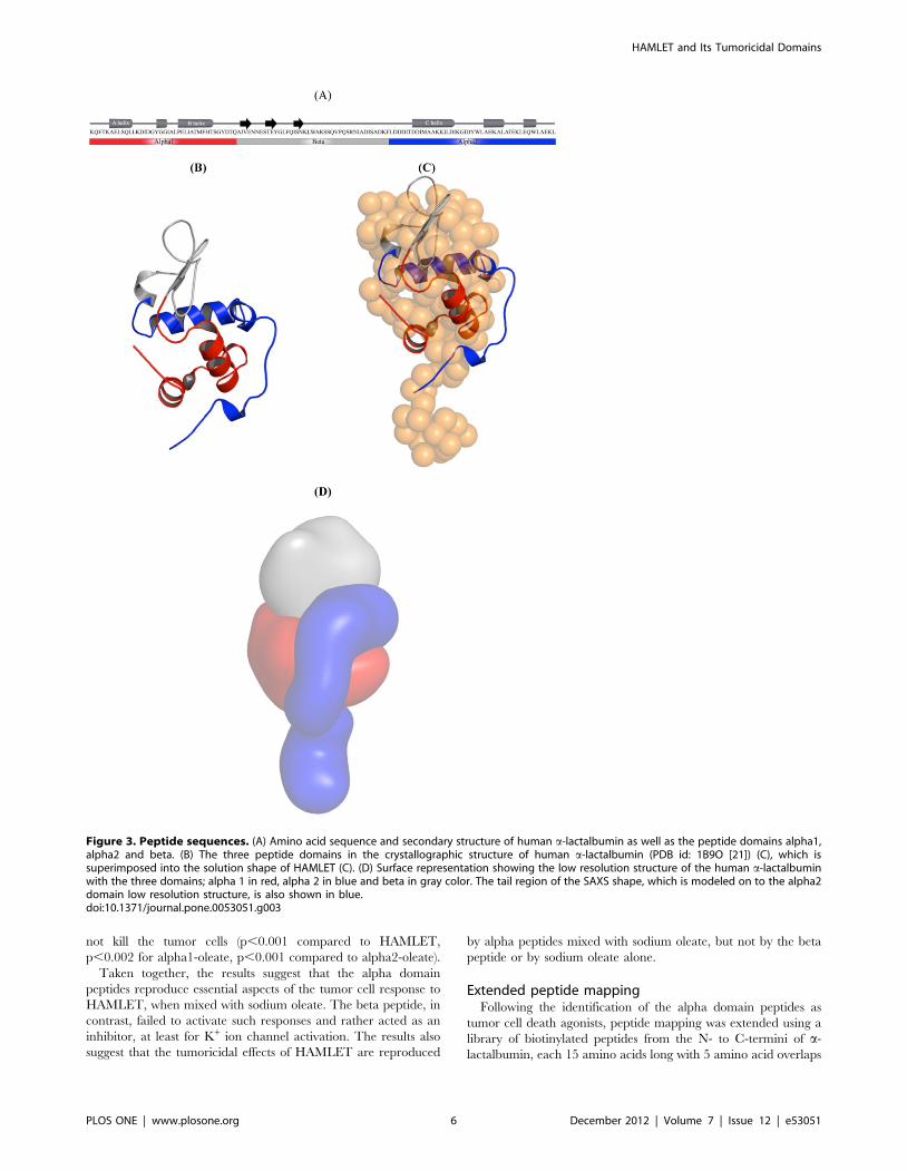
Effects of HAMLET peptides on tumor cellsRecent Far-UV CD spectroscopy studies have shown that the
secondary structural features in HAMLET are similar to those in
a-lactalbumin [10], which include an alpha1 and alpha2 domain,
consisting of the amino acid residues K1-Q39 and L81-L123,
respectively, as well as the beta domain, formed by the residues
A40-F80 (Fig. 3A–B). The first low resolution solution structure of
HAMLET shows that the major domain of HAMLET is
composed by the alpha1 and beta domain. As described above
the alpha2 domain forms a part of the globular domain (residues
L81 to W104) as well as the C-terminal tail with the amino acids
L105 to L123. In order to identify epitopes involved in the
biological effects of the HAMLET, the peptides of the three
domains alpha1, alpha2 and beta were synthesized (Fig. 3A) with
substitutions of alanine to cysteine residues. The design of peptides
took our previous work into consideration, which demonstrated
that recombinant human a-lactalbumin mutant (rHLAAll-ala),
when deprived of all disulphide bridges by cysteine to alanine
substitution, retains the ability to form a complex (rHLAAll-ala-OA)
with full tumoricidal activity, similar to HAMLET [10].
To analyze the cellular uptake of the alpha1, alpha2 and beta
peptides, lung carcinoma cells were exposed to biotinylated
peptides in the presence or absence of sodium oleate, and
counterstained with AlexaFluor568-conjugated Steptavidin for
confocal microscopy visualization (Fig. 4A). The peptide-oleate
complex was prepared by mixing 35 mM of peptide with 175 mM
of sodium oleate prior to the incubation with tumor cells. A
decrease in the turbidity of the sodium oleate solution upon mixing
with the peptide solution indicates the complexation of sodium
oleate with the peptide, as previously reported in other systems
[31,32].
In the oleate-bound form, the alpha1 peptide was internalized
showing a uniform cytoplasmic staining pattern and nuclear
Figure 1. SAXS structure analysis. (A) Experimental scattering curves (o) with error bars in gray color and (insert) the Guinier plot with the linearfit (red line) are shown. (B) The fitting curves (—; green: fit for the experimental data, red: fit for the calculated ab initio model) of HAMLET derivedfrom SAXS data. (C) Distance distribution functions of HAMLET. (D) The Kratky plot indicates that the protein is globular and folded. The increase athigher angles might reflect that the protein is slightly flexible.doi:10.1371/journal.pone.0053051.g001
HAMLET and Its Tumoricidal Domains
PLOS ONE | www.plosone.org 4 December 2012 | Volume 7 | Issue 12 | e53051
accumulation. In addition, the peptide accumulated in the nuclear
periphery, adjacent to the inner leaflet of the nuclear membrane,
as shown by counterstaining with WGA, which stains cytoplasmic
and nuclear membranes [33–35]. The fluorescence intensity
reflecting peptide internalization was increased 6-fold compared
to the Streptavidin/dimethyl sulfoxide (DMSO) control (P,0.001)
(Fig. S3). In comparison, no significant internalization of the
peptide was observed in the absence of sodium oleate. The alpha1
peptide-oleate complex was also shown to influence cell morphol-
ogy. Transmission light differential interference contrast (DIC)
images (Fig. S2) revealed that the cells rounded up, while cells
exposed to the peptides alone maintained the extended morphol-
ogy of the control cells.
By holographic imaging, which constructs a 3D image from
modified interference patterns, the temporal changes in cellular
shape and density were followed in real time, without fixation of
the cells. Adherent cells, exposed to HAMLET showed an increase
in cellular optical thickness and a reduction in adherent surface
area after 30 minutes, followed by cell detachment at later time
points. The alpha1 peptide-oleate complex caused a morphologic
response with similar kinetics, but detachment was delayed
compared to HAMLET (Fig. 4B).
The cellular interactions of the alpha2 peptide resembled those
of the alpha1 peptide. The alpha2 peptide-oleate bound form was
internalized with similar efficiency (6-fold increase in fluorescence
intensity as compared to control, p,0.001), and with a similar
staining pattern as the alpha1 peptide-oleate mixture (Fig. S3).
Morphological changes were similar as observed by transmission
DIC (Fig. S2). Holographic imaging of tumor cells treated with the
alpha2 peptide-oleate mixture showed a rapid increase in cellular
optical thickness and a reduction in adherent surface area after
30 minutes, followed by cell detachment at later time points,
resembling the response to the alpha1-oleate complex (Fig. 4B).
In contrast, tumor cells treated with the beta peptide-oleate
mixture showed no internalization of the beta peptide (Fig. 4A). In
addition, both transmission DIC images (Fig. S2) and holographic
images (Fig. 4B) showed that the cells remained morphologically
intact and fully adherent.
Peptide-oleate mixtures trigger ion fluxes in tumor cellsHAMLET triggers rapid and sustained K+ and Ca2+ fluxes in
lung carcinoma cells and Na+ fluxes in Jurkat cells (unpublished).
To identify HAMLET peptide epitopes responsible for ion
channel activation, cells were preloaded with Na+, K+, or Ca2+
fluorophores and exposed to peptides or peptide-oleate mixtures.
Changes in intracellular ion concentrations were characterized by
fluorometry. The alpha1-oleate mixture triggered a brief Ca2+ flux
and sustained Na+ fluxes (p,0.01), which were weaker than
responses to HAMLET (p,0.05) (Fig. 5A). Both, the alpha1- as
well as the alpha2-oleate complex stimulated K+ fluxes, which
were similar to those triggered by HAMLET. The K+ efflux was
monitored as the influx of thallium, which is used as a surrogate
marker for K+.
In comparison, the alpha2 peptide-oleate mixture triggered a
profile of ion fluxes, which resembled that of the alpha1 peptide-
oleate mixture and HAMLET. However, the Ca2+ fluxes caused
by the alpha2 peptide-oleate mixture were weaker than for
HAMLET (p,0.01).
Without sodium oleate, the alpha peptides were inactive.
Sodium oleate alone triggered significant K+ fluxes but Ca2+
and Na+ fluxes were not observed. The beta peptide-oleate
mixture suppressed the K+ flux triggered by sodium oleate alone
(p,0.003) but did not modify Na+ or Ca2+ fluxes.
Peptide-oleate mixtures trigger tumor cell deathTo examine, if the peptide and sodium oleate mixtures have
tumoricidal activity, lung carcinoma or leukemia cells were
exposed to each peptide with or without sodium oleate and cell
death was quantified as a reduction in ATP levels or decrease in
PrestoBlue fluorescence. HAMLET was used as a positive control,
reducing cell viability by 90% after 3 hours. The alpha1 peptide
triggered cell death, when mixed with sodium oleate (Fig. 5B),
reducing cell viability by about 60% after 3 hours in lung
carcinoma (p,0.001) and leukemia cells (p,0.05). In analogy,
the alpha2 peptide triggered cell death when mixed with sodium
oleate. Viability was reduced by about 80% after 3 hours
(p,0.001). This tumoricidal effect was lower than for HAMLET
(p,0.05 compared to alpha1 peptide-oleate mixture and p,0.06
for alpha2 peptide-oleate mixture). The beta peptide-oleate
mixture showed no significant effects on the viability of lung
carcinoma cells but marginally reduced the ATP levels in Jurkat
cells (p,0.05). In the absence of peptide, sodium oleate alone did
Figure 2. SAXS structure of HAMLET and native human a-lactalbumin crystal structure. (A) Low resolution structures ofHAMLET in brown spheres and (B) its 180u view. (C–D) Superposition ofHAMLET with human a-lactalbumin (PDB id: 1B9O [21]). The C-terminalresidues from L105 to L123 of the crystal structure of the human a-lactalbumin, which form a flexible loop in the crystal structure of humana-lactalbumin, are colored red. We suggest that this region L105 toL123 takes up an extended conformation in HAMLET by forming a tail.doi:10.1371/journal.pone.0053051.g002
HAMLET and Its Tumoricidal Domains
PLOS ONE | www.plosone.org 5 December 2012 | Volume 7 | Issue 12 | e53051
not kill the tumor cells (p,0.001 compared to HAMLET,
p,0.002 for alpha1-oleate, p,0.001 compared to alpha2-oleate).
Taken together, the results suggest that the alpha domain
peptides reproduce essential aspects of the tumor cell response to
HAMLET, when mixed with sodium oleate. The beta peptide, in
contrast, failed to activate such responses and rather acted as an
inhibitor, at least for K+ ion channel activation. The results also
suggest that the tumoricidal effects of HAMLET are reproduced
by alpha peptides mixed with sodium oleate, but not by the beta
peptide or by sodium oleate alone.
Extended peptide mappingFollowing the identification of the alpha domain peptides as
tumor cell death agonists, peptide mapping was extended using a
library of biotinylated peptides from the N- to C-termini of a-
lactalbumin, each 15 amino acids long with 5 amino acid overlaps
Figure 3. Peptide sequences. (A) Amino acid sequence and secondary structure of human a-lactalbumin as well as the peptide domains alpha1,alpha2 and beta. (B) The three peptide domains in the crystallographic structure of human a-lactalbumin (PDB id: 1B9O [21]) (C), which issuperimposed into the solution shape of HAMLET (C). (D) Surface representation showing the low resolution structure of the human a-lactalbuminwith the three domains; alpha 1 in red, alpha 2 in blue and beta in gray color. The tail region of the SAXS shape, which is modeled on to the alpha2domain low resolution structure, is also shown in blue.doi:10.1371/journal.pone.0053051.g003
HAMLET and Its Tumoricidal Domains
PLOS ONE | www.plosone.org 6 December 2012 | Volume 7 | Issue 12 | e53051
(Fig. 3A). Peptides 1–4 corresponded to the alpha1-, peptides 5–8
to the beta- and peptides 9–12 to the alpha2 peptide.
To examine the cellular uptake, A549 lung carcinoma cells were
treated with each peptide (105 uM, except for peptide 6, which
was added at 12 mM due to its lower solubility) and probed with
AlexaFluor568-conjugated Streptavidin for confocal microscopy
visualization (Fig. 6A). Peptides 1, 10 and 11 showed significant
internalization by the tumor cells and the strongest nuclear
localization. This effect was enhanced in the presence of sodium
oleate for peptides 10 and 11, but not for peptide 1, demonstrating
that certain peptides have independent cell-penetrating properties.
Remaining peptides were negative or internalized to a much lesser
extent (Fig. S4).
The effects on ion fluxes were examined in fluorophore-
preloaded cells (Fig. 6B–D). The two cell penetrating peptides 1
(p,0.01) and 11 (p,0.05) triggered K+ fluxes independent of
sodium oleate. Peptide 11 also increased Ca2+ fluxes in the
absence and presence of sodium oleate. Peptide10 induced a
weaker Ca2+ flux in the absence and presence of sodium oleate, as
did the peptide 6-oleate mixture. The baseline for K+ fluxes was
increased by sodium oleate and all ion fluxes generally by DMSO,
used to solubilize the biotinylated peptides. Weak Na+ fluxes were
recorded for peptide 6 and peptide 10 when mixed with sodium
Figure 4. Internalization of peptides into tumor cells and changes in tumor cell morphology. (A) Internalization of peptides. A549 lungcarcinoma cells cultured on glass slides, were incubated with peptide-oleate mixtures for 1 hour, fixed and stained with AlexaFluor568-streptavidin,counterstained with WGA and examined by confocal microscopy. Alpha1 and alpha2 peptides, mixed with oleate, were internalized as shown by thered fluorescence. The beta peptide was not internalized. Scale bar 20 mm. (B) Morphological changes in A549 lung carcinoma cells treated withHAMLET, alpha1 peptide+oleate, alpha2 peptide+oleate and beta peptide+oleate recorded by holography imaging. Cells treated with HAMLETstarted to round up after 30 minutes and after 60 minutes, many cells had detached. Alpha1 peptide+oleate mixture triggers similar morphologicalchanges as that by HAMLET. Alpha2 peptide+oleate mixture triggers similar morphological changes. Beta peptide+oleate mixture did not change cellmorphology.doi:10.1371/journal.pone.0053051.g004
HAMLET and Its Tumoricidal Domains
PLOS ONE | www.plosone.org 7 December 2012 | Volume 7 | Issue 12 | e53051
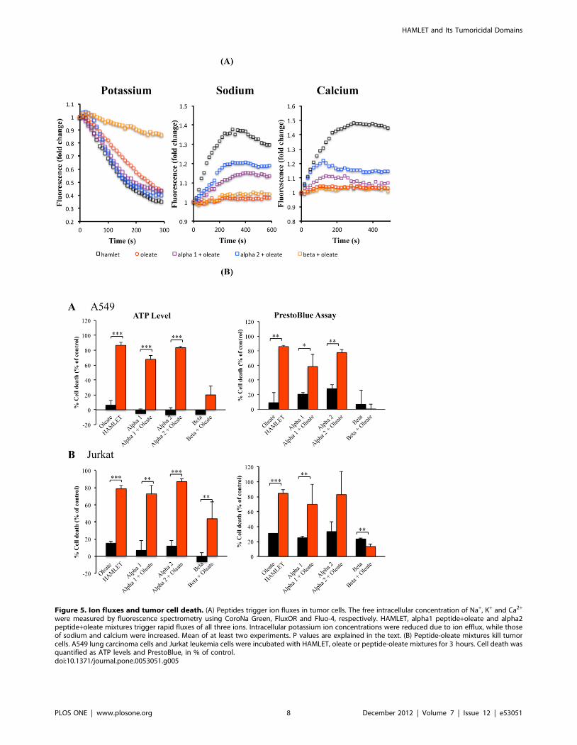
Figure 5. Ion fluxes and tumor cell death. (A) Peptides trigger ion fluxes in tumor cells. The free intracellular concentration of Na+, K+ and Ca2+
were measured by fluorescence spectrometry using CoroNa Green, FluxOR and Fluo-4, respectively. HAMLET, alpha1 peptide+oleate and alpha2peptide+oleate mixtures trigger rapid fluxes of all three ions. Intracellular potassium ion concentrations were reduced due to ion efflux, while thoseof sodium and calcium were increased. Mean of at least two experiments. P values are explained in the text. (B) Peptide-oleate mixtures kill tumorcells. A549 lung carcinoma cells and Jurkat leukemia cells were incubated with HAMLET, oleate or peptide-oleate mixtures for 3 hours. Cell death wasquantified as ATP levels and PrestoBlue, in % of control.doi:10.1371/journal.pone.0053051.g005
HAMLET and Its Tumoricidal Domains
PLOS ONE | www.plosone.org 8 December 2012 | Volume 7 | Issue 12 | e53051
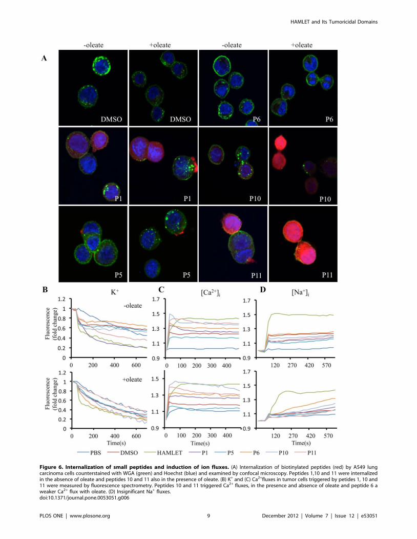
Figure 6. Internalization of small peptides and induction of ion fluxes. (A) Internalization of biotinylated peptides (red) by A549 lungcarcinoma cells counterstained with WGA (green) and Hoechst (blue) and examined by confocal microscopy. Peptides 1,10 and 11 were internalizedin the absence of oleate and peptides 10 and 11 also in the presence of oleate. (B) K+ and (C) Ca2+fluxes in tumor cells triggered by petides 1, 10 and11 were measured by fluorescence spectrometry. Peptides 10 and 11 triggered Ca2+ fluxes, in the presence and absence of oleate and peptide 6 aweaker Ca2+ flux with oleate. (D) Insignificant Na+ fluxes.doi:10.1371/journal.pone.0053051.g006
HAMLET and Its Tumoricidal Domains
PLOS ONE | www.plosone.org 9 December 2012 | Volume 7 | Issue 12 | e53051
oleate. K+ fluxes were comparable to those induced by HAMLET
but Ca2+and Na+ fluxes were weaker. A summary of the effects of
these peptides is included in table S1.
Cell viability was examined after a 3-hour exposure of lung
carcinoma cells to the peptides alone or peptide-oleate mixtures.
No tumoricidal activity was detected, suggesting that the
combined effect of several domains of the protein is needed to
trigger tumor cell death (Fig. S5).
Discussion
HAMLET represents a new family of protein-lipid complexes
with tumoricidal activity, whose structures have remained
unsolved. It has also been debated to what extent the lipid and
protein components of these complexes contribute to the cellular
responses leading to death. The presented low-resolution solution
structure of HAMLET shows that this protein has a two-domain
feature in solution in contrast to its compact precursor protein a-
lactalbumin. We suggest that the flexible loop at the C-terminal
region of the protein enables the amino acids L105 to L123 to
form an extended conformation in HAMLET and increase
thereby the overall surface and accessibility for the lipid as well
as the binding partner in the tumor cell (Fig. 3D).
Peptide mapping identified the alpha1 and alpha2 domains as
tumor cell ligands, involved in internalization, ion channel
activation and cell death. The SAXS structure of HAMLET
predicted that both of these domains might become more
accessible in HAMLET than in native a-lactalbumin. In the
native state, the alpha2 domain folds back onto the alpha1
domain, thus creating the globular shape [21]. The extended
conformation of the C-terminal residues from L105 to L123 in
HAMLET may thus have the dual effect of exposing novel peptide
epitopes in the alpha2 domain per se and of uncovering epitopes in
the alpha1 domain, which normally are protected in the native
protein structure. These findings extend and offer a structural
context to the results of Tolin et al, using peptide digests and oleic
acid [32]. As the fragments used did not exclusively cover the
alpha or the beta domain, a generalization of the role of amino
acid sequences led to the conclusion that oleic acid is the
biologically active entity. Our results define the existence of at least
two functional domains in alpha-lactalbumin that interact with
sodium oleate and form tumoricidal complexes. The tumoricidal
activity thus resides in both termini of the alpha domain but not
the beta domain, demonstrating that specific peptides are
involved. Further support for peptide specificity was obtained by
Baumann et. al. [36], studying the differential interaction of alpha-
lactalbumin fragments with phospholipid monolayers. In partic-
ular, Peptide A (residues 1–18) and Peptide C (75–100) showed
positive interactions and annular HAMLET structures were
observed on artificial lipid surfaces rather than the monomeric
structure observed in solution. The molecular shapes of HAM-
LET, when in contact with host cells, remain to be defined.
The general cytotoxicity of unsaturated fatty acids is well
recognized, including oleic acid [37–41]. High lipid concentrations
are cytolytic and mitochondrial membrane depolarization by
lipids may activate a caspase-independent cascade with inactiva-
tion of the Bcl-2-associated death promoter (BAD). HAMLET-like
protein-lipid complexes produced by other methods, such as
BAMLET (the HAMLET counterpart made from bovine alpha-
lactalbumin) with much higher lipid-protein ratios [42,43] may
therefore have properties different from those of the defined
HAMLET complex, which carries 5–8 oleic acid residues per
protein molecule [10]. Importantly, oleic acid alone is not
cytotoxic at the concentrations present in HAMLET and extensive
comparisons between oleic acid and HAMLET treated cells,
including genome-wide transcription analyses, have demonstrated
that oleic acid per se is virtually inert, as opposed to HAMLET
(unpublished). It is also important to note that other types of fatty
acids are less efficient in forming cytotoxic complexes and less
active than HAMLET [44], further suggesting that the protein
and lipid are both required for the specific response to HAMLET
and the increased effect on tumor cells.
Crystallographic and NMR structural comparisons between
bovine apo- and holo-a-lactalbumin have revealed structural
differences in the cleft between the two domains [45,46]. The
expansion in apo-a-lactalbumin of the Ca2+ binding loop tilts the
310 helix towards the C helix and disrupts the hydrophobic box
(aromatic cluster II) the interface between the two domains [24],
which involves residues W26, F53, W60, Y103 and W104 [47,48].
The current SAXS structure identifies additional structural
alterations in HAMLET. The extended conformation of L105–
L123 will inevitably alter the adjacent hydrophobic box and
thereby expanding the solvent exposed hydrophobic surface
compared to the native protein, as shown by increased 8-
anilinonaphthalene-1-sulfonate (ANS) binding [10]. A secondary
effect of this major structural alteration is in the exposure of the
alpha domain, represented by the alpha1 peptide in the current
study. Alterations in the alpha1 region potentially expose aromatic
cluster I in a-lactalbumin [47,49]. The biological activities shown
by the alpha domain peptides together with sodium oleate suggest
that both of these domains bind oleate and present the lipid to
tumor cells in a unique way, not reproduced by either constituent
alone. However, there was no evidence that exposure of the cleft
region by structural alterations created a new biological function
for the beta domain, which lacked activating activity in the cellular
assays. It has also been reported that a-lactalbumin forms high
molecular weight complexes with oleic acid [43]. Such complexes,
which also include complex formed from equine lysozyme [50],
contain much higher amounts of oleic acid than HAMLET and
may thus perturb cell membranes in a manner that resembles lysis
rather than programmed cell death.
HAMLET is rapidly internalized by tumor cells, which change
morphology, with rounding up, loss of cytoplasm and nuclear
condensation followed by death. The alpha-domain peptide-oleate
mixtures reproduced these end points. They were rapidly
internalized and caused significant changes in cell morphology
and loss of viability. Several of these responses may be attributable
to ion channel activation as ion channel inhibitors block
HAMLET internalization, morphologic change, gene expression
and death (unpublished). Like HAMLET, the alpha domain
peptide-oleate mixtures activated Na+, K+ and Ca2+ fluxes and
while these responses were weaker than in HAMLET treated cells,
they were qualitatively and quantitatively different from the
responses to sodium oleate alone. Sodium oleate alone only
activated K+ fluxes, suggesting that the peptides extend the ion
channel repertoire, despite being inactive on their own. Weaker
cellular responses might reflect a partial attack of each peptide
compared to HAMLET. Preliminary results suggest that when the
two alpha domains were combined, ion channel activation was
comparable to HAMLET, suggesting that several alpha-HAM-
LET epitopes may act in concert and that multiple interactions
may facilitate cellular attack.
Independent, cell-penetrating properties were detected in
peptides 1 and 11, from the alpha1 and alpha2 domains,
respectively (Fig. S6). These peptides triggered K+ fluxes, suggest
direct effects on tumor cell membranes. Peptide 11 also triggered
Ca2+ fluxes, suggesting a difference in specificity between peptides
1 and 11. It is interesting to speculate that different peptides
HAMLET and Its Tumoricidal Domains
PLOS ONE | www.plosone.org 10 December 2012 | Volume 7 | Issue 12 | e53051
activate different facets of the HAMLET ion flux repertoire. The
Na+, K+ and Ca2+ fluxes accompanying cell death were
reproduced by the alpha1- and alpha2 peptides, which were
cytotoxic in the presence of sodium oleate. The smaller peptides
triggered a more restricted repertoire of ion fluxes and were not
cytotoxic.
HAMLET is the first example of a protein that in its native state
exhibits a well-defined function but also acquires a different,
beneficial function after partial unfolding [11,51]. The properties
of HAMLET suggest a mechanism of structure-function variation,
which might generate significant structural diversity to meet
functional demands in different tissues. Moonlighting proteins, in
contrast, serve one main and several additional functions, while
undergoing a structural change to a different, well-folded state
[52]. More recently, intrinsically unstructured proteins were
included in the moonlighting group [53]. These proteins lack a
well-defined tertiary structure but can fulfill specific biological
functions. HAMLET is distinct from the moonlighting proteins
and from the intrinsically unfolded proteins, because it undergoes
partial unfolding and retains a defined three dimensional shape to
fulfill its new biological function as shown by the determined low
resolution solution structure of HAMLET. The low resolution
SAXS structure presented here supports the notion of HAMLET
as a largely monomeric molecular entity with disrupted globular-
ity, alteration of its tertiary structure in the domain remaining
globular and gain of a tail domain due to an extension of the C-
terminal portion of the molecule.
Supporting Information
Figure S1 Determination of the native molecular massby gel filtration analysis. (A), Superdex 75 gel filtration
analysis of HAMLET was performed as described under
‘‘Materials and Methods’’. The insert shows an SDS-PAGE of
the HAMLET fractions (grey area in the chromatogram), which
have been used for the SAXS experiments. (B) Proteins used as
molecular size standards (X) were BSA ((I), 67 kDa), ovalbumin
((II), 45 kDa), b-chymotrypsin A ((III), 25 kDa), ribonuclease A
((IV), 13.7 kDa) and subunit F (15 kDa) from the Methanosarcina
mazei Go1 A-ATP synthase (o). (B), for each protein, a Kav
parameter was derived as described under ‘‘EXPERIMENTAL
PROCEDURES’’. The Kav for HAMLET is indicated by (&).
(TIF)
Figure S2 Transmission light DIC images of cells inFig. 4A. A549 cells incubated for 1 h with alpha1-oleate or
alpha2-oleate show a round morphology, while cells incubated
with oleate-free alpha1-, alpha2-, beta peptides or with oleate
remain as flat and extended.
(TIF)
Figure S3 Quantification of peptide internalization. Low
magnification images of A549 cells incubated with DMSO,
alpha1-, alpha2- or beta peptides in the presence (A) or absence
(B) of oleate. The alpha1 and alpha2 peptides are internalized, if
incubated together with oleate. Without oleate, the peptides are
not internalized. Scale bar 100 mm. (C) Quantification of
internalization as measured by fluorescence intensity.
(TIF)
Figure S4 Quantification of red fluorescence intensityfor cells in Figure 6A by fluorescence microscopy.(TIF)
Figure S5 Effects of peptides on the viability of A549lung carcinoma cells. Cell viability was quantified as ATP
levels (A) and PrestoBlue (B) in % of control after 3 hours of
incubation.
(TIF)
Figure S6 Mapping of biologically active extendedpeptides. Peptide 1 (red) and peptide 11 (blue) are highlighted
in the crystallographic structure of human a-lactalbumin (PDB id:
1B9O [21]).
(TIF)
Table S1 A summary of the biological activities of theextended peptides.(DOC)
Acknowledgments
We acknowledge the EMBL-Outstation, Hamburg, Germany, for
provision of synchrotron radiation facility, and we thank Dr. M. Roessle
for his help in collecting SAXS-data.
Author Contributions
Conceived and designed the experiments: JHCS AR MSSM CS GG.
Performed the experiments: JHCS AR MSSM. Analyzed the data: JHCS
AR MSSM CS GG. Contributed reagents/materials/analysis tools: CS
GG. Wrote the paper: JHCS AR MSSM CS GG.
References
1. Fischer W, Gustafsson L, Mossberg AK, Gronli J, Mork S, et al. (2004) Human
alpha-lactalbumin made lethal to tumor cells (HAMLET) kills human
glioblastoma cells in brain xenografts by an apoptosis-like mechanism and
prolongs survival. Cancer Res 64: 2105–2112.
2. Gustafsson L, Leijonhufvud I, Aronsson A, Mossberg AK, Svanborg C (2004)
Treatment of Skin Papillomas with Topical a-Lactalbumin-Oleic Acid. New
Engl J Med 350: 2663–2672.
3. Mossberg AK, Hou Y, Svensson M, Holmqvist B, Svanborg C (2010) HAMLET
treatment delays bladder cancer development. J Urol 183: 1590–1597.
4. Storm P, Aits S, Puthia MK, Urbano A, Northen T, et al. (2011) Conserved
features of cancer cells define their sensitivity to HAMLET-induced death; c-
Myc and glycolysis. Oncogene 30: 4765–4779.
5. Duringer C, Hamiche A, Gustafsson L, Kimura H, Svanborg C (2003)
HAMLET interacts with histones and chromatin in tumor cell nuclei. J Biol
Chem 278: 42131–42135.
6. Svanborg C, Agerstam H, Aronson A, Bjerkvig R, Duringer C, et al. (2003)
HAMLET kills tumor cells by an apoptosis-like mechanism–cellular, molecular,
and therapeutic aspects. Adv Cancer Res 88: 1–29.
7. Aits S, Gustafsson L, Hallgren O, Brest P, Gustafsson M, et al. (2009) HAMLET
(human alpha-lactalbumin made lethal to tumor cells) triggers autophagic tumor
cell death. Int J Cancer 124: 1008–1019.
8. Gustafsson L, Aits S, Onnerfjord P, Trulsson M, Storm P, et al. (2009) Changes
in proteasome structure and function caused by HAMLET in tumor cells. PLoS
ONE 4: e5229.
9. Trulsson M, Yu H, Gisselsson L, Chao Y, Urbano A, et al. (2011) HAMLET
binding to a-actinin facilitates tumor cell detachment. PLoS One 6: e17179–
e17179.
10. Pettersson-Kastberg J, Mossberg AK, Trulsson M, Yong YJ, Min S, et al. (2009)
alpha-Lactalbumin, engineered to be nonnative and inactive, kills tumor cells
when in complex with oleic acid: a new biological function resulting from partial
unfolding. J Mol Biol 394: 994–1010.
11. Svensson M, Hakansson A, Mossberg AK, Linse S, Svanborg C (2000)
Conversion of alpha-lactalbumin to a protein inducing apoptosis. Proc Natl
Acad Sci U S A 97: 4221–4226.
12. Pettersson J, Mossberg AK, Svanborg C (2006) alpha-Lactalbumin species
variation, HAMLET formation, and tumor cell death. Biochem Biophys Res
Commun 345: 260–270 Epub 2006 Apr 2027.
13. Wilhelm K, Darinskas A, Noppe W, Duchardt E, Mok KH, et al. (2009) Protein
oligomerization induced by oleic acid at the solid-liquid interface–equine
lysozyme cytotoxic complexes. Febs J 276: 3975–3989.
14. Liskova K, Auty MAE, Chaurin V, Min S, Mok KH, et al. (2011) Cytotoxic
complexes of sodium oleate with b-lactoglobulin. Eur J Lipid Sci Technol 113:
1207–1218.
HAMLET and Its Tumoricidal Domains
PLOS ONE | www.plosone.org 11 December 2012 | Volume 7 | Issue 12 | e53051
15. Brodbeck U, Ebner KE (1966) Resolution of a soluble lactose synthetase into two
protein components and solubilization of microsomal lactose synthetase. J Biol
Chem 241: 762–764.
16. Brew K, Vanaman TC, Hill RL (1967) Comparison of the amino acid sequence
of bovine alpha-lactalbumin and hens egg white lysozyme. J Biol Chem 242:
3747–3749.
17. Svensson M, Fast J, Mossberg AK, Duringer C, Gustafsson L, et al. (2003)
Alpha-lactalbumin unfolding is not sufficient to cause apoptosis, but is required
for the conversion to HAMLET (human alpha-lactalbumin made lethal to
tumor cells). Protein Sci 12: 2794–2804.
18. Boulin C, Kempf R, Koch MHJ, McLaughlin SM (1986) Data appraisal,
evalution and display for synchrotron radiation experiments: hardware and
software. Nucl Instr & Meth in Phys Res 249: 399–407.
19. Roessle MW, Klaering R, Ristau U, Robrahn B, Jahn D, et al. (2007) Upgrade
of the small-angle X-ray scattering beamline X33 at the European Molecular
Biology Laboratory, Hamburg. J Appl Crystallogr 40: 190–194.
20. Round AR, Franke D, Moritz S, Huchler R, Fritsche M, et al. (2008) Automated
sample-changing robot for solution scattering experiments at the EMBL
Hamburg SAXS station X33. J Appl Crystallogr 41: 913–917.
21. Svergun DI (1993) A direct indirect method of small-angle scattering data
treatment. J Appl Crystallogr 26: 258–267.
22. Guinier A, Fournet G (1955) Small-angle Scattering of X-rays, Wiley, New
York.
23. Svergun DI, Petoukhov MV, Koch MH (2001) Determination of domain
structure of proteins from X-ray solution scattering. Biophys J 80: 2946–2953.
24. Harata K, Abe Y, Muraki M (1999) Crystallographic evaluation of internal
motion of human alpha-lactalbumin refined by full-matrix least-squares method.
J Mol Biol 287: 347–358.
25. Wu LC, Kim PS (1997) Hydrophobic sequence minimization of the a-
lactalbumin molten globule. Proc Natl Acad Sci U S A 94: 14314–14319.
26. Cuche E, Bevilacqua F, Depeursinge C (1999) Digital holography for
quantitative phase-contrast imaging. Opt Lett 24: 291–293.
27. Gustafsson M, Sebesta M, Bengtsson B, Pettersson SG, Egelberg P, et al. (2004)
High-resolution digital transmission microscopy – a Fourier holography
approach. Opt Lasers Eng 41: 553–563.
28. Schnars U, Juptner W (2005) Digital Holography: Digital Hologram Recording,
Numerical Reconstruction, and Related Techniques, Springer Berlin.
29. Sebesta M, Gustafsson M (2005) Object characterization with refractometric
digital Fourier holography. Opt Lett 30: 471–473.
30. Dubois F, Yourassowsky C, Monnom O, Legros JC, Debeir O, et al. (2006)
Digital holographic microscopy for the three-dimensional dynamic analysis of in
vitro cancer cell migration. J Biomed Opt 11: 054032.
31. Campbell J, Martucci AD, Green GR (1964) Plasma albumin as an acceptor of
free fatty acids. Biochem J 93: 183–189.
32. Tolin S, De Franceschi G, Spolaore B, Frare E, Canton M, et al. (2010) The
oleic acid complexes of proteolytic fragments of alpha-lactalbumin display
apoptotic activity. FEBS J 277: 163–173.
33. Fitzgerald LA, Denny JB, Baumbach GA, Ketcham CM, Roberts RM (1984)
Effect of altered oligosaccharide structure on the cell surface number,
distribution and turnover of the high molecular weight acidic glycoproteins of
CHO cells. J Cell Sci 67: 1–23.
34. Wright CS (1984) Structural comparison of the two distinct sugar binding sites in
wheat germ agglutinin isolectin II. J Mol Biol 178: 91–104.
35. Nakano Y, Banfi B, Jesaitis AJ, Dinauer MC, Allen LA, et al. (2007) Critical
roles for p22phox in the structural maturation and subcellular targeting of Nox3.Biochem J 403: 97–108.
36. Baumann A, Gjerde AU, Ying M, Svanborg C, Holmsen H, et al. (2012)
HAMLET forms annular oligomers when deposited with phospholipidmonolayers. J Mol Biol 418: 90–102.
37. Dymkowska D, Szczepanowska J, Wojtczak L (2004) Fatty-Acid-inducedapoptosis in ehrlich ascites tumor cells. Toxicol Mech Methods 14: 73–77.
38. Zhu Y, Schwarz S, Ahlemeyer B, Grzeschik S, Klumpp S, et al. (2005) Oleic
acid causes apoptosis and dephosphorylates Bad. Neurochem Int 46: 127–135.39. Dymkowska D, Szczepanowska J, Wieckowski MR, Wojtczak L (2006) Short-
term and long-term effects of fatty acids in rat hepatoma AS-30D cells: the wayto apoptosis. Biochim Biophys Acta 1763: 152–163.
40. Fernanda Cury-Boaventura M, Cristine Kanunfre C, Gorjao R, Martins deLima T, Curi R (2006) Mechanisms involved in Jurkat cell death induced by
oleic and linoleic acids. Clin Nutr 25: 1004–1014.
41. Yu F, Lu S, Shi J, McGuire PM, Wang R (2008) Cytotoxic activity of anoctadecenoic acid extract from Euphorbia kansui (Euphorbiaceae) on human
tumour cell strains. J Pharm Pharmacol 60: 253–259.42. Brinkmann CR, Heegaard CW, Petersen TE, Jensenius JC, Thiel S (2011) The
toxicity of bovine alpha-lactalbumin made lethal to tumor cells is highly
dependent on oleic acid and induces killing in cancer cell lines and noncancer-derived primary cells. Febs J 278: 1955–1967.
43. Spolaore B, Pinato O, Canton M, Zambonin M, Polverino de Laureto P, et al.(2010) a-Lactalbumin Forms with Oleic Acid a High Molecular Weight
Complex Displaying Cytotoxic Activity. Biochemistry 49: 8658–8667.44. Svensson M, Mossberg AK, Pettersson J, Linse S, Svanborg C (2003) Lipids as
cofactors in protein folding: stereo-specific lipid-protein interactions are required
to form HAMLET (human alpha-lactalbumin made lethal to tumor cells).Protein Sci 12: 2805–2814.
45. Chrysina ED, Brew K, Acharya KR (2000) Crystal structures of apo- and holo-bovine alpha-lactalbumin at 2.2 A resolution reveal an effect of calcium on inter-
lobe interactions. J Biol Chem 275: 37021–37029.
46. Wijesinha-Bettoni R, Dobson CM, Redfield C (2001) Comparison of thestructural and dynamical properties of holo and apo bovine alpha-lactalbumin
by NMR spectroscopy. J Mol Biol 307: 885–898.47. Koga K, Berliner LJ (1985) Structural elucidation of a hydrophobic box in
bovine alpha-lactalbumin by NMR: nuclear Overhauser effects. Biochemistry24: 7257–7262.
48. Acharya KR, Stuart DI, Walker NPC, Lewis M, Phillips DC (1989) Refined
structure of baboon a-lactalbumin at 1.7 A resolution: Comparison with C-typelysozyme. J Mol Biol 208: 99–127.
49. Alexandrescu AT, Broadhurst RW, Wormald C, Chyan C-L, Baum J, et al.(1992) 1H-NMR assignments and local environments of aromatic residues in
bovine, human and guinea pig variants of a-lactalbumin. Eur J Biochem 210:
699–709.50. Wilhelm K, Darinskas A, Noppe W, Duchardt E, Mok KH, et al. (2009) Protein
oligomerization induced by oleic acid at the solid-liquid interface–equinelysozyme cytotoxic complexes. Febs J 276: 3975–3989.
51. Svensson M, Duringer C, Hallgren O, Mossberg AK, Hakansson A, et al. (2002)Hamlet – a complex from human milk that induces apoptosis in tumor cells but
spares healthy cells. Adv Exp Med Biol 503: 125–132.
52. Jeffery CJ (2003) Moonlighting proteins: old proteins learning new tricks. TrendsGenet 19: 415–417.
53. Jeffery CJ (2009) Moonlighting proteins – an update. Mol Biosyst 5: 345–350.
HAMLET and Its Tumoricidal Domains
PLOS ONE | www.plosone.org 12 December 2012 | Volume 7 | Issue 12 | e53051