ECG Interpretation - Amarillo College 5 Junctional Escape Rhythm Occurs if significant sinus...
31
1 ECG Interpretation Part 2 Junctional Rhythms Junctional Escape Rhythm
Transcript of ECG Interpretation - Amarillo College 5 Junctional Escape Rhythm Occurs if significant sinus...
1
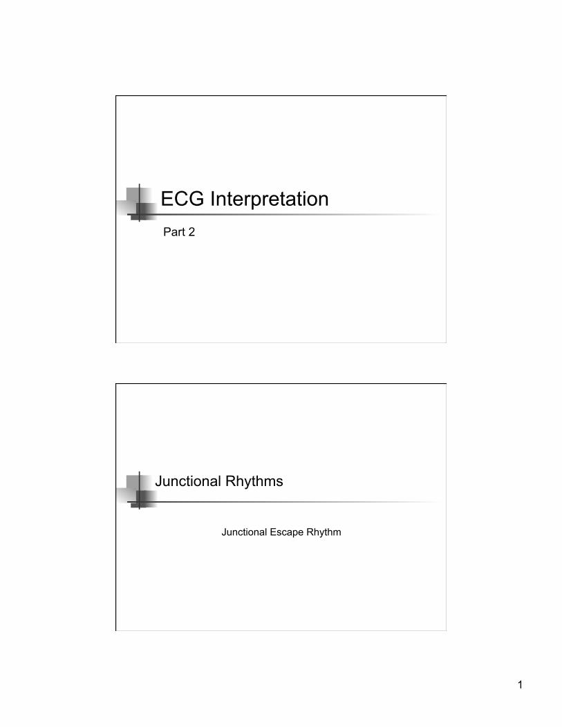
ECG Interpretation
Part 2
Junctional Rhythms
Junctional Escape Rhythm
2
3
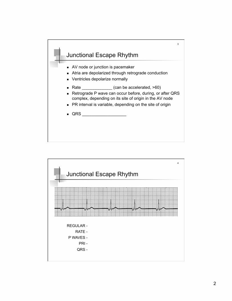
Junctional Escape Rhythm
AV node or junction is pacemaker Atria are depolarized through retrograde conduction Ventricles depolarize normally
Rate _____________ (can be accelerated, >60) Retrograde P wave can occur before, during, or after QRS
complex, depending on its site of origin in the AV node PR interval is variable, depending on the site of origin
QRS ___________________
4
Junctional Escape Rhythm
REGULAR - RATE -
P WAVES - PRI -
QRS -

3
5
Junctional Escape Rhythm
Occurs if significant sinus bradycardia or complete heart block is present
Sometimes exceeds the sinus node rate at a time when the sinus rate would be normal (dig toxicity)
Heart rate determines presence of symptoms Morbidity & mortality related more to cause (sick sinus
node or complete heart block)
6
Junctional Escape Rhythm
Causes Sick sinus syndrome (including drug-induced) Digoxin toxicity Ischemia of the AVN, especially with acute inferior infarction Acutely after cardiac surgery Acute inflammatory processes (eg, acute rheumatic fever),
which may involve the conduction system Other drugs (eg, beta-blockers, calcium blockers, most
antiarrhythmic agents) that cause sinus bradycardia Treatment (depends on cause)
permanent pacemaker, if sick sinus or 3° block atropine, if dig toxicity

4
Bundle Branch Blocks
8
Bundle Branch Blocks
Right and left bundle branches send the electrical impulse to the right and left ventricle simultaneously
When the bundle branches are functioning normally, the right and left ventricles contract at the same time
BBB occurs when one of the bundle branches becomes diseased or damaged, and stops conducting electrical impulses; that is, a bundle branch becomes “blocked”
5
9
Bundle Branch Blocks
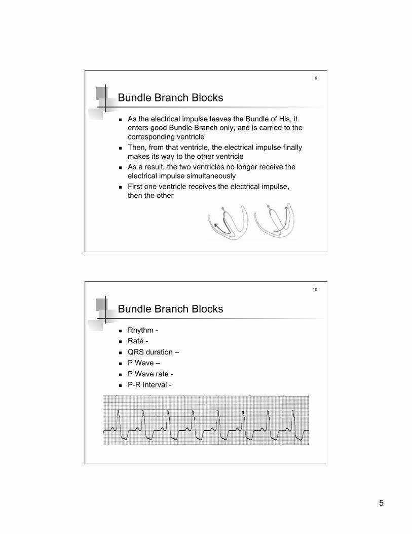
As the electrical impulse leaves the Bundle of His, it enters good Bundle Branch only, and is carried to the corresponding ventricle
Then, from that ventricle, the electrical impulse finally makes its way to the other ventricle
As a result, the two ventricles no longer receive the electrical impulse simultaneously
First one ventricle receives the electrical impulse, then the other
10
Bundle Branch Blocks
Rhythm - Rate - QRS duration – P Wave – P Wave rate - P-R Interval -

6
11
Right Bundle Branch Block
Occurs in medical conditions that affect the right side of the heart or the lungs pulmonary embolus chronic lung disease cardiomyopathy atrial and ventricular septal defects
Observation of RBBB should trigger a screening exam for above conditions
RBBB is also commonly occurs in normal, healthy individuals
12
Left Bundle Branch Block
Usually indicates underlying cardiac pathology cardiomyopathy hypertension aortic valve disease coronary artery disease a variety of other cardiac conditions
Its appearance should trigger a thorough search (as opposed to a simple screening) for underlying cardiac problems
7
13
Identify BBB
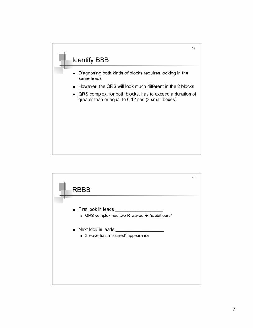
Diagnosing both kinds of blocks requires looking in the same leads
However, the QRS will look much different in the 2 blocks
QRS complex, for both blocks, has to exceed a duration of greater than or equal to 0.12 sec (3 small boxes)
14
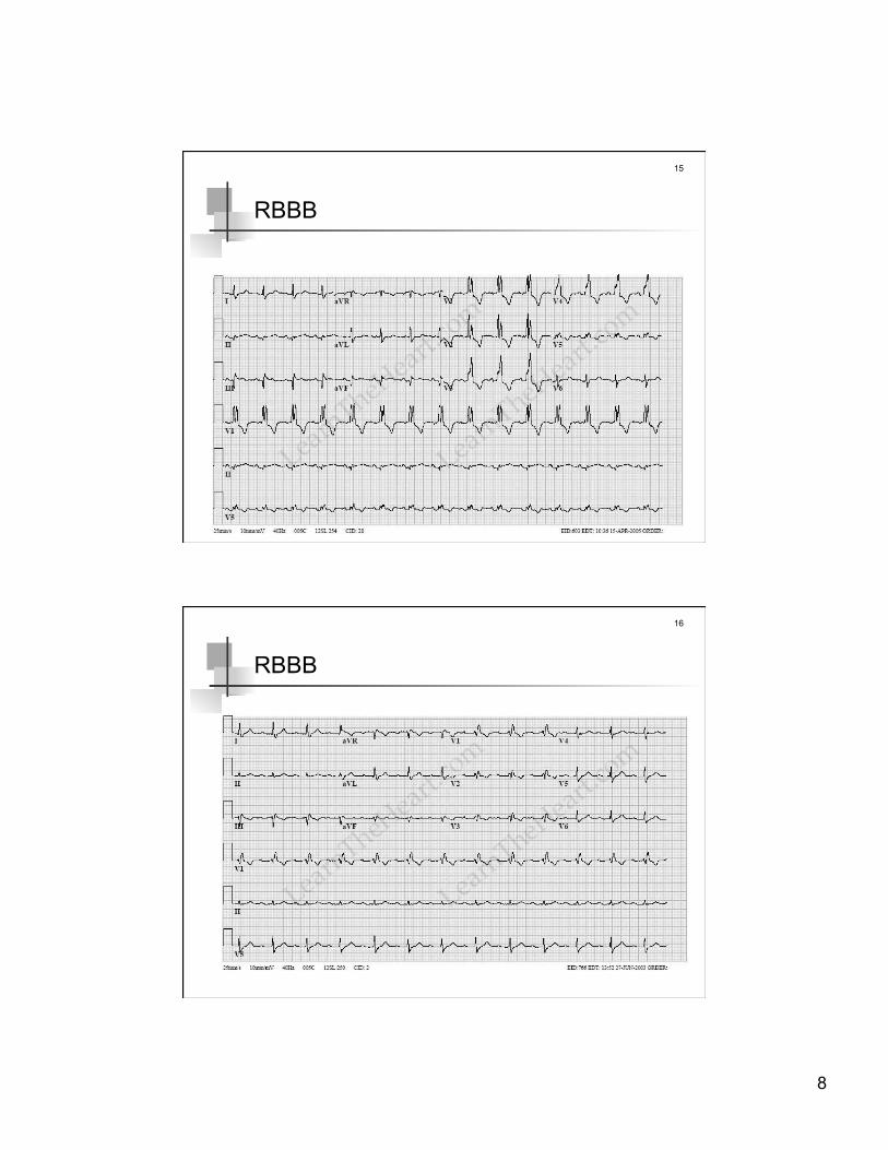
RBBB
First look in leads ___________________ QRS complex has two R-waves “rabbit ears”
Next look in leads ___________________ S wave has a “slurred” appearance
8
15
RBBB
16
RBBB

9
17
LBBB
First look in leads ___________________ QRS is wide, mostly upright, and the T waves are inverted
Next look in leads ___________________ if QRS complexes are mostly negative (like big Q waves)
and the T waves are upright, then you are for sure looking at LBBB
18
LBBB

10
19
LBBB
20
Bundle Branch Block
Treatment heart depends on the bundle branches without them, the electrical impulse is not delivered to the
ventricles block in both bundle branches, therefore (a condition called
complete heart block) can be fatal but is rare
if RBBB or LBBB is accompanied by syncope _________
block of both BB ___________________
11
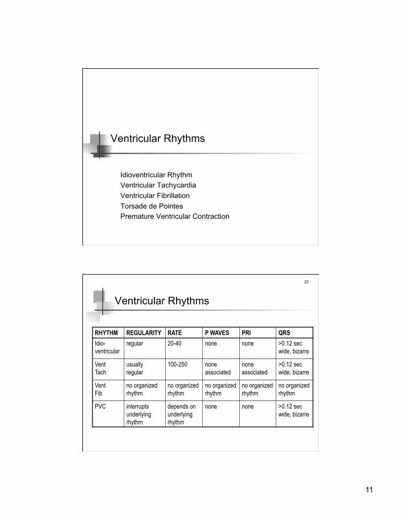
Ventricular Rhythms
Idioventricular Rhythm Ventricular Tachycardia Ventricular Fibrillation Torsade de Pointes Premature Ventricular Contraction
22
Ventricular Rhythms
RHYTHM REGULARITY RATE P WAVES PRI QRS Idio- ventricular
regular 20-40 none none >0.12 sec wide, bizarre
Vent Tach
usually regular
100-250 none associated
none associated
>0.12 sec wide, bizarre
Vent Fib
no organized rhythm
no organized rhythm
no organized rhythm
no organized rhythm
no organized rhythm
PVC interrupts underlying rhythm
depends on underlying rhythm
none none >0.12 sec wide, bizarre
12
23
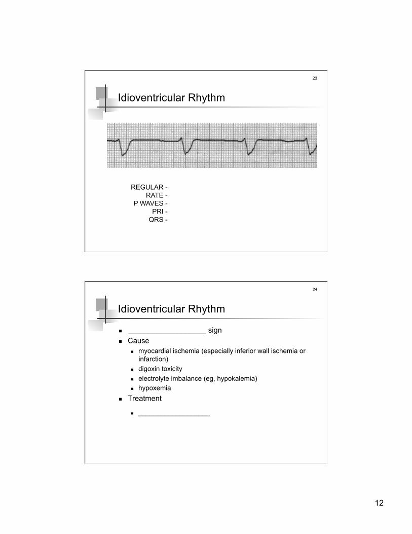
Idioventricular Rhythm
REGULAR - RATE -
P WAVES - PRI -
QRS -
24
Idioventricular Rhythm
___________________ sign Cause
myocardial ischemia (especially inferior wall ischemia or infarction)
digoxin toxicity electrolyte imbalance (eg, hypokalemia) hypoxemia
Treatment
___________________

13
25
Ventricular Tachycardia
REGULAR - RATE -
P WAVES - PRI -
QRS -
26
Ventricular Tachycardia (Monomorphic)

14
27
Ventricular Tachycardia (Polymorphic)
= torsade de pointes
28
Ventricular Tachycardia
Results from abnormal tissues in the ventricles generating a rapid and irregular heart rhythm coronary artery disease hypokalemia cocaine use
Poor cardiac output is usually associated with this rhythm thus causing the patient to go into cardiac arrest
If loss of consciousness, hypotension, no pulse -
_____________________________________ If hemodynamic status is stable + no evidence of coronary
ischemia or infarction --> rhythm conversion
_____________________________________

15
29
Ventricular Fibrillation
REGULAR - RATE -
P WAVES - PRI -
QRS -
30
Ventricular Fibrillation
Causes cardiac
myocardial ischemia or infarction due to coronary artery disease
cardiomyopathy, myocarditis aortic stenosis pericardial tamponade congenital heart disease electrical accidents heart block
respiratory bronchospasm aspiration primary pulmonary hypertension pulmonary embolism tension pneumothorax

16
31
Ventricular Fibrillation
Causes (con’t) metabolic or toxic
electrolyte disturbances and acidosis medications or drug ingestion environmental poisoning sepsis
neurologic seizure cerebrovascular accident (intracranial hemorrhage or ischemic
stroke) drowning
32
Ventricular Fibrillation
Treatment prehospital care is vital for arrests due to VF that occur
outside the hospital witnessed or early recognition of an arrest early activation of emergency medical services (EMS) system bystander CPR slows the degeneration of VF and improves
survival automated external defibrillator (AED) application and
defibrillation by trained personnel in the field early access to trained EMS personnel capable of performing
CPR, defibrillation, and advanced cardiac life support (ACLS)

17
33
Ventricular Fibrillation
Treatment hospital care (ACLS) = SCREAM
S Shock 360J monophasic, 200J biphasic, 1st and subsequent shocks. (Shock every 2 minutes if indicated)
C CPR After shock, immediately begin chest compressions followed by respirations (30:2 ratio) for 2 minutes. (Do not check rhythm or pulse)
R Rhythm Rhythm check after 2 minutes of CPR (and after every 2 minutes of CPR thereafter) and shock again if indicated. Check pulse only if an organized or non-shockable rhythm is present.
34
Ventricular Fibrillation
E Epinephrine 1 mg IV/IO q3-5 min. Or vasopressin 40 U IV/IO, once, in place of the 1st or 2nd dose of epi.
AM Antiarrhythmic Medications
Consider antiarrhythmics: Amiodarone 300mg IV/IO, may repeat once at 150mg in 3-5 min. if VF/PVT persists or Lidocaine (if amiodarone unavailable) 1.0-1.5 mg/kg IV/IO, may repeat X 2, q5-10 min. at 0.5-0.75 mg/kg, (3mg/kg max. loading dose) if VF/PVT persists, or Magnesium Sulfate1-2 g IV/IO diluted in 10mL D5W (5-20 min. push) for torsades de pointes or suspected/ known hypomagnesemia.

18
35
Premature Ventricular Contractions
Odd QRS waveforms = ventricles depolarizing prematurely in response to increased automaticity within the ventricles
Electrical impulse does not pass over the ventricles in the regular, organized way --> result may be a heart beat that is less effective in pumping blood
In addition, since the PVC comes early, the left ventricle is not properly filled, so the cardiac output from these premature beats is lower than a normal sinus beat
36
Premature Ventricular Contractions
Occur in normal heart anxiety excessive use of alcohol, caffeine, tobacco, cocaine meds (epinephrine, theophylline)
Occur in diseased heart ischemia
Other causes acidosis, electrolyte imbalance CHF, MI hypoxia

19
37
Premature Ventricular Contractions
Single PVC poses no problems, but may signal serious problems
Causes for concern increased frequency (> 6/minute) multifocal couplets salvos R-on-T phenomenon
38
Premature Ventricular Contractions
REGULAR - RATE -
P WAVES - PRI -
QRS -
20
39
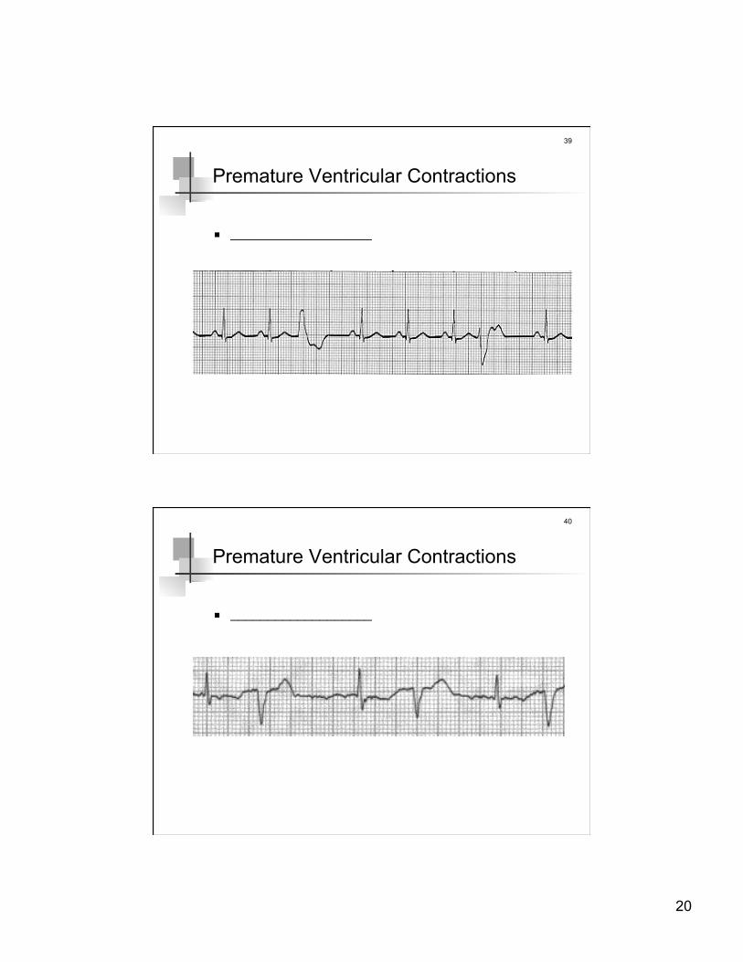
Premature Ventricular Contractions
___________________
40
Premature Ventricular Contractions
___________________

21
41
Premature Ventricular Contractions
___________________
42
Premature Ventricular Contractions
___________________

22
43
Premature Ventricular Contractions
___________________
44
Premature Ventricular Contractions
___________________
23
45
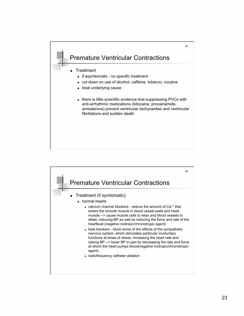
Premature Ventricular Contractions
Treatment if asymtomatic - no specific treatment cut down on use of alcohol, caffeine, tobacco, cocaine treat underlying cause
there is little scientific evidence that suppressing PVCs with anti-arrhythmic medications (lidocaine, procainamide, amiodarone) prevent ventricular tachycardias and ventricular fibrillations and sudden death
46
Premature Ventricular Contractions
Treatment (if symtomatic) normal hearts
calcium channel blockers - reduce the amount of Ca++ that enters the smooth muscle in blood vessel walls and heart muscle --> cause muscle cells to relax and blood vessels to dilate, reducing BP as well as reducing the force and rate of the heartbeat (negative inotropic/chronotropic agent)
beta blockers - block some of the effects of the sympathetic nervous system, which stimulates particular involuntary functions at times of stress, increasing the heart rate and raising BP --> lower BP in part by decreasing the rate and force at which the heart pumps blood(negative inotropic/chronotropic agent)
radiofrequency catheter ablation

24
47
Premature Ventricular Contractions
Treatment (with structural heart disease) PVCs may be a sign of an oncoming dangerous heart
rhythm, loss of consciousness, or even cardiac arrest treatment strategy depends upon the severity of the
underlying heart disease if heart muscle function is severely decreased (due to prior
heart attack, cardiomyopathy or significant hypertrophic cardiomyopathy), an implantable cardioverter defibrillator (ICD) may be recommended
Others

25
49
Pulseless Electrical Activity
Formerly - electromechanical dissociation (EMD) Dissociation of electrical & mechanical activity = electrical
activity --> ___________________ Is rare Causes - ___________________
Hypovolemia Toxins (OD)
Hypoxia Tamponade, cardiac
Hydrogen ion (acidosis) Tension pneumothorax
Hypo-/Hyperkalemia Thrombosis, pulmonary
Hypoglycemia Trauma
Hypothermia
50
Pulseless Electrical Activity
Treatment CPR (intubate), monitor, IV confirm in another lead consider possible causes & treatments epinephrine
1 mg IV every 3-5 minutes atropine
1 mg IV every 3-5 minutes to a total of 0.04 mg/kg consider termination of efforts

26
51
Asystole
Is an arrhythmia, not a dysrhythmia
= ___________________ Usually fatal unless rhythm rapidly restored Straight or almost straight line
52
Asystole
Causes ___________________
Hypovolemia Toxins (OD)
Hypoxia Tamponade, cardiac
Hydrogen ion (acidosis) Tension pneumothorax
Hypo-/Hyperkalemia Thrombosis, pulmonary
Hypoglycemia Trauma
Hypothermia
27
53
Asystole
Treatment CPR (intubate), monitor, IV confirm in another lead consider possible causes & treatments epinephrine
1 mg IV every 3-5 minutes atropine
1 mg IV every 3-5 minutes to a total of 0.04 mg/kg consider termination of efforts
Agonal Rhythm
28
55
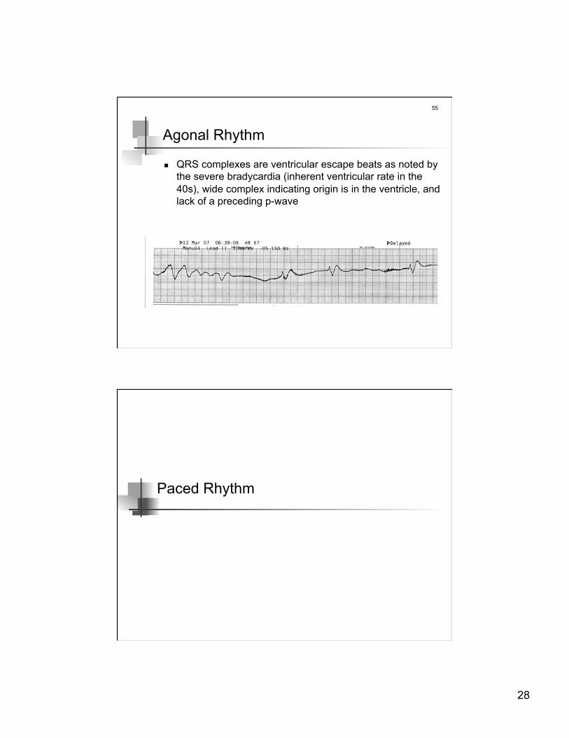
Agonal Rhythm
QRS complexes are ventricular escape beats as noted by the severe bradycardia (inherent ventricular rate in the 40s), wide complex indicating origin is in the ventricle, and lack of a preceding p-wave
Paced Rhythm
29
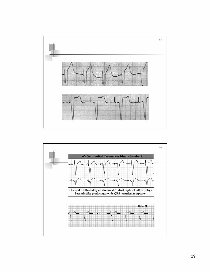
57
58

30
COPD
60
COPD
COPD patients typically have ECG abnormalities Right axis deviation
due to hyperinflation of lungs & flattening of diaphragms --> more vertical position of heart
Right ventricular hypertrophy due to pulmonary hypertension --> right heart enlargement
(cor pulmonale) Dysrhythmias often seen
tachycardia multifocal atrial tachycardia PVCs RBBB

31
61
practice strips http://monroecc.edu/Depts/pstc/backup/prandekg.htm http://www.skillstat.com/Flash/ECGSim531.html http://sprojects.mmi.mcgill.ca/heart/puz990914r1.html
ACLS http://www.acls.net/aclsalg.htm