A thesis by - COnnecting REpositories and pharmacological investigation of a benzimidazole...
109
Clinical and pharmacological investigation of a benzimidazole anthelmintic against donkeys’ worm infestation. A thesis by Sawsan Mohmmed Ahmed Imam B. V. Sc. 2005 University of Nyala Supervisor Prof. Tigani Hassan Al amin Department of Medicine, pharmacology and toxicology. Faculty of Veterinary Medicine, University of Khartoum. CoSupervisor Dr. Hisham Ismail Seri Department of Clinical Studies. Faculty of Veterinary Science University of Nyala A thesis submitted to the graduate College University of Khartoum in partial fulfillment of the requirements of master degree in veterinary Medicine (Pharmacology) March 2009
Transcript of A thesis by - COnnecting REpositories and pharmacological investigation of a benzimidazole...
Clinical and pharmacological investigation
of a benzimidazole anthelmintic against
donkeys’ worm infestation.
A thesis by
Sawsan Mohmmed Ahmed Imam
B. V. Sc. 2005 University of Nyala
Supervisor
Prof. Tigani Hassan Al amin
Department of Medicine, pharmacology and toxicology. Faculty of
Veterinary Medicine, University of Khartoum.
CoSupervisor
Dr. Hisham Ismail Seri
Department of Clinical Studies. Faculty of Veterinary Science
University of Nyala
A thesis submitted to the graduate College University of Khartoum in
partial fulfillment of the requirements of master degree in veterinary
Medicine (Pharmacology)
March 2009

Dedication
This work is dedicated to
Soul of my Father, my great Mother
my kind brothers, my lovely aunties
Asmaa and Mariam

TABLE OF CONTENTS
Table of contents………………………………..….………………………. I
List of tables…………….…………………………..…………..………….. IV
List of figures………………………………………..…….……….…...... VI
Acknowledgements……………….………………….…………………… VIII
English Abstract………………………………………………………......... IX
Arabic abstract………………………………………….…………….….. XI
Chapter one: Introduction and literature review
Introduction ………….…….………………………………...….……….. 1
1.1 Albendazole ……………………………………………………. 4
1.1.1 Identity…………………………………..……….…..………... 4
1.1.2 Chemical name……………………….……….…….…...……… 5
1.1.3 Molecular formulae…………………………………..…………. 5
1.1.4 Molecular weight………………………………………………. 5
1.1.5 Appearance……………………………………………………. 5
1.1.6 Structural formula………………………………..….…...…...... 5
1.1.7 Mode of action…………………..…………………...………… 5
1.1.8 Toxicity of Albendazole…………………………………..….... 6
1.1.9 Pharmacokinetics………………………….………………….. 9
1.1.10 Use of Albendazole in animals……………………....……...... 13
1.2 Ivermectin ……………………..…………...……………….... 16
1.2.1 Efficacy of Ivermectin against equine nematodes………….. 17
1.3 Prevalence of gastro- intestinal nematodes in donkeys and
horses…………………………………………………………..
19
Chapter two: Materials and Methods
2.1 Survey of gastrointestinal nematodes in donkeys and horses… 21
2.1.1 Study area.……………………………………...…………….. 21
2.1.2 Samples collection and examination….…………..…….……… 21
2.1.3 Intensity of infection.…………………………….…................ 21
2.1.4 Parasitological techniques …….……………………………… 23
2.1.4.1 The modified McMaster technique……………………………. 23
2.1.4.2 Faecal culture and identification of larvae……………………... 23
2.2 Therapeutic efficacy of Albendazole against donkey’s worm
infestation ……………………………………………………..
23
2.2.1 Experimental animals……………………………………......... 24
2.2.2 Experimental drugs…………………………………………… 24
2.2.3 Design of the study……………………………………………. 24
2.2.4 Sampling and Time schedule…………………………………. 24
2.2.5 Necropsy of animals and sample preparation………………… 26
2.2.6 Data analysis…………………………………………………. 26
2.3 An assay of some biochemical parameters in donkeys
medicated with benzimidazoles………….……………………
26
2.3.1 Collection of samples ….…………………………………….. 26
2.3.2 Biochemical methods…………………………………...……… 32
2.3.2.1 Total serum protein…………………………………………… 32
2.3.2.2 Serum albumin………………………………………………… 32
2.3.2.3 Serum urea…………………………………………….………. 32
2.3.2.4 Serum creatinine………………………………………………. 33
2.3.2.5 Serum calcium ………………………..…………………........ 33
2.3.3.6 Serum inorganic phosphorus ………………………………… 34
2.4 Statistical methods…………………………………………….. 34
Chapter three: Results
3.1 Survey of gastrointestinal nematodes in donkeys (Equus asinus)
and horses in South Darfur State……………………………….
35
3.2 Therapeutic efficacy of Albendazole against gastrointestinal
nematodes in donkeys (Equus asinus)…………………....……..
48
3.3 An assay of some biochemical parameters in donkeys (Equus
asinus) medicated with Albendazole….. ……………………….
61
CHAPTER FOUR: DISCUSSION
4 Discussion …………………………………………………….. 68
CHAPTER FIVE: CONCLUSION AND RECOMMENDATIONS
5.1 Conclusion………………………………………………………. 74
5.2 Recommendations……………………………………………….. 74
References …………………………………………..……….………….. 76

LIST OF TABLES
No. Table Page
3.1 Overall prevalence of gastrointestinal nematodes in donkeys and horses in South Darfur State……………........................................
36
3.2 Mean± SD and range of egg per gram of faeces (epg) in donkeys and
horses infested with gastro-intestinal nematodes……………….…..
39
3.3 Severity of infection with gastro-intestinal nematodes in donkeys and
horses per month…………………………………………………….
40
3.4 Prevalence of gastro-intestinal nematodes in donkeys and horses per
month…………………………………………………………………
45
3.5 Mean± SD and range of egg per gram of faeces (epg) count in
donkeys and horses infested with gastro-intestinal nematodes per
month………………………………………………………………..
46
3.6 Severity of infection with gastro-intestinal nematodes in donkeys and
horses per month…………………………………………………….
47
3.7 Mean faecal egg counts (±SD) and reduction percentage for
Albendazole-treated donkeys……………………………………….
49
3.8 Mean faecal egg counts (±SD) and reduction percentage for Albendazole twice-treated donkeys……………………………….
50
3.9 Mean faecal egg counts (±SD) and reduction percentage for
Ivermectin-treated donkeys…………………………………………
51
3.10 Summary of harvested worms from control and animals treated with
Albendazole (ABZT1) drench at necropsy…………………………
52
3.11 Summary of harvested worms from control and animals treated with Albendazole Twice (ABZT2) drench at necropsy……………………………………………………………
53
3.12 Summary of harvested worms from control and animals treated with
Ivermectin (IVMT) drench at necropsy……………………………..
54
3.13 Changes in total protein concentration (g/dL) following oral
administration of experimental anthelmintics compounds……….
62
3.14 Changes in albumin concentration (g/dL) following oral
administration of experimental anthelmintics compounds…..……
63
3.15 Changes in urea concentration (mg/dL) following oral administration
of experimental anthelmintics compounds……………………..….
64
3.16 Changes in creatinine concentration (mg/dL) following oral
administration of experimental anthelmintics compounds…..……
65
3.17 Changes in calcium concentration due (mg/dL) following oral
administration of experimental anthelmintic compounds…………
66
3.18 Changes in inorganic-phosphorus concentration (mg/dl) following
oral administration of experimental anthelmintics compounds……
67
LIST OF FIGURES
No. Figure Page
2.1 Map of South Darfur state, Sudan………………………………. 22
2.2 Experimental animals allocated and penned in the clinic of Faculty
Veterinary Sciences, Nyala University.........................................
25
2.3 Preparing a donkey for Euthanasia.............................................. 27
2.4 Bleeding of a donkey from the group treated with Albendazole... 28
2.5 Abdominal cavity of Euthanized donkey treated with Albendazole. 29
2.6 Cranial mesenteric artery removed from euthanized donkey treated
with Albendazole…………………………………………………..
30
2.7 Stomach removed from euthanized donkey treated with
Albendazole.................................................................................
31
3.1 Percentage of donkeys to horses in the population sampled……. 37
3.2 Prevalence of gastro-intestinal nematodes in donkeys and horses
per month………………………………………………………….
38
3.3 Severity of infection with gastro-intestinal nematodes in donkeys
and horses per month……………………………………………..
41
3.4 The percentage efficacy of Albendazole (Different dose regimen) and Ivermectin at 14 days post treatment…………………………..
55
3.5 larvae of Strongylus vulgaris in cranial mesntric artry removed form Albendazole treated donkey………………………………..
56
3.6 larvae of gasterophilus in a stomach removed from Albendazole treated donkey…………………………………………………….
57
3.7 Parascaris equorum removed from small intestine of donkey in the control group………………………………………………….
58
3.8 large and small strongyles in a Caecum removed from donkey in the control group…………………………………………………..
59
3.9 Encysted larvae of small strongyles in colon of donkey in the 60

control group…………………………………………………….

ACKNOWLEDGEMENTS
I would like to express my deepest gratitude and sincere appreciation to
my supervisor Prof. Tigani Hassan Department of Medicine, Pharmacology and
Toxicology. Faculty of Veterinary Medicine University of Khartoum and my
co-supervisor Dr. Hisham Ismail Seri Department of Clinical studies, Faculty of
Veterinary Science, University of Nyala, for their unfailing guidance,
constructive criticism, provision of facilities, encouragement and for their
considerable assistance throughout this work.
Special thanks are due to staff members of the faculty of Veterinary
Science, University of Nyala. Special thanks are due to Dr. Hidaia B. Zolain.
Tech. Ahmed Alam Eldin and Mr. A/Allah Younis for the help with
exsanguinations and necropsy of donkeys and to Mr. El Tahir Shueib for the
help with statistical analysis.
My thanks are also due to all members of the Nyala Veterinary Research
Laboratory specially Dr. M. B. A/Wahab and Mr. S. S. Suleiman A. Noga.
The thanks extend to Dr. Awad El kareem A/Allah the head of animal
production development, Federal Ministry of Animal Resource and Fishery; Dr.
M. Tahir (CHF development organization), Mr. Mustafa Arabi for the help in
collection of faecal samples. And to my colleagues in the Ministry of Animal
Resource and Fishery, South Darfur State for all kinds of help they provided.
The author also would like to appreciate the kind help he received from
the staff members of the Department of Radioisotopes, Central Veterinary
Research Laboratory (CVRL) Soba, in this regard special thanks are due to Dr.
Yousif H. A. Elmansoury.
Above all, praise is to Allah, the Compassionate, and the most Merciful
for giving me the health and strength to carry out this work.

English abstract
In this study we decided to throw light on some aspects of donkey’s
helminthes infestation and control and the impact of treatment on liver and
kidney function.
In this study, 1256 animals were examined, 446 horses (Equus cabalus)
and 810 donkeys (Equus asinus) during the period October 2006 to September
2007, in South Darfur State, to investigate infection with gastro-intestinal
nematodes. The overall prevalence of gastro-intestinal nematodes was 29.20%.
November showed the highest incidence of infection (41.50%) while June the
lowest incidence of infection (13.92%). The average arithmetic mean of egg per
gram of faeces (epg) count was 589.97 ± 986.02 and the highest range reported
was on April of 50-13450. The animals harbouring mild infection reported the
highest incidence of 81.35%, while moderate infection reported 8.11% and
10.54 for severe infection. The most prevalent genera of gastro-intestinal
nematodes were Strongylus spp, Cyathostomes spp, Trichostrongylus spp, and
Strongyloides westeri.
Horses showed prevalence of 15.37%. In August showed we observed the
highest incidence (100%), while in March was the lowest incidence (5.26%).
Severity of infection reported were 82.35% for mild, 8.82% for moderate and
8.82% for severe infection. Arithmetic mean of egg per gram of faeces (epg)
count was 975.37±1099 and the highest range reported on November of 50-
13450.
Donkeys showed 37.48% prevalence of gastro-intestinal nematodes. In
January we observed the highest incidence of 55.79%, while in May we
reported the lowest incidence 14.89%. Severity of infection showed 81.25% for
mild, 7.89% for moderate and 10.86% for severe infection. Arithmetic mean of
egg per gram of faeces (epg) count was 750.14±1071.95 and the highest range
reported was in April 50-11800.
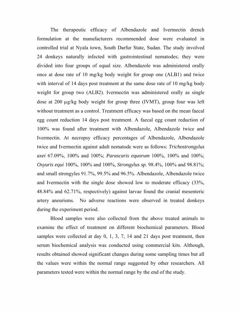
The therapeutic efficacy of Albendazole and Ivermectin drench
formulation at the manufacturers recommended dose were evaluated in
controlled trial at Nyala town, South Darfur State, Sudan. The study involved
24 donkeys naturally infected with gastrointestinal nematodes; they were
divided into four groups of equal size. Albendazole was administered orally
once at dose rate of 10 mg/kg body weight for group one (ALB1) and twice
with interval of 14 days post treatment at the same dose rate of 10 mg/kg body
weight for group two (ALB2). Ivermectin was administered orally as single
dose at 200 µg/kg body weight for group three (IVMT), group four was left
without treatment as a control. Treatment efficacy was based on the mean faecal
egg count reduction 14 days post treatment. A faecal egg count reduction of
100% was found after treatment with Albendazole, Albendazole twice and
Ivermectin. At necropsy efficacy percentages of Albendazole, Albendazole
twice and Ivermectin against adult nematode were as follows: Trichostrongylus
axei 67.09%, 100% and 100%; Parascaris equorum 100%, 100% and 100%;
Oxyuris equi 100%, 100% and 100%, Strongylus sp. 98.4%, 100% and 98.81%;
and small strongyles 91.7%, 99.5% and 96.5%. Albendazole, Albendazole twice
and Ivermectin with the single dose showed low to moderate efficacy (33%,
48.84% and 62.71%, respectively) against larvae found the cranial mesenteric
artery aneurisms. No adverse reactions were observed in treated donkeys
during the experiment period.
Blood samples were also collected from the above treated animals to
examine the effect of treatment on different biochemical parameters. Blood
samples were collected at day 0, 1, 3, 7, 14 and 21 days post treatment, then
serum biochemical analysis was conducted using commercial kits. Although,
results obtained showed significant changes during some sampling times but all
the values were within the normal range suggested by other researchers. All
parameters tested were within the normal range by the end of the study.

بسم اهللا الرمحن الرحيم
اخلالصة
بالديـدان الطفيليـة اجلوانب املتعلقـة باإلصـابة مت تسليط الضوء على بعض يف هذه الدراسة
.حيوية يف احليوانات املعاجلةوم بعض القيم الكيمي عالجها و تقي واالسطوانية املعدية املعوية
شـهر سـبتمرب و إىل 2006 مت إجراء مسح و بائي خالل سنة كاملة امتدت من شهر أكتوبر
حيوان من الفصيلة اخليلية و قد كـان عـدد 1256مشل املسح ، الية جنوب دارفور ينة نياال و مبد 2007
خضعت مجيعها للفحص عن اإلصابة الطبيعية بالديدان الطفيلية االسطوانية 810 بينما احلمري 446اخليول
شهر نوفمرب النـسبة سجل، %29.20نسبة اإلصابة : كم يلي" و قد كانت النتيجة إمجاال. املعدية املعوية
متوسـط تعـداد ،%13.92"بينما سجل يونيو النسبة األكثر اخنفاضا % 41.50األعلى بني كل الشهور
-50 يف شهر ابريـل هليسجقد مت ت لتعداد البيض و كان املدى األعلى986.02 ±589.97البيض كان
بينما سجلت اإلصابة % 81.35 النسبة االعلي إلصابة اخلفيفة شكلت ا من ناحية حدة اإلصابة، . 13450
االسـطوانية األجناس التالية من الديدان %. 8.11" توسطة كانت اقل حظا و اإلصابة امل % 10.45احلادة
,Strongylus spp, Cyathostomes spp: التالية سجلت الغالبيـة يف العينـات الـيت مت فحـصها
Trichostrongylus spp, and Strongyloides westeri .
بينمـا % 100سجل شهر أغسطس النسبة األعلى ، %15.37 نسبة اإلصابة يف اخليول كانت
% 8.82لإلصابة اخلفيفة و % 82.35و توزعت حدة اإلصابة ما بني %. 5.26شهر مارس كان األدىن
و 1099 ±975.37لكل من اإلصابة املتوسطة و احلادة و كان متوسط تعداد البيض لزنة جرام من الروث
.نوفمرب سجل يف شهر 13450-50املدى األعلى لتعداد البيض
كانـت يف % 55.79لكل العام و لكن النسبة األعلى % 37.48 نسبة اإلصابة يف احلمري كانت
املتوسـطة ، % 10.86اإلصابة احلادة شـكلت . كانت يف شهر مايو % 14.89شهر يناير بينما األدىن

±750.14متوسط تعداد البيض لزنة جـرام مـن الـروث كـان %. 81.25بينما الطفيفة % 7.89
.11800-50على لتعداد البيض سجل يف شهر ابريل و كان م املدى اال1071.95
و ) املعلـق ( البنـدازول ي من ذكور احلمري لتقدير جناعة كل من عقار 24 أجريت جتربة مشلت
ملجرام للكيلو جـرام لعقـار 10( صى ا من املصنع للعقارين حسب اجلرعات املو ) الشراب( االيفرمكتني
يف عـالج اإلصـابة الطبيعيـة بالديـدان ) مايكروجرام للكيلو جرام لعقار االيفرمكتني 200البندازول و
تلقت اموعـة األوىل وانات بالتساوي إيل أربع جمموعات، قسمت احلي . االسطوانية الطفيلية املعدية املعوية
14 بالفم بفاصل زمين قدره وية واحدة من عقار البندازول بينما تلقت اموعة الثانية جرعتني أيضاً جرعة فم
اموعة الثالثة تلقت جرعة فموية واحدة من عقار االيفرمكتني بينما مت االحتفـاظ يوم من عقار البندازول،
التجربة يف تقدير فعالية الـدوائني علـي اعتمدت . باموعة الرابعة دون عالج للتحكم و املقارنة فيما بعد
القتل الرحيم يف اليـوم مت إجراء يوما من جتريع الدواء و من مثة 14االخنفاض يف تعداد البيض الروث خالل
احلادي و العشرين من جتريع الدواء و ذلك لفحص حمتوي القناة اهلضمية من الديدان االسـطوانية املعديـة
.املعوية
و . للثالثة جمموعات املعاجلـة % 100اض تعداد البيض يف زنة جرام من الروث كان بنسبة اخنف
% 100و % 100و % 67.09بلغــت نــسبة الفعاليــة يف معاجلــة أنــواع الديــدان االســطوانية
Trichostrongylus axei .100% ،100 % ــنس % 100و . Parascaris equorumجلـ
جلــنس %98.81 و Oxyuris equi .98.4%، 100%جلــنس % 100و% 100، 100%
Strongylus spp. . 91.7%، 99.5%، كانت ألجناس % 96.5 وsmall Strongyles لكل من
العقاران أبديا درجة فعالية . عقار البندازول باجلرعة العادية مث اجلرعة املضاعفة و عقار االيفرمكتني على التوايل
% 62.71للبندازول باجلرعـة املـضاعفة و % 48.84للبندازول باجلرعة العادية % 33متوسطة بلغت

املتغذيـة علـى الـدماء بالـشريان Strongylus sppلاليفرمكتني يف عالج يرقات الطور الثالث لديدان
.املساريقي القحفي و فروعه
18 االستطبايب بعقاري البندازول املعلق و االيفرمكتني الشراب يف أجريت دراسة ثانية لتقيم األثر
قسمت احليوانات بالتـساوي ايل . من ذكور احلمري ذات اإلصابة الطبيعية بالديدان االسطوانية املعدية املعوية
تلقت اموعة االوىل جرعة فموية واحدة من عقار البندازول بينما تلقت اموعة الثانيـة ، ثالث جمموعات
اموعة الثالثة تلقت جرعة فموية واحدة ، ندازول يوم من عقار الب 14 بالفم بفاصل زمين قدره جرعتني أيضاً
من العالج و من بعد 21يوم " و أخريا 14 ،7، 3 ،1 يوم صفر، مجعت العينات يف . من عقار االيفرمكتني
اختالفات النتائجسجلت . اليت مت مجعها خالل التجربة ذلك اجري التحليل الكيموحيوي على عينات املصل
و لكن غالب االختالفات كان ضمن املـستوى الطبيعـي و الزمنية خالل التجربة الفترات معنوية يف بعض
. كل القراءات كانت ضمن املستوى الطبيعي يف اية التجربة" عموما

CHAPTER ONE

Introduction
In Sudan, donkeys are becoming increasingly important in view of their
increased use instead of horses in labour as drought animals as well as carrying
water, and in transportation. This present situation can be observed in many
urban and suburban areas. Moreover, donkeys are used as pack beasts and in
ploughing. As in many other developing countries donkeys in Sudan play and
constitute an important source of cheap energy for agricultural production by
way of traction for cultivation and transport of produce, goods and labour.
With regard to livestock population, Sudan constitutes one of the richest
African and Arab countries. Most of these animals are owned by nomadic or
semi-nomadic tribes living in the semi arid regions. Livestock population was
reported to be about 121 million head, composed of 35.825, 44.802, 37.346,
3.031, 0.65 and 6.35 million head of cattle, sheep, goats, camels, horses and
donkeys respectively (SBAR, 2000). However, donkeys’ population is
relatively large compared to camels and horses. About 30.9% of donkeys’ are
found in Darfur States.
Horses, ponies and donkeys are hosts to a large population of parasites
(Duncan, 1983). Lichtenfels (1975) stated that; equids are hosts for helminths
belonging to 28 genera and 75 species of nematodes, 2 genera and 5 species of
trematodes as well as 3 genera and 24 species of cestodes.
Helminth parasite infection was the main problem reported in donkeys
admitted to veterinary clinic (Ali et al., 2001). Seri and his colleagues (2004)
showed that the prevalence of nematodes infection in donkeys in Khartoum
state was 70.1%. Kheir and Kheir (1981) conducted field survey in South
Darfur State, Sudan, and stated that the prevalence was 56.2% and overall
prevalence of infection with nematode parasites was (58%) in town animals and
(22%) in nomadic areas. In Sennar; of the 218 donkeys examined for parasitic
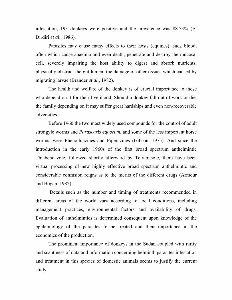
infestation, 193 donkeys were positive and the prevalence was 88.53% (El
Dirdiri et al., 1986).
Parasites may cause many effects to their hosts (equines): suck blood,
often which cause anaemia and even death; penetrate and destroy the mucosal
cell, severely impairing the host ability to digest and absorb nutrients;
physically obstruct the gut lumen; the damage of other tissues which caused by
migrating larvae (Brander et al., 1982).
The health and welfare of the donkey is of crucial importance to those
who depend on it for their livelihood. Should a donkey fall out of work or die,
the family depending on it may suffer great hardships and even non-recoverable
adversities.
Before 1960 the two most widely used compounds for the control of adult
strongyle worms and Parascaris equorum, and some of the less important horse
worms, were Phenothiazines and Piperazines (Gibson, 1975). And since the
introduction in the early 1960s of the first broad spectrum anthelmintic
Thiabendazole, followed shortly afterward by Tetramisole, there have been
virtual processing of new highly effective broad spectrum anthelmintic and
considerable confusion reigns as to the merits of the different drugs (Armour
and Bogan, 1982).
Details such as the number and timing of treatments recommended in
different areas of the world vary according to local conditions, including
management practices, environmental factors and availability of drugs.
Evaluation of anthelmintics is determined consequent upon knowledge of the
epidemiology of the parasites to be treated and their importance in the
economics of the production.
The prominent importance of donkeys in the Sudan coupled with rarity
and scantiness of data and information concerning helminth parasites infestation
and treatment in this species of domestic animals seems to justify the current
study.

Objectives
The main objectives of this study are:
1. To determine the prevalence of the gastrointestinal helminths of equines in
South Darfur State.
2. To investigate the therapeutic efficacy of the drugs under study against
gastrointestinal nematodes of donkeys.
3. To report on the post treatment biochemical changes, if any.
Literature review
1.1 Albendazole
Benzimidazole anthelmintics have a broad spectrum of activity against
gastrointestinal helminths, including migrating strongyle larvae and lungworm
infections, and are well tolerated by mammals (McKellar and Scott, 1990).
All benzimidazoles have been developed as a result of the extensive
studies that were carried out in a number of laboratories to modify the structure
of thiabendazole. A feature of the newer structures is that they are metabolised
and excreted much more slowly than thiabendazole. This is achieved by
blocking the 5-position. The substitutions of another group in the 5-position,
and the replacement of the thiazole ring by methyl carbamate, have a very
significant effect on the rate of elimination (Brander et al., 1982). The
differences in efficiency between members of this class of drugs against groups
of parasites probably reflect differences in bioavailability of the drugs within
the host animal (McKellar and Scott, 1990).
Benzimidazoles have a low mammalian toxicity and it have proved
impossible to find an LD50 for some of these compounds. Oxenfendazole and
albendazole are teratogenics, and depending on the dose rate, should not be used
in early pregnancy (Armour and Bogan, 1982). In vitro turbidimetric techniques
and competitive colchicine-binding studies demonstrated the inhibitory power
of benzimidazoles on the polymerization of tubulin into microtubules. The
difference in the sensitivity of host and parasites to the effects of
benzimidazoles may be due to difference in the structure of microtubules in
their cells (Davis and Gull, 1983).
1.1.1 Identity
Albendazole is a member of the benzimidazole group. It is orally
administered broad-spectrum anthelmintic, soluble in dimethylsulphoxid, strong
acids and strong bases. It is slightly soluble in methanol, chloroform, ethyl
acetate and acetonitrile. Albendazole is practically insoluble in water.
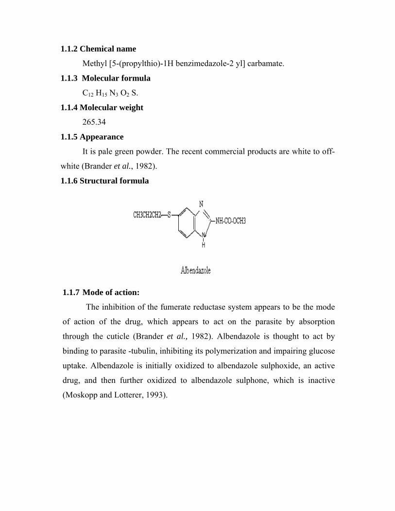
1.1.2 Chemical name
Methyl [5-(propylthio)-1H benzimedazole-2 yl] carbamate.
1.1.3 Molecular formula
C12 H15 N3 O2 S.
1.1.4 Molecular weight
265.34
1.1.5 Appearance
It is pale green powder. The recent commercial products are white to off-
white (Brander et al., 1982).
1.1.6 Structural formula
1.1.7 Mode of action:
The inhibition of the fumerate reductase system appears to be the mode
of action of the drug, which appears to act on the parasite by absorption
through the cuticle (Brander et al., 1982). Albendazole is thought to act by
binding to parasite -tubulin, inhibiting its polymerization and impairing glucose
uptake. Albendazole is initially oxidized to albendazole sulphoxide, an active
drug, and then further oxidized to albendazole sulphone, which is inactive
(Moskopp and Lotterer, 1993).

1.1.8Toxicity of Albendazole
Acute toxicological effects
World health organization (WHO), the food additive series 25 reported
findings in dead rats included, urinary staining of abdomen, bloody discharge
around nose, and intestinal haemorrhage. Also in another study, necropsy of
dead rabbits showed intestines containing fluid dilated with gas.
Short term toxicological effects
Mice
Two groups of both males and females treated with albendazole in
different doses for three months. All the females and 50% of the males of the
highest dose 1600 mg/kg bw (160 times the recommended dose) died
spontaneously or killed in a moribund condition. From week 9, ear lesions
involving thickening and/or encrustation of the lip were observed in 20% of the
males and 22.2% of the females at 800 mg/kg bw (80 times the recommended
dose), and 100% of the males at 1600 mg/kg bw. Food consumption was
generally decreased in males given 400 mg/kg bw (40 times the recommended
dose) or more but body weight gain was depressed only at1600 mg/kg bw. At
the end of the study haemoglobin (Hb), haematocrit and erythrocyte levels were
reduced at dose of 800 mg/kg bw or more also decreased leucocyte counts
observed in males at (800 mg/kg bw) (Daly and Rinehart, 1980).
Post mortem examination revealed increase in absolute and relative liver
weight at 40 mg/kg bw (4 times the recommended dose) in males and 80 mg/kg
bw (8 times the recommended dose) in females (Daly and Rinehart 1980).
Rats
The toxic signs following administration of higher doses more than 48
mg/kg bw, (4.8 times the recommended dose) were diarrhoea, piloerection,
nasal swelling with blood stained, nasal discharge, death and body weight
depressed. Haemoglobin, haematocrit, erythrocyte and leucocyte counts were
reduced (Simon, 1979a).
At high doses 48 and 168 mg/kg bw, (4.8 and 16.8 times the
recommended dose respectively), adrenal size was increased particularly in
females. Testes were reduced in size at 48 mg/kg bw (Simon, 1979a).
Histopathology revealed hypoplasia in testes, bone marrow, spleen and lymph
nodes at 48 and 168 mg/kg bw (Simon, 1979).
At dose of 45 mg/kg bw (4.5 times the recommended dose) plasma
cholesterol was increased in males and females, potassium was increased in
males and albumin, plasma and erythrocyte cholinesterase were decreased in
females, and urinary protein was increased at 30 and 45 mg/kg bw (3 and 4.5
times of recommended dose, respectively) (Daly and Hogan, 1982).
Numerous gross alternations were noted with high doses in post mortem
including discoloration and/or nodules in the lungs, heart, lymph nodes, spleen,
pancreas, liver, adrenal and kidneys. Some of these organs were also enlarged
or showed adhesions. And thymic tissue was often absent and testes were small
and flaccid (Daly and Hogan, 1982).
Histopathology carried out identified a number of instances, where
bacterial colonization in lung, spleen, kidney, and heart was associated with
necrosis without the usual acute inflammatory response (Daly and Hogan,
1982).
Long term toxicological effects
Mice
Long-term toxicological studies in mice revealed decrease in erythrocyte
and leucocyte counts without toxic signs or effects on food intake and body
weight (Selwyn, 1987). Histopathological tests carried out indicated flaccid or
small testes, testicular tubular degeneration and oligospermia (Selwyn, 1987).
Rats
Studies carried in rats, showed decrease of total leucocytes neutrophil
counts, serum cholesterol was increased in some sampling times (Sauer 1985).
Post mortem findings and histopathology revealed in rats treated at 20mg/kg bw
(2 times of recommended dose) were increased incidence of flaccid testes (Daly
and Knezevich, 1987). Degeneration, atrophy of germinal and relative liver
weight in males and hepatic fatty metamorphosis in both males and females
(Daly and Knezevich, 1987).
Reproductive toxicological effects
In a group of rats fed diet containing albendazole at different doses for
three successive generations. There were no toxic sings, body weight, food
consumption, mating, fertility, pregnancy rate and gestation length, were
normal. During lactation weight gain depressed but only in F1 and F2
(Schroeder and Rinehart, 1980). In female rats uterine examinations in gestation
day 13 showed fewer implantation (not significant) at dose of 30 mg/kg bw (3
times the recommended dose) with no effect on resorption (Boutemy, 1980).
Genotoxic effects
Studies on Chinese hamster ovary cells indicated negative result for
genotoxicosis by albendazole (Galloway, 1981).
Teratogenic effects
Mice
There was no overt maternal toxicity or effects on resorption incidence,
foetal weight and external-visceral and skeletal development of foetus following
administration of albendazole (Killeen and Rapp, 1975).
Rats
At dose of 6 - 62 mg/kg bw (0.6 and 6.2 times of recommended dose)
skeletal abnormalities were increased and greater with increases in resorption
and external malformations, decreased foetal weight. The major malformations
were cranio-facial and bone defect (Martin, 1980). The dose of 30 mg/kg bw (3
times the recommended dose) showed foetal limbs abnormalities (Christian,
1984).

At 40 mg/kg bw (4 times the recommended dose) no toxicity or effect on
gestation and parturition. Small lungs and anasarca possibly related to treatment
in foetus (Johnson, 1981).
Rabbits
In rabbits, the maternal mortality was increased in 30 mg/kg bw (3 times
the recommended dose) and reduction in implantation in gestation day 7 – 19
(Killeen and Rapp, 1975).
Sheep
The pregnant ewes given albendazole in gestation day 17 were allowed to
deliver naturally, but premature delivery in more ewes was noted. All premature
lambs were stillborn; consequently the number of live lambs was reduced. Post-
mortem revealed increased incidence of prognathia, scoliosis, spinal bifid,
reduced tail and poorly developed or absent kidneys (Tash and Harper, 1977).
1.1.9 Pharmacokinetics
Mice
The single gavage dose of albendazole C14-ring labelled albendazole
given to mice revealed, over 72 hours period, that 20.5% of the administered
dose was recovered in the urine. Albendazole sulphoxide and two other
metabolites accounted for 81% of label. Low level of parent drug, albendazole
sulphoxide and three other metabolites were detected (Parish et al., 1979a). The
sulphone metabolite has higher plasma level than sulphoxide (Souhaili El Amri
et al., 1988). Both the sulphoxide and the sulphone metabolites decreased to
very low levels at 18 hours (Delatour et al., 1984).
Albendazole induces certain hepatic drug-metabolising enzymes, which
may be responsible for the enhancing degradation of sulphoxide to sulphone
following repeated administration (Souhaili El Amri et al., 1988).
Rats
In rats 31% of administered radioactive dose was recovered in the urine
over 72 hours period. Sulphoxide, 2-aminosulphone and three other metabolites

accounted for 89% of the label. Albendazole sulphone and two other
metabolites were also found (Parish and Gyurik, 1979). The sulphoxide and the
sulphone derivatives resulted in the urinary excretion. The urinary metabolites
were qualitatively similar to those observed after albendazole administration
(Parish and Gyurik, 1979).
Sheep
Albendazole was absorbed unchanged from the rumen and passed
through the stomach while the metabolites were secreted or diffused in this
organ (Marianer and Bogan, 1980).
After single oral dose of labelled albendazole on day 1, in the liver the
label was mainly on form of sulphoxide and sulphone metabolites, which were
progressively converted to 2-amino sulphone which was the primary metabolite
on day 8. Over 72 hours the sulphoxide and 2-amino sulphone accounted 60 –
70% of urinary excretion of the drug. Low levels of parent drug and the
sulphone with 6 other metabolites were detected (Colman et al., 1977).
Cattle
The metabolism to the sulphoxide and the sulphone was rapid and the
compounds were present in plasma for up to 40 hours (Prichard et al, 1985).
In calves at dose of 20 mg/kg bw (4 times the recommended dose), over 7 days
period 47% of the administered dose was recovered in urine; the sulphoxide,
sulphone and 2-amino sulphone accounted 70% of the excreted dose (Parish et
al.,1977 )
Although albendazole excreted in the animal’s milk, biliary elimination
presumably accounts for a portion of the elimination as evidenced by biliary
concentrations of albendazole similar to those achieved in the plasma.
Limited examinations of kidney revealed a similar metabolic profile (Kaeer et
al., 1977)
Human
Oral bioavailability of albendazole appears to be enhanced when it is co-
administered with a fatty meal as evidenced by higher plasma concentrations of
albendazole sulphoxide as compared to the fast state. Albendazole not detected
in plasma, but its main metabolites could be found and the half-life of
albendazole sulphoxide is 10 and 15 hours (Jung et al., 1992).
Albendazole sulphoxide is 70% bound to plasma protein and is widely
distributed through the body; it has been detected in urine, bile, liver, cyst wall,
cyst fluid and cerebral spinal fluid (CSF). Analysis of the urine showed the
presence of the sulphoxide, the sulphone and their amino derivatives with three
other metabolites (Rossignol and Maisonneuve, 1984).
Equine
Albendazole when given to donkeys at dose of 10 mg/kg bw; the parent
molecule of the drug was not detected in the plasma, but its sulphoxide and
sulphone metabolites were detected, demonstrating that albendazole was
completely metabolised by first-pass mechanism in donkeys. The sulphoxide
metabolite of albendazole was significantly higher than that of the sulphone
metabolite (Gokbulut et al., 2005).
Faecal concentration of parent molecule of the drug after albendazole
administration was significantly metabolised, probably by gastro-intestinal
microflora, to its sulphoxide metabolites that showed a similar profile to the
parent molecule in the faecal sample (Gokbulut et al., 2005). Plasma
concentration of albendazole was significantly lower when compared with
oxifendazole (Gokbulut et al, 2005).
Benzimidazoles are extensively metabolized in all animal species.
Generally, the plasma elimination half-lives of the parent drugs are short and
the metabolic moieties predominate in plasma and tissues and in the excretion
of the host as well as in parasites recovered from Benzimidazole-treated animals
(Lanusse and Prichard, 1993; Delatour and Parish, 1986).
The less soluble Benzimidazole compounds have a longer residence in
the fore stomachs of ruminants from which they are absorbed over prolonged
periods and thus remain in the plasma for a relatively long time. Since an
equilibrium exists between the plasma and gastrointestinal tract, the duration of
exposure of gut parasites to an effective concentration of the drug is extended
(Mackellar and Scott, 1990). Extremely insoluble anthelmintics may be less
effective, since they may not be absorbed and are excreted unchanged in the
faeces. This may explain the difference between the plasma concentrations of
oxfendazole (fenbendazole sulphoxide, FBZSO) following oral administration
and its inter-convertible metabolite fenbendazole (FBZ) (Ngomuo et al., 1984).
A large proportion of the less soluble FBZ is known to be excreted in the faeces
of ruminants (Duwel, 1977).
Oxfendazole, fenbendazole and albendazole (ABZ) are commercially
available sulphur-containing benzimidazoles that commonly undergo
microsomal oxidation in liver. Sulphide benzimidazoles (FBZ and ABZ) are
reversibly metabolized to their sulphoxide derivatives (Marriner and Bogan,
1980, 1981; Gyurick et al., 1981; Mohammed Ali et al., 1987). Irreversible
sulphonation follows sulphoxidation and is a slower oxidative step resulting in a
sulphone metabolite (Averkin et al., 1975).
The pharmacokinetics of FBZ and FBZSO have been studied in horses in
which the metabolic inter-conversion of those compounds appears to differ
substantially from ruminants. In the horse, the bioavailability and residence time
of the tested benzimidazoles (and metabolites) were lower and shorter,
respectively, than in ruminants (Mckellar et al., 2002; Gokbulut, 2000; Marriner
and Bogan, 1985, 1981).
Sulphide and sulphoxide benzimidazoles are known to bind to nematode
tubulin (Lacey et al., 1987) and therefore have activity against nematode
although sulphides exert an inhibitory activity on tubulin at lower
concentrations than sulphoxides. In most species examined, the sulphoxide
moiety predominates in plasma and is thought to confer activity against gut
dwelling nematodes following secretion across the gastrointestinal wall into the
gut lumen where it may undergo sulpho-reduction (Mckellar and Scott, 1990).
Maximum plasma concentration (Cmax) of albendazole sulphoxide
(ALBSO) (0.08µg/mL) and albendazole sulphone (ALBSO2) (0.04 µg/mL)
were obtained at (tmax) 5.71 and 8.00 h respectively, following administration of
albendazole. The area under the curve (AUC) of the sulphoxide metabolite
(0.84 µg.h/mL) of ABZ was significantly higher than that of the sulphone
metabolite (0.50 µg.h/mL).
Although (ABZ) is not licensed for use in Equidae. Its metabolites
presented a greater plasma kinetic profile than FBZ which is licensed for use in
horses. A higher metabolic capacity, first-pass effects and lower absorption of
benzimidazoles in donkeys decrease bioavailability and efficacy compared to
ruminants (Gokbulut et al., 2005).
1.1.10 Use of Albendazole in animals
Cattle
Albendazole had been shown to be highly effective against lungworms;
also it is very active against adult liver flukes in cattle when given at twice the
recommended dosage rate for roundworm therapy (Armour and Bogan, 1982).
Sheep
Albendazole is effective in chronic fasciolaisis treatment, also
albendazole is certainly effective against the adult and developing larval stage
of D. filarial and naturally occurring infection with the other small lungworms
in sheep and goat (Armour and Bogan, 1982).
Equine
There is a paucity of data available in the literature on the toxic or side
effects in equine and albendazole is not used in these species (Gokbulut et al.,
2005). A higher metabolic capacity, first-pass effects and lower absorption of

benzimidazoles in donkeys decrease bioavailability and efficacy compared to
ruminants (Gokbulut et al., 2005).
High intestinal concentrations could be effective against gastrointestinal
nematodes that inhabit the gut lumen, but very low plasma concentrations of
albendazole may not be effective against migrating fourth larval stages of large
strongyles or lung worms. Repeated dosage regimes of albendazole or co-
administration with metabolic inhibitors could be used to treat migrating larval
or tissue stages of strongyles and lungworms in donkeys (Gokbulut et al, 2005).
10 mg/kg BW of albendazole may be used for treatment of
strongyloidosis of horses and donkeys (Kassai, 1999). In Kentucky agricultural
experiment station, tests were conducted between February 1977 and June 1981
using 10 adolescent horses which were proved to be naturally infected with
population B small strongyles. Effective reductions (97% to 100%) were
recorded for treatments with Oxibendazole, Albendazole, and Thiabendazole.
The larval count indicated that all of the compounds were 100% effective in
removing larvae of S. vulgaris and S. edentates. Albendazole removed 94% to
100% of 11 species and 84% of the 12th species (Cyathostomum coronatum).
Removal of immature small strongyles by oxibendazole and albendazole in the
tests (15% and 38%) was somewhat less than the 71% for Oxibendazole in an
earlier study (Drudge et al., 1979). Population B small strongyles have been
reported to be resistant to thiabendazole, fenbendazole, and oxfenbendazole but
not to oxibendazole (Drudge et al., 1979). Five species of small strongyles were
resistant, whereas 19 species of small strongyles and 2 species of large
strongyles (Strongylus vulgaris and Strongylus edentates) were removed
efficaciously by all six benzimidazoles. Albendazole is closely related analogue
of oxibendazole and is active against the 5 species of Benzimidazole resistant
small strongyles.
Worm counts indicated that the efficacy of albendazole against larval stages
of small strongyles, measured as the percentage of worms removed by

albendazole treatment was limited, particularly for L3. The efficacy of
albendazole against these helminth adults was somewhat higher, but
considerable differences between species also were observed (Eysker et al.,
1988).
The effect of albendazole against larval stages was lower than that against
adult worms; this low effect mainly is the result of a limited efficacy of
albendazole against larval stages (Drudge et al., 1984). Low efficacy of
albendazole treatment can be a result of anthelmintic resistance (Eysker et al.,
1988)
Benzimidazole resistance is a common phenomenon in Cyathostomes. It
has been recorded in 5 species of Cyathostomes by Drudge et al., (1979).
In a series of experiments on the epidemiology and control of
cyathostome infections in Shetland ponies between 1984 and 1986, the effect of
repeated early-season albendazole (Eysker et al., 1986, 1988) and oxfendazole
(Eysker et al., 1989) treatments was evaluated. The results of these studies
invariably showed reduction in faecal egg output after the first treatment but
poor effect of later treatments.
Apparently the repeated use of one Benzimidazole within one grazing
season is of little prophylactic value (Eysker et al., 1989a). An important aspect
of the epidemiology of cyathostome infections in equids is the occurrence of
inhibited development. Gibson (1953) observed that some weeks after each of a
series of phenothiazine treatments, strongyle-type eggs reappeared in the faeces
of horses, even after housing period under helminth-free conditions of 3 years.
His explanation of this phenomenon was the existence of an equilibrium
between inhibited larvae in, and adult worm on, the gut wall leading to
resumption of development of inhibited larvae after removal of adult worms.
Later, Ogburne (1975) found evidence for an emergence of fourth-stage
larvae (L4) from the intestinal wall at the end of the winter, indicating a

seasonal pattern of inhibited development similar to many Trichostrongylidae of
ruminants.
Anthelmintics resistance in cyathostomin nematodes (‘small strongyles’) of
horses is a well-known phenomenon worldwide. Benzimidazole (BZ) resistant
cyathostomins have been reported from North and South America, South
Africa, Australia. New Zealand and many European countries (reviewed by
Kaplan, 2002) including Greece (Papadopoulos et al., 2000).
Benzimidazole resistant populations of cyathostomin nematodes were
detected on seven of ten Turkish horse farms (Cirak et al., 2004). According to
FECRT data, Benzimidazole resistance in stronglyids was observed only at
Dubrovsky (Ukraine) horse farm (FECRT=68%). No resistance to macrocyclic
lactones in strongylids or in Parascaris equorum was observed (Kuzmina and
Kharchenko, 2008). BZ resistance in horse strongylid nematodes in Ukraine
was found only in cyathostomins, or small strongyles (Borgsteede et al., 1997,
Kuzmina et al., 2002).
Horse strongylids are known to be resistant to Benzimidazoles (BZ) and
tetrahydropyrimidines (Kaplan, 2002, 2004), while the only case of resistance in
cyathostomins to microcyclic lactones was reported in donkey in the UK
(Trawford et al., 2005).
1.2 Ivermectin
Ivermectin was the first macrocyclic lactone anthelmintic, introduced as
a veterinary anti-parasitic agent in France in 1981 and marketed as a mixture of
22, 23 dihydro- B1a (>80%) and 22, 23 dihydro- B1b (<20%) (Fisher and
Morzik, 1989).
Ivermectin is today elixir in the world of parasites chemotherapy, it is a
potent agent, active against many internal and external parasites in domestic
animals, and it is vogue and highly effective with a wide margin of activity and
safety. In equines, a variety of adverse reactions have been reported in horses
after parenteral administration of Ivermectin at the recommended dosage of
200µg/kg body weight (Reed, 1983). These reactions have occurred in a small
percentage of treated horses and the drug is now sold as a paste for oral
administration. Ivermectin have an excellent efficacy for an important range of
gastro-intestinal nematodes of equine (Campbell et al., 1989).
1.2.1 Efficacy of Ivermectin against equine endoparasites:
Draschia spp and Habronema spp
Three species, H. musca, H. microstoma and D. megastoma, occur in the
stomach of equine. The largest one is the H. microstom. The first 2 are free in
the host’s stomach; D. megastoma lives in nodules in the stomach wall.
Diagnosis of Habronemiasis is often impossible because the larvae can seldom
be found in the faeces (Hall, 1985).
In horse, Herd and Donham (1984), showed 1-2 intramuscular doses of
ivermectin at 200 µg/kg were highly effective against Draschia spp and
Habronema spp., while DiPietro (1982) reported 100% efficacy.
Gasterophilus spp
The larvae of several species of this genus are parasites of equines and are
known as bots fly, the third- stage larvae causes inflammation and ulceration of
the mucous membrane of the stomach and duodenum in addition to blood
sucking (Soulsby, 1982; Hall, 1985).
At dose rate of 200-300 µg/kg when Ivermectin given intramuscularly to
horses, the efficacy was 99% against larvae of Gasterophilus spp, the few larvae
in the treated horses were located in the large intestine contents and were not
attached to the intestinal epithelium (DiPietro, 1982).
Parascaris equorum
This is found in the small intestine of horse kind. The infection and
migration is through the blood stream via the right ventricle of the heart to the
lungs (Hall, 1985).
When given orally as paste formulation, Ivermectin at 200 µg/kg totally
eliminated the passage of P. equorum in the natural infected horses (Cobra et
al., 1986). Ivermectin is also active against immature stages of P. equorum, in
ponies, larval burdens were determined at necropsy following treatment with
the paste at 200 µg/kg. When necropsy was performed 2 weeks after treatment,
the reduction in lung larvae burden was 100% (French et al., 1988) and the
reduction in intestinal larvae burden was also 100 % (DiPietro et al., 1987).
Both the paste and the liquid Ivermectin formulations have been shown to
be highly active against both the early (L3) tissue phase of P. equorum and
against the later intestinal (L4) phase of P. equorum. The efficacy has been
discussed by Boraski (1987).
Strongloides westeri
This is occurs in the small intestine, mainly in the duodenum and jejunum
which causes irritation, resulting in diarrhea, especially in young foals. 200
µg/kg of Ivermectin was effective against S. westeri in foals in a paste
formulation (Ryan and Best 1985).
Trichostrongylus axei
A small worm (3-8mm) found in the stomach and duodenum of horse
kind. In horses T. axie causes chronic catarrhal gastritis (Hall, 1985).
Stongylus spp
There are three species, Strongylus equinus, S. vulgaris and S. edentatus
occur in the large intestine including the caecum of equines (Soulsby, 1982).
Generally strongylidae worms are not severe pathogens, unless they occur in
large numbers when their mouth parts cause extensive damage by sucking in the
mucous membrane, thus causing ulcers. Larvae, however, in the characteristic
nodules, produced damage, which result in bacterial invasion and consequently
ulcers. When larvae leave the nodules there is extensive bleeding (Hall, 1985).
Fourth-stage larvae S. vulgaris penetrate the intima of the sub-mucosal
arterioles and migrate in the vessels towards the cranial mesenteric artery they

are to be found here from 14 days after infection onwards associated with
thrombi and later aneurysms (Soulsby, 1982). The oral paste and injectable
formulations of Ivermectin at 200 µg/kg was 100% effective against 8-week-old
Stongylus vulgaris, as revealed by necropsy 5 weeks after treatment (Klei et al.,
1984). Also as intramuscular injectable formulation at dose rate 200 µg/kg body
weight Seri and his colleagues (2005) showed that efficacy of Ivermectin
against arterial stages of Strongylus vulgaris was only 69, 23%.
Small Strongyles
Known as Cyathostomes worldwide, they occur in the large intestine of
the equides all over the world (Soulsby 1982). Burrows and his colleagues
(1985) used faecal examination to demonstrate the efficacy of Ivermectin paste
formulation at 200 µg/kg against benzimidazole-resistant small Strongylus, the
egg counts remained zero at least 3 weeks in horses. Also 200-300 µg/kg were
effective against Cyathostomes in percentage of 99% and the efficacy was 86%-
97% against immature stages of Cyathostomes according to 200-300 µg/kg dose
rate respectively (DiPietro 1982).
Oxyuris equi
A worm lives in the large intestine of horses; males are about 9 – 12 mm
and females up to 150 mm. According to Seri et al., (2005) the efficacy of
Ivermectin injectable formulation at dose of 200 µg/kg was 100%.
1.3 Prevalence of gastro- intestinal nematodes in donkeys and horses
Horses, ponies and donkeys are hosts to a large population of parasites
(Duncan, 1983). Helminths parasite infection was the main problem reported in
donkeys admitted to veterinary clinic (Ali et al., 2001).
Swedish nationwide study showed that strongyle infections were highly
prevalent in Swedish horse herds: 78% of 1183 examined horses on 110 farms
were found to shed nematode eggs (Lind, 2005).

In Ethiopia, All of 338 samples examined were found positive for
helminth eggs. Strongyle spp. (100%), Parascaris equorum (50%) and Oxuris
equi (3%). 81.7% of donkeys sampled were severely infected, 8.3% heavily,
3.8% moderately and 6.2% mildly (Ayele et al., 2006).
In Chad, Garber (1970) reported prevalence of 89% for Strongyle spp.
and Cyathostomes spp. 72% for Parascaris equorum, and 6% for Strongloides
westeri. In Kenya, Strongyle spp. prevalence was 57.6%, Cyathostomes spp.
was 15.4% and 20.7% reported for Parascaris equorum (Mukhwana, 1994). In
Morocco, the prevalence of different gastro-intestinal nematodes species
reported as follow: Cyathostomes spp. 52%, Parascaris equorum 37% and
93.5% was for Trichuris spp. (Abdelkarim, 1991).
In Sudan, Seri and his colleagues (2004) reported that, the prevalence of
nematodes infection was 70.1% in Khartoum state, Strongylus spp. 35.8%,
Cyathostomes spp 36.7% Parascais equrum 10.7%, Trichostrongylus axei 12%
Strongloides westeri 3.4%.
Kheir and Kheir (1981) conducted a field survey in South Darfur State,
and stated that the overall prevalence was 56.2% and overall incidence of
infection with nematode parasites was found (58%) in town animals and (22%)
in nomadic areas. Also five nematode genera were encountered in donkeys in
the same area: Strongylus spp 43.6%, Oxyuris spp 10.5%, Strongyloides spp.
3.9%, Parascaris eqourum 4.4% and Trichuris spp. 0.9%. In addition to
donkeys in south Darfur state, prevalence of gastrointestinal nematodes in
horses in Bahr Al Arab area was 18.5%.
Another study in Sennar state revealed that out of the 218 donkeys
examined for the parasitic infestation, 193 donkeys were positive and the
prevalence was 88.53%, the prevalence of Strongylus spp was 100% Oxyuris
spp. was 0.5%, both highly and moderately infested animals noted 36.27% and
27.46% for slightly infection (El Dirdiri et al., 1986).

Recently by postmortem examination, Ahmed (2008) reported that 97.8%
of examined donkeys at Nyala town were infected with one ore more of gastro-
intestinal parasite. He found that the distribution recovered parasites from
different parts of GIT were as follows: Stomach (92%) Small intestine (19.6%)
Caecum (88%) Colon (80%) and Rectum (73.9%). The author reported the
occurrence of Habronema SPP (40%), Trichostrongylus axei (30%), G.
Intestinales (92%), G. Nasalis (77%), Parascaris equorum (18%),
Anaplocephala perfoliata (4.4%), Gastrodiscus aegyptacus (8.7), Large
Strongyles (84%) and Small Strongyles (65%).

CHAPTER TWO

Chapter two
Materials and Methods
2.1. Survey of gastro-intestinal helminths in donkeys and horses
A field survey was conducted to study the prevalence of helminth
parasites in donkeys (Equus asinus) and horses (Equus cabalus). The current
study was conducted to spot light on the prevalence and intensity of infection
with gastro-intestinal helminth parasites in South Darfur State during the period
October 2006 to September 2007.
2.1.1 Study area
The study was conducted in South Darfur State which is located in
western Sudan between latitudes 9º- 30º N and longitudes 13º-15º E (Figure
2.1). Faecal samples were collected from donkeys and horses from different
locations in South Darfur State.
2.1.2 Sample collection and examination
A total number of 1256 animals (446 horses and 810 donkeys) were
sampled for fresh faecal samples, fresh faecal samples were collected directly
from the recta of individual donkeys and horses in long plastic bags, after
labeling of the bags, the samples were submitted as soon as possible to the
diagnostic laboratory of the Veterinary Research laboratory, Ministry of
Sciences and Technology in Nyala. Egg count was done using modified
McMaster technique (Anonymous, 1986) and the eggs were identified
according to Soulsby (1982).
2.1.3 Intensity of infection
The severity of the infection was obtained from the number of egg per
gram of faeces and was classified according to Soulsby (1982) as follows:
500 egg per gram of faeces Mild infection
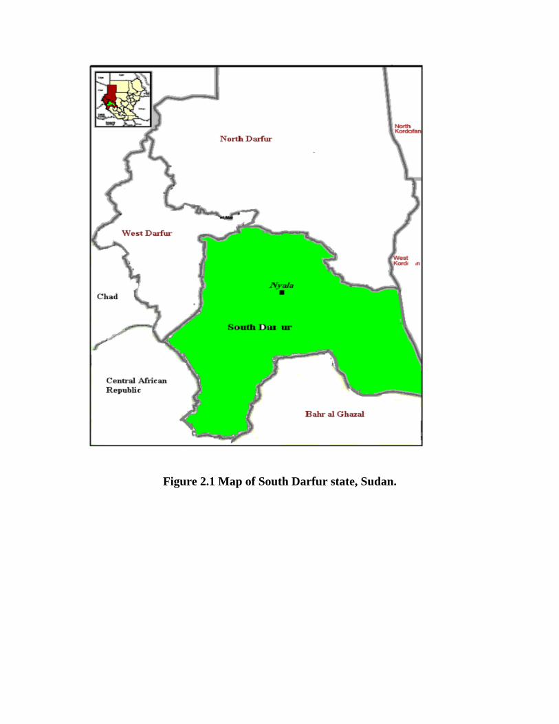
Figure 2.1 Map of South Darfur state, Sudan.

800-1000 egg per gram of faeces Moderate infection
1500-2000 egg per gram of faeces severe infection
2.1.4 Parasitological techniques
2.1.4.1 The modified McMaster technique
A modified McMaster technique (Anonymous, 1986) was used to count
the egg per gram (epg) of faeces as following:
1. Three grams of faeces were mixed with 42 ml of tap water and the faecal
suspension was passed through the 80 µm square sieve to remove debris.
2. The filtrate was collected in a clean dry container.
3. 15 ml of this filtrate was taken into a centrifuge tube and centrifuged at
1500 rpm for 2 minutes and the supernatant was then discarded.
4. The sediment was emulsified by gentle agitation and saturated Na Cl was
added until the volume became equal to the initial aliquot of the filtrate.
5. The centrifuged tube was inverted several times to obtain an even
suspension of the contents.
6. The two chambers of the McMaster slide were filled using a clean Pasteur
pipette.
7. The average number of eggs present in the chambers was multiplied by
100 to obtain the number of egg per gram of faeces (epg).
2.1.4.2 Faecal culture and identification of larvae
A pooled culture of positive faecal samples was examined for larval
identification, and where possible 100 third stage larvae were identified as
described by Anonymous (1986).
2.2 Therapeutic efficacy of Albendazole against donkey’s worm infestation
The aim of this study was to investigate the therapeutic efficacy of
Albendazole at two different dose regimens as an anthelmintic in donkeys

harbouring natural worm infestation and to compare the results obtained with
that of Ivermectin.
2.2.1 Experimental Animals
In this study we utilized 24 male donkeys (3-10 years). Before starting the
study, animals were examined to prove infestation with gastrointestinal
helminth parasites. Animals were kept in the premises of the Department of
Clinical Studies, Faculty of Veterinary Science, and University of Nyala (Figure
2.2). They were provided with tap water and allowed to graze in pasture freely.
2.2.2 Experimental drugs
Albendazole suspension: Albendex 25mg/ml (Avico®. Jordon).
Ivermectin drench: Avimec liquid (Avico®. Jordon)
2.2.3 Design of the study
Experimental animals were allocated into four groups and penned
according to treatment groups. The first three groups were treated and the last
group was remained untreated as control group.
The animals in the three treatment groups received treatment as follows:
- Albendazole – treated group 1 (ABZT1) received a single oral dose of
Albendazole at the manufacturer recommended dose i-e 10 mg/kg body weight.
- Albendazole – treated group 2 (ABZT2) received two oral doses of
Albendazole 14 days apart, at the manufacturer recommended dose i-e 10
mg/kg body weight.
- Ivermectin treated group (IVMT) received a single oral dose of Ivermectin
drench at the manufacturer recommended dose i-e 200 µg/kg body weight.
Then donkeys were monitored for possible adverse reactions for 2 hours
after administration of each drug.
2.2.4. Sampling and Time schedule
The experiment extended for 21 days. Faecal samples were collected at 0
(before treatment), 1, 3, 7, 14, and 21 days post treatment. Necropsy of the
animals was done at day 21 post treatment for all donkeys.
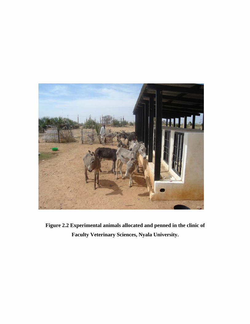
Figure 2.2 Experimental animals allocated and penned in the clinic of
Faculty Veterinary Sciences, Nyala University.
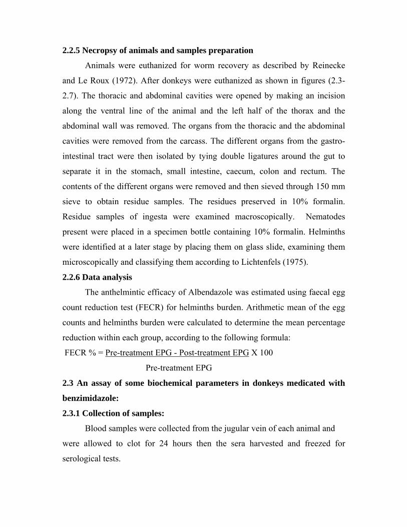
2.2.5 Necropsy of animals and samples preparation
Animals were euthanized for worm recovery as described by Reinecke
and Le Roux (1972). After donkeys were euthanized as shown in figures (2.3-
2.7). The thoracic and abdominal cavities were opened by making an incision
along the ventral line of the animal and the left half of the thorax and the
abdominal wall was removed. The organs from the thoracic and the abdominal
cavities were removed from the carcass. The different organs from the gastro-
intestinal tract were then isolated by tying double ligatures around the gut to
separate it in the stomach, small intestine, caecum, colon and rectum. The
contents of the different organs were removed and then sieved through 150 mm
sieve to obtain residue samples. The residues preserved in 10% formalin.
Residue samples of ingesta were examined macroscopically. Nematodes
present were placed in a specimen bottle containing 10% formalin. Helminths
were identified at a later stage by placing them on glass slide, examining them
microscopically and classifying them according to Lichtenfels (1975).
2.2.6 Data analysis
The anthelmintic efficacy of Albendazole was estimated using faecal egg
count reduction test (FECR) for helminths burden. Arithmetic mean of the egg
counts and helminths burden were calculated to determine the mean percentage
reduction within each group, according to the following formula:
FECR % = Pre-treatment EPG - Post-treatment EPG X 100
Pre-treatment EPG
2.3 An assay of some biochemical parameters in donkeys medicated with
benzimidazole:
2.3.1 Collection of samples:
Blood samples were collected from the jugular vein of each animal and
were allowed to clot for 24 hours then the sera harvested and freezed for
serological tests.
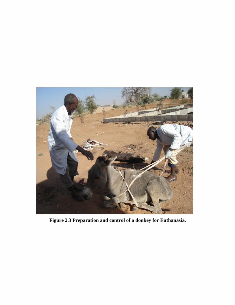
Figure 2.3 Preparation and control of a donkey for Euthanasia.
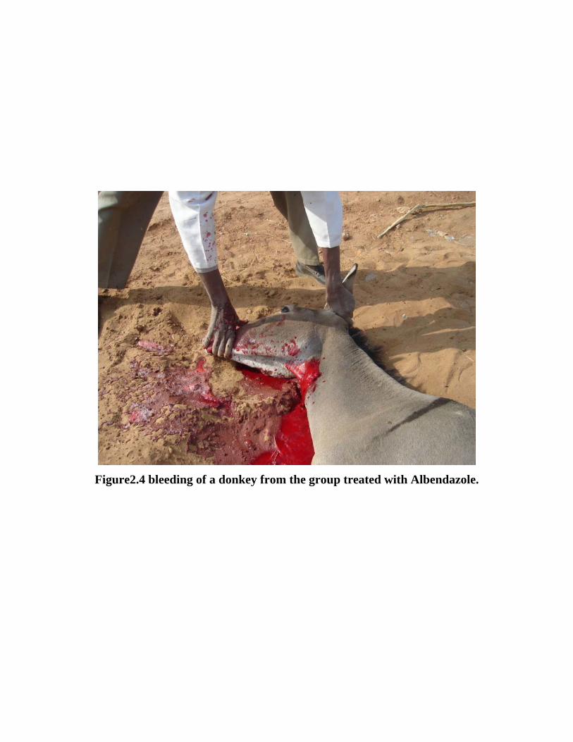
Figure2.4 bleeding of a donkey from the group treated with Albendazole.

Figure 2.5 Abdominal cavity of Euthanized donkey treated with
Albendazole.

Figure 2.6 Cranial mesenteric artery removed from euthanized donkey
treated with albendazole.
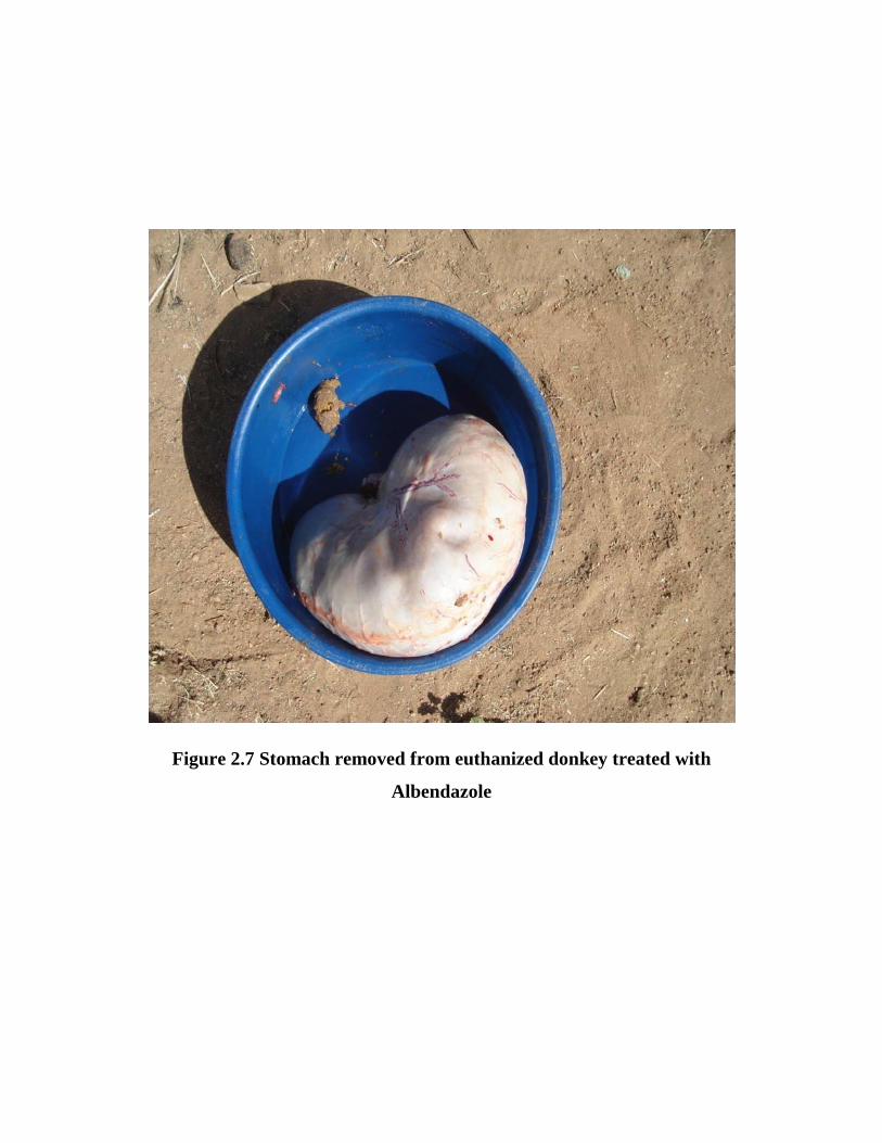
Figure 2.7 Stomach removed from euthanized donkey treated with
Albendazole
2.3.2 Biochemical methods
2.3.2.1 Total Serum Protein (TSP)
The Biuret method as described by King and Wooton (1956) was utilized
to assess TSP using commercial kits (Spectrum, Egypt).
Principle of the test
In alkaline medium the copper reacts with the peptide bonds of protein to
form the characteristic pink to purple biuret complex. Sodium potassium
tartarate prevent copper hydroxide precipitation and potassium iodide prevents
the auto-reduction of copper. The colour intensity is directly proportional to the
protein concentration. The change in colour was measured using
spectrophotometer (Jenway 6105 U.V./vis. Spectrophotometer, U.K.).
2.3.2.2 Serum Albumin
Concentration of serum albumin was measured by Bromocresol green
method according to Bartholomew and Delany (1966).
Principle of the test
Measurement of serum Albumin based on its binding to the indicator dye
bromochresol green (BCG) in pH 4.3 to form a blue-green coloured complex.
The intensity of the blue-green colour is directly proportional to the
concentration of albumin in the sample. The assay was conducted using
commercial kits (Spectrum, Egypt).
2.3.2.3 Serum Urea
Urea is the major end product of protein nitrogen metabolism. It is
synthesized by the urea cycle in the liver and excreted through the kidneys. The
circulating levels of the urea depend upon protein intake, protein catabolism and
kidneys function. Elevated urea level can occur due to renal impairment or in
some diseases such as diabetes, infection, congestive heart failure, and during
different liver diseases. Determination of blood urea nitrogen is most widely
used screening test for renal function together with serum creatinine.

The principle of the test
Urea is hydrolyzed in the presence of water and urease to produce:
Urea + H2O urease › 2NH3 + CO2
The free ammonia in an alkaline pH and in the presence of the indicator
forms coloured complex proportional to the urea concentration (mg/dl) in the
specimen.
Serum urea concentration was measured by an enzymatic colorimetric
method using a commercial kit (Randox laboratories Ltd., United Kingdom)
according to Fawcett and Scott (1960). The intensity of the developing colour
was measured at 600 nm using Jenway spectrophotometer (Jenway 6105 U. V.
/vis. Spectrophotometer, U. K.).
2.3.2.4 Serum creatinine
Creatinine is derived from creatine and the creative phosphate in the
muscle tissue, and may be defined as a nitrogenous waste product. Creatinine is
not reutilized but it is excreted from the body through the urine via the kidneys.
Principle
Creatinine reacts with picric acid under alkaline condition to form a
yellow-red complex. The intensity of the developing colour was measured at
wavelength 492nm, using Jenway spectrophotometer (Jenway 6105 U. V. /vis.
Spectrophotometer, U. K) according to Bartels et al., (1972).The commercial
kits used were also from the same manufacturer (Spectrum, Egypt).
2.3.2.5 Serum Calcium
Calcium is the fifth most common element in the body, most of which
(98%) is present in the skeleton. One half of the remaining calcium is found in
extracellular fluid and the rest in tissue. Calcium has a crucial role in bone
mineralization and is also vital for basic physiological processes such as blood
coagulation, neuromuscular conduction, and normal muscle tone.

The principle of the test
Calcium ions react with O-cresolphthalein complexone (O-CPC) under
alkaline condition to form violet coloured complex. The colour intensity of the
complex formed is directly proportional to the calcium concentration (mg/dl). It
is determined by measuring the increase in absorbance at 578nm using
commercial kit (Spectrum, Egypt). The calcium values were calculated in
mmol/l of serum according to Sarkar and Chauhan (1967) and Barnett et al.,
(1973).
2.3.2.6 Serum inorganic phosphorus
The body contains phosphorus entirely in the form of phosphates
distributed fairly equally between extracellular and intracellular compartments.
About 85% of extracellular phosphate occurs in inorganic forms as
hydroxyapatite. In plasma or serum, most phosphate exists in the inorganic
form; this fraction is present as the mono- and dihydrogen forms. The relative
proportion is varying with the pH. Intracellular phosphate occurs mainly in as
phospholipids and phosphoproteins; this fraction is termed organic phosphate.
Assay principle
Inorganic phosphate reacts with ammonium molybdate in sulphuric acid to
form no reduced phosphomolbdate. The concentration of phosphomolbdate
formed is directly proportional to the inorganic phosphate concentration. It is
determined by measuring the increase in absorbance at 340 nm. The serum
inorganic phosphorus was determined according to Taussky and Shorr (1953)
and Goldenburg and Fernandez, (1966).
2.4. Statistical Methods
SPSS 11.5 for windows computer package was utilized to assess
significant differences, if any. Paired T-test was used to compare between
means.

CHAPTER THREE

Chapter three
Results
3.1 Survey of gastro-intestinal nematodes in donkeys and horses:
In the current study a total of 1256 donkeys and horses were examined
for helminth gastrointestinal nematodes, during the period October 2006 to
September 2007. The overall prevalence of infection with gastro-intestinal
nematodes in equines was 29.20% (Table 3.1). During the survey period,
November showed the highest incidence of infection (41.50%), while June
showed the lowest percentage 13.92% (Figure 3.2).
Considering severity of infection, animals with mild infection were
dominant (81.35%), while animals with severe infection were 10.54%, and
animals with moderate infection were the lowest one 8.11% (Table 3.3). The
highest incidence of mild infection was reported in August (93.75%) and the
lowest incidence was on April 64.29%. In animals harbouring moderate
infection, the lowest infection level was 0% and reported in August and the
highest moderate infection level was reported in March 20.69%. Severe
infection reported 0% in the lowest incidence level in June while 28.57% in
April as the highest (Table 3.3).
The results of faecal culture and identification of larvae revealed the
dominance of following helminth genera: Strongylus spp, Cyathostomum spp,
Trichostrongylus spp, and Strongyloides westeri. No efforts to larvae count had
done.
In this study, a total of 443 horses were involved in the current survey.
The prevalence of infection was 15.35% (Table 3.5). March reported the lowest
prevalence level of 5.26% and in November was the highest one 50% (Table
3.5). In February we failed to examine any horse. A total number of 810
donkeys were involved in this study. The prevalence of infection was 37.48%.
The highest prevalence infection was reported in January 55.79% and the lowest
one was 14.89% reported in May (Table 3.5).
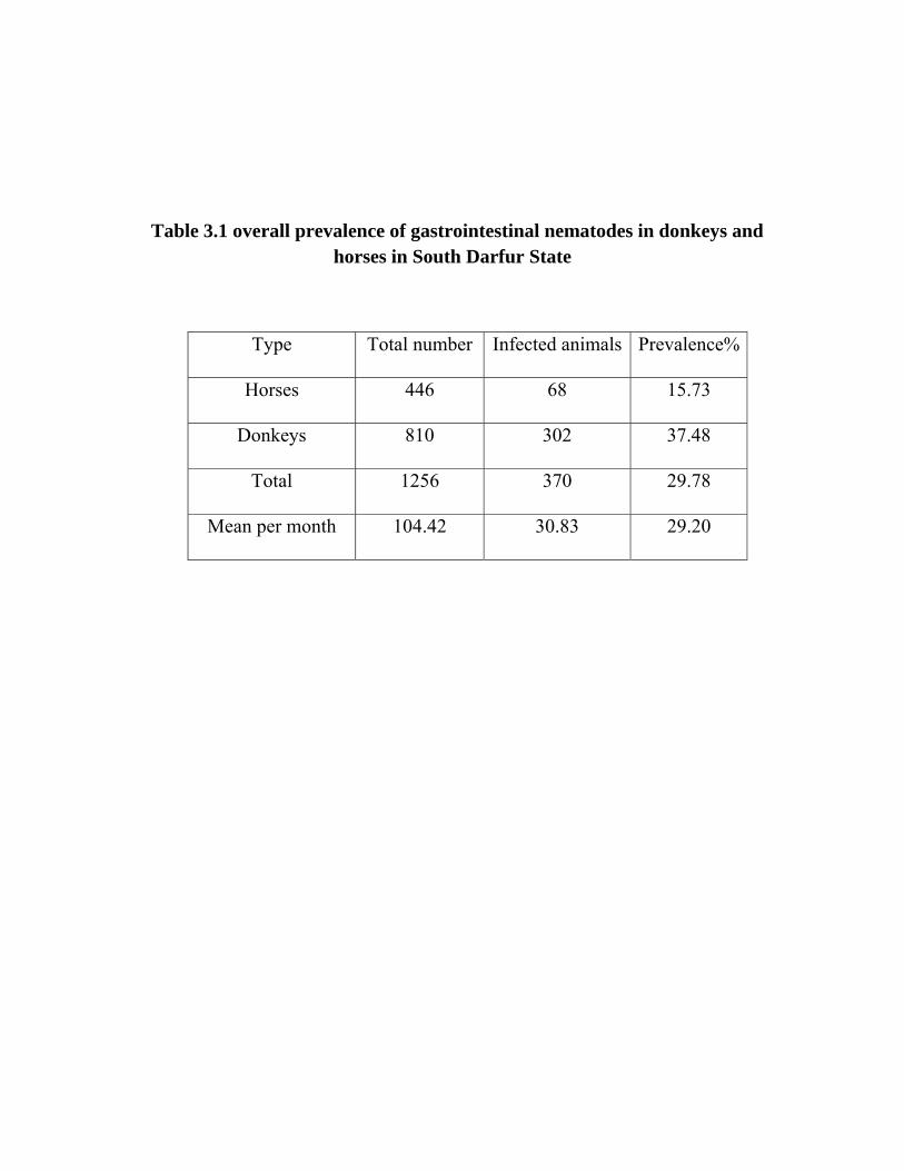
Table 3.1 overall prevalence of gastrointestinal nematodes in donkeys and horses in South Darfur State
Type Total number Infected animals Prevalence%
Horses 446 68 15.73
Donkeys 810 302 37.48
Total 1256 370 29.78
Mean per month 104.42 30.83 29.20

Figure 3.1Percentage of donkeys to horses in the population sampled.
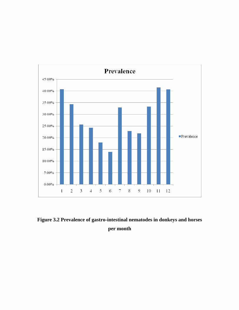
Figure 3.2 Prevalence of gastro-intestinal nematodes in donkeys and horses
per month

Table 3.2 Mean± SD and range of egg per gram of faeces (epg) in donkeys
and horses infested with gastro-intestinal nematodes
Month Mean ± SD Range
January 400.86 ± 640.41 50-3800
February 890.91±1,231.63 50-4500
March 655.17 ±859.65 50-3600
April 1390.03 ±2401.26 50-11800
May 457.89 ±526.32 50-1700
June 290.00± 242.44 50-900
July 283.75 ±290.53 50-1600
August 340.00± 364.10 50-1500
September 565.71 ±1366.55 50-8000
October 889.29 ±1,641.45 50-10400
November 608.49 ±1,725.63 50-13450
December 307.58 ±542.29 50-2400
Total 589.97 ±986.02 50-13450
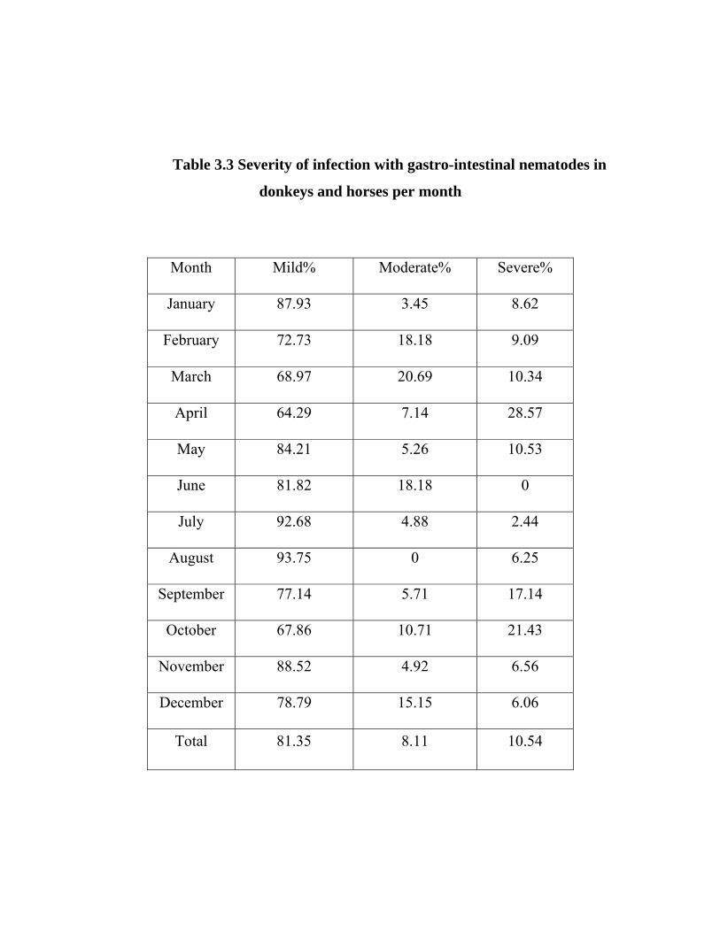
Table 3.3 Severity of infection with gastro-intestinal nematodes in
donkeys and horses per month
Month Mild% Moderate% Severe%
January 87.93 3.45 8.62
February 72.73 18.18 9.09
March 68.97 20.69 10.34
April 64.29 7.14 28.57
May 84.21 5.26 10.53
June 81.82 18.18 0
July 92.68 4.88 2.44
August 93.75 0 6.25
September 77.14 5.71 17.14
October 67.86 10.71 21.43
November 88.52 4.92 6.56
December 78.79 15.15 6.06
Total 81.35 8.11 10.54
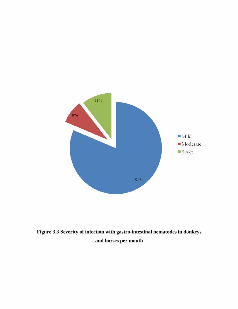
Figure 3.3 Severity of infection with gastro-intestinal nematodes in donkeys
and horses per month
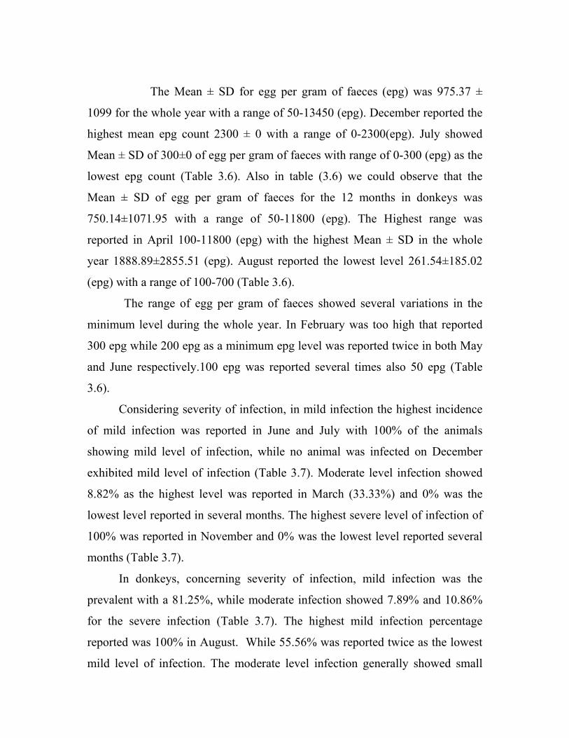
The Mean ± SD for egg per gram of faeces (epg) was 975.37 ±
1099 for the whole year with a range of 50-13450 (epg). December reported the
highest mean epg count 2300 ± 0 with a range of 0-2300(epg). July showed
Mean ± SD of 300±0 of egg per gram of faeces with range of 0-300 (epg) as the
lowest epg count (Table 3.6). Also in table (3.6) we could observe that the
Mean ± SD of egg per gram of faeces for the 12 months in donkeys was
750.14±1071.95 with a range of 50-11800 (epg). The Highest range was
reported in April 100-11800 (epg) with the highest Mean ± SD in the whole
year 1888.89±2855.51 (epg). August reported the lowest level 261.54±185.02
(epg) with a range of 100-700 (Table 3.6).
The range of egg per gram of faeces showed several variations in the
minimum level during the whole year. In February was too high that reported
300 epg while 200 epg as a minimum epg level was reported twice in both May
and June respectively.100 epg was reported several times also 50 epg (Table
3.6).
Considering severity of infection, in mild infection the highest incidence
of mild infection was reported in June and July with 100% of the animals
showing mild level of infection, while no animal was infected on December
exhibited mild level of infection (Table 3.7). Moderate level infection showed
8.82% as the highest level was reported in March (33.33%) and 0% was the
lowest level reported in several months. The highest severe level of infection of
100% was reported in November and 0% was the lowest level reported several
months (Table 3.7).
In donkeys, concerning severity of infection, mild infection was the
prevalent with a 81.25%, while moderate infection showed 7.89% and 10.86%
for the severe infection (Table 3.7). The highest mild infection percentage
reported was 100% in August. While 55.56% was reported twice as the lowest
mild level of infection. The moderate level infection generally showed small
percentage 7.89% when compared to mild and severe infection. The highest
percentage of moderate infection was reported in June 20%, and the lowest
moderate infection 0% was reported in August and September respectively.
Severe infection reported the highest percentage in April 38.89%. Both June
and August reported 0% as the lowest percentage (Table 3.7).
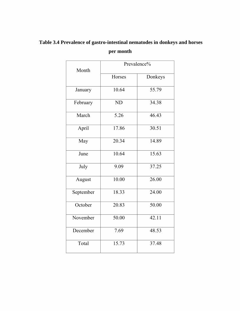
Table 3.4 Prevalence of gastro-intestinal nematodes in donkeys and horses
per month
Prevalence% Month
Horses Donkeys
January 10.64 55.79
February ND 34.38
March 5.26 46.43
April 17.86 30.51
May 20.34 14.89
June 10.64 15.63
July 9.09 37.25
August 10.00 26.00
September 18.33 24.00
October 20.83 50.00
November 50.00 42.11
December 7.69 48.53
Total 15.73 37.48
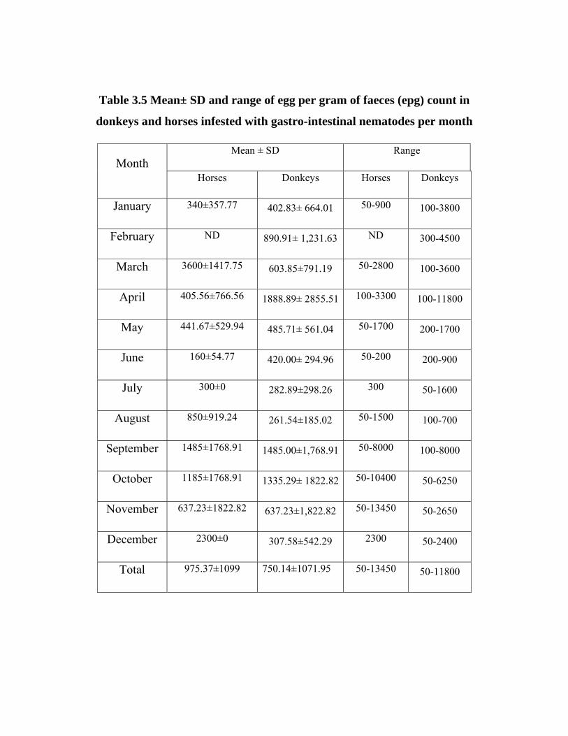
Table 3.5 Mean± SD and range of egg per gram of faeces (epg) count in
donkeys and horses infested with gastro-intestinal nematodes per month
Mean ± SD Range Month
Horses Donkeys Horses Donkeys
January 340±357.77 402.83± 664.01 50-900 100-3800
February ND 890.91± 1,231.63 ND 300-4500
March 3600±1417.75 603.85±791.19 50-2800 100-3600
April 405.56±766.56 1888.89± 2855.51 100-3300 100-11800
May 441.67±529.94 485.71± 561.04 50-1700 200-1700
June 160±54.77 420.00± 294.96 50-200 200-900
July 300±0 282.89±298.26 300 50-1600
August 850±919.24 261.54±185.02 50-1500 100-700
September 1485±1768.91 1485.00±1,768.91 50-8000 100-8000
October 1185±1768.91 1335.29± 1822.82 50-10400 50-6250
November 637.23±1822.82 637.23±1,822.82 50-13450 50-2650
December 2300±0 307.58±542.29 2300 50-2400
Total 975.37±1099 750.14±1071.95 50-13450 50-11800
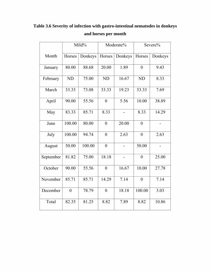
Table 3.6 Severity of infection with gastro-intestinal nematodes in donkeys
and horses per month
Mild% Moderate% Severe%
Month Horses Donkeys Horses Donkeys Horses Donkeys
January 80.00 88.68 20.00 1.89 0 9.43
February ND 75.00 ND 16.67 ND 8.33
March 33.33 73.08 33.33 19.23 33.33 7.69
April 90.00 55.56 0 5.56 10.00 38.89
May 83.33 85.71 8.33 - 8.33 14.29
June 100.00 80.00 0 20.00 0 -
July 100.00 94.74 0 2.63 0 2.63
August 50.00 100.00 0 - 50.00 -
September 81.82 75.00 18.18 - 0 25.00
October 90.00 55.56 0 16.67 10.00 27.78
November 85.71 85.71 14.29 7.14 0 7.14
December 0 78.79 0 18.18 100.00 3.03
Total 82.35 81.25 8.82 7.89 8.82 10.86
3.2 Therapeutic efficacy of Albendazole and Ivermectin drench in donkeys
3.2.1 Egg per gram reduction
The result of mean egg per gram of faeces and the range in addition to the
reduction percentage of egg per gram of faeces for the three treated groups from
day zero to day 21 are presented in tables (3.8, 3.9 and 3.10).
On day 3, Albendazole showed reduction of 97.13% egg per gram count
(EPGC), while Albendazole with the two doses showed 98.90%. In Ivermectin
treated group 98.94% of egg per gram count (EPGC) was reported.
All three groups reported 100% reduction of egg per gram of faeces on
day 7 and till day 21 when animals were euthanized.
3.2.2 Post-mortem findings
The results of post-mortem findings are presented in tables (3.11, 3.12
and 3.13) for the three groups. Albendazole reported efficacy of 97.13% at the
single dose while albendazole at the two doses reported 98.90%, but the
efficacy against L4 Strongylus vulgaris found in cranial mesenteric arteries was
33% for Albendazole and 59.08 %for Albendazole twice. On the other side,
Ivermectin reported 62.71% efficacy against L4 Strongylus vulgaris.

Table 3.7 Mean faecal egg counts (±SD) and reduction percentage for
Albendazole-treated donkeys
Days Arithmetic mean
(epg) Range Reduction%
0 3621.43±3583.05 500-10400 -
1 1221.43±1526.94 200-4500 66.27%
3 21.43±39.34 50-100 98.25%
7 0 0 100%
14 0 0 100%
21 0 0 100%

Table3.8 Mean faecal egg counts (±SD) and reduction percentage for Albendazole twice-treated donkeys
Days Arithmetic mean (epg) Range Reduction %
0 2462.50 ± 2137.50 1300-4500 -
1 1250.00 ± 574.46 900-2100 49.24%
3 100.00 ±141.42 0-300 92%
7 0 0 100%
14 0 0 100%
21 0 0 100%
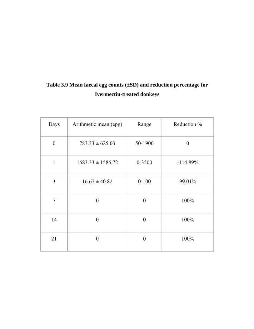
Table 3.9 Mean faecal egg counts (±SD) and reduction percentage for
Ivermectin-treated donkeys
Days Arithmetic mean (epg) Range Reduction %
0 783.33 ± 625.03 50-1900 0
1 1683.33 ± 1586.72 0-3500 -114.89%
3 16.67 ± 40.82 0-100 99.01%
7 0 0 100%
14 0 0 100%
21 0 0 100%

Table 3.10 Summary of harvested worms from control and animals treated with Albendazole (ABZT1) drench at necropsy.
Albendazole Organs examined Control
No. Reduction %
Cranial mesenteric artery
Strongylus vulgaris 303 203 33.00
Stomach
Gasterophilus spp. 349 273 21.78
Habronema spp. 291 147 49.48
Trichostrongylus axei 79 26 67.09
Small Intestine
Parascaris equorum 25 0 100.00
Strongloid westeri 31 10 67.74
Caecum
Gasterophilus spp. 3151 59 98.13
Strongylus spp. 841 157 81.33
Cyathostomes spp. 6012 0 100.00
Colon and Rectum
Strongylus spp. 6800 109 98.40
Cyathostomes spp. 37660 701 98.14
Oxyuris equi 3151 0 100.00
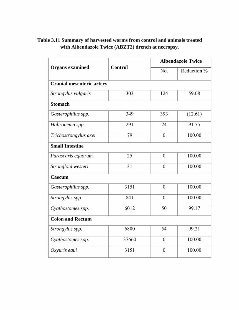
Table 3.11 Summary of harvested worms from control and animals treated with Albendazole Twice (ABZT2) drench at necropsy.
Albendazole Twice Organs examined Control
No. Reduction %
Cranial mesenteric artery
Strongylus vulgaris 303 124 59.08
Stomach
Gasterophilus spp. 349 393 (12.61)
Habronema spp. 291 24 91.75
Trichostrongylus axei 79 0 100.00
Small Intestine
Parascaris equorum 25 0 100.00
Strongloid westeri 31 0 100.00
Caecum
Gasterophilus spp. 3151 0 100.00
Strongylus spp. 841 0 100.00
Cyathostomes spp. 6012 50 99.17
Colon and Rectum
Strongylus spp. 6800 54 99.21
Cyathostomes spp. 37660 0 100.00
Oxyuris equi 3151 0 100.00
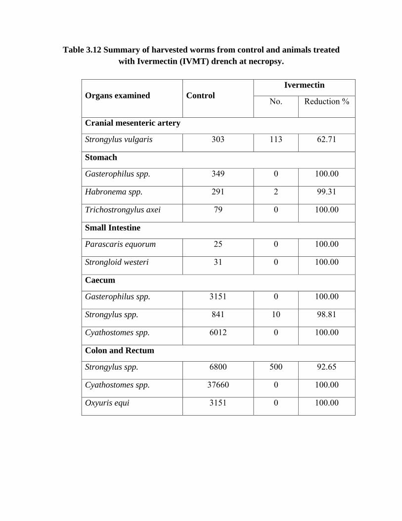
Table 3.12 Summary of harvested worms from control and animals treated with Ivermectin (IVMT) drench at necropsy.
Ivermectin Organs examined Control
No. Reduction %
Cranial mesenteric artery
Strongylus vulgaris 303 113 62.71
Stomach
Gasterophilus spp. 349 0 100.00
Habronema spp. 291 2 99.31
Trichostrongylus axei 79 0 100.00
Small Intestine
Parascaris equorum 25 0 100.00
Strongloid westeri 31 0 100.00
Caecum
Gasterophilus spp. 3151 0 100.00
Strongylus spp. 841 10 98.81
Cyathostomes spp. 6012 0 100.00
Colon and Rectum
Strongylus spp. 6800 500 92.65
Cyathostomes spp. 37660 0 100.00
Oxyuris equi 3151 0 100.00
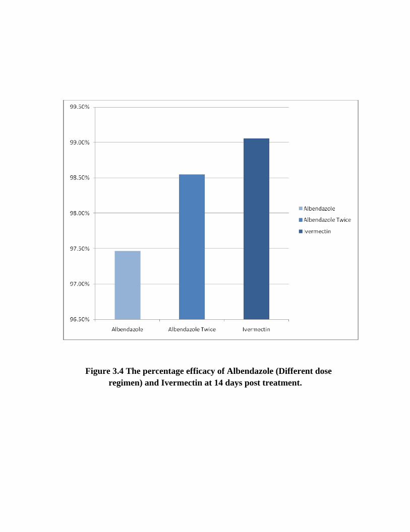
Figure 3.4 The percentage efficacy of Albendazole (Different dose regimen) and Ivermectin at 14 days post treatment.

Figure 3.5 larvae of Strongylus vulgaris in cranial mesntric artry removed form Albendazole treated donkey.

Figure 3.6 larvae of gasterophilus in a stomach removed from Albendazole
treated donkey.
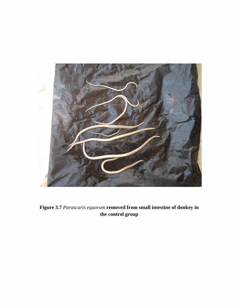
Figure 3.7 Parascaris equorum removed from small intestine of donkey in the control group

Figure 3.8 Parascaris equorum expeled from small intestine of donkey in the
Ivermectin Drench treated group on day 3 post treatment.
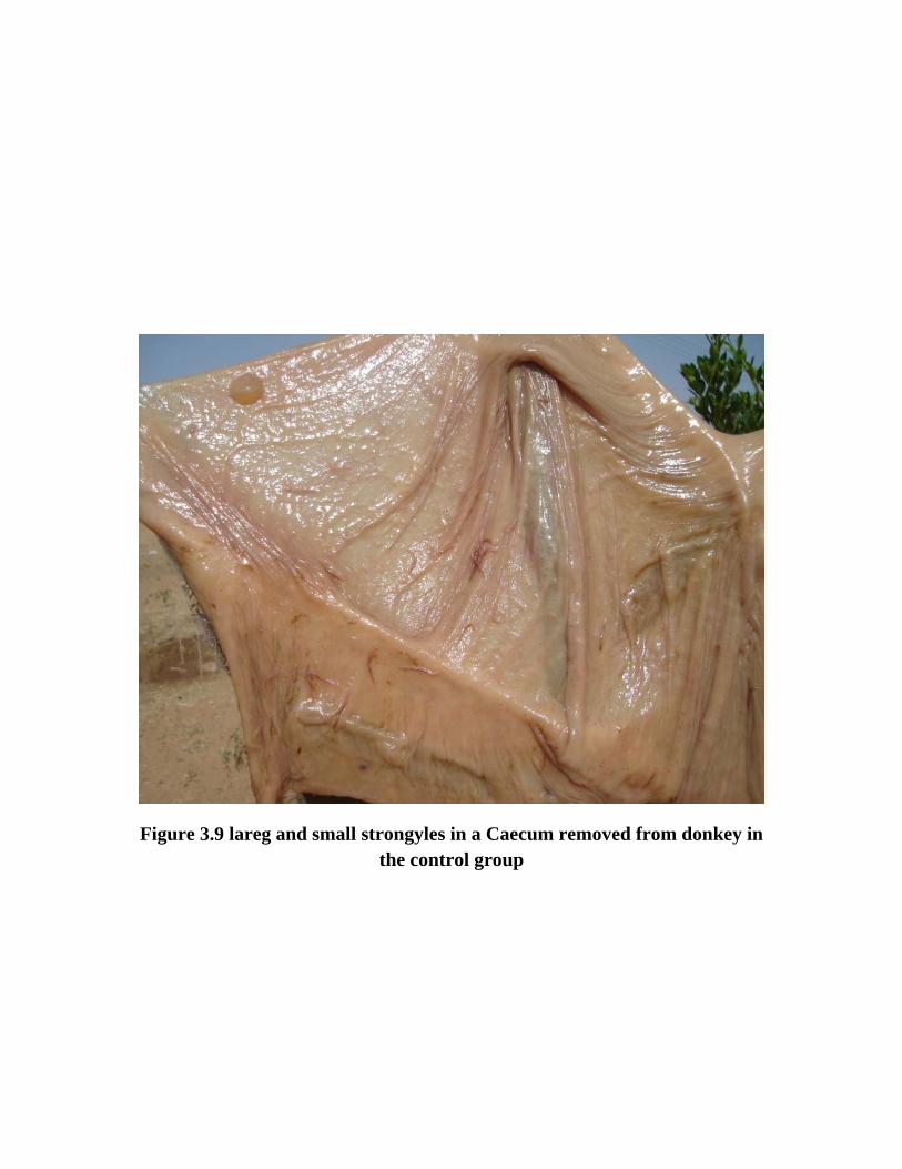
Figure 3.9 lareg and small strongyles in a Caecum removed from donkey in
the control group

Figure 3.10 Encysted larvae of small strongyles in colon of donkey in the
control group.
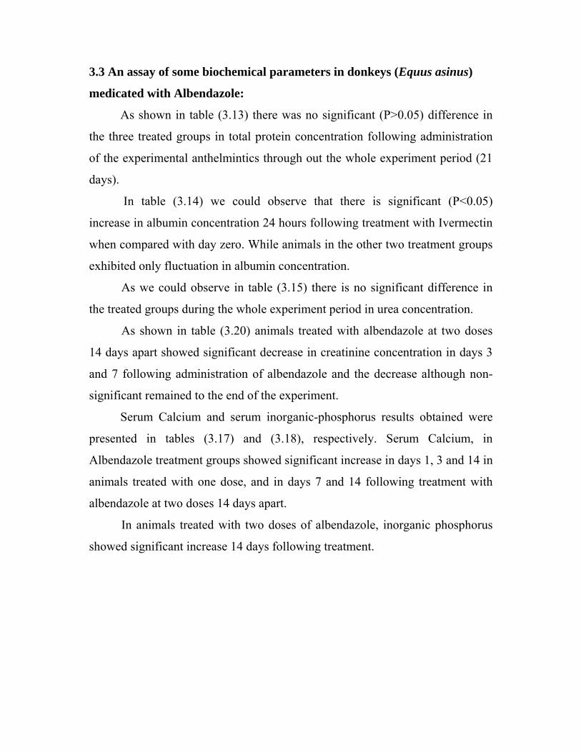
3.3 An assay of some biochemical parameters in donkeys (Equus asinus)
medicated with Albendazole:
As shown in table (3.13) there was no significant (P>0.05) difference in
the three treated groups in total protein concentration following administration
of the experimental anthelmintics through out the whole experiment period (21
days).
In table (3.14) we could observe that there is significant (P<0.05)
increase in albumin concentration 24 hours following treatment with Ivermectin
when compared with day zero. While animals in the other two treatment groups
exhibited only fluctuation in albumin concentration.
As we could observe in table (3.15) there is no significant difference in
the treated groups during the whole experiment period in urea concentration.
As shown in table (3.20) animals treated with albendazole at two doses
14 days apart showed significant decrease in creatinine concentration in days 3
and 7 following administration of albendazole and the decrease although non-
significant remained to the end of the experiment.
Serum Calcium and serum inorganic-phosphorus results obtained were
presented in tables (3.17) and (3.18), respectively. Serum Calcium, in
Albendazole treatment groups showed significant increase in days 1, 3 and 14 in
animals treated with one dose, and in days 7 and 14 following treatment with
albendazole at two doses 14 days apart.
In animals treated with two doses of albendazole, inorganic phosphorus
showed significant increase 14 days following treatment.

Table 3.13 Changes in total protein concentration (g/dL) following oral
administration of experimental anthelmintics compounds
ABZT1 ABZT2 IVMT DAY
Mean ± SD Mean ± SD Mean ± SD
0 09.94±2.24 08.47±0.88 8.02±1.22
1 09.06±1.50 09.64±1.24 8.76±0.73
3 10.96±2.56 12.52±5.55 8.49±0.70
7 08.53±0.73 10.30±0.95 8.56±0.84
14 08.21±0.33 08.97±0.58 8.02±0.51
21 09.53±0.39 10.74±1.87 8.86±0.72

Table 3.14 Changes in albumin concentration (g/dL) following oral
administration of experimental anthelmintics compounds
ABZT1 ABZT2 IVMT DAY
Mean ± SD Mean ± SD Mean ± SD
0 3.14±0.36 2.88±0.44 2.88±0.44
1 3.15±0.33 3.25±0.37 3.88±0.43*
3 3.06±0.36 3.18±0.42 2.91±0.41
7 2.95±0.59 3.84±0.66 3.74±0.81
14 2.95±0.50 3.28±0.28 2.73±0.33
21 3.16±0.21 3.66±0.86 3.19±0.57
(*) Means on the same column have asterisk are significantly different with day
zero

Table 3.15 Changes in urea concentration (mg/dL) following oral
administration of experimental anthelmintics compounds
ABZT1 ABZT2 IVMT DAY
Mean ± SD Mean ± SD Mean ± SD
0 8.57±1.61 09.26±1.94 7.47±1.37
1 7.01±0.35 09.16±3.14 7.10±0.73
3 7.60±1.18 09.19±3.28 8.14±0.82
7 7.68±1.13 11.57±3.86 8.41±1.01
14 8.12±3.80 06.89±2.14 7.57±1.42
21 9.71±2.82 09.55±2.72 8.35±2.12
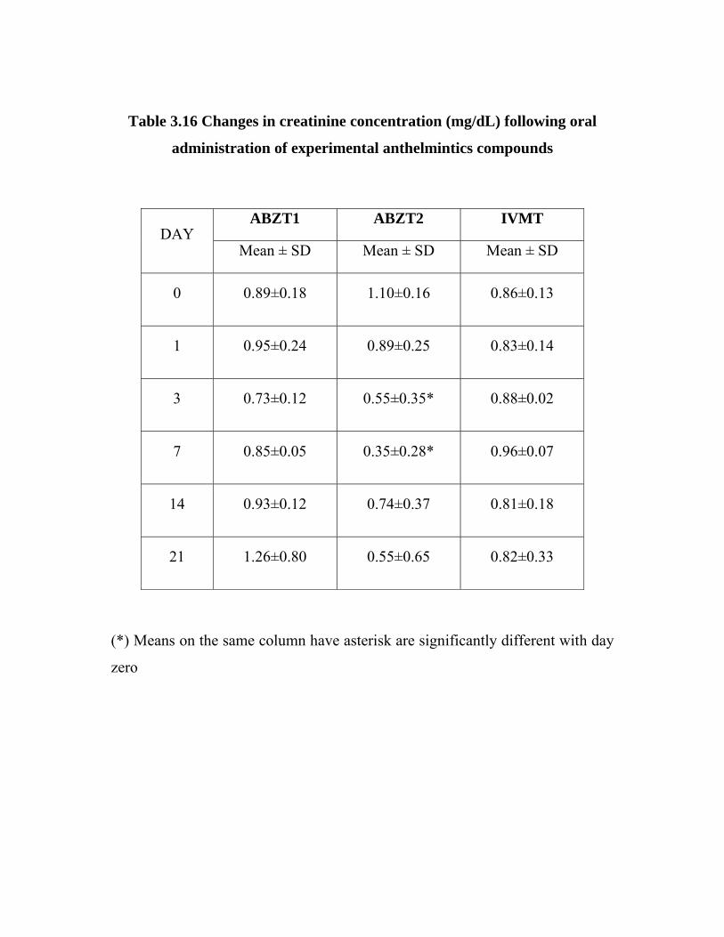
Table 3.16 Changes in creatinine concentration (mg/dL) following oral
administration of experimental anthelmintics compounds
ABZT1 ABZT2 IVMT DAY
Mean ± SD Mean ± SD Mean ± SD
0 0.89±0.18 1.10±0.16 0.86±0.13
1 0.95±0.24 0.89±0.25 0.83±0.14
3 0.73±0.12 0.55±0.35* 0.88±0.02
7 0.85±0.05 0.35±0.28* 0.96±0.07
14 0.93±0.12 0.74±0.37 0.81±0.18
21 1.26±0.80 0.55±0.65 0.82±0.33
(*) Means on the same column have asterisk are significantly different with day
zero
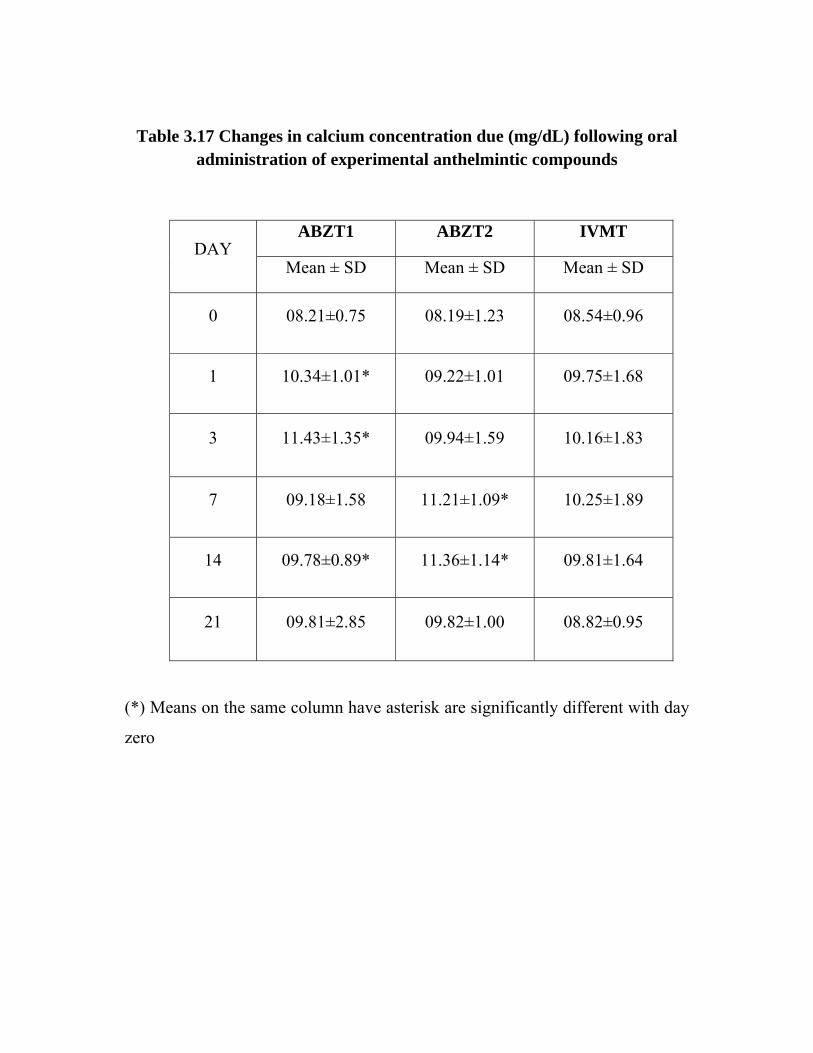
Table 3.17 Changes in calcium concentration due (mg/dL) following oral administration of experimental anthelmintic compounds
ABZT1 ABZT2 IVMT DAY
Mean ± SD Mean ± SD Mean ± SD
0 08.21±0.75 08.19±1.23 08.54±0.96
1 10.34±1.01* 09.22±1.01 09.75±1.68
3 11.43±1.35* 09.94±1.59 10.16±1.83
7 09.18±1.58 11.21±1.09* 10.25±1.89
14 09.78±0.89* 11.36±1.14* 09.81±1.64
21 09.81±2.85 09.82±1.00 08.82±0.95
(*) Means on the same column have asterisk are significantly different with day
zero

Table 3.18 Changes in inorganic-phosphorus concentration (mg/dl)
following oral administration of experimental anthelmintics compounds
ABZT1 ABZT2 IVMT DAY
Mean ± SD Mean ± SD Mean ± SD
0 4.63±2.57 3.81±1.79 3.90±0.84
1 5.18±1.58 5.75±2.53 4.11±1.26
3 7.26±2.97 7.58±3.99 5.79±1.56
7 6.59±2.10 9.34±5.47 5.74±3.38
14 4.79±0.71 6.46±0.74* 5.30±2.35
21 5.39±1.87 9.66±5.24 5.60±1.18
(*) Means on the same column have asterisk are significantly different with day
zero .

CHAPTER FOUR

Chapter four
Discussion
From the results obtained in this study, it is interesting to note that, the
overall prevalence for both donkeys and horses with CIT parasited was found to
be 29.785%, this is in harmony with the results obtained by Kheir and Kheir
(1981) in Bahr El Arab (22%), while it is less than that reported in Nyala town
(58%), this may be referred to the large number of animal examined in this
study (1256 animal) when compared to Kheir and Kheir (390 animals), and also
to the availability of anthelmintics compounds. In Sennar-Sudan also El Dirdiri
et al., (1986) reported a similar percentage of infection with gastro intestinal
diseases (27%).
Prevalence of gastrointestinal nematodes in horses noticed in this study
was 15.73%, which is in close agreement with that reported by Kheir and Kheir
(1981) in Bahr El Arab (18.5%). Results obtained in this study concerning the
prevalence of helminth nematodes in donkeys was 37.84% this result is very
low when compared with that reported by Seri et al., (2004) in Khartoum state-
Sudan (70.1%), while Kheir and Kheir (1981) reported that the overall
incidence of infection with nematode parasites was found higher in town
animals (58%) than in animals kept in nomadic areas (22%).
The overall Mean ± SD for egg per gram of faeces (epg) was 589.97 ±
986.02 and the range was 50-11800 (epg), this is low when compared with Seri
et al., (2004) (1016.6 ± 363.6) (epg), this may be attributed to the large
percentage of mild infection obtained in this study (81.35%) when compared to
that of Seri et al., (2004) who reported 58.6% in donkeys examined in
Khartoum state.
The highest Mean ± SD was observed in April (hot season) 1390.03 ±
2401.26 with a range of 50-11800 (epg), and the lowest was in June (rainy
season) 290.00 ± 242.44 with a range of 50-900 (epg).
Concerning severity of infection reported in this study, 81.35% for mild
infection, both moderate and severe burdens shared the lower incidence (8.11%
and10.54% respectively) these findings are in agreement with Seri et al., (2004)
in donkeys (58.6%, 21.9% 19.5%) for mild, moderate and severe infections
respectively.
However, all the findings obtained in the survey during the whole year
were affected with the extensive miss-use of anthelmintics by animal’s owners
in absence of veterinary authorities where sometimes sub doses, over dosage of
anthelmintics, wrong timing of de-worming and a lot of misconceptions of de-
worming were applied, what shows apparent differences in the results in this
study.
Therapeutic efficacy of Albendazole in donkeys in this study, in both
groups of animals treated with either single dose or two doses of Albendazole at
10 mg/kg were resulted in 100% reduction in (epg) count on day 7 post
treatment. Kuzmina and Kharchenko (2008) obtained a similar result at day10
post treatment in horses treated with a dose rate of 5mg/kg body weight in
Ukraine to control Strongylus spp.
Following post-mortem, Albendazole administered at single dose resulted
in low efficacy of 49.48% against Habronema sp., and Trichostrongylus axei
67.09%, Strongyloid westeri 67.74%; but expressed high efficacy against
Parascaris equorum100%; and 81.33%, 98.40% for Strongylus spp. Present in
caecum, colon and rectum respectively; Cyathostomes spp 100% 98.14% for
caecum, colon and rectum respectively. This result explained by the
justification of Gokbulut et al., (2005) who stated that, a higher metabolic
capacity, first-pass effect and lower absorption of benzimidazoles in donkeys
decrease bioavailability and efficacy compared to ruminants, this explanation
also justify the increase in efficacy when we used the double dose for the
second group in this study

Albendazole at single and multiple dose regimen showed efficacy of
21.78% and (12.61)% against Gasterophilus spp. Albendazole at two doses 14
days apart showed the following efficacy percentages against: Habronema spp.
91.75%; Trichostrongylus axei100.%; Parascaris equorum 100% , Strongloid
westeri 100% ; Strongylus spp .in the caecum, colon and rectum 100%, 99.21%
respectively. Cyathostomes spp in the caecum, colon and rectum 99.17% and
100% respectively; and 100% for Oxyuris equi. These results are in agreement
with that obtained by Kuzmina and Kharchenko (2008) in Ukraine. Besides the
strongylid nematodes, eggs of Parascaris equorum, Oxuyris equi, Strongyloides
westeri and Habronema spp. were found in faecal samples a day before
treatment. No eggs of these nematodes were found in horse faeces on the 10th
and 14th days after treatment with the both drugs.
In this study, Ivermectin showed 100% faecal egg count reduction on day
7 this is in agreement with Seri et al., (2005) who reported the same result when
used ivermectin injectable formulation intramusclarlly at dose rate of 200µg/kg
body weight in donkeys in sudan, the efficacy against Habronema spp was
99.31% this result is in close agreement with that reported in horse by Herd and
Donham (1984), who showed 1-2 intramuscular doses of Ivermectin at 200
µg/kg were highly effective against Draschia spp and Habronema spp., while
DiPietro (1982) revealed 100% efficacy. When Ivermectin given
intramuscularly to donkeys at dose rate of 200 µg/kg, the efficacy was 100%
against larvae of Gasterophilus spp. (Seri et al., 2005), this result is in
agreement with the results (100%) obtained in this study. Ivermectin
successfully (100%) removed P. equorum from the small intestine when given
orally as paste formulation at 200 µg/kg. Ivermectin totally eliminated the
passage of P. equorum in the natural infected horses (Cobra et al., 1986). In
case of T. axie Ivermectin (100%) eliminated T. axie from the small intestine of
donkeys this also the same condition when used at dose rate of 200 µg/kg in
intramuscular formulation (Seri et al., 2005). In this study, Strongylus spp were
98.81% eliminated from caecum and 92.65% from the colon and rectum, which
is in close agreement when compared with that reported by Seri et al.,
(2005)100% when they used Ivermectin injectable formulation intramuscularly
for donkeys at dose rat of 200 µg/kg, a result which may be attributed to the
route of administration that affects the bioavailability of the drug in this study.
Cyathostomes spp were 100% eliminated from both caecum and colon in
this study, this result also is in agreement with that obtained by Seri et al.,
(2005). Costa et al., (1998) when utilized Ivermectin at same dose rate of 200
µg/kg for equine in Brazil as a paste formulation reported an efficacy of 100%
against Oxiuris equi, the same result was also obtained in this study. In this
study, Ivermectin expressed moderate efficacy against Strongylus vulgaris
larvae which found in the cranial mesenteric arteries 62.71% this result is to be
considered in the same range with that reported by Costa et al., (1998) and Seri
et al., (2005) and the results were 67.8%, 69,23% respectively.
In this study, the values obtained for the total protein values in the three
treatment groups were higher than reference values obtained by other
researchers worldwide and not in harmony with the values reported by Zinkl et
al., (1990) 7.2±0.7 in American donkeys and that 6.82 ±0.40 reported by Mori
et al., (2003) in Brazilian donkeys, we attribute this difference to the breed
and/or the technical method used. Daly and Hogan (1982) reported increasing of
urinary protein in rats treated with albendazole at 30 and 45 mg/kg bw (3 and
4.5 times of recommended dose, respectively).
In the current study, Albumin concentration fluctuated within the normal
level for albendazole treated groups which ranged between 2.88±0.44 g/L on
day 0 for Albendazole treated group 1 to 3.66±0.86 g/L on day 21 for the
animals treated with two doses with non significant differences, and this is on
line with reference range 3.48 ± 0.148 reported in India by Gupta (1994) and
not in harmony with Daly and Hogan (1982), who reported increased level of
albumin in male rats when treated with a dose of 45 mg/kg. We referred that to
the difference between the two doses. Ivermectin treated animals showed
significant increase in day one, this finding is in agreement with Seri et al
(2006), who reported significant increase in day 2 post treatment with
Doramectin in donkeys and also in horses Herd et al (1985) reported similar
results.
Urea concentration showed no significant difference in the groups treated
with Albendazole with the tow doses and Ivermectin. Herd and Kociba (1985)
reported that in horses treated with Ivermectin and also Seri et al., (2006) in
donkeys treated with Doramectin had different result which revealed significant
increase and decrease of Urea some sampling times. We may refer this to the
tow different routes of administration which affected the bioavailability of the
drugs.
Serum creatine concentration was decreased in two occasions
respectively on days 3 and 7 for the animals treated with Albendazole at two
doses.
In this study serum calcium concentration showed significant increase in
two occasions on days 1and 3, respectively, then a significant decrease once in
day 14 for Albendazole recommended dose treated group and also significant
increase in days 7 and 14 for Albendazole treated twice. Martin (1980) observed
that at dose of 6 - 62 mg/kg bw albendazole a skeletal abnormalities were
increased and external malformations and major malformations were cranio-
facial and bone defects in rats. No significant difference in calcium level in
Ivermectin treated group and this is in harmony with Seri et al., (2006).
Serum inorganic phosphorus concentration in the animals treated with
double dose of Albendazole showed significant increase in day 14. No
significant differences in inorganic phosphorus level in Ivermectin treated
group, this is not in harmony with Seri et al., (2006) and Herd and Kociba

(1985). We attributed this to the different route of administration for the drug
between the studies.
At the end we may conclude that administration of albendazole and
Ivermectin did not show any toxic or adverse effects on liver and kidney
functions.

CHAPTER FIVE
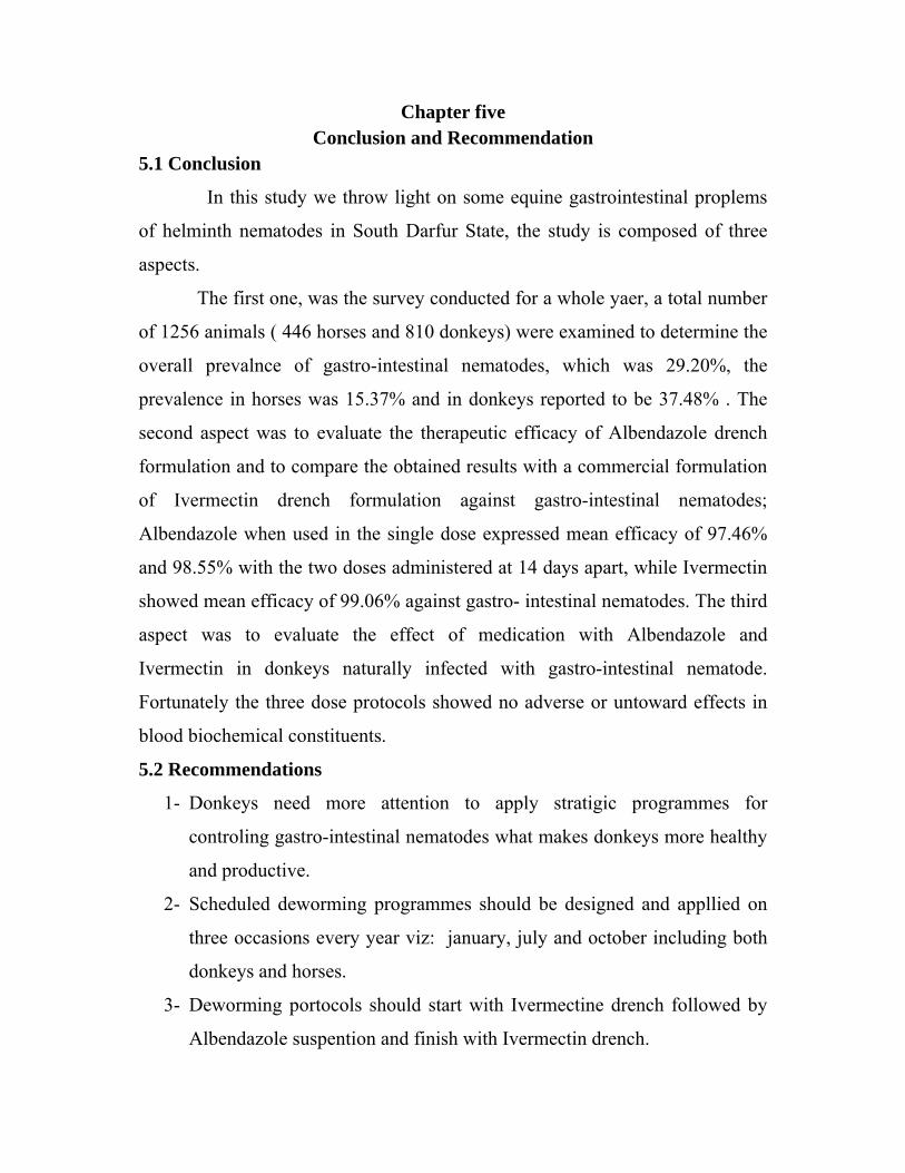
Chapter five Conclusion and Recommendation
5.1 Conclusion
In this study we throw light on some equine gastrointestinal proplems
of helminth nematodes in South Darfur State, the study is composed of three
aspects.
The first one, was the survey conducted for a whole yaer, a total number
of 1256 animals ( 446 horses and 810 donkeys) were examined to determine the
overall prevalnce of gastro-intestinal nematodes, which was 29.20%, the
prevalence in horses was 15.37% and in donkeys reported to be 37.48% . The
second aspect was to evaluate the therapeutic efficacy of Albendazole drench
formulation and to compare the obtained results with a commercial formulation
of Ivermectin drench formulation against gastro-intestinal nematodes;
Albendazole when used in the single dose expressed mean efficacy of 97.46%
and 98.55% with the two doses administered at 14 days apart, while Ivermectin
showed mean efficacy of 99.06% against gastro- intestinal nematodes. The third
aspect was to evaluate the effect of medication with Albendazole and
Ivermectin in donkeys naturally infected with gastro-intestinal nematode.
Fortunately the three dose protocols showed no adverse or untoward effects in
blood biochemical constituents.
5.2 Recommendations
1- Donkeys need more attention to apply stratigic programmes for
controling gastro-intestinal nematodes what makes donkeys more healthy
and productive.
2- Scheduled deworming programmes should be designed and appllied on
three occasions every year viz: january, july and october including both
donkeys and horses.
3- Deworming portocols should start with Ivermectine drench followed by
Albendazole suspention and finish with Ivermectin drench.
4- The new anthelmintic drugs (Doramectin, Abmectin and Moxidectin)
should be tested and used in equines.
5‐ Research institues should encourage studies in both sudanese donkeys and horses in the coming future.

REFERNCES
Abdelkarim, M. (1991). Variation saisonnières des populations vermineuses et
gastrophiliennes chez les asines de la region de settat (chaouia). These
Doctorate Vétérinaire, institute Agronomique et Vétérinaire Hassan II,
Rebat, Maroc.
Ahmed. N. k (2008). Epidemiology of donkey parasite based on postmortem
examination in South Darfur state, MVSc thesis. University of Nyala.PP
83.
Ali, T.M.O.; K.E.E. Ibrahim; E. H.A. Eltom; M.E. Hamid (2001). Animal
disease diagnosed at the University of Khartoum Veterinary Teaching
hospital (1995-1998). Sud. J. Vet. Sci. Anim. Husb. 40, 38-44.
Anonymous (1986). Manual of Veterinary Parasitological Techniques. Ministry
of Agriculture, Fisheries and Food. Reference Book 418, (3rd Ed).
HMSO, p. 160.
Armour, J.; and Bogan, J. (1982). Diagnostic and therapeutic check list.
Anthelmintics for ruminants. Br. Vet. J. 138: 371-381.
Armour, J.; Bairden, K. and Preston, J. M. (1980). Anthelmintic efficiency of
Ivermectin against naturally acquired bovine gastrointestinal nematodes.
Vet. Rec. 107(10): 226-227.
Averkin, E.; Beard, C.; Dvorak, C.; Edwards, J.; Fried, J.; Schiltz, R.; kistner,
T. P.; Drudge, J. H.; Lyons, E. T.; Sharp, M. L.; Corvin, R. M. (1975).
Methyl 5(6)-phenylsulfinyl-2-benzimidazole carbamate: a new potent
anthelmintics. J. Med. Chemis. 19: 1164-1166.

Ayele, G.; Feseha, G.; Bojia, E.; and Joe, A. (2006). Prevalence of gastro-
intestinal parasites of donkeys in Dugda Bora District, Ethiopia. Addis
Ababa University, Faculty of veterinary medicine, P. O. Box 1973,
Debre Zeit, Ethiopia Donkey sanctuary UK Sidmouth Devon, UK
Barnett, R. N.; Skodon, S. B.; Goldberg, M. H. (1973). Performance of “kits”
used for clinical chemical analysis of calcium in serum. Am. J. Clin.
Pathol. 59 (6): 836-45.
Bartels, H.; Bohmer, M.; Heierli, C. (1972). Serum creatinine determination
without protein precipitation. Clin. Chim. Acta; 37: 193 - 197.
Bartholomew, R. J.; Delany, A. M. (1966). Blood albumin determination.
Proceedings of Australian Association of Clinical Biochemists. 1-214.
Boraski, E. A. (1987) Ivermectin, efficacy against ascarids-new information.
Calif. Vet. 41:16-17
Borgsteede, F. H. M.; Dvojnos, G. M.; Kharchenko, V. A. (1997).
Benzimidazole resistance in cyathostomes in horses in the Ukraine. Vet.
Parasitol. 68: 113-117.
Boutemy, C. (1980). Expert study of the action of the compound albendazole on
the fertility of the male rat, per os. Unpublished study No. 2477 RSR
from IFM Research Center. Submitted to WHO by SmithKline and
French.
Brander, G. C; Pugh D. M.; Bywater, R. J. (1982). Veterinary Applied
Pharmacology & Therapeutic. 4th Edition. W. B. Saunders. London. 582
pages.
Burrows, R. O.; Thomson, B. M.; Lindesy, M. J. (1985). Efficacy of Ivermectin
against nematodes of horses including small Strongyles resistant to
benzimidazoles. Austral. Vet. J. 61:343-344

Campbell, W.C., W. H. D. Leaning, R. L. Seward (1989): Use of Ivermectin in
Horses. In: Ivermectin and Abmectin. (W. C. Campbell, Ed.), Springer
Verlag. New York Inc. 234-244.
Christian, M.S. (1984). Review of albendazole rat teratogenicity studies.
Unpublished report from Argus International. Submitted to WHO by
SmithKline and French.
Cirak, V. Y.; Gulegen, E.; Bauer, C. (2004). Benzimidazole resistance in
cyathostomin populations on horse farms in western Anatolia, Turkey.
Parasitol. Res. 93: 392-395.
Cobra, J.; Andrasko, H.; Stoffa, P., Holakovsky, P. (1986) Efficacy of
EqvalanTM and Panacur TM against gastrointestinal nematodes of horses.
Veterinary 36:79-80 (in Slovak).
Colman, W.; Towner, C.; Towner, D.; and Sokolek, J. (1977). Metabolic fate of
albendazole in sheep tissues. Unpublished report from Apple brook
Research Centre. Submitted to WHO by SmithKline and French.
Costa, A. J.; Barbosa, O. F.; Moraes, F. R.; Acuna, A. H.; Rocha, U. F.; Soares,
V. E.; Paullilo, A. C.; Sanches, A. (1998). Comparative efficacy
evaluation of Moxidectin gel and Ivermectin paste against internal
parasites of equine in Brazil. Vet. Parasitol. 80(1):29-36.
Daly, I. W.; and Knezevich, A. L. (1987). A long-term oral (dietary)
carcinogenicity study of albendazole in mice. Unpublished project No.80-
2480 from Biodynamics Incorporated. Submitted to WHO by SmithKline
and French.
Daly, I. W.; and Rinehart, W. E. (1980). A three month dose range finding
study with albendazole in mice. Unpublished project No. 79-2368 from
Biodynamics Incorporated. Submitted to WHO by SmithKline and
French.

Daly, I.W.; and Hogan, G. K. (1982). A long-term oral dietary toxicity
carcinogenicity study of albendazole in rats. Unpublished project No.79-
2383 from Biodynamics Incorporated. Submitted to WHO by SmithKline
Davis, C. ; and Gull, K. (1983). Protofilament number in microtubules in cells
of two parasitic nematodes. J. Parasitol., 69: 1094-1099.
Delatour, P.; Garnier, F.; Benoit, E.; and Longin, Ch. (1984). A correlation of
toxicity of albendazole and oxfendazole with their free metabolites and
bound residues. J. Vet. Pharmacol. Therap., 7, 139-145.
Delatour, P.; Parish, R. (1986). Benzimidazole anthelmintics and related
compounds: toxicity and evaluation of residues. In: Rico, A. G. (Ed.).
Drug residues in animals. Academic press, New York, pp. 175-203.
DiPietro, J. A.; Lock, T. F.; Todd, K. S.; Reuter, V. E. (1987). Evaluation of
Ivermectin paste in the treatment of ponies for Parascaris equourm. J.
Am. Vet. Med. Assoc 190:1181-1183.
DiPietro, J. A.; Todd, K. S.; Lock, T. F.; McPherron, T. A. (1982). Anthelmintic
efficacy of Ivermectin given intramuscularly in horses. Am. J. Vet. Res.
43: 145-148.
Drudge, J. H.; Lyons, E. T.; Tolliver, S. C. (1979). Benzimidazole resistance of
equine strongyles-critical tests of six compounds against population B.
Am. J. Vet. Res. 40: 590-594.
Drudge, J. H.; Tolliver, S. C.; Lyons, E. T. (1984). Benzimidazole resistant
strongyles: Critical tests of several classes of compounds against
population B strongyles from 1977-1981. Am. J. Vet. Res. 45 (4): 804-
809.
Duncan, J. (1983). Anthelmintics for use in equine practice. In:
Pharmacological basis of large animal medicine. Edited by J. A. Bogan;
P. Lees; and A. T. Yoxall. Blackwell Scientific publications, USA. pp
565.
Duwel, D. (1977). Fenbedazole II, biological properties and activity. Pesticide
Science. 8: 550-555.
El Dirdiri, N. I.; Abu Damir, H.; and Wahbi, A. A. (1986). Disease incidence in
donkeys (Equus asinus asinus) with emphasis on Strongyle infection.
Acta Vet. (Belgr) 36:313-320.
Eysker, M.; Boersema, J. H.; and kooyman, F. N. J. (1989). The effect of
repeated oxfendazole treatments on small strongyle infections in Shetland
ponies. Res. Vet. Sci. 46: 409-412.
Eysker, M.; Boersema, J. H.; and kooyman, F. N. J. (1989a). Emergence from
inhibited development of cyathostome larvae in ponies following failure
to remove them by repeated treatments with Benzimidazole compounds.
Vet. Parasitol. 34: 87-93.
Eysker, M.; Boersema, J. H.; kooyman, F. N. J.; Berghen, P. (1988). Possible
resistance of small strongyles from female ponies in the Netherlands
against albendazole. Am. J. Vet. Res. 49(7): 995-999.
Eysker, M.; Jansen, J.; kooyman, F. N. J.; Mirk, M. H.; and Wensing, Th.
(1986). Comparison of two control systems for Cyathostome infections in
the horse and further aspects of the epidemiology of these infections. Vet.
Parasitol. 22: 105-112.
Fawcett, J. K.; Scott, J. E. (1960). A rapid and precise method for the
determination of urea. J Clin Pathol. 13: 156-9.
Fisher, M; Morzik, H. (1989) Chemistry In: Ivermectin and Abamectin.
Campbell, W.C. (Ed). Springer Verlag, New York, USA. P. 1-23.
French, D. D.; Klei, T. R.; Taylor, H.W., Chapman, M. R.; Wright, F. R.
(1988). Efficacy of Ivermectin in the oral paste formulation against
acquired adult and larval stage of Parascaris equourm in pony foal. Am.
J. Vet. Res. 49:1000-1003.
Galloway, S.M. (1981). Mutagenicity evaluation of albendazole (SK&F 62979)
in an in vitro cytogenetic assay measuring chromosome aberration
frequencies in Chinese hamster ovary (CHO) cells. Unpublished project
No. 22000 from Litton Bionetics. Submitted to WHO by SmithKline and
French.
Garber, M. (1970). Helminth et helminthasis dis equides ( anes et chevaux) de
la republique du Tchad. Rev. Elve. Med.Vet. Pay. Trop., 23: 207-222
Gibson, T. E. (1953). The effect of repeated anthelmintics treatment with
phenothiazine on the faecal egg counts of housed horses, with some
observations on the life-cycle of Trichonema spp. in the horse. J.
Helminthol., 27: 29-40.
Gibson, T. E. (1975). Veterinary anthelmintic medication, 3rd edition.
Commonwealth Institute of Helminthology. Farnham Royal,
Buckinghamshire.
Gokbulut, C. (2000). Pharmacokinetic disposition, faecal excretion, metabolism
and chirality of anthelmintics drugs in horses. Ph. D. thesis, University of
Glasgow, Faculty of Veterinary Medicine, Department of Pharmacology.
Glasgow, Scotland, UK.
Gokbulut, C.; Akar, F.; Mckellar, Q. A. (2005). Plasma disposition and faecal
excretion of oxfendazole, fenbendazole and albendazole following oral
administration to donkeys. The Vet. J. 172: 166-172.
Goldenburg, H.; Fernandez, A. (1966). Simplified method for the estimation of
inorganic phosphorus in body fluids. Clin. Chem. 12 (12): 871-82.
Gupta, A. K.; Varshney, J. P.; Uppal, P. K. (1994) Comparative studies on
biochemical indices in different breeds of equine. Indian Vet. J. 71: 26-
30.
Hall. H. T. B. (1985). Disease and parasites of livestock in the tropics.
Intermediate tropical agriculture series.

Herd, R. P.; Donham, J. C. (1984). Control of equine cutaneous nematodiasis by
Ivermectin. Proc. MSD AGVET Symposium: recent developments in the
control of animal parasites. XXII World Vet Congress. Perth Australia,
Aug. 25-26, 1983, pp 286-295.a
Herd, R. P.; Kociba, G. L. (1985). Effect of Ivermectin on equine blood
constituents. Equ. Vet. J. 17: 142-144.
sJohnson, D. E. (1981). Perinatal and postnatal study in rats. Unpublished report
No. 483-001 from International Research and Development Corporation.
Submitted to WHO by SmithKline and French.
Jung. H.; M. Hurtado; M. Sanchez; M. T. Medina; and J. Sotelo (1992). Clinical
pharmacokinetics of albendazole in patients with brain cysticercosis. The
Journal of Clinical Pharmacology. 32:28-31
Kaeer, P.; Towner, D.; and Sokolek, J. (1977). Metabolic fate of albendazole in
cattle tissues. Unpublished report from Applebrook Research Center.
Submitted to WHO by SmithKline and French.
Kaplan, R. M. (2002). Anthemintic resistance in nematodes of horses. Vet. Res.
33: 491-507.
Kaplan, R. M. (2004). Drug resistance in nematodes of veterinary importance.
Trends Parasitol. 20: 477-481.
Kassai, T. (1999). Veterinary helminthology. Butter worth Heinemann. Oxford.
Kheir, S. M.; Kheir, H. S. M. (1981). Gastrointestinal nematodes of equines in
the Southern Darfur region of the Sudan. Sud. J. Vet. Res. (3): 53-57.
Killeen, J. C. and Rapp, W. R. (1975). A three month oral toxicity study of
SK&F 62979 in rats. Unpublished project No. 75-1109 from
Biodynamics Incorporated. Submitted to WHO by SmithKline and
French.
King, E. S.; Wooton, J. G. P. (1956). Microanalysis in: Medical Biochemistry.
3rd edition. Church hill, J. A. 57-60.

Klei , T.R.; Torbert, B.J.; Chapman, M. R.; and Turk, M.A.M (1984) Efficacy
of Ivermectin in injectable and oral paste formulation against 8-week old
Strongylus vulgaris larvae in ponies. Am. J. Vet. Res. 45:183-185.
Kuzmina, T. A.; Kharchenko, V. O. (2008). Anthelmintic resistance in
cyathostomins of brood horses in Ukraine and influence of anthelmintic
treatments on strongylid community structure. Veterinary Parasitology.
154:277–288.
Kuzmina, T. A.; Negrutsa, K. A.; Dvjnos, G. M.; Berezovsky, A. V. (2002).
The resistance in horse cyathostomes to Benzimidazole preparations.
Trudy VIGIS. 38: 189-194 (in Russian).
Lacey, E.; Brady, R. L.; Prichard, R. K.; Watson, T. R. (1987). Comparison of
inhibition of polymeraisation of mammalian tubulin and helminth
ovicidal activity by Benzimidazole carbonates. Vet. Parasitol. 23: 105-
119.
Lanusse, C. E.; Prichard, R. K. (1993). Relationship between pharmacological
properties and clinical efficacy of ruminants anthelmintics. Vet. Parasitol.
49: 123-158.
Lichtenfels, J. R. (1975). Helminths of domestic equids. Proceedings of
helmointhological Society of Washington, special issue, 42, p. 92.
Lind, Eva Osterman (2005). Faculty of Veterinary Medicine and Animal
Science Department of Biomedical Sciences and Veterinary Public
Health Division of Parasitology and Virology Uppsala. Doctoral thesis
Swedish University of Agricultural Sciences Uppsala.
Mariner, S. E.; Bogan, J. A. (1980). Pharmacokinetics of albendazole in sheep.
Am. J. Vet. Res. 41: 483-491.
Mariner, S. E.; Bogan, J. A. (1981). Pharmacokinetics of fenbendazole in sheep.
Am. J. Vet. Res. 42: 1146-1148.
Mariner, S. E.; Bogan, J. A. (1985). Plasma concentration of fenbendazole and
oxfenbendazole in the horse. Equine Veterinary Journal. 17: 58-61.
Martin, D. (1980). Albendazole: embryo-toxic study of ten metabolites. Thesis
for D. Vet. Sci. Claude Bernard University, Lyons. Submitted to WHO by
SmithKline and French.
Mckellar, Q. A.; Gokbulut, C.; benchaoui, H. A.; Muzandu, K. M. (2002).
Fenbendazole pharmacokinetics, metabolism and potentiation in horses.
Drug Metabolism Disposition. 30: 1230-1239.
McKellar, Q. A.; Scott, E. W. (1990). The Benzimidazole anthelmintic agents:
J. Vet. Pharma. Therap. 13, 223-247.
Mohammed Ali, N. A. K.; Bogan, J. A.; Marriner, S. E.; Richards, R. J. (1987).
Pharmacokinetics of triclabendazole alone or in combination with
fenbendazole in sheep. J. Vet. Pharma. Therp. 9: 442-445.
Mori, E.; Fernandes, W. R.; Mirandola, R. M. S.; Kubo, G.; Ferreira, R. R.;
Oliveira, J. V.; Garcek, F. (2003). Reference values on serum
biochemical parameters of Brazilian donkey (Equus asinus) breed. J. Equ.
Vet. S. 23(8): 358: 364.
Moskopp, D. ; and Lotterer, E. (1993). Concentrations of albendazole in serum,
cerebrospinal fluid and hydatidous brain cyst. Neurosurgical Review 16,
35–7.
Mukhwana, E. J.; (1994). Helminth Parasites of Donkeys (Equus asinus),
Burchell’s zebra (Equus burchelli) and camels (Camelus dromedaries) in
a selected area of northern Kenya. In proceedings of a 2nd Colloquium on
working equines (Eds. Bakkoury M. and Prentis. R.A.), Institute
Agronomique et vetrinaire Hassan II, Rebat, Morocco. pp: 45-50.
Ngomuo, A. J.; Marriner, S. E.; Bogan, J. A. (1984). The pharmacokinetics of
fenbendazole and oxfenbendazole in cattle. Vet. Res. Communi. 8: 187-
193.
Ogburne, C. P. (1975). Epidemiological studies on horses infected with
nematodes of the family trichonematidae (Witenberg, 1925). Int. J.
Parasitol. 5: 667-672.

Papadopoulos, E.; Hamhougias, K.; Himonas, C.; Dorchies, P. (2000).
Strongyle anthelmintics resistance in horses and cattle in Greece. Rev.
Med. Vet. 151: 1139-1142.
Parish, R.C., Chow, A.W. and Gyurik, R. J. (1977). The isolation and
characterization of radioactive metabolites from the urine of steers treated
orally with C-14 labelled SK&F 62979 (Albendazole). Unpublished
report from Applebrook Research Centre. Submitted to WHO by
SmithKline and French.
Parish, R.C.; and Gyurik, R. (1979a). Urinary excretion and identification of
metabolites from urine of Sprague-Dawley rats orally dosed with
albendazole C-14 albendazole sulfoxide C-14 and albendazole sulfone -
14°C. Unpublished report No. A-4003-78 from Applebrook Research
Centre. Submitted to WHO by SmithKline and French.
Parish, R.C.; Gyurik, R.; and Bruner, E. (1979). The isolation and quantitation
of metabolites from urine of Charles River mice treated orally with C-14
labelled SK&F 62979 (Albendazole). Unpublished report No. A-4006-78
from Applebrook Research Centre. Submitted to WHO by SmithKline
and French.
Prichard, R.K.; Hennessy, D.R.; Steel, J.W.; and Lacey, E. (1985). Metabolite
concentrations in plasma following treatment of cattle with five
anthelmintics. Res. Vet. Sci., 39, 173-178.
Reed. S. M. (1983). Ivermectin and CNS signs. Mod. Vet. Pract. 64, 783-784.
References
Reinecke, R. K.; Le Roux, D. J. (1972). Anthelmintic activity of Menbendazle
in Equine. Journal of Veterinary Medical Association 43:287-294.
Rossignol, J.F.; and Maisonneuve, H. (1984). Albendazole: a new concept in
the control of intestinal helminthiasis. Gastroenterol. Clin. Biol., 8, 569-
576.
Ryan, W. G.; Best, P. J. (1985) Efficacy of Ivermectin Best against Strongloides
westeri in foals. Vet. Rec. 117:169-170.
Sarkar, B. C.; Chauhan, U. P. (1967). A new method for determining micro
quantities of calcium in biological materials. Anal. Biochem. 20 (1): 155-
66.
Sauer, R.M. (1985). Pathology Working Group report on albendazole in
Sprague-Dawley rats and CD-1 mice. Unpublished report from Pathco
Incorporated. Submitted to WHO by SmithKline and French.
SBAR (2000). Statistical Bulletin for Animal Resources. Ministry of animal
resources. 10:14.
Schroeder, R.E. and Rinehart, W. E. (1980). A three-generation reproduction
study with albendazole in rats. Unpublished project No. 77-2019 from
Biodynamics Incorporated. Submitted to WHO by SmithKline and
French.
Selwyn, M.R. (1987). Statistical analysis for oral carcinogenicity studies of
albendazole in mice and rats. Unpublished study from Statistics
Unlimited Inc. Submitted to WHO by SmithKline and French.
Seri, H. I.; A. A. Ismail; A. D. Abakar; T. A. Tigani (2005). Efficacy of
Ivermectin in an injectable formulation against gastrointestinal nematodes
of donkeys (Equus asinus).Vet. arhiv 75 (4), 369-374.
Seri, H. I.; T, Hassan.; M. M. Salih.; Y. H. A. Elmansoury.; (2006). Effect of
Doramectin in donkeys blood constituents (Equus asinus) in Khatoum
state. Sudan. International Journal of pharmacology 2(5): 547- 550.
Seri, H. I.; T. Hassan; M.M. Salih; A.D. Abakar (2004). A survey of
gastrointestinal nematodes in donkeys (Equus asinus) in Khartoum State,
Sudan. Journal of Animal and Veterinary Advances. 3(11): 736-739.

Simon, J.C. (1979). Sub-acute toxicity study in dogs. Unpublished study No.
2204 TSC from IFM Research Centre. Submitted to WHO by SmithKline
and French.
Simon, J.C. (1979a). 4-week sub-acute oral toxicity study in rats. Unpublished
study No. 2206 TSR from IFM Research Centre. Submitted to WHO by
SmithKline and French.
Souhaili-El Amri, H.; Fargetton, X.; Benoit, E.; Totis, M.; and att, A-M. (1988).
Inducing effect of albendazole on rat liver drug metabolising enzymes
and metabolite pharmacokinetics. Toxicol. App. Pharmacol. 92, 141-149.
Soulsby, E. J. L. (1982). Helminth, arthropods and protozoa of domesticated
animals. 7th edition, Bailliere Tindal, Philadelphia.
Tash, J. M.; and Harper, J.A. (1977). Albendazole (SK&F 62979): Effects of
single oral administration upon pregnancy in sheep. Unpublished report
No. 77/SAF210/205 from Life Science Research. Submitted to WHO by
SmithKline and French.
Taussky, H. H.; Shorr, E. (1953). A microcolorimetric method for the
determination of inorganic phosphorus. J. Biol. Chem. 202 (2): 675-85.
Trawford, A. F.; Burden, F.; Hodgkinson, J. E. (2005). Suspected moxidectin
resistance in cyathostomes in two donkey herds at the donkey sanctuary.
UK. In: Proceedings of the 20th International Conference of the World
Association for the Advancement of Veterinary Parasitology,
Christchurch, New Zealand, 16-20 October 2005, p. 196.
WHO . World health organization. The food additive series 25.
Zinkl, J. G.; Mae, D.; Merida, P. G.; Farver, T. B.; Humble, J. A. (1990)
Reference range and the influence of age and sex on haematologic and
serum biochemical values in donkeys (Equus asinus). Am. J. Vet. Res.
51(3): 4.8: 413.