PDF - arXiv.org e-Print archive · BioMed Research International into four classes according to the...
9
Research Article Computer-Aided Decision Support for Melanoma Detection Applied on Melanocytic and Nonmelanocytic Skin Lesions: A Comparison of Two Systems Based on Automatic Analysis of Dermoscopic Images Kajsa Møllersen, 1 Herbert Kirchesch, 2 Maciel Zortea, 3 Thomas R. Schopf, 1 Kristian Hindberg, 3 and Fred Godtliebsen 3 1 Norwegian Centre for Integrated Care and Telemedicine, University Hospital of North Norway, 9038 Tromsø, Norway 2 Private Office, Venloer Straße 107, 50259 Pulheim, Germany 3 Department of Mathematics and Statistics, UiT e Arctic University of Norway, 9037 Tromsø, Norway Correspondence should be addressed to Kajsa Møllersen; [email protected] Received 11 September 2015; Accepted 3 November 2015 Academic Editor: Elisabeth Roider Copyright © 2015 Kajsa Møllersen et al. is is an open access article distributed under the Creative Commons Attribution License, which permits unrestricted use, distribution, and reproduction in any medium, provided the original work is properly cited. Commercially available clinical decision support systems (CDSSs) for skin cancer have been designed for the detection of melanoma only. Correct use of the systems requires expert knowledge, hampering their utility for nonexperts. Furthermore, there are no systems to detect other common skin cancer types, that is, nonmelanoma skin cancer (NMSC). As early diagnosis of skin cancer is essential, there is a need for a CDSS that is applicable to all types of skin lesions and is suitable for nonexperts. Nevus Doctor (ND) is a CDSS being developed by the authors. We here investigate ND’s ability to detect both melanoma and NMSC and the opportunities for improvement. An independent test set of dermoscopic images of 870 skin lesions, including 44 melanomas and 101 NMSCs, were analysed by ND. Its sensitivity to melanoma and NMSC was compared to that of Mole Expert (ME), a commercially available CDSS, using the same set of lesions. ND and ME had similar sensitivity to melanoma. For ND at 95% melanoma sensitivity, the NMSC sensitivity was 100%, and the specificity was 12%. e melanomas misclassified by ND at 95% sensitivity were correctly classified by ME, and vice versa. ND is able to detect NMSC without sacrificing melanoma sensitivity. 1. Introduction Melanoma is the deadliest of all skin cancers. When it is detected early, the treatment is excision of the tumour, and the survival rate is high. Recent developments in melanoma treatment are promising [1, 2], but the survival rate for patients with metastasised melanoma is still poor [3, 4]. Early diagnosis is crucial, but challenging, since early stage melanomas resemble benign skin lesions. Other types of skin cancer, like basal cell carcinoma (BCC) and squamous cell carcinoma (SCC), have high incidence rates but low mortality rates [4]. Early detection is beneficiary for the patient to get early treatment and avoid further damaging of the surrounding skin. e dermoscope (dermatoscope, epiluminescence micro- scope) reveals structures not visible to the naked eye and has proven to raise diagnostic accuracy of melanoma when used by properly trained personnel [5]. Dermoscopy has not reached widespread use among general practitioners (GPs), except in Australia [6]. In view of increased melanoma incidence rates, national screening programmes, and growing awareness of skin cancer in the public, there seems to be a need for clinical decision support systems (CDSSs) for GPs not familiar with dermoscopy. For the past few decades, many CDSSs (also referred to as computer-aided diagnostic (CAD) systems) have been developed for melanoma detection. For a description of the major steps and an overview of different technologies, see, Hindawi Publishing Corporation BioMed Research International Volume 2015, Article ID 579282, 8 pages http://dx.doi.org/10.1155/2015/579282
Transcript of PDF - arXiv.org e-Print archive · BioMed Research International into four classes according to the...

Research ArticleComputer-Aided Decision Support for MelanomaDetection Applied on Melanocytic and Nonmelanocytic SkinLesions: A Comparison of Two Systems Based on AutomaticAnalysis of Dermoscopic Images
Kajsa Møllersen,1 Herbert Kirchesch,2 Maciel Zortea,3 Thomas R. Schopf,1
Kristian Hindberg,3 and Fred Godtliebsen3
1Norwegian Centre for Integrated Care and Telemedicine, University Hospital of North Norway, 9038 Tromsø, Norway2Private Office, Venloer Straße 107, 50259 Pulheim, Germany3Department of Mathematics and Statistics, UiT The Arctic University of Norway, 9037 Tromsø, Norway
Correspondence should be addressed to Kajsa Møllersen; [email protected]
Received 11 September 2015; Accepted 3 November 2015
Academic Editor: Elisabeth Roider
Copyright © 2015 Kajsa Møllersen et al.This is an open access article distributed under theCreative CommonsAttribution License,which permits unrestricted use, distribution, and reproduction in any medium, provided the original work is properly cited.
Commercially available clinical decision support systems (CDSSs) for skin cancer have been designed for the detection ofmelanomaonly. Correct use of the systems requires expert knowledge, hampering their utility for nonexperts. Furthermore, there are nosystems to detect other common skin cancer types, that is, nonmelanoma skin cancer (NMSC). As early diagnosis of skin cancer isessential, there is a need for a CDSS that is applicable to all types of skin lesions and is suitable for nonexperts. Nevus Doctor (ND) isa CDSS being developed by the authors.We here investigate ND’s ability to detect bothmelanoma andNMSC and the opportunitiesfor improvement. An independent test set of dermoscopic images of 870 skin lesions, including 44 melanomas and 101 NMSCs,were analysed by ND. Its sensitivity to melanoma and NMSC was compared to that of Mole Expert (ME), a commercially availableCDSS, using the same set of lesions. ND and ME had similar sensitivity to melanoma. For ND at 95% melanoma sensitivity, theNMSC sensitivity was 100%, and the specificity was 12%. The melanomas misclassified by ND at 95% sensitivity were correctlyclassified by ME, and vice versa. ND is able to detect NMSC without sacrificing melanoma sensitivity.
1. Introduction
Melanoma is the deadliest of all skin cancers. When it isdetected early, the treatment is excision of the tumour, andthe survival rate is high. Recent developments in melanomatreatment are promising [1, 2], but the survival rate forpatients with metastasised melanoma is still poor [3, 4].Early diagnosis is crucial, but challenging, since early stagemelanomas resemble benign skin lesions. Other types ofskin cancer, like basal cell carcinoma (BCC) and squamouscell carcinoma (SCC), have high incidence rates but lowmortality rates [4]. Early detection is beneficiary for thepatient to get early treatment and avoid further damaging ofthe surrounding skin.
Thedermoscope (dermatoscope, epiluminescencemicro-scope) reveals structures not visible to the naked eye andhas proven to raise diagnostic accuracy of melanoma whenused by properly trained personnel [5]. Dermoscopy hasnot reached widespread use among general practitioners(GPs), except in Australia [6]. In view of increasedmelanomaincidence rates, national screening programmes, and growingawareness of skin cancer in the public, there seems to be aneed for clinical decision support systems (CDSSs) for GPsnot familiar with dermoscopy.
For the past few decades, many CDSSs (also referredto as computer-aided diagnostic (CAD) systems) have beendeveloped for melanoma detection. For a description of themajor steps and an overview of different technologies, see,
Hindawi Publishing CorporationBioMed Research InternationalVolume 2015, Article ID 579282, 8 pageshttp://dx.doi.org/10.1155/2015/579282

2 BioMed Research International
for example, [7]. The review paper of Rosado et al. [8] from2003 concluded that the diagnostic accuracy of the CDSSswas statistically not superior to that of human diagnosis.Vestergaard and Menzies [9] reported the same in 2008, andKorotkov and Garcia [10] made a similar conclusion in 2012.Simultaneously high sensitivity and specificity scores havebeen reported [11], but the scores droppedwhen the same sys-tems were tested in clinical-like settings with an independenttest set of consecutively collected images. Dreiseitl et al. [12]concluded that the performance of their system in a clinical-like setting was significantly lower than the result obtainedduring training. Elbaum et al. [13] reported 100% sensitivityand 85% specificity on the training set, with a dramatic dropin specificity to 9% on an independent test set [14]. Also,Bauer et al. reported better results for the training set [15] thanthose achieved with an independent test set [16]. The goodresults of Hoffmann et al. [17] were reproduced in a smallstudy with only 6 melanomas [18] but declined in anothersmall study [19]. In two other small studies with independenttest sets [20, 21], the performance reported by Blum et al. [22]declined.There are several possible explanations for the dropin performance. In studies where cross-validation has beenused to validate the performance, the whole data set is usedfor feature selection or model selection, which gives overlyoptimistic results [23, 24]. Also, several studies exclude non-melanocytic lesions post hoc based on the pathology reports,which introduces bias.
Differentiating between melanocytic and nonmelano-cytic lesions can be challenging even for experienced dermo-scopy users [25], and it is recommended that a CDSS formelanoma detection can handle nonmelanocytic lesions aswell [8, 11, 26]. A lesion is classified as suspicious if it resem-bles amelanoma, but lesions that resembleNMSC should alsobe classified as suspicious, especially when used by GPs [27],and not be classified together with nonsuspicious lesions.
In this report we present the performance of a CDSS formelanoma detection, Nevus Doctor (ND), when applied tobothmelanocytic and nonmelanocytic lesions. To our knowl-edge, this has not been previously presented for image-basedCDSSs. Shimizu et al. [28] included BCC but excluded otherNMSCs. Several studies have included nonmelanocyticlesions, but without reporting sensitivity to NMSC [14–17,29–31] or with less than three NMSCs [20, 32]. Recently,non-image-based technologies for melanoma detection haveincludedNMSC sensitivity in their reported findings [33, 34].
There is no apparent ranking of the CDSSs for melanomadetection. In practice, CDSSs can only be compared if testedon the same set of lesions, as done by Perrinaud et al. [32].We therefore compare the performance of ND to that of acommercially available system, Mole Expert (ME), on thesame set of lesions. This methodology potentially identifiesthe diagnostic difficulty of the data set, supplementing theinformation on the proportion of melanomas in situ, Breslowdepth, clinical diagnoses, and so forth. It also allows forindirect comparison of CDSSs if, in the future, some studychose to compare another system to ME.
2. Materials and Methods
2.1. Data. From March to December 2013, patients wererecruited at a private dermatology practice in Pulheim,Germany. Adult patients scheduled for excision of a pig-mented skin lesion were eligible for inclusion. Further-more, patients who were having nonpigmented skin lesionsexcised were eligible for inclusion if melanoma, BCC, orSCC was a potential differential diagnosis. Patients attendingthe German skin cancer screening programme [35] werealso eligible for inclusion if they had skin lesions selectedfor excision. Informed written consent was obtained fromall patients prior to inclusion. Skin lesions were excisedbecause of concern about malignancy or when requested bythe patient for other reasons. All skin lesions were photo-graphed prior to excision with a digital camera (Canon G10,Canon Inc., Tokyo, Japan) with an attached dermoscope(DermLite FOTO, 3Gen LLC, California, USA) and with avideodermoscope (DermoGenius ultra, DermoScan GmbH,Regensburg, Germany). All excised lesions were examined bya dermatopathologist. In the case of a malignant diagnosis, asecond dermatopathologist examined the excised lesion anda consensus diagnosis was set.
2.2. Automatic Image Analysis. ND takes a dermoscopicimage from the Canon/DermLite device as input and clas-sifies the lesion. ND is still in an experimental phase. In aprevious study, ND performed as well as three independentdermatologists in terms of melanoma sensitivity and speci-ficity [36]. In another study, ND performed as well as anindependent dermatologist on an independent test set con-sisting of 21melanomas and 188 benign lesions [37].The dataset in this study partly overlaps with the test set in Møllersenet al. [37], but not with the training set. ND has not beenretrained, so the present data set is independent. ND outputsa probability of malignancy for each lesion image, and thesensitivity can be tuned with a parameter 𝛼 (see [36–38] fordetails).
ME (MoleExpert micro Version 3.3.30.156) takes a der-moscopic image from the DermoGenius device as input. Theoutput is a number between −5.00 and 5.00, where highvalues indicate suspicion of melanoma, and the sensitivitycan be tuned by adjusting a threshold 𝑡. ME is intendedfor use on melanocytic lesions only. ME was chosen as thecomparison system due to availability and the fact that it hasbeen tested and compared to other commercial systems inclinical settings on a small scale [29, 32]. To our knowledge,the study of Perrinaud et al. [32] is the only study thatcompares several systems on the same set of lesions, and itis therefore not possible to pick the most adequate referencesystem.
2.3. Statistical Analysis. All excised lesions for which apathology report was available were included in the analysis.Cases were excluded if either of the CDSSs could not givean output or if images were missing. Presence of hairs andbubbles, lesion size, inadequate segmentation, and so forthwere not used as exclusion criteria. The data was divided

BioMed Research International 3
into four classes according to the histopathological diag-nosis: melanoma, NMSC, benign melanocytic lesions, andbenign nonmelanocytic lesions.Themelanoma class includeslesions where malignancy could not be ruled out by thedermatopathologists (ICD-10 D48.5), and the NMSC classincludes precancerous lesions, as done in other studies [33,34]. Precancerous lesions should be classified as suspicious,since the patient should receive treatment or follow-up.The sensitivity and specificity scores of ND and ME werecalculated by adjusting parameters 𝛼 and 𝑡 and classifyingthe lesions accordingly. Sensitivity refers to the ratio ofmalignant lesions classified as suspicious to the total numberof malignant lesions. Since there are two classes of malignantlesions, the termsmelanoma sensitivity andNMSC sensitivityare used for clarification when needed.
The clinical diagnoses are taken from the dermatologist’sreferrals to pathology. The clinical malignant class includeslesions where malignancy is a differential diagnosis. Theclinical benign class consists of lesions that were given abenign diagnosis by the dermatologist but were referred topathology, which indicates that malignancy could not beruled out by the dermatologist, even if this was not explicitlystated.
3. Results and Discussion
3.1. Results. There were 516 consultations (47% women)included in the study and a total of 877 excised lesions.The minimum age was 18, the maximum age was 93, andmedian age was 53 years. Table 1 shows the histopathologicaldiagnoses. The ratio of benign lesions per melanoma was16 : 1, which is within the range for dermatologists [39, 40].In total, 5% of the lesions were melanomas. The medianBreslow depth for the 23 invasive melanomas was 0.50mm,and the maximum Breslow depth was 2.25mm. Table 2shows the diagnoses and the Breslow depths according tothe 2009 American Joint Committee on Cancer (AJCC)staging [41]. About 70% of the melanomas and about 90%of the NMSCs were clinically diagnosed as malignant. Onelesion lacked clinical diagnosis. Of the 875 lesions withhistopathological diagnosis, four were excluded because MEdid not give an output (one naevus, one seborrheic keratosis,one BCC, and one SCC) and one was excluded because theCanon/DermLite image was lost (melanoma in situ), whichcorresponds to less than 1% of the images. In comparison,10% of the lesions were excluded due to lesions size or devicemalfunctions in the study by Malvehy et al. [33].
Figure 1(a) shows receiver operating characteristic (ROC)curves for ND and ME. The red and blue solid curves showsensitivity versus specificity for the whole data set wherethe malignant class includes both melanoma and NMSC,and a clear distinction between ND and ME can be seen.The pink and turquoise dotted curves show sensitivity versusspecificity for melanocytic lesions only, and there is nosignificant difference betweenND andME. Figure 1(b) showsNMSC sensitivity as a function of melanoma sensitivity.
The boxplots in Figure 2 illustrate each CDSS’s ability todiscriminate between the different classes of lesions. For both
Table 1: Histopathological diagnoses for the 877 skin lesions.
Histopathological diagnosis Total: 877Benign lesions 727Melanocytic lesions 596Naevus 574Naevoid lentigo 9Blue naevus 8Spitz naevus 4Sutton naevus 1
Nonmelanocytic lesions 118Seborrheic keratosis 80Lentigo senilis 9Neurofibroma 5Dermatofibroma 8Hemangioma 4Verruca 2Acanthoma 3Papilloma, fibroma molle, sebaceous glandhyperplasia, scar, eczema, folliculitis, anddiskoid lupus erythematodes
7
Collision tumours 13Malignant and precancerous lesions 148Melanoma 45Invasive melanoma 25Melanoma in situ 20
Nonmelanoma skin cancers 103Adnexal carcinoma 1Actinic keratosis 13Basal cell carcinoma 71Squamous cell carcinoma 7Bowen’s disease 11
No histopathological diagnosis 2
Table 2: Characteristics of the 45melanomas.
Diagnosis/Breslow NumberIn situ 13Lentigo maligna 7Cutaneous metastasis 2≤1.00mm 191.01–2.00mm 32.01–4.00mm 1>4.00mm 0
ND and ME, there is an overlap between melanomas in situand benignmelanocytic lesions, whereas invasivemelanomascan be separated from benign melanocytic lesions. ND hashigh scores for NMSC, but also for benign nonmelanocyticlesions. ME has greater relative variety for all categories thanND has which lead to larger overlaps and thusmore difficultyin distinguishing the different categories.

4 BioMed Research International
1
0.9
0.9
0.8
0.8
0.7
0.7
0.6
0.6
0.5
0.5
0.4
0.4
0.3
0.3
0.2
0.2
0.1
0.1 0
Sens
itivi
ty
Specificity
ND-all lesionsND-melanocytic
ME-all lesionsME-melanocytic
(a)
0.90.80.70.60.50.40.30.2
1
1
0.9
0.8
0.7
0.6
0.5
0.4
0.3
0.2
0.1
0.1
NDME
Sens
itivi
ty to
NM
SCSensitivity to melanoma
(b)
Figure 1: (a) Receiver operating characteristic curves for Nevus Doctor and Mole Expert. (b) Nonmelanoma skin cancer sensitivity as afunction of melanoma sensitivity.
1
0.8
0.6
0.4
0.2
0
Oth
erM
elano
cytic
(22)
Nae
vus (
573)
In si
tum
elan
oma (
19)
Inva
sive
mel
anom
a (25
)
Sebk
er (7
9)
Oth
erN
onm
elan
ocyt
ic (3
8)
Col
lisio
n (1
3)
BCC
(70)
SCC
(6)
Oth
erN
MSC
(25)
(a)
5
3
1
−1
−3
−5
Oth
erM
elano
cytic
(22)
Nae
vus (
573)
In si
tum
elan
oma (
19)
Inva
sive
mel
anom
a (25
)
Sebk
er (7
9)
Oth
erN
onm
elan
ocyt
ic (3
8)
Col
lisio
n (1
3)
BCC
(70)
SCC
(6)
Oth
erN
MSC
(25)
(b)
Figure 2: Boxplots for (a) Nevus Doctor and (b) Mole Expert.The horizontal lines inside the boxes are the medians; the boxes are defined bythe 25th and 75th percentiles. The whiskers are situated at two standard deviations from the mean. The crosses indicate outlier observations.
For ND at 95%melanoma sensitivity, the overall sensitiv-ity was 99%, the specificity was 12%, the positive predictivevalue (PPV) was 18%, and the negative predictive value(NPV) was 98%, where sensitivity, specificity, PPV, and NPVare defined in terms of malignant (melanoma and NMSC)and benign histopathological diagnosis. Figure 3 shows thetwo melanomas that were misclassified at 95% melanomasensitivity for each CDSS. They were all in situ and were allclinically diagnosed as benign.
3.2. Discussion. The ROC curves for ND and ME inFigure 1(a) show that ND performed similar to ME whennonmelanocytic lesions were excluded, and by that we
have shown that ND performed similarly to ME underthe circumstances for which ME is intended. When non-melanocytic lesions were included, ND performed betterthan ME. Figure 1(b) shows that ND’s NMSC sensitivityreached 100% at melanoma sensitivity of 86%, which meansthat, at reasonably high melanoma sensitivity, all NMSCswere classified as suspicious.
The majority of the benign lesions were excised due tosuspicion of malignancy, and about 30% of the melanomashad a benign clinical diagnosis, so the overlap betweenbenignmelanocytic lesions andmelanomas in situ, as seen inFigure 2, was expected. NDmisclassifies seborrheic keratosesas suspicious, similar to other CDSSs [20, 31–33]. GPs excise

BioMed Research International 5
(a) (b) (c) (d)
(e) (f) (g) (h)
Figure 3: (a)–(d) Lesions photographed with Canon/DermLite. (e)–(h) Lesions photographed with DermoGenius. ((a)-(b) and (e)-(f)) Thetwo melanomas misclassified by ND at 95% melanoma sensitivity. ((c)-(d) and (g)-(h)) The two melanomas misclassified by ME at 95%melanoma sensitivity.
ND, all lesionsND, melanocytic
ME, all lesionsME, melanocytic
1
0.95
0.9
0.85
0.8
0.5 0.45 0.4 0.35 0.3 0.25 0.2 0.15 0.1 0.05 0
Sens
itivi
ty
Specificity
Figure 4: Receiver operating characteristic curves forNevusDoctorand Mole Expert. Upper right part of Figure 1(a).
more seborrheic keratoses per melanoma than dermatolo-gists do [39, 40, 42], so there is a potential for saving pathol-ogy resources if a CDSS can classify them correctly. Devel-opment of image analysis features to discriminate seborrheickeratosis from melanoma has just begun [28].
Only the segment of the ROC curve with high sensitivityto melanoma, as shown in Figure 4, is clinically relevant,and therefore the area under the curve (AUC) is not anadequate summary of a CDSS’s performance. The area underthe clinically relevant segment of the ROC curve would be amore adequate summary statistic; however, then the clinicallyrelevant segment must first be defined.
Inadequate segmentation was not used as exclusion cri-terion, although it seems unlikely that a user would trustthe outcome if the segmentation fails. But since there is noground truth for segmentation, it is less suited as exclusioncriterion. The decision to carry out excision was based on
only one dermatologist’s opinion, which is a drawback sinceinterobserver agreement is only moderate for dermatologists[25]. Short-term follow-up of the patient or consensus diag-nosis based on dermoscopic and clinical images can be usedas the gold standard for nonexcised lesions, but it requireslarge resources.The data set used in this study is independentof all stages of ND’s development, but the lesions in this setare possibly more similar to the lesions ND has been trainedon than to the lesions ME has been trained on, and this ispotentially an advantage for ND.
The present data set consisted of 44% melanomas insitu and median Breslow depth of 0.50mm, which indicatehigh diagnostic difficulty. A proportion of 31% melanomasin situ was reported on a similar population in Germany[43]. The data set consists of 44 melanomas, which is morethan most reported studies of CDSSs with independent testsets, with some exceptions [14, 33]. With 44 melanomas,95% melanoma sensitivity means that only two melanomasare misclassified, and the results are therefore very sensitiveto small changes in the data set. Confidence intervals forhighly data sensitive results can give a false impression ofgeneralisability and are therefore not reported.
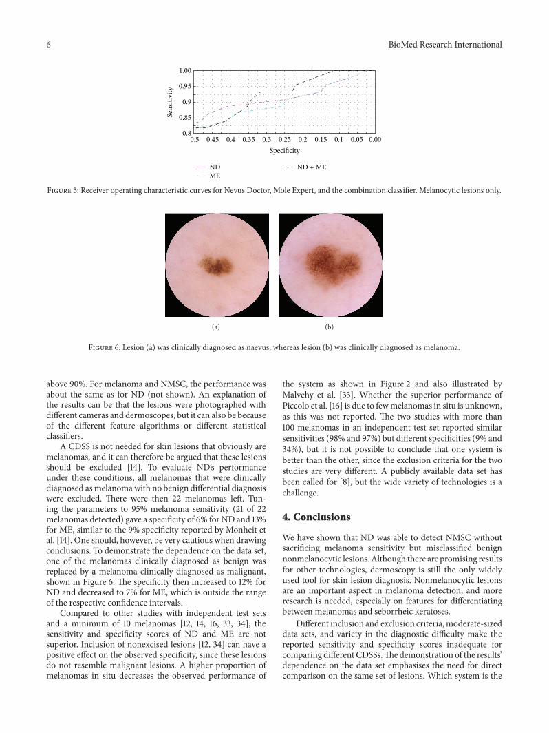
ND andMEmisclassified different melanomas, as shownin Figure 3, which is not unexpected since the two CDSSshave been developed independently of each other. It has beenreported that the sensitivity and specificity for dermatologistsimprove whenmajority vote or consensus is used to diagnoseskin lesions [5, 25]. We have investigated whether a combi-nation of ND and ME will increase the sensitivity withoutdecreasing the specificity. Figure 5 shows the ROC curve fora classifier which classifies a lesion as suspicious if eitherND or ME classifies it as suspicious. For melanocytic lesionsonly, this classifier outperformedND andME for sensitivities
6 BioMed Research International
1.00
0.95
0.9
0.85
0.8
Sens
itivi
ty
0.5 0.45 0.4 0.35 0.3 0.25 0.2 0.15 0.1 0.05 0.00
Specificity
NDME
ND + ME
Figure 5: Receiver operating characteristic curves for Nevus Doctor, Mole Expert, and the combination classifier. Melanocytic lesions only.
(a) (b)
Figure 6: Lesion (a) was clinically diagnosed as naevus, whereas lesion (b) was clinically diagnosed as melanoma.
above 90%. For melanoma and NMSC, the performance wasabout the same as for ND (not shown). An explanation ofthe results can be that the lesions were photographed withdifferent cameras and dermoscopes, but it can also be becauseof the different feature algorithms or different statisticalclassifiers.
A CDSS is not needed for skin lesions that obviously aremelanomas, and it can therefore be argued that these lesionsshould be excluded [14]. To evaluate ND’s performanceunder these conditions, all melanomas that were clinicallydiagnosed asmelanomawith no benign differential diagnosiswere excluded. There were then 22 melanomas left. Tun-ing the parameters to 95% melanoma sensitivity (21 of 22melanomas detected) gave a specificity of 6% for ND and 13%for ME, similar to the 9% specificity reported by Monheit etal. [14]. One should, however, be very cautious when drawingconclusions. To demonstrate the dependence on the data set,one of the melanomas clinically diagnosed as benign wasreplaced by a melanoma clinically diagnosed as malignant,shown in Figure 6. The specificity then increased to 12% forND and decreased to 7% for ME, which is outside the rangeof the respective confidence intervals.
Compared to other studies with independent test setsand a minimum of 10 melanomas [12, 14, 16, 33, 34], thesensitivity and specificity scores of ND and ME are notsuperior. Inclusion of nonexcised lesions [12, 34] can have apositive effect on the observed specificity, since these lesionsdo not resemble malignant lesions. A higher proportion ofmelanomas in situ decreases the observed performance of
the system as shown in Figure 2 and also illustrated byMalvehy et al. [33]. Whether the superior performance ofPiccolo et al. [16] is due to fewmelanomas in situ is unknown,as this was not reported. The two studies with more than100 melanomas in an independent test set reported similarsensitivities (98% and 97%) but different specificities (9% and34%), but it is not possible to conclude that one system isbetter than the other, since the exclusion criteria for the twostudies are very different. A publicly available data set hasbeen called for [8], but the wide variety of technologies is achallenge.
4. Conclusions
We have shown that ND was able to detect NMSC withoutsacrificing melanoma sensitivity but misclassified benignnonmelanocytic lesions. Although there are promising resultsfor other technologies, dermoscopy is still the only widelyused tool for skin lesion diagnosis. Nonmelanocytic lesionsare an important aspect in melanoma detection, and moreresearch is needed, especially on features for differentiatingbetween melanomas and seborrheic keratoses.
Different inclusion and exclusion criteria,moderate-sizeddata sets, and variety in the diagnostic difficulty make thereported sensitivity and specificity scores inadequate forcomparing different CDSSs.The demonstration of the results’dependence on the data set emphasises the need for directcomparison on the same set of lesions. Which system is the

BioMed Research International 7
most adequate for comparison will remain unknown untilmore studies are reported.
According to a study by Dreiseitl and Binder [44], physi-cians are willing to follow the recommendation of a CDSS,especially if they are not confident in their own diagnosis, andFruhauf et al. [20] reported that patients accept the use of aCDSS. Hence, there is a potential for CDSSs in melanomadetection if the systems can give reliable recommendationsfor all kinds of lesions.
Conflict of Interests
The authors declare that there is no conflict of interestsregarding the publication of this paper.
References
[1] C. Garbe, T. K. Eigentler, U. Keilholz, A. Hauschild, and J. M.Kirkwood, “Systematic review of medical treatment in mela-noma: current status and future prospects,”The Oncologist, vol.16, no. 1, pp. 5–24, 2011.
[2] A. Niezgoda, P. Niezgoda, and R. Czajkowski, “Novelapproaches to treatment of advanced melanoma: a review ontargeted therapy and immunotherapy,” BioMed Research Inter-national, vol. 2015, Article ID 851387, 16 pages, 2015.
[3] American Cancer Society, “Cancer facts & figures 2014,” Tech.Rep., American Cancer Society, 2014.
[4] Cancer Registry of Norway, “Cancer in Norway 2013—cancerincidence, mortality, survival and prevalence in Norway,” Tech.Rep., Cancer Registry of Norway, 2015.
[5] H. Kittler, H. Pehamberger, K. Wolff, and M. Binder, “Diagnos-tic accuracy of dermoscopy,”The Lancet Oncology, vol. 3, no. 3,pp. 159–165, 2002.
[6] C. Rosendahl, G. Williams, D. Eley et al., “The impact of sub-specialization and dermatoscopy use on accuracy of melanomadiagnosis among primary care doctors in Australia,” Journal ofthe American Academy of Dermatology, vol. 67, no. 5, pp. 846–852, 2012.
[7] A. Masood and A. A. Al-Jumaily, “Computer aided diagnosticsupport system for skin cancer: a review of techniques and algo-rithms,” International Journal of Biomedical Imaging, vol. 2013,Article ID 323268, 2013.
[8] B. Rosado, S.Menzies, A.Harbauer et al., “Accuracy of computerdiagnosis of melanoma: a quantitative meta-analysis,” Archivesof Dermatology, vol. 139, no. 3, pp. 361–367, 2003.
[9] M. E. Vestergaard and S. W. Menzies, “Automated diagnosticinstruments for cutaneous melanoma,” Seminars in CutaneousMedicine and Surgery, vol. 27, no. 1, pp. 32–36, 2008.
[10] K. Korotkov and R. Garcia, “Computerized analysis of pig-mented skin lesions: a review,”Artificial Intelligence inMedicine,vol. 56, no. 2, pp. 69–90, 2012.
[11] A. Blum, I. Zalaudek, and G. Argenziano, “Digital image anal-ysis for diagnosis of skin tumors,” Seminars in Cutaneous Medi-cine and Surgery, vol. 27, no. 1, pp. 11–15, 2008.
[12] S. Dreiseitl, M. Binder, K. Hable, and H. Kittler, “Computerversus humandiagnosis ofmelanoma: evaluation of the feasibil-ity of an automated diagnostic system in a prospective clinicaltrial,”Melanoma Research, vol. 19, no. 3, pp. 180–184, 2009.
[13] M. Elbaum, A. W. Kopf, H. S. Rabinovitz et al., “Automatic dif-ferentiation of melanoma from melanocytic nevi with multi-spectral digital dermoscopy: a feasibility study,” Journal of the
American Academy of Dermatology, vol. 44, no. 2, pp. 207–218,2001.
[14] G.Monheit, A. B. Cognetta, L. Ferris et al., “The performance ofMelaFind: a prospective multicenter study,” Archives of Derma-tology, vol. 147, no. 2, pp. 188–194, 2011.
[15] P. Bauer, P. Cristofolini, S. Boi et al., “Digital epiluminescencemicroscopy: usefulness in the differential diagnosis of cuta-neous pigmentary lesions. A statistical comparison betweenvisual and computer inspection,” Melanoma Research, vol. 10,no. 4, pp. 345–349, 2000.
[16] D. Piccolo, A. Ferrari, K. Peris, R. Daidone, B. Ruggeri, and S.Chimenti, “Dermoscopic diagnosis by a trained clinician vs.a clinician with minimal dermoscopy training vs. computer-aided diagnosis of 341 pigmented skin lesions: a comparativestudy,” British Journal of Dermatology, vol. 147, no. 3, pp. 481–486, 2002.
[17] K. Hoffmann, T. Gambichler, A. Rick et al., “Diagnostic andneural analysis of skin cancer (DANAOS). A multicentre studyfor collection and computer-aided analysis of data from pig-mented skin lesions using digital dermoscopy,” British Journalof Dermatology, vol. 149, no. 4, pp. 801–809, 2003.
[18] M. Barzegari, H. Ghaninezhad, P. Mansoori, A. Taheri, Z.S. Naraghi, and M. Asgari, “Computer-aided dermoscopy fordiagnosis of melanoma,” BMC Dermatology, vol. 5, no. 1, article8, 2005.
[19] J. C. Boldrick, C. J. Layton, J. Nguyen, and S. M. Swetter, “Eval-uation of digital dermoscopy in a pigmented lesion clinic: clini-cian versus computer assessment of malignancy risk,” Journal ofthe American Academy of Dermatology, vol. 56, no. 3, pp. 417–421, 2007.
[20] J. Fruhauf, B. Leinweber, R. Fink-Puches et al., “Patient accep-tance and diagnostic utility of automated digital image analysisof pigmented skin lesions,” Journal of the European Academy ofDermatology and Venereology, vol. 26, no. 3, pp. 368–372, 2012.
[21] A. Fueyo-Casado, F. Vazquez-Lopez, J. Sanchez-Martin, B.Garcia-Garcia, andN. Perez-Oliva, “Evaluation of a program forthe automatic dermoscopic diagnosis of melanoma in a generaldermatology setting,” Dermatologic Surgery, vol. 35, no. 2, pp.257–259, 2009.
[22] A. Blum, R. Hofmann-Wellenhof, H. Luedtke et al., “Value ofthe clinical history for different users of dermoscopy comparedwith results of digital image analysis,” Journal of the EuropeanAcademy of Dermatology and Venereology, vol. 18, no. 6, pp.665–669, 2004.
[23] P. Smialowski, D. Frishman, and S. Kramer, “Pitfalls of super-vised feature selection,” Bioinformatics, vol. 26, no. 3, pp. 440–443, 2009.
[24] T. Hastie, R. Tibshirani, and J. Friedman,The Elements of Statis-tical Learning: Data mining, Inference, and Prediction, SpringerSeries in Statistics, Springer, New York, NY, USA, 2nd edition,2009.
[25] G. Argenziano, H. P. Soyer, S. Chimenti et al., “Dermoscopy ofpigmented skin lesions: results of a consensus meeting via theinternet,” Journal of the American Academy of Dermatology, vol.48, no. 5, pp. 679–693, 2003.
[26] S. M. Rajpara, A. P. Botello, J. Townend, and A. D. Ormerod,“Systematic review of dermoscopy and digital dermoscopy/artificial intelligence for the diagnosis of melanoma,” BritishJournal of Dermatology, vol. 161, no. 3, pp. 591–604, 2009.

8 BioMed Research International
[27] F. M.Walter, H. C. Morris, E. Humphrys et al., “Effect of addinga diagnostic aid to best practice to manage suspicious pig-mented lesions in primary care: randomised controlled trial,”BritishMedical Journal, vol. 345, no. 7866,Article ID e4110, 2012.
[28] K. Shimizu, H. Iyatomi, M. E. Celebi, K.-A. Norton, and M.Tanaka, “Four-class classification of skin lesions with taskdecomposition strategy,” IEEE Transactions on Biomedical Engi-neering, vol. 62, no. 1, pp. 274–283, 2015.
[29] M. J. Jamora, B. D.Wainwright, S. A.Meehan, and J.-C. Bystryn,“Improved identification of potentially dangerous pigmentedskin lesions by computerized image analysis,” Archives of Der-matology, vol. 139, no. 2, pp. 195–198, 2003.
[30] M. Carrara, A. Bono, C. Bartoli et al., “Multispectral imagingand artificial neural network: mimicking the managementdecision of the clinician facing pigmented skin lesions,” Physicsin Medicine and Biology, vol. 52, no. 9, pp. 2599–2613, 2007.
[31] S. W. Menzies, L. Bischof, H. Talbot et al., “The performance ofsolarscan: an automated dermoscopy image analysis instrumentfor the diagnosis of primary melanoma,” Archives of Dermatol-ogy, vol. 141, no. 11, pp. 1388–1396, 2005.
[32] A. Perrinaud, O. Gaide, L. E. French, J.-H. Saurat, A. A.Marghoob, andR. P. Braun, “Can automated dermoscopy imageanalysis instruments provide added benefit for the dermatol-ogist? A study comparing the results of three systems,” BritishJournal of Dermatology, vol. 157, no. 5, pp. 926–933, 2007.
[33] J. Malvehy, A. Hauschild, C. Curiel-Lewandrowski et al.,“Clinical performance of the Nevisense system in cutaneousmelanoma detection: an international, multicentre, prospectiveand blinded clinical trial on efficacy and safety,” The BritishJournal of Dermatology, vol. 171, no. 5, pp. 1099–1107, 2014.
[34] H. Lui, J. Zhao, D. McLean, and H. Zeng, “Real-time Ramanspectroscopy for in vivo skin cancer diagnosis,” CancerResearch, vol. 72, no. 10, pp. 2491–2500, 2012.
[35] A. Waldmann, S. Nolte, M. A. Weinstock et al., “Skin cancerscreening participation and impact on melanoma incidencein Germany—an observational study on incidence trends inregions with and without population-based screening,” BritishJournal of Cancer, vol. 106, no. 5, pp. 970–974, 2012.
[36] M. Zortea, T. R. Schopf, K. Thon et al., “Performance of a der-moscopy-based computer vision system for the diagnosis ofpigmented skin lesions compared with visual evaluation byexperienced dermatologists,” Artificial Intelligence in Medicine,vol. 60, no. 1, pp. 13–26, 2014.
[37] K. Møllersen, M. Zortea, K. Hindberg, T. R. Schopf, S. O.Skrøvseth, and F. Godtliebsen, “Improved skin lesion diagnos-tics for general practice by computer aided diagnostics,” inDer-moscopy Image Analysis, Digital Imaging and Computer Vision,pp. 247–292, CRC Press, 2015.
[38] M. Zortea, S. O. Skrøvseth, T. R. Schopf, H.M. Kirchesch, and F.Godtliebsen, “Automatic segmentation of dermoscopic imagesby iterative classification,” International Journal of BiomedicalImaging, vol. 2011, Article ID 972648, 19 pages, 2011.
[39] R. Marks, D. Jolley, C. McCormack, and A. P. Dorevitch, “Whoremoves pigmented skin lesions? A study of the ratio of mela-noma to other benign pigmented tumors removed by differentcategories of physicians in Australia in 1989 and 1994,” Journalof the American Academy of Dermatology, vol. 36, no. 5, pp. 721–726, 1997.
[40] B. Lindelof, M.-A. Hedblad, and U. Ringborg, “Nevus ellermalignt melanom? Ratt kompetens vid diagnostik ger lagrekostnader,” Lakartidningen, vol. 105, no. 39, pp. 2666–2669,2008.
[41] C.M. Balch, J. E. Gershenwald, S.-J. J. Soong et al., “Final versionof 2009 AJCC melanoma staging and classification,” Journal ofClinical Oncology, vol. 27, no. 36, pp. 6199–6206, 2009.
[42] M. I. Duque, J. R. Jordan, A. B. Fleischer Jr. et al., “Frequency ofseborrheic keratosis biopsies in the United States: a benchmarkof skin lesion care quality and cost effectiveness,” DermatologicSurgery, vol. 29, no. 8, pp. 796–801, 2003.
[43] A. Katalinic, A. Waldmann, M. A. Weinstock et al., “Does skincancer screening save lives?: an observational study comparingtrends in melanoma mortality in regions with and withoutscreening,” Cancer, vol. 118, no. 21, pp. 5395–5402, 2012.
[44] S. Dreiseitl and M. Binder, “Do physicians value decisionsupport? A look at the effect of decision support systems onphysician opinion,”Artificial Intelligence inMedicine, vol. 33, no.1, pp. 25–30, 2005.

Submit your manuscripts athttp://www.hindawi.com
Stem CellsInternational
Hindawi Publishing Corporationhttp://www.hindawi.com Volume 2014
Hindawi Publishing Corporationhttp://www.hindawi.com Volume 2014
MEDIATORSINFLAMMATION
of
Hindawi Publishing Corporationhttp://www.hindawi.com Volume 2014
Behavioural Neurology
EndocrinologyInternational Journal of
Hindawi Publishing Corporationhttp://www.hindawi.com Volume 2014
Hindawi Publishing Corporationhttp://www.hindawi.com Volume 2014
Disease Markers
Hindawi Publishing Corporationhttp://www.hindawi.com Volume 2014
BioMed Research International
OncologyJournal of
Hindawi Publishing Corporationhttp://www.hindawi.com Volume 2014
Hindawi Publishing Corporationhttp://www.hindawi.com Volume 2014
Oxidative Medicine and Cellular Longevity
Hindawi Publishing Corporationhttp://www.hindawi.com Volume 2014
PPAR Research
The Scientific World JournalHindawi Publishing Corporation http://www.hindawi.com Volume 2014
Immunology ResearchHindawi Publishing Corporationhttp://www.hindawi.com Volume 2014
Journal of
ObesityJournal of
Hindawi Publishing Corporationhttp://www.hindawi.com Volume 2014
Hindawi Publishing Corporationhttp://www.hindawi.com Volume 2014
Computational and Mathematical Methods in Medicine
OphthalmologyJournal of
Hindawi Publishing Corporationhttp://www.hindawi.com Volume 2014
Diabetes ResearchJournal of
Hindawi Publishing Corporationhttp://www.hindawi.com Volume 2014
Hindawi Publishing Corporationhttp://www.hindawi.com Volume 2014
Research and TreatmentAIDS
Hindawi Publishing Corporationhttp://www.hindawi.com Volume 2014
Gastroenterology Research and Practice
Hindawi Publishing Corporationhttp://www.hindawi.com Volume 2014
Parkinson’s Disease
Evidence-Based Complementary and Alternative Medicine
Volume 2014Hindawi Publishing Corporationhttp://www.hindawi.com