
PDF (615 KB) - IOPscience
5
Journal of Physics: Conference Series OPEN ACCESS Multilayer silicon rich oxy-nitride films characterization by SIMS, VASE and AFM To cite this article: M Barozzi et al 2008 J. Phys.: Conf. Ser. 100 012016 View the article online for updates and enhancements. You may also like Effect of post-deposition annealing on the interfacial chemical bonding states and band alignment of atomic layer deposited neodymium oxide on silicon Xiaojiao Fan, Hongxia Liu and Chenxi Fei - Structural parameters effect on the electrical and electroluminescence properties of silicon nanocrystals/SiO 2 superlattices J López-Vidrier, Y Berencén, S Hernández et al. - Characterization of ProtonInduced Color Transition in an Amorphous Tungsten Oxide Film H. RaflaYuan, J. G. H. Mathew and B. P. Hichwa - Recent citations Applicability of Child–Langmuir collision laws for describing a dc cathode sheath in N2O V. A. Lisovskiy et al - This content was downloaded from IP address 61.228.180.167 on 21/12/2021 at 11:56
Transcript of PDF (615 KB) - IOPscience

Journal of Physics Conference Series
OPEN ACCESS
Multilayer silicon rich oxy-nitride filmscharacterization by SIMS VASE and AFMTo cite this article M Barozzi et al 2008 J Phys Conf Ser 100 012016
View the article online for updates and enhancements
You may also likeEffect of post-deposition annealing on theinterfacial chemical bonding states andband alignment of atomic layer depositedneodymium oxide on siliconXiaojiao Fan Hongxia Liu and Chenxi Fei
-
Structural parameters effect on theelectrical and electroluminescenceproperties of silicon nanocrystalsSiO2superlatticesJ Loacutepez-Vidrier Y Berenceacuten S Hernaacutendezet al
-
Characterization of ProtonInduced ColorTransition in an Amorphous TungstenOxide FilmH RaflaYuan J G H Mathew and B PHichwa
-
Recent citationsApplicability of ChildndashLangmuir collisionlaws for describing a dc cathode sheath inN2OV A Lisovskiy et al
-
This content was downloaded from IP address 61228180167 on 21122021 at 1156
Multilayer silicon rich oxy-nitride films characterization by SIMS VASE and AFM
M Barozzi1 L Vanzetti1 E Iacob1 M Bersani1 M Anderle1 G Pucker2 C Kompocholis2 M Ghulinyan2 and P Bellutti2
1 Physics Chemistry of Surfaces and Interfaces Division Fondazione Bruno Kessler-IRST Via Sommarive 18 38050 Trento Italy
2 Microtechnologies Laboratory Fondazione Bruno Kessler-IRST Via Sommarive 18 38050 Trento Italy
barozziitcit
Abstract In this work secondary ion mass spectrometry (SIMS) variable angle spectroscopy ellipsometry (VASE) and atomic force microscopy (AFM) are used to investigate the structure composition and morphology of multilayer SRON films Threefour SRON sequential layers were deposited on silicon wafers by PECVD and silicon nitrogen and oxygen content was varied by changing the N2OSiH4 ratio The total thickness of the resulting SRON stack is about 50nm SIMS analyses of NCs+ OCs+ SiCs+ in MCs+ methodology are performed by a Cameca SC-ultra instrument Depth profiles are obtained at 500eV of primary beam impact energy with sample rotation An approximate method to obtain silicon concentration is used Total layer thickness are obtained from both SIMS and VASE measurements In addition we compare the thickness of the single layers obtained from VASE with the SIMS depth profiles A detailed analysis of films morphology is obtained by AFM The SRON stack is sputtered by SIMS until a certain layer is exposed which is then analyzed by AFM The sputtered layers are then etched in HF solution to better resolve the exposed nano-crystals
1 Introduction Multilayer silicon rich oxide films (SRO) or silicon rich oxy-nitride (SRON) films are attractive for their application in opto-electronic devices [1] The electro-optical characterization of the devices has to be correlated to the compositional structure of the films in order to improve the fabrication process and as ultimate goal to enhance the device efficiency These multi-layers SRON stacks can be incorporated in MOS structures to obtain LEDs
Silicon quantification in SRON by SIMS analyses is not a trivial task because of matrix effects SIMS analyses are affected by sputtering rate variations and ion yield changes throughout the various layers An approximate method is here adopted to quantify silicon in atomic concentrations as proposed by the same authors in a previous publication [2] AFM can provide detailed information about the morphology of the nano-crystals In particular the structures embedded in a particular SRON layer can be revealed after SIMS sputtering process and etching in HF solution
Spectroscopic ellipsometry was used by different groups to investigate dielectric matrices (SiO2 or Si3N4) containing Si-nanocrystals either to establish the thickness of the layer(s) containing Si-nano-
IVC-17ICSS-13 and ICN+T2007 IOP PublishingJournal of Physics Conference Series 100 (2008) 012016 doi1010881742-65961001012016
ccopy 2008 IOP Publishing Ltd 1
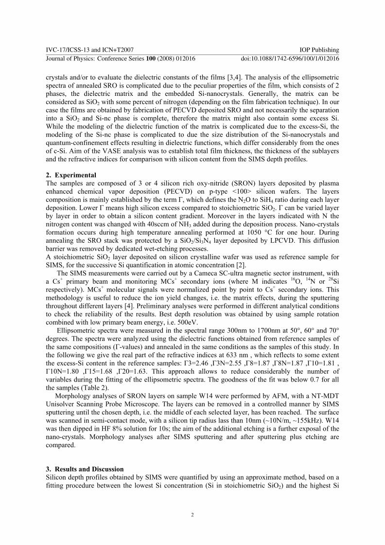
crystals andor to evaluate the dielectric constants of the films [34] The analysis of the ellipsometric spectra of annealed SRO is complicated due to the peculiar properties of the film which consists of 2 phases the dielectric matrix and the embedded Si-nanocrystals Generally the matrix can be considered as SiO2 with some percent of nitrogen (depending on the film fabrication technique) In our case the films are obtained by fabrication of PECVD deposited SRO and not necessarily the separation into a SiO2 and Si-nc phase is complete therefore the matrix might also contain some excess Si While the modeling of the dielectric function of the matrix is complicated due to the excess-Si the modeling of the Si-nc phase is complicated to due the size distribution of the Si-nanocrystals and quantum-confinement effects resulting in dielectric functions which differ considerably from the ones of c-Si Aim of the VASE analysis was to establish total film thickness the thickness of the sublayers and the refractive indices for comparison with silicon content from the SIMS depth profiles
2 Experimental The samples are composed of 3 or 4 silicon rich oxy-nitride (SRON) layers deposited by plasma enhanced chemical vapor deposition (PECVD) on p-type lt100gt silicon wafers The layers composition is mainly established by the term Γ which defines the N2O to SiH4 ratio during each layer deposition Lower Γ means high silicon excess compared to stoichiometric SiO2 Γ can be varied layer by layer in order to obtain a silicon content gradient Moreover in the layers indicated with N the nitrogen content was changed with 40sccm of NH3 added during the deposition process Nano-crystals formation occurs during high temperature annealing performed at 1050 degC for one hour During annealing the SRO stack was protected by a SiO2Si3N4 layer deposited by LPCVD This diffusion barrier was removed by dedicated wet-etching processes A stoichiometric SiO2 layer deposited on silicon crystalline wafer was used as reference sample for SIMS for the successive Si quantification in atomic concentration [2]
The SIMS measurements were carried out by a Cameca SC-ultra magnetic sector instrument with a Cs+ primary beam and monitoring MCs+ secondary ions (where M indicates 16O 14N or 28Si respectively) MCs+ molecular signals were normalized point by point to Cs+ secondary ions This methodology is useful to reduce the ion yield changes ie the matrix effects during the sputtering throughout different layers [4] Preliminary analyses were performed in different analytical conditions to check the reliability of the results Best depth resolution was obtained by using sample rotation combined with low primary beam energy ie 500eV
Ellipsometric spectra were measured in the spectral range 300nm to 1700nm at 50deg 60deg and 70deg degrees The spectra were analyzed using the dielectric functions obtained from reference samples of the same compositions (Γ-values) and annealed in the same conditions as the samples of this study In the following we give the real part of the refractive indices at 633 nm which reflects to some extent the excess-Si content in the reference samples Γ3=246 Γ3N=255 Γ8=187 Γ8N=187 Γ10=181 Γ10N=180 Γ15=168 Γ20=163 This approach allows to reduce considerably the number of variables during the fitting of the ellipsometric spectra The goodness of the fit was below 07 for all the samples (Table 2)
Morphology analyses of SRON layers on sample W14 were performed by AFM with a NT-MDT Unisolver Scanning Probe Microscope The layers can be removed in a controlled manner by SIMS sputtering until the chosen depth ie the middle of each selected layer has been reached The surface was scanned in semi-contact mode with a silicon tip radius lass than 10nm (~10Nm ~155kHz) W14 was then dipped in HF 8 solution for 10s the aim of the additional etching is a further exposal of the nano-crystals Morphology analyses after SIMS sputtering and after sputtering plus etching are compared
3 Results and Discussion Silicon depth profiles obtained by SIMS were quantified by using an approximate method based on a fitting procedure between the lowest Si concentration (Si in stoichiometric SiO2) and the highest Si
IVC-17ICSS-13 and ICN+T2007 IOP PublishingJournal of Physics Conference Series 100 (2008) 012016 doi1010881742-65961001012016
2
concentration (Si in Si bulk) [2] Example of the result obtained on sample W14 is reported in figure 1 Depth scale was established from reference sample SiO2 Single layer thicknesses differ from the nominal values as expected because of SIMS sputtering proceeds faster in Si matrix than in SiO2 Depth corrections are anyway difficult to be applied because of heterogeneous matrices Silicon atomic concentrations obtained by SIMS are reported in table 1 SIMS results depend on analytical conditions mainly on the primary beam impact energy Matrix effects and RSF variations throughout the layers are definitely unavoidable in SIMS analyses Therefore the reported Si contents were calculated as mean values between the concentrations obtained at 3keV and 500eV
The total layer thickness and the thickness of the single stacks obtained from analysis of the VASE spectra are given in table 2 The total thickness of the samples is very close to the ones obtained from SIMS The thickness of the single layer differs from SIMS analysis In same cases if the difference in the dielectric functions (refractive indices) is low layers might even vanish in the VASE modeling (eg sample W8) The SIMS depth profiles show that in some cases due to diffusion of Si at high temperatures among the single layers the Si-depth profile is not well represented by a 3 or 4 layer model
AFM images obtained on Γ20 and Γ3N sputtered layers are compared in figure 2 (a) and 2 (b) The parameter considered to compare the layers surface is their roughness rout mean square Sq The morphology of the layers seems nearly the same but in Γ3N layer the structures appear laterally connected The silicon aggregation shapes seem not well separated and in particular they could be not spherical but form elongated and connected structures as proposed by Műller [5] Possible mutual interferences of the structured layers have to be pointed out because of their shallowness In fact the bumps periodicity estimated for silicon nano-crystals in the buried layers (about 30nm) exceeds the nominal layers thickness Moreover SIMS sputtering starts on uneven Γ20 surface and follows its morphology and similarly it follows the buried layers morphology even if the sputtering process tends to smooth the surface The smoothing effect during sputtering is well confirmed by reduced roughness Sq=033nm in the silicon substrate compared to 057nm in the above Γ8N layer Bumps in the silicon substrate are still present but they are artefacts probably originated by SIMS analyses with sample rotation and they can be assimilated to smoothed traces of the above sputtered layers (figure 2 (c)) No bumps are visible on flat silicon wafer (figure 2 (d)) where Sq=01nm The additional etching process in HF solution (8 for 10s) is selective for SiO2 and increases Sq in the SRON layers less affecting Sq in the silicon substrate (table 1)
a b
c d
0 10 20 30 40 50 60 70 80 901022
1x10²³
Silicon subΓ3NΓ8NΓ20 Γ15
Si
[ac
m3 ]
[nm]0 20 40 60 80
101
102
103
104
105
106
107
CsN CsO
[cts
s]
Figure 1 SIMS depth profiles on sample W14 obtained at 500eV Cs+ with rotating stage Si is quantified by an approximate fitting method
Figure 2 AFM images 500nm x 500nm on Γ20 (a) Γ3N (b) Silicon sub (c) on flat
silicon wafer (d)
IVC-17ICSS-13 and ICN+T2007 IOP PublishingJournal of Physics Conference Series 100 (2008) 012016 doi1010881742-65961001012016
3
Table 1 Silicon atomic concentration and roughness in SRON layers calculated by SIMS and AFM analysesdata obtained on sample W14
matrix
Si-content [atcm3] SIMS
Roughness Sq [nm] sputtered layer
Roughness Sq [nm] after etching
Si sub 506x1022 033 046 Γ3N 40x1022 053 095 Γ8N 37x1022 057 11 Γ10 35x1022 -- -- Γ15 33x1022 048 087 Γ20 28x1022 047 053 SiO2 265x1022 -- --
Table 2 Multilayer structures of the samples with nominal layer thickness(1) and the layer thickness
obtained from SIMS(2) and VASE(3) measurements Γ =N2OSiH4 N= 40 sccm of NH3
sample Total thickness 4th layer 3rd layer 2nd layer 1st layer
W2 50 nm(1) 39nm(2) 373nm(3) Γ3 15nm
13nm 78nm Γ15 20nm
15nm 232nm Γ3 15nm
11nm 62nm
W8 50 nm 46 nm 453nm -- Γ15 30nm
22nm 329nmΓ10 15nm
11nm 0nm Γ3 15nm
13 nm 123nm
W14 60 nm 49 nm 500nm
Γ20 15nm 10nm 0 nm
Γ15 15nm 11nm 158 nm
Γ8N 15nm 11nm 204 nm
Γ3N 15nm 17nm 138nm
W20 50 nm 41 nm 432 nm -- Γ3 15nm
11nm 68nm Γ10 15nm
15nm 262nm Γ15 15nm
15nm 101nm
W22 51 nm 42 nm 392nm
Γ15 15nm 10nm 187nm
Γ10 20nm 16nm 78nm
Γ3 15nm 16nm 128nm
Native oxide 1nm -- --
SiO2 -- -- -- SiO2 53nm
Si
licon
subs
trat
e
Conclusions SIMS VASE and AFM are pointed out as fruitfully combined analytical techniques which can provide physic-chemical and morphological characterization of SRON multilayer
The fitting method applied on silicon depth profiles obtained by SIMS is confirmed as an approximate method but useful to reduce the spreading of the raw data We find an excellent agreement for the total sample thickness for both SIMS and VASE analysis The correspondence of the thickness of each single layer in the stacks obtained by the 2 different methods is sometimes bad The origin is inter-diffusion of excess Si between the sub-layers resulting in a Si-concentration depth profile which might differ considerable from the ones assumed in VASE modeling
SIMS sputtering combined with a chemical etching was used to finely tune the layer position where AFM analysis has to be performed A chemical etching alone shall not be able to precisely target a depth and stop within a particular layer if multilayer with a gradient in the silicon content is present as occurs in SRON stack
References [1] R J Walters P G Kik J D Casperson H A Atwater R Lindstedt M Giorgi and G
Bourianoff Appl Phys Letters 85 (2004) 2622 [2] M Barozzi E Iacob L Vanzetti M Bersani M Anderle G Pucker and C Kompocholis Rev
Adv Mater Sci 15 (2007) 56 [3] PBasa PPetrik M Fried LDobos B Pecz L Toth Physica E 38 (2007) 76-79 [4] Y Gao J Appl Phys 64 (1988) 3760 [5] T Muller KH Heinig W Moller Mat Sci and Eng B 101 (2003) 49-54
IVC-17ICSS-13 and ICN+T2007 IOP PublishingJournal of Physics Conference Series 100 (2008) 012016 doi1010881742-65961001012016
4

Multilayer silicon rich oxy-nitride films characterization by SIMS VASE and AFM
M Barozzi1 L Vanzetti1 E Iacob1 M Bersani1 M Anderle1 G Pucker2 C Kompocholis2 M Ghulinyan2 and P Bellutti2
1 Physics Chemistry of Surfaces and Interfaces Division Fondazione Bruno Kessler-IRST Via Sommarive 18 38050 Trento Italy
2 Microtechnologies Laboratory Fondazione Bruno Kessler-IRST Via Sommarive 18 38050 Trento Italy
barozziitcit
Abstract In this work secondary ion mass spectrometry (SIMS) variable angle spectroscopy ellipsometry (VASE) and atomic force microscopy (AFM) are used to investigate the structure composition and morphology of multilayer SRON films Threefour SRON sequential layers were deposited on silicon wafers by PECVD and silicon nitrogen and oxygen content was varied by changing the N2OSiH4 ratio The total thickness of the resulting SRON stack is about 50nm SIMS analyses of NCs+ OCs+ SiCs+ in MCs+ methodology are performed by a Cameca SC-ultra instrument Depth profiles are obtained at 500eV of primary beam impact energy with sample rotation An approximate method to obtain silicon concentration is used Total layer thickness are obtained from both SIMS and VASE measurements In addition we compare the thickness of the single layers obtained from VASE with the SIMS depth profiles A detailed analysis of films morphology is obtained by AFM The SRON stack is sputtered by SIMS until a certain layer is exposed which is then analyzed by AFM The sputtered layers are then etched in HF solution to better resolve the exposed nano-crystals
1 Introduction Multilayer silicon rich oxide films (SRO) or silicon rich oxy-nitride (SRON) films are attractive for their application in opto-electronic devices [1] The electro-optical characterization of the devices has to be correlated to the compositional structure of the films in order to improve the fabrication process and as ultimate goal to enhance the device efficiency These multi-layers SRON stacks can be incorporated in MOS structures to obtain LEDs
Silicon quantification in SRON by SIMS analyses is not a trivial task because of matrix effects SIMS analyses are affected by sputtering rate variations and ion yield changes throughout the various layers An approximate method is here adopted to quantify silicon in atomic concentrations as proposed by the same authors in a previous publication [2] AFM can provide detailed information about the morphology of the nano-crystals In particular the structures embedded in a particular SRON layer can be revealed after SIMS sputtering process and etching in HF solution
Spectroscopic ellipsometry was used by different groups to investigate dielectric matrices (SiO2 or Si3N4) containing Si-nanocrystals either to establish the thickness of the layer(s) containing Si-nano-
IVC-17ICSS-13 and ICN+T2007 IOP PublishingJournal of Physics Conference Series 100 (2008) 012016 doi1010881742-65961001012016
ccopy 2008 IOP Publishing Ltd 1
crystals andor to evaluate the dielectric constants of the films [34] The analysis of the ellipsometric spectra of annealed SRO is complicated due to the peculiar properties of the film which consists of 2 phases the dielectric matrix and the embedded Si-nanocrystals Generally the matrix can be considered as SiO2 with some percent of nitrogen (depending on the film fabrication technique) In our case the films are obtained by fabrication of PECVD deposited SRO and not necessarily the separation into a SiO2 and Si-nc phase is complete therefore the matrix might also contain some excess Si While the modeling of the dielectric function of the matrix is complicated due to the excess-Si the modeling of the Si-nc phase is complicated to due the size distribution of the Si-nanocrystals and quantum-confinement effects resulting in dielectric functions which differ considerably from the ones of c-Si Aim of the VASE analysis was to establish total film thickness the thickness of the sublayers and the refractive indices for comparison with silicon content from the SIMS depth profiles
2 Experimental The samples are composed of 3 or 4 silicon rich oxy-nitride (SRON) layers deposited by plasma enhanced chemical vapor deposition (PECVD) on p-type lt100gt silicon wafers The layers composition is mainly established by the term Γ which defines the N2O to SiH4 ratio during each layer deposition Lower Γ means high silicon excess compared to stoichiometric SiO2 Γ can be varied layer by layer in order to obtain a silicon content gradient Moreover in the layers indicated with N the nitrogen content was changed with 40sccm of NH3 added during the deposition process Nano-crystals formation occurs during high temperature annealing performed at 1050 degC for one hour During annealing the SRO stack was protected by a SiO2Si3N4 layer deposited by LPCVD This diffusion barrier was removed by dedicated wet-etching processes A stoichiometric SiO2 layer deposited on silicon crystalline wafer was used as reference sample for SIMS for the successive Si quantification in atomic concentration [2]
The SIMS measurements were carried out by a Cameca SC-ultra magnetic sector instrument with a Cs+ primary beam and monitoring MCs+ secondary ions (where M indicates 16O 14N or 28Si respectively) MCs+ molecular signals were normalized point by point to Cs+ secondary ions This methodology is useful to reduce the ion yield changes ie the matrix effects during the sputtering throughout different layers [4] Preliminary analyses were performed in different analytical conditions to check the reliability of the results Best depth resolution was obtained by using sample rotation combined with low primary beam energy ie 500eV
Ellipsometric spectra were measured in the spectral range 300nm to 1700nm at 50deg 60deg and 70deg degrees The spectra were analyzed using the dielectric functions obtained from reference samples of the same compositions (Γ-values) and annealed in the same conditions as the samples of this study In the following we give the real part of the refractive indices at 633 nm which reflects to some extent the excess-Si content in the reference samples Γ3=246 Γ3N=255 Γ8=187 Γ8N=187 Γ10=181 Γ10N=180 Γ15=168 Γ20=163 This approach allows to reduce considerably the number of variables during the fitting of the ellipsometric spectra The goodness of the fit was below 07 for all the samples (Table 2)
Morphology analyses of SRON layers on sample W14 were performed by AFM with a NT-MDT Unisolver Scanning Probe Microscope The layers can be removed in a controlled manner by SIMS sputtering until the chosen depth ie the middle of each selected layer has been reached The surface was scanned in semi-contact mode with a silicon tip radius lass than 10nm (~10Nm ~155kHz) W14 was then dipped in HF 8 solution for 10s the aim of the additional etching is a further exposal of the nano-crystals Morphology analyses after SIMS sputtering and after sputtering plus etching are compared
3 Results and Discussion Silicon depth profiles obtained by SIMS were quantified by using an approximate method based on a fitting procedure between the lowest Si concentration (Si in stoichiometric SiO2) and the highest Si
IVC-17ICSS-13 and ICN+T2007 IOP PublishingJournal of Physics Conference Series 100 (2008) 012016 doi1010881742-65961001012016
2
concentration (Si in Si bulk) [2] Example of the result obtained on sample W14 is reported in figure 1 Depth scale was established from reference sample SiO2 Single layer thicknesses differ from the nominal values as expected because of SIMS sputtering proceeds faster in Si matrix than in SiO2 Depth corrections are anyway difficult to be applied because of heterogeneous matrices Silicon atomic concentrations obtained by SIMS are reported in table 1 SIMS results depend on analytical conditions mainly on the primary beam impact energy Matrix effects and RSF variations throughout the layers are definitely unavoidable in SIMS analyses Therefore the reported Si contents were calculated as mean values between the concentrations obtained at 3keV and 500eV
The total layer thickness and the thickness of the single stacks obtained from analysis of the VASE spectra are given in table 2 The total thickness of the samples is very close to the ones obtained from SIMS The thickness of the single layer differs from SIMS analysis In same cases if the difference in the dielectric functions (refractive indices) is low layers might even vanish in the VASE modeling (eg sample W8) The SIMS depth profiles show that in some cases due to diffusion of Si at high temperatures among the single layers the Si-depth profile is not well represented by a 3 or 4 layer model
AFM images obtained on Γ20 and Γ3N sputtered layers are compared in figure 2 (a) and 2 (b) The parameter considered to compare the layers surface is their roughness rout mean square Sq The morphology of the layers seems nearly the same but in Γ3N layer the structures appear laterally connected The silicon aggregation shapes seem not well separated and in particular they could be not spherical but form elongated and connected structures as proposed by Műller [5] Possible mutual interferences of the structured layers have to be pointed out because of their shallowness In fact the bumps periodicity estimated for silicon nano-crystals in the buried layers (about 30nm) exceeds the nominal layers thickness Moreover SIMS sputtering starts on uneven Γ20 surface and follows its morphology and similarly it follows the buried layers morphology even if the sputtering process tends to smooth the surface The smoothing effect during sputtering is well confirmed by reduced roughness Sq=033nm in the silicon substrate compared to 057nm in the above Γ8N layer Bumps in the silicon substrate are still present but they are artefacts probably originated by SIMS analyses with sample rotation and they can be assimilated to smoothed traces of the above sputtered layers (figure 2 (c)) No bumps are visible on flat silicon wafer (figure 2 (d)) where Sq=01nm The additional etching process in HF solution (8 for 10s) is selective for SiO2 and increases Sq in the SRON layers less affecting Sq in the silicon substrate (table 1)
a b
c d
0 10 20 30 40 50 60 70 80 901022
1x10²³
Silicon subΓ3NΓ8NΓ20 Γ15
Si
[ac
m3 ]
[nm]0 20 40 60 80
101
102
103
104
105
106
107
CsN CsO
[cts
s]
Figure 1 SIMS depth profiles on sample W14 obtained at 500eV Cs+ with rotating stage Si is quantified by an approximate fitting method
Figure 2 AFM images 500nm x 500nm on Γ20 (a) Γ3N (b) Silicon sub (c) on flat
silicon wafer (d)
IVC-17ICSS-13 and ICN+T2007 IOP PublishingJournal of Physics Conference Series 100 (2008) 012016 doi1010881742-65961001012016
3
Table 1 Silicon atomic concentration and roughness in SRON layers calculated by SIMS and AFM analysesdata obtained on sample W14
matrix
Si-content [atcm3] SIMS
Roughness Sq [nm] sputtered layer
Roughness Sq [nm] after etching
Si sub 506x1022 033 046 Γ3N 40x1022 053 095 Γ8N 37x1022 057 11 Γ10 35x1022 -- -- Γ15 33x1022 048 087 Γ20 28x1022 047 053 SiO2 265x1022 -- --
Table 2 Multilayer structures of the samples with nominal layer thickness(1) and the layer thickness
obtained from SIMS(2) and VASE(3) measurements Γ =N2OSiH4 N= 40 sccm of NH3
sample Total thickness 4th layer 3rd layer 2nd layer 1st layer
W2 50 nm(1) 39nm(2) 373nm(3) Γ3 15nm
13nm 78nm Γ15 20nm
15nm 232nm Γ3 15nm
11nm 62nm
W8 50 nm 46 nm 453nm -- Γ15 30nm
22nm 329nmΓ10 15nm
11nm 0nm Γ3 15nm
13 nm 123nm
W14 60 nm 49 nm 500nm
Γ20 15nm 10nm 0 nm
Γ15 15nm 11nm 158 nm
Γ8N 15nm 11nm 204 nm
Γ3N 15nm 17nm 138nm
W20 50 nm 41 nm 432 nm -- Γ3 15nm
11nm 68nm Γ10 15nm
15nm 262nm Γ15 15nm
15nm 101nm
W22 51 nm 42 nm 392nm
Γ15 15nm 10nm 187nm
Γ10 20nm 16nm 78nm
Γ3 15nm 16nm 128nm
Native oxide 1nm -- --
SiO2 -- -- -- SiO2 53nm
Si
licon
subs
trat
e
Conclusions SIMS VASE and AFM are pointed out as fruitfully combined analytical techniques which can provide physic-chemical and morphological characterization of SRON multilayer
The fitting method applied on silicon depth profiles obtained by SIMS is confirmed as an approximate method but useful to reduce the spreading of the raw data We find an excellent agreement for the total sample thickness for both SIMS and VASE analysis The correspondence of the thickness of each single layer in the stacks obtained by the 2 different methods is sometimes bad The origin is inter-diffusion of excess Si between the sub-layers resulting in a Si-concentration depth profile which might differ considerable from the ones assumed in VASE modeling
SIMS sputtering combined with a chemical etching was used to finely tune the layer position where AFM analysis has to be performed A chemical etching alone shall not be able to precisely target a depth and stop within a particular layer if multilayer with a gradient in the silicon content is present as occurs in SRON stack
References [1] R J Walters P G Kik J D Casperson H A Atwater R Lindstedt M Giorgi and G
Bourianoff Appl Phys Letters 85 (2004) 2622 [2] M Barozzi E Iacob L Vanzetti M Bersani M Anderle G Pucker and C Kompocholis Rev
Adv Mater Sci 15 (2007) 56 [3] PBasa PPetrik M Fried LDobos B Pecz L Toth Physica E 38 (2007) 76-79 [4] Y Gao J Appl Phys 64 (1988) 3760 [5] T Muller KH Heinig W Moller Mat Sci and Eng B 101 (2003) 49-54
IVC-17ICSS-13 and ICN+T2007 IOP PublishingJournal of Physics Conference Series 100 (2008) 012016 doi1010881742-65961001012016
4