PCSK9 Dominant Negative Mutant Results in Increased LDL ... · CEA, Gif sur Yvette, France; APHP,...
25
Costet Dufernez, Maud Chétiveaux, Patrizia Tarugi, Michel Krempf, Pascale Benlian and Philippe Kourimate, Isabelle Benoit, Cédric Le May, Constance Gayet, Khaldia Belabbas, Fabienne Bertrand Cariou, Khadija Ouguerram, Yassine Zaïr, Raphael Guerois, Cédric Langhi, Sanae Hypobetalipoproteinemia PCSK9 Dominant Negative Mutant Results in Increased LDL Catabolic Rate and Familial Print ISSN: 1079-5642. Online ISSN: 1524-4636 Copyright © 2009 American Heart Association, Inc. All rights reserved. Greenville Avenue, Dallas, TX 75231 is published by the American Heart Association, 7272 Arteriosclerosis, Thrombosis, and Vascular Biology doi: 10.1161/ATVBAHA.109.194191 2009; 2009;29:2191-2197; originally published online September 17, Arterioscler Thromb Vasc Biol. http://atvb.ahajournals.org/content/29/12/2191 World Wide Web at: The online version of this article, along with updated information and services, is located on the http://atvb.ahajournals.org//subscriptions/ at: is online Arteriosclerosis, Thrombosis, and Vascular Biology Information about subscribing to Subscriptions: http://www.lww.com/reprints Information about reprints can be found online at: Reprints: document. Question and Answer Permissions and Rights page under Services. Further information about this process is available in the which permission is being requested is located, click Request Permissions in the middle column of the Web Copyright Clearance Center, not the Editorial Office. Once the online version of the published article for can be obtained via RightsLink, a service of the Arteriosclerosis, Thrombosis, and Vascular Biology in Requests for permissions to reproduce figures, tables, or portions of articles originally published Permissions: by guest on October 26, 2013 http://atvb.ahajournals.org/ Downloaded from by guest on October 26, 2013 http://atvb.ahajournals.org/ Downloaded from by guest on October 26, 2013 http://atvb.ahajournals.org/ Downloaded from by guest on October 26, 2013 http://atvb.ahajournals.org/ Downloaded from by guest on October 26, 2013 http://atvb.ahajournals.org/ Downloaded from by guest on October 26, 2013 http://atvb.ahajournals.org/ Downloaded from by guest on October 26, 2013 http://atvb.ahajournals.org/ Downloaded from by guest on October 26, 2013 http://atvb.ahajournals.org/ Downloaded from by guest on October 26, 2013 http://atvb.ahajournals.org/ Downloaded from by guest on October 26, 2013 http://atvb.ahajournals.org/ Downloaded from by guest on October 26, 2013 http://atvb.ahajournals.org/ Downloaded from by guest on October 26, 2013 http://atvb.ahajournals.org/ Downloaded from by guest on October 26, 2013 http://atvb.ahajournals.org/ Downloaded from by guest on October 26, 2013 http://atvb.ahajournals.org/ Downloaded from by guest on October 26, 2013 http://atvb.ahajournals.org/ Downloaded from by guest on October 26, 2013 http://atvb.ahajournals.org/ Downloaded from by guest on October 26, 2013 http://atvb.ahajournals.org/ Downloaded from by guest on October 26, 2013 http://atvb.ahajournals.org/ Downloaded from by guest on October 26, 2013 http://atvb.ahajournals.org/ Downloaded from by guest on October 26, 2013 http://atvb.ahajournals.org/ Downloaded from by guest on October 26, 2013 http://atvb.ahajournals.org/ Downloaded from by guest on October 26, 2013 http://atvb.ahajournals.org/ Downloaded from by guest on October 26, 2013 http://atvb.ahajournals.org/ Downloaded from by guest on October 26, 2013 http://atvb.ahajournals.org/ Downloaded from
Transcript of PCSK9 Dominant Negative Mutant Results in Increased LDL ... · CEA, Gif sur Yvette, France; APHP,...
CostetDufernez, Maud Chétiveaux, Patrizia Tarugi, Michel Krempf, Pascale Benlian and PhilippeKourimate, Isabelle Benoit, Cédric Le May, Constance Gayet, Khaldia Belabbas, Fabienne Bertrand Cariou, Khadija Ouguerram, Yassine Zaïr, Raphael Guerois, Cédric Langhi, Sanae
HypobetalipoproteinemiaPCSK9 Dominant Negative Mutant Results in Increased LDL Catabolic Rate and Familial
Print ISSN: 1079-5642. Online ISSN: 1524-4636 Copyright © 2009 American Heart Association, Inc. All rights reserved.
Greenville Avenue, Dallas, TX 75231is published by the American Heart Association, 7272Arteriosclerosis, Thrombosis, and Vascular Biology
doi: 10.1161/ATVBAHA.109.1941912009;
2009;29:2191-2197; originally published online September 17,Arterioscler Thromb Vasc Biol.
http://atvb.ahajournals.org/content/29/12/2191World Wide Web at:
The online version of this article, along with updated information and services, is located on the
http://atvb.ahajournals.org//subscriptions/
at: is onlineArteriosclerosis, Thrombosis, and Vascular Biology Information about subscribing to Subscriptions:
http://www.lww.com/reprints
Information about reprints can be found online at: Reprints:
document. Question and AnswerPermissions and Rightspage under Services. Further information about this process is available in the
which permission is being requested is located, click Request Permissions in the middle column of the WebCopyright Clearance Center, not the Editorial Office. Once the online version of the published article for
can be obtained via RightsLink, a service of theArteriosclerosis, Thrombosis, and Vascular Biologyin Requests for permissions to reproduce figures, tables, or portions of articles originally publishedPermissions:
by guest on October 26, 2013http://atvb.ahajournals.org/Downloaded from by guest on October 26, 2013http://atvb.ahajournals.org/Downloaded from by guest on October 26, 2013http://atvb.ahajournals.org/Downloaded from by guest on October 26, 2013http://atvb.ahajournals.org/Downloaded from by guest on October 26, 2013http://atvb.ahajournals.org/Downloaded from by guest on October 26, 2013http://atvb.ahajournals.org/Downloaded from by guest on October 26, 2013http://atvb.ahajournals.org/Downloaded from by guest on October 26, 2013http://atvb.ahajournals.org/Downloaded from by guest on October 26, 2013http://atvb.ahajournals.org/Downloaded from by guest on October 26, 2013http://atvb.ahajournals.org/Downloaded from by guest on October 26, 2013http://atvb.ahajournals.org/Downloaded from by guest on October 26, 2013http://atvb.ahajournals.org/Downloaded from by guest on October 26, 2013http://atvb.ahajournals.org/Downloaded from by guest on October 26, 2013http://atvb.ahajournals.org/Downloaded from by guest on October 26, 2013http://atvb.ahajournals.org/Downloaded from by guest on October 26, 2013http://atvb.ahajournals.org/Downloaded from by guest on October 26, 2013http://atvb.ahajournals.org/Downloaded from by guest on October 26, 2013http://atvb.ahajournals.org/Downloaded from by guest on October 26, 2013http://atvb.ahajournals.org/Downloaded from by guest on October 26, 2013http://atvb.ahajournals.org/Downloaded from by guest on October 26, 2013http://atvb.ahajournals.org/Downloaded from by guest on October 26, 2013http://atvb.ahajournals.org/Downloaded from by guest on October 26, 2013http://atvb.ahajournals.org/Downloaded from by guest on October 26, 2013http://atvb.ahajournals.org/Downloaded from

http://atvb.ahajournals.org/content/suppl/2009/09/17/ATVBAHA.109.194191.DC1.htmlData Supplement (unedited) at:
http://atvb.ahajournals.org//subscriptions/
at: is onlineArteriosclerosis, Thrombosis, and Vascular Biology Information about subscribing to Subscriptions:
http://www.lww.com/reprints
Information about reprints can be found online at: Reprints:
document. Question and AnswerPermissions and Rightspage under Services. Further information about this process is available in the
which permission is being requested is located, click Request Permissions in the middle column of the WebCopyright Clearance Center, not the Editorial Office. Once the online version of the published article for
can be obtained via RightsLink, a service of theArteriosclerosis, Thrombosis, and Vascular Biologyin Requests for permissions to reproduce figures, tables, or portions of articles originally publishedPermissions:
by guest on October 26, 2013http://atvb.ahajournals.org/Downloaded from
PCSK9 Dominant Negative Mutant Results in IncreasedLDL Catabolic Rate and Familial Hypobetalipoproteinemia
Bertrand Cariou, Khadija Ouguerram, Yassine Zaïr, Raphael Guerois, Cedric Langhi, Sanae Kourimate,Isabelle Benoit, Cedric Le May, Constance Gayet, Khaldia Belabbas, Fabienne Dufernez,
Maud Chetiveaux, Patrizia Tarugi, Michel Krempf, Pascale Benlian, Philippe Costet
Objective—Proprotein convertase subtilisin/kexin type 9 (PCSK9) is a central player in the regulation of cholesterolhomeostasis, increasing the low-density lipoprotein (LDL) receptor degradation. Our study aimed at exploring thepathogenic consequences in vivo and in vitro of a PCSK9 prodomain mutation found in a family with hypobetalipo-proteinemia (FHBL).
Methods and Results—A white 49-year-old diabetic man had profound FBHL (LDLC: 16 mg/dL) whereas his daughterand sister displayed a milder phenotype (LDLC 44 mg/dL and 57 mg/dL, respectively), all otherwise healthy with anormal liver function. A monoallelic PCSK9 double-mutant R104C/V114A cosegregated with FBHL, with no mutationfound at other FHBL-causing loci. A dose-effect was also found in FBHL relatives for plasma APOB and PCSK9(very-low to undetectable in proband, �50% decreased in sister and daughter) and LDL catabolic rate (256% and 88%increased in proband and daughter). Transient transfection in hepatocytes showed severely impaired processing andsecretion of the double mutant which acted as a dominant negative over secretion of wild-type PCSK9.
Conclusion—These results show that heterozygous PCSK9 missense mutations may associate with profound hypobeta-lipoproteinemia and constitute the first direct evidence in human that decrease of plasma LDLC concentrationsassociated to PCSK9 LOF mutations are attributable to an increased clearance rate of LDL. (Arterioscler Thromb VascBiol. 2009;29:2191-2197.)
Key Words: PCSK9 � LDL � mutation � hypobetalipoproteinemia
Hypobetalipoproteinemia (HBL) refers to a heteroge-neous group of monogenic disorders characterized by
very low plasma concentrations of low-density lipoproteincholesterol (LDLC) and apolipoprotein B (apoB) (ie, �5percentile of the distribution in the population; for reviewsee1). HBL includes 3 inherited disorders: (1) familial hypo-betalipoproteinemia (FHBL; OMIM 107730), (2) abetali-poproteinemia (ABL; OMIM 200100), and (3) chylomicronretention disease (CRD; OMIM 246700). The frequency ofsubjects with heterozygous FHBL has been estimated to be1:500/1:1000.2 FHBL heterozygotes are often asymptomaticor express mild clinical manifestations such as fatty liverdisease and intestinal fat malabsorption.3 FHBL are oftencaused by APOB gene mutations.1,4 However, a substantialnumber of FHBL (varying to 36%1 to 56%5 in the literature)do not harbor apoB mutations.
In the last 5 years, proprotein convertase subtilisin/kexin 9(PCSK9) has emerged as a crucial modulator of cholesterol
metabolism.6 PCSK9 is primarily expressed in the liver andthe intestine. PCSK9 inhibits the LDL receptor (LDLR)pathway in a posttranscriptional manner.6 In human, PCSK9was initially reported as the third gene causing autosomaldominant hypercholesterolemia, in addition to LDLR andAPOB mutations.7 Indeed, PCSK9 gain-of-function (GOF)mutations lead to increased plasma LDLC levels and prema-ture atherosclerosis.7,8 In contrast, PCSK9 loss-of-function(LOF) mutations are associated with low LDLC levels andprotection against coronary diseases.9,10 To date, only 2individuals fully deficient in PCSK9 have been identifiedwith LOF mutations causing very low plasma LDLC (14mg/dL and 15 mg/dL).11,12 Although PCSK9 truncating mu-tations are more prevalent in FHBL subjects of Africanancestry, LOF missense mutations associated with loweredplasma LDLC in the general population were reported on allcontinents.6
Received July 10, 2009; revision accepted September 9, 2009.From INSERM, U915 (B.C., K.O., Y.Z., C.L., S.K., I.B., C.L.M., C.G., M.C., M.K., P.C.), Nantes F-44000, France; Universite de Nantes, Faculte de
Medecine, l’Institut du Thorax (B.C., K.O., Y.Z., C.L., S.K., I.B., M.C., M.K.), Nantes, France; Clinique d’Endocrinologie, Maladies Metaboliques etNutrition, l’Institut du Thorax (B.C., Y.Z., M.K., P.C.), Nantes F-44000, France; Laboratoire de Biologie Structurale et Radiobiologie, iBiTec-S (R.G.),CEA, Gif sur Yvette, France; APHP, Hopital Saint-Antoine, Biochimie B, Laboratoire de Reference pour le Diagnostic Genetique des Maladies Rares(K.B., F.D., P.B.), Paris, France; UPMC Univ Paris 06, Faculte de Medecine Pierre et Marie Curie, Departement de Biochimie et Biologie Moleculaire(P.B.), Paris, France; and the Department of Biomedical Sciences (P.T.), University of Modena and Reggio Emilia, Modena, Italy.
B.C. and K.O. contributed equally to this study.Correspondence to Philippe Costet, IRT-UN l’institut du Thorax LI7R415. 8, Quai Moncousu BP70721, 44007 Nantes Cedex 1, France. E-mail
[email protected]© 2009 American Heart Association, Inc.
Arterioscler Thromb Vasc Biol is available at http://atvb.ahajournals.org DOI: 10.1161/ATVBAHA.109.194191
2191

In this study, we identified and characterized a novelPCSK9 LOF double mutant that acts as a dominant negative.By performing lipoprotein kinetics, we demonstrated for thefirst time that FHBL linked to PCSK9 deficiency is attribut-able to an increase of LDL clearance in human.
Subjects and MethodsFor a complete “Subject and Methods” section please see the supple-mental materials (available online at http//atvb.ahajournals.org).
SubjectsThe experimental protocol was approved by the ethic committee ofthe Nantes University Hospital and written consents were obtainedfrom each volunteer before inclusion in the study (Protocol refer-enced as No. 15/06 - BRD 06/3-E), including members of theproband’s family and healthy normolipidemic controls studied by thesame compartmental method.13
ResultsClinical Findings in the FHBL Probandand RelativesThe proband is a 49-year-old French white man who wasinitially hospitalized for the rapid-onset of an insulin-requiring diabetes-mellitus. He exhibited extremely lowplasma LDLC levels (7 mg/dL) on admission and also atdistance of diabetes onset (16 mg/dL). Abdominal ultra-sonography showed a moderate liver steatosis. However,hepatic enzymes levels and liver function tests were notaltered. There was neither history of diarrhea nor eye orneurological abnormalities related to any vitamin deficiency.To date, the etiology of the diabetes of the proband remainsuncertain. HbA1C was initially increased at 11.5%. Onadmission, ketonuria (���) was detected, evocative of atransient insulin-deficient state. However, islet-related auto-antibodies (anti-GAD and anti-IA2) were negative, probablyexcluding autoimmune type 1 diabetes. There was neitherargument for endocrinopathy nor pancreas exocrine disease,with a normal abdominal computed tomography. He was freeof microvascular and macrovascular diabetes-related compli-cations. The patient was transiently treated with a basal-bolusinsulin therapy regimen, and he is currently controlled(HbA1C�6.5%) under sitagliptin (100 mg/j) only. In accor-dance with a persistent endogenous insulin secretion, fastingplasma C-peptide levels were in the normal range. Proband’smother was deceased at 66 from dementia, whereas his fatherwas healthy at age 79. His grandparents died at the age of 79,87, 91, and 94 years, suggesting familial longevity.
Clinical and biological parameters were explored in fastedvolunteer family members of the proband (II.3; Table 1).Circulating PCSK9 was undetectable in the proband. Both hissister (II.2) and his daughter (III.1) had much less plasmaPCSK9 concentrations than the noncarrier II.4 and II.1(respectively, 100 and 125 ng/mL versus 216 and 435ng/mL). For comparison, PCSK9 circulating concentrationwas determined in 10 normolipemic fasted controls (5 womenand 5 men, 25.7�4.7 year old, total cholesterol: 173�3mg/dL, LDLC: 96�2 mg/dL, HDLC: 62�1 mg/dL, TG:72�2 mg/dL). Mean PCSK9 concentration was 238�80ng/mL, representing approximately twice as much as deter-
mined for II.2 and III.1. More information on proband’srelatives is presented as supplemental Results.
A fast protein liquid chromatography performed on plasmafrom mutation-carriers II.3 and III.1 is presented as supple-mental Figure I and confirms low level of LDLC in thesesubjects. Gel-electrophoresis analysis of apoB-rich lipopro-tein fractions isolated by ultracentrifugation did not revealany abnormal apoB isoform in the proband and his daughter.
Genotypic Findings in the Proband and FamilyThe proband was heterozygous for two PCSK9 missensemutations R104C and V114A in exon 2 (Figure 1A). Bothmutations were absent from 600 and 300 chromosomes fromFrench hypercholesterolemic patients and normolipidemicblood donors, respectively. Cosegregation analysis (Figure1B) revealed that FHBL relatives II.2 and III.1 were alsoheterozygous for both mutations, whereas normolipidemicrelatives were noncarriers of these mutations. Haplotypinginformative single nucleotide polymorphism (SNP) and mic-
Table 1. Clinical and Biological Characteristics in Carriers ofthe R104C/V114A PCSK9 Mutant and in Their Relatives
Individuals
II.1 II.2* II.3* II.4 III.1*
Age, y 51 37 49 48 29
Sex M F M F F
BMI, kg/m2 nd nd 28.8 21.0 22.9
Total serumcholesterol, mg/dl
167 152 100 235 143
LDLC, mg/dl 93 57 16 140 58
HDLC, mg/dl 57 81 75 82 83
Triglycerides, mg/dl 84 71 49 68 71
PCSK9, ng/ml 435 100 Und. 216 125
ApoA1, mg/dl 141 218 188 203 226
ApoB, mg/dl 72 52 25 87 44
ApoB/apoA1 0.51 0.24 0.13 0.43 0.19
ApoC2, mg/l(n:14–54)
nd nd 62 44 48
ApoC3, mg/l(n:66–171)
nd nd 155 89 135
ApoE, mg/l(n:25–53)
nd nd 34 51 35
Lp(a), g/l nd nd �0.12 �0.12 �0.12
Fasting plasmaglucose, mmol/l
5.4 4.8 7.0 4.5 5.2
HbA1c, % 5.4 4.8 5.6 ND ND
ASAT (multiple ofthe upper limit ofthe normal range)
0.67 0.51 0.93 0.88 0.74
ALAT (multiple ofthe upper limit ofthe normal range)
0.85 0.37 0.74 0.64 0.66
BMI indicates body mass index; Apo, apolipoprotein; LDLC, low densitylipoprotein–associated cholesterol; HDLC, high density lipoprotein–associatedcholesterol; Lp(a), lipoprotein a; HbA1c, glycated haemoglobin A1c; ASAT,aspartate amino transferase; ALAT, alanine amino transferase; Und, undetec-ted; nd, not determined. *Carrier of the R104C/V114A amino acid substitution.
2192 Arterioscler Thromb Vasc Biol December 2009

rosatellite at the PCSK9 locus confirmed that both mutationswere allelic and therefore adjacent on the same DNA strand.The proband’s normolipidemic spouse (II.4) and HBL daugh-ter (III.1) were respectively homozygous and heterozygouscarriers of a rare conservative L483V variation (c.1447C�G,in exon 9). The PCSK9 proximal promoter and 5�-UTR DNAsequences matched the reference wild-type genomic se-quence. Extensive analysis of the APOB coding and splicingsequences in the family did not reveal any pathogenicmutation causing FHBL. Analysis of common polymor-phisms excluded segregation of any APOB haplotype withFHBL in the family (see supplemental Figure II). ExtensiveDNA sequencing of similar regions and proximal promoter atthe LDLR locus was normal. All family members werecarriers of the E3E3 genotype of APOE.
Computed Estimation of Mutation FunctionalityComputed estimation of functional changes induced by theR104C and V114A mutations, although discrepant when eachmutation was considered alone, suggested a potential delete-rious effect induced by the double mutant (see supplemental
Methods). Indeed, the presence of 2 adjacent DNA changeswould generate a mutant protein harboring 2 amino acidsubstitutions within a 10-aa stretch of the PCSK9 prodomain,which is highly conserved in 3 primate species (see supple-mental Figure III). In contrast, the L483V conservativegenetic variation found in the normolipidemic spouse (II.4)and FHBL daughter (III.1) was consistently estimated asneutral by 4 algorithms.
The X-ray structure of PCSK9 (protein data bank code:3BPS) can be used to map the positions of the mutationsR104C and V114A and shed light on their potential impact(supplemental Figure IVA). PCSK9 is composed of 3 do-mains, the N-terminal domain corresponding to the prodo-main known to inhibit the proteolytic activity of the enzyme,the catalytic domain itself, and a C-terminal domain adoptinga 3 6-stranded �-sheet adipokine resistin-like structure. Bothpoint mutations are located in the prodomain, remote from thecatalytic site of the enzyme. R104 is rather solvent exposed,whereas V114 is part of the hydrophobic core of the prodo-main suggesting that this mutation may decrease the stabilityof the prodomain.
Figure 1. Genotypic findings and cosegrega-tion analysis in the HBL family. A, DNAsequence chromatogram (Gensearch output)centered on PCSK9 mutations found in FHBLproband. Exon 2 reference sequence is shownin black letters (bottom line), patient sequenceis colored according to PHRED base-calling(top line). Alternate bases found in patient atspecific positions are shown in between. Posi-tions with high probability of mutation are high-lighted in yellow. B, Segregation analysis ofPCSK9 exon 2 mutations (indicated by *) andinformative polymorphic markers, with HBL inthe family. HBL subjects are represented byfilled symbols, normolipidemic subjects byopen symbols. Subjects represented by dotted-line symbols filled with “ND” were not availablefor genetic studies. Age (years) and plasmaLDLC, apoB concentrations (mg/dL) is indi-cated below symbols (ND indicates not deter-mined). Single nucleotide polymorphisms(SNPs) are described according to codingsequence numbering, with correspondingaccession number in dbSNP. The number (n) ofshort tandem repeats in intron 9 (GT)n is indi-cated within haplotypes (above or below indi-vidual symbols). Haplotype segregating withexon 2 allelic mutations and HBL is coloredin red.
Cariou et al PCSK9 Dominant Negative Mutant Increases LDL Clearance 2193

It has been demonstrated that the final structure of thecatalytic domain of subtilisin-like proteases is dependent onthe folding process guided by their prodomain. Notably, aprevious study showed that a conservative mutation from Ileto Val (I-48V) in the hydrophobic core of the prodomain ofBacillus subtilis subtilisin could change the structure andspecificity of the active folded enzyme by altering the foldingprocess.14 A remarkable feature of the V114A mutation is thatit affects the same region of the prodomain hydrophobic coreas the I-48V mutation in the bacterial subtilisin (supplementalFigure IVB).
Processing and Secretion of PCSK9 R104C/V114ATo assess the effect of R104C and V114 amino acid substi-tutions, alone or in combination, on PCSK9 processing andsecretion, immortalized human hepatocytes (IHH) were tran-siently transfected with the corresponding expression vectorsflagged with a V5 epitope. In parallel, wild-type PCSK9(WT-PCSK9) and the catalytically inactive S386A mutant15
were also transfected in IHH cells (Figure 2). In lysates fromcells expressing WT-PCSK9, 2 bands of 73 and 64 kDA weredetected, corresponding to the pro-PCSK9 and the maturecleaved PCSK9 protein, respectively. The relative amounts ofpro- and mature PCSK9 for each variant were determined byWestern blot, independently of each other in order to avoidany bias that could be due to the level of expression of theconstructs. Mutant R104C appeared to have decreased auto-catalytic activity, whereas the autocatalytic cleavage of theV114A mutant was dramatically abolished when comparedwith that observed in cells expressing WT-PCSK9 (percent-age of PCSK9 maturation at 62%, 13% and 2% in WT,R104C and V114A, respectively). Moreover, combination ofboth amino acid substitutions R104C and V114A totallyabolished autocatalytic cleavage, in a similar extent to that of
the catalytically inactive S386A mutant. Secreted matureWT-PCSK9 was detected in cell culture medium by Westernblot analysis using anti-V5 antibody (Figure 2, lower). Aspreviously described,16 cells expressing the S386A mutantappeared to secrete the uncleaved form of PCSK9 in themedium. A lower band appeared in the cells transfected withWT-PCSK9, potentially corresponding to the previouslydescribed cleavage product by furin and PC5/6A atRFHR2182.17 Compared with WT-PCSK9, amino acid sub-stitution R104C slightly reduced and the V114A stronglyimpaired the secretion of the protein. Strikingly, the combi-nation of both V114A and R104C resulted in no immunode-tectable PCSK9 in the media, suggesting an additive effect.Altogether, these results indicate that the combination of bothR104C and V114A amino acid substitutions, observed inheterozygous carriers, is associated with a severe defect ofprocessing and secretion of PCSK9 in vitro.
PCSK9 R104C/V114A Acts as aDominant NegativeBecause the proband had undetectable levels of circulatingPCSK9 and both mutations were found on a single allele, wehypothesized that R104C/V114A might exhibit dominantnegative activity over the wild-type allele. We coexpressedWT-PCSK9 with increasing amounts of R104C, V114A, orR104C/V114A carrying the same V5 tag to avoid any biasattributable to the nature of the tag (Figure 3A). SingleR104C and V114A mutations had no effect on WT-PCSK9processing or secretion. The double mutant, however, obvi-ously impaired PCSK9 protein secretion. The effect waspatent when R104C/V114A expression represented as little as25% that of WT-PCSK9 and was more pronounced whenboth were transfected in equal quantity (last lane). Next, weverified whether other PCSK9 variants would affect WT-PCSK9 secretion. Figure 3B presents the results obtainedwith the LOF mutant S386A and the GOF mutants S127Rand D374Y. None of these variants seemed to affect PCSK9secretion, suggesting that our findings are specific to theR104C/V114A mutant.
PCSK9 R104C/V114A Increases LDL ClearanceIn VivoThere is no demonstration yet that PCSK9 LOF mutationsincrease LDL catabolism in human. To show that PCSK9R104C/V114A double mutant could alter LDL metabolism inhuman, in vivo kinetics of apoB100-containing lipoproteinsusing a 14-hour primed constant infusion of [2H] leucinewere conducted in the proband (II.3) and his daughter (III.1),as well as in healthy control subjects (n�11, 34�12 yearsold; body mass index, 26�4.4 kg/m). The proband’s sister,II.2, was not available for further investigation. Time courseof enrichments in VLDL, IDL, and LDL apoB100 for thecarriers and controls are shown as supplemental Figure VI.Model fitted lines and experimental points showed closeagreement. Kinetic parameters are shown in Table 2.
For the proband, the apoB100 production is lower com-pared to controls rate (�25%; Table 2). This low apoB100production is related to a lower proportion of apoB100secreted in VLDL. Importantly, the fractional catabolic rate
Figure 2. PCSK9 processing is abolished by the double R104C/V114A mutation. Human immortalized hepatocytes were trans-fected with the plasmid pcDNA3.1 empty (control) or coding forwild-type PCSK9 (WT), the loss-of-function variants S386A,R104C, V114A, or the double mutant R104C/V114A. Proteinswere extracted from cell lysates or media and analyzed byWestern blot using an anti-V5 antibody. Within cell lysates, theintensity of each band (proPCSK9 and PCSK9) was determinedby densitometry and the percentage of maturation was calcu-lated for each variant independently of each other, as the contri-bution of the lower band (PCSK9) to the sum of the signalsobtained for both bands (proPCSK9 and PCSK9). A star indi-cates the band corresponding to the cleavage of PCSK9 byfurin and PC5/6A.17
2194 Arterioscler Thromb Vasc Biol December 2009

(FCR) of these lipoproteins is higher than controls particu-larly in LDL (�256% for LDL compared with controls). Thehigher FCR in VLDL and IDL is related to a higher directremoval of VLDL (0.395 versus 0.05�0.02 hour) and ahigher conversion rate of IDL (0.51 versus 0.31�0.10 hour),respectively.
For the proband’s daughter III.1, the apoB100 total pro-duction rate was higher than controls (�88%). However, theproportion of apoB100 secreted in VLDL, IDL, and LDL wassimilar to controls (respectively, 72%, 9%, and 17%), sug-gesting a higher production rate but a normal distribution ofapoB100 on each lipoprotein class. This subject also dis-played a higher FCR for VLDL, IDL, and LDL (�88%).Once again, this higher FCR is related to a higher directremoval (0.22 hour versus 0.05�0.02 hour for VLDL and0.92 hour versus 0.20�0.15 hour for IDL).
DiscussionWe report a French FHBL family with several individualsexhibiting low plasma LDLC, apoB, and PCSK9 concentra-tions, potential familial longevity, and normal liver function.Mutation carriers were heterozygous for R104C and V114Aamino acid substitutions in PCSK9 prodomain. In the presentcase, family analysis revealed that both mutations segregatedon a single-allele in subjects with a phenotype compatiblewith a mutation at a FHBL disease-causing locus. Bothmutations were previously reported independently, as part ofpopulation screening by PCSK9 gene sequencing in healthy
individuals. The R104C was reported as a heterozygous rareallele (frequency �0.005) in a single hypercholesterolemicindividual living in the area of Osaka, Japan.18 The V114Amutation was also found heterozygous and rare (frequency�0.005) in a single hypocholesterolemic blood donor origi-nating from Sicily, Italy.19 A similar picture of very rare allelefrequency was found in the French population because noneof these mutations were found in both hypercholesterolemicand normolipidemic individuals. We also looked for potentialmutations at other known FHBL-causing loci and found noevidence for any genetic complementation on LDL metabo-lism. In addition, the clinical presentation of a late dominantFHBL with very limited clinical consequences (ie, absence ofimpaired neurological development, of severe intestinal mal-absorption, or severe liver steatosis) made complete defi-ciency rather unlikely at the APOB or Microsomal triglycer-ide transfer protein loci.
We used stable isotope tracer methodology to study in vivokinetics of apoB100 in 2 subjects carrying the double muta-tion R104C/V114A. Our study demonstrates for the first timean accelerated LDL catabolism in patients affected by aPCSK9 LOF mutation. Taking into account previous studiesin PCSK9�/� mice,20 it is very probable that the increasedFCRs in human are related to the overexpression of thehepatic LDLR. Indeed it was shown that not only LDL butalso VLDL can be recaptured by the liver via the LDLR.21
The increased apoB100-rich lipoprotein catabolism inPCSK9 LOF mutation carriers mirrors the deficient lipopro-
Figure 3. PCSK9R104C/V114A acts as adominant negative. Human immortalizedhepatocytes were transfected with wild-type PCSK9 or mutants coding for PCSK9variants R104C, V114A, R104C V114A,S127R, S386A, D374Y. Lanes 1 to 5 (A) or1 to 6 (B) correspond to cells transfectedwith 5 �g/well empty pcDNA3.1 or 2.5�g/well of WT PCSK9 or indicated variants�2.5 �g/well empty pcDNA3.1. Dark col-ored triangles indicate lanes correspondingto cells transfected with constant amount ofWTPCSK9 (2.5 �g/well) with indicated vari-ants at 0.62, 1.25, and 2.5 �g/well from theleft top to the right of the panel and adjust-able doses of empty pcDNA3.1 for a totalof 5 �g/well of plasmid. After proteinextraction, equal amounts were analyzedby Western blots with anti-V5 epitopeantibodies.
Table 2. Lipoprotein Kinetic Parameters in Carriers of PCSK9 R104C/V114A Mutation and Controls
VLDL IDL LDL
TPR PR FCR CR PR PRd FCR CR PR PRd FCR
II.3 0.45 0.22 0.71 0.32 0.24 0.07 0.79 0.51 0.24 0.09 0.089
III.1 1.13 0.82 0.87 0.65 0.67 0.10 1.22 0.30 0.34 0.19 0.047
Controls
Mean 0.60 0.43 0.38 0.33 0.47 0.06 0.51 0.31 0.43 0.11 0.025
SD 0.31 0.15 0.18 0.15 0.14 0.04 0.17 0.17 0.13 0.11 0.008
TPR indicates total production rate (in mg/kg/h); PR, production rate; PRd, direct production rate; FCR, fractional catabolic rate (inh-1); CR, conversion rate (in h-1).
Cf. supplemental Methods and supplemental Figure III for more explanation on the model used for rate constant calculations.
Cariou et al PCSK9 Dominant Negative Mutant Increases LDL Clearance 2195
tein catabolism we previously reported in patients with theGOF mutation S127R.13 However, the picture is not the samefor the apoB100 production. We had found an apoB100production twice that of FH patients in patients with theS127R mutation,13 suggesting that PCSK9 could be involvedin the packaging and plasma delivery of VLDL. Interestingly,circulating PCSK9 plasma concentration was correlated withplasma triglycerides in 2 recent studies.22,23 However, in thepresent case, the daughter presented with increased apoB100production compared with controls. The fact that estrogensincrease apoB100 production while androgens attenuate thiseffect24,25 supports possible effect of gender on variability oflipoprotein kinetics.
FHBL carriers of the R104C/V114A mutation had loweredto undetectable fasting plasma PCSK9 concentrationswhereas noncarriers displayed no difference compared withlevels found in unrelated fasted normolipidemic subjects. Ourdata fit with several recent reports, even though there is alarge heterogeneity in the literature concerning PCSK9plasma concentration range and absolute values.22,23,26,27 Ofnote, its association with gender is not consistent.22,23 Tran-sient transfections of human hepatocytes by recombinantvectors showed that R104C somehow impaired PCSK9cleavage, but V114A appeared even more deleterious onprocessing and overall secretion of PCSK9.
There was an obvious difference in the phenotype dis-played by the proband with profound FHBL exhibiting about250% increase in LDL catabolic rate and virtual absence ofPCSK9 in plasma, and a phenotype of mild FHBL displayedby his sister and daughter also heterozygous for the samePCSK9 mutation but with appreciable PCSK9 plasma con-centrations. Independent measurements were made at a dis-tance from each other and confirmed this trait. A firstexplanation for lower LDLC in the proband comes from theapoB production that is increased in the daughter, as dis-cussed above. The FHBL phenotype in the proband appearssimilar to that reported by Zhao et al (ie, 14 mg/dL) in ahealthy compound heterozygote for 2 PCSK9 mutations(R97 and Y142X) or by Hooper et al in a homozygote forthe C679X mutation resulting in the absence of PCSK9 in thebloodstream.11,12 Therefore, profound FHBL cannot be ex-plained solely by the presence of a single heterozygous LOFPCSK9 missense mutation, in the family described herein.We cannot exclude another unidentified mutation at thePCSK9 locus, impairing PCSK9 expression. However thishypothesis is rather unlikely for several reasons. First, theseverely affected proband and his mildly affected sisterdisplayed exactly identical PCSK9 genotype and parentalhaplotypes, ruling out the en face PCSK9 allele for bearinganother disease-causing mutation. Second, the daughter andsister both displaying a similar phenotype of mild FHBL haddifferent en face PCSK9 alleles, with 1 clearly inherited fromproband’s normolipidemic spouse, consistent with them be-ing heterozygous carriers of a FHBL disease-causing muta-tion. Third, all relatives displayed heterozygosity for differentPCSK9 haplotypes spanning a large portion of the PCSK9locus, ruling out for very large gene rearrangement causingapparent haplo-insufficiency. Therefore the hypothesis of anymutation altering PCSK9 expression (proximal promoter,
cryptic intronic, flanking, etc) present on the en face parentalallele within the PCSK9 gene appears rather unlikely in theproband.
Witztum et al identified an increased clearance rate ofglycated LDL in diabetic patients that could be related to thepresence of antibodies directed toward glycated proteins intheir plasma, suggesting additional immune-mediated clear-ance of LDL in the proband.28 However, we believe that thishypothesis is unlikely. Indeed, we and others have observedwith kinetic studies a substantial decrease of LDL catabolism(20 to 30%) in diabetic patients.29,30 Furthemore the pro-band’s diabetes was well controlled under diet only at thetime of the present kinetics, as illustrated by his level ofHbA1c (5.6%). Since then, despite a very good glycaemiccontrol (HbA1c �6.5%), his LDLC has remained in the verylow concentration range.
Another plausible explanation for discrepant FHBL phe-notype between proband and relatives is that the R104C/V114A mutant not only is a LOF mutation but could act as adominant negative impairing wild-type allele PCSK9 func-tion or secretion. This might explain why the intact PCSK9allele is apparently silent in the proband, whereas its expres-sion remains partially preserved in his sister and daughter.Indeed, we found in vitro evidence of such an effect fromliver cells transiently cotransfected with several PCSK9mutants together with wild-type PCSK9 allele. Only theR104C/V114A double mutant displayed a dominant negativedosage effect on wild-type PCSK9 secretion. There arenumerous examples in human disease that dominant negativemutations may display highly variable disease penetrance andexpressivity in heterozygous carriers of the same mutation.For example, patients with the same dominant negativemutations of PPARgamma—a nuclear receptor involved incarbohydrate and lipid metabolism targeted by thiazo-lidinediones—display a variable phenotype of partial lipo-dystrophy, insulin resistance, and metabolic syndrome.31
Nevertheless, we cannot isolate the dominant negative effectfrom the other factors influencing LDLC beside the LDLrpathway (like apoB production) and determine to whichextent it really contributes to the phenotype.
Thus the R104C/V114A is the first example of a LOF PCSK9missense mutation displaying dominant negative activity, whichcould account for the variable FHBL phenotype observed in thisfamily. This is also the first demonstration that PCSK9 LOFmutations are accompanied by increased LDL catabolic rate inhuman, providing an additional argument for developing inhib-itors of PCSK9 to treat hypercholesterolemia.
AcknowledgmentsWe are indebted to the patients and their families for their cooper-ation in this study. We thank Chantal Bernard, Christine Morel, andMarjorie Jodar (Laboratoire de Reference pour le Diagnostic desMaladies Rares), APHP, for excellent technical assistance.
Sources of FundingThis work was supported by a specific funding program “PlanMaladies Rares,” of the French Ministry of Health (DHOS), and byAssistance Publique Hopitaux de Paris (APHP), by Agence Nation-ale de la Recherche (Physiopathologies humaines 2006 R0651ONS),Fondation de France, ALFEDIAM, PNR-Maladies Cardiovascu-
2196 Arterioscler Thromb Vasc Biol December 2009
laires 2006. S.K. is sponsored by Regions Pays de La Loire andCentre de Recherche en Nutrition Humaine de Nantes. P.C. benefitsfrom a “Contrat d’Interface” INSERM –CHU de Nantes.
DisclosuresP.C. received research Grant ANR-06-Physio-027-01 in the amountof �$10 000.
References1. Tarugi P, Averna M, Di LE, Cefalu AB, Noto D, Magnolo L, Cattin L,
Bertolini S, Calandra S. Molecular diagnosis of hypobetalipoproteinemia:an ENID review. Atherosclerosis. 2007;195:e19–e27.
2. Linton MF, Farese RV Jr, Young SG. Familial hypobetalipoproteinemia.J Lipid Res. 1993;34:521–541.
3. Schonfeld G. Familial hypobetalipoproteinemia: a review. J Lipid Res.2003;44:878–883.
4. Hooper AJ, van Bockxmeer FM, Burnett JR. Monogenic hypocholester-olaemic lipid disorders and apolipoprotein B metabolism. Crit Rev ClinLab Sci. 2005;42:515–545.
5. Fouchier SW, Sankatsing RR, Peter J, Castillo S, Pocovi M, Alonso R,Kastelein JJ, Defesche JC. High frequency of APOB gene mutationscausing familial hypobetalipoproteinaemia in patients of Dutch andSpanish descent. J Med Genet. 2005;42:e23.
6. Costet P, Krempf M, Cariou B. PCSK9 and LDL cholesterol: Unravellingthe target to design the bullet. Trends Biochem Sci. 2008;33:426–434.
7. Abifadel M, Varret M, Rabes JP, Allard D, Ouguerram K, Devillers M,Cruaud C, Benjannet S, Wickham L, Erlich D, Derre A, Villeger L,Farnier M, Beucler I, Bruckert E, Chambaz J, Chanu B, Lecerf JM, LucG, Moulin P, Weissenbach J, Prat A, Krempf M, Junien C, Seidah NG,Boileau C. Mutations in PCSK9 cause autosomal dominant hypercholes-terolemia. Nat Genet. 2003;34:154–156.
8. Allard D, Amsellem S, Abifadel M, Trillard M, Devillers M, Luc G,Krempf M, Reznik Y, Girardet JP, Fredenrich A, Junien C, Varret M,Boileau C, Benlian P, Rabes JP. Novel mutations of the PCSK9 genecause variable phenotype of autosomal dominant hypercholesterolemia.Hum Mutat. 2005;26:497.
9. Berge KE, Ose L, Leren TP. Missense Mutations in the PCSK9 Gene AreAssociated With Hypocholesterolemia and Possibly Increased Responseto Statin Therapy. Arterioscler Thromb Vasc Biol. 2006;26:1094–1100.
10. Cohen JC, Boerwinkle E, Mosley TH Jr, Hobbs HH. Sequence variationsin PCSK9, low LDL, and protection against coronary heart disease.N Engl J Med. 2006;354:1264–1272.
11. Zhao Z, Tuakli-Wosornu Y, Lagace TA, Kinch L, Grishin NV, HortonJD, Cohen JC, Hobbs HH. Molecular Characterization of loss-of-functionmutations in PCSK9 and identification of a compound heterozygote. Am JHum Genet. 2006;79:514–523.
12. Hooper AJ, Marais AD, Tanyanyiwa DM, Burnett JR. The C679Xmutation in PCSK9 is present and lowers blood cholesterol in a SouthernAfrican population. Atherosclerosis. 2007;193:445–448.
13. Ouguerram K, Chetiveaux M, Zair Y, Costet P, Abifadel M, Varret M,Boileau C, Magot T, Krempf M. Apolipoprotein B100 metabolism inautosomal-dominant hypercholesterolemia related to mutations inPCSK9. Arterioscler Thromb Vasc Biol. 2004;24:1448–1453.
14. Shinde UP, Liu JJ, Inouye M. Protein memory through altered foldingmediated by intramolecular chaperones. Nature. 1997;389:520–522.
15. Naureckiene S, Ma L, Sreekumar K, Purandare U, Lo CF, Huang Y,Chiang LW, Grenier JM, Ozenberger BA, Jacobsen JS, Kennedy JD,DiStefano PS, Wood A, Bingham B. Functional characterization of Narc1, a novel proteinase related to proteinase K. Arch Biochem Biophys.2003;420:55–67.
16. Sun XM, Eden ER, Tosi I, Neuwirth CK, Wile D, Naoumova RP, SoutarAK. Evidence for effect of mutant PCSK9 on apolipoprotein B secretionas the cause of unusually severe dominant hypercholesterolaemia. HumMol Genet. 2005;14:1161–1169.
17. Benjannet S, Rhainds D, Hamelin J, Nassoury N, Seidah NG. The proproteinconvertase PCSK9 is inactivated by furin and/or PC5/6A: Functional conse-quences of natural mutations and post-translational modifications. J BiolChem. 2006;281:30561–30572.
18. Miyake Y, Kimura R, Kokubo Y, Okayama A, Tomoike H, Yamamura T,Miyata T. Genetic variants in PCSK9 in the Japanese population: Raregenetic variants in PCSK9 might collectively contribute to plasma LDLcholesterol levels in the general population. Atherosclerosis. 2007;196:29–36.
19. Fasano T, Cefalu AB, Di LE, Noto D, Pollaccia D, Bocchi L, Valenti V,Bonardi R, Guardamagna O, Averna M, Tarugi P. A novel loss offunction mutation of PCSK9 gene in white subjects with low-plasmalow-density lipoprotein cholesterol. Arterioscler Thromb Vasc Biol. 2007;27:677–681.
20. Rashid S, Curtis DE, Garuti R, Anderson NN, Bashmakov Y, Ho YK,Hammer RE, Moon YA, Horton JD. Decreased plasma cholesterol andhypersensitivity to statins in mice lacking Pcsk9. Proc Natl Acad SciU S A. 2005;102:5374–5379.
21. Twisk J, Gillian-Daniel DL, Tebon A, Wang L, Barrett PH, Attie AD.The role of the LDL receptor in apolipoprotein B secretion. J Clin Invest.2000;105:521–532.
22. Dubuc G, Tremblay M, Pare G, Jacques H, Hamelin J, Benjannet S,Boulet L, Genest J, Bernier L, Seidah NG, Davignon J. A new method formeasurement of total plasma PCSK9-Clinical applications. J Lipid Res. Inpress.
23. Lakoski SG, Lagace TA, Cohen JC, Horton JD, Hobbs HH. Plasma levelsof PCSK9 in a large multiethnic population. J Clin Endocrinol Metab.2009;94:2537–2543.
24. Croston GE, Milan LB, Marschke KB, Reichman M, Briggs MR. Androgenreceptor-mediated antagonism of estrogen-dependent low density lipoproteinreceptor transcription in cultured hepatocytes. Endocrinology. 1997;138:3779–3786.
25. Smith PM, Cowan A, White BA. The low-density lipoprotein receptor isregulated by estrogen and forms a functional complex with the estrogen-regulated protein ezrin in pituitary GH3 somatolactotropes. Endocrinology.2004;145:3075–3083.
26. Alborn WE, Cao G, Careskey HE, Qian YW, Subramaniam DR, DaviesJ, Conner EM, Konrad RJ. Serum proprotein convertase subtilisin kexintype 9 is correlated directly with serum LDL cholesterol. Clin Chem.2007;53:1814–1819.
27. Lagace TA, Curtis DE, Garuti R, McNutt MC, Park SW, Prather HB,Anderson NN, Ho YK, Hammer RE, Horton JD. Secreted PCSK9decreases the number of LDL receptors in hepatocytes and inlivers ofparabiotic mice. J Clin Invest. 2006;116:2995–3005.
28. Witztum JL, Steinbrecher UP, Kesaniemi YA, Fisher M. Autoantibodiesto glucosylated proteins in the plasma of patients with diabetes mellitus.Proc Natl Acad Sci U S A. 1984;81:3204–3208.
29. Duvillard L, Pont F, Florentin E, Galland-Jos C, Gambert P, Verges B.Metabolic abnormalities of apolipoprotein B-containing lipoproteins innon-insulin-dependent diabetes: a stable isotope kinetic study. Eur J ClinInvest. 2000;30:685–694.
30. Ouguerram K, Magot T, Zair Y, Marchini JS, Charbonnel B, Laouenan H,Krempf M. Effect of atorvastatin on apolipoprotein B100 containinglipoprotein metabolism in type-2 diabetes. J Pharmacol Exp Ther. 2003;306:332–337.
31. Semple RK, Chatterjee VK, O’Rahilly S. PPAR gamma and humanmetabolic disease. J Clin Invest. 2006;116:581–589.
Cariou et al PCSK9 Dominant Negative Mutant Increases LDL Clearance 2197

SUPPLEMENTAL MATERIAL
Supplemental Methods
BIOCHEMISTRY
Plasma marker analyzes.
Biochemical analysis was performed by the Biochemical Analysis Facility of Nantes
University Hospital. PCSK9 concentration in plasma from overnight-fasted individuals,
collected in an EDTA tube, was determined in duplicates using an ELISA kit targeting human
PCSK9, according to the manufacturer recommendations (CycLex Co, Japan). The ELISA kit
employs a quantitative sandwich enzyme immunoassay technique.
Fast Protein Liquid Chromatography.
Lipoproteins were isolated by FPLC (Fast Protein Liquid Chromatography). Two
hundred microliters of plasma were injected into a MV-7 multi-injection loop and separation
was performed on 2 superose 6 HR 10/30 columns in series with an elution flow rate of 0.35
mL/min; 0.5 mL was collected for each fraction and entire profile was completed within 105
minutes. The system was controlled by FPLC DIRECTOR software (Amersham Pharmacia
Biotech Inc). Cholesterol and triglycerides levels were measured on FPLC fractions using
commercially available enzymatic kits (Biomérieux, Marcy l’Etoile, France).
MOLECULAR GENETICS
DNA was extracted from blood leucocytes by a phase exchange method (Puregene, Gentra
systems, Minneapolis, USA) in family members and in 150 healthy blood donors from the
Etablissement Français du Sang (Paris, Saint Antoine). Genomic sequences of PCSK9
(http://www.ncbi.nlm.nih.gov, RefSeq# NC_000001.9, GeneID 255738), LDLR (NCBI

Refseq # NC_000019.8, GeneID 3949) and APOB (GenBank accession number
NM_000384), were PCR-amplified and sequenced as previously described 1, 2
.
Proximal promoter, exonic and periexonic regions were sequenced on forward and reverse
strands, by the Big-Dye terminator cycle-sequencing protocol on a 16-capillary DNA
sequencer (ABI 3130, Applied Biosystems, France). For mutation detection, electronic DNA
sequence tracks were read using software Seqscape™ (Applied Biosystems). For APOB
mutation identification amino acid sequence changes in apoB are described according to the
National Center for Biotechnology Information reference sequence (NP_ 000375.2,
GI:105990532). APOE polymorphism (alleles E2, E3 and E4; dbSNP rs429358 and rs7412,
http://www.ncbi.nlm.nih.gov/SNP/) was assessed by allele-specific hybridization of PCR
products on nitrocellulose strips (InnoGenetics, Belgium). Quality control analyses provided
consistent genotyping results from three independent blood samples obtained in family
members. A panel of informative Short Tandem Repeats (STR) from 6 independent human
chromosomes (AmpliFlSTR COfiler, Applied Biosystems) was used to check for genetic
relatedness and for quality control purposes.
BIOINFORMATICS
The Gensearch® software (v3.5, Phenosystems, Belgium) was used to analyze raw
sequence data for mutation detection and functionality qualification against HUGO criteria
and public genomic databases (http://www.phenosystems.com). The algorithm uses base
caller “Phred” (www.phrap.com/Phred) and incorporates computed criteria for DNA variation
(substitutions or In/Del) qualification as possible or probable heterozygous or homozygous
mutations, after filtering for known DNA polymorphisms. These criteria derive from
computed comparisons of peak areas and relative positions along the sequence track, with
genomic reference sequences and with wild-type sequence tracks. In addition, the software
provides links with open-access tools to public databases for genomic comparisons, structure
and ontology analyses. In the present study, the BLAT and PhastCons
(http://genome.ucsc.edu/) tools were used for estimation of evolutionary sequence
conservation and genetic polymorphism analysis. Protein sequence alignments (NCBI
accession#: NP_777596 (human/ Homo Sapiens Sapiens); NP_001104592 (Chimpanzee/Pan
Troglodytes); NP_001106130 (Rhesus Monkey/Macaca Mulatta); NP_705793 (Mouse/Mus
Musculus), were performed with BLAST (http://blast.ncbi.nlm.nih.gov/Blast.cgi). Several
amino acid substitution prediction algorithms based on protein sequence comparisons and
estimated functional changes from amino-acid structural differences, were used: POLYPHEN
http://genetics.bwh.harvard.edu/pph; PMut http://mmb2.pcb.ub.es:8080/PMut; SNPs3D
http://www.snps3d.org; SIFT http://blocks.fhcrc.org/sift. Estimation of potential functionality
of an amino acid change was considered as valid, if at least 3 algorithms provided consistent
predictions.
MOLECULAR AND CELLULAR BIOLOGY
Expression constructs for PCSK9-WT and mutant forms of PCSK9
Wild type human PCSK9 (genebank AX207686) with a V5 epitope flag was inserted
into pcDNA3.1 and used as a backbone in order to produce various mutants (Genscript,
Piscataway, NJ, USA).
Western blot analysis of transfected IHH
Immortalized human hepatocytes (IHH) 3 were cultured on collagen-coated flasks in
William’s E medium in the presence of a 10% fetal calf serum (FCS). They were seeded (1 X
106 cells/well) in 6-well
plates. Expression plasmids (2.5 µg/well) were transiently transfected
into IHH cells with lipofectamine (5µL/well) in Dulbecco’s modified Eagle Medium
(DMEM) with 4.5 mM glucose, according to the manufacturer’s protocol (InVitrogen). For
co-transfection experiments, a total quantity of 5µg/well was used using empty pcDNA3.1 to
reach this value. After 4.5 h, transfection medium was replaced with William’s E medium
without FCS for 24 h. Cells were scraped and homogenized in 1x phosphate-buffered saline
containing 0.25% sodium-deoxycholate, 1% Triton X-100, and a protease inhibitor mixture
(Roche Diagnostic). Proteins were analysed by western blot (70 µg per lane) as described
elsewhere 4, using a mouse anti-V5 monoclonal antibody (InVitrogen). For secretion analysis,
proteins from 500 µl of cell culture media were precipitated with acetone and 30 µg of
proteins used for Western blot.
KINETICS OF LIPOPROTEINS
The endogenous labelling of apoB100 was carried out by constant infusion of [2H3]-
leucine in subjects fasted overnight for 12 h prior to the study, and who remained fasting
during the entire procedure. Each subject received intravenously a prime of 10 µmol.kg-1
of
tracer immediately followed by a constant tracer infusion (10 µmol.kg-1
.h-1
) for 14 h. Venous
blood samples were drawn into EDTA tubes (Venoject, Paris, France) at baseline and at 15,
30, 45 min, 1, 1.5, 2, 2.5 h, and then hourly until 14 h. Sodium azide, an inhibitor of bacterial
growth, and Pefabloc SC (Interchim, Montluçon, France), a protease inhibitor, were added to
blood samples at a final concentration of 1.5 and 0.5 mmol/L, respectively.
• Isolation of apoB100 and measurement of enrichment.
Lipoproteins were separated by ultracentrifugation and apoB100 was isolated by
sodium dodecylsulfate polyacrylamide gel electrophoresis. The amino acids obtained by
hydrolysis were esterified and derivatized and analyzed by ectron-impact gas
chromatography-mass spectrometry (5891A gas chromatograph connected with a 5971A
quadrupole mass spectrometer). The isotopic ratio was determined by selected ion-monitoring
at m/z of 282 and 285. Calculations of apoB100 kinetic parameters were based on the tracer-
to-tracee mass ratio 5.
• Measurements of lipids and apoB100
Cholesterol and triacylglycerol concentrations were measured using commercially
available enzymatic kits (Boehringer Mannheim GmbH, Mannheim, Germany) at three
different sampling times. The percentage recovery of cholesterol, triacylglycerol and apoB100
after FPLC separation was higher than 85%. ApoB100 concentrations were obtained in
lipoprotein fractions by combining selective precipitation and mass spectrometry (Beghin,
2000).
• Modeling
Kinetic analysis of tracer-to-tracee ratios was achieved using computer software for
simulation, analysis and modeling (SAAMII, Resource Facility for Kinetic Analysis, SAAM
Institute, Seattle, WA). The model used (supplemental figure V) and the measure of the
kinetic parameters were previously described 6. This model takes into account the
heterogeneity of VLDLs, e.g. large (VLDL1) and small VLDL (VLDL2) and the shunt
between VLDL and LDL. In this model, a forcing function, corresponding to the time course
of plasma leucine enrichment, was used to drive the appearance of leucine tracer into
apoB100 during the process of synthesis. Methods provided identified values ± standard
deviation obtained by iterative least squares fitting for individual kinetic parameters. Standard
deviations were less than 30% for most of the parameters (data not shown). The use of more
complex models did not provide significant improvement in the fitting from F test and Akaike
information criterion 7. For comparison with controls, the VLDL1 and VLDL2 data were
presented as VLDL conversion rate (CR) and VLDL fractional catabolic rate (FCR), which
represents the sum of conversion rate and direct removal rate. The VLDL conversion rate was
calculated as VLDL2 delipidation flux divided by total VLDL mass. The VLDL direct
removal was calculated as a sum of VLDL1 and VLDL2 direct removal divided by total
VLDL mass. The apoB100 production rate (PR) in mg/kg/h represents the product of FCR
and pool size of apoB100 in lipoprotein fractions. Pools of apoB100 in VLDL, IDL, LDL
were calculated by multiplying the apoB100 concentration by 0.045 (L/kg) assuming a plasma
volume of 4.5% of body weight 8.
STATISTICS
Results are representative of 2 or more experiments. Error bars represent means ± SD.
A Student t test was used to estimate statistical significance.
Supplemental Results
Clinical and biological parameters of the proband’s family members
Clinical and biological parameters were explored in volunteer family members of the
proband (II.3) (Table 1). Of note, his son (III.2) was victim of an accidental death and one
daughter (III.3) was not available for further investigations. One of his daughters (III.1,
LDLC: 44 mg/dl) and his sister (II.2, LDLC: 57 mg/dl) had also FHBL. His wife (II.4, LDLC:
140 mg/dl) and his brother (II-1, LDLC: 93 mg/dl) were normolipidemic. Except for the
proband (II.3), fasting plasma glucose was strictly normal in FHBL relatives (II.2: 4.8 mmol/l,
III.1: 4.7 mmol/l). As expected, FHBL subjects had also low plasma apoB concentrations.
Consequently, FHBL subjects exhibited highly cardioprotective apoB/apoA1 ratio (<0.25).
The proband appeared to have low fasted plasma triglycerides levels for a type 2 diabetic
patient, while both his sister (II.2) and his daughter (III.1) had near normal levels. In addition,

there was no striking difference in plasma levels of other apolipoproteins (i.e. apoC2, apoC3,
apoE, Lp[a]), between FHBL subjects and normolipidemic relatives.
Fast Protein Liquid Chromatography (FPLC)
Plasma from mutation-carriers II.3 and III.1 and from two healthy volunteers, a 36
year old male with a LDLC of 133 mg/dl and a 25 year-old women with a LDLC of 77 mg/dl
were analysed by FPLC (supplemental figure I). Subjects fasted for 12 h and cholesterol and
triglycerides were measured in the various fractions. As expected, controls exhibited peaks of
LDLC higher than that of HDLC but this was not the case for FHLB mutation-carriers
(supplemental figure I, upper panel). Of note, the peak of HDL for III.1 was shifted to the
left, suggesting larger particles. This was probably due to a larger content of triglycerides in
the HDLs of III.1, (supplemental figure I, lower panel).
Independent analysis of amino acid sequence conservation across animal species
The independent analysis of amino acid sequence conservation across animal species
suggested a potential functional effect induced in human, by the single allele double
R104C/V114A mutation (supplemental figure III). Indeed, the presence of two adjacent
DNA changes would generate a mutant protein harbouring 2 amino acid substitutions within a
10 amino acid stretch of the PCSK9 prodomain which is highly conserved in 3 primate
species, with lower level of amino acid sequence conservation in other mammals (8 species).
In contrast, when the R104C or the V114A mutations were considered alone, amino-acid
substitution prediction algorithms failed to provide consistent estimations of functionality,
both mutations being alternately predicted as probably damaging, possibly deleterious or
neutral. The only valid result was obtained for the L483V genetic variant, being consistently
estimated as neutral by the four algorithms.
SUPPLEMENTAL REFERENCE LIST
(1) Allard D, Amsellem S, Abifadel M, Trillard M, Devillers M, Luc G, Krempf M, Reznik
Y, Girardet JP, Fredenrich A, Junien C, Varret M, Boileau C, Benlian P, Rabes JP.
Novel mutations of the PCSK9 gene cause variable phenotype of autosomal dominant
hypercholesterolemia. Hum Mutat. 2005;26:497.
(2) Amsellem S, Briffaut D, Carrie A, Rabes JP, Girardet JP, Fredenrich A, Moulin P,
Krempf M, Reznik Y, Vialettes B, de Gennes JL, Brukert E, Benlian P. Intronic
mutations outside of Alu-repeat-rich domains of the LDL receptor gene are a cause of
familial hypercholesterolemia. Hum Genet. 2002;111:501-510.
(3) Schippers IJ, Moshage H, Roelofsen H, Muller M, Heymans HS, Ruiters M, Kuipers F.
Immortalized human hepatocytes as a tool for the study of hepatocytic (de-
)differentiation. Cell Biol Toxicol. 1997;13:375-386.
(4) Costet P, Cariou B, Lambert G, Lalanne F, Lardeux B, Jarnoux AL, Grefhorst A, Staels
B, Krempf M. Hepatic PCSK9 expression is regulated by nutritional status via insulin
and sterol regulatory-element binding protein 1c. J Biol Chem. 2006;281:6211-6218.
(5) Cobelli C, Toffolo G, Foster DM. Tracer-to-tracee ratio for analysis of stable isotope
tracer data: link with radioactive kinetic formalism. Am J Physiol. 1992;262:E968-E975.
(6) Ouguerram K, Magot T, Zair Y, Marchini JS, Charbonnel B, Laouenan H, Krempf M.
Effect of atorvastatin on apolipoprotein B100 containing lipoprotein metabolism in type-
2 diabetes. J Pharmacol Exp Ther. 2003;306:332-337.
(7) Pont F, Duvillard L, Verges B, Gambert P. Development of compartmental models in
stable-isotope experiments: application to lipid metabolism. Arterioscler Thromb Vasc
Biol. 1998;18:853-860.
(8) Dagher FJ, Lyons JH, Finlayson DC, Shamsai J, Moore FD. Blood volume
measurement: a critical study prediction of normal values: controlled measurement of
sequential changes: choice of a bedside method. Adv Surg. 1965;1:69-109.
(9) Kwon HJ, Lagace TA, McNutt MC, Horton JD, Deisenhofer J. Molecular basis for LDL
receptor recognition by PCSK9. Proc Natl Acad Sci U S A. 2008;105:1820-1825.

Supplemental Figure Legend
Figure I: Fast-protein liquid chromatography of R104C/V114A carriers
Fast-protein liquid chromatography (FPLC) profile of plasma samples from the proband (II-3,
black square) and his daughter (III-1, black circle), carrying the R104C/V114A amino acid
substitutions, and a 36-year old male (empty square) and 24-year old female (empty circle)
controls. Cholesterol (upper panel) and triglycerides (lower panel) concentrations in the eluted
fractions are indicated on the y axis.
Figure II: Segregation Analysis of APOB Polymorphic Markers in the Family.
HBL subjects are represented by filled symbols, normolipidemic subjects by open
symbols. Subjects represented by dotted-line symbols filled with “ND”, were not available for
genetic studies. Age (years) and plasma LDLC (mg/dl) is indicated below symbols (ND: not
determined). Single nucleotide polymorphisms (SNPs) are described according to coding
sequence numbering.
Figure III: Amino acid sequence alignment across species
Amino acid sequence alignment of PCSK9 N-terminal domain for human (homo
sapiens), primates (Chimpanzee: Pan troglodytes, and Rhesus monkey: Macaca mulatta) and
mouse (Mus musculus). Residues corresponding to the prodomain are coloured in blue.
Residues non-identical to human sequence are coloured in red, and marked by a red “*”
below sequences; identical residues are marked by a “-“. R104 and V114 residues are
highlighted in green and indicated by an arrow.
Figure IV. R104C and V114A amino acid substitutions
A) Ribbon representation of the complex between PCSK9 and EGF-A domain of LDLR using
the PDB code 3BPS 9. PCSK9 pro-domain is coloured from blue to red from the N- to the C-
terminus. The catalytic domain is coloured in gray, the EGF-A domain of is coloured in
yellow. The locations of various mutants are shown as sticks. The C-terminal domain of
PCSK9 is not shown. B) Comparison between the structures of the Bacillus subtilis subtilisin
(blue ribbon) and of the human PCSK9. The location of the I-48V mutant is shown as pink
sticks. Images were produced using PYMOL
Figure V: Model used for the lipoprotein kinetic analyses
This model takes into account the heterogeneity of VLDLs.
Figure VI: Time course of enrichment of VLDL (x), IDL (●), and LDL (■) apolipoprotein
B100 in a representative control subject and in the proband (II.3) and his daughter (III.1)
subjects during [2H3]-leucine perfusion.

Cholesterol
0.10
0.20
0.30
0.40
1 11 21 31 41 51
Plasma fractions
co
nce
ntr
ation (
g/l)
Triglycerides
0.05
0.10
0.15
0.20
1 11 21 31 41 51
Plasma fractions
co
nce
ntr
ation (
g/l)
Figure I
A
B
LDL HDL
VLDL
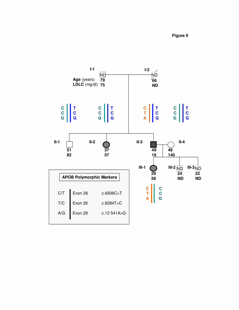
Figure II
II-1
ND
II-2 II-3 II-4
III-1 III-2
ND ND
I-1 I-2
ND III-3
Age (years)
LDLC (mg/dl)7975
66
ND
51
82
37
57
49
16
48
140
29
58
22
ND
24
ND
C
C
G
T
C
G
C
C
G
T
C
G
C
T
A
T
C
G
C
C
G
T
C
G
CTA
CCG
C/T Exon 26 c.6936C>T
T/C Exon 26 c.9294T>C
A/G Exon 29 c.12 541A>G
APOB Polymorphic Markers

Figure III
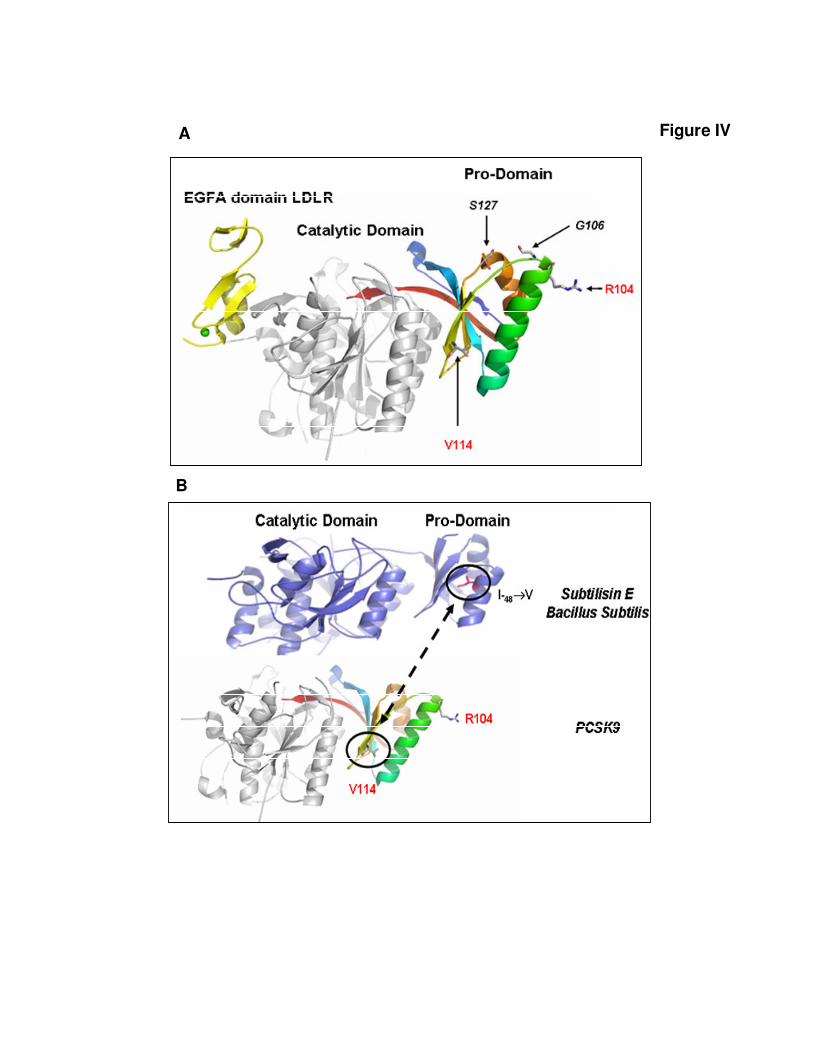
Figure IV A
B

Figure V
1
Plasma
leucine
10
VLDL1
20
VLDL2
30
IDL
40
LDL
Delay of apoB
production
K(20,10)
K(0,20)
K(0,30)
K(0,40)
K(0,10)
K(40,30)
K(30,20)
K(40,20)

Figure VI
II.3
III.1
Hours
(Ta
cer*
Tra
ce)*
10
0
Control
(Ta
cer*
Tra
ce)*
10
0
0
2
4
6
8
10
0 2 4 6 8 10 12 14
0
1
2
3
4
5
6
7
8
9
0 2 4 6 8 10 12 14
0
2
4
6
8
10
12
0 2 4 6 8 10 12 14
(Ta
cer*
Tra
ce
)*10
0
X VLDL
IDLLDL