pBAD/gIII A, B, and C - Thermo Fisher Scientific
32
USER GUIDE For Research Use Only. Not intended for any animal or human therapeutic or diagnostic use. pBAD/gIII A, B, and C Vectors for Regulated, Secreted Expression of Recombinant Proteins Containing C-Terminal 6xHis Tags in E. coli Catalog number V450-01 Revision date 2 March 2012 Publication Part number 25-0207 MAN0000058
Transcript of pBAD/gIII A, B, and C - Thermo Fisher Scientific

user guide
For Research Use Only. Not intended for any animal or human therapeutic or diagnostic use.
pBAD/gIII A, B, and C Vectors for Regulated, Secreted Expression of Recombinant Proteins Containing C-Terminal 6xHis Tags in E. coli
Catalog number V450-01
Revision date 2 March 2012 Publication Part number 25-0207
MAN0000058

ii

iii
Contents
Kit Contents and Storage ..................................................................................................................................... iv
Introduction ................................................................................................................... 1
Product Overview .................................................................................................................................................. 1
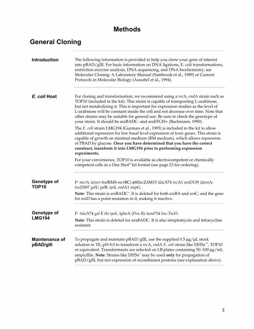
Methods ......................................................................................................................... 3
General Cloning ...................................................................................................................................................... 3
Cloning into pBAD/gIII ........................................................................................................................................ 4
E. coli Transformation ............................................................................................................................................ 8
Expression ............................................................................................................................................................... 9
Osmotic Shock ...................................................................................................................................................... 13
Purification ............................................................................................................................................................ 16
Appendix ...................................................................................................................... 17
pBAD/gIII Vector................................................................................................................................................. 17
Recipes ................................................................................................................................................................... 20
Accessory Products .............................................................................................................................................. 23
Technical Support ................................................................................................................................................. 24
Purchaser Notification ......................................................................................................................................... 25
References .............................................................................................................................................................. 26

iv
Kit Contents and Storage
Shipping and Storage
Kits are shipped at room temperature. Upon receipt, store the plasmids and the 20% L-arabinose solution at –20°C. Store stabs 4°C.
Kit Contents This kit contains the following items:
Contents Cat. No. 20 µg each pBAD/gIII A, B, and C, at 0.5 µg/μL in 10 mM Tris-HCl, 1 mM EDTA, pH 8.0 in a total volume of 40 μL.
V450-01
20 µg pBAD/gIII/calmodulin at 0.5 μg/μL in 10 mM Tris-HCl, 1 mM EDTA, pH 8.0 in a total volume of 40 μL.
1 mL sterile, 20% L-arabinose
1 stab LMG194
1 stab TOP10
The E. coli stabs supplied with the kit are guaranteed until the expiration date marked on tube when stored at 4°C. We recommend you prepare a set of glycerol master stocks prior to using your E. coli cells.
For research use only. Not intended for any human or animal therapeutic or diagnostic use.

1
Introduction
Product Overview
Description of the System
The pBAD/gIII plasmids are pBR322-derived expression vectors designed for regulated, secreted recombinant protein expression and purification in E. coli. The gene III signal sequence is utilized for secretion of the recombinant protein into the periplasmic space. Optimum levels of secreted, recombinant protein are possible using the araBAD promoter (PBAD) from E. coli. The regulatory protein, AraC, is provided on pBAD/gIII vectors allowing regulation of PBAD.
Gene III Secretion Signal
Gene III encodes pIII, one of the minor capsid proteins from the filamentous phage fd (similar to M13 and f1). pIII is synthesized with an 18 amino acid, amino terminal signal sequence and requires the bacterial Sec system for insertion into the membrane (Boeke and Model, 1982; Boeke et al., 1982; Davis et al., 1985; Rapoza and Webster, 1993). The signal sequence is removed after crossing the inner membrane, and most proteins will be retained in the periplasmic space.
Regulation of Expression by L-arabinose
In the presence of L-arabinose, expression from PBAD is turned on while the absence of L-arabinose produces very low levels of transcription from PBAD (Lee, 1980; Lee et al., 1987). Uninduced levels are repressed even further by growth in the presence of glucose. Glucose reduces the levels of 3´,5´-cyclic AMP, thus lowering expression of the catabolite-repressed PBAD promoter (Miyada et al., 1984). By varying the concentration of L-arabinose, protein expression levels can be manipulated to optimize expression of soluble, secreted protein. In addition, the tight regulation of PBAD by AraC is useful for expression of potentially toxic or essential genes (Carson et al., 1991; Dalbey and Wickner, 1985; Guzman et al., 1992; Kuhn and Wickner, 1985; Russell et al., 1989; San Millan et al., 1989). For more information on the mechanism of expression and repression of the ara regulon, refer to Schleif, 1992.

2
Product Overview, Continued
Experimental Outline
The table below describes the basic steps needed to clone and express your protein using pBAD/gIII. For more details, refer to the page(s) indicated.
Step Action Page
1 Develop a cloning strategy to ligate your gene of interest into pBAD/gIII A, B, or C.
4
2 Propagate and maintain the empty vectors by transforming them into a recA, endA E. coli host (i.e. TOP10).
3
3 Ligate your gene of interest into pBAD/gIII, transform into TOP10 or LMG194, and select on 50–100 µg/mL ampicillin.
4–8
4 Sequence your construct to ensure that it is in frame with the C-terminal peptide if you elect to create a fusion protein.
8
5 Perform a 4-hour expression using a 10,000-fold range of L-arabinose concentrations (e.g. 0.00002%, 0.0002%, 0.002%, 0.02%, and 0.2%).
9–10
6 Optimize expression by varying L-arabinose concentration or the time of induction.
11
7 Purify your recombinant protein by chromatography on metal-chelating resin (e.g. ProBond™).
13–16
Detection of Recombinant Proteins
Expression of your recombinant protein can be detected using an antibody to the appropriate epitope. The table below describes the antibodies available for use with pBAD/gIII (see page 23 for ordering). Horseradish peroxidase (HRP)-conjugated antibodies allow one-step detection using colorimetric or chemiluminescent detection methods.
Vector Epitope Antibody
pBAD/gIII c-myc Anti-Myc
Anti-Myc-HRP
C-terminal polyhistidine tag Anti-His(C-term)
Anti-His(C-term)-HRP
Purification of Recombinant Protein
The metal binding domain encoded by the polyhistidine tag allows simple, easy purification of your recombinant protein by Immobilized Metal Affinity Chromatography (IMAC) using ProBond™ Resin. To purify proteins expressed using pBAD/gIII, the ProBond™ Purification System or the ProBond™ resin in bulk are available separately. See page 23 for ordering information.
3
Methods
General Cloning
Introduction The following information is provided to help you clone your gene of interest into pBAD/gIII. For basic information on DNA ligations, E. coli transformations, restriction enzyme analysis, DNA sequencing, and DNA biochemistry, see Molecular Cloning: A Laboratory Manual (Sambrook et al., 1989) or Current Protocols in Molecular Biology (Ausubel et al., 1994).
E. coli Host For cloning and transformation, we recommend using a recA, endA strain such as TOP10 (included in the kit). This strain is capable of transporting L-arabinose, but not metabolizing it. This is important for expression studies as the level of L-arabinose will be constant inside the cell and not decrease over time. Note that other strains may be suitable for general use. Be sure to check the genotype of your strain. It should be araBADC- and araEFGH+ (Bachmann, 1990).
The E. coli strain LMG194 (Guzman et al., 1995) is included in the kit to allow additional repression for low basal level expression of toxic genes. This strain is capable of growth on minimal medium (RM medium), which allows repression of PBAD by glucose. Once you have determined that you have the correct construct, transform it into LMG194 prior to performing expression experiments.
For your convenience, TOP10 is available as electrocompetent or chemically competent cells in a One Shot® kit format (see page 23 for ordering).
Genotype of TOP10
F- mcrA ∆(mrr-hsdRMS-mcrBC) φ80lacZ∆M15 ∆lacX74 recA1 araD139 ∆(araA-leu)7697 galU galK rpsL endA1 nupG.
Note: This strain is araBADC-. It is deleted for both araBA and araC, and the gene for araD has a point mutation in it, making it inactive.
Genotype of LMG194
F- ∆lacX74 gal E thi rpsL ∆phoA (Pvu II) ∆ara714 leu::Tn10.
Note: This strain is deleted for araBADC. It is also streptomycin and tetracycline resistant.
Maintenance of pBAD/gIII
To propagate and maintain pBAD/gIII, use the supplied 0.5 μg/μL stock solution in TE, pH 8.0 to transform a recA, endA E. coli strain like DH5α™, TOP10 or equivalent. Transformants are selected on LB plates containing 50–100 µg/mL ampicillin. Note: Strains like DH5α™ may be used only for propagation of pBAD/gIII, but not expression of recombinant proteins (see explanation above).

4
Cloning into pBAD/gIII
To generate secreted, recombinant proteins that are expressed correctly and contain the C-terminal fusion peptide, it is necessary to clone in frame with BOTH the gene III secretion signal and the C-terminal peptide. The initiation ATG of the secretion signal is correctly spaced from the optimized RBS to ensure optimal translation.
To facilitate cloning, the pBAD/gIII vector is provided in three different reading frames. They differ only in the spacing between the signal sequence and the multiple cloning site. For proper expression, first determine which restriction sites are appropriate for ligation and then which vector will preserve the reading frame at BOTH the 5´ and the 3´ ends. You may have to use PCR to create a fragment with the appropriate restriction sites to clone in frame at both ends. Be sure that there is no stop codon in the open reading frame of your gene (except as noted below).
If you wish to express your protein WITHOUT the C-terminal peptide, be sure to include a stop codon at the end of your gene.
pBAD/gIII Multiple Cloning Site
The multiple cloning sites of each version of pBAD/gIII are provided on the following pages. Restriction sites are labeled to indicate cleavage site. The boxed sequence is the variable region that facilitates in-frame cloning with the gene III signal sequence.
Features of the araBAD and araC promoters are marked and described as follows. For more information see Lee, 1980; Miyada, et al., 1984; Lee, et al., 1987; and Schleif, 1992.
• O2 region: Binding site of AraC that represses transcription from PBAD.
• O1 region: Binding site of AraC that represses transcription of the araC promoter (PC) (transcribed on the opposite strand; not shown).
• CAP binding site: Site where CAP (cAMP binding protein) binds to help activate transcription from PBAD and PC.
• I2 and I1 regions: Binding sites of AraC that activate transcription from PBAD.
• –10 and –35 regions: Binding sites of RNA polymerase for transcription of PBAD.
Each multiple cloning site has been confirmed by sequencing and functional testing.
Cleavage of the Gene III Signal
Cleavage of the gene III signal occurs after Ser-His-Ser (see the multiple cloning site diagrams on the following pages). To minimize the number of additional amino acids at the N-terminus of your protein, we recommend using the Nco I site to clone your gene of interest closest to the gene III signal sequence. If you use the Nco I site, correct cleavage of the gene III will leave a threonine in front of the methionine.
Continued on next page
5
Cloning into pBAD/gIII, Continued
pBAD/gIII A
Continued on next page

6
Cloning into pBAD/gIII, Continued
pBAD/gIII B

7
Cloning into pBAD/gIII, Continued
pBAD/gIII C

8
E. coli Transformation
E. coli Transformation
After ligating your insert into the appropriate vector, transform your ligation mixtures into TOP10 cells and select on LB plates containing 50–100 µg/mL ampicillin. Select 10–20 clones and analyze for the presence and orientation of your insert.
We recommend that you sequence your construct to confirm that your gene is in frame with the appropriate tag.
Glycerol Stock Once you have obtained your desired construct, we recommend that you store your clone as a glycerol stock.
1. Grow 1–2 mL of the strain containing your construct in pBAD/gIII to log phase (OD600 = 0.5–0.7) in LB containing 50–100 µg/mL ampicillin.
2. Combine 0.85 mL of the culture with 0.15 mL of sterile glycerol.
3. Mix the solution by vortexing.
4. Transfer to an appropriate vial for freezing and cap.
5. Freeze in an ethanol/dry ice bath or liquid nitrogen and then transfer to -70°C for long-term storage.

9
Expression
Introduction Since each recombinant protein has different characteristics that may affect optimum expression, it is helpful to vary the L-arabinose concentration and/or run a time course of expression to determine the best conditions for optimal expression of your particular protein. A mock expression consisting of the pBAD/gIII vector alone should be done as a negative control. pBAD/gIII/calmodulin is included for use as a positive expression control (see page 19). TOP10 may be used as a general host for expression. LMG194 should be used if your protein is toxic or essential to E. coli.
Basic Strategy We recommend that you check for expression of your protein first, then check for solubility and secretion. Use the following strategy to determine the optimal expression level.
1. Pilot Expression. In this expression experiment you will vary the amount of L-arabinose over a 10,000-fold range (0.00002% to 0.2%) to determine the approximate amount of L-arabinose needed for maximum expression of your protein. See next page for protocol.
2. To optimize expression of your protein, you may wish to try L-arabinose concentrations spanning the amount determined in Step 1. Or you may wish to perform a time course.
Note: If you transformed your pBAD/gIII construct into LMG194, be sure to perform your expression experiments in RM medium with glucose (see page 21 for recipe) to ensure low basal levels of your protein.
Expression of your protein with the C-terminal tag will increase the size of your protein by ~2 kDa. Be sure to account for any additional amino acids between the tag and your protein.
Materials Needed • SOB or LB containing 50 µg/mL ampicillin (see Recipes, pages 20)
• RM medium containing glucose (see Recipes, page 21)
• 37°C shaking incubator
• 20% L-arabinose (provided)
• 37°C heat block or water bath
• 42°C water bath
• Liquid nitrogen
• 1X and 2X SDS-PAGE sample buffer
• Reagents and apparatus for SDS-PAGE gel
• 70°C water bath
• Lysis Buffer (see page 22 for recipe)
• Sterile water
Continued on next page

10
Expression, Continued
Pilot Expression This experiment is designed to test for and optimize expression of your recombinant protein. Remember to include a negative control (cells only or cells containing the empty vector) and a positive control (pBAD/gIII/calmodulin) to evaluate your expression experiment.
1. For each transformant or control, inoculate 2 mL of SOB or LB containing 50 µg/mL ampicillin with a single recombinant E. coli colony. Note: If you are using LMG194 as a host, use RM medium containing glucose and 50-100 µg/mL ampicillin at all steps.
2. Grow overnight at 37°C with shaking (225–250 rpm) to OD600 = 1–2.
3. The next day, label five tubes 1 through 5 and add 10 mL of medium containing 50 µg/mL ampicillin.
4. Inoculate each tube with 0.1 mL of the overnight culture.
5. Grow the cultures at 37°C with vigorous shaking to an OD600 = ~0.5 (the cells should be in mid-log phase).
6. While the cells are growing, prepare four 10-fold serial dilutions of 20% L-arabinose with sterile water (e.g. 2%, 0.2%, 0.02%, and 0.002%).
7. Remove a 1 mL aliquot of cells from each tube, centrifuge at maximum speed in a microcentrifuge for 30 seconds, and aspirate the supernatant.
8. Freeze the cell pellet at –20°C. This is the zero time point sample.
9. Add L-arabinose to the five 10 mL cultures as follows:
Tube Volume (mL) Stock Solution Final Concentration
1 0.1 0.002% 0.00002%
2 0.1 0.02% 0.0002%
3 0.1 0.2% 0.002%
4 0.1 2% 0.02%
5 0.1 20% 0.2% 10. Grow at 37°C with shaking for 4 hours.
11. Take 1 mL samples at 4 hours and treat as in Step 7 and 8. In general, four hours is sufficient for expression of most proteins. You may discard the remaining culture or you may continue on with the time course.
Continued on next page

11
Expression, Continued
Preparing Samples
Before starting, prepare SDS-PAGE gels to analyze all the samples you collected. Note: If you already know that your protein is insoluble, use the protocol on the next page to analyze your samples.
1. When all the samples have been collected from Steps 8 and 11, previous page, resuspend each pellet in 100 µL of 1X SDS-PAGE sample buffer.
2. Heat 5 minutes at 70°C and centrifuge briefly.
3. Load 5 µL of each sample on an SDS-PAGE gel and electrophorese. Save your samples by storing at –20°C.
Sample Analysis 1. Stain the gel with Coomassie blue and look for a band of increasing intensity in the expected size range for the recombinant protein.
2. Use a negative control (empty vector) to distinguish recombinant proteins from background proteins.
3. Use the positive control (pBAD/gIII/calmodulin) to confirm that growth and induction was done properly. The positive control should yield a 30 kDa protein.
4. You should be able to determine the approximate L-arabinose concentration for maximum expression.
Low Expression If you don't see any expression on a Coomassie-stained gel, re-run your samples on an SDS-PAGE gel and perform a western blot. Use antibody to your protein or the Anti-Myc antibodies to detect expression of your protein. Note: Proteins expressed using pBAD/gIII may also be detected with the Anti-His (C-term) Antibody which recognizes histidine tags with a free carboxyl group (see page 23 for ordering).
If you still don't see expression of your protein, sequence your construct and make sure it is in frame with the C-terminal peptide.
Optimizing Expression
Once you have detected expression of your protein, you may wish to perform some experiments to further optimize expression. Use the Pilot Expression protocol, but vary the L-arabinose concentration over a smaller range. For example, if you obtained the best expression at 0.002%, try 0.0004%, 0.0008%, 0.001%, 0.004%, and 0.008%.
Also you may perform a time course of induction over a 5–6 hour time period, taking time points every hour, to determine if varying the time increases expression.
Remember to store your time points at –20°C.
If your protein is insoluble, you may wish to analyze the supernatant and pellet of lysed cells when you vary the L-arabinose concentration. Refer to the protocol on the next page to prepare samples.
Continued on next page
12
Expression, Continued
Insoluble Proteins
If you suspect that your protein may be insoluble, analyze your expression samples using the following protocol.
1. When all the samples have been collected, thaw and resuspend each pellet in 100 µL of Lysis Buffer (see Recipes, page 22).
2. Incubate on ice for 30 minutes.
3. Perform 3 cycles of freeze-thaw (freeze in liquid nitrogen or a dry ice bath, then thaw at 42°C).
4. Incubate at 37°C for 30 minutes.
5. Centrifuge samples to pellet insoluble proteins. Transfer supernatant to a fresh tube and store on ice.
6. Mix together equal amounts of supernatant and 2X SDS Sample buffer and heat for 5 minutes at 70°C.
7. Add 200 µL of 1X SDS-PAGE sample buffer to pellets from Step 5 and heat 5 minutes at 70°C.
8. Load 10 µL of the supernatant sample and 10 µL of the pellet sample onto an SDS-PAGE and electrophorese.
9. Analyze for optimal, soluble expression of your protein. Ideally, most of your recombinant protein should be soluble. If the majority of your protein is still insoluble, try the induction at 28°C or 30°C instead of 37°C. Expressing at a lower temperature may help proteins fold correctly.
Analyzing Secreted Protein
The gene III signal sequence is designed to direct your protein to the periplasmic space. To test for secretion, you will need to osmotically shock your cells to release the recombinant protein from the periplasmic space. Refer to the protocol on pages 13–15 for details.
Expressing Toxic Proteins
To ensure low levels of expression, you may find it useful to utilize glucose to repress the araBAD promoter further. Follow the steps below to express your protein.
1. Transform your construct into LMG194. LMG194 can be grown in RM medium, which enables repression of PBAD by glucose.
2. Follow the Pilot Expression on page 10, substituting RM Medium + Glucose medium (see page 21) to grow the cells.
3. Be sure to monitor the OD600 as the cells will grow more slowly in RM medium.
4. Induce with various concentrations of L-arabinose as described in the Pilot Expression.
5. Monitor OD600 over time be sure cells are growing.

13
Osmotic Shock
Introduction The procedure below may be used to check for secretion of your recombinant protein and/or partial purification of your recombinant protein. The positive control vector pBAD/gIII/calmodulin may be used to evaluate secretion and the osmotic shock procedure.
Before Starting • SOB or LB containing 50 µg/mL ampicillin
• Osmotic Shock Solution 1 (see page 22)
• Osmotic Shock Solution 2 (see page 22)
Control pBAD/gIII/calmodulin is provided as a positive control for osmotic shock. About 50% of the total expressed calmodulin (~0.2 mg/mL) can be recovered in the shock fluid after osmotic shock.
Osmotic shock works best on fresh cells. Do not store cells before shocking. Do not shock frozen cells.
Growth and Induction of Cells
Use the conditions you developed in the Expression section to grow and induce expression of your protein.
1. Inoculate 2 mL of SOB or LB containing 50 µg/mL ampicillin with a single recombinant E. coli colony. Note: If you are using LMG194 as a host, use RM medium containing glucose and 50–100 µg/mL ampicillin.
2. Grow overnight at 37°C with shaking (225–250 rpm) to OD600 = 1–2. 3. The next day, inoculate 10–25 mL of SOB or LB containing 50 µg/mL
ampicillin with 0.1–0.25 mL of the overnight culture.
4. Grow the culture at 37°C with vigorous shaking to an OD600 = ~0.5 (the cells should be in mid-log phase). Record the OD600.
5. Remove a 1 mL sample of cells, centrifuge at maximum speed in a microcentrifuge for 30 seconds, aspirate the supernatant, and store the cells on ice. Do not freeze the cells. This is the zero time point sample.
6. Add the appropriate amount of L-arabinose determined previously to induce expression of your protein.
7. Grow the cells to the optimal time point as previously determined. Read and record the OD600.
8. Remove a 1 mL sample, centrifuge as described in Step 5, aspirate the supernatant, and store the cells on ice.
9. Take the rest of the culture and discard. Do not save the cells. Proceed to the next section.
Continued on next page

14
Osmotic Shock, Continued
Osmotic Shock 1. Resuspend cell pellets from Growth and Induction of Cells, Steps 5 and 8 (previous page) in Osmotic Shock Solution 1 (with sucrose) to an OD600 of 5.0. Use the OD600 value you recorded for each time point to determine in what volume you should resuspend the cells.
Formula: VR = (OD600 of sample/5.0) × VS Where VR is the volume to resuspend the cell pellet and VS is the original sample volume of the cell suspension
Example: If a 1 mL sample of your cells has an OD600 of 0.5 for the zero time point, then:
VR = (0.5/5.0) × 1 mL = 0.1 mL or 100 µL. This is the volume in which to resuspend your cells. Note that each pellet may need to be resuspended in a different volume.
2. Incubate cells on ice for 10 minutes. Centrifuge for 1 minute at 4°C and decant the supernatant.
3. Resuspend cell pellets in Osmotic Shock Solution 2 using the same volumes from Step 1. Incubate on ice for 10 minutes.
4. Centrifuge for 10 minutes at 4°C. Transfer the supernatant (shock fluid) to a clean tube and keep on ice.
5. Resuspend the pellets from Step 4 in the same volume of Osmotic Shock Solution 2 as was used in Step 1. Note that each pellet may be resuspended in a different volume.
6. You now have four samples – a supernatant (shock fluid) and a pellet sample (cells) for the zero time point and a supernatant and pellet sample for the optimal time point. If you included the positive control, you will have four more samples for a total of eight.
7. These samples may be frozen at –20°C if you do not want to run a gel the same day you prepare samples. Proceed to Analysis of Osmotic Shock Samples on the next page.
Continued on next page

15
Osmotic Shock, Continued
Analysis of Osmotic Shock Samples
1. The samples from Osmotic Shock, step 6 (previous page) are analyzed on an SDS-PAGE gel. Use 10 µL aliquots for each sample. Prepare and load the samples onto the gel so you can compare shock fluid with cells for each time point. Run the gel and process.
2. Use the following table to evaluate your experiment:
IF the sample
containing calmodulin ...
AND the sample containing the
fusion protein ...
THEN...
is in the supernatant fraction
is also in the supernatant fraction
the fusion protein is secreted and released by osmotic shock. Osmotic shock can be used as a purification step.
is in the supernatant fraction
is in the pellet fraction the fusion protein is either not secreted or is not released by osmotic shock. See next section below.
is in the pellet fraction
is also in the pellet fraction
review the osmotic shock procedure and make sure the correct buffers were used in the correct order. If the osmotic shock step was done properly, calmodulin should be in the supernatant.
Recombinant Protein Appears not to be Secreted
It may happen that because of the nature of your protein, it is not properly secreted or shocked out. The protein may be retained in the cytoplasm or associated with the inner cell membrane. You may need to prepare a whole cell lysate prior to purifying your protein. For additional information, refer to Guide to Protein Purification, pages 147–153 (Deutscher, 1990).
Note: If the recombinant protein is retained in the cytoplasm, the gene III signal sequence will not be removed from the protein.
Activity Assay If your fusion protein was successfully purified by osmotic shock, you may wish to assay for the activity of your desired protein. If the fusion protein retains significant levels of activity, you may scale-up your purification to produce more fusion protein.

16
Purification
Scale-up of Expression for Purification
Use the conditions determined in the previous section to grow and induce 50 mL of cells. This is the largest culture volume to use with the 2 mL prepacked columns included in the ProBond™ System. If you need to purify larger amounts of recombinant protein, you may need more ProBond™ resin. See page 23 for ordering information. Note: Remember to use RM medium (page 21) with LMG194.
1. Inoculate 2 mL of SOB or LB containing 50 µg/mL ampicillin with a single recombinant E. coli colony.
2. Grow overnight at 37°C with shaking (225–250 rpm) to OD600 = 1–2.
3. The next day, inoculate 50 mL of SOB or LB containing 50 µg/mL ampicillin with 1 mL of the overnight culture.
4. Grow the culture at 37°C with vigorous shaking to an OD600 = ~0.5 (the cells should be in mid-log phase).
5. Add the optimal amount of L-arabinose to induce expression.
6. Grow at 37°C with shaking until the optimal time point is reached. Harvest the cells by centrifugation (3000 × g for 10 minutes at 4°C).
7. At this point, you may proceed directly to purification (ProBond™ Purification System manual) or store at –80°C for future use.
Purification For help with purification of your recombinant protein, refer to the ProBond™ Purification System manual.
If you are using another type of resin, refer to the manufacturer's recommendations.

17
Appendix
pBAD/gIII Vector
Map of pBAD/gIII The figure below summarizes the features of the pBAD/gIII vector. Vector sequences for all three pBAD/gIII vectors can be downloaded from www.lifetechnologies.com or by contacting Technical Support (see page 24). Details of each multiple cloning site are shown on pages 5–7.

18
pBAD/gIII Vectors, Continued
Features of pBAD/gIII
The important elements of pBAD/gIII A (4145 bp), pBAD/gIII B (4147 bp), and pBAD/gIII C (4149 bp) are described in the following table. All features have been functionally tested.
Feature Benefit
araBAD promoter (PBAD) Provides tight, dose-dependent regulation of heterologous gene expression (Guzman et al., 1995)
Optimized ribosome binding site Increases efficiency of recombinant fusion protein expression
Initiation ATG Provides a translational initiation site for the fusion protein
Gene III secretion signal Permits secretion of recombinant protein into the periplasmic space (Rapoza and Webster, 1993)
Multiple cloning site Allows insertion of your gene for expression
C-terminal myc epitope tag (Glu-Gln-Lys-Leu-Ile-Ser-Glu-Glu-
Asp-Leu)
Allows detection of the fusion protein by the Anti-Myc Antibody (see page 23) (Evans et al., 1985)
C-terminal polyhistidine region Forms metal-binding site for affinity purification of recombinant fusion protein on metal-chelating resin (i.e. ProBond™) In addition, it allows detection of the recombinant protein with Anti-His (C-term) Antibody (see page 2)
rrnB transcription termination region Strong transcription termination region
Ampicillin resistance gene (β-lactamase)
Allows selection of the plasmid in E. coli
pBR322 origin Low copy replication and growth in E. coli
araC gene Encodes the regulatory protein for tight regulation of the PBAD promoter (Lee, 1980; Schleif, 1992)
Continued on next page

19
Map of pBAD/gIII/calmodulin
Description pBAD/gIII is a 4556 bp control vector containing the gene for calmodulin fused to the C-terminal peptide. It was constructed by digesting pBAD/gIII A with Sac I and Xba I, and ligating an 455 bp Sac I-Xba I fragment containing the calmodulin gene. The calculated molecular weight of calmodulin fused to the gene III signal sequence is 21.3 kDa. The observed molecular weight from an SDS-PAGE gel is 25–30 kDa.
Map of Control Vector
The figure below summarizes the features of the pBAD/gIII/calmodulin vector. The nucleotide sequence for pBAD/gIII/calmodulin may be downloaded from www.lifetechnologies.com or by contacting Technical Support (see page 24).

20
Recipes
Low Salt LB Medium (with Ampicillin)
LB Medium (per liter)
1% Tryptone
0.5% Yeast Extract
0.5% NaCl
pH 7.0
1. For 1 liter, dissolve 10 g tryptone, 5 g yeast extract, and 5 g NaCl in 950 mL deionized water.
2. Adjust the pH of the solution to 7.0 with 5 M NaOH and bring the volume to 1 liter.
3. Autoclave for 20 minutes on liquid cycle.
4. Let solution cool to ~55°C. Add ampicillin to a final concentration of 50 µg/mL. Store the medium at 4°C. Medium is stable for only 1–2 weeks.
Low Salt LB Agar Plates with Ampicillin
LB Medium (per liter)
1% Tryptone
0.5% Yeast Extract
0.5% NaCl
1.5% Agar
pH 7.0
1. For 1 liter, dissolve 10 g tryptone, 5 g yeast extract, and 5 g NaCl in 950 mL deionized water.
2. Adjust the pH of the solution to 7.0 with 5 M NaOH, add 15 g agar, and bring the volume to 1 liter.
3. Autoclave for 20 minutes on liquid cycle.
4. Let agar cool to ~55°C. Add ampicillin to a final concentration of 50 µg/mL.
5. Pour into 10 cm petri plates. Let the plates harden, then invert and store at 4°C. Plates containing ampicillin are stable for 1–2 weeks.
Continued on next page

21
Recipes, Continued
SOB Medium (with Ampicillin)
SOB (per liter)
2% Tryptone
0.5% Yeast Extract
0.05% NaCl
2.5 mM KCl
10 mM MgCl2
1. Dissolve 20 g tryptone, 5 g yeast extract, and 0.5 g NaCl in 950 mL deionized water.
2. Make a 250 mM KCl solution by dissolving 1.86 g of KCl in 100 mL of deionized water. Add 10 mL of this stock KCl solution to the solution in Step 1.
3. Adjust pH to 7.5 with 5 M NaOH and add deionized water to 1 liter.
4. Autoclave this solution, cool to ~55°C, and add 10 mL of sterile 1 M MgCl2. You may also add ampicillin to 50 µg/mL.
5. Store at 4°C. Medium is stable for only 1–2 weeks.
RM Medium + Glucose
1X M9 Salts (See below for recipe for 10X M9 Salts)
2% Casamino Acids
0.2% glucose
1 mM MgCl2
50–100 µg/mL ampicillin
1. For 1 liter of RM medium, mix 20 g Casamino Acids and 890 mL deionized water.
2. Autoclave 20 minutes on liquid cycle.
3. After the autoclaved solution has cooled, add the following sterile solutions aseptically:
10X M9 Salts 100 mL
1 M MgCl2 1 mL
20% glucose 10 mL
100 mg/mL ampicillin 0.5 to 1 mL
4. Mix well and store medium containing ampicillin at 4°C. Medium is good for 1 month at 4°C.
Continued on next page

22
Recipes, Continued
10X M9 Salts For 1 liter:
Na2HPO4 60 g
KH2PO4 30 g
NaCl 5 g
NH4Cl 10 g
Water 900 mL
1. Dissolve reagents in the water and adjust the pH to 7.4 with 10 M NaOH.
2. Add water to 1 liter and autoclave for 20 minutes on liquid cycle.
3. Add 1 mL of 1 M thiamine (filter-sterilize) per 1 L 1X M9 medium.
4. Store at room temperature.
Lysis Buffer 10 mM Tris-HCl, pH 8
1 mM EDTA
0.5 mg/mL lysozyme
0.1 mg/mL DNase I
10 mM CaCl2
1. Prepare just before use. Take 10 mL of TE buffer and add 5 mg of lysozyme, 1 mg of DNase I, and 0.1 mL of 1 M CaCl2.
2. Gently mix and store on ice. Use immediately.
Osmotic Shock Solution 1
20 mM Tris-HCl, pH 8
2.5 mM EDTA
20% Sucrose
1. For 1 liter, combine 200 g sucrose, 20 mL 1 M Tris-HCl pH 8.0, and 5 mL 0.5 M EDTA and bring up to a final volume of 1 liter with water.
2. Stir to dissolve sucrose.
3. Autoclave or filter-sterilize.
4. Store at room temperature or at 4°C.
Osmotic Shock Solution 2
20 mM Tris-HCl, pH 8
2.5 mM EDTA
1. For 1 liter, combine 20 mL 1 M Tris-HCl pH 8.0 and 5 mL 0.5 M EDTA and bring up to a final volume of 1 liter with water.
2. Autoclave or filter-sterilize.
3. Store at room temperature or at 4°C.

23
Accessory Products
Additional Products
Many products suitable for use with pBAD/gIII are available separately. Ordering information for these reagents is provided below.
Item Quantity Cat. no.
One Shot® TOP10 Electrocomp™ E. coli 21 × 50 µL C4040-52
One Shot® TOP10 Chemically Competent E. coli 21 × 50 µL C4040-03
Detection of Recombinant Fusion Proteins
You can detect expression of your recombinant fusion protein from pBAD/gIII using the Anti-Myc and Anti-His antibodies available from Life Technologies.
Epitope Antibody Cat. no.
c-myc Anti-Myc R950-25
Anti-Myc-HRP R951-25
C-terminal polyhistidine tag Anti-His(C-term) R930-25
Anti-His(C-term)-HRP R931-25
Purification of Recombinant Protein
The presence of the polyhistidine tag in pBAD/gIII allows purification of your recombinant fusion protein using a nickel-charged agarose resin such as ProBond™. Ordering information is provided below.
Item Quantity Cat. no.
ProBond™ Nickel-Chelating Resin 50 mL 150 mL
R801-01 R801-15
ProBond™ Purification System 6 purifications K850-01
Purification Columns (10 mL polypropylene columns) 50 R640-50
24
Technical Support
Obtaining support
For the latest services and support information for all locations, go to www.lifetechnologies.com/support.
At the website, you can: • Access worldwide telephone and fax numbers to contact Technical Support
and Sales facilities • Search through frequently asked questions (FAQs) • Submit a question directly to Technical Support ([email protected]) • Search for user documents, SDSs, vector maps and sequences, application
notes, formulations, handbooks, certificates of analysis, citations, and other product support documents
• Obtain information about customer training • Download software updates and patches
Safety Data Sheets (SDS)
Safety Data Sheets (SDSs) are available at www.lifetechnologies.com/support.
Certificate of Analysis
The Certificate of Analysis provides detailed quality control and product qualification information for each product. Certificates of Analysis are available on our website. Go to www.lifetechnologies.com/support and search for the Certificate of Analysis by product lot number, which is printed on the box.
Limited warranty Life Technologies Corporation is committed to providing our customers with high-quality goods and services. Our goal is to ensure that every customer is 100% satisfied with our products and our service. If you should have any questions or concerns about a Life Technologies product or service, contact our Technical Support Representatives. All Life Technologies products are warranted to perform according to specifications stated on the certificate of analysis. The Company will replace, free of charge, any product that does not meet those specifications. This warranty limits the Company’s liability to only the price of the product. No warranty is granted for products beyond their listed expiration date. No warranty is applicable unless all product components are stored in accordance with instructions. The Company reserves the right to select the method(s) used to analyze a product unless the Company agrees to a specified method in writing prior to acceptance of the order. Life Technologies makes every effort to ensure the accuracy of its publications, but realizes that the occasional typographical or other error is inevitable. Therefore the Company makes no warranty of any kind regarding the contents of any publications or documentation. If you discover an error in any of our publications, report it to our Technical Support Representatives. Life Technologies Corporation shall have no responsibility or liability for any special, incidental, indirect or consequential loss or damage whatsoever. The above limited warranty is sole and exclusive. No other warranty is made, whether expressed or implied, including any warranty of merchantability or fitness for a particular purpose.

25
Purchaser Notification
Limited Use Label License No: Research Use Only
The purchase of this product conveys to the purchaser the limited, non-transferable right to use the purchased amount of the product only to perform internal research for the sole benefit of the purchaser. No right to resell this product or any of its components is conveyed expressly, by implication, or by estoppel. This product is for internal research purposes only and is not for use in commercial applications of any kind, including, without limitation, quality control and commercial services such as reporting the results of purchaser's activities for a fee or other form of consideration. For information on obtaining additional rights, please contact [email protected] or Out Licensing, Life Technologies, 5791 Van Allen Way, Carlsbad, California 92008.
Limited Use Label License 6X His Tag
This product is licensed from Hoffmann-La Roche, Inc., Nutley, NJ and/or Hoffmann-LaRoche Ltd., Basel, Switzerland and is provided only for use in research. Information about licenses for commercial use is available from QIAGEN GmbH, Max-Volmer-Str. 4, D-40724 Hilden, Germany.
Limited Use Label License araB Promoter
Inquiries for any commercial use, including production of material to be sold commercially or used in production or in product development efforts, which includes efforts toward regulatory approval, should be made directly to Xoma Corporation, 2910 Seventh Street, Berkeley, CA 94710, Tel: 1-510-644-1170 Fax: 1-510-649-7571.
26
References
Ausubel, F. M., Brent, R., Kingston, R. E., Moore, D. D., Seidman, J. G., Smith, J. A., and Struhl, K. (1994). Current Protocols in Molecular Biology (New York: Greene Publishing Associates and Wiley-Interscience).
Bachmann, B. J. (1990). Linkage map of Escherichia coli K-12, edition 8. Microbiol. Rev 54, 130-197.
Boeke, J. D., and Model, P. (1982). A Prokaryotic Membrane Anchor Sequence: Carboxyl Terminus of Bacteriophage f1 Gene III Protein Retains it in the Membrane. Proc. Natl. Acad. Sci. USA 79, 5200-5204.
Boeke, J. D., Model, P., and Zinder, N. D. (1982). Effects of Bacteriophage f1 Gene III Protein on the Host Cell Membrane. Mol. Gen. Genet. 186, 185-192.
Carson, M. J., Barondess, J. J., and Beckwith, J. (1991). The FtsQ Protein of Escherichia coli: Membrane Topology, Abundance, and Cell Division Phenotypes Due to Overproduction and Insertion Mutations. J. Bacteriol. 173, 2187-2195.
Dalbey, R. E., and Wickner, W. (1985). Leader Peptidase Catalyzes the Release of Exported Proteins from the Outer Surface of the Escherichia coli Plasma Membrane. J. Biol. Chem. 260, 15925-15931.
Davis, N. G., Boeke, J. D., and Model, P. (1985). Fine Structure of a Membrane Anchor Domain. J. Mol. Biol. 181, 111-121.
Deutscher, M. P. (1990) Guide to Protein Purification. In Methods in Enzymology, Vol. 182. (J. N. Abelson and M. I. Simon, eds.) Academic Press, San Diego, CA.
Evans, G. I., Lewis, G. K., Ramsay, G., and Bishop, V. M. (1985). Isolation of Monoclonal Antibodies Specific for c-myc Proto-oncogene Product. Mol. Cell. Biol. 5, 3610-3616.
Guzman, L.-M., Barondess, J. J., and Beckwith, J. (1992). FtsL, an Essential Cytoplasmic Membrane Protein Involved in Cell Division in Escherichia coli. J. Bacteriol. 174, 7716-7728.
Guzman, L.-M., Belin, D., Carson, M. J., and Beckwith, J. (1995). Tight Regulation, Modulation, and High-Level Expression by Vectors Containing the Arabinose PBAD Promoter. J. Bacteriol. 177, 4121-4130.
Kuhn, A., and Wickner, W. (1985). Isolation of Mutants in M13 Coat Protein That Affect its Synthesis, Processing and Assembly into Phage. J. Biol. Chem. 260, 15907-15913.
Lee, N. (1980) Molecular Aspects of ara Regulation. In The Operon, J. H. Miller and W. S. Reznikoff, eds. (Cold Spring Harbor, N.Y.: Cold Spring Harbor Laboratory), pp. 389-410.
Lee, N., Francklyn, C., and Hamilton, E. P. (1987). Arabinose-Induced Binding of AraC Protein to araI2 Activates the araBAD Operon Promoter. Proc. Natl. Acad. Sci. USA 84, 8814-8818.
Miyada, C. G., Stoltzfus, L., and Wilcox, G. (1984). Regulation of the araC Gene of Escherichia coli: Catabolite Repression, Autoregulation, and Effect on araBAD Expression. Proc. Natl. Acad. Sci. USA 81, 4120-4124.
Rapoza, M. P., and Webster, R. E. (1993). The Filamentous Bacteriophage Assembly Proteins Require the Bacterial SecA protein for Correct Localization to the Membrane. J. Bacteriol. 175, 1856-1859.
Russell, C. B., Stewart, R. C., and Dahlquist, F. W. (1989). Control of Transducer Methylation Levels in Escherichia coli: Investigation of Components Essential for Modulation of Methylation and Demethylation Reactions. J. Bacteriol. 171, 3609-3618.
Sambrook, J., Fritsch, E. F., and Maniatis, T. (1989). Molecular Cloning: A Laboratory Manual, Second Edition (Plainview, New York: Cold Spring Harbor Laboratory Press).
San Millan, J. L., Boyd, D., Dalbey, R., Wickner, W., and Beckwith, J. (1989). Use of phoA Fusions to Study the Topology of the Escherichia coli Inner Membrane Protein Leader Peptidase. J. Bacteriol. 171, 5536-5541.
Schleif, R. S. (1992). DNA Looping. Ann. Rev. Biochem. 61, 199-223.
©2012 Life Technologies Corporation. All rights reserved. The trademarks mentioned herein are the properties of Life Technologies Corporation or their respective owners.