Pax2 and Pax8 cooperate in mouse inner ear morphogenesis and
17
RESEARCH ARTICLE Open Access Pax2 and Pax8 cooperate in mouse inner ear morphogenesis and innervation Maxime Bouchard 1† , Dominique de Caprona 2 , Meinrad Busslinger 3 , Pinxian Xu 4 , Bernd Fritzsch 2*† Abstract Background: Pax2;5;8 transcription factors play diverse roles in vertebrate and invertebrate organogenesis, including the development of the inner ear. Past research has suggested various cochlear defects and some vestibular defects in Pax2 null mice but the details of the cochlear defects and the interaction with other Pax family members in ear development remain unclear. Results: We show that Pax2;8 double null mice do not develop an ear past the otocyst stage and show little to no sensory as well as limited and transient neuronal development, thus indicating that these two family members are essential for overall ear morphogenesis and sustained neurosensory development. In support of functional redundancy between Pax proteins, Pax2 can be substituted by a Pax5 minigene, a gene normally not expressed in the embryonic mouse ear. There is no detectable morphological defect in Pax8 null mice suggesting that Pax2 expression can compensate for Pax8. Conversely, Pax8 cannot compensate for Pax2 leading to a cochlear phenotype not fully appreciated previously: Cochlear development is delayed until E15.5 when the cochlea extrudes as a large sack into the brain case. Immunocytochemistry and tracing from the brain show that a cochlear spiral ganglia form as a small addition to the inferior vestibular ganglion. However, the empty cochlear sack, devoid of any sensory epithelium development as indicated by the absence of Sox2 or MyoVII expression, nevertheless develop a dense innervation network of small neurons situated in the wall of the cochlear sack. Conclusions: Combined these data suggest that Pax2 is needed for organ of Corti formation and is directly or indirectly involved in the coordination of spiral ganglion formation which is partially disrupted in the Pax2 null ears. All three Pax genes can signal redundantly in the ear with their function being determined primarily by the spatio- temporal expression driven by the three distinct promoters of these genes. Background Pax2;5;8 genes are vertebrate Pax orthologs that evolved out of an ancestral Pax2;5;8;6 gene of sponges [1] that became associated with ocelli and statocysts in coelente- rates [2]. Pax2;5;8 became associated with ear develop- ment in vertebrates and sensilla development in flies among additional expressions domains in brain, kidney and other organs. In vertebrates, Pax8 is among the ear- liest genes unequivocally expressed in the developing otic placode of fish, frog and mice and appears to be later largely co-expressed with Pax2 in the mouse ear. Several papers have at least partially characterized the effects of Pax2 inactivation in the mouse ear [3-5]. The data agree that Pax2 function is essential for cochlear development in mice and human but vary in the degree of vestibular defects and in the degree of loss of sensory neurons. However, as the cochlea is a mammalian novelty[6], the expression of Pax2 in the ear of bony fish that have not evolved a cochlea [7] reflects a more ancient function of Pax2 in vertebrate ear development. Moreover, Pax2 reduction in zebrafish ear development results in hair cell defects that may be initiated at the level of the otic placode [2,8]. In this system, Pax2 and Pax5 seem to regulate ear development downstream of Fgf3/8 with a similar near complete loss of ear differen- tiation in knock-down experiments [8-10]. In contrast to Pax2, previous work on Pax8 null mice has shown a thyroid phenotype but no obvious ear defect was identi- fied [11]. Pax5 is apparently not expressed in the ear of * Correspondence: [email protected] † Contributed equally 2 Department of Biology, College of Liberal Arts and Sciences, 143 Biology Building, Iowa City, IA 52242-1324, USA Full list of author information is available at the end of the article Bouchard et al. BMC Developmental Biology 2010, 10:89 http://www.biomedcentral.com/1471-213X/10/89 © 2010 Bouchard et al; licensee BioMed Central Ltd. This is an Open Access article distributed under the terms of the Creative Commons Attribution License (http://creativecommons.org/licenses/by/2.0), which permits unrestricted use, distribution, and reproduction in any medium, provided the original work is properly cited.
Transcript of Pax2 and Pax8 cooperate in mouse inner ear morphogenesis and

RESEARCH ARTICLE Open Access
Pax2 and Pax8 cooperate in mouse inner earmorphogenesis and innervationMaxime Bouchard1†, Dominique de Caprona2, Meinrad Busslinger3, Pinxian Xu4, Bernd Fritzsch2*†
Abstract
Background: Pax2;5;8 transcription factors play diverse roles in vertebrate and invertebrate organogenesis,including the development of the inner ear. Past research has suggested various cochlear defects and somevestibular defects in Pax2 null mice but the details of the cochlear defects and the interaction with other Paxfamily members in ear development remain unclear.
Results: We show that Pax2;8 double null mice do not develop an ear past the otocyst stage and show little to nosensory as well as limited and transient neuronal development, thus indicating that these two family members areessential for overall ear morphogenesis and sustained neurosensory development. In support of functionalredundancy between Pax proteins, Pax2 can be substituted by a Pax5 minigene, a gene normally not expressed inthe embryonic mouse ear. There is no detectable morphological defect in Pax8 null mice suggesting that Pax2expression can compensate for Pax8. Conversely, Pax8 cannot compensate for Pax2 leading to a cochlearphenotype not fully appreciated previously: Cochlear development is delayed until E15.5 when the cochleaextrudes as a large sack into the brain case. Immunocytochemistry and tracing from the brain show that a cochlearspiral ganglia form as a small addition to the inferior vestibular ganglion. However, the empty cochlear sack,devoid of any sensory epithelium development as indicated by the absence of Sox2 or MyoVII expression,nevertheless develop a dense innervation network of small neurons situated in the wall of the cochlear sack.
Conclusions: Combined these data suggest that Pax2 is needed for organ of Corti formation and is directly orindirectly involved in the coordination of spiral ganglion formation which is partially disrupted in the Pax2 null ears.All three Pax genes can signal redundantly in the ear with their function being determined primarily by the spatio-temporal expression driven by the three distinct promoters of these genes.
BackgroundPax2;5;8 genes are vertebrate Pax orthologs that evolvedout of an ancestral Pax2;5;8;6 gene of sponges [1] thatbecame associated with ocelli and statocysts in coelente-rates [2]. Pax2;5;8 became associated with ear develop-ment in vertebrates and sensilla development in fliesamong additional expressions domains in brain, kidneyand other organs. In vertebrates, Pax8 is among the ear-liest genes unequivocally expressed in the developingotic placode of fish, frog and mice and appears to belater largely co-expressed with Pax2 in the mouse ear.Several papers have at least partially characterized the
effects of Pax2 inactivation in the mouse ear [3-5]. Thedata agree that Pax2 function is essential for cochleardevelopment in mice and human but vary in the degreeof vestibular defects and in the degree of loss of sensoryneurons. However, as the cochlea is a mammaliannovelty[6], the expression of Pax2 in the ear of bonyfish that have not evolved a cochlea [7] reflects a moreancient function of Pax2 in vertebrate ear development.Moreover, Pax2 reduction in zebrafish ear developmentresults in hair cell defects that may be initiated at thelevel of the otic placode [2,8]. In this system, Pax2 andPax5 seem to regulate ear development downstream ofFgf3/8 with a similar near complete loss of ear differen-tiation in knock-down experiments [8-10]. In contrast toPax2, previous work on Pax8 null mice has shown athyroid phenotype but no obvious ear defect was identi-fied [11]. Pax5 is apparently not expressed in the ear of
* Correspondence: [email protected]† Contributed equally2Department of Biology, College of Liberal Arts and Sciences, 143 BiologyBuilding, Iowa City, IA 52242-1324, USAFull list of author information is available at the end of the article
Bouchard et al. BMC Developmental Biology 2010, 10:89http://www.biomedcentral.com/1471-213X/10/89
© 2010 Bouchard et al; licensee BioMed Central Ltd. This is an Open Access article distributed under the terms of the CreativeCommons Attribution License (http://creativecommons.org/licenses/by/2.0), which permits unrestricted use, distribution, andreproduction in any medium, provided the original work is properly cited.

mouse embryos and no defects have been reported inPax5 mutants [12] in contrast to zebrafish embryos [8].In chicken, Pax8 is lost and Pax2 appears to be the onlygene relevant for placode invagination [13], clearly indi-cating the importance of these Pax genes for ear induc-tion and development.Consistent with the idea of functional conservation, an
allelic series of Pax2 and Pax8 mutation indeed showeda dose dependent effect indicative of compensation ofeither gene by the other in the kidney [14]. We presenthere data on Pax2;8 double null mice showing a com-parable gene dosage effect in the ear. We also show thatloss of both Pax2 and Pax8 results in disruption of sen-sory formation, whereas the evolutionary novelty of thevertebrate ear, the sensory neurons [15,16] form at leasttransiently. Later roles of Pax2 in cochlear and sensoryneuron patterning are revealed by the analysis of Pax2null and Pax25ki/5ki mice, a mouse in which the Pax2gene was replaced by a Pax5 minigene [17]. Combined,these data show that, as observed in the kidney, multiplesteps of ear development are exquisitely sensitive to Paxgene dosage.
ResultsPrevious work has shown that Pax8 is upregulated inzebrafish prior to Pax2 and a very early expression ofPax8 was also noted in mice in the otic region prior tosomite formation but not directly compared with Pax2expression in the same mouse lines. We verified thatindeed this sequence of expression is also true for miceas previously suggested based on sections. Our data con-firm that Pax8 is one of the earliest markers of the oticplacode region that is prominently upregulated at the 7somite stage (Fig. 1). In contrast, Pax2 is barely visiblein the otic placode at this stage but has a much moreprofound expression in the brain. These data confirmand extend previous work and demonstrate that bothPax2 and Pax8 are expressed in early stages of placodeformation in mice and zebrafish around the timemesenchyme specification induces the first postoticsomites.We next examined the further development of the oto-
cyst in Pax2;8 double null mice and found that thesemice have defects in the otic placode invagination at E9.5as revealed by b-Galactosidase immunostainingexpressed from the Pax2 targeted allele. At this stage, thevesicle has fully invaginated and detached from the ecto-derm in wild-type embryos (Fig. 2A,B) but instead wasdefective in invagination and remained continuous withthe ectoderm the Pax2;8 double mutant embryos. Thissuggests that the combined Pax2;8 double null affectsotocyst morphogenesis (Fig. 2A,B), leading to a smallerotocyst, possibly through aberrant cell movements.
At E10.5 the overall size differences in the otocystwere more dramatic between Pax2;Pax8 double null andeither Pax2 or wild type (Fig. 2C-N). Interestingly, noneof the genes known to play a role in ear morphogeneis(Eya1, Dll) nor neurosensory development (Jag1, Notch)was lost in Pax2;8 double mutant ears. However, thesemarkers revealed a somewhat ventralized fate of doublemutant otocysts, in concordance with the loss ofplacode tissue due to incomplete invagination of theplacode (see Fig. 2A,B). Combined these data suggestthat the initial expression of Pax2 and Pax8 is support-ing a process of otocyst invagination that fails to becompleted in the absence of those two Pax geneswhereas the expression of the tested genes for furthersensory ear development is almost unaffected.We next examined overall morphology of the develop-
ing ear using whole-mounts and sections. Consistentwith previous work we found that the cochlear ductgrowth was reduced as early as E11.5 (Fig. 3) in Pax2null mice. Neither Pax8 null mice nor combinations of
Figure 1 Early expression of Pax2 and Pax8 compared. Thisimage shows the earliest expression of Pax8 (A) and Pax2 (B) in 7somite (S1, S7) mouse embryos of approximately 8 embryonic days.Note that at this stage Pax8 is more profoundly expressedthroughout the area of the future otic placode (OP) just anterior tothe first somite (S1). In contrast, Pax2 is more prominently expressedin the brain with only very limited expression in the area of the oticplacode (OP). At this stage the heart is anterior to the future oticplacode. Bar indicates 100 um.
Bouchard et al. BMC Developmental Biology 2010, 10:89http://www.biomedcentral.com/1471-213X/10/89
Page 2 of 17

Figure 2 Pax2;8 affects invagination but not early marker expression. Shortly after the otocyst is detached from the ectoderm, Pax2expression is throughout the ventral and medial aspect of the developing ear (as revealed by a LacZ-reporter for Pax2). In contrast, the reporterfor Pax2 shows in the Pax2;8 double null mouse a much wider expression including areas outside the partially invaginated otic placode.Expression profile of four genes known to be important for overall ear development (Eya1, Dll) and for neurosensory development (Jag1, Notch)show no expression changes in simple Pax2 or compound Pax2;8 double mutants, suggesting that Pax2 and Pax8 have little effect on theseearly markers. However, there is a reduction in otocyst size and a delayed detachment from the ectoderm in Pax2;8 double null mice. Barindicates 100 um.
Bouchard et al. BMC Developmental Biology 2010, 10:89http://www.biomedcentral.com/1471-213X/10/89
Page 3 of 17

Figure 3 Overview of ear defects and sections. Whole mounted (A-D, I-K) and coronally sectioned ears (E-H) show the mutant effects onmorphology. The cochlear duct is truncated in Pax2 null whereas Pax2;8 double null mice have only an otic vesicle at E11.5 (A, B; outlined by adotted line). Coronal sections (see vertical line in C) show no cochlear outgrowth in Pax2 null mice as late as E13.5 (E). Numerous apoptotic cellsin and around the cochlear duct (F, G) are indicated by chromatin condensation. Cartilage is obvious lateral but not medial to the ear (E). Highermagnification shows also apoptotic cells in the ganglion including in the more ventral, presumably spiral ganglion part (H). At E18.5 wild-typeears have developed a coiled cochlea, three distinct canals and recesses for the saccule and the utricle (I). The utricle is separated by theutriculo-saccular foramen from the saccule (USF in I) and the cochlea is separated from the saccule by the ductus reunions (DR in I). Pax2 nullmice have an inflated saccule and cochlear duct with shortened semicircular canals (J, K). The only constriction resulting in two adjacent vesiclesis near the saccule. Facial (VII) and vestibular (VIII) nerves are abutting the ear adjacent to the constriction separating the superior from theinferior division. AC, anterior canal/crista; CC, common crus; HC, horizontal canal/crista; Co, cochlear duct/sack; DR, ductus reunions; Ggl, ganglion;PC, posterior canal/crista; S, Saccule; U, utricle; USF, utriculo-saccular foramen; VII, facial nerve; VIII vestibular/cochlear nerve. Bar indicates 100 umin A-F, I-K, 10 um in G, L.
Bouchard et al. BMC Developmental Biology 2010, 10:89http://www.biomedcentral.com/1471-213X/10/89
Page 4 of 17

Pax8 null with Pax2 heterozygosity showed such defectsindicating that the cochlear defect is exclusive to thePax2 function. This is consistent with absence ofexpression of Pax8 in the developing cochlea. However,combined deletion of Pax2;Pax8 resulted in small earvesicles that were only a fraction of the size of wild typeor Pax2 null ears (Fig. 3A-D).Previous work had suggested that cochlear truncation
in Pax2 mutant embryos comes about through extensivecell death consistent with a function of Pax2 in stabiliz-ing cell survival [3,8]. Our data confirm these earlier find-ings on our specific mutant background for the growingcochlear duct (Fig. 3E-H). However, while cell death asshown by pyknotic profiles was clearly associated withthe cochlear duct, a large proportion of dying cells was inthe mesenchyme around the growing cochlear duct (Fig.3E-G). This distribution is suggestive of a non-cell auton-omous survival role of Pax genes as suggested in therenal system. In addition to these cells, the forming spiraland vestibular neurons also showed massive cell deaththat was, however, less localized compared to the mor-phogenetic cell death in the cochlea (Fig. 3H). Consistentwith a previous expression study reporting Pax2 expres-sion in the medial wall of the otic capsule separating theforming cochlear duct from the brain, we found no evi-dence for the formation of cartilage in this area in Pax2null embryos, therefore abutting the forming spiral andvestibular neurons and the cochlea next to the brain. Incontrast to the absent medial cartilage, lateral cartilagewas normal in Pax2 null inner ears (Fig. 3E).These data are in close agreement with previous work,
except that a cochlear duct apparently forms at laterstages of development in Pax2 null mice. In fact, in allE18.5 Pax2 null mice examined (10 ears total), we founda tubular expansion without any separation of the super-ior part of the ear crowned by what appeared to beshortened semicircular canals (Fig. 3I-K). In addition wefound an inferior part of the ear consisting of anenlarged sack instead of a coiled cochlea. This sack wasextruded through a small foramen medial into the braincase (Fig. 4A). We also investigated whether this unu-sual and previously unnoticed morphology of the earcould be enhanced with Pax8 haploinsufficiency butfound nearly identical ears in Pax2 null mice combinedwith Pax8 heterozygosity. In addition to the facial nervepassing around the constriction between the enlargedupper and lower half of the ear we found a sizable,albeit distorted vestibular ganglion (Fig. 3I-K).We next wanted to understand the organization of
these ears and conducted a detailed histological serialanalysis (Fig. 4). Previous work on comparably old earsshowed agenesis of the cochlea but near normal devel-opment of the vestibular region [5] whereas othersfound also vestibular expansion [3,4] or a prolapsed part
of the ear in the brain case in some older mice [18].Our histological data showed an expanded vestibularsystem with shortened canals and a continuous utricle,saccule, and cochlea (Fig. 4). The saccule continued intoa sack-like expansion (Fig. 4C) of what should be acoiled cochlear duct (Fig. 4F). This sack or large vesicleextruded in almost all of our older samples from theotic capsule into the cranium (Fig. 4A-E). The saccule,identified based on its topology opposite to the stapesfoot plate, continued across a foramen into the cochlearsack (Fig. 4C, D). All three sensory epithelia of the canalcrista could be identified but only the anterior canalcrista was near normal in organization and distribution(Additional file 1). The horizontal canal crista was foundto be an isolated patch near but not adjacent to thehorizontal canal opening (Fig. 4A).Obviously, neither the utriculo-saccular foramen nor
the ductus reunions form in the Pax2 null mice thusblocking the separation of utricle from saccule andcochlea (Fig. 4A,F). As will be apparent from the inner-vations described below, it is likely that the extrudedsack represents the cochlear duct. However, there is noindication of the formation of an organ of Corti. A con-tinuous patch of hair cells that extended from the sac-cule through the cochlea foramen and into the cochleamight be a combined saccule/cochlea (Additional File 2)or simply an expansion of saccular hair cells. In sum-mary, while Pax2 null mice show clear evidence forformation of sensory epithelium, including hair cellsimmediately next to the opening, this epithelium clearlydoes not extent any further into the large vesicle in thebrain leading to what appears essentially an emptyvesicle lined by generalized otic epithelium.
Nerve fibers reveal the presence of all sensory epitheliaprimordia during developmentWe next wanted to visualize the pattern of innervationas an indicator of the potential nature of the sensoryepithelia. While the molecular details of their navigationacross the ear to reach their specific sensory epitheliaremains unclear, multiple sets of data show that growingnerve fibers can identify their target even if they arepoorly segregated [19], do not develop sensory organs[20] or lack hair cell development [21].The earliest consistent fiber growth to the developing
ear can be imaged at E11.5 [22,23]. At this stage fiberswere already targeting the undeveloped regions of thefuture canal cristae, utricle and saccule in the wild-type(Fig. 5A). While a vestibular ganglion with fibers extend-ing to the small otic vesicle formed in Pax2;8 double nullmice (Fig. 5B.B’) there was no indication of branching ofthese fibers at the otocyst. Consistent with previous workon Eya1 null mice [24], these data suggest that someneurons can at least transiently form in these mutants.
Bouchard et al. BMC Developmental Biology 2010, 10:89http://www.biomedcentral.com/1471-213X/10/89
Page 5 of 17

Using tracing with carbocyanine dyes [25] on thevarious mutant lines we found no defects in the Pax8null mice (Fig. 5C,F) with a normal formation ofspiral ganglia innervating the developing cochlea andother sensory epithelia like the saccule and posteriorcanal. In contrast, there was a massive defect in thePax2 null mice which showed disorganized fibers pro-jecting to the saccule and posterior canal and few
dispersed spiral ganglion neurons (Fig. 5D). Injectionof dye into the ear resulted in prominent labeling ofvestibular neurons but also a few fibers emanatingfrom the truncated ventral part of the ear that weresuggestive of cochlear afferents (Fig. 5E). Efferentfibers could also be traced into the ear of Pax2 nullmice and showed a pattern very similar to that ofafferents (Fig. 5G).
Figure 4 Overview of sections through E18.5 ear. These near serial coronal sections through the ear show the unusual morphology with avesicle within the otic capsule (top of each section) and the large ventral vesicle that is expanded into the brain cavity, labeled as cochlea (C inA-E) instead of the multiple cross sections through the cochlear duct found in wild-type (F). Note the brain has been removed to verify beforesectioning that a vesicle was present. Canals and canal cristae can be found along the superior vesicle surrounded by periodic mesenchyme.However, the horizontal crista is not in the horizontal canal (HC in A, D) whereas it sits in wild-type at the orifice of the horizontal canal (insert inD). Hair cells of the utircular and saccular macula are continuous and extend into the ventral, extruded sack of the ear (A-D). While adjacent toeach other, the endolymphatic duct (ED) and the common crus (CC) are nevertheless distinct anatomical entities (E). AC, anterior canal/crista; CC,common crus; FN, facial nerve; ED, endolymphatic duct; HC, horizontal canal/crista; Co, cochlea duct/sack; OC, organ of Corti; PC, posterior canal/crista; S, saccule; TN, trigeminal nerve; U, utricle; VG, vestibular ganglion. Bar indicates 100 um.
Bouchard et al. BMC Developmental Biology 2010, 10:89http://www.biomedcentral.com/1471-213X/10/89
Page 6 of 17
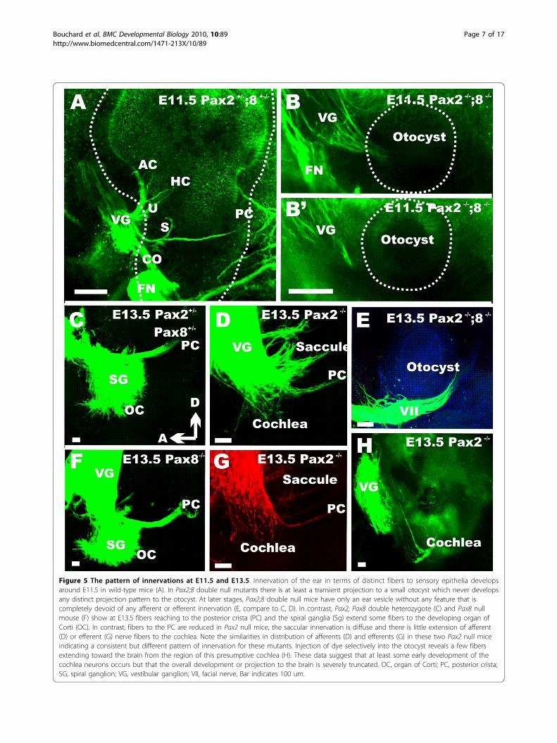
Figure 5 The pattern of innervations at E11.5 and E13.5. Innervation of the ear in terms of distinct fibers to sensory epithelia developsaround E11.5 in wild-type mice (A). In Pax2;8 double null mutants there is at least a transient projection to a small otocyst which never developsany distinct projection pattern to the otocyst. At later stages, Pax2;8 double null mice have only an ear vesicle without any feature that iscompletely devoid of any afferent or efferent innervation (E, compare to C, D). In contrast, Pax2; Pax8 double heterozygote (C) and Pax8 nullmouse (F) show at E13.5 fibers reaching to the posterior crista (PC) and the spiral ganglia (Sg) extend some fibers to the developing organ ofCorti (OC). In contrast, fibers to the PC are reduced in Pax2 null mice, the saccular innervation is diffuse and there is little extension of afferent(D) or efferent (G) nerve fibers to the cochlea. Note the similarities in distribution of afferents (D) and efferents (G) in these two Pax2 null miceindicating a consistent but different pattern of innervation for these mutants. Injection of dye selectively into the otocyst reveals a few fibersextending toward the brain from the region of this presumptive cochlea (H). These data suggest that at least some early development of thecochlea neurons occurs but that the overall development or projection to the brain is severely truncated. OC, organ of Corti; PC, posterior crista;SG, spiral ganglion; VG, vestibular ganglion; VII, facial nerve, Bar indicates 100 um.
Bouchard et al. BMC Developmental Biology 2010, 10:89http://www.biomedcentral.com/1471-213X/10/89
Page 7 of 17

As expected from the small vesicle we found in Pax2;8double null mice at E10.5 and 11.5 (Figs. 2B, 3D, 5B)only a small vesicle was present at E13.5 (Fig. 5H). Incontrast to the earlier stages where we detected someganglion cells with fibers extending toward the ear (Fig.5B), no nerve fibers were found extending to the smallvesicle at E13.5 (Fig. 5H). Considering that Pax2;8 dou-ble mutant ears showed Jag1 and Eya1 expression indouble mutant inner ears (Fig. 2), this suggests a rapidloss of sensory neurons likely by apoptosis as in Pax2null mice (Fig. 3H). Combined these data suggest that inaddition to promote otic vesicle formation, Pax2 andPax8 combined are needed for neurosensory systemdifferentiation and/or maintenance.We next investigated the innervation of the ear in
later stage Pax2 mutant embryos. Starting at E14.5 (Fig.6A,B) we found prominent differences in the develop-ment of the cochlea spiral ganglion. In wild-type thespiral ganglion formed an elongated extension curvedalong the cochlear duct whereas the spiral ganglionformed a tear drop addition to the vestibular ganglionwith little longitudinal extension. The difference intopology was particularly obvious when comparing thetopology of nerve fibers projecting to the posteriorcanal. These were reduced in Pax2 null mice (Fig. 6B)and emanated from vestibular neurons mixed to whatmight be part of the spiral ganglion. In contrast to thesemassive differences, the innervation to the anterior canalcrista and utricle were only different in terms of sizecompared to wild-type littermates (Fig. 6C,D). The sac-cule consisted of two small patches of innervation butalso numerous fibers radiating around the medial wallof the otocyst, including a few fibers extending towardthe posterior canal crista. Even compared to the youngerwild-type ears (Fig. 5) this fiber tract was reduced. Thisdata suggests that spiral neurons form but never segre-gate from the inferior vestibular ganglion.At an even later stage (E15.5) we found many fibers
radiating from the tear-drop shaped spiral gangliontoward the cochlear sack (Fig. 6G,H). Investigating var-ious focal planes following dye injection (Fig. 7I-L), wefound that spiral ganglia were clearly distinct from infer-ior vestibular ganglia; the latter projected to the sacculeand the posterior canal at this stage. As in youngerstages, the fibers to the posterior canal were disorga-nized and few. Together these data show that Pax2 nullmutant embryos can form some cochlear sensory neu-rons which extend projections to the brain. However,these never develop to a typical spiral ganglion.
Utricular, saccular and cochlear hair cells are continuousWe next investigated at E18.5 the pattern of innervationand the distribution of nerve fibers using anti-tubulin andanti-Myo VII to highlight nerve fibers and hair cells,
respectively. As was the case with the lipophilic dye tracingexperiments, the pattern of innervation of the anterior andhorizontal canal crista and utricle was fairly normal inPax2 mutant embryos (Fig. 7A-C). However, there was noclear pattern of innervation suggestive of distinct bound-aries of the saccule and the fibers to the posterior canalcrista were reduced or entirely absent. After physicallyremoving the facial nerve and the vestibular ganglion, anunusual and occasionally dense innervation of the ventralcochlear expansion was found (Fig. 7E). These fibers con-centrated on the anterior aspect of the large ventral sackwithout any clear boundary recognition. Comparing sev-eral ears showed that fibers were always present but theirdistribution varied dramatically (Fig. 7,8).Hair cells were identified using Myosin VII immunocyto-
chemistry and found in all three canal cristae, but theshape of the cristae and their size deviated variably fromnormal. Hair cells of the utricle and saccule were contigu-ous and extended across the constriction separating thesuperior from the inferior part of the ear (Fig. 7C,D).Assuming that only hair cells inferior to the constrictionare cochlear hair cells, a cochlear duct, some cochlear ductinnervation, a spiral ganglion but apparently no cochlearhair cells form in these ears. This suggests that the cochleaduct forms, but with a delay and in an unusual shapeincluding the separation of all elements and a completeabsence of any organ of Corti like histogenesis of hair cells.At this stage we also studied the Pax2 (5ki/5ki) genotype
(Fig. 7F) as this allelic combination acts as a hypomorphin the embryos due to a gene dosage level betweenPax2-/- and Pax2+/- [17]. While the overall organizationand innervation of Pax2 (5ki/5ki) ears was indistinguish-able from normal mice, the detailed patterning of haircells in the cochlea showed minor aberrations such asmultiple and somewhat disorganized rows of hair cells(Fig. 7G,H) indicative of a role for Pax2 and Pax5 inhair cell patterning as also noticed in zebrafish [8].An interesting aspect of the Pax2 mutant ear pheno-
type was the dense innervations of the cochlear sack(Fig. 8), which showed a meshwork of criss-crossingfibers instead of radial bundles emanating from thespiral ganglion to reach the organ of Corti. This mesh-work of what appeared to be nerve fibers spinningaround the cochlear sack proved upon closer inspectionto be a network of fibers emanating from small, MYO7positive cells, which likely are neurons embedded intothe wall of the cochlear sack (Fig. 8C-F). These datasuggest that cochlear sensory neuron formation mayoccur but that there is no migration of those neuronsaway from the cochlear sack and they do not developany clear pattern of innervation reminiscent of the radialfibers in normal innervation (Fig. 8C and insert). It isnot clear what neurotrophic support maintains thoseneurons as neither supporting cells nor hair cells are
Bouchard et al. BMC Developmental Biology 2010, 10:89http://www.biomedcentral.com/1471-213X/10/89
Page 8 of 17

found in the area of the cochlear sack supplied by thesenerve fibers. Clearly, there is cooperation between Paxproteins and even functional equivalence as revealed bythe Pax2 5ki/5ki data. Recent work in flies has suggestedthat dPax2 has a very complicated promoter regulation[26] and it is likely that similar complexity might play arole for the differential expression regulation in mouse(and mammalian) Pax2;5;8 expression.
DiscussionPrevious work on several Pax2 mutant mice has sug-gested that Pax2 affects the vestibular system to a
variable degree [3-5]. We provide here the first detailedhistological and whole mount analysis of the Pax2 nullphenotype, which revealed new unappreciated features.Our data suggest a Mondini type dysplasia of thecochlea and vestibular system with short canals and ashortened endolymphatic sack and common crus, muchlike in Foxi1 null mice [27]. In this paper we analyzedfor the first time the innervation defects in Pax2 null,Pax2;Pax8 double null mice and show that innervationof the Pax2 null mouse ear has a delayed and truncatedformation of spiral ganglia. As previously reported [4],cell death occurs in both the sensory neurons and the
Figure 6 The pattern of innervation at E14.5 and E15.5. Wild-type animals show afferents (in green) to the growing cochlear spiral (A).Efferent fibers, shown in red, begin to form the intraganglionic spiral bundle. There is no cochlear spiral or spiral ganglion in Pax2 null mice (E).Instead there is an aggregation of neurons near the reduced fibers leading to the posterior canal crista (B), the presumed spiral ganglion (SG). Inmutants, efferent fibers reroute from the spiral ganglion to the facial nerve (B). The innervation to the anterior canal crista (AC in C, D) isreduced, but not that to the horizontal crista (C, D). The saccule shows near normal organization of fibers in the sensory epithelium (F) as well asan overshooting projection at a different focal plane (E). At E15.5, sensory neurons that can be filled with brain injections of lipophilic dyes forma tear drop arrangement of spiral sensory neurons (G, H). Multiple nerve fibers radiate away for a short distance toward the cochlear expansion(CO), but no pattern reminiscent of radial fibers in the cochlea can be observed (G, H, J, K). There is a variable reduction of nerve fibers to theanterior and posterior canal crista (D, G) and the innervation of the saccule is variable (G-L). A focal series (I-L) shows that spiral ganglionneurons are distinct in their distribution from vestibular ganglion cells as is evidenced by. AC, anterior canal crista; HC, horizontal canal crista; Co,cochlear duct/sack; Gen Ggl, geniculate ganglion; PC, posterior canal crista; VG, vestibular ganglion; IX, glossopharyngeal nerve. Bar indicates100 um.
Bouchard et al. BMC Developmental Biology 2010, 10:89http://www.biomedcentral.com/1471-213X/10/89
Page 9 of 17
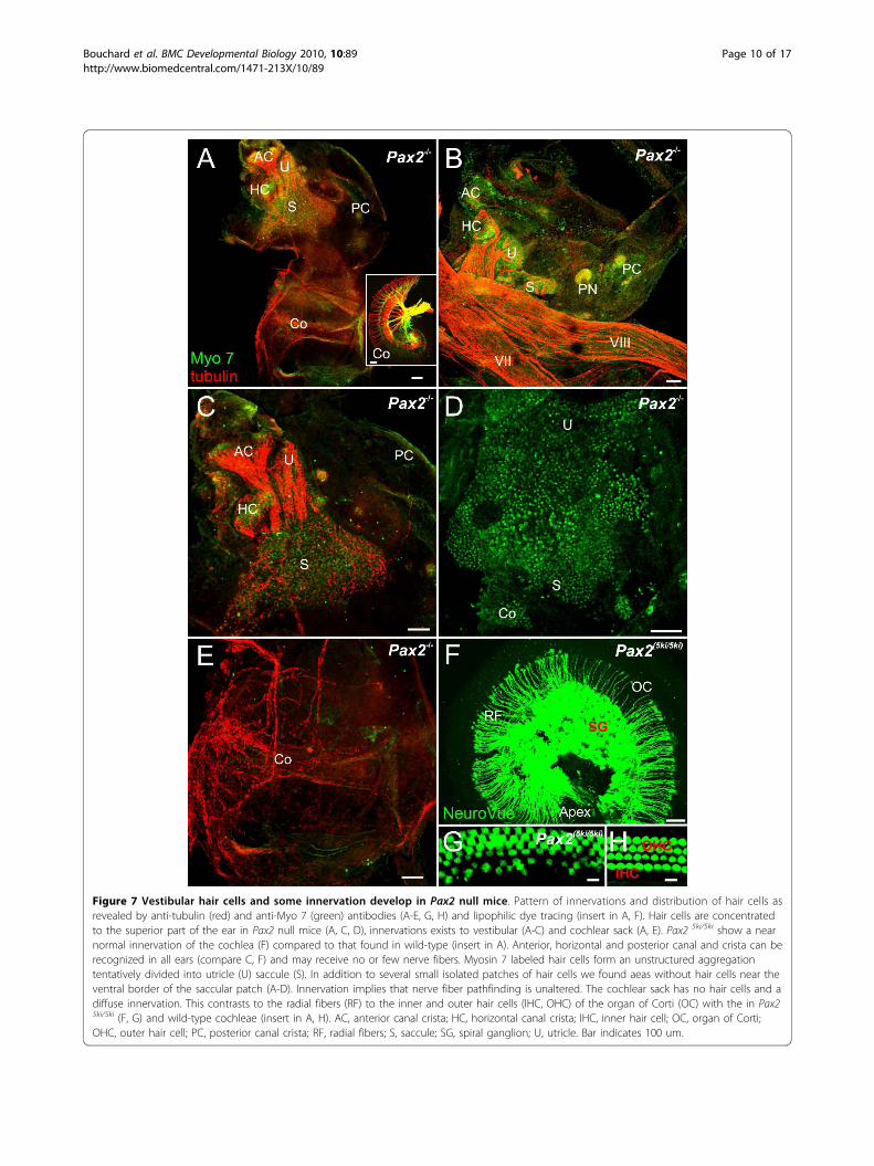
Figure 7 Vestibular hair cells and some innervation develop in Pax2 null mice. Pattern of innervations and distribution of hair cells asrevealed by anti-tubulin (red) and anti-Myo 7 (green) antibodies (A-E, G, H) and lipophilic dye tracing (insert in A, F). Hair cells are concentratedto the superior part of the ear in Pax2 null mice (A, C, D), innervations exists to vestibular (A-C) and cochlear sack (A, E). Pax2 5ki/5ki show a nearnormal innervation of the cochlea (F) compared to that found in wild-type (insert in A). Anterior, horizontal and posterior canal and crista can berecognized in all ears (compare C, F) and may receive no or few nerve fibers. Myosin 7 labeled hair cells form an unstructured aggregationtentatively divided into utricle (U) saccule (S). In addition to several small isolated patches of hair cells we found aeas without hair cells near theventral border of the saccular patch (A-D). Innervation implies that nerve fiber pathfinding is unaltered. The cochlear sack has no hair cells and adiffuse innervation. This contrasts to the radial fibers (RF) to the inner and outer hair cells (IHC, OHC) of the organ of Corti (OC) with the in Pax25ki/5ki (F, G) and wild-type cochleae (insert in A, H). AC, anterior canal crista; HC, horizontal canal crista; IHC, inner hair cell; OC, organ of Corti;OHC, outer hair cell; PC, posterior canal crista; RF, radial fibers; S, saccule; SG, spiral ganglion; U, utricle. Bar indicates 100 um.
Bouchard et al. BMC Developmental Biology 2010, 10:89http://www.biomedcentral.com/1471-213X/10/89
Page 10 of 17
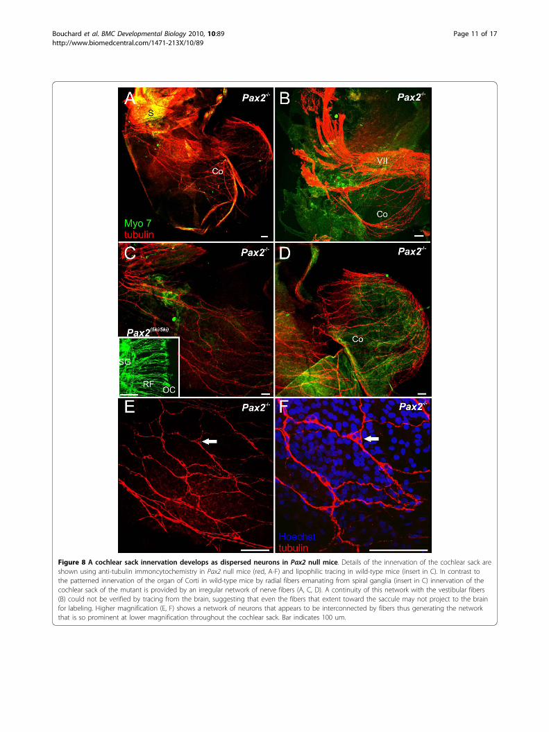
Figure 8 A cochlear sack innervation develops as dispersed neurons in Pax2 null mice. Details of the innervation of the cochlear sack areshown using anti-tubulin immoncytochemistry in Pax2 null mice (red, A-F) and lipophilic tracing in wild-type mice (insert in C). In contrast tothe patterned innervation of the organ of Corti in wild-type mice by radial fibers emanating from spiral ganglia (insert in C) innervation of thecochlear sack of the mutant is provided by an irregular network of nerve fibers (A, C, D). A continuity of this network with the vestibular fibers(B) could not be verified by tracing from the brain, suggesting that even the fibers that extent toward the saccule may not project to the brainfor labeling. Higher magnification (E, F) shows a network of neurons that appears to be interconnected by fibers thus generating the networkthat is so prominent at lower magnification throughout the cochlear sack. Bar indicates 100 um.
Bouchard et al. BMC Developmental Biology 2010, 10:89http://www.biomedcentral.com/1471-213X/10/89
Page 11 of 17

cochlear duct [3] but initial formation of sensory neu-rons is near normal as suggested previously [3]. How-ever, the Pax2 phenotype described thus far has beenoverstating the degree of loss of the cochlea which infact forms as a featureless sack that extrudes in thebrain case lumen in older mouse embryos.
Effects of Pax2; Pax8 double null on ear developmentWe show for the first time the phenotype of the Pax2;Pax8 double null mutants. These data show that Pax2;Pax8 mutant otic vesicles are much smaller than theirnormal counterpart possibly because the Pax2; Pax8double null ear has an incomplete invagination withextensive cell death of non-invaginated tissue. Thisinterpretation is consistent with recent findings inchicken that suggest that Pax2 affects cell shape and earmorphogenesis [13]. Surprisingly, even these reducedvesicles show in mice several transcription factorsalmost normally expressed, and develop initially someneurons but seems to be unable to sustain neurosensorydevelopment. Pax2;8 mutants are in that respect remi-niscent of Eya1 null mice which show also a transientformation of neurons but no indication of sensorydifferentiation [24].A limited development of only a vesicle with no evi-
dence for sustained neurosensory formation as wedescribe here in Pax2;8 double null (Fig. 1,2,4) has thusfar been reported for a small number of ear mutantssuch as the Fgf3;10 double null mice [28,29], the Gata3null mice [30] and the Eya1 null mice [18]. Previouswork has established that Fgf3 and Fgf10 together areneeded to develop an ear past the placode and otocyststage [28,29] possibly by guiding neuronal differentiationof the otic ectoderm through suppression of BMP sig-naling [16,31]. Consistent with this interpretation is thatPax2;8 are regulated by Fgf’s and appear to be down-stream to the Fgf activation [32]. Indeed, some reductionof Pax2 expression has been noted in Fgf3;10 null ears[28]. The data on Pax2;8 double null mice clearly showthat the ear is truncated in development at the level ofthe otocyst (Fig. 1,2,4). Pax2;8 double mutants aredelayed in otic placode invagination, and the remainingotocyst shows near normal expression of some genes,suggesting that the otocyst is still responsive to ventra-lizing signals such as sonic hedgehog [33]. Yet, neuronaldevelopment is only transient and could not proceednormally beyond E11.5 (Fig. 5). Further work will needto demonstrated the overall reduction in crucial genesfor sensory development such as Neurog1 [34,35] andNeurod1 [36,37] in such Pax2;8 double null mice.Previous work [18] has compared the early embryonic
expression pattern of Eya1, Pax2 and Pax8 using in situhybridization and investigated the ear in Pax2, Eya1 andSix1 compound mutants with latex paintfilling. These
data suggest that Pax2 interacts with Eya1 during innerear morphogenesis, in particular in sensory areas of theinner ear. Our data, however, indicate that Eya1 is not agenetic target of Pax2;8 in this system as Pax2;8 doesnot regulate the expression of Eya1 (Fig. 2) and somePax2 expression persists in Eya1 null mice [38]. In renaldevelopment, Pax2 and Eya1 proteins are known tocomplex on the Gdnf promoter [39] and Gdnf is alsoexpressed in the ear [40,41]. Further work is needed toshow the remaining expression of Gdnf in Pax2;Eya1compound mice. Pax2;8 combined are needed for sus-tained neurosensory ear development and normal innerear morphogenesis. Pax2 and Pax5 maintain hair cellsand direct nerve fiber growth in zebrafish [8] and atleast Pax2 seems to play a similar role for the cochlea ofmice. Hence, as previously suggested based on expres-sion of a Pax2;6 fusion protein in ‘eyes’ and ‘ears’ of theboxed jelly fish [2] as well as other expression similari-ties in bHLH and other genes [42], these data clearlyestablish that Pax gene expression is essential for nor-mal neurosensory development of eyes and ears, twomajor sensory systems of the vertebrate head.
Pax8 alone has no defects on embryonic eardevelopmentPax8 is one of the first genes expressed in the develop-ing mouse ear [7,10,43] but this early placodal expres-sion is rapidly replaced by Pax2 in ear development.Lineage tracing experiments previously showed that theentire inner ear is derived from Pax8-positive cells [44].In spite of this, we could not detect any defects in sen-sorineural development in the absence of Pax8 (Fig. 3,5)suggesting that the early expression of Pax8 can befunctionally compensated by Pax2 expression. However,previous work [45] has shown that Pax8 null mice areprofoundly deaf due to athyriodism [11] because ofmaturation defects of the ribbon synapses connectinghair cells to afferents [46,47]. This defect can be nearlyfully compensated for by postnatal thyroid T(4) treat-ment [45], preferentially already in late embryos toensure normal hair cell maturation and synaptogenesis.As is apparent through the data of the Pax2;Pax8 dou-
ble null and the clear defects known in Pax2 null mice,Pax8 cannot compensate for Pax2 in late sensorineuraldevelopment but is necessary in combination with Pax2for early ear development. What exactly the function ofPax8 is in ear development and how the absence ofboth cause the incomplete invagination and at the mosttransient formation of some neurons remains to bedemonstrated. In analogy with other systems [48] weassume that Pax8 simply cannot compensate for Pax2because it is not expressed any more in the ear whenPax2 is needed, in particular in the cochlea [18]. Testingthis assumption would require to replace Pax2 by Pax8
Bouchard et al. BMC Developmental Biology 2010, 10:89http://www.biomedcentral.com/1471-213X/10/89
Page 12 of 17

which likely will result in normal ear development asalready shown for Pax5 replacement of Pax2 [17] andverified here (Fig. 8,9) As with the Pax2 5ki/5ki mice,there may be subtle differences like alignment of haircells that would allow to elucidate further the differen-tial function, if any, of Pax2 and Pax8 in ear develop-ment that is independent of the expression difference.However, it is very likely that the regulatory differencesin expression through the complex promoter systemrecently described in dPax2 [26] rather than the minorsequence differences in protein will determine the func-tion of Pax genes.
Pax2 has defects in vestibular and cochlear developmentVestibular defectsPrevious work has identified a variable phenotype in thevestibular system of Pax2 null mice [4,5] that was laterrelated to indirect effects as most vestibular developmentappeared to progress normally [3]. Our data on earlydevelopment is in line with these considerations. How-ever, there are clear defects in later development thatcannot be related to a near normal vestibular systemdevelopment. Most notably are the disorganization of thecanal cristae and alterations in the relative position of thehorizontal canal relative to the horizontal canal crista.The latter may form in the lateral wall of the superiorpart of the bipartite otic vesicle several hundred micronsaway from the horizontal canal. In agreement with two ofthe previous three papers those alterations are compara-tively minor and require serial sectioning to be detectedand thus could have been missed easily. In contrast toclaims raised based on paint fillings suggesting a fusionof the common crus with the endolymphatic duct [3],our data show that both structures form as distinct albeitclosely associated entities that most likely could not bediscriminated at the lower resolution of a whole mountedpaint filled ear (Fig. 4). In summary, the overall morpho-logical changes in the superior part of the bipartite oticvesicle are mild and it remains unclear how direct orindirect they relate to the lack of Pax2.A major alteration is the organization and separation of
the utricle, saccule and possibly cochlea sensory epithe-lium. Data on early gene expression show that Pax2expression overlaps with the Lfng expression as early asE10.5 and thus could affect at least the utricular and sac-cular maculae [3]. Indeed, some defects were noted pre-viously in the saccule but since this study terminated atE15.5 it could not fully evaluate the effects of Pax2 onhair cell distribution and differentiation. We show herean expansion and fusion of hair cell patches that, judgingfrom their pattern of innervations, belong to the utricle,saccule and possibly cochlea (Fig. 7). Major effects onhair cell formation were noted previously in zebrafish butnone have been reported in the three previous analysis of
ear development in Pax2 null mice likely either becauseno specific hair cell markers were used [4,5] or the analy-sis was not extended into older embryos were this defectis so obvious [3]. Indeed, recent data suggest a long termexpression of Pax2 in post-hatchling chicken in differen-tiated hair cells [49] and Pax2 and Pax5 are importantfor normal hair cell development in zebrafish [8] andPax2 is later expressed in hair cells [3,50].Current data suggest that non-sensory epithelium spe-
cification is as tightly regulated as sensory epitheliumspecification and this specification is needed for segrega-tion of sensory epithelia and their normal developmentrequiring a multitude of genes to achieve this segrega-tion [51-53]. As was recently shown in Lmx1a null mice[19,54], expression outside the sensory epithelia cancause major defects in overall ear development and seg-regation of sensory epithelium. Clearly, the maculae ofthe utricle and saccule are not segregated in Pax2 nullmice implying that Pax2 interacts with other genes thusfar unidentified in this sensory segregation through thespecification of non-sensory domains in the ear.Cochlear defectsOur data agree with previous work that cochlear out-growth is delayed and this delay correlates with exces-sive cell death [3] in the immature cochlear duct (Fig.3). As previously noted [4,5,18] there is the possibility ofa late formation of a cochlear sack instead of a cochlearduct. We show here that this is actually a fairly regularfeature of late development and in most instances leadsto the prolaps of this sack into the brain case, extendingboth ‘cochlear sacks’ underneath the brainstem to meetnear the midline. This prolaps seems to happen at areasof excessive cell death in the mesenchyme around thegrowing cochlear duct and may relate to the absence ofcartilage formation medial to the cochlea that normallyseparates the ear from the brain case. This expandedventral ‘cochlear sack’ shows little differentiation inPax2 null mice with sensory epithelium forming onlyadjacent to the constriction through which it expands(Figs. 3,4,7,8). This sensory epithelium is in continuationwith the saccule and it remains unclear if this is a non-segregated part of the cochlea or a part of the saccule.Likewise, normal ear development requires formation ofthe utriculo-saccular foramen to segregate the utricleform the saccule [52,55] and the formation of the ductusreunions that constricts the ear between the saccule andthe basal turn of the cochlea. Molecularly, the utriculo-saccular foramen depends in part on Otx1 [53,56]whereas the ductus reunions depends on Lmx1a [19].
Innervation defectsPrevious work has demonstrated very early overlap ofEya1 and Pax2 in early ear development [18] whereas laterstages show no expression of Pax2 in sensory neurons but
Bouchard et al. BMC Developmental Biology 2010, 10:89http://www.biomedcentral.com/1471-213X/10/89
Page 13 of 17

in hair cells [49,50]. Pax8 and Pax2 reporter expressionshow extensive expression in all sensory neurons as wellas sensory epithelia [57,58] suggesting that at least at sometime during development there is expression of either ofthese Pax genes in neurosensory precursors. Our datashow little defects in the innervations of several vestibularepithelia of Pax2 mutant ears, except for the saccule andposterior canal (Fig. 5, 6,7,8). Whether these defects are aconsequence of the patchy absence of hair cells in the con-fluent utriculo-saccular epithelium or reflect a primarydefect in these vestibular sensory epithelia remainsunclear. Disorganized innervations of the saccule couldrelate to alterations in numerous genes now identified inneuronal differentiation, in particular Neurod1 [37].Most interesting is the pattern of innervation of the
‘cochlear sack’ and the distribution of sensory neuronsassociated with this innervation. There was a clear indi-cation of spiral ganglion formation near the inferior ves-tibular ganglion innervating the saccule and posteriorcanal which radiated a few fibers to the cochlear sack(Figs. 5,6). In contrast to this tracing data, later examina-tion of the innervations using tubulin as a neuronal mar-ker showed a dense meshwork of innervations inparticular of the medial part of the ‘cochlear sack’ (Figs.7, 8), suggesting that the distributed sensory neuronsmay not project to the brain and can thus not be filled bydye tracing (Figs. 5, 6). Interestingly these fibers ema-nated from neurons embedded singly or in clusters inthis network (Fig. 8) suggesting that sensory neuronsform and survive. At E18.5 sensory neurons that are notin contact with neurotrophin releasing sensory epitheliaare either dead or dying [59]. How these neurons survivebeyond the normal loss induced by the absence of neuro-trophins emitted from sensory epithelia remains unclear.However, given that Ntf3 is the main supporting neuro-trophin for spiral neurons [48,60] and is expressed insupporting cells it may be possible that a certain level ofdifferentiation of the organ of Corti supporting cellstakes place in the absence of any hair cells and that thisdifferentiation is sufficient to express enough Ntf3 tosupport neuronal survival. Similar data are known forAtoh1 null mice in which a targeted innervation of thecochlea develops in the absence of hair cell differentia-tion and enough neurotrophin is expressed to maintaininnervation at least until birth [21].This diffuse formation of what appears to be spiral
sensory neurons distributed within the wall of the‘cochlear sack’ needs to be further investigated in termsof their birthdate using BrdU [61] and survival throughneurotrophic factors.
ConclusionOur data demonstrate that all three Pax2;5;8 genes cansignal redundantly in ear development with their
respective function depending on spatio-temporalexpression pattern. Pax2 can compensate for Pax8, butloss of both results in otocyst formation with only tran-sient expression of sensory markers and development ofsome neurons. Likewise, a Pax5 minigene knocked intoPax2 can nearly completely compensate for Pax2. Wealso show that Pax2 null does not affect the early neuro-genesis, but results in the formation of a cochlear sackwith rather aberrant innervation patterns later indevelopment.
MethodsMiceThe Pax2 and Pax8 null mice have been previouslydescribed [14,17] and these lines were bred tohomozygosity as single or double knockout mice gener-ating the expected frequency of mutant embryos inMendelian ratios. Pax2;8 compound female carriershave defects in their genital tracks and only a small por-tion are fertile thus leaving us with a very small numberof Pax2;8 double null mutants to work with. ThePax25ki/5ki mice, in which the Pax2 gene was replacedby a Pax5 minigene, were also previously described andbred according to those description [17].Embryos were collected from timed pregnant females
at E8, 9.5, 10.5, 11.5, E13.5, E14.5, E15.5 and E18.5counting noon of the day the vaginal plug was found asE0.5. Pregnant mothers were anesthetized with a lethaldose of Avertin (1.25% of 2.2.2-tribromoethanol at adose of 0.025 ml/g of body weight). Embryos were per-fused with 4% paraformaldehyde (PFA) in 0.1 M phos-phate buffer (pH 7.4). Heads were isolated and fixed in4% PFA for further analysis. Offspring was genotyped byPCR analysis of tail DNA as previously described [17].All animal procedures were approved by the Universityof Iowa animal care committee according to IACUCguidelines for use of laboratory animals in biologicalresearch (ACURF #0804066).
X-gal stainingHeads of mice perfused with 4% PFA were hemisected.Ears were dissected and then briefly washed with 0.1 Mphosphate buffer. The samples were stained in a solutioncontaining 0.1 M phosphate buffer, 0.01% deoxycholicacid, 0.02% NP40, 2 mM magnesium chloride, 5 mMpotassium ferricyanide, 5 mM potassium ferrocyanideand 0.1 mg/ml X-gal (5-bromo-4-chloro-3-indolyl-b-D-galactoside) for about 24 hours at room temperature [62].
In situ hybridizationIn situ hybridization was performed as described [63]using the RNA probe labeled with digoxigenin. Thecover slipped slides were viewed in a Nikon Eclipse 800
Bouchard et al. BMC Developmental Biology 2010, 10:89http://www.biomedcentral.com/1471-213X/10/89
Page 14 of 17

microscope and images were captured with Image-Pro software.
ImmunofluorescenceFor immunofluorescence staining, the ears were dehy-drated in 100% ethanol overnight and rehydrated andblocked with 0.25% normal goat serum in PBS contain-ing 0.01% Triton-X-100 for 1 hour. Then the primaryantibodies for Tubulin (Cell Signaling Technology) andMyo VII (Myosin VIIa, Proteus Biosciences) were usedin dilutions of 1:800 and 1:200 respectively, and incu-bated for 48 hours at 4°C. After several washes withPBS, corresponding secondary antibodies (1:500) (Alexafluor molecular probe 647 or 532; Invitrogen) wereadded and incubated overnight at 4°C. The ears werewashed with PBS and mounted in glycerol and imageswere taken with a Leica TCS SP5 confocal microscope.
Lipophilic dye tracingThe heads of the mice were cut sagittally along the mid-line and different colored NeuroVue dyes were insertedto label the afferent and efferent fibers from and to theinner ear. Lipophilic dye-soaked filter strips [25] wereinserted into the alar plate of the brainstem to label theeighth cranial nerve afferent fibers. Efferent fibers werelabeled by applying dye into the olivo-cochlear efferentbundle as it crosses the floor plate in rhombomere 4.Half heads were kept in 60°C oven for about 3-7 daysdepending on the age of the mice for proper diffusion.In the E14.5 mice the dyes were injected in rhombo-mere 2, 5 and 7 to label the afferent and efferent fibers.Then the ears, vestibular ganglia and brains were dis-sected out for analysis and images were taken by LeicaTCS SP5 confocal microscope.
Additional material
Additional file 1: Detail of cochlear and saccule. The highermagnified images show the formation of sensory epithelia with hair cellsthrough (C) and on either side of the closed foramen though which theprolapsed cochlear sack extrudes. Co, cochlear sack; S, saccule. Barindicates 100 um.
Additional file 2: Detail of canal organization. All canal cristae can beidentified, but the posterior canal crista is drastically reduced (A). Notethat the horizontal crista is, like the anterior crista, very close to theutricle on the lateral wall of the utricular recess. However, this crista isnot close to the horizontal canal opening. AC, anterior canal crista; HC,horizontal canal cirista; PC, posterior canal crista; U, utricle. Bar indicates100 um.
AcknowledgementsThis work was supported by a NIH grant (R01 DC 005590) to B.F, a grantfrom the Canadian Institutes for Health Reseach (CIHR, MOP-84470) toM. Bouchard and a NIH grant (R01 DC005824) to P.X. M. Bouchard holds aCanada Research Chair in Developmental Genetics. We thank J. Kersigo forexpert assistance.
Author details1Biochemistry Department, Goodman Cancer Centre, McGill University,Quebec, Canada. 2Department of Biology, College of Liberal Arts andSciences, 143 Biology Building, Iowa City, IA 52242-1324, USA. 3ResearchInstitute of Molecular Pathology, Vienna Biocenter, Vienna, Austria.4Department of Genetics and Genomic Sciences, Mount Sinai School ofMedicine of New York University, New York, NY 10029, USA.
Authors’ contributionsAll investigators planned the experiments, MBo bred and collected theembryos, BF did the tract tracing and imaging, DdC did the histology andfigure assembly; these three authors shared the majority of the writing, PXand MBu read and commented on the manuscript. All authors read andapproved the final version.
Received: 20 May 2010 Accepted: 20 August 2010Published: 20 August 2010
References1. Srivastava M, Simakov O, Chapman J, Fahey B, Gauthier ME, Mitros T,
Richards GS, Conaco C, Dacre M, Hellsten U, et al: The Amphimedonqueenslandica genome and the evolution of animal complexity. Nature2010, 466(7307):720-726.
2. Kozmik Z, Daube M, Frei E, Norman B, Kos L, Dishaw LJ, Noll M,Piatigorsky J: Role of Pax genes in eye evolution: a cnidarian PaxB geneuniting Pax2 and Pax6 functions. Dev Cell 2003, 5(5):773-785.
3. Burton Q, Cole LK, Mulheisen M, Chang W, Wu DK: The role of Pax2 inmouse inner ear development. Dev Biol 2004, 272(1):161-175.
4. Favor J, Sandulache R, Neuhauser-Klaus A, Pretsch W, Chatterjee B, Senft E,Wurst W, Blanquet V, Grimes P, Sporle R, et al: The mouse Pax2(1Neu)mutation is identical to a human PAX2 mutation in a family with renal-coloboma syndrome and results in developmental defects of the brain,ear, eye, and kidney. Proc Natl Acad Sci USA 1996, 93(24):13870-13875.
5. Torres M, Gomez-Pardo E, Gruss P: Pax2 contributes to inner earpatterning and optic nerve trajectory. Development 1996,122(11):3381-3391.
6. Fritzsch B, Pauley S, Feng F, Matei V, Nichols DH: The evolution of thevertebrate auditory system: transformations of vestibularmechanosensory cells for sound processing is combined with newlygenerated central processing neurons., 19:1-24. International Journal ofComparative Psychology 2006, 19:1-24.
7. Pfeffer PL, Gerster T, Lun K, Brand M, Busslinger M: Characterization ofthree novel members of the zebrafish Pax2/5/8 family: dependency ofPax5 and Pax8 expression on the Pax2.1 (noi) function. Development1998, 125(16):3063-3074.
8. Kwak SJ, Vemaraju S, Moorman SJ, Zeddies D, Popper AN, Riley BB:Zebrafish pax5 regulates development of the utricular macula andvestibular function. Dev Dyn 2006, 235(11):3026-3038.
9. Hans S, Liu D, Westerfield M: Pax8 and Pax2a function synergistically inotic specification, downstream of the Foxi1 and Dlx3b transcriptionfactors. Development 2004, 131(20):5091-5102.
10. Mackereth MD, Kwak SJ, Fritz A, Riley BB: Zebrafish pax8 is required forotic placode induction and plays a redundant role with Pax2 genes inthe maintenance of the otic placode. Development 2005, 132(2):371-382.
11. Mansouri A, Chowdhury K, Gruss P: Follicular cells of the thyroid glandrequire Pax8 gene function. Nat Genet 1998, 19(1):87-90.
12. Urbanek P, Wang ZQ, Fetka I, Wagner EF, Busslinger M: Complete block ofearly B cell differentiation and altered patterning of the posteriormidbrain in mice lacking Pax5/BSAP. Cell 1994, 79(5):901-912.
13. Christophorou NA, Mende M, Lleras-Forero L, Grocott T, Streit A: Pax2coordinates epithelial morphogenesis and cell fate in the inner ear. DevBiol 2010.
14. Bouchard M, Souabni A, Mandler M, Neubuser A, Busslinger M: Nephriclineage specification by Pax2 and Pax8. Genes Dev 2002, 16(22):2958-2970.
15. Fritzsch B, Beisel KW: Keeping sensory cells and evolving neurons toconnect them to the brain: molecular conservation and novelties invertebrate ear development. Brain Behav Evol 2004, 64(3):182-197.
16. Fritzsch B, Beisel KW, Hansen LA: The molecular basis of neurosensory cellformation in ear development: a blueprint for hair cell and sensoryneuron regeneration? Bioessays 2006, 28(12):1181-1193.
Bouchard et al. BMC Developmental Biology 2010, 10:89http://www.biomedcentral.com/1471-213X/10/89
Page 15 of 17

17. Bouchard M, Pfeffer P, Busslinger M: Functional equivalence of thetranscription factors Pax2 and Pax5 in mouse development. Development2000, 127(17):3703-3713.
18. Zou D, Silvius D, Rodrigo-Blomqvist S, Enerback S, Xu PX: Eya1 regulatesthe growth of otic epithelium and interacts with Pax2 during thedevelopment of all sensory areas in the inner ear. Dev Biol 2006,298(2):430-441.
19. Nichols DH, Pauley S, Jahan I, Beisel KW, Millen KJ, Fritzsch B: Lmx1a isrequired for segregation of sensory epithelia and normal earhistogenesis and morphogenesis. Cell Tissue Res 2008, 334(3):339-358.
20. Pauley S, Wright TJ, Pirvola U, Ornitz D, Beisel K, Fritzsch B: Expression andfunction of FGF10 in mammalian inner ear development. Dev Dyn 2003,227(2):203-215.
21. Fritzsch B, Matei VA, Nichols DH, Bermingham N, Jones K, Beisel KW,Wang VY: Atoh1 null mice show directed afferent fiber growth toundifferentiated ear sensory epithelia followed by incomplete fiberretention. Dev Dyn 2005, 233(2):570-583.
22. Fritzsch B: Development of inner ear afferent connections: formingprimary neurons and connecting them to the developing sensoryepithelia. Brain Res Bull 2003, 60(5-6):423-433.
23. Maklad A, Kamel S, Wong E, Fritzsch B: Development and organization ofpolarity-specific segregation of primary vestibular afferent fibers in mice.Cell Tissue Res 2010, 340(2):303-321.
24. Zou D, Silvius D, Fritzsch B, Xu PX: Eya1 and Six1 are essential for earlysteps of sensory neurogenesis in mammalian cranial placodes.Development 2004, 131(22):5561-5572.
25. Fritzsch B, Muirhead KA, Feng F, Gray BD, Ohlsson-Wilhelm BM: Diffusionand imaging properties of three new lipophilic tracers, NeuroVueMaroon, NeuroVue Red and NeuroVue Green and their use for doubleand triple labeling of neuronal profile. Brain Res Bull 2005, 66(3):249-258.
26. Swanson CI, Evans NC, Barolo S: Structural rules and complex regulatorycircuitry constrain expression of a Notch- and EGFR-regulated eyeenhancer. Dev Cell 18(3):359-370.
27. Hulander M, Kiernan AE, Blomqvist SR, Carlsson P, Samuelsson EJ,Johansson BR, Steel KP, Enerback S: Lack of pendrin expression leads todeafness and expansion of the endolymphatic compartment in innerears of Foxi1 null mutant mice. Development 2003, 130(9):2013-2025.
28. Wright TJ, Mansour SL: Fgf3 and Fgf10 are required for mouse oticplacode induction. Development 2003, 130(15):3379-3390.
29. Alvarez Y, Alonso MT, Vendrell V, Zelarayan LC, Chamero P, Theil T, Bosl MR,Kato S, Maconochie M, Riethmacher D, et al: Requirements for FGF3 andFGF10 during inner ear formation. Development 2003, 130(25):6329-6338.
30. Karis A, Pata I, van Doorninck JH, Grosveld F, de Zeeuw CI, de Caprona D,Fritzsch B: Transcription factor GATA-3 alters pathway selection ofolivocochlear neurons and affects morphogenesis of the ear. J CompNeurol 2001, 429(4):615-630.
31. Rogers CD, Moody SA, Casey ES: Neural induction and factors thatstabilize a neural fate. Birth Defects Res C Embryo Today 2009,87(3):249-262.
32. Goode DK, Elgar G: The PAX258 gene subfamily: a comparativeperspective. Dev Dyn 2009, 238(12):2951-2974.
33. Riccomagno MM, Martinu L, Mulheisen M, Wu DK, Epstein DJ: Specificationof the mammalian cochlea is dependent on Sonic hedgehog. Genes Dev2002, 16(18):2365-2378.
34. Ma Q, Anderson DJ, Fritzsch B: Neurogenin 1 null mutant ears developfewer, morphologically normal hair cells in smaller sensory epitheliadevoid of innervation. J Assoc Res Otolaryngol 2000, 1(2):129-143.
35. Ma Q, Chen Z, del Barco Barrantes I, de la Pompa JL, Anderson DJ:neurogenin1 is essential for the determination of neuronal precursorsfor proximal cranial sensory ganglia. Neuron 1998, 20(3):469-482.
36. Kim WY, Fritzsch B, Serls A, Bakel LA, Huang EJ, Reichardt LF, Barth DS,Lee JE: NeuroD-null mice are deaf due to a severe loss of the inner earsensory neurons during development. Development 2001, 128(3):417-426.
37. Jahan I, Kersigo J, Pan N, Fritzsch B: Neurod1 regulates survival andformation of connections in mouse ear and brain. Cell Tissue Res 2010,34:95-110.
38. Xu PX, Adams J, Peters H, Brown MC, Heaney S, Maas R: Eya1-deficientmice lack ears and kidneys and show abnormal apoptosis of organprimordia. Nat Genet 1999, 23(1):113-117.
39. Gong KQ, Yallowitz AR, Sun H, Dressler GR, Wellik DM: A Hox-Eya-Paxcomplex regulates early kidney developmental gene expression. Mol CellBiol 2007, 27(21):7661-7668.
40. Hashino E, Dolnick RY, Cohan CS: Developing vestibular ganglion neuronsswitch trophic sensitivity from BDNF to GDNF after target innervation. JNeurobiol 1999, 38(3):414-427.
41. Stover T, Gong TL, Cho Y, Altschuler RA, Lomax MI: Expression of theGDNF family members and their receptors in the mature rat cochlea.Brain Res Mol Brain Res 2000, 76(1):25-35.
42. Fritzsch B, Piatigorsky J: Ancestry of photic and mechanic sensation?Science 2005, 308(5725):1113-1114, author reply 1113-1114.
43. Abello G, Khatri S, Radosevic M, Scotting PJ, Giraldez F, Alsina B:Independent regulation of Sox3 and Lmx1b by FGF and BMP signalinginfluences the neurogenic and non-neurogenic domains in the chickotic placode. Dev Biol 339(1):166-178.
44. Grote D, Souabni A, Busslinger M, Bouchard M: Pax 2/8-regulated Gata 3expression is necessary for morphogenesis and guidance of the nephricduct in the developing kidney. Development 2006, 133(1):53-61.
45. Christ S, Biebel UW, Hoidis S, Friedrichsen S, Bauer K, Smolders JW: Hearingloss in athyroid pax8 knockout mice and effects of thyroxinesubstitution. Audiol Neurootol 2004, 9(2):88-106.
46. Sendin G, Bulankina AV, Riedel D, Moser T: Maturation of ribbon synapsesin hair cells is driven by thyroid hormone. J Neurosci 2007,27(12):3163-3173.
47. Brandt N, Kuhn S, Munkner S, Braig C, Winter H, Blin N, Vonthein R,Knipper M, Engel J: Thyroid hormone deficiency affects postnatal spikingactivity and expression of Ca2+ and K+ channels in rodent inner haircells. J Neurosci 2007, 27(12):3174-3186.
48. Farinas I, Jones KR, Tessarollo L, Vigers AJ, Huang E, Kirstein M, deCaprona DC, Coppola V, Backus C, Reichardt LF, et al: Spatial shaping ofcochlear innervation by temporally regulated neurotrophin expression. JNeurosci 2001, 21(16):6170-6180.
49. Warchol ME, Richardson GP: Expression of the Pax2 transcription factor isassociated with vestibular phenotype in the avian inner ear. DevNeurobiol 2009, 69(2-3):191-202.
50. Lawoko-Kerali G, Rivolta MN, Holley M: Expression of the transcriptionfactors GATA3 and Pax2 during development of the mammalian innerear. J Comp Neurol 2002, 442(4):378-391.
51. Daudet N, Lewis J: Two contrasting roles for Notch activity in chick innerear development: specification of prosensory patches and lateralinhibition of hair-cell differentiation. Development 2005, 132(3):541-551.
52. Fritzsch B, Beisel KW, Jones K, Farinas I, Maklad A, Lee J, Reichardt LF:Development and evolution of inner ear sensory epithelia and theirinnervation. J Neurobiol 2002, 53(2):143-156.
53. Bok J, Chang W, Wu DK: Patterning and morphogenesis of the vertebrateinner ear. Int J Dev Biol 2007, 51(6-7):521-533.
54. Koo SK, Hill JK, Hwang CH, Lin ZS, Millen KJ, Wu DK: Lmx1a maintainsproper neurogenic, sensory, and non-sensory domains in themammalian inner ear. Dev Biol 2009, 333(1):14-25.
55. Morsli H, Choo D, Ryan A, Johnson R, Wu DK: Development of the mouseinner ear and origin of its sensory organs. J Neurosci 1998, 18(9):3327-3335.
56. Fritzsch B, Signore M, Simeone A: Otx1 null mutant mice show partialsegregation of sensory epithelia comparable to lamprey ears. Dev GenesEvol 2001, 211(8-9):388-396.
57. Bouchard M, Souabni A, Busslinger M: Tissue-specific expression of crerecombinase from the Pax8 locus. Genesis 2004, 38(3):105-109.
58. Ohyama T, Groves AK: Generation of Pax2-Cre mice by modification of aPax2 bacterial artificial chromosome. Genesis 2004, 38(4):195-199.
59. Fritzsch B, Tessarollo L, Coppola E, Reichardt LF: Neurotrophins in the ear:their roles in sensory neuron survival and fiber guidance. Prog Brain Res2004, 146:265-278.
60. Fritzsch B, Farinas I, Reichardt LF: Lack of neurotrophin 3 causes losses ofboth classes of spiral ganglion neurons in the cochlea in a region-specific fashion. J Neurosci 1997, 17(16):6213-6225.
61. Matei V, Pauley S, Kaing S, Rowitch D, Beisel KW, Morris K, Feng F, Jones K,Lee J, Fritzsch B: Smaller inner ear sensory epithelia in Neurog1 null miceare related to earlier hair cell cycle exit. Dev Dyn 2005, 234(3):633-650.
62. Matei VA, Feng F, Pauley S, Beisel KW, Nichols MG, Fritzsch B: Near-infraredlaser illumination transforms the fluorescence absorbing X-Gal reaction
Bouchard et al. BMC Developmental Biology 2010, 10:89http://www.biomedcentral.com/1471-213X/10/89
Page 16 of 17
product BCI into a transparent, yet brightly fluorescent substance. BrainRes Bull 2006, 70(1):33-43.
63. Henrique D, Adam J, Myat A, Chitnis A, Lewis J, Ish-Horowicz D: Expressionof a Delta homologue in prospective neurons in the chick. Nature 1995,375(6534):787-790.
doi:10.1186/1471-213X-10-89Cite this article as: Bouchard et al.: Pax2 and Pax8 cooperate in mouseinner ear morphogenesis and innervation. BMC Developmental Biology2010 10:89.
Submit your next manuscript to BioMed Centraland take full advantage of:
• Convenient online submission
• Thorough peer review
• No space constraints or color figure charges
• Immediate publication on acceptance
• Inclusion in PubMed, CAS, Scopus and Google Scholar
• Research which is freely available for redistribution
Submit your manuscript at www.biomedcentral.com/submit
Bouchard et al. BMC Developmental Biology 2010, 10:89http://www.biomedcentral.com/1471-213X/10/89
Page 17 of 17