Pathway Analysis Report - Gene - HSLSfiles.hsls.pitt.edu/files/molbio/Reactome_report.pdfCollagen...
73
Pathway Analysis Report Gene This report contains the pathway analysis results for the submitted sample 'Gene'. Analysis was per- formed against Reactome version 68 on 10/04/2019. The web link to these results is: https://reactome.org/PathwayBrowser/#/ANALYSIS=MjAxOTA0MTAxMDU2NDZfODQzNg%3D%3D Please keep in mind that analysis results are temporarily stored on our server. The storage period depends on usage of the service but is at least 7 days. As a result, please note that this URL is only valid for a limited time period and it might have expired.
Transcript of Pathway Analysis Report - Gene - HSLSfiles.hsls.pitt.edu/files/molbio/Reactome_report.pdfCollagen...

Pathway Analysis Report
Gene
This report contains the pathway analysis results for the submitted sample 'Gene'. Analysis was per-formed against Reactome version 68 on 10/04/2019. The web link to these results is:
https://reactome.org/PathwayBrowser/#/ANALYSIS=MjAxOTA0MTAxMDU2NDZfODQzNg%3D%3D
Please keep in mind that analysis results are temporarily stored on our server. The storage period depends on usage of the service but is at least 7 days. As a result, please note that this URL is only
valid for a limited time period and it might have expired.

Table of Contents
Introduction1.
Properties2.
Genome-wide overview3.
Most significant pathways4.
Pathways details5.
Identifiers found6.
Identifiers not found7.
https://reactome.org Page 2
1. Introduction
Reactome is a curated database of pathways and reactions in human biology. Reactions can be con-sidered as pathway 'steps'. Reactome defines a 'reaction' as any event in biology that changes the state of a biological molecule. Binding, activation, translocation, degradation and classical bio-chemical events involving a catalyst are all reactions. Information in the database is authored by expert biologists, entered and maintained by Reactome’s team of curators and editorial staff. Re-actome content frequently cross-references other resources e.g. NCBI, Ensembl, UniProt, KEGG (Gene and Compound), ChEBI, PubMed and GO. Orthologous reactions inferred from annotation for Homo sapiens are available for 17 non-human species including mouse, rat, chicken, puffer fish, worm, fly, yeast, rice, and Arabidopsis. Pathways are represented by simple diagrams follow-ing an SBGN-like format.
Reactome's annotated data describe reactions possible if all annotated proteins and small mo-lecules were present and active simultaneously in a cell. By overlaying an experimental dataset on these annotations, a user can perform a pathway over-representation analysis. By overlaying quantitative expression data or time series, a user can visualize the extent of change in affected pathways and its progression. A binomial test is used to calculate the probability shown for each result, and the p-values are corrected for the multiple testing (Benjamini–Hochberg procedure) that arises from evaluating the submitted list of identifiers against every pathway.
To learn more about our Pathway Analysis, please have a look at our relevant publications:
Fabregat A, Sidiropoulos K, Garapati P, Gillespie M, Hausmann K, Haw R, … D’Eustachio P (2016). The reactome pathway knowledgebase. Nucleic Acids Research, 44(D1), D481–D487.
https://doi.org/10.1093/nar/gkv1351.
Fabregat A, Sidiropoulos K, Viteri G, Forner O, Marin-Garcia P, Arnau V, … Hermjakob H (2017). Reactome pathway analysis: a high-performance in-memory approach. BMC Bioinformatics, 18.

https://reactome.org Page 3
2. Properties
This is an expression analysis: The numbers are used to produce a scaled coloured overlay over Reactome pathway diagrams, as a means to visualize relative expression levels. Note that the numeric values do not have to be expression data, for instance by using gene association
scores the same analysis can be used to visualize genotyping results.
•
337 out of 461 identifiers in the sample were found in Reactome, where 1601 pathways were hit by at least one of them.
•
All non-human identifiers have been converted to their human equivalent. •
IntAct interactors were included to increase the analysis background. This greatly increases the size of Reactome pathways, which maximises the chances of matching your submitted identifiers to the expanded pathway, but will include interactors that have not undergone manual curation by Reactome and may include interactors that have no biological signific-ance, or unexplained relevance.
•
This report is filtered to show only results for species 'Homo sapiens' and resource 'all re-sources'.
•
The unique ID for this analysis (token) is MjAxOTA0MTAxMDU2NDZfODQzNg%3D%3D. This ID is valid for at least 7 days in Reactome’s server. Use it to access Reactome services with your data.
•
https://reactome.org Page 4
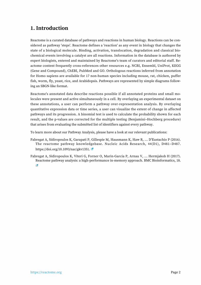
3. Genome-wide overview
Cell CycleDNA Replication
Musclecontraction
Disease
Circadian ClockDNA Repair
Geneexpression (Transcription)
Organelle biogenesisand maintenance
Vesicle-mediatedtransport
DevelopmentalBiology Signal
Transduction
Reproduction
Extracellularmatrix organization
Immune SystemMetabolism
of RNA
Cellular responsesto external stimuli
Proteinlocalization
Hemostasis
Neuronal System
ProgrammedCell Death
Digestionand absorption
Metabolism Transport ofsmall molecules
Chromatinorganization
Cell-Cellcommunication
Metabolismof proteins
9.98E0
-5.88E0
This figure shows a genome-wide overview of the results of your pathway analysis. Reactome path-ways are arranged in a hierarchy. The center of each of the circular "bursts" is the root of one top-
level pathway, for example "DNA Repair". Each step away from the center represents the next level lower in the pathway hierarchy. The color code denotes over-representation of that pathway in your input dataset. Light grey signifies pathways which are not significantly over-represented.

https://reactome.org Page 5
4. Most significant pathways
The following table shows the 25 most relevant pathways sorted by p-value.
Entities ReactionsPathway name
found ratio p-value FDR* found ratio
Nuclear Receptor transcription pathway
13 / 101 0.005 1.05e-04 0.184 2 / 2 1.65e-04
Interleukin-4 and Interleukin-13 signaling
30 / 365 0.018 4.71e-04 0.412 16 / 46 0.004
Laminin interactions 6 / 33 0.002 0.001 0.786 14 / 15 0.001
Antagonism of Activin by Follistatin 6 / 14 6.99e-04 0.002 0.786 2 / 2 1.65e-04
Anchoring fibril formation 4 / 15 7.49e-04 0.002 0.806 4 / 4 3.30e-04
Defective B3GALTL causes Peters-plus syndrome (PpS)
6 / 39 0.002 0.003 0.806 1 / 1 8.25e-05
Metallothioneins bind metals 6 / 39 0.002 0.003 0.806 14 / 27 0.002
O-glycosylation of TSR domain-containing proteins
6 / 41 0.002 0.004 0.894 2 / 2 1.65e-04
Collagen chain trimerization 6 / 44 0.002 0.006 0.966 5 / 28 0.002
Response to metal ions 6 / 47 0.002 0.008 0.966 14 / 31 0.003
Regulation of FZD by ubiquitination 5 / 34 0.002 0.008 0.966 6 / 6 4.95e-04
Microtubule-dependent trafficking of connexons from Golgi to the plasma membrane
4 / 22 0.001 0.009 0.966 1 / 2 1.65e-04
Transport of connexons to the plasma membrane
4 / 23 0.001 0.01 0.966 1 / 3 2.48e-04
Collagen degradation 7 / 76 0.004 0.022 0.966 12 / 34 0.003
FOXO-mediated transcription of oxidative stress, metabolic and neuronal genes
14 / 132 0.007 0.023 0.966 32 / 34 0.003
Diseases associated with O-glycosylation of proteins
7 / 79 0.004 0.026 0.966 2 / 9 7.43e-04
Post-chaperonin tubulin folding pathway
4 / 35 0.002 0.039 0.966 9 / 9 7.43e-04
O-linked glycosylation 12 / 170 0.008 0.047 0.966 5 / 28 0.002
Activation of AMPK downstream of NMDARs
7 / 74 0.004 0.054 0.966 3 / 3 2.48e-04
Assembly of collagen fibrils and other multimeric structures
6 / 74 0.004 0.054 0.966 23 / 26 0.002
Crosslinking of collagen fibrils 3 / 24 0.001 0.057 0.966 12 / 13 0.001
Molecules associated with elastic fibres
4 / 40 0.002 0.058 0.966 6 / 10 8.25e-04
TGFBR2 MSI Frameshift Mutants in Cancer
1 / 2 9.98e-05 0.07 0.966 1 / 1 8.25e-05

https://reactome.org Page 6
Entities ReactionsPathway name
found ratio p-value FDR* found ratio
VEGF ligand-receptor interactions 6 / 27 0.001 0.075 0.966 3 / 4 3.30e-04
VEGF binds to VEGFR leading to receptor dimerization
6 / 27 0.001 0.075 0.966 2 / 3 2.48e-04
* False Discovery Rate

https://reactome.org Page 7
5. Pathways details
For every pathway of the most significant pathways, we present its diagram, as well as a short sum-mary, its bibliography and the list of inputs found in it.
Nuclear Receptor transcription pathway (R-HSA-383280)1.
A classic example of bifunctional transcription factors is the family of Nuclear Receptor (NR) pro-teins. These are DNA-binding transcription factors that bind certain hormones, vitamins, and oth-er small, diffusible signaling molecules. The non-liganded NRs recruit specific corepressor com-plexes of the NCOR/SMRT type, to mediate transcriptional repression of the target genes to which they are bound. During signaling, ligand binding to a specific domain the NR proteins induces a conformational change that results in the exchange of the associated CoR complex, and its replace-ment by a specific coactivator complex of the TRAP / DRIP / Mediator type. These coactivator com-plexes typically nucleate around a MED1 coactivator protein that is directly bound to the NR tran-scription factor.
A general feature of the 49 human NR proteins is that in the unliganded state, they each bind dir-ectly to an NCOR corepressor protein, either NCOR1 or NCOR2 (NCOR2 was previously named "SMRT"). This NCOR protein nucleates the assembly of additional, specific corepressor proteins, depending on the cell and DNA context. The NR-NCOR interaction is mediated by a specific protein interaction domain (PID) present in the NRs that binds to specific cognate PID(s) present in the NCOR proteins. Thus, the human NRs each take part in an NR-NCOR binding reaction in the ab-sence of binding by their ligand.
A second general feature of the NR proteins is that they each contain an additional, but different PID that mediates specific binding interactions with MED1 proteins. In the ligand-bound state, NRs each take part in an NR-MED1 binding reaction to form an NR-MED1 complex. The bound MED1 then functions to nucleate the assembly of additional specific coactivator proteins, depending on the cell and DNA context, such as what specific target gene promoter they are bound to, and in what cell type.

https://reactome.org Page 8
The formation of specific MED1-containing coactivator complexes on specific NR proteins has been well-characterized for a number of the human NR proteins (see Table 1 in (Bourbon, 2004)). For ex-ample, binding of thyroid hormone (TH) to the human TH Receptor (THRA or THRB) was found to result in the recruitment of a specific complex of Thyroid Receptor Associated Proteins - the TRAP coactivator complex - of which the TRAP220 subunit was later identified to be the Mediator 1 (MED1) homologue.
Similarly, binding of Vitamin D to the human Vitamin D3 Receptor was found to result in the re-cruitment of a specific complex of D Receptor Interacting Proteins - the DRIP coactivator complex, of which the DRIP205 subunit was later identified to be human MED1.
References
Edit history
Date Action Author
2008-11-20 Authored Caudy M
2008-12-03 Created Caudy M
2009-05-27 Edited Caudy M
2009-08-29 Reviewed Freedman LP
2019-03-13 Modified Weiser D
Entities found in this pathway (4)
Input UniProt Id #FC
PPARG P37231 1.03
NR4A1 P22736 1.34
NR4A3 Q92570-1, Q92570-2 2.11
NR3C1P04150-1, P04150-2, P04150-3, P04150-4, P04150-5, P04150-6, P04150-7, P04150-8,
P04150-9-1.01e+00
https://reactome.org Page 9
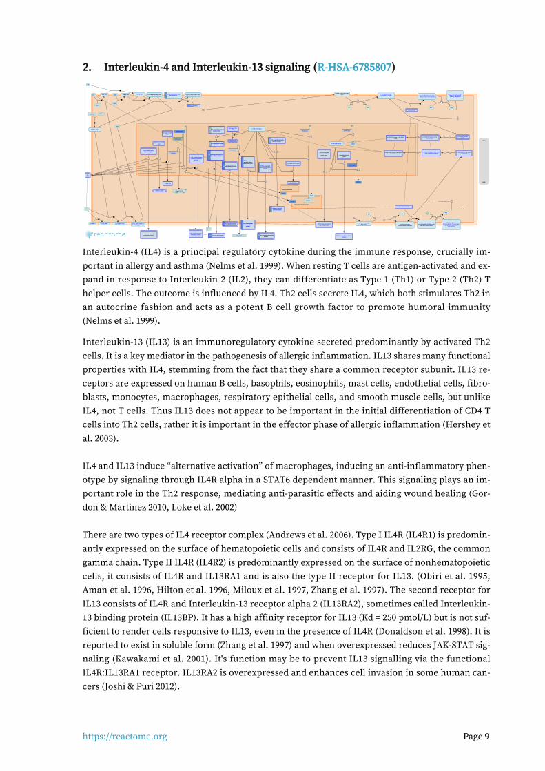
Interleukin-4 and Interleukin-13 signaling (R-HSA-6785807)2.
Interleukin-4 (IL4) is a principal regulatory cytokine during the immune response, crucially im-portant in allergy and asthma (Nelms et al. 1999). When resting T cells are antigen-activated and ex-pand in response to Interleukin-2 (IL2), they can differentiate as Type 1 (Th1) or Type 2 (Th2) T helper cells. The outcome is influenced by IL4. Th2 cells secrete IL4, which both stimulates Th2 in an autocrine fashion and acts as a potent B cell growth factor to promote humoral immunity (Nelms et al. 1999).
Interleukin-13 (IL13) is an immunoregulatory cytokine secreted predominantly by activated Th2 cells. It is a key mediator in the pathogenesis of allergic inflammation. IL13 shares many functional properties with IL4, stemming from the fact that they share a common receptor subunit. IL13 re-ceptors are expressed on human B cells, basophils, eosinophils, mast cells, endothelial cells, fibro-blasts, monocytes, macrophages, respiratory epithelial cells, and smooth muscle cells, but unlike IL4, not T cells. Thus IL13 does not appear to be important in the initial differentiation of CD4 T cells into Th2 cells, rather it is important in the effector phase of allergic inflammation (Hershey et al. 2003). IL4 and IL13 induce “alternative activation” of macrophages, inducing an anti-inflammatory phen-otype by signaling through IL4R alpha in a STAT6 dependent manner. This signaling plays an im-portant role in the Th2 response, mediating anti-parasitic effects and aiding wound healing (Gor-don & Martinez 2010, Loke et al. 2002) There are two types of IL4 receptor complex (Andrews et al. 2006). Type I IL4R (IL4R1) is predomin-antly expressed on the surface of hematopoietic cells and consists of IL4R and IL2RG, the common gamma chain. Type II IL4R (IL4R2) is predominantly expressed on the surface of nonhematopoietic cells, it consists of IL4R and IL13RA1 and is also the type II receptor for IL13. (Obiri et al. 1995, Aman et al. 1996, Hilton et al. 1996, Miloux et al. 1997, Zhang et al. 1997). The second receptor for IL13 consists of IL4R and Interleukin-13 receptor alpha 2 (IL13RA2), sometimes called Interleukin-13 binding protein (IL13BP). It has a high affinity receptor for IL13 (Kd = 250 pmol/L) but is not suf-ficient to render cells responsive to IL13, even in the presence of IL4R (Donaldson et al. 1998). It is reported to exist in soluble form (Zhang et al. 1997) and when overexpressed reduces JAK-STAT sig-naling (Kawakami et al. 2001). It's function may be to prevent IL13 signalling via the functional IL4R:IL13RA1 receptor. IL13RA2 is overexpressed and enhances cell invasion in some human can-cers (Joshi & Puri 2012).
https://reactome.org Page 10
The first step in the formation of IL4R1 (IL4:IL4R:IL2RB) is the binding of IL4 with IL4R (Hoffman et al. 1995, Shen et al. 1996, Hage et al. 1999). This is also the first step in formation of IL4R2 (IL4:IL4R:IL13RA1). After the initial binding of IL4 and IL4R, IL2RB binds (LaPorte et al. 2008), to form IL4R1. Alternatively, IL13RA1 binds, forming IL4R2. In contrast, the type II IL13 complex (IL13R2) forms with IL13 first binding to IL13RA1 followed by recruitment of IL4R (Wang et al. 2009).
Crystal structures of the IL4:IL4R:IL2RG, IL4:IL4R:IL13RA1 and IL13:IL4R:IL13RA1 complexes have been determined (LaPorte et al. 2008). Consistent with these structures, in monocytes IL4R is tyr-osine phosphorylated in response to both IL4 and IL13 (Roy et al. 2002, Gordon & Martinez 2010) while IL13RA1 phosphorylation is induced only by IL13 (Roy et al. 2002, LaPorte et al. 2008) and IL2RG phosphorylation is induced only by IL4 (Roy et al. 2002).
Both IL4 receptor complexes signal through Jak/STAT cascades. IL4R is constitutively-associated with JAK2 (Roy et al. 2002) and associates with JAK1 following binding of IL4 (Yin et al. 1994) or IL13 (Roy et al. 2002). IL2RG constitutively associates with JAK3 (Boussiotis et al. 1994, Russell et al. 1994). IL13RA1 constitutively associates with TYK2 (Umeshita-Suyama et al. 2000, Roy et al. 2002, LaPorte et al. 2008, Bhattacharjee et al. 2013).
IL4 binding to IL4R1 leads to phosphorylation of JAK1 (but not JAK2) and STAT6 activation (Takeda et al. 1994, Ratthe et al. 2007, Bhattacharjee et al. 2013).
IL13 binding increases activating tyrosine-99 phosphorylation of IL13RA1 but not that of IL2RG. IL4 binding to IL2RG leads to its tyrosine phosphorylation (Roy et al. 2002). IL13 binding to IL4R2 leads to TYK2 and JAK2 (but not JAK1) phosphorylation (Roy & Cathcart 1998, Roy et al. 2002).
Phosphorylated TYK2 binds and phosphorylates STAT6 and possibly STAT1 (Bhattacharjee et al. 2013).
A second mechanism of signal transduction activated by IL4 and IL13 leads to the insulin receptor substrate (IRS) family (Kelly-Welch et al. 2003). IL4R1 associates with insulin receptor substrate 2 and activates the PI3K/Akt and Ras/MEK/Erk pathways involved in cell proliferation, survival and translational control. IL4R2 does not associate with insulin receptor substrate 2 and consequently the PI3K/Akt and Ras/MEK/Erk pathways are not activated (Busch-Dienstfertig & González-Rodríguez 2013).
References
Nelms K, Keegan AD, Zamorano J, Ryan JJ & Paul WE (1999). The IL-4 receptor: signaling mechan-
isms and biologic functions. Annu. Rev. Immunol., 17, 701-38.
Hershey GK (2003). IL-13 receptors and signaling pathways: an evolving web. J. Allergy Clin. Im-
munol., 111, 677-90; quiz 691.
Edit history
Date Action Author
2015-07-01 Authored Jupe S
2015-07-01 Created Jupe S
2016-09-02 Edited Jupe S
2016-09-02 Reviewed Leibovich SJ
https://reactome.org Page 11
Date Action Author
2019-03-13 Modified Weiser D
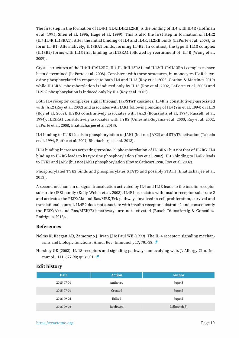
Entities found in this pathway (15)
Input UniProt Id #FC
VCAM1 P19320 -3.60e+00
IL12A P29459 -2.01e+00
LIF P15018 -2.54e+00
HPS5 P0DJI8 1.59
CEBPD P49716 1.48
FOXO1 Q12778 2.31
CDKN1A P38936 -1.03e+00
FOXO3 O43524 1.26
PIK3R1 P27986 1.62
MAOA P21397 3.28
SOCS1 O15524 -1.15e+00
HMOX1 P09601 -1.35e+00
S1PR1 P21453 -1.47e+00
MAOB P21397 1.22
VEGFA P15692 -1.23e+00
Input Ensembl Id #FC
VCAM1 ENSG00000162692 -3.60e+00
IL12A ENSG00000168811 -2.01e+00
LIF ENSG00000128342 -2.54e+00
CEBPD ENSG00000221869 1.48
FOXO1 ENSG00000150907 2.31
CDKN1A ENSG00000124762 -1.03e+00
FOXO3 ENSG00000118689 1.26
PIK3R1 ENSG00000145675 1.62
MAOA ENSG00000189221 3.28
SOCS1 ENSG00000185338 -1.15e+00
HMOX1 ENSG00000100292 -1.35e+00
S1PR1 ENSG00000170989 -1.47e+00
VEGFA ENSG00000112715 -1.23e+00
Interactors found in this pathway (3)
Input UniProt Id Interacts with #FC
PIK3R1 P27986-2, P27986 P48023, O15524 1.62
THBS1 P07996-PRO_0000035842 P16671 1.94
Input ChEBI Id Interacts with #FC
FAM20A Q96MK3 15422 1.09

https://reactome.org Page 12
Laminin interactions (R-HSA-3000157)3.
Laminins are a large family of conserved, multidomain trimeric basement membrane proteins. There are many theoretical trimer combinations but only 18 have been described (Domogatskaya et al. 2012, Miner 2008, Macdonald et al. 2010) and the existence of isoforms laminin-212 and/or laminin-222 (Durbeej et al. 2010) awaits further confirmation. The chains assemble through coiled-coil domains at their C-terminal end. Alpha chains additionally have a large C-terminal globular do-main containing five LG subdomains (LG1-5). The N termini are often referred to as the short arms. These have varying numbers of laminin-type epidermal growth factor-like (LE) repeats. Trimer as-sembly is controlled by highly specific coiled-coil interactions (Domogatskaya et al. 2012). Some laminin isoforms are modified extracellularly by proteolytic processing at the N- or C-terminal ends prior to their binding to cellular receptors or other matrix molecules (Tzu & Marinkovitch 2008).
The cell adhesion properties of laminins are mediated primarily through the alpha chain G domain to integrins, dystroglycan, Lutheran glycoprotein, or sulfated glycolipids. The N-terminal globular domains of the alpha-1 (Colognato-Pyke et al. 1995) and alpha-2 chains (Colognato et al. 1997) and globular domains VI (Nielsen & Yamada 2001) and IVa (Sasaki & Timpl 2001) of the alpha-5 chain can bind to several integrin isoforms (alpha1beta1, alpha2beta1, alpha3beta1, and alphaVbeta3), which enables cell binding at both ends of laminins with these alpha chains.
References
Domogatskaya A, Rodin S & Tryggvason K (2012). Functional diversity of laminins. Annu. Rev. Cell
Dev. Biol., 28, 523-53.
Edit history
Date Action Author
2008-05-07 Reviewed Hynes R, Humphries MJ, Yamada KM
2012-08-08 Authored Jupe S
2013-01-24 Created Jupe S
2013-08-13 Edited Jupe S

https://reactome.org Page 13
Date Action Author
2013-08-13 Reviewed Ricard-Blum S
2019-03-08 Modified Weiser D
Entities found in this pathway (6)
Input UniProt Id #FC
COL7A1 Q02388 1.22
COL4A4 P53420 2.02
COL4A1 P02462 1.33
LAMA2 P24043 2.07
ITGA2 P17301 -1.04e+00
NID1 P14543 1.54

https://reactome.org Page 14
Antagonism of Activin by Follistatin (R-HSA-2473224)4.
Cellular compartments: extracellular region.
Both Follistatin (FST) and Follistatin-like-3 (FSTL3) irreversibly bind Activin dimers and prevent Activin from interacting with its receptor (reviewed in Schneyer et al. 2004, Xia and Schneyer 2009). Though functionally similar in vitro, FST and FSTL3 do not function identically in vivo. Mice lack-ing FST die shortly after birth due to defects in muscle and bone (Matzuk et al. 1995); mice lacking FSTL3 are viable but have altered glucose metabolism (Mukherjee et al. 2007).
References
Schneyer A, Sidis Y, Xia Y, Saito S, del Re E, Lin HY & Keutmann H (2004). Differential actions of
follistatin and follistatin-like 3. Mol. Cell. Endocrinol., 225, 25-8.
Xia Y & Schneyer AL (2009). The biology of activin: recent advances in structure, regulation and
function. J. Endocrinol., 202, 1-12.
Matzuk MM, Lu N, Vogel H, Sellheyer K, Roop DR & Bradley A (1995). Multiple defects and perinat-
al death in mice deficient in follistatin. Nature, 374, 360-3.
Mukherjee A, Sidis Y, Mahan A, Raher MJ, Xia Y, Rosen ED, ... Schneyer AL (2007). FSTL3 deletion reveals roles for TGF-beta family ligands in glucose and fat homeostasis in adults. Proc. Natl.
Acad. Sci. U.S.A., 104, 1348-53.
Edit history
Date Action Author
2012-09-21 Edited May B
2012-09-21 Authored May B

https://reactome.org Page 15
Date Action Author
2012-09-22 Created May B
2012-11-14 Reviewed Chen YG
2019-03-08 Modified Weiser D
Entities found in this pathway (4)
Input UniProt Id #FC
INHBB P09529 3.64
INHBA P08476 1.07
FST P19883 -1.78e+00
FSTL3 O95633 1.83
Interactors found in this pathway (2)
Input UniProt Id Interacts with #FC
ADAM12 O43184-2 O95633 -1.84e+00
CREB5 Q02930-3 P19883 -1.98e+00

https://reactome.org Page 16
Anchoring fibril formation (R-HSA-2214320)5.
Cellular compartments: extracellular region.
Collagen VII forms anchoring fibrils, composed of antiparallel dimers that connect the dermis to the epidermis (Bruckner-Tuderman 2009, Has & Kern 2010). During fibrillogenesis, the nascent type VII procollagen molecules dimerize in an antiparallel manner. The C-propeptide is then re-moved by Bone morphogenetic protein 1 (Rattenholl et al. 2002) and the processed antiparallel di-mers laterally aggregate (Villone et al. 2008, Gordon & Hahn 2010).
References
Chung HJ & Uitto J (2010). Type VII collagen: the anchoring fibril protein at fault in dystrophic epi-
dermolysis bullosa. Dermatol Clin, 28, 93-105.
Edit history
Date Action Author
2012-04-30 Authored Jupe S
2012-04-30 Created Jupe S
2012-10-08 Reviewed Kalamajski S, Raleigh S
2012-11-12 Edited Jupe S
2012-11-19 Reviewed Ricard-Blum S
2019-03-08 Modified Weiser D
Entities found in this pathway (4)
Input UniProt Id #FC
COL7A1 Q02388 1.22
COL4A4 P53420 2.02

https://reactome.org Page 17
Input UniProt Id #FC
COL4A1 P02462 1.33
COL1A1 P02452 -1.31e+00

https://reactome.org Page 18
Defective B3GALTL causes Peters-plus syndrome (PpS) (R-HSA-5083635)6.
Diseases: eye disease, orofacial cleft.
Human beta-1,3-glucosyltransferase like protein (B3GALTL, HGNC Approved Gene Symbol: B3GLCT; MIM:610308; CAZy family GT31), localised on the ER membrane, glucosylates O-fucosylated proteins. The resultant glc-beta-1,3-fuc disaccharide modification on thrombospondin type 1 repeat (TSR1) domain-containing proteins is thought to assist in the secretion of many of these proteins from the ER lumen, and mediate an ER quality-control mechanism of folded TSRs (Vasudevan et al. 2015). Defects in B3GALTL can cause Peters plus syndrome (PpS; MIM:261540), an autosomal recessive disorder characterised by anterior eye chamber defects, short stature, delay in growth and mental developmental and cleft lip and/or palate (Heinonen & Maki 2009).
References
Heinonen TY & Maki M (2009). Peters'-plus syndrome is a congenital disorder of glycosylation caused by a defect in the beta1,3-glucosyltransferase that modifies thrombospondin type 1 re-
peats. Ann. Med., 41, 2-10.
Vasudevan D, Takeuchi H, Johar SS, Majerus E & Haltiwanger RS (2015). Peters plus syndrome
mutations disrupt a noncanonical ER quality-control mechanism. Curr. Biol., 25, 286-95.
Edit history
Date Action Author
2013-11-07 Edited Jassal B
2013-11-07 Authored Jassal B
2013-11-07 Created Jassal B
2015-12-18 Modified Jassal B
2015-12-18 Reviewed Hansen L, Joshi HJ
Entities found in this pathway (6)

https://reactome.org Page 19
Input UniProt Id #FC
ADAMTS1 Q9UHI8 2.28
ADAMTS14 Q8WXS8 -2.42e+00
ADAMTSL1 Q8N6G6 -1.05e+00
ADAMTS5 Q9UNA0 2.12
SPON1 Q9HCB6 1.8
THBS1 P07996 1.94

https://reactome.org Page 20
Metallothioneins bind metals (R-HSA-5661231)7.
Metallothioneins are highly conserved, cysteine-rich proteins that bind metals via thiolate bonds (recent general reviews in Capdevila et al. 2012, Blindauer et al. 2014, reviews of mammalian metal-lothioneins in Miles et al. 2000, Maret 2011, Vasak and Meloni 2011, Thirumoorthy et al. 2001, Bab-ula et al. 2012). Mammals contain 4 general metallothionein isoforms (MT1,2,3,4). The MT1 isoform has radiated in primates to 8 or 9 functional proteins (depending on classification of MT1L). Each mammalian metallothionein binds a total of 7 divalent metal ions in two clusters, the alpha and beta clusters. Though the functions of metallothioneins have not been fully elucidated, they appear to participate in detoxifying heavy metals (reviewed in Sharma et al. 2013), storing and transporting zinc, and redox biochemistry. Metallothioneins interact with many other cellular proteins, with most interactions involving proteins of the central nervous system (reviewed in Atrian and Capdev-ila 2013).
References
Miles AT, Hawksworth GM, Beattie JH & Rodilla V (2000). Induction, regulation, degradation, and biological significance of mammalian metallothioneins. Crit. Rev. Biochem. Mol. Biol., 35, 35-70
.
Babula P, Masarik M, Adam V, Eckschlager T, Stiborova M, Trnkova L, ... Kizek R (2012). Mammali-
an metallothioneins: properties and functions. Metallomics, 4, 739-50.
Vašák M & Meloni G (2011). Chemistry and biology of mammalian metallothioneins. J. Biol. Inorg.
Chem., 16, 1067-78.
Thirumoorthy N, Shyam Sunder A, Manisenthil Kumar K, Senthil Kumar M, Ganesh G & Chatterjee M (2011). A review of metallothionein isoforms and their role in pathophysiology. World J Surg
Oncol, 9, 54.

https://reactome.org Page 21
Maret W (2011). Redox biochemistry of mammalian metallothioneins. J. Biol. Inorg. Chem., 16,
1079-86.
Edit history
Date Action Author
2015-01-07 Edited May B
2015-01-07 Authored May B
2015-01-10 Created May B
2015-09-19 Reviewed Atrian S
2019-03-08 Modified Weiser D
Entities found in this pathway (4)
Input UniProt Id #FC
MT1X P80297 3.24
MT1M P13640, Q8N339 2.4
MT2A P02795, P04731 2.19
MT1E P04732 2.16

https://reactome.org Page 22
O-glycosylation of TSR domain-containing proteins (R-HSA-5173214)8.
The O-fucosylation of proteins containing thrombospondin type 1 repeat (TSR) domains is an im-portant PTM, regulating many biological processes such as Notch signalling, inflammation, wound healing, angiogenesis amd neoplasia (Adams & Tucker 2000, Moremen et al. 2012). Fucose addition is carried out by two protein fucosyltransferases, POFUT1 and 2. Only POFUT2 recognises the con-sensus sequence CSXS/TCG found in TSR1 domains and the fucosyl residue is attached to the hy-droxyl group of conserved serine (S) or threonine (T) residues within the consensus sequence. The modification was first demonstrated on thrombospondin 1, found in platelets and the ECM (Hof-steenge et al. 2001, Luo et al. 2006). The resulting O-fucosyl-protein is subsequently a substrate for beta-1,3-glucosyltransferase-like protein (B3GALTL), which adds a glucosyl moiety to form the rare disaccharide modification Glc-beta-1,3-Fuc. More than 60 human proteins contain TSR1 domains, The disaccharide modification has been demonstrated on a small number of these TSR1 domain-containing proteins such as thrombospondin 1 (Hofsteenge et al. 2001, Luo et al. 2006), properdin (Gonzalez de Peredo et al. 2002) and F-spondin (Gonzalez de Peredo et al. 2002). The ADAMTS (a disintegrin-like and metalloprotease domain with thrombospondin type-1 repeats) superfamily consists of 19 secreted metalloproteases (ADAMTS proteases) and at lease five ADAMTS-like pro-teins in humans. Five members of the ADAMTS superfamily have also had experimental confirma-tion of the disaccharide modification. Examples are ADAMTS13 (Ricketts et al. 2007) and ADAMTSL1 (Wang et al. 2007). In the two reactions described here, the TSR1 domain-containing proteins with similarity to the experimentally confirmed ones are included as putative substrates.
References
Adams JC & Tucker RP (2000). The thrombospondin type 1 repeat (TSR) superfamily: diverse pro-
teins with related roles in neuronal development. Dev. Dyn., 218, 280-99.
Moremen KW, Tiemeyer M & Nairn AV (2012). Vertebrate protein glycosylation: diversity, synthes-
is and function. Nat. Rev. Mol. Cell Biol., 13, 448-62.
Hofsteenge J, Huwiler KG, Macek B, Hess D, Lawler J, Mosher DF & Peter-Katalinic J (2001). C-man-nosylation and O-fucosylation of the thrombospondin type 1 module. J. Biol. Chem., 276, 6485-
98.

https://reactome.org Page 23
Luo Y, Nita-Lazar A & Haltiwanger RS (2006). Two distinct pathways for O-fucosylation of epiderm-
al growth factor-like or thrombospondin type 1 repeats. J. Biol. Chem., 281, 9385-92.
Ricketts LM, Dlugosz M, Luther KB, Haltiwanger RS & Majerus EM (2007). O-fucosylation is re-
quired for ADAMTS13 secretion. J. Biol. Chem., 282, 17014-23.
Edit history
Date Action Author
2013-11-25 Edited Jassal B
2013-11-25 Authored Jassal B
2013-11-25 Created Jassal B
2014-02-07 Reviewed D'Eustachio P
2019-03-08 Modified Weiser D
Entities found in this pathway (6)
Input UniProt Id #FC
ADAMTS1 Q9UHI8 2.28
ADAMTS14 Q8WXS8 -2.42e+00
ADAMTSL1 Q8N6G6 -1.05e+00
ADAMTS5 Q9UNA0 2.12
SPON1 Q9HCB6 1.8
THBS1 P07996 1.94

https://reactome.org Page 24
Collagen chain trimerization (R-HSA-8948216)9.
The C-propeptides of collagen propeptide chains are essential for the association of three peptide chains into a trimeric but non-helical procollagen. This initial binding event determines the com-position of the trimer, brings the individual chains into the correct register and initiates formation of the triple helix at the C-terminus, which then proceeds towards the N-terminus in a zipper-like fashion (Engel & Prockop 1991). Most early refolding studies were performed with collagen type III, which contains a disulfide linkage at the C-terminus of its triple helix (Bächinger et al. 1978, Bruck-ner et al. 1978) that acts as a permanent linker even after removal of the non-collagenous domains.
Mutations within the C-propeptides further suggest that they are crucial for the correct interaction of the three polypeptide chains and for subsequent correct folding (refs. in Boudko et al. 2011).
References
Byers PH, Click EM, Harper E & Bornstein P (1975). Interchain disulfide bonds in procollagen are located in a large nontriple-helical COOH-terminal domain. Proc Natl Acad Sci U S A, 72, 3009-13
.
Bächinger HP, Brückner P, Timpl R & Engel J (1978). The role of cis-trans isomerization of peptide bonds in the coil leads to and comes from triple helix conversion of collagen. Eur J Biochem, 90,
605-13.
Edit history
Date Action Author
2012-04-11 Authored Jupe S
2012-05-24 Reviewed Canty-Laird EG
2016-11-03 Edited Jupe S
2016-11-11 Created Jupe S

https://reactome.org Page 25
Date Action Author
2019-03-08 Modified Weiser D
Entities found in this pathway (5)
Input UniProt Id #FC
COL7A1 Q02388 1.22
COL4A4 P53420 2.02
COL4A1 P02462, Q03692 1.33
COL11A1 P12107 2.36
COL1A1 P02452 -1.31e+00

https://reactome.org Page 26
Response to metal ions (R-HSA-5660526)10.
Though metals such as zinc, copper, and iron are required as cofactors for cellular enzymes they can also catalyze damaging metal substitution or unspecific redox reactions if they are not se-questered. The transcription factor MTF1 directs the major cellular response to zinc, cadmium, and copper. MTF1 activates gene expression to up-regulate genes encoding proteins, such as metallo-thioneins and glutamate-cysteine ligase (GCLC), involved in sequestering metals. MTF1 represses gene expression to down-regulate genes encoding transporters that import the metals into the cell (reviewed in Laity and Andrews 2007, Jackson et al. 2008, Günther et al. 2012, Dong et al. 2015). Dur-ing activation MTF1 in the cytosol binds zinc ions and is translocated into the nucleus, where it binds metal response elements in the promoters of target genes. Activation of MTF1 by cadmium and copper appears to be indirect as these metals displace zinc from metallothioneins and the dis-placed zinc then binds MTF1.
Metallothioneins bind metals and participate in detoxifying heavy metals, storing and transporting zinc, and redox biochemistry.
References
Dong G, Chen H, Qi M, Dou Y & Wang Q (2015). Balance between metallothionein and metal re-sponse element binding transcription factor 1 is mediated by zinc ions (review). Mol Med Rep,
11, 1582-6.
Günther V, Lindert U & Schaffner W (2012). The taste of heavy metals: gene regulation by MTF-1.
Biochim. Biophys. Acta, 1823, 1416-25.
Jackson KA, Valentine RA, Coneyworth LJ, Mathers JC & Ford D (2008). Mechanisms of mammalian
zinc-regulated gene expression. Biochem. Soc. Trans., 36, 1262-6.
Laity JH & Andrews GK (2007). Understanding the mechanisms of zinc-sensing by metal-response
element binding transcription factor-1 (MTF-1). Arch. Biochem. Biophys., 463, 201-10.
Edit history

https://reactome.org Page 27
Date Action Author
2014-12-28 Edited May B
2014-12-28 Authored May B
2015-01-05 Created May B
2015-09-19 Reviewed Atrian S
2019-03-08 Modified Weiser D
Entities found in this pathway (4)
Input UniProt Id #FC
MT1X P80297 3.24
MT1M P13640, Q8N339 2.4
MT2A P02795, P04731 2.19
MT1E P04732 2.16

https://reactome.org Page 28
Regulation of FZD by ubiquitination (R-HSA-4641263)11.
WNT responsiveness is influenced by expression levels of FZD and LRP proteins. Levels of these receptors at the cell surface are regulated in part by endocytosis, but the mechanisms are not fully elucidated (Garliardi et al, 2008). A number of recent studies have identified a role for ubiquitina-tion in the localization and turnover of WNT receptors at the plasma membrane. ZNRF3 and RNF43 are E3 ligases that have been shown to ubiquitinate FZD proteins and promote their lyso-somal degradation, while the deubiquitinating enzyme USP8 promotes recycling of the receptor back to the plasma membrane (Hao et al, 2012; Mukai et al, 2010). This balance of ubiquitination and deubiquitination is in turn regulated by the R-spondin (RSPO) proteins, agonists of WNT signal-ing which appear to act by downregulating ZNRF3 and RNF43, thus potentiating both canonical and non-canonical pathways (Hao et al, 2012; reviewed in Abo and Clevers, 2012; Fearon and Spence, 2012, Papartriantafyllou, 2012).
References
Hao HX, Xie Y, Zhang Y, Charlat O, Oster E, Avello M, ... Cong F (2012). ZNRF3 promotes Wnt re-
ceptor turnover in an R-spondin-sensitive manner. Nature, 485, 195-200.
Mukai A, Yamamoto-Hino M, Awano W, Watanabe W, Komada M & Goto S (2010). Balanced ubi-quitylation and deubiquitylation of Frizzled regulate cellular responsiveness to Wg/Wnt. EMBO
J., 29, 2114-25.
Gagliardi M, Piddini E & Vincent JP (2008). Endocytosis: a positive or a negative influence on Wnt
signalling?. Traffic, 9, 1-9.
Abo A & Clevers HC (2012). Modulating WNT receptor turnover for tissue repair. Nat. Biotechnol.,
30, 835-6.
Fearon ER & Spence JR (2012). Cancer biology: a new RING to Wnt signaling. Curr. Biol., 22, R849-
51.
Edit history
Date Action Author
2007-09-04 Edited Matthews L
2013-09-24 Authored Rothfels K

https://reactome.org Page 29
Date Action Author
2013-09-28 Created Rothfels K
2013-10-03 Edited Gillespie ME
2014-01-22 Reviewed Rajakulendran N
2014-02-15 Reviewed van Amerongen R
2014-04-22 Reviewed Kikuchi A
2019-03-08 Modified Weiser D
Entities found in this pathway (4)
Input UniProt Id #FC
LGR4 Q9BXB1 -1.10e+00
FZD8 Q9H461 2.44
RSPO2 Q2I0M5, Q6UXX9 -1.48e+00
RSPO1 Q2MKA7 1.33

https://reactome.org Page 30
Microtubule-dependent trafficking of connexons from Golgi to the plasma
membrane (R-HSA-190840)
12.
Cellular compartments: cytosol.
Through videomicroscopy, a saltatory transport of connexon vesicles along curvilinear microtu-bules from the Golgi to the plasma membrane has been observed (Lauf et al., 2002). Such a trans-port system has been described for similar secretory vesicles (Toomre et al., 1999).
References
Lauf U, Giepmans BN, Lopez P, Braconnot S, Chen SC & Falk MM (2002). Dynamic trafficking and delivery of connexons to the plasma membrane and accretion to gap junctions in living cells.
Proc Natl Acad Sci U S A, 99, 10446-51.
Edit history
Date Action Author
2007-01-03 Authored Gilleron J, Segretain D, Falk MM
2007-01-09 Created Matthews L
2007-04-12 Edited Matthews L
2019-03-08 Modified Weiser D
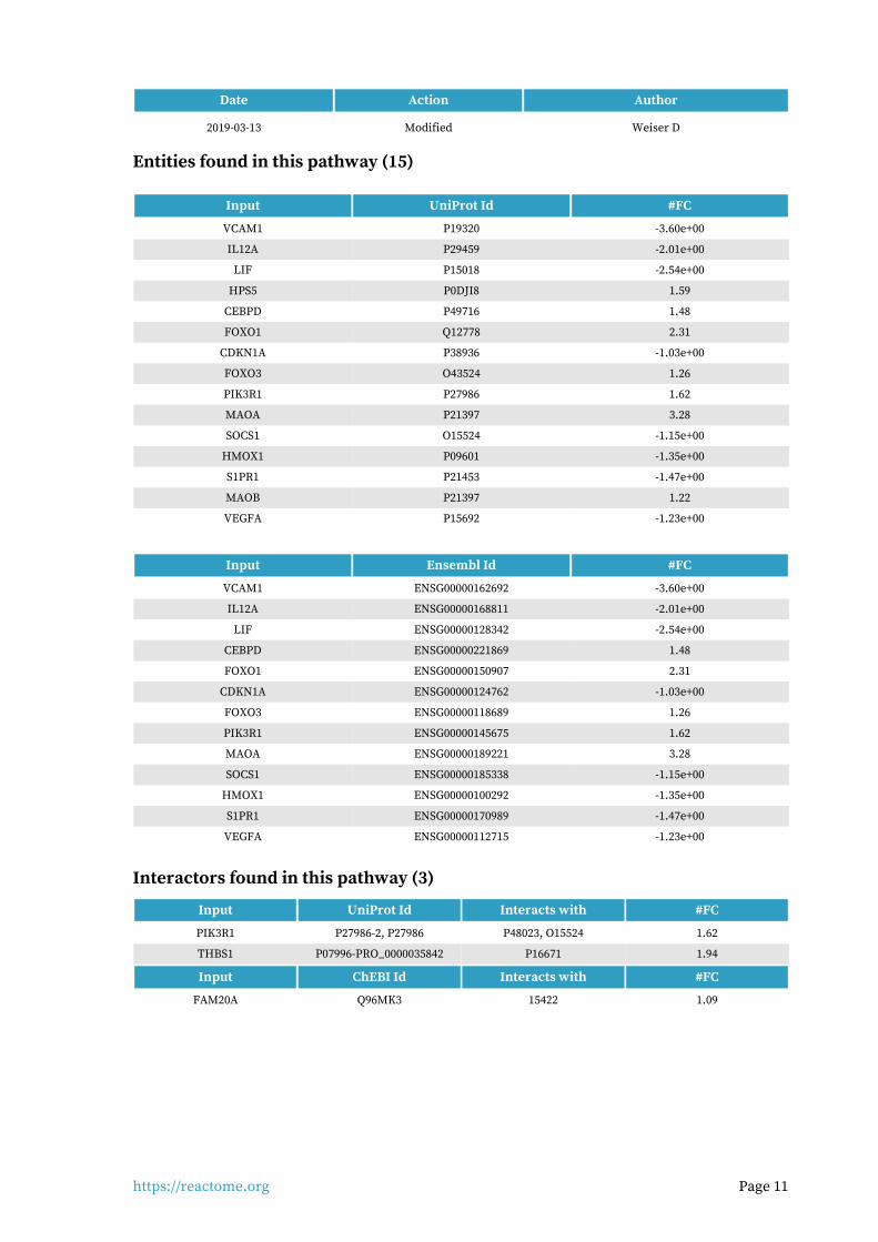
Entities found in this pathway (3)
Input UniProt Id #FC
TUBA4A P68363, P68366 -1.45e+00
TUBB3 Q13509 -1.49e+00
TUBA1A P68363, Q71U36 -1.29e+00

https://reactome.org Page 32
Transport of connexons to the plasma membrane (R-HSA-190872)13.
Cellular compartments: cytosol.
Following connexon oligomerization, the hemichannels must be transported to the plasma mem-brane. This has been shown to occur in transport vesicles called "cargo containers". Most of post-Golgi cargo containers have a diameter of of 50- 200 nm (Lauf et al., 2002). Recently direct transport of connexins to GJ assembly sides has been described (Shaw et al., 2007). Besides microtuble-de-pendent trafficking, a microtubule-independent delivery pathway may exist as concluded from studies using the secretory transport inhibitor, Brefeldin A (Musil and Goodenough 1993; De Sousa et al. 1993; Laird et al. 1995).
References
De Sousa PA, Valdimarsson G, Nicholson BJ & Kidder GM (1993). Connexin trafficking and the con-trol of gap junction assembly in mouse preimplantation embryos. Development, 117, 1355-67.
Lauf U, Giepmans BN, Lopez P, Braconnot S, Chen SC & Falk MM (2002). Dynamic trafficking and delivery of connexons to the plasma membrane and accretion to gap junctions in living cells.
Proc Natl Acad Sci U S A, 99, 10446-51.
Laird DW, Castillo M & Kasprzak L (1995). Gap junction turnover, intracellular trafficking, and phosphorylation of connexin43 in brefeldin A-treated rat mammary tumor cells. J Cell Biol, 131,
1193-203.
Shaw RM, Fay AJ, Puthenveedu MA, von Zastrow M, Jan YN & Jan LY (2007). Microtubule plus-end-tracking proteins target gap junctions directly from the cell interior to adherens junctions. Cell,
128, 547-60.
Musil LS & Goodenough DA (1993). Multisubunit assembly of an integral plasma membrane chan-
nel protein, gap junction connexin43, occurs after exit from the ER. Cell, 74, 1065-77.

https://reactome.org Page 33
Edit history
Date Action Author
2007-01-03 Authored Gilleron J, Segretain D, Falk MM
2007-01-09 Created Matthews L
2007-04-12 Edited Matthews L
2019-03-08 Modified Weiser D
Entities found in this pathway (3)
Input UniProt Id #FC
TUBA4A P68363, P68366 -1.45e+00
TUBB3 Q13509 -1.49e+00
TUBA1A P68363, Q71U36 -1.29e+00

https://reactome.org Page 34
Collagen degradation (R-HSA-1442490)14.
Collagen fibril diameter and spatial organisation are dependent on the species, tissue type and stage of development (Parry 1988). The lengths of collagen fibrils in mature tissues are largely un-known but in tendon can be measured in millimetres (Craig et al. 1989). Collagen fibrils isolated from adult bovine corneal stroma had ~350 collagen molecules in transverse section, tapering down to three molecules at the growing tip (Holmes & Kadler 2005).
The classical view of collagenases is that they actively unwind the triple helical chain, a process termed molecular tectonics (Overall 2002, Bode & Maskos 2003), before preferentially cleaving the alpha2 chain followed by the remaining chains (Chung et al. 2004). More recently it has been sug-gested that collagen fibrils exist in an equilibrium between protected and vulnerable states (Stultz 2002, Nerenberg & Stultz 2008). The prototypical triple-helical structure of collagen does not fit into the active site of collagenase MMPs. In addition the scissile bonds are not solvent-exposed and are therefore inaccessible to the collagenase active site (Chung et al. 2004, Stultz 2002). It was realized that collagen must locally unfold into non-triple helical regions to allow collagenolysis. Observa-tions using circular dichroism and differential scanning calorimetry confirm that there is consider-able heterogeneity along collagen fibres (Makareeva et al. 2008) allowing access for MMPs at physiological temperatures (Salsas-Escat et al. 2010).
Collagen fibrils with cut chains are unstable and accessible to proteinases that cannot cleave intact collagen strands (Woessner & Nagase 2000, Somerville et al. 2003). Continued degradation leads to the formation of gelatin (Lovejoy et al. 1999). Degradation of collagen types other than I-III is less well characterized but believed to occur in a similar manner.

https://reactome.org Page 35
Metalloproteinases (MMPs) play a major part in the degradation of several extracellular macro-molecules including collagens. MMP1 (Welgus et al. 1981), MMP8 (Hasty et al. 1987), and MMP13 (Knauper et al. 1996), sometimes referred to as collagenases I, II and III respectively, are able to ini-tiate the intrahelical cleavage of the major fibril forming collagens I, II and III at neutral pH, and thus thought to define the rate-limiting step in normal tissue remodeling events. All can cleave ad-ditional substrates including other collagen subtypes. Collagenases cut collagen alpha chains at a single conserved Gly-Ile/Leu site approximately 3/4 of the molecule's length from the N-terminus (Fields 1991, Chung et al. 2004). The cleavage site is characterised by the motif G(I/L)(A/L); the G-I/L bond is cleaved. In collagen type I this corresponds to G953-I954 in the Uniprot canonical alpha chain sequences (often given as G775-I776 in literature). It is not clear why only this bond is cleaved, as the motif occurs at several other places in the chain. MMP14, a membrane-associated MMP also known as Membrane-type matrix metalloproteinase 1 (MT-MMP1), is able to cleave colla-gen types I, II and III (Ohuchi et al. 1997).
References
Hadler-Olsen E, Fadnes B, Sylte I, Uhlin-Hansen L & Winberg JO (2011). Regulation of matrix metal-
loproteinase activity in health and disease. FEBS J, 278, 28-45.
Steinhusen U, Weiske J, Badock V, Tauber R, Bommert K & Huber O (2001). Cleavage and shedding
of E-cadherin after induction of apoptosis. J. Biol. Chem., 276, 4972-80.
Shapiro SD (1998). Matrix metalloproteinase degradation of extracellular matrix: biological con-
sequences. Curr Opin Cell Biol, 10, 602-8.
Edit history
Date Action Author
2011-07-12 Authored Jupe S
2011-07-12 Created Jupe S
2012-10-08 Reviewed Sorsa T
2012-11-12 Edited Jupe S
2019-03-08 Modified Weiser D
Entities found in this pathway (6)
Input UniProt Id #FC
MMP15 P51511 1.28
COL7A1 Q02388 1.22
COL4A4 P53420 2.02
COL4A1 P02462, Q03692 1.33
COL11A1 P12107 2.36
COL1A1 P02452 -1.31e+00

https://reactome.org Page 36
FOXO-mediated transcription of oxidative stress, metabolic and neuronal
genes (R-HSA-9615017)
15.
FOXO6, the least studied member of the FOXO family, directly stimulates transcription of PLXNA4 gene, encoding a co-factor for the semaphorin SEMA3A receptor. FOXO6-mediated regulation of PLXNA4 expression plays an important role in radial glia migration during cortical development (Paap et al. 2016).
FOXO-mediated up-regulation of genes involved in reduction of the oxidative stress burden is not specific to neurons, but plays an important role in neuronal survival and neurodegenerative dis-eases. FOXO3 and FOXO4, and possibly FOXO1, directly stimulate transcription of the SOD2 gene, encoding mitochondrial manganese-dependent superoxide dismutase, which converts superoxide to the less harmful hydrogen peroxide and oxygen (Kops et al. 2002, Hori et al. 2013, Araujo et al. 2011, Guan et al. 2016). FOXO4 stimulates SOD2 gene transcription in collaboration with ATXN3, a protein involved in spinocerebellar ataxia type 3 (SCA3) (Araujo et al. 2011). FOXO3 and FOXO6, and possibly FOXO1, directly stimulate transcription of the CAT gene, encoding catalase, an enzyme that converts hydrogen peroxide to water and oxygen, thus protecting cells from the oxidative stress (Awad et al. 2014, Kim et al. 2014, Rangarajan et al. 2015, Song et al. 2016, Liao et al. 2016, Guo et al. 2016).
FOXO transcription factors regulate transcription of several genes whose protein products are secreted from hypothalamic neurons to control appetite and food intake: NPY gene, AGRP gene and POMC gene. At low insulin levels, characteristic of starvation, FOXO transcription factors bind to insulin responsive elements (IRES) in the regulatory regions of NPY, AGRP and POMC gene. FOXO1 directly stimulates transcription of the NPY gene, encoding neuropeptide-Y (Kim et al. 2006, Hong et al. 2012), and the AGRP gene, encoding Agouti-related protein (Kitamura et al. 2006, Kim et al. 2006), which both stimulate food intake. At the same time, FOXO1 directly represses transcrip-tion of the POMC gene, encoding melanocyte stimulating hormone alpha , which suppresses food intake (Kitamura et al. 2006, Kim et al. 2006). When, upon food intake, blood insulin levels rise, in-sulin-mediated activation of PI3K/AKT signaling inhibits FOXO transcriptional activity.

https://reactome.org Page 37
In liver cells, FOXO transcription factors regulate transcription of genes involved in gluconeogen-esis: G6PC gene, encoding glucose-6-phosphatase and PCK1 gene, encoding phosphoenolpyruvate carboxykinase. Actions of G6PC and PCK1 enable steady glucose blood levels during fasting. FOXO1, FOXO3 and FOXO4 directly stimulate PCK1 gene transcription (Hall et al. 2000, Yang et al. 2002, Puigserver et al. 2003), while all four FOXOs, FOXO1, FOXO3, FOXO4 and FOXO6 directly stimulate G6PC gene transcription (Yang et al. 2002, Puigserver et al. 2003, Onuma et al. 2006, Kim et al. 2011). FOXO-mediated induction of G6PC and PCK1 genes is negatively regulated by insulin-induced PI3K/AKT signaling.
FOXO1, FOXO3 and FOXO4 directly stimulate transcription of the IGFBP1 gene, encoding insulin growth factor binding protein 2 (Tang et al. 1999, Kops et al. 1999, Hall et al. 2000, Yang et al. 2002), which increases sensitivity of cells to insulin.
FOXO1 and FOXO3 directly stimulate transcription of the ABCA6 (ATP-binding cassette sub-family A member 6) gene, encoding a putative transporter protein that is thought to be involved in lipid homeostasis (Gai et al. 2013). The GCK (glucokinase) gene is another gene involved in lipid homeo-stasis that is regulated by FOXOs. FOXO1, acting with the SIN3A:HDAC complex, directly represses the GCK gene transcription, thus repressing lipogenesis in the absence of insulin (Langlet et al. 2017). The SREBF1 (SREBP1) gene, which encodes a transcriptional activator required for lipid homeostasis, is directly transcriptionally repressed by FOXO1 (Deng et al. 2012). Transcription of the RETN gene, encoding resistin, an adipocyte specific hormone that suppresses insulin-mediated uptake of glucose by adipose cells, is directly stimulated by FOXO1 (Liu et al. 2014).
Transcription of two genes encoding E3 ubiquitin ligases FBXO32 (Atrogin-1) and TRIM63 (MURF1), involved in degradation of muscle proteins and muscle wasting during starvation, is positively reg-ulated by FOXO transcription factors (Sandri et al. 2004, Waddell et al. 2008, Raffaello et al. 2010, Senf et al. 2011, Bollinger et al. 2014, Wang et al. 2017).
References
Paap RH, Oosterbroek S, Wagemans CM, von Oerthel L, Schellevis RD, Vastenhouw-van der Linden AJ, ... Smidt MP (2016). FoxO6 affects Plxna4-mediated neuronal migration during mouse cortic-
al development. Proc. Natl. Acad. Sci. U.S.A..
Kops GJ, de Ruiter ND, De Vries-Smits AM, Powell DR, Bos JL & Burgering BM (1999). Direct control
of the Forkhead transcription factor AFX by protein kinase B. Nature, 398, 630-4.
Kops GJ, Dansen TB, Polderman PE, Saarloos I, Wirtz KW, Coffer PJ, ... Burgering BM (2002). Fork-head transcription factor FOXO3a protects quiescent cells from oxidative stress. Nature, 419,
316-21.
Hori YS, Kuno A, Hosoda R & Horio Y (2013). Regulation of FOXOs and p53 by SIRT1 modulators
under oxidative stress. PLoS ONE, 8, e73875.
Araujo J, Breuer P, Dieringer S, Krauss S, Dorn S, Zimmermann K, ... Evert BO (2011). FOXO4-de-pendent upregulation of superoxide dismutase-2 in response to oxidative stress is impaired in
spinocerebellar ataxia type 3. Hum. Mol. Genet., 20, 2928-41.
Edit history
Date Action Author
2018-07-31 Created Orlic-Milacic M
2018-10-11 Authored Orlic-Milacic M

https://reactome.org Page 38
Date Action Author
2018-10-17 Reviewed Donlon T
2018-10-26 Reviewed Bertaggia E
2018-10-31 Edited Orlic-Milacic M
2019-03-08 Modified Weiser D
Entities found in this pathway (7)
Input UniProt Id #FC
PLXNA4 Q9HCM2 2.9
ABCA6 Q8N139 1.02
FOXO1 Q12778 2.31
INS-IGF2 P01308 -5.88e+00
FBXO32 Q969P5 1.09
FOXO3 O43524 1.26
NR3C1 P04150 -1.01e+00
Input Ensembl Id #FC
PLXNA4 ENSG00000221866 2.9
ABCA6 ENSG00000154262 1.02
FBXO32 ENSG00000156804 1.09
Interactors found in this pathway (3)
Input UniProt Id Interacts with #FC
PPARG P37231 Q9UBK2 1.03
ARHGEF2 Q92974 O43524 -1.02e+00
KLHL42 Q9P2K6 P01189 1.04

https://reactome.org Page 39
Diseases associated with O-glycosylation of proteins (R-HSA-3906995)16.
Diseases: congenital disorder of glycosylation.
Glycosylation is the most abundant modification of proteins, variations of which occur in all living cells. Glycosylation can be further categorized into N-linked (where the oligosaccharide is conjug-ated to Asparagine residues) and O-linked glycosylation (where the oligosaccharide is conjugated to Serine, Threonine and possibly Tyrosine residues). Within the family of O-linked glycosylation, the oligosaccharides attached can be further categorized according to their reducing end residue: Gal-NAc (often described as mucin-type, due to the abundance of this type of glycosylation on mucins), Mannose and Fucose. This section reviews currently known congenital disorders of glycosylation associated with defects of protein O-glycosylation (Cylwik et al. 2013, Freeze et al. 2014).
References
Cylwik B, Lipartowska K, Chrostek L & Gruszewska E (2013). Congenital disorders of glycosylation.
Part II. Defects of protein O-glycosylation. Acta Biochim. Pol., 60, 361-8.
Freeze HH, Chong JX, Bamshad MJ & Ng BG (2014). Solving glycosylation disorders: fundamental
approaches reveal complicated pathways. Am. J. Hum. Genet., 94, 161-75.
Edit history
Date Action Author
2013-07-17 Edited Jassal B
2013-07-17 Authored Jassal B
2013-07-17 Created Jassal B
2015-12-18 Reviewed Hansen L, Joshi HJ
2016-07-30 Modified Gillespie ME
Entities found in this pathway (7)
Input UniProt Id #FC
ADAMTS1 Q9UHI8 2.28
ADAMTS14 Q8WXS8 -2.42e+00
ADAMTSL1 Q8N6G6 -1.05e+00
ADAMTS5 Q9UNA0 2.12
GALNT12 Q8IXK2 -1.16e+00
SPON1 Q9HCB6 1.8
THBS1 P07996 1.94

https://reactome.org Page 40
Post-chaperonin tubulin folding pathway (R-HSA-389977)17.
Cellular compartments: cytosol.
Alpha and beta tubulin folding intermediates are formed through ATP-dependent interaction with TriC/CCT. In order to form a functional heterodimer, these folding intermediates undergo a series of interactions with five proteins: (cofactors A-E) following release from TriC/CCT (reviewed in Cowan and Lewis et al., 2001). These interactions are described in the reactions below. Ultimately, alpha tubulin, when associated with cofactor E, interacts with cofactor D-bound beta-tubulin. The entry of cofactor C into this complex results in the discharge of native heterodimer triggered by GTP hydrolysis in beta tubulin (Tian et al., 1997).
References
Lewis SA, Tian G & Cowan NJ (1997). The alpha- and beta-tubulin folding pathways. Trends Cell Biol
, 7, 479-84.
Edit history
Date Action Author
2008-12-01 Authored Matthews L
2009-01-21 Reviewed Cowan NJ
2009-01-22 Created Matthews L
2009-02-21 Edited Matthews L
2017-04-03 Modified Matthews L
Entities found in this pathway (3)

https://reactome.org Page 41
Input UniProt Id #FC
TUBA4A P68363, P68366 -1.45e+00
TUBB3 Q13509 -1.49e+00
TUBA1A P68363, Q71U36 -1.29e+00

https://reactome.org Page 42
O-linked glycosylation (R-HSA-5173105)18.
O-glycosylation is an important post-translational modification (PTM) required for correct func-tioning of many proteins (Van den Steen et al. 1998, Moremen et al. 2012). The O-glycosylation of proteins containing thrombospondin type 1 repeat (TSR) domains and O-glycosylation of mucins are currently described here.
References
Van den Steen P, Rudd PM, Dwek RA & Opdenakker G (1998). Concepts and principles of O-linked
glycosylation. Crit. Rev. Biochem. Mol. Biol., 33, 151-208.
Moremen KW, Tiemeyer M & Nairn AV (2012). Vertebrate protein glycosylation: diversity, synthes-
is and function. Nat. Rev. Mol. Cell Biol., 13, 448-62.
Edit history
Date Action Author
2013-11-25 Edited Jassal B
2013-11-25 Authored Jassal B
2013-11-25 Created Jassal B
2014-02-07 Reviewed D'Eustachio P
2019-03-08 Modified Weiser D
Entities found in this pathway (10)
Input UniProt Id #FC
GCNT4 Q9P109 1.97
ADAMTS1 Q9UHI8 2.28
GALNT16 Q8N428 -1.06e+00
ADAMTS14 Q8WXS8 -2.42e+00
GALNT15 Q7Z4T8, Q8N3T1 1.54
ADAMTSL1 Q8N6G6 -1.05e+00
ADAMTS5 Q9UNA0 2.12

https://reactome.org Page 43
Input UniProt Id #FC
GALNT12 Q8IXK2 -1.16e+00
SPON1 Q9HCB6 1.8
THBS1 P07996 1.94
Interactors found in this pathway (1)
Input ChEBI Id Interacts with #FC
FAM20A Q96MK3 15422 1.09

https://reactome.org Page 44
Activation of AMPK downstream of NMDARs (R-HSA-9619483)19.
Cellular compartments: cytosol.
Activation of NMDA receptors (NMDARs) leads to activation of AMP-activated kinase (AMPK) in a CAMKK2-dependent manner. Overactivation of CAMKK2 or AMPK in neurons can lead to dendritic spine loss and is implicated in synaptotoxicity of beta-amyloids in Alzheimer's disease (Mairet-Coello et al. 2013).
References
Mairet-Coello G, Courchet J, Pieraut S, Courchet V, Maximov A & Polleux F (2013). The CAMKK2-AMPK kinase pathway mediates the synaptotoxic effects of A oligomers through Tau phos-
phorylation. Neuron, 78, 94-108.
Edit history
Date Action Author
2018-09-18 Created Orlic-Milacic M
2018-10-11 Authored Orlic-Milacic M
2018-11-02 Reviewed Hansen KB, Yi F
2018-11-07 Edited Orlic-Milacic M
2019-03-13 Modified Weiser D
Entities found in this pathway (4)
Input UniProt Id #FC
PRKAG2 P54619, Q9UGJ0 1.57
TUBA4A P68363, P68366 -1.45e+00
https://reactome.org Page 45
Input UniProt Id #FC
TUBB3 Q13509 -1.49e+00
TUBA1A P68363, Q71U36 -1.29e+00
Interactors found in this pathway (1)
Input ChEBI Id Interacts with #FC
FAM20A Q96MK3 15422 1.09

https://reactome.org Page 46
Assembly of collagen fibrils and other multimeric structures (R-HSA-2022090
)
20.
Collagen trimers in triple-helical form, referred to as procollagen or collagen molecules, are expor-ted from the ER and trafficked through the Golgi network before secretion into the extracellular space. For fibrillar collagens namely types I, II, III, V, XI, XXIV and XXVII (Gordon & Hahn 2010, Ricard-Blum 2011) secretion is concomitant with processing of the N and C terminal collagen propeptides. These processed molecules are known as tropocollagens, considered to be the units of higher order collagen structures. They form within the extracellular space via a process that can proceed spontaneously, but in the cellular environment is regulated by many collagen binding pro-teins such as the FACIT (Fibril Associated Collagens with Interrupted Triple helices) family colla-gens and Small Leucine-Rich Proteoglycans (SLRPs). The architecture formed ultimately depends on the collagen subtype and the cellular conditions. Structures include the well-known fibrils and fibres formed by the major structural collagens type I and II plus several different types of supra-molecular assembly (Bruckner 2010). The mechanical and physical properties of tissues depend on the spatial arrangement and composition of these collagen-containing structures (Kadler et al. 1996, Shoulders & Raines 2009, Birk & Bruckner 2011).
Fibrillar collagen structures are frequently heterotypic, composed of a major collagen type in asso-ciation with smaller amounts of other types, e.g. type I collagen fibrils are associated with types III and V, while type II fibrils frequently contain types IX and XI (Wess 2005). Fibres composed exclus-ively of a single collagen type probably do not exist, as type I and II fibrils require collagens V and XI respectively as nucleators (Kadler et al. 2008, Wenstrup et al. 2011). Much of the structural un-derstanding of collagen fibrils has been obtained with fibril-forming collagens, particularly type I, but some central features are believed to apply to at least the other fibrillar collagen subtypes (Wess 2005). Fibril diameter and length varies considerably, depending on the tissue and collagen types (Fang et al. 2012). The reasons for this are poorly understood (Wess 2005).

https://reactome.org Page 47
Some tissues such as skin have fibres that are approximately the same diameter while others such as tendon or cartilage have a bimodal distribution of thick and thin fibrils. Mature type I collagen fibrils in tendon are up to 1 cm in length, with a diameter of approx. 500 nm. An individual fibrillar collagen triple helix is less than 1.5 nm in diameter and around 300 nm long; collagen molecules must assemble to give rise to the higher-order fibril structure, a process known as fibrillogenesis, prevented by the presence of C-terminal propeptides (Kadler et al. 1987). In electron micrographs, fibrils have a banded appearance, due to regular gaps where fewer collagen molecules overlap, which occur because the fibrils are aligned in a quarter-stagger arrangement (Hodge & Petruska 1963). Collagen microfibrils are believed to have a quasi-hexagonal unit cell, with tropocollagen ar-ranged to form supertwisted, right-handed microfibrils that interdigitate with neighbouring mi-crofibrils, leading to a spiral-like structure for the mature collagen fibril (Orgel et al. 2006, Holmes & Kadler 2006).
Neighbouring tropocollagen monomers interact with each other and are cross-linked covalently by lysyl oxidase (Orgel et al. 2000, Maki 2006). Mature collagen fibrils are stabilized by lysyl oxidase-mediated cross-links. Hydroxylysyl pyridinoline and lysyl pyridinoline cross-links form between (hydroxy) lysine and hydroxylysine residues in bone and cartilage (Eyre et al. 1984). Arginoline cross-links can form in cartilage (Eyre et al. 2010); mature bovine articular cartilage contains roughly equimolar amounts of arginoline and hydroxylysyl pyridinoline based on peptide yields. Mature collagen fibrils in skin are stabilized by the lysyl oxidase-mediated cross-link histidino-hydroxylysinonorleucine (Yamauch et al. 1987). Due to the quarter-staggered arrangement of colla-gen molecules in a fibril, telopeptides most often interact with the triple helix of a neighbouring collagen molecule in the fibril, except for collagen molecules in register staggered by 4D from an-other collagen molecule. Fibril aggregation in vitro can be unipolar or bipolar, influenced by tem-perature and levels of C-proteinase, suggesting a role for the N- and C- propeptides in regulation of the aggregation process (Kadler et al. 1996). In vivo, collagen molecules at the fibril surface may re-tain their N-propeptides, suggesting that this may limit further accretion, or alternatively repres-ents a transient stage in a model whereby fibrils grow in diameter through a cycle of deposition, cleavage and further deposition (Chapman 1989).
In vivo, fibrils are often composed from more than one type of collagen. Type III collagen is found associated with type I collagen in dermal fibrils, with the collagen III on the periphery, suggesting a regulatory role (Fleischmajer et al. 1990). Type V collagen associates with type I collagen fibrils, where it may limit fibril diameter (Birk et al. 1990, White et al. 1997). Type IX associates with the surface of narrow diameter collagen II fibrils in cartilage and the cornea (Wu et al. 1992, Eyre et al. 2004). Highly specific patterns of crosslinking sites suggest that collagen IX functions in interfibril-lar networking (Wess 2005). Type XII and XIV collagens are localized near the surface of banded collagen I fibrils (Nishiyama et al. 1994). Certain fibril-associated collagens with interrupted triple helices (FACITs) associate with the surface of collagen fibrils, where they may serve to limit fibril fusion and thereby regulate fibril diameter (Gordon & Hahn 2010). Collagen XV, a member of the multiplexin family, is almost exclusively associated with the fibrillar collagen network, in very close proximity to the basement membrane. In human tissues collagen XV is seen linking banded collagen fibers subjacent to the basement membrane (Amenta et al. 2005). Type XIV collagen, SLRPs and discoidin domain receptors also regulate fibrillogenesis (Ansorge et al. 2009, Kalamajski et al. 2010, Flynn et al. 2010).

https://reactome.org Page 48
Collagen IX is cross-linked to the surface of collagen type II fibrils (Eyre et al. 1987). Type XII and XIV collagens are found in association with type I (Walchli et al. 1994) and type II (Watt et al. 1992, Eyre 2002) fibrils in cartilage. They are thought to associate non-covalently via their COL1/NC1 do-mains (Watt et al. 1992, Eyre 2002).
Some non-fibrillar collagens form supramolecular assemblies that are distinct from typical fibrils. Collagen VII forms anchoring fibrils, composed of antiparallel dimers that connect the dermis to the epidermis (Bruckner-Tuderman 2009). During fibrillogenesis, the nascent type VII procollagen molecules dimerize in an antiparallel manner. The C-propeptides are then removed by Bone morphogenetic protein 1 (Rattenholl et al. 2002) and the processed antiparallel dimers aggregate laterally. Collagens VIII and X form hexagonal networks and collagen VI forms beaded filament (Gordon & Hahn 2010, Ricard-Blum et al. 2011).
References
Kadler KE, Holmes DF, Trotter JA & Chapman JA (1996). Collagen fibril formation. Biochem J, 316,
1-11.
Orgel JP, San Antonio JD & Antipova O (2011). Molecular and structural mapping of collagen fibril
interactions. Connect. Tissue Res., 52, 2-17.
Edit history
Date Action Author
2011-08-05 Authored Jupe S
2011-11-25 Created Jupe S
2012-10-08 Reviewed Kalamajski S, Raleigh S
2012-11-12 Edited Jupe S
2012-11-19 Reviewed Ricard-Blum S
2019-03-08 Modified Weiser D
Entities found in this pathway (5)
Input UniProt Id #FC
COL7A1 Q02388 1.22
COL4A4 P53420 2.02
COL4A1 P02462, Q03692 1.33
COL11A1 P12107 2.36
COL1A1 P02452 -1.31e+00

https://reactome.org Page 49
Crosslinking of collagen fibrils (R-HSA-2243919)21.
After removal of the N- and C-procollagen propeptides, fibrillar collagen molecules aggregate into microfibrillar arrays, stabilized by covalent intermolecular cross-links. These depend on the oxidat-ive deamination of specific lysine or hydroxylysine residues in the telopeptide region by lysyl oxi-dase (LOX) with the subsequent spontaneous formation of covalent intermolecular cross-links (Pin-nell & Martin 1968, Siegel et al. 1970, 1974, Maki 2009, Nishioka et al. 2012). Hydroxylysine is formed intracellularly by lysine hydroxylases (LH). There are different forms of LH responsible for hydroxylation of helical and telopeptide lysines (Royce & Barnes 1985, Knott et al.1997, Takaluoma et al. 2007, Myllyla 2007). The chemistry of the cross-links formed depends on whether lysines or hydroxylysines are present in the telopeptides (Barnes et al. 1974), which depends on the propor-tion of collagen lysines post-translationally converted to hydroxylysine by LH. The lysine pathway predominates in adult skin, cornea and sclera while the hydroxylysine pathway occurs primarily in bone, cartilage, ligament, tendons, embryonic skin and most connective tissues (Eyre 1987, Eyre & Wu 2005, Eyre et al. 2008). Oxidative deamination of lysine or hydroxylysine residues by LOX gener-ates the allysine and hydroxyallysine aldehydes respectively. These can spontaneously react with either another aldehyde to form an aldol condensation product (intramolecular cross-link), or with an unmodified lysine or hydroxylysine residue to form intermolecular cross-links.
The pathway of cross-linking is regulated primarily by the hydroxylation pattern of telopeptide and triple-helix domain lysine residues. When lysine residues are the source of aldehydes formed by lysyl oxidase the allysine cross-linking pathway leads to the formation of aldimine cross-links (Eyre & Wu 2005). These are stable at physiological conditions but readily cleaved at acid pH or elevated temperature. When hydroxylysine residues are the source of aldehydes formed by lysyl oxidase the hydroxyallysine cross-linking pathway leads to the formation of more stable ketoimine cross-links.

https://reactome.org Page 50
Telopeptide lysine residues can be converted by LOX to allysine, which can react with a helical hy-droxylysine residue forming the lysine aldehyde aldimine cross-link dehydro hydroxylysino norleu-cine (deHHLNL) (Bailey & Peach 1968, Eyre et al. 2008). If the telopeptide residue is hydroxylysine, the hydroxyallysine formed by LOX can react with a helical hydroxylysine forming the Schiff base, which spontaneously undergoes an Amadori rearrangement resulting in the ketoimine cross link hydroxylysino 5 ketonorleucine (HLKNL). This stable cross-link is formed in tissues where te-lopeptide residues are predominanly hydroxylated, such as foetal bone and cartilage, accounting for the relative insolubility of collagen from these tissues (Bailey et al. 1998). In bone, telopeptide hydroxyallysines can react with the epsilon-amino group of a helical lysine (Robins & Bailey 1975). The resulting Schiff base undergoes Amadori rearrangement to form lysino-hydroxynorleucine (LHNL). An alternative mechanism of maturation of ketoimine cross-links has been reported in cartilage leading to the formation of arginoline (Eyre et al. 2010).
These divalent crosslinks greatly diminish as connective tissues mature, due to further spontan-eous reactions (Bailey & Shimokomaki 1971, Robins & Bailey 1973) with neighbouring peptides that result in tri- and tetrafunctional cross-links. In mature tissues collagen cross-links are predomin-antly trivalent. The most common are pyridinoline or 3-hydroxypyridinium cross-links, namely hy-droxylysyl-pyridinoline (HL-Pyr) and lysyl-pyridinoline (L-Pyr) cross-links (Eyre 1987, Ogawa et al. 1982, Fujimoto et al. 1978). HL-Pyr is formed from three hydroxylysine residues, HLKNL plus a fur-ther hydroxyallysine. It predominates in highly hydroxylated collagens such as type II collagen in cartilage. L-Pyr is formed from two hydroxylysines and a lysine, LKNL plus a further hydroxyallys-ine, found mostly in calcified tissues (Bailey et al. 1998). Trivalent collagen cross-links can also form as pyrroles, either Lysyl-Pyrrole (L-Pyrrole) or hydroxylysyl-pyrrole (HL-Pyrrole), respectively formed when LKNL or HLKNL react with allysine (Scott et al. 1981, Kuypers et al. 1992). A further three-way crosslink can form when DeH-HLNL reacts with histidine to form histidino-hydroxylys-inonorleucine (HHL), found in skin and cornea (Yamauchi et al. 1987, 1996). This can react with an additional lysine to form the tetrafunctional cross-link histidinohydroxymerodesmosine (Reiser et al. 1992, Yamauchi et al. 1996).
Another mechanism which could be involved in the cross-linking of collagen IV networks is the sul-filimine bond (Vanacore et al. 2009), catalyzed by peroxidasin, an enzyme found in basement mem-brane (Bhave 2012).
To improve clarity inter-chain cross-linking is represented here for Collagen type I only. Although the formation of each type of cross-link is represented here as an independent event, the partial and random nature of lysine hydroxylation and subsequent lysyl oxidation means that any combin-ation of these cross-linking events could occur within the same collagen fibril .
References
Bailey AJ, Paul RG & Knott L (1998). Mechanisms of maturation and ageing of collagen. Mech Age-
ing Dev, 106, 1-56.
Edit history
Date Action Author
2012-04-30 Authored Jupe S
2012-05-09 Created Jupe S
2012-10-08 Reviewed Kalamajski S, Raleigh S

https://reactome.org Page 51
Date Action Author
2012-11-12 Edited Jupe S
2012-11-19 Reviewed Ricard-Blum S
2019-03-08 Modified Weiser D
Entities found in this pathway (3)
Input UniProt Id #FC
COL4A4 P53420 2.02
COL4A1 P02462 1.33
COL1A1 P02452 -1.31e+00

https://reactome.org Page 52
Molecules associated with elastic fibres (R-HSA-2129379)22.
Proteins found associated with microfibrils include vitronectin (Dahlback et al. 1990), latent trans-forming growth factor beta-binding proteins (Kielty et al. 2002, Munger & Sheppard 2011), emilin (Bressan et al. 1993, Mongiat et al. 2000), members of the microfibrillar-associated proteins (MFAPs, Gibson et al.1996), and fibulins (Roark et al. 1995, Yanagisawa et al. 2002). The significance of these interactions is not well understood but may help mediate elastin-fibrillin interactions dur-ing elastic fibre assembly.
Proteoglycans such as versican (Isogai et al. 2002), biglycan, and decorin (Reinboth et al. 2002) can interact with the microfibrils. They confer specific properties including hydration, impact absorp-tion, molecular sieving, regulation of cellular activities, mediation of growth factor association, and release and transport within the extracellular matrix (Buczek-Thomas et al. 2002). In addition, glyc-osaminoglycans have been shown to interact with tropoelastin through its lysine side chains (Wu et al. 1999) regulating tropoelastin assembly (Tu and Weiss, 2008).
References
Wagenseil JE & Mecham RP (2007). New insights into elastic fiber assembly. Birth Defects Res. C
Embryo Today, 81, 229-40.
Kielty CM, Sherratt MJ & Shuttleworth CA (2002). Elastic fibres. J Cell Sci, 115, 2817-28.
Edit history
Date Action Author
2012-02-21 Created Jupe S
2012-04-30 Authored Jupe S
2012-11-02 Reviewed Muiznieks LD
2012-11-12 Edited Jupe S
2019-03-08 Modified Weiser D

https://reactome.org Page 53
Entities found in this pathway (4)
Input UniProt Id #FC
FBN2 P35556 2.07
TGFB2 P61812 -1.62e+00
GDF5 P43026 -1.27e+00
ITGA8 P53708 1.07

https://reactome.org Page 54
TGFBR2 MSI Frameshift Mutants in Cancer (R-HSA-3642279)23.
Diseases: cancer.
The short adenine repeat in the coding sequence of TGF-beta receptor II (TGFBR2) gene is fre-quently targeted by loss-of-function frameshift mutations in colon cancers with microsatellite in-stability (MSI). The 1- or 2-bp deletions in the adenine stretch of TGFBR2 cDNA introduce a prema-ture stop codon that leads to degradation of the majority of mutant transcripts through nonsense-mediated decay or to production of a truncated TGFBR2 that cannot be presented on the cell sur-face. Cells that harbor TGFBR2 MSI frameshift mutations are resistant to TGF-beta (TGFB1)-medi-ated growth inhibition.
References
Markowitz S, Wang J, Myeroff L, Parsons R, Sun L, Lutterbaugh J, ... Vogelstein B (1995). Inactiva-tion of the type II TGF-beta receptor in colon cancer cells with microsatellite instability. Science
, 268, 1336-8.
Wang J, Sun L, Myeroff L, Wang X, Gentry LE, Yang J, ... Willson JK (1995). Demonstration that mutation of the type II transforming growth factor beta receptor inactivates its tumor suppress-
or activity in replication error-positive colon carcinoma cells. J. Biol. Chem., 270, 22044-9.
Edit history
Date Action Author
2013-05-31 Created Orlic-Milacic M
2013-08-08 Edited Orlic-Milacic M
2013-08-08 Reviewed Meyer S, Akhurst RJ
2013-08-08 Authored Meyer S, Akhurst RJ, Orlic-Milacic M
2019-03-13 Modified Weiser D
Entities found in this pathway (1)
Input UniProt Id #FC
TGFBR2 P37173 1.44

https://reactome.org Page 56
VEGF ligand-receptor interactions (R-HSA-194313)24.
Cellular compartments: plasma membrane.
The VEGF family is encoded by seven genes (VEGF-A, B, C, D, E: PLGF (Placenta Growth Factor)-1, 2). Six isoforms of VEGF-A protein, containing 121, 145, 165, 183, 189, and 206 amino acid residues, and two isoforms of VEGF-B (167 and 186 residues) are specified by alternatively spliced mRNAs. The active form of each of these proteins is a homodimer.
The specificities of the three VEGF tyrosine kinase receptors, VEGFR-1, VEGFR-2 and VEGFR-3, for these ligands are shown in the figure (Hicklin and Ellis 2005). All VEGF-A isoforms bind both VEG-FR-1 and VEGFR-2; PLGF-1 and -2, and VEGF-B isoforms bind only VEGFR-1; VEGF-E binds VEGFR-2; and VEGF-C and -D bind both VEGFR-2 and -3. VEGF-D undergoes a complex series of post-trans-lational modifications that results in secreted forms with increased activity toward VEGFR-3 and VEGFR-2.
Two co-receptor proteins in the cell membrane, neuropilin (NRP)-1 and NRP-2, interact with VEG-FR proteins to increase the affinity of the latter for their ligands (Neufeld et al.,2002). They differ from VEGFR proteins in not having intracellular signaling domains.
References
Olsson AK, Dimberg A, Kreuger J & Claesson-Welsh L (2006). VEGF receptor signalling - in control
of vascular function. Nat Rev Mol Cell Biol, 7, 359-71.
Shibuya M & Claesson-Welsh L (2006). Signal transduction by VEGF receptors in regulation of an-
giogenesis and lymphangiogenesis. Exp Cell Res, 312, 549-60.
Matsumoto T & Mugishima H (2006). Signal transduction via vascular endothelial growth factor
(VEGF) receptors and their roles in atherogenesis. J Atheroscler Thromb, 13, 130-5.
Cross MJ, Dixelius J, Matsumoto T & Claesson-Welsh L (2003). VEGF-receptor signal transduction.
Trends Biochem Sci, 28, 488-94.
Edit history
Date Action Author
2007-03-13 Created Gopinathrao G
2008-02-28 Reviewed Claesson-Welsh L
2013-08-30 Edited Garapati P V
2013-08-30 Authored Garapati P V
2019-03-08 Modified Weiser D
Entities found in this pathway (2)
Input UniProt Id #FC
PGF P49763 -1.27e+00

https://reactome.org Page 57
Input UniProt Id #FC
VEGFA P15692, P49765 -1.23e+00
Interactors found in this pathway (3)
Input UniProt Id Interacts with #FC
PIK3R1 P27986 P17948 1.62
PGF P49763 P17948 -1.27e+00
VEGFA P15692-4 P35968, P17948 -1.23e+00

https://reactome.org Page 58
VEGF binds to VEGFR leading to receptor dimerization (R-HSA-195399)25.
Cellular compartments: plasma membrane.
The binding of VEGF ligands to VEGFR receptors in the cell membrane induces dimerization and activation of the latter, initiating intracellular signaling cascades that result in proliferation, surviv-al, migration and increased permeability of vascular endothelial cells (Matsumoto and Mugishima, 2006). The receptors predominantly form homodimers but heterodimers between VEGFR-1 and -2 have been observed. Although both VEGFR-1 and -2 are expressed in the vascular endothelium, the angiogenic activities of VEGFs are transduced mainly through VEGFR-2 in vivo.
References
Matsumoto T & Mugishima H (2006). Signal transduction via vascular endothelial growth factor
(VEGF) receptors and their roles in atherogenesis. J Atheroscler Thromb, 13, 130-5.
Olsson AK, Dimberg A, Kreuger J & Claesson-Welsh L (2006). VEGF receptor signalling - in control
of vascular function. Nat Rev Mol Cell Biol, 7, 359-71.
Cross MJ, Dixelius J, Matsumoto T & Claesson-Welsh L (2003). VEGF-receptor signal transduction.
Trends Biochem Sci, 28, 488-94.
Edit history
Date Action Author
2007-04-06 Created Gopinathrao G
2008-02-28 Reviewed Claesson-Welsh L
2013-08-30 Edited Garapati P V
2013-08-30 Authored Garapati P V
2019-03-08 Modified Weiser D
Entities found in this pathway (2)
Input UniProt Id #FC
PGF P49763 -1.27e+00
VEGFA P15692, P49765 -1.23e+00
Interactors found in this pathway (3)
Input UniProt Id Interacts with #FC
PIK3R1 P27986 P17948 1.62
PGF P49763 P17948 -1.27e+00
VEGFA P15692-4 P35968, P17948 -1.23e+00

https://reactome.org Page 59
6. Identifiers found
Below is a list of the input identifiers that have been found or mapped to an equivalent element in Reactome, classified by resource.
Entities (337)
Input UniProt Id #FC
AASS Q9UDR5 1.09
ABAT P80404 1.15
ABCA10 Q8WWZ4 -1.44e+00
ABCA6 Q8N139 1.02
ABCC9 O60706 1.02
ABHD5 Q8WTS1 1.06
ABLIM1 O14639 1.39
ACKR4 Q9NPB9 -1.46e+00
ACSL1 P33121 1.6
ACSS1 Q9NUB1 1.86
ACTA2 P62736, P63267 1.07
ACTG2 P63267 1.91
ADAM12 O43184 -1.84e+00
ADAMTS1 Q9UHI8 2.28
ADAMTS14 Q8WXS8 -2.42e+00
ADAMTS5 Q9UNA0 2.12
ADAMTSL1 Q8N6G6 -1.05e+00
ADARB1 P78563 2.29
ADAT2 Q7Z6V5 -1.11e+00
ADCY1 Q08828 -2.24e+00
ADH1B P00325 1.18
ADHFE1 Q8IWW8 1.42
ADM2 Q7Z4H4 -2.59e+00
ADRA1B P35368 3.74
AGTRAP Q6RW13 1.08
AHCTF1 Q8WYP5 1.28
AKR1C1 P42330 1.06
ALDH1B1 P30837 1
ALDH6A1 Q02252 1.2
ALOX15B O15296 9.98
AMOT Q4VCS5-1 -2.25e+00
AMPH P49418 1.14
ANPEP P15144 1.07
ANXA4 P04156 1.17
AOX1 Q06278 1.45
AP1M1 Q9BXS5 1.08
AQP3 Q92482 -2.97e+00
ARHGAP28 Q9P2N2 -1.52e+00
ARHGAP29 Q52LW3 1.2
ARHGEF2 Q92974 -1.02e+00
ARHGEF28 Q8N1W1 1.06

https://reactome.org Page 60
Input UniProt Id #FC
ARMC8 Q8IUR7 1.33
ARSI Q5FYB1 -1.70e+00
ASNS P08243 -1.27e+00
ATF3 P18847 1.98
ATP10A O60312 1.3
ATP2A2 P16615 1.37
AXIN2 Q9Y2T1 1.17
BDKRB1 P46663 -1.93e+00
BDKRB2 P30411 -2.00e+00
BRCA1 P38398 -1.20e+00
C1QTNF1 Q8NFI3 1.21
C7 P10643 3.2
C9orf3 Q8N6M6 1.09
CACNB2 Q08289 3.37
CARMIL1 Q5VZK9 1.17
CCKAR P32238 -2.22e+00
CCNA2 P20248 1.09
CDC42EP3 Q9UKI2 1.5
CDH6 P55285 -1.31e+00
CDKN1A P38936 -1.03e+00
CDKN2B P42772 -1.50e+00
CDON Q4KMG0 -1.63e+00
CEBPD P49716 1.48
CFLAR O15519-1, O15519-2 1.19
CH25H O95992 -1.60e+00
CHST7 Q9NS84 1.56
CILP O75339 1.35
CIT O14578, O14578-3 -1.58e+00
CITED2 Q99967 1.6
CITED4 Q96RK1 1.44
CLDN11 O75508 -1.74e+00
CLIC2 O15247 -1.24e+00
COL11A1 P12107 2.36
COL1A1 P02452 -1.31e+00
COL4A1 P02462 1.33
COL4A4 P53420 2.02
COL7A1 Q02388 1.22
CORIN Q9Y5Q5 1.93
COX11 Q9Y6N1 1.14
CPM P14384 1.49
CRABP2 P29373 -1.69e+00
CREB3L1 Q96BA8 -1.01e+00
CRISPLD2 Q9H0B8 2.68
CTGF P29279 1.37
CTPS1 P17812 1.51
CXCL12 P48061 -1.93e+00
CYFIP2 Q96F07 -1.00e+00
CYR61 O00622 1.8
CYTH3 O43739 1.18
DAAM1 Q9Y4D1 -1.02e+00

https://reactome.org Page 61
Input UniProt Id #FC
DAPK2 Q9UIK4 1.55
DCXR Q7Z4W1 2.12
DDAH1 O94760 1.32
DDI2 Q92681 1.07
DDIT3 P35638 -1.05e+00
DHRS3 O75911 1.1
DIO2 Q92813 1.53
DLL4 Q9NR61 3.75
DNM1 Q05193 -1.74e+00
DOK6 Q6PKX4 -1.14e+00
DUSP1 P28562 2.94
DUSP10 Q9Y6W6 -1.22e+00
DUSP5 Q16690 1.28
EBF1 Q9UH73 1.12
ENDOD1 O94919 1.96
ENPP1 P22413 1.35
EPB41 P11171 -1.02e+00
EPHA4 P54764 1.35
EPHB2 P29323 -1.77e+00
EPHB3 P54753 -1.45e+00
EPHB6 O15197 1.29
FAM20A Q96MK3 1.09
FBN2 P35556 2.07
FBXO32 Q969P5 1.09
FDXR P22570 -1.00e+00
FGD4 Q96M96 2.26
FGF14 Q92915 1.11
FKBP5 Q13451 3.93
FLRT3 Q9NZU0 -2.35e+00
FOXO1 Q12778 2.31
FOXO3 O43524 1.26
FST P19883 -1.78e+00
FSTL3 O95633 1.83
FZD8 Q9H461 2.44
G0S2 P27469 -2.73e+00
GALNT12 Q8IXK2 -1.16e+00
GALNT15 Q7Z4T8, Q8N3T1 1.54
GALNT16 Q8N428 -1.06e+00
GCH1 P30793 1.61
GCHFR P30047 1.91
GCLM P48507 1.24
GCNT4 Q9P109 1.97
GDF5 P43026 -1.27e+00
GDNF P39905 1.83
GFPT2 O94808 1.3
GGT5 P36269 2.36
GLS O94925 1.19
GLUL P15104 2.99
GNG2 P50150, P59768 -1.11e+00
GPAT3 Q53EU6 1.21

https://reactome.org Page 62
Input UniProt Id #FC
GPR37 O15354 1.46
GPR68 Q15743 -3.20e+00
GPX3 O75715, P22352 3.72
HBA1 P69905 -3.38e+00
HIF3A Q9Y2N7 2.53
HIGD1A Q9Y241 1.19
HIST2H2AA4 Q6FI13 3.83
HMGB2 P26583 1.22
HMOX1 P09601 -1.35e+00
HPS5 P0DJI8 1.59
HSD17B6 O14756 -2.15e+00
HSPA12B Q96MM6 -2.37e+00
HSPA2 P54652 2.55
HSPA4L O95757 -1.28e+00
IER3 P46695 -1.18e+00
IFIT1 P09914 -1.63e+00
IGFBP2 P18065 2.05
IL12A P29459 -2.01e+00
IL34 Q6ZMJ4 -2.10e+00
IMPA2 O14732 2.31
ING2 Q9H160 1.27
INHBA P08476 1.07
INHBB P09529 3.64
INPP5A Q14642 1.37
INS-IGF2 P01308 -5.88e+00
IRS2 Q9Y4H2 2.15
ITGA10 O75578 3.83
ITGA11 Q9UKX5 -1.60e+00
ITGA2 P17301 -1.04e+00
ITGA8 P53708 1.07
ITGBL1 O95965 1.07
JADE1 Q6IE81 1.22
JUN P05412 -1.08e+00
KANK1 Q14678 1.22
KCNE3 Q9Y6H6 -1.30e+00
KCNK15 Q9H427 -1.43e+00
KCNK6 Q9Y257 2.88
KLF15 Q9UIH9 4.56
KLF5 Q13887 2.55
KLHL42 Q9P2K6 1.04
LAMA2 P24043 2.07
LEP P41159 3.28
LGR4 Q9BXB1 -1.10e+00
LHFPL2 Q6ZUX7 -1.26e+00
LIF P15018 -2.54e+00
LIG1 P18858 -1.01e+00
LIMS2 Q7Z4I7 1.27
LMCD1 Q9NZU5 1.61
LMOD1 P29536 1.12
MAFF Q9ULX9 1.26

https://reactome.org Page 63
Input UniProt Id #FC
MAOA P21397 3.28
MAOB P21397 1.22
MAP1LC3C Q9BXW4 2.53
MAP3K8 P41279 1.12
MARCKS P29966 -1.19e+00
METTL7A Q9H8H3 2.31
MFGE8 Q08431 1.07
MMP15 P51511 1.28
MORF4L2 Q15014 1
MPST P25325 1.01
MT1E P04732 2.16
MT1M P13640, Q8N339 2.4
MT1X P80297 3.24
MT2A P02795, P04731 2.19
MX1 P20591 -1.01e+00
NAMPT P43490 1.03
NCOA3 Q9Y6Q9 1.41
NEDD4L Q96PU5 -1.06e+00
NEGR1 Q7Z3B1 1.46
NF2 P35240 1.02
NFIL3 Q16649 1.14
NFYB P25208 1.03
NID1 P14543 1.54
NNMT P40261 2.15
NOV Q9UIW2 -2.55e+00
NPAS2 Q99743 1.12
NR3C1P04150-1, P04150-2, P04150-3, P04150-4, P04150-5, P04150-6, P04150-7, P04150-8,
P04150-9-1.01e+00
NR4A1 P22736 1.34
NR4A3 Q92570-1, Q92570-2 2.11
NRP2 O60462 1.03
NUAK1 O60285 1.37
OLFML1 Q8ND30 -1.02e+00
OMD Q99983 1.79
OMG P23515 -1.74e+00
OSBPL3 Q9H4L5 -1.09e+00
P2RX7 Q99572 -1.94e+00
PAFAH1B3 Q15102 -1.26e+00
PCYT2 Q99447 1.12
PDE5A O76074 -1.17e+00
PDK4 Q16654 2.56
PDLIM5 Q96HC4 1.59
PDPN Q86YL7 1.85
PER1 O15534 3.15
PGF P49763 -1.27e+00
PHC2 Q8IXK0 1.37
PHGDH O43175 -1.44e+00
PHLDA1 Q8WV24 -1.08e+00
PIK3R1 P27986 1.62

https://reactome.org Page 64
Input UniProt Id #FC
PIK3R3 Q92569 -1.70e+00
PLA2G4A P47712 -2.45e+00
PLK2 Q9NYY3 -1.05e+00
PLPP3 O14495 -1.02e+00
PLXNA4 Q9HCM2 2.9
PNPLA2 Q96AD5 1.07
PPARG P37231 1.03
PPP1CB P62140 1.06
PPP1R14A Q96A00 2.81
PPP1R1B Q9UD71 -3.12e+00
PRDM1 O75626 1.26
PREB Q9HCU5 1.11
PRKAG2 P54619, Q9UGJ0 1.57
PRKCE Q02156 -1.23e+00
PRKG2 Q13237 -1.65e+00
PSAT1 Q9Y617 -1.28e+00
PTK2B Q14289 1.11
PTX3 P26022 2.21
QPCT Q16769 1.14
QPRT Q15274 -1.23e+00
RAB7B Q96AH8 -1.85e+00
RAC3 P60763 1.47
RAP2B P61225 -1.28e+00
REV3L O60673 1.12
RGCC Q9H4X1 3.17
RGL1 Q9NZL6 -1.10e+00
RHOB P62745 1.17
RHOBTB3 O94955 1.04
RHOJ Q9H4E5 -1.20e+00
RNF144B Q7Z419 2.2
RNF217 Q8TC41 1.08
ROBO1 Q9Y6N7 -1.14e+00
RSPO1 Q2MKA7 1.33
RSPO2 Q2I0M5, Q6UXX9 -1.48e+00
S1PR1 P21453 -1.47e+00
SAMHD1 Q9Y3Z3 3.84
SAP30 O75446 1.22
SAT1 Q9H2B4 1.32
SCD O00767 -1.98e+00
SCN7A Q01118 1.43
SEMA3A Q14563 -1.22e+00
SEMA6D Q8NFY4 -1.31e+00
SERPINA3 P01011 3.29
SLC19A2 O60779 1.1
SLC38A2 Q96QD8 1.23
SLC38A5 Q8WUX1 -1.01e+00
SLC39A10 Q9ULF5 -1.23e+00
SLC5A6 Q9Y289 1.2
SLC6A6 P31641 -2.10e+00
SLC6A9 P48067 -2.23e+00

https://reactome.org Page 65
Input UniProt Id #FC
SLC7A5 Q01650 -1.19e+00
SLC7A6 Q92536 1.64
SMARCD2 Q92925 1.26
SOCS1 O15524 -1.15e+00
SORT1 Q99523 2.21
SOX4 Q06945 -2.38e+00
SOX9 P48436 -1.28e+00
SPARCL1 Q14515 4.62
SPINT2 O43291 1.8
SPON1 Q9HCB6 1.8
SQOR Q9Y6N5 1.01
STARD7 Q9NQZ5 1.24
STEAP2 Q8NFT2 1.98
STOM P27105 1.42
STON1 Q9Y6Q2 1.38
SUN2 Q9UH99 1.88
SYDE2 Q5VT97 1.22
TGFB2 P61812 -1.62e+00
TGFBR2 P37173 1.44
THBS1 P07996 1.94
TJP2 Q9UDY2 1.97
TNFAIP6 P98066 -2.50e+00
TNFRSF11B O00300, O95407 -1.54e+00
TNFSF13B Q9Y275 -1.95e+00
TNIK Q9UKE5 -1.15e+00
TRIB3 Q96RU7 -1.10e+00
TRIM45 Q9H8W5 -1.97e+00
TRPC6 Q9Y210 1.94
TSC22D3 Q99576 3.32
TSLP Q969D9 -2.80e+00
TST Q16762 1.29
TUBA1A P68363, Q71U36 -1.29e+00
TUBA4A P68363, P68366 -1.45e+00
TUBB P07437 -1.21e+00
TUBB3 Q13509 -1.49e+00
TXNRD1 Q16881 1.53
TYMP P19971 1.43
UNC5B Q8IZJ1 -1.03e+00
VCAM1 P19320 -3.60e+00
VEGFA P15692 -1.23e+00
WARS P23381 -1.33e+00
WASF3 Q9UPY6 1.08
WNT2 P09544 -3.15e+00
ZBTB16 Q05516 7.05
ZCCHC6 Q5VYS8 1.11
ZNF135 P52742 -1.22e+00
ZNF521 Q96K83 -1.02e+00

https://reactome.org Page 66
Input Ensembl Id #FC
ABCA6 ENSG00000154262 1.02
ACSL1 ENSG00000151726 1.6
ACTA2 ENSG00000107796 1.07
ASNS ENSG00000070669 -1.27e+00
ATF3 ENSG00000162772 1.98
AXIN2 ENSG00000168646, ENST00000307078 1.17
BRCA1 ENSG00000012048, ENST00000357654 -1.20e+00
CCNA2 ENSG00000145386 1.09
CDKN1A ENSG00000124762 -1.03e+00
CDKN2B ENSG00000147883 -1.50e+00
CEBPD ENSG00000221869 1.48
CITED2 ENSG00000164442 1.6
COL1A1 ENSG00000108821 -1.31e+00
CTGF ENSG00000118523 1.37
CXCL12 ENSG00000107562 -1.93e+00
DDIT3 ENSG00000175197 -1.05e+00
EBF1 ENSG00000164330 1.12
FBXO32 ENSG00000156804 1.09
FKBP5 ENSG00000096060 3.93
FOXO1 ENSG00000150907 2.31
FOXO3 ENSG00000118689 1.26
G0S2 ENSG00000123689 -2.73e+00
HIGD1A ENSG00000181061 1.19
HMOX1 ENSG00000100292 -1.35e+00
IFIT1 ENSG00000185745 -1.63e+00
IL12A ENSG00000168811 -2.01e+00
ITGBL1 ENSG00000198542 1.07
KLF15 ENSG00000163884 4.56
KLF5 ENSG00000102554 2.55
LEP ENSG00000174697 3.28
LIF ENSG00000128342 -2.54e+00
MAOA ENSG00000189221 3.28
MT2A ENSG00000125148 2.19
MX1 ENSG00000157601 -1.01e+00
NAMPT ENSG00000105835 1.03
NPAS2 ENSG00000170485 1.12
NR4A3 ENSG00000119508 2.11
PER1 ENSG00000179094, ENST00000317276 3.15
PIK3R1 ENSG00000145675 1.62
PLK2 ENSG00000145632 -1.05e+00
PLXNA4 ENSG00000221866 2.9
PPARG ENSG00000132170 1.03
PRDM1 ENSG00000057657 1.26
PREB ENSG00000138073 1.11
RGCC ENSG00000102760 3.17
RGL1 ENSG00000143344 -1.10e+00
ROBO1 ENSG00000169855 -1.14e+00
S1PR1 ENSG00000170989 -1.47e+00
SAMHD1 ENSG00000101347 3.84
SCD ENSG00000099194 -1.98e+00
https://reactome.org Page 67
Input Ensembl Id #FC
SOCS1 ENSG00000185338 -1.15e+00
THBS1 ENSG00000137801 1.94
TNIK ENSG00000154310 -1.15e+00
TRIB3 ENSG00000101255 -1.10e+00
TRIM45 ENSG00000134253 -1.97e+00
TSC22D1 ENSG00000102804 1.18
TXNRD1 ENSG00000198431 1.53
VCAM1 ENSG00000162692 -3.60e+00
VEGFA ENSG00000112715 -1.23e+00
Interactors (225)
Input UniProt Id Interacts with #FC
ABHD5 Q8WTS1 Q99541 1.06
ABLIM1 O14639-4 Q14192 1.39
ADAM12 O43184-2 O95633 -1.84e+00
ADARB1 P78563-4 P19525 2.29
AGTRAP Q6RW13 Q00059 1.08
AKR1C1 Q04828 P26045 1.06
ALOX15B O15296 P19474 9.98
AMOT A2BDD9 Q14192 -2.25e+00
AMPH P49418 Q05193 1.14
AP1M1 Q9BXS5 P18848 1.08
APBB2 Q92870 P00533 1.08
APCDD1 Q8J025 O75197 1.74
AQP3 Q92482 Q6IN84 -2.97e+00
ARHGEF2 Q92974 O43524 -1.02e+00
ARMC8 Q8IUR7 Q6VN20, Q96S59 1.33
ASNS P08243 P11142 -1.27e+00
ATF3 P18847 P35638, P18847, P18848 1.98
ATP2A2 P16615 Q13418 1.37
AXIN2 Q9Y2T1 P49841 1.17
BRCA1 P38398 Q9ULW0, O75330 -1.20e+00
BRIX1 Q8TDN6 P62753 1.03
C1QTNF1 Q9BXJ1-2 P22735 1.21
C4orf46 Q504U0 Q9UL45 -1.38e+00
CACNB2 Q08289 O14744 3.37
CALCOCO2 Q13137 P30626 1.14
CARMIL1 Q5VZK9 Q00613 1.17
CBSL P0DN79 O75928-2 1.36
CCDC107 Q8WV48 Q9H1C4 1
CCDC34 Q96HJ3-2 Q8N661 -1.29e+00
CCNA2 P20248 P38936 1.09
CD302 Q8IX05 Q3SXY8 1.82
CD82 P27701 O15552 1.26
CDC42EP3 Q9UKI2 Q5S007 1.5
CDKN1A EBI-1550784 Q09472 -1.03e+00
CDKN2B P42772 Q9UI12 -1.50e+00
CDON Q4KMG0 P00519 -1.63e+00
CEBPD P49716 Q9BXW9 1.48
CFLAR O15519 Q14790 1.19
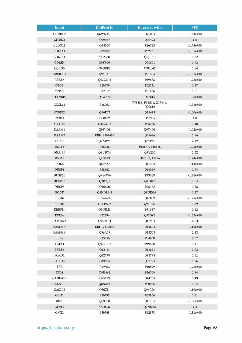
https://reactome.org Page 68
Input UniProt Id Interacts with #FC
CHRDL2 Q6WN34-2 O75093 -1.44e+00
CITED2 Q99967 Q09472 1.6
CLDN11 O75508 P26715 -1.74e+00
COL1A1 P02452 P02751 -1.31e+00
COL7A1 Q02388 Q5JRA6 1.22
CORIN Q9Y5Q5 O00501 1.93
CORO6 Q6QEF8 Q9UL45 2.34
CREB3L1 Q96BA8 P21854 -1.01e+00
CREB5 Q02930-3 P19883 -1.98e+00
CTGF P29279 P02751 1.37
CTPS1 P17812 P01106 1.51
CTTNBP2 Q8WZ74 O43815 -1.08e+00
CXCL12 P48061P78556, P13501, O15444,
Q99616-1.93e+00
CYFIP2 Q96F07 Q13485 -1.00e+00
CYR61 O00622 O60906 1.8
CYTH3 O43739-2 P53365 1.18
DAAM1 Q9Y4D1 Q9Y4D1 -1.02e+00
DAAM2 EBI-12904486 Q08426 1.66
DCXR Q7Z4W1 Q7Z4W1 2.12
DDIT3 P35638 P18847, P18848 -1.05e+00
DNAJB4 Q9UDY4 Q9Y230 1.52
DNM1 Q05193 Q05193, 15996 -1.74e+00
DOK6 Q6PKX4 Q16288 -1.14e+00
DUSP1 P28562 Q16539 2.94
DUSP10 Q9Y6W6 P49639 -1.22e+00
DUSP23 Q9BVJ7 Q8TBC4 1.34
DUSP5 Q16690 P28482 1.28
EOGT Q5NDL2-3 Q9UKG4 1.27
EPHB2 P54763 Q13009 -1.77e+00
EPHB6 O15197-2 Q96RU7 1.29
ERRFI1 Q9UJM3 P31947 2.42
EVI2A P22794 Q9Y5Z9 -1.62e+00
FAM107A O95990-4 Q12933 4.63
FAM46A EBI-12149039 O14503 -1.15e+00
FAM46B Q96A09 O14503 2.33
FBN2 P35556 P04608 2.07
FGF14 Q92915-2 P49638 1.11
FKBP5 Q13451 Q15831 3.93
FOXO1 Q12778 Q92793 2.31
FOXO3 O43524 Q92793 1.26
FST P19883 P10599 -1.78e+00
FZD8 Q9H461 P56704 2.44
GADD45B O75293 O14733 1.43
GALNT15 Q8N3T1 P30825 1.54
GAS2L3 Q86XJ1 Q96GD4 -1.10e+00
GCH1 P30793 P63104 1.61
GDF15 Q99988 Q13185 -1.86e+00
GFPT2 O94808 Q9NUX5 1.3
GNG2 P59768 P62873 -1.11e+00

https://reactome.org Page 69
Input UniProt Id Interacts with #FC
GPM6B P35803 P31645 2.63
GPR37 O15354 Q01959 1.46
GRAMD4 Q6IC98 Q96QG7 -1.18e+00
HBA1 P69905 P0DMV8 -3.38e+00
HMGB2 P26583 P48729 1.22
HMOX1 P09601 Q9NUX5 -1.35e+00
HSPA12B Q96MM6 Q12933 -2.37e+00
HSPA2 P54652 P11142 2.55
HSPA4L O95757 P11142 -1.28e+00
IER3 P46695 Q9UKG4 -1.18e+00
IFIT1 P09914 Q86WV6 -1.63e+00
IL12A P29459 P29460 -2.01e+00
IL34 Q6ZMJ4-1 P07333 -2.10e+00
IMPA2 O14732 Q8TBC4 2.31
ING2 Q9H160 P12004 1.27
INS-IGF2 F8WCM5 P16150 -5.88e+00
IRS2 Q9Y4H2 Q96EB6 2.15
ITGA2 P17301 P05556 -1.04e+00
JADE1 Q6IE81 O75161 1.22
JUN P05412 P18847, P18848 -1.08e+00
KANK1 Q14678 Q9UQB8 1.22
KIAA0408 Q6ZU52 O43639 1.5
KLF15 Q9UIH9 Q9UKG1 4.56
KLF5 Q13887 P0CG48 2.55
KLF6 Q99612 Q04206 1.09
KLHL42 Q9P2K6 P01189 1.04
LDB2 O43679 O00560 -1.32e+00
LHFPL2 Q6ZUX7 P08034 -1.26e+00
LIF P15018 P40189 -2.54e+00
LIMS2 Q7Z4I7-5 Q96PM5 1.27
MAP1LC3C Q9BXW4 Q96A56 2.53
MAP3K8 P41279 P19838, Q8NFZ5 1.12
MEST Q5EB52 Q05329 -1.81e+00
MFSD6 Q6ZSS7 P29033 1.04
MMD Q15546 Q3SXY8 1.64
MORF4L2 Q15014 Q99856 1
MPP3 Q13368 Q9HAP6 1.52
MPST P25325 O96006 1.01
MX1 P20591 Q13507 -1.01e+00
MYADM Q96S97 Q9Y624 1.48
NAMPT P43490 P43490 1.03
NCOA3 Q9Y6Q9 Q09472, Q92793 1.41
NEDD4L Q96PU5-5 O15105 -1.06e+00
NEDD9 Q14511 Q8IZD9 1.65
NEGR1 Q7Z3B1 P61916 1.46
NEK11 Q8NG66 P51955 -1.30e+00
NF2 P35240 Q8NI35 1.02
NFIL3 Q16649 P16220 1.14
NFYB P25208 P04637 1.03
NKD2 Q969F2 Q96PM5 1.38

https://reactome.org Page 70
Input UniProt Id Interacts with #FC
NOV P48745 O43559 -2.55e+00
NPAS2 Q99743 O43639 1.12
NR3C1 P04150 P31948 -1.01e+00
NR4A1 P22736 Q9Y4D7 1.34
NR4A3 Q92570 Q9H2F3 2.11
NRP2 Q99435-2 P49639 1.03
NUAK1 O60285 P04637 1.37
OSBPL3 Q9H4L5 P31946 -1.09e+00
P2RX7 Q99572 P14373 -1.94e+00
PAFAH1B3 Q15102 P19320 -1.26e+00
PER1 J3QSH9 O15055 3.15
PER3 P56645 O96017 -2.25e+00
PGF P49763 P17948 -1.27e+00
PHC2 Q8IXK0 P53350 1.37
PIK3R1 P27986-2, P27986 P48023, O15524 1.62
PIK3R3 Q92569 Q06187 -1.70e+00
PKIA P61925 O43663 -1.34e+00
PLEKHG4 Q58EX7 Q9NQ94 -1.35e+00
PLPP3 O14495 O60716 -1.02e+00
PPARG P37231 Q9UBK2 1.03
PPP1CB P62140 Q9UQK1 1.06
PPP1R14A Q96A00 P48729 2.81
PPTC7 Q8NI37 P41182 1.19
PRDM1 O75626 O75925 1.26
PRKCE Q02156 P08238 -1.23e+00
PRR5L Q6MZQ0 Q14192 1.61
PTK2B Q14289 P06241 1.11
PTX3 P26022 P09038 2.21
QPRT Q15274 Q15274 -1.23e+00
RAC3 Q9Y6Q9 Q09472, Q92793 1.47
RAP2B P61225 P19320 -1.28e+00
RASD1 Q9Y272 O75525 2.17
RASSF2 P50749 O60341 -1.09e+00
REV3L O60673 Q9UI95 1.12
RHOBTB3 O94955 O14503 1.04
RHOJ Q9H4E5 Q13177 -1.20e+00
RNF144B Q7Z419 Q07812 2.2
ROBO1 Q9Y6N7 O75044 -1.14e+00
RSPO2 Q6UXX9-2 Q92504 -1.48e+00
SAMD4A Q9UPU9-3 O95429 1.19
SAMHD1 Q9Y3Z3 O75923 3.84
SAP30 O75446 Q16576 1.22
SAT1 Q9H2H9 P24593 1.32
SCD O00767 P08034 -1.98e+00
SLC22A23 A1A5C7-2 P27352 -1.28e+00
SLC7A14 Q8TBB6 P63027 -2.84e+00
SLC7A6 Q92536 Q9UPQ8 1.64
SMIM3 Q9BZL3 Q9H2H9 1.26
SOCS1 O15524 P46109 -1.15e+00
SORT1 Q99523 P01138 2.21

https://reactome.org Page 71
Input UniProt Id Interacts with #FC
SOX4 Q06945 P04637 -2.38e+00
SQOR Q9Y6N5 P01106 1.01
STOM P27105 P50281 1.42
SUN2 Q9UH99 P52292 1.88
SYNPO2 Q9UMS6 Q13418 1.72
TBX18 O95935 Q13077 -1.81e+00
TCEAL4 Q96EI5 Q93009 1.52
TENM4 Q6N022 Q92608 -1.45e+00
TGFB2 P61812 P05067 -1.62e+00
TGFBR2 P37173 Q8IX30 1.44
THBS1 P07996-PRO_0000035842 P16671 1.94
TJP2 Q9UDY2 Q14160 1.97
TMCC2 O75069 P02649 -1.05e+00
TMEM119 EBI-17423904 Q14973 -1.19e+00
TMEM35A Q53FP2 P07204 -1.67e+00
TNFAIP6 P98066 P10145 -2.50e+00
TNFRSF11B O00300 Q9Y6Q6 -1.54e+00
TNIK Q9UKE5 O14920 -1.15e+00
TRIB3 Q96RU7 P18848 -1.10e+00
TSC22D1 Q15714 Q15047 1.18
TSC22D3 Q99576-3 Q9Y6D9 3.32
TUBA1A Q71U36 Q9Y241 -1.29e+00
TUBA4A P68366 P30622 -1.45e+00
TUBB P07437 P05412 -1.21e+00
TUBB3 Q13509 Q5S007 -1.49e+00
TXNRD1 Q16881 Q03135 1.53
UBASH3B Q8TF42 P29353 1.1
USP53 Q70EK8 P46109 2.3
VCAM1 P19320 P31948 -3.60e+00
VEGFA P15692-4 P35968, P17948 -1.23e+00
VGLL3 A8MV65-2 Q8TDS5 1.34
WASF3 Q9UPY6-2 P61024 1.08
YIF1B Q5BJH7 O15354 1.13
ZBTB16 Q05516 P05231 7.05
ZHX3 Q9H4I2 Q9UBE8 1.13
Input ChEBI Id Interacts with #FC
CYTH3 O43739 16618 1.18
DNM1 Q05193 15996 -1.74e+00
FAM20A Q96MK3 15422 1.09
FGD4 Q96M96 17283 2.26
MX1 P20591 18348 -1.01e+00
PLA2G4A P47712 18348 -2.45e+00

https://reactome.org Page 72
7. Identifiers not found
These 124 identifiers were not found neither mapped to any entity in Reactome.
10-Mar AC005674.2 AC007620.2 AC010255.1 AC011611.3 AC012349.1 AC027237.3 AC079467.1
AC090673.1 AC097534.2 AC098847.2 AC138866.2 AFAP1L1 AIF1L AKAP2 AL035681.1
AL133415.1 AL133453.1 ANGPTL1 ANGPTL7 AP001542.3 C10orf10 C14orf132 C20orf96
C2CD2 CADPS2 CCDC69 CCDC71L CLIC6 CORO2B CPA4 CPED1
CRIM1 DANCR DNM1P47 ENC1 EVA1C FAIM2 FAM107B FAM171B
FAM198B FAM43A FAM46C FER1L6 FIBIN FRZB GALM GDF7
GPRC5B GRAMD2B GXYLT2 HAPLN3 HSPB3 IGDCC4 IGFLR1 IGSF10
KAZALD1 KCTD12 KIAA1755 KLF9 KLHL23 LBH LINC00968 LINC02202
LRRC15 LRRC17 MAMDC2 MARCKSL1 MEX3B MICAL2 MINDY2 MXRA5
NAV3 NEK10 NEXN NXPH3 NYNRIN OLFML2A PAPLN PHOSPHO2
PKDCC PLXDC1 PLXDC2 POM121L9P PRSS35 PXDC1 RAB11FIP1 RASL10B
RASL11B RASL12 RELL1 RGMB-AS1 RND2RPL36A-
HNRNPH2RRAD RTL3
RWDD4 SAMD5 SGCD SIPA1L2 SLC45A1 SMTNL2 SSH2 STC1
STEAP1 STEAP4 STIM2 STK17B TAGLN TCEAL3 TEX2 TIMP4
TLDC2 TM4SF1 TMEM200A TMEM217 TRNP1 TSPAN11 ULK2 VWCE
WDR63 WFDC1 YPEL4 ZNF821