Pathophysiology and therapy of pruritus in allergic and ...
17
REVIEW ARTICLE Pathophysiology and therapy of pruritus in allergic and atopic diseases J. Buddenkotte 1 & M. Steinhoff 2 1 Deparment of Dermatology, Boltzmann Institute for Cell- and Immunobiology of the Skin, University Hospital Mu ¨ nster, Mu ¨ nster, Germany; 2 Depatments of Dermatology and Surgery, University of California, San Francisco, CA, USA To cite this article: Buddenkotte J, Steinhoff M. Pathophysiology and therapy of pruritus in allergic and atopic diseases. Allergy 2010; 65: 805–821. Itch transmission by the nervous system The skin constitutes a barrier between ‘outside’ environment and ‘inner’ body. Therefore, one of its main tasks is to pro- tect the organism against harmful influences from the out- side. To fullfil this task, the skin is armed with an effective communication and control system. In all layers of the skin, specialized sensory and efferent nerve branches appear to form an overall dense nerval network. One main ‘outside- to-inside’ interaction causes sensations of itch. Pruritus is reg- ularly defined as an unpleasant sensation provoking the desire to scratch (1) and constitutes an essential feature of atopic dermatitis (AD) (2, 3). Based on early psychophysical studies on itch (4), it was believed that itch is nothing but a low-intensity pain. Con- cepts from those times declared that itch is enciphered in spe- cific patterns of action potentials running through ‘pain fibres’ or that itch emerges from combinations of other pri- mary sensory signals. However, it is clear at this stage that pruritoception is a distinct entity just as nociception is a dis- tinct entity (5–8). Therefore, the new concept of itch trans- mission is based on an important proposition: the existence of a central itch-specific neuronal pathway, in other words, it envisages the existence of a sensory system for pruritoception that is distinct from the sensory system for nociception (Fig. 1). Pruritus can be triggered by localized, systemic, peripheral or central stimuli. To relay itch information to different cere- bric areas is the specific function of a subpopulation of the dense nerval network in the skin, the unmyelienated C-polymo- dal nociceptive neurons (in general being histamine-sensitive). The free nerve endings referred to as cutaneous terminals reside in the epidermis, papillary dermis and around skin appendages and are qualified to apprehend endogenous or exogenous itch causing agents through an armada of relevant receptors. These receptors detect their corresponding ‘itchy’ ligands and send either an electrical signal to the central Keywords atopic dermatitis; itch; nerve; pathophysiology; pruritus; therapy. Correspondence Martin Steinhoff, MD, PhD, Departments of Dermatology and Surgery, University of California San Francisco, 513 Parnassus Ave, Room S-1268, 94143 San Fransico, CA, USA. Tel.: +1 415 476 6978 Fax: +1 415 476 0936 E-mail: [email protected] DOI:10.1111/j.1398-9995.2010.01995.x Edited by: Jean Bousquet Abstract Pruritus (itch) is a major characteristic and one of the most debiliating symptoms in allergic and atopic diseases and the diagnostic hallmark of atopic dermatitis. Pruri- tus is regularly defined as an unpleasant sensation provoking the desire to scratch. Although we achieved rather good knowledge about certain inducers of itch such as neuropeptides, amines, l-opioids, cytokines and proteases, for example, less is known about the pathophysiological specifities among the different diseases, and the therapeutic consequences which may derive thereoff. This review dissects the role of mediators, receptors and itch inhibitors on peripheral nerve endings, dorsal root ganglia, the spinal cord and the CNS leading to the amplification or – vice versa – suppression of pruritus. As the treatment of pruritus in allergic and atopic skin dis- ease is still not satisfactory, knowing these pathways and mechanisms may lead to novel therapeutic approaches against this frequently encountered skin symptom. Abbreviations ACh, acetylcholine; AD, atopic dermatitis; CB, cannabinoid receptor; CGRP, calcitonin gene-related peptide; CyA, cyclosporin A; GRP, gastrin-releasing peptide; GRPR, gastrin-releasing peptide receptor; IL, interleukin; IFN, interferon; NGF, nerve growth factor; NKR, neurokinin receptor; NPY, neuropeptide Y; NT, neurotrophin; PAR, protease-activated receptor; PGP, protein gene product; SP, substance P; TRPV1, transient receptor potential vanilloid (capsaicin) receptor 1; VIP, vasoactive intestinal polypeptide. Allergy Allergy 65 (2010) 805–821 ª 2010 John Wiley & Sons A/S 805
Transcript of Pathophysiology and therapy of pruritus in allergic and ...

REVIEW ARTICLE
Pathophysiology and therapy of pruritus in allergic andatopic diseasesJ. Buddenkotte1 & M. Steinhoff2
1Deparment of Dermatology, Boltzmann Institute for Cell- and Immunobiology of the Skin, University Hospital Munster, Munster, Germany;2Depatments of Dermatology and Surgery, University of California, San Francisco, CA, USA
To cite this article: Buddenkotte J, Steinhoff M. Pathophysiology and therapy of pruritus in allergic and atopic diseases. Allergy 2010; 65: 805–821.
Itch transmission by the nervous system
The skin constitutes a barrier between ‘outside’ environment
and ‘inner’ body. Therefore, one of its main tasks is to pro-
tect the organism against harmful influences from the out-
side. To fullfil this task, the skin is armed with an effective
communication and control system. In all layers of the skin,
specialized sensory and efferent nerve branches appear to
form an overall dense nerval network. One main ‘outside-
to-inside’ interaction causes sensations of itch. Pruritus is reg-
ularly defined as an unpleasant sensation provoking the
desire to scratch (1) and constitutes an essential feature of
atopic dermatitis (AD) (2, 3).
Based on early psychophysical studies on itch (4), it was
believed that itch is nothing but a low-intensity pain. Con-
cepts from those times declared that itch is enciphered in spe-
cific patterns of action potentials running through ‘pain
fibres’ or that itch emerges from combinations of other pri-
mary sensory signals. However, it is clear at this stage that
pruritoception is a distinct entity just as nociception is a dis-
tinct entity (5–8). Therefore, the new concept of itch trans-
mission is based on an important proposition: the existence
of a central itch-specific neuronal pathway, in other words, it
envisages the existence of a sensory system for pruritoception
that is distinct from the sensory system for nociception
(Fig. 1).
Pruritus can be triggered by localized, systemic, peripheral
or central stimuli. To relay itch information to different cere-
bric areas is the specific function of a subpopulation of the
dense nerval network in the skin, the unmyelienated C-polymo-
dal nociceptive neurons (in general being histamine-sensitive).
The free nerve endings referred to as cutaneous terminals
reside in the epidermis, papillary dermis and around skin
appendages and are qualified to apprehend endogenous or
exogenous itch causing agents through an armada of relevant
receptors. These receptors detect their corresponding ‘itchy’
ligands and send either an electrical signal to the central
Keywords
atopic dermatitis; itch; nerve;
pathophysiology; pruritus; therapy.
Correspondence
Martin Steinhoff, MD, PhD, Departments of
Dermatology and Surgery, University of
California San Francisco, 513 Parnassus
Ave, Room S-1268, 94143 San Fransico,
CA, USA.
Tel.: +1 415 476 6978
Fax: +1 415 476 0936
E-mail: [email protected]
DOI:10.1111/j.1398-9995.2010.01995.x
Edited by: Jean Bousquet
Abstract
Pruritus (itch) is a major characteristic and one of the most debiliating symptoms in
allergic and atopic diseases and the diagnostic hallmark of atopic dermatitis. Pruri-
tus is regularly defined as an unpleasant sensation provoking the desire to scratch.
Although we achieved rather good knowledge about certain inducers of itch such as
neuropeptides, amines, l-opioids, cytokines and proteases, for example, less is
known about the pathophysiological specifities among the different diseases, and the
therapeutic consequences which may derive thereoff. This review dissects the role of
mediators, receptors and itch inhibitors on peripheral nerve endings, dorsal root
ganglia, the spinal cord and the CNS leading to the amplification or – vice versa –
suppression of pruritus. As the treatment of pruritus in allergic and atopic skin dis-
ease is still not satisfactory, knowing these pathways and mechanisms may lead to
novel therapeutic approaches against this frequently encountered skin symptom.
Abbreviations
ACh, acetylcholine; AD, atopic dermatitis; CB, cannabinoid
receptor; CGRP, calcitonin gene-related peptide; CyA, cyclosporin
A; GRP, gastrin-releasing peptide; GRPR, gastrin-releasing peptide
receptor; IL, interleukin; IFN, interferon; NGF, nerve growth factor;
NKR, neurokinin receptor; NPY, neuropeptide Y; NT, neurotrophin;
PAR, protease-activated receptor; PGP, protein gene product; SP,
substance P; TRPV1, transient receptor potential vanilloid
(capsaicin) receptor 1; VIP, vasoactive intestinal polypeptide.
Allergy
Allergy 65 (2010) 805–821 ª 2010 John Wiley & Sons A/S 805

nervous system or trigger a direct inflammatory response by
antidromic impulse transmission. From the foregoing, it is
apparent that the nature of the ligand present and the corre-
sponding accessory receptor determine the nerval reaction (7).
Sensory cutaneous nerves transmit the pruritic information
to dorsal root ganglions and from there it reaches the spinal
cord where it can be modulated. From the lamina I, a specific
side within the dorsal horn of the spinal cord, the signal is
projected to the thalamus (6, 9). On this passage, the signal
crosses to the contralateral side. Nerval structures pertinent to
the transmission of pruritic information in the spine are not
clarified to date, but a subset of histamine- and GRP-sensitive
neurons probably executes this assignment (5, 7, 10). A neuro-
toxic destruction approach carried out in rodents supplies evi-
dence for a key role of NK-1 receptor expressing neurons in
the transmission of itch information in the superficial spinal
dorsal horn (11). Notably, ablation of NK1R-positive neurons
compromises chronic pain behaviours in rat (12, 13), although
the confirmation for a direct role of the NK-1 receptor in
human spinal itch transmission is still lacking. From the thala-
mus, direct excitatory connections that consist of the anterior
cingulate cortex, the insular cortex (insula) and primary and
secondary somatosensory cortices take over (6, 9, 14–16).
Itch-specific mediators in the central nervous system are
elusive and so far not known. However, recent evidence
emerged that gastrin-releasing peptide receptor (GRPR), a
bombesin-like peptide receptor homologue that is specifically
expressed in the lamina I of the dorsal horn, might play a
crucial role in mediating itch sensations in the spinal cord
and might furthermore constitute a marker for central itch-
selective neurons (10). Strikingly, selective ablation of lamina
I neurons expressing GRPR in the spinal cord of mice
resulted in severe deficiencies in scratching responses to an
amarda of pruritogenic stimuli such as histamine, compound
48/80, serotonin, endothelin-1, PAR2-activating peptide and
the anti-malaria drug chloroquine (17). The severity of this
Spinal cord
DRG
pru
ritu
s / p
ain
e. g. GPR/GPRPPAR2?, opioids?,
NK1R?
Protons, kinins,proteases, amines,
prostaglandins, leukotriens,cannabinoids, endothelins,
neutrophins, drugsNeuropeptides
Immune cells(mast cells,
T cells)
Bloodvessel
Langerhans cell
Epidermis
IL-2, IL-8, IL-31,IFN-γ ?
Antigen-presentation
Keratinocyte activation and apoptosiscytokine, chemokine, neurotrophin, pro- stanoid, opioid, protease releasereceptor upregulation
AChβ-endorphinproteases
Hyperkeratosisplasma extravastionoedema
Peripheralnerve ending
(C-fibre)
Allergens,house dust mite,
Staphylococcus aureus,dermatophytes
Topical / systemic drugs,injury
Brain
Figure 1 General neuroanatomical and neurophysiologcial path-
ways activated during pruritus (pruritogenic itch). Exogenous or
endogenous meditors stimulate selective subtypes of peripheral C
fibre nerve endings of primary afferent neurons in the epidermis or
dermis. High-affinity receptors for various pruritogenic mediators
transmit the stimulus, via not completely understood intracellular
signaling pathways, from the periphery to the dorsal root ganglia
(DRG), and the spinal cord. DRGs can modulate this stimulus on
the transcriptional and posttranscriptional level, thereby modulating
peripheral and central nerve endings. Within the spinal cord, itch
signals can be also modulated. From lamina 1, a selective area
within the the dorsal horn of spinal cord, the signal will be trans-
mitted to the CNS after crossing to the contralateral side. Activa-
tion of specific areas in the CNS results in the perception of itch
leading to ‘discomfort’ and an acute or chronic scratch response.
Additionally, the associated peripheral axon reflex may lead to the
release of mast cell-stimulating neuropeptides (e.g. substance P)
thereby amplifying pruritus via release of histamine, tryptase and
IL-31, for example. This figure does not consider the complex inter-
action between pain and itch fibres on the spinal cord level where
GRPR, opioid receptors, NK1R (post-synaptic), PAR2 (pre-synaptic
primary afferents) and probably other mediators/ receptors can
exert exciatatory or inhibitory influences.
Pruritus in allergic and atopic diseases Buddenkotte and Steinhoff
806 Allergy 65 (2010) 805–821 ª 2010 John Wiley & Sons A/S

observation is that GPRP transmits itch signals independent
of the nature of the pruritogenic stimulus. In fact, GRPR
constitutes the long-sought labelled line for itch sensation in
the spinal cord.
Cutaneous neuroreceptors and mediators: induction of
pruritus
The induction of itch is accomplished by a variety of agents
that ineract with a multitude of receptors on free nerve end-
ings. Over the last decade of itch research, the number of
pruritogenic agents has grown far beyond the usual suspect
histamine. But it is the lot of several newly reported pruritic
agents to lack appropriate recognition by the scientific com-
munity, while the potency of histamine to induce itch contin-
ues to be overestimated. In the following, the main mediators
of cutaneous pruritus will be introduced and briefly described
(see also Table 1).
Histamine
About 80 years ago, Lewis reported that intracutaneous
injection of histamine causes symptoms characteristic of neu-
rogenic inflammation – redness, wheal and flare – along with
pruritus (18, 19). Furthermore, Williams (20) suggested that
histamine may play a role in the pathogenesis of AD because
intramuscular histamine injections resulted in pruritus. Based
on these initial experiments, histamine has doubtlessly
become the most exhaustingly investigated ‘itchy’ agonist.
Over the years, elevated histamine levels in lesional and non-
lesion al skin were detected (21, 22), topographically associat-
ing histamine and itch. The distinct source of skin histamine
was attributed to mast cells and keratinocytes (23–25). Also
to date, four distinct histamine receptors (H1-4R) have been
identified that histamine is capable of activating (26). At least
two of them, H1R and H2R, are present on cutaneous sen-
sory nerve fibres (23, 27). Therewith, all ingredients have
been provided to support histamine in being an important
pruritogen, especially as H1R- (and less so H2R) antagonists
have been demonstrated to reduce itch in numerous clinical
trials (7, 28–30). However, re-evaluating the histamine con-
tent of the skin, recent investigations could not verify
increased histamine levels in all pruritic diseases indicating a
selective role of various mediators among the different pruri-
tic diseases (31). And while re-evaluating the potency of
histamine to induce itch, it was shown in recent studies that
small doses of histamine fail to produce itch but still are suf-
ficient to produce oedema and erythema upon intracutaneous
Table 1 Mediators of itch in atopic dermatitis
Substrate Provocation of itch Mechanism
Spinal inductor of itch
GRP + Binding to GRPR of the spinal cord
Cutaneous inductors of itch
Histamine (+) Binding to histamine receptors on sensory nerve fibres
Neuropeptides (e.g. substance P) + Mast cell degranulation, increased concentration in
lesional skin
Acetylcholine + Central sensitization?
Tryptase kallikreins, cathepsin S + Binding to PAR2 on sensory nerve fibres
Cytokines: Interleukin 2 + Possible release of various mediators
Interleukin 8 )Interleukin 31 +
Neurotrophin-4 + m.n.n.
Eosinophils +/? Release mediators like PAF, leucotriens; histamine,
proteinase liberation
Platelet activating factor + Histamine liberator
Leukotriens + m.n.n. (LTB4?)
Cutaneous suppressors of itch
Cannabinoids Interruption of itch transmission Binding to CB1 and CB2 on cutaneous sensory nerve
fibres
Opioid peptides Induction of itch-inhibiting neurons
on spinal level; suppression in the skin?
Binding to opioid receptors
TRP channels (Vanilloids) Suppression of itch transmission TRPV1, TRPV3 involved in itch
Direct or indirect effects on sensory nerves
suppressing itch
Interferon gamma Suppression of pruritus m.n.n. (IFN-c receptor on nerves?)
Calcineurin inhibitors Interruption of itch transmission Downregulation of pruritic cytokines by effecting T cells
Binding to TRPV1 on cutaneous sensory nerve fibres
Ameliorating neuropeptide release
Decreasing effects of neuropeptides on mast cells?
), no induction of itch; (+), induction of weak itch; +, clear induction of itch; m.n.n., mechanism not known.
Buddenkotte and Steinhoff Pruritus in allergic and atopic diseases
Allergy 65 (2010) 805–821 ª 2010 John Wiley & Sons A/S 807
injection (32–35). Although in urticaria, the itchy role of
histamine is well established, yet in patients with AD, intra-
cutaneous injection and also iontophoretical application of
histamine did not provoke an increase but a reduction in itch
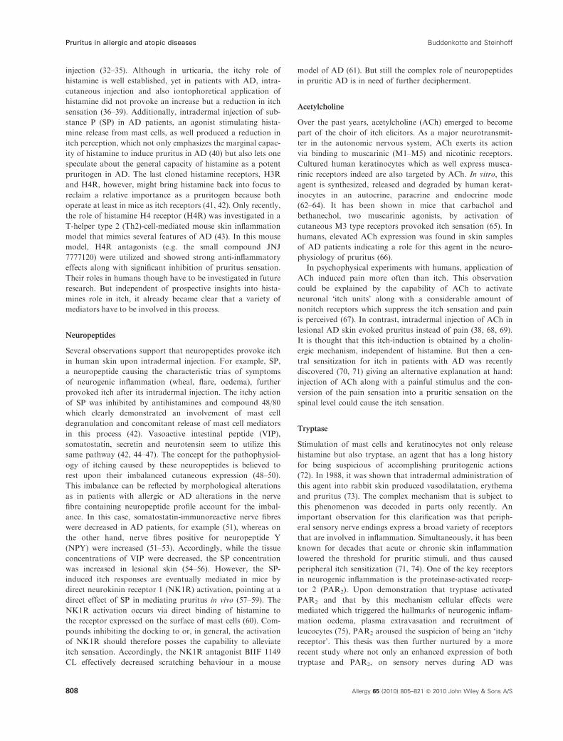
sensation (36–39). Additionally, intradermal injection of sub-
stance P (SP) in AD patients, an agonist stimulating hista-
mine release from mast cells, as well produced a reduction in
itch perception, which not only emphasizes the marginal capac-
ity of histamine to induce pruritus in AD (40) but also lets one
speculate about the general capacity of histamine as a potent
pruritogen in AD. The last cloned histamine receptors, H3R
and H4R, however, might bring histamine back into focus to
reclaim a relative importance as a pruritogen because both
operate at least in mice as itch receptors (41, 42). Only recently,
the role of histamine H4 receptor (H4R) was investigated in a
T-helper type 2 (Th2)-cell-mediated mouse skin inflammation
model that mimics several features of AD (43). In this mouse
model, H4R antagonists (e.g. the small compound JNJ
7777120) were utilized and showed strong anti-inflammatory
effects along with significant inhibition of pruritus sensation.
Their roles in humans though have to be investigated in future
research. But independent of prospective insights into hista-
mines role in itch, it already became clear that a variety of
mediators have to be involved in this process.
Neuropeptides
Several observations support that neuropeptides provoke itch
in human skin upon intradermal injection. For example, SP,
a neuropeptide causing the characteristic trias of symptoms
of neurogenic inflammation (wheal, flare, oedema), further
provoked itch after its intradermal injection. The itchy action
of SP was inhibited by antihistamines and compound 48/80
which clearly demonstrated an involvement of mast cell
degranulation and concomitant release of mast cell mediators
in this process (42). Vasoactive intestinal peptide (VIP),
somatostatin, secretin and neurotensin seem to utilize this
same pathway (42, 44–47). The concept for the pathophysiol-
ogy of itching caused by these neuropeptides is believed to
rest upon their imbalanced cutaneous expression (48–50).
This imbalance can be reflected by morphological alterations
as in patients with allergic or AD alterations in the nerve
fibre containing neuropeptide profile account for the imbal-
ance. In this case, somatostatin-immunoreactive nerve fibres
were decreased in AD patients, for example (51), whereas on
the other hand, nerve fibres positive for neuropeptide Y
(NPY) were increased (51–53). Accordingly, while the tissue
concentrations of VIP were decreased, the SP concentration
was increased in lesional skin (54–56). However, the SP-
induced itch responses are eventually mediated in mice by
direct neurokinin receptor 1 (NK1R) activation, pointing at a
direct effect of SP in mediating pruritus in vivo (57–59). The
NK1R activation occurs via direct binding of histamine to
the receptor expressed on the surface of mast cells (60). Com-
pounds inhibiting the docking to or, in general, the activation
of NK1R should therefore posses the capability to alleviate
itch sensation. Accordingly, the NK1R antagonist BIIF 1149
CL effectively decreased scratching behaviour in a mouse
model of AD (61). But still the complex role of neuropeptides
in pruritic AD is in need of further decipherment.
Acetylcholine
Over the past years, acetylcholine (ACh) emerged to become
part of the choir of itch elicitors. As a major neurotransmit-
ter in the autonomic nervous system, ACh exerts its action
via binding to muscarinic (M1–M5) and nicotinic receptors.
Cultured human keratinocytes which as well express musca-
rinic receptors indeed are also targeted by ACh. In vitro, this
agent is synthesized, released and degraded by human kerat-
inocytes in an autocrine, paracrine and endocrine mode
(62–64). It has been shown in mice that carbachol and
bethanechol, two muscarinic agonists, by activation of
cutaneous M3 type receptors provoked itch sensation (65). In
humans, elevated ACh expression was found in skin samples
of AD patients indicating a role for this agent in the neuro-
physiology of pruritus (66).
In psychophysical experiments with humans, application of
ACh induced pain more often than itch. This observation
could be explained by the capability of ACh to activate
neuronal ‘itch units’ along with a considerable amount of
nonitch receptors which suppress the itch sensation and pain
is perceived (67). In contrast, intradermal injection of ACh in
lesional AD skin evoked pruritus instead of pain (38, 68, 69).
It is thought that this itch-induction is obtained by a cholin-
ergic mechanism, independent of histamine. But then a cen-
tral sensitization for itch in patients with AD was recently
discovered (70, 71) giving an alternative explanation at hand:
injection of ACh along with a painful stimulus and the con-
version of the pain sensation into a pruritic sensation on the
spinal level could cause the itch sensation.
Tryptase
Stimulation of mast cells and keratinocytes not only release
histamine but also tryptase, an agent that has a long history
for being suspicious of accomplishing pruritogenic actions
(72). In 1988, it was shown that intradermal administration of
this agent into rabbit skin produced vasodilatation, erythema
and pruritus (73). The complex mechanism that is subject to
this phenomenon was decoded in parts only recently. An
important observation for this clarification was that periph-
eral sensory nerve endings express a broad variety of receptors
that are involved in inflammation. Simultaneously, it has been
known for decades that acute or chronic skin inflammation
lowered the threshold for pruritic stimuli, and thus caused
peripheral itch sensitization (71, 74). One of the key receptors
in neurogenic inflammation is the proteinase-activated recep-
tor 2 (PAR2). Upon demonstration that tryptase activated
PAR2 and that by this mechanism cellular effects were
mediated which triggered the hallmarks of neurogenic inflam-
mation oedema, plasma extravasation and recruitment of
leucocytes (75), PAR2 aroused the suspicion of being an ‘itchy
receptor’. This thesis was then further nurtured by a more
recent study where not only an enhanced expression of both
tryptase and PAR2, on sensory nerves during AD was
Pruritus in allergic and atopic diseases Buddenkotte and Steinhoff
808 Allergy 65 (2010) 805–821 ª 2010 John Wiley & Sons A/S

detected but also the triggering of itching in AD patients by
PAR2 agonists was demonstrated (76). It is not clear to date
whether the remaining PAR family members, foremost PAR1
and PAR4, are also involved in the itch pathway, and under
which circumstances proteases may induce pain, inflammation
or pruritus in patients suffering from AD.
Mrgpr
Several G protein-coupled receptors have been shown to act
as key receptors in generating itch including histamine recep-
tors and PARs. This group has been extended by the work of
Liu et al. (77): Mrgprs (also termed Mrg/SNSR) are orphan
receptors grouped into several subfamilies (MrgprA1–A22,
MrgprB1–B13, MrgprC1–C14 and MrgprD–G). Mouse gen-
ome analysis revealed an existence of more than 50 members
distributed to these subfamilies. The function of Mrgprs
in vivo was so far an enigma but it was known that the expres-
sion of MrgprAs, MrgprB4, MrgprB5, MrgprC11, and
MrgprD, is restricted to subsets of small-diameter sensory
neurons in DRG and trigeminal ganglia (78, 79). To unveil
the Mrgpr function, targetet deletion of an Mrgpr gene cluster
located on mouse chromosome 7 was performed. Mrgpr-clus-
terD)/) mice were then challenged with pruritogenic agents.
Histamine and compound 48/80 induced itch behaviour in
Wildtype mice and Mrgpr-clusterD)/) mice of similar intensity.
Strikingly, chloroquine, an anti-malaria drug that is known
for pruritic side-effects, elicited itch in wildtype mice only
(77). The group could show that the essential receptor to
mediate the itchy action of chloroquine is MrgrpA3 and that
chloroquine-sensitive neurons (3–4% of total DRG neurons)
also respond to histamine and capsaicin. Interestingly, the
human Mrgpr family member MrgprX1 shares a similar
expression pattern with mouse MrgrpA3 (80) and also
responds to chloroquine treatment indicating a role for this
receptor in nonhistaminergic itch transmission in humans. But
still it will be interesting to see how these findings translate to
the human situation: chloroquine induced itch is common
among black Africans but less common among other races
(81). Still there is no doubt that the putative itchy action of
MrgprX1 and its importance in the pathophysiology of pru-
ritic diseases such as AD will be investigated soon. Alongside,
it is tempting to speculate which endogenous agonist(s) acti-
vate this subset of MrgprX1-positive primary sensory fibres in
the skin and whether an imbalance of its/their expression
might affect the outcome of chronic pruritus. Certainly the
discovery of Liu et al. may establish the ground for novel
anti-itch drugs targeted against itch-selective neurons.
Cutaneous neuroreceptors and mediators: suppresion
of pruritus
Endovanilloids and the TRPV ion channel family
Endovanilloids interact with TRPV1, an ion channel that
belongs to the superfamily of transient receptor potential
(TRP) channels. To date, six groups of molecules complete
this superfamily: the canonical (TRPC), the melastatin
(TRPM), the polycystin (TRPP), the ankyrin transmembrane
protein 1 (TRPA), the mucolipin (TRPML) and the vanilloid
(TRPV) subfamilies. All members of this superfamily act as
nonselective calcium-permeable sensory transduction channels
(82). Endovanilloids constitute a group of itch mediators to
which heterogenous agents such as eicosanoids, histamine,
bradykinin, ATP and various neurotrophins (NTs) (83–86)
belong where all agents share endovanilloid functions (87).
These agents either directly and/or indirectly activate/sensitize
TRPV1 (84, 88, 89).
Originally, TRPV1 was found to be expressed by nocicep-
tive sensory neurons (89) as an integrator of different pain-
inducing stimuli. Its most well-known activator is capsaicin,
the pungent ingredient of hot chili peppers. Administration
of this compound excites (88) but then desensitize sensory
afferents via TRPV1 activation, a mechanism that is utilized
to alleviate pain and itch in numerous skin diseases (88,
90–92). More precisely, vanilloid administration leads to a
depletion of neuropeptides in the C-fibres, which disrupts the
communication between mast cells and skin sensory neurons
(91–93). Interestingly, also the calcineurin inhibitors tacroli-
mus (94) and pimecrolimus (95) bind to TRPV1 suggesting a
mode of action for these clinically important compounds.
Only recently, functional TRPV1 channels were reported
on numerous nonneuronal cell types (96–98), including
human epidermal and hair follicle keratinocytes, endothelial
cells, dermal mast cells and dendritic cells (99–102). TRPV1
activation resulted in the release of pruritogenic cytokine
mediators from several of these nonneuronal cells. In kerati-
nocyte, TRPV1 was furthermore reported to mediate prolifer-
ation, differentiation and apoptosis, respectively (103, 104)
but recent results utilizing a functional approach with both
systemic and local resiniferatoxin (RTX) treatment question
a functional expression of TRPV1 in primary human
keratinocyte (105). It will be a subject of further detailed
research to show whether endovanilloid itch mediators,
besides acting on their cognate receptors, activate/sensitize
TRPV1 expressed on itch-mediating sensory neurons only or
also address TRPV1 expressed on other skin cells since the
specificity of current antibodies against TRPV1 are question-
able. In other words, topically applied capsaicin may not
only desensitize TRPV1-mediated signalling in neuronal cells
but may also provoke same in the many other skin cells to
counteract a pruritogenic outcome.
Next to TRPV1, itching sensitization might also be related
to the activation of other TRPs expressed in the skin, sensory
fibres and keratinocytes including TRPV2, TRPV3, TRPV4,
TRPA1 and TRPM8 (106) (Fig. 2). In fact, an important role
for TRPV3 in pruritus has been shown recently. Asakawa
and colleagues discovered an amino-acid substitution
(G573S) in TRPV3 that led to an increase in ion channel
activity in keratinocytes which caused a spontaneous allergic
and pruritic dermatitis in mice (107).
Cannabinoids
Another suspect in the itch department is the cannabinoid
system. Cannabinoid receptor-1 (CB1) is co-localized with
Buddenkotte and Steinhoff Pruritus in allergic and atopic diseases
Allergy 65 (2010) 805–821 ª 2010 John Wiley & Sons A/S 809
TRPV1 in sensory neurons (108) and cannabinoids interact
with the TRPV1-signalling pathway. This interaction triggers
a switch of their neuronal effect from inhibition (109) to exci-
tation and sensitization (110) under inflammatory conditions.
In fact, a topically applied synthetic cannabinoid, HU210,
suppressed histamine-induced pruritus and reduced axon
reflex erythema (111). CB1 as well as CB2 were also found to
be expressed in nonneuronal cells of the skin such as mast
cells (112, 113). In consequence, cannabinoid receptors may
be involved in the neuronal–nonneuronal cellular network of
pruritogenic stimuli arising in/from skin. In addition, these
findings support an antipruritic role of the cannabinoid sys-
tem which could be exploited for new therapy approaches in
itch-accompanied skin diseases. In fact, preliminary studies
with a cannabinoid (palmitoylethanolamin) containing cream
assign anti-inflammtory and antipruritic properties to this
compound in AD (114). A very appealing consideration for
administration of cannabinoid in itch therapy is the possibil-
ity of co-administration with a TRPV1 agonist. Thereby, the
patient would benefit from (i) the antipruritic impact of both
agents and (ii) the mitigative effect of the cannabinoid on an
acute burning sensation that is elicited by exclusive capsaicin
administration.
Opioids
It is a common habit to counteract itch by scratching. To be
more precise, itch is counteracted only by a painful stimulus.
In experimental settings, this painful stimulus can be mim-
icked by various painful thermal, mechanical and chemical
stimuli (115). For instance, it is well demonstrated that electri-
cal stimuli can reduce an itch sensation for hours (116). The
potency of itch-inhibition though appears to be dependent on
the nature of the applied stimulus: noxious heat stimuli and
scratching produce a stronger itch inhibition than noxious
cold stimuli (117). Conversely, it is imaginable that analgesia
may reduce the competence of pain to inhibit itch whereby
pruritic sensation is amplified (118). This phenomenon is
observed when l-opioid receptor agonists are spinally admin-
istered as segmental pruritus arouses along with the desired
segmental analgesia (119–125). Accordingly, opioid receptor
antagonists may have antipruritic effects in pruritic diseases
(126–132). The pruritic l-opioid receptor is expressed by
C-fibres. During pain perception, l-opioid receptor antago-
nists such as naxolone are not ideal antipuritics because such
compounds can reverse opioid analgesia concurrently (133,
134). However, a combination of high-dose intrathecal opioids
with postoperative intravenous naloxone provided excellent
analgesia with minor pruritic side-effects (135).
The antipruritic j-opioid receptor (KOR), expressed by
Ad-fibres, seems to be a more promising target to ameliorate
itching after spinal analgesia administration without engaging
in the desired antinociception (136). When treated chronically
with U-50488H, a selective KOR agonist, treated monkeys
displayed an excessive scratching activity upon agonist-
withdrawal (137) indicating an antipruritic role of KOR
Stimuli:Cold, heat, chemical agonists,
irritants, mechanical stress (geralding) ?,allergens (indirect) ?
Human:TRPV1
TRPV3 ?TRPV 4 ?TRPA1 ?
Human:TRPV1, TRPV2 ?,TRPV3, TRPV4 ?,TRPM8 ?, TRPA1 ?
Na+
Ca2- Na+
Ca2+
Ca2+
CaV
Fibreterminal
AP
Keratinocyte
Human epidermis:TRPV1TRPV3TRPV4TRPA1
Dorsal rootganglion To spinal
cord
?
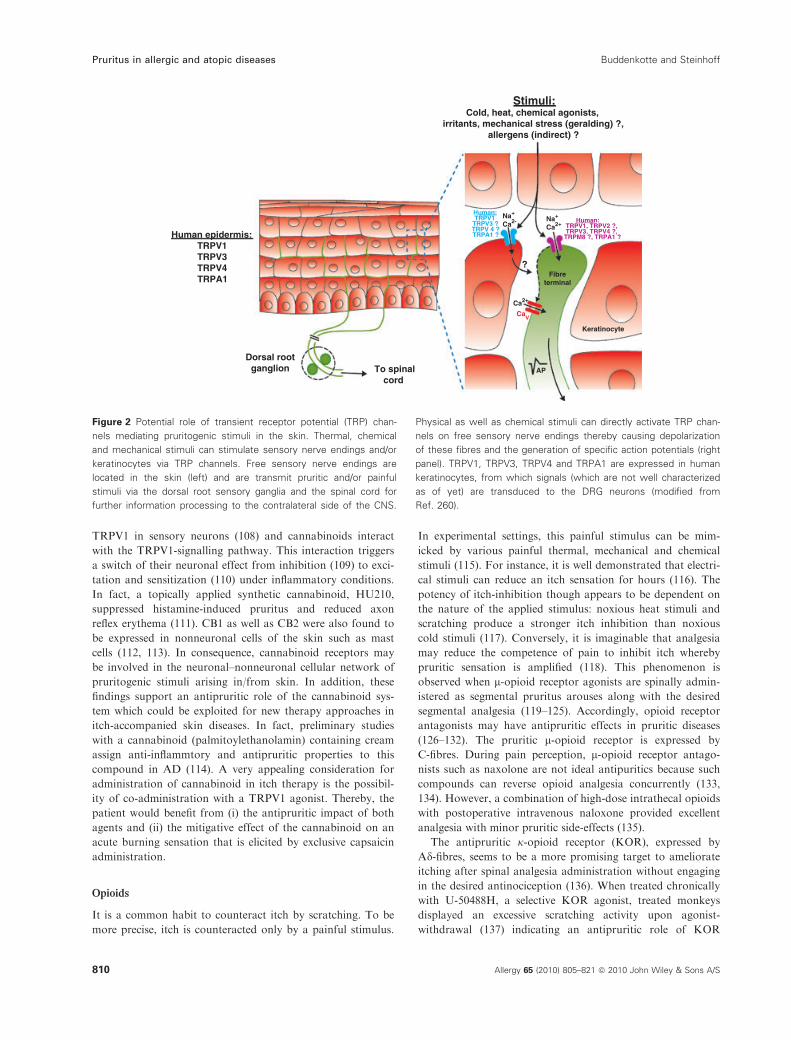
Figure 2 Potential role of transient receptor potential (TRP) chan-
nels mediating pruritogenic stimuli in the skin. Thermal, chemical
and mechanical stimuli can stimulate sensory nerve endings and/or
keratinocytes via TRP channels. Free sensory nerve endings are
located in the skin (left) and are transmit pruritic and/or painful
stimuli via the dorsal root sensory ganglia and the spinal cord for
further information processing to the contralateral side of the CNS.
Physical as well as chemical stimuli can directly activate TRP chan-
nels on free sensory nerve endings thereby causing depolarization
of these fibres and the generation of specific action potentials (right
panel). TRPV1, TRPV3, TRPV4 and TRPA1 are expressed in human
keratinocytes, from which signals (which are not well characterized
as of yet) are transduced to the DRG neurons (modified from
Ref. 260).
Pruritus in allergic and atopic diseases Buddenkotte and Steinhoff
810 Allergy 65 (2010) 805–821 ª 2010 John Wiley & Sons A/S

when holding in mind that many withdrawal symptoms from
opioids appear to be opposite to the acute effects of the
administered agonist (138, 139). In another pharmacological
monkey study, KOR agonists prevented/reversed intrathecal
morphine-induced itch/scratching responses without interfer-
ing with intrathecal morphine analgesia (140). This outcome
led to a clinical trial of a new KOR agonist, nalfurafine
(TRK-820), in hemodialysis patients suffering from uraemic
pruritus resulting in a successfull amelioration of the itch sen-
sation in these patients indicating an important therapeutical
potential of KOR agonists as antipruritic agents (141).
In patients with pruritic AD, the itchy pathogenesis of the
disease might in parts be ascribed to the involvement of
opioids because b-endorphin serum levels were found to
be markedly elevated (142).
To date, not much is known about a participation of opi-
oid-receptors in the neuronal–nonneuronal cellular network
of pruritogenic stimuli. It has been speculated though that
downregulated epidermal l-opiate receptor of AD patients
increases the pool of available opioid ligands, which then in
turn induce histamine-unrelated chronic pruritus (143, 144).
Cytokines and inflammatory cells
Under inflammatory conditions, cytokines are, among numer-
ous other factors, released from cutaneous and immune cells.
Some of these cytokines are well capable of triggering pruritic
sensations and release of neuropeptides from sensory nerves.
In the following, such cytokines will be briefly described.
Interleukins
Although the contribution of many interleukins in atopic and
allergic diseases is well established, the precise role of these
immune mediators in pruritus is still unclear. For instance, a
pleithropic inhibition of cytokine production achieved by
usage of cyclosporine A resulted in the mitigation of itch in
patients suffering from AD (145, 146). Also, upon prick test-
ing, supernatants of mitogen-stimulated leucocytes were pru-
ritic in AD patients but not in controls (147). Cyclosporin A
(CyA), among other interleukins, effectively blocks the pro-
duction of IL-2. Gaspari and co-workers showed that actu-
ally this distinct interleukin is a potent activator of pruritus.
In their study, all cancer patients treated with IL-2 developed
macular erythema with burning sensation and cutaneous pru-
ritus (147). In AD patients, as well as in healthy individuals,
a single intracutaneous injection of IL-2 resulted in a low-
intensity intermittent local itch perception (148, 149). It has
been demonstarated that bradykinin is involved in the mech-
anism to modulate the intensity of IL-2-induced pruritus on
sensory nerves (150) but the mechanism for the induction of
itch by IL-2 itself remains to be uncovered. Because the pru-
ritogenic response initiated by IL-2 administration is faster in
AD patients than in healthy individuals, an indirect mecha-
nism of action via other mediators is likely. Recently, a role
for IL-8 in the pathogenesis of pruritus was postulated.
Enhanced level of this chemokine was detected in lesional
skin (151), plasma (152) and blood mononuclear cells (153,
154) of AD patients. In addition, these findings indicate a
role for IL-8 in the generation of pruritus but so far no study
provided direct evidence for this thesis. Future research has
to clarify the connection of IL-8 and ‘itchy outcome’. A
novel T-cell-derived cytokine was reported to induce severe
pruritus and dermatitis in transgenic mice: IL-31. Its targeted
receptor to initiate itch was shown to consist of a heterdimer-
ic receptor composition of IL-31 receptor and oncostatin
receptor (155) and to cause pro-inflammatory effects of acti-
vated human monocytes and macrophages which may have
implications for cutaneous inflammation in eczema (156).
Subsequent to this initial study, IL-31 was also found to be
overexpressed in pruritic atopic skin (157). Especially in
patients with prurigo nodularis, one of the most pruritic
forms of chronic skin inflammation, IL-31 levels were
severely upregulated. Also a role for IL-31 in the pruritus of
atopiform dermatitis is under consideration (158). In vivo,
staphylococcal superantigen rapidly induced IL-31 expression
in atopic individuals indicating a new link among staphylo-
coccal colonization, subsequent T-cell recruitment/activation
and pruritus induction in patients with AD.
Interferon gamma (IFN-c)
While long-term treatment with IFN-b appears to cause
pruritic side-effects (159), IFN-a seems to have a beneficial
effect on pruritus in various diseases (160–162). In patients
suffering from AD, IFN-c treatment effectively relieved pruri-
tus (163). This relief was reported by AD patients to still exist
after long-term treatment with IFN-c (164). The distinct
mechanism of action for IFN-c to modulate pruritus, how-
ever, has still to be identified. It is likely that the diminished
IFN-c production in peripheral blood mononuclear cells of
AD patients (165) accounts for the pruritus phenomenon.
Neurotrophin-4
Recent studies suggest a contribution of NT-4 to inflamma-
tory and itch responses of patients with AD. NT-4 is
produced by keratinocyte and highly expressed under inflam-
matory conditions and acts growth-promoting on nerve cells.
Accordingly, NT-4 expression was found to be significantly
increased in lesional skin of patients with AD and in prurigo
lesions of AD skin (166). Interestingly, IFN-c, itself a potent
anti-pruritic agent, can initiate NT-4 production. These find-
ings suggest a close link between immune and neurotrophic
factors in the development of pruritus in AD.
Eosinophils and basophils
The role of eosinophils in the pathogenesis of AD is well
established but how they engage in the pathophysiology of
pruritus during AD is still unclear. It is likely that factors
released by eosinophils such as prostanoids, kinins, cytokines,
leucotrienes, platelet-activating factor and proteases adopt
the pruritogenic effect of eosinophils on a molecular level
(167–172). But it is also imaginable that the itch reponse is
elicited indirectly by activation of mast cells which in turn
Buddenkotte and Steinhoff Pruritus in allergic and atopic diseases
Allergy 65 (2010) 805–821 ª 2010 John Wiley & Sons A/S 811

trigger the release of histamine or proteinases from eosinoph-
ils. In summary, although some reports are in favour for a
role of eosinophils during pruritus in various diseases (167,
170, 172), direct evidence for a role of eosinophils for itch
responses during AD is still lacking. Peripheral blood ba-
sophils are inconspicuous in patients suffering from AD.
However, in vitro analysis of basophil function demonstrated
faster histamine releasability upon stimulation (173, 174).
These in vitro results could not be confirmed in patients with
AD, though (175). The contribution of basophils to the
development of itch and erythema in patients with AD is still
in need of detailed investigation.
Platelet-activating factor
The lipid mediator Platelet-activating factor is a component of
several inflammatory cells such as mast cells and granulocytes
with proinflammatory activity (176). One of its actions upon
intradermal injection is to increase vascular permeability and
thereby to cause a wheal and flare reaction along with pruri-
tus. The underlying mechanism debated is based on an indirect
pruritogenic effect via histamine release (177). Platelet-acti-
vating factor antagonists have proved, in a double-blind study,
to be able to reduce pruritus in AD patients when applied
topically already within the initial weeks of treatment (178).
Leukotriens
Leukotriens are mediators with proinflammatory properties
generated from arachidonic acid, an essential fatty acid found
in the membrane of all cells (179). A synthesis pathway
whose key enzyme is 5-lipoxygenase provides all known leu-
kotriens. Their cellular origin is reflected by 5-lipoxygenase
expression and essentially restricted to various myeloid cells
such as neutrophils, monocytes/macrophages, B lymphocztes
and mast cells. Leukotriens bind to three receptor subtypes:
BLT, CysLT1 or CysLT2.
The contribution of leukotriens to the pathophysiology of
inflammatory diseases, in particular asthma, is well established
whereas their role in the pathogenesis of pruritus is still sub-
ject of debate. However, intradermally injected leukotriene B4
was demonstrated to provoke scratching in mice (180) and
high urinary leukotriene E4 levels could be correlated to noc-
turnal itch (181). An increased abundance of leukotriens could
therefore account for itch induction in AD. In fact, inhibition
of leukotrien receptor by zafirlukast and zileuton resulted in a
reduction of pruritus in patients with AD (182–184).
Trigger factors aggravating pruritus perception in
atopic dermatitis
The skin of AD patients reveals a higher tendency to itch
upon minimal provocation because of reduced itch threshold
and prolonged itch duration to pruritic stimuli as compared
with healthy skin (185–187). A series of pruritus triggering
factors are known (186), which release mast cell mediators or
vasomotor and sweat reactions to cause itch, and all may be
subjected to emotional influences (185).
Scratching
It is a recurring debate whether scratching itselfs precedes the
induction of AD or if itch and scratching are a consequence
of the presence of eczemas. The final answer to this debate is
indeed not found so far, in particular the itch–scratch order
crucial for the pathophysiology of AD remains to be an
enigma. However, results obtained from an animal study
were in favour of scratching behaviour anteceding and
thereby contributing to the development of dermatitis (188).
This AD mouse model investigated the development of spon-
taneous dermatitis by comparison of mice neonatally treated
with capsaicin (Cap-NC mice) to ablate capsaicin-sensetive
sensory nerves or vehicle. In the treated mice, scratching
behaviour was hardly observed and the development of der-
matitis determined by elevation of the serum IgE level and
the numbers of infiltrating eosinophils and mast cells was sig-
nificantly suppressed. Also the capability of spleen T cells to
produce both T-helper Th1 (IFN-c) and Th2 (IL-5, IL-13)
cytokines appeared to be constrained in Cap-NC mice, indi-
cating a direct correlation of scratching and subsequent
immunological responses. Clinically, the prevention of itch
sensation and/or itch-associated scratching behaviour may be
an additional important step in the treatment of AD.
Epidermal barrier
Xerosis is a common problem of the skin of patients suffering
from AD. It constitutes a keratinization disorder that reflects
a dysfunctional epidermal barrier. This dysfunction results in
an increased transepidermal water loss and a decreased ability
of the stratum corneum to bind water (189) which may be due
to incomplete arrangement of intercellular lipid lamellae in
the stratum corneum (190, 191). It is well-established knowl-
edge that a disturbed epidermal barrier constitutes an activa-
tor of pruritus. In fact, scratching behaviour and induction of
pruritus are triggered by water content below 10% (192). The
precise mechanisms for the pruritogenic effect of a disturbed
epidermal barrier remain unknown. One possibility may be
that an impaired skin barrier simplifies the penetration of irri-
tants and itchy agents (193, 194). Animal studies demon-
strated that the epidermal barrier homeostasis and stratum
corneum integrity is furthermore affected by psychoemotional
stress: decrease of lamellar bodies’ formation and secretion
along with decrease corneodesmosomes production was
observed (195). These findings suggested a correlation between
stress factors and decreased barrier function and might be of
relevance for patients with AD (see Stress section).
Stress
It has long been appreciated that both acute stress and
chronic psychoemotional stress can trigger or modulate pruri-
tus (196–203).
To understand this correlation requires a deeper under-
standing of the neuroendocrinology and neuroimmunology
of stress responses. Stress responses are learned, involve
the cortical centres but can be reprogrammed by behavioural
Pruritus in allergic and atopic diseases Buddenkotte and Steinhoff
812 Allergy 65 (2010) 805–821 ª 2010 John Wiley & Sons A/S

and neuropharmacological/neurendocrine therapy. In patients
with AD, a close relationship between psychological factors,
pruritus and scratching has been shown (198, 199, 204–206)
– intriguing 81% of AD patients attribute emotional stress
to aggravate their pruritus (46). In turn, an experience of
increased itch upon stressful events might lead to conditioned
itch in AD patients, which thereby could give way to a
vicious circle of aggravation and perpetuation of stress-
induced itch. Relaxation therapies that are also operant for
pain patients like autogenic training or hypnosis have been
found to ameliorate itch and eczema in AD patients by dis-
tinctly treating mental stress factors (207, 208). The mecha-
nism of stress-induced pruritus in AD patients is not
unravelled as of yet but an activation of the psycho-neuroen-
docrine system seems likely (198, 199). For instance, immobi-
lization stress applied to rats resulted in mast cell
degranulation (209) supporting the thesis that stress tension
may lead to increased release of pruritogenic mediators from
mast cells in AD patients which further may results in inten-
sified scratching behaviour and subsequent skin lesions (199).
In another study, AD patients undergoing a stress test
revealed a responsive increase of IgE, blood eosinophils,
IFN-c and IL-4 (210). Pruritus intensity may be modulated
by vasodilators responses and increased skin temperature in
consequence to emotional stress (185, 211).
Sweating
Generalized itching evoked by any stimulus to sweating
(thermal, emotional stimuli) is a typical hallmark and repre-
sents the most common trigger factor of itch in patients
with AD (185, 212, 213). Increased sweating was observed
in lichenified skin of patients with AD. Causative factor
might be a lowered threshold for sweat stimulation in chron-
ically pruritic and altered skin (214). The exact mechanism
underlying sweat-induced pruritus is far from being resolved,
but recent evidence points to a role of ACh. ACh-induced
eccrine sweating (215), is found to be increased in the skin
of AD patients (64), and finally acts as a sensitizer or pruri-
togenic in AD patients (68).
Microcirculation
Clinically, itching is mostly associated with erythema and
hyperthermia. A variety of mediators for itching such as
histamine, tryptase, ACh, SP, prostaglandins are potent
vasodilatators, rarely vasoconstrictors (NPY or catecholam-
ines). Neuropeptide-induced itching does not vary between
atopic and nonatopic patients, whereas vascular responses
obviously show a significant difference between these two
groups. Moreover, patients with AD were more susceptible to
stress and showed increased vasodilatation as compared with
controls (216). Pruritic mediators which also acts as potent
vasodilatators may be histamine and tryptase. Certain prosta-
noids are effective sensitizers and vasodilatators. In sum, which
receptors are the most important ones to regulate itch and
vasodilatation among the different pruritic diseases is still a
matter of debate.
Exogenous factors
Pruritus elicited by direct contact with wool in patients with
AD is a characteristic and reproducible phenomenon (217,
218). It is likely that the irritation is caused by the spiky
nature of wool fibres itself. Mechanical vibration seems to be
irrelevant for induction of itch because it inhibits experimen-
tal, histamine-induced itch (219). Interestingly, thicker wool
fibres were found to provoke more intense itching than thin-
ner fibres (220). Other irritants like lipid solvents, disinfec-
tants (221) may additionally contribute to aggravate xerosis.
Contact- and aero-allergens as dust mites or pollens (212)
may also provoke pruritus. Microbiological agents like bacte-
ria (Staphylococcus aureus) or yeast may exacerbate both
dermatitis and pruritus (186, 212).
Pruritus and erythema may be also triggered by exogenous
substances like proteinases from bacteria and dermatophytes
increasing blood flow, conduct vasodilatation or release hista-
mine. Among those, heat, hot and spicy foods, hot drinks
and alcohol are most likely to generate itch in AD patients
(186, 212, 222). In early childhood, food allergies exacerbate
eczematous skin lesions, but these food allergies mostly
resolve during ageing in older children and adults (222).
Management of pruritus in allergic and atopic skin
diseases
The handling and treatment of severe itch is one of the major
challenges in the management of patients with allergic and
AD (Table 2). Concerning a successful suppression of pruritus,
several levels have to be considered. First of all, identification
and elimination of individual trigger factors must be appreci-
ated as the primary goal of the management (200, 223). As
patients frequently develop some harmful self-treatments, e.g.
alcohol-containing solutions, these misconceived therapies
must be eliminated. Lotions and creams lubricating the skin
have to be recommended. To combat skin dryness, applica-
tion of hydrophilic emollients and bathing with oily bath
additives is additionally helpful (223). Lipid based repair for-
mulations based on ceramide-dominant contents have been
described to be superior in reduction of disease severity (224–
226). Adding substances such as urea, menthol, camphor and
polidocanol to these cremas leads to an immediate short-term
interruption of the itch. These creams can be applied by the
patients each time the itch starts to worsen (227). Unspecific
physical modalities are described to be beneficial like
acupuncture (228) and cutaneous field stimulation (116).
Another level of therapy is the handling of the scratch
artefacts. Chronic pruritus induces chronic scratching or
rubbing. Accordingly, erosions, ulcerations, bleding, crusts,
lichenifications up to prurigo nodularis may develop. Stage-
dependent, disinfections, antimicrobials and topical corticos-
teroids have to be applied. In patients with prurigo nodularis
or lichen simplex associated to AD, frequently an automatic
scratching behaviour develops. These patients additionally
need education to control scratch behaviour (229). For exam-
ple, the behaviour method ‘habit reversal’ can be employed
(230). First, patients become aware of their scratching behav-
Buddenkotte and Steinhoff Pruritus in allergic and atopic diseases
Allergy 65 (2010) 805–821 ª 2010 John Wiley & Sons A/S 813

iour by counting scratch movements. In a second step, they
learn a new behaviour by reacting to scratch impulses.
Scratch-induced skin damage caused by nocturnal scratch
movements may be improved by using cotton gloves. Also
controlled physical exercise like gymnastics or ball games
were demonstrated in a controlled study to teach patients to
cope better with itch attacks (231).
As chronic scratching represents also a trigger factor and
maintains the itch-scratch-cycle, the most important step in
the management of the AD patients is the interruption of
itch by an effective symptomatical topical and/or systemical
therapy.
Symptomatical topical and systemical therapy
Studies concerning the pathophysiology of pruritus clearly
demonstrated that different nociceptive mechanisms are
involved in AD. Thus, conventional therapeutic modalities
like antihistamines often fail to ameliorate pruritus in AD
(33). This is comprehensive with the idea that histamine is
not the major mediator of pruritus in AD (35). Placebo-
controlled studies concerning the antipruritic effect of oral
antihistamines have shown conflicting results in AD. In some
studies, no superior effect was observed as compared with
placebo (232–234) whereas others showed a significant anti-
pruritic effect (38, 235–237). In recent experimental studies,
the H1-antihistamine cetirizine could be demonstrated to
focally reduce itch (38). However, an evidence-based review
concerning the efficacy of antihistamines in relieving pruritus
in AD concluded that little objective evidence exists for
H1-antihistamines to demonstrate improvement of pruritus
(33). Topical application of the tricyclic antidepressant
doxepin suggested to have antipruritic effects because of its high
affinity to H1 histamine receptors. In fact, 5% doxepin cream
revealed improvement of histamine-induced and SP-mediated
cutaneous responses but also evoked sedative effects in some
patients (238, 239). Unfortunately, doxepin was accompanied
by contact allergies after long-term application (240).
In general, anti-inflammatory, immunomodulating thera-
pies as regularly applied in AD often result also in cessation
of pruritus, because they suppress the inflammatory mecha-
nisms underlying the induction of itch. So far, most effective
and consistent antipruritics remain systemic immunomodula-
tors such as glucocorticoids, CyA, tacrolimus, pimecrolimus
and ultraviolet radiation therapy (146, 241–244). Moreover,
there are no evident and efficient alternatives to topical appli-
cation of corticosteroids or calcineurin inhibitors for the con-
trol of acute episodes in AD (244–246). With reduction of
skin lesions, a decreased itch intensity results probably
because of reduction of inflammatory cells and protection of
depolarization of nerve fibres mediated directly by the steroid
(247). However, treatment of a patient suffering from amyloi-
dosus lichen (LA) associated with AD by a combination of
narrowband ultraviolet B phototherapy, topical corticoster-
Table 2 Therapeutic strategies alleviating pruritus in atopic dermatitis
Therapeutical modalities Examples
Elimination of trigger factors Perspiration, xerosis, emotional stress, scratching, wearing wool fibres,
using of mild soaps, detergents, hot, spicy food, hot drinks, alcoholics
Skin barrier protection and restauration, itch control Emollients
Bathing with oily additives
Lotions, creams or sprays containing menthol, local anaesthetics,
camphor, polidocanol, urea, pimecrolimus, cooling
Skin care to reduce sweating-induced itch
Therapy of scratch artefacts Desinfection, antibiotics, topical steroids, pimecrolimus, tacrolimus,
doxepin cream 5%, amitryptiline 4% / ketamine 2% cream
Interruption of itch-scratch-cycle: behaviour therapy against scraching
Physical exercise
Acupuncture, hypnosis
Cutaneous field stimulation
Symptomatic therapy: anti-inflammatory therapy Corticosteroids, topical and systemical
Cyclosporin A
Tacrolimus, pimecrolimus
(Interferon gamma)
Immunoglobulin therapy
Ultraviolet irradiation (UVA1, UVA/UVB)
Symptomatic therapy: interfering with
pathophysiology of pruritus in AD
l-Opioid antagonists, j-opioid agonist
Capsaicin
Cannabinoid agonists combination of high-dose antihistamines
(neurokinin-1 receptor antagonist)
Contradictory results Antihistamines, leukotriene antagonists
Doxepin (potential: contact allergy upon long-term application)
Mycophenolat mofetil
Systemic therapy Gabapentin, pregabalin
Pruritus in allergic and atopic diseases Buddenkotte and Steinhoff
814 Allergy 65 (2010) 805–821 ª 2010 John Wiley & Sons A/S

oids and an oral antihistamine led to an improvement of AD
as well as LA symptoms (248). Cyclosporin A, a cyclic poly-
peptide with potent immunosuppressive effects, has been
reported to have a considerable itch-reliving effect in various
diseases including AD. In a randomised study, CyA was
demonstrated to significantly reduce itch intensity (146).
After discontinuation of this therapy, pruritus recurred
immediately. As oral CyA has demonstrated to be effective
in AD, a topical CyA formulation has been developed to
avoid systemic adverse effects. However, no significant
improvement of AD was found upon clinical application
(249).
Recently, much interest has been drawn to tacrolimus and
pimecrolimus, both effective immunomodulators and calcineu-
rin inhibitors. Although the mode of action is similar to that
of CyA, the molecular weight is lower and its potency of
inhibiting T-cell activation is higher. Multiple, large rando-
mised studies of the last years confirmed topical administra-
tion of tacrolimus and pimecrolimus to interrupt acute
attacks of AD, reduce fastly pruritus and prevent exacerba-
tion after cessation of eczemas in adults and even children
with AD (250–253). This beneficial effect of pimecrolimus is
also detected in children suffering from Netherton syndrome
(254). Upon treatment with a 1% pimecrolimus cream, rapid
marked improvements were observed in the Netherton Area
and Severity Assessment, Eczema Area and Severity Index,
and pruritus scores. Treatment with IFN-c has been shown
to be effective not only for the improvement of erythema,
excoriations and lichenifications, but also of pruritus (164,
255, 256). In addition, this effect maintained up to 2 years
after therapy (164). Amelioration of pruritus has also been
described under intravenous immunoglobulin therapy in few
cases of AD (257, 258). As of yet, however, no controlled
studies were performed.
Also other therapeutical modalities such as TRPV1 recep-
tor antagonists/or agonists (90, 259), l-opiate receptor antag-
onists (128–130), j-opioid receptor agonists, PAR2 receptor
antagonists, histamine-3 receptor or histamine-4 receptor tar-
geting molecules, cannabinoid agonists, nerve-growth factor
or nerve-growth factor receptor antagonists, neurokinin-1
receptor antagonists, GRPR antagonists, certain prostaglan-
din and leukotriene antagonists (182–184) appear to be prom-
ising new approaches for the therapy of AD and certain
allergic diseases, but will have to prove their safety and prac-
ticability in further controlled studies. In conclusion, the
pathopysiology of pruritus in AD has not been evaluated
completely. Accordingly, no specific antipruritic agent has
been developed and management of itch in AD is confined to
mainly immunomodulating therapies. However, the consider-
ation of several levels may improve this distressing situation
for the patients. Further investigations are necessary to estab-
lish antipruritic substances influencing the centrally and
peripherally altered itch perception to interfere with the com-
plex pathophysiology of pruritus in AD.
Conclusion and perspectives
Amelioration of pruritus is a major goal in the treatment of
patients suffering from allergic and atopic skin diseases.
Identification of a single effective pharmacological treatment
is an old continuing demand of physicians handling and
managing itch symptoms for this disease. Promising new
approaches have been made, but recent insight into the ori-
gin and onset of pruritus leads to the conclusion that the
single treatment/compound that universally combats the itch
symptom in AD patients may not be found in the near
future. Because itch pathophysiology is too complex involv-
ing neurophysiological and neuroimmunological aspects,
more has to be learned about the mediators, receptors, mul-
tidirectional pathways in the near future to reach a valid
golden standard for therapy. However, the complexity of
interactions between the central and peripheral nervous sys-
tem and the skin in generating this symptom has catapulted
an indeed broad but clearly delineated spectrum of molecu-
lar targets into focus which, when successfully exploited,
could serve to treat the itch perception in AD patients.
Once these molecules will be explored systematically and in
detail, we undoubtedly will hold sophisticated and more
effective therapeutic strategies for pruritus management in
AD in our hands. In particular, combining approaches that
target both the peripheral production of inflammation-
induced itch signals and the peripherally driven cycles that
perpetuate itch and provoke spinal and central sensitization
to itch in AD are promising new strategies. The direction of
the development of innovative and more effective itch man-
agement is to unequivocally extend the scope of pharmaco-
logical targets far beyond the ubiquitous usual suspect
histamine.
References
1. Rothman S. Physiology of itching. Physiol
Rev 1941;21:357–381.
2. Hanifin JM, Rajka G. Diagnostic features
of atopic dermatitis. Acta Derm Venereol
Suppl (Stockh) 1980;92:44–47.
3. Koblenzer CS. Itching and the atopic skin.
J Allergy Clin Immunol 1999;104(3 Pt 2):
S109–S113.
4. von Frey M. [On the physiology of pruri-
tus]. Arch Neerland Physiol 1922;7:142–145.
5. Schmelz M, Schmidt R, Bickel A, Hand-
werker HO, Torebjork HE. Specific C-
receptors for itch in human skin. J Neuro-
sci 1997;17:8003–8008.
6. Andrew D, Craig AD. Spinothalamic lam-
ina I neurons selectively sensitive to hista-
mine: a central neural pathway for itch.
Nat Neurosci 2001;4:72–77.
7. Paus R, Schmelz M, Biro T, Steinhoff
M. Frontiers in pruritus research:
scratching the brain for more effective
itch therapy. J Clin Invest 2006;116:1174–
1186.
8. Handwerker HO, Schmelz M. Pain: itch
without pain-a labeled line for itch sensa-
tion? Nat Rev Neurol 2009;5:640–641.
9. Carstens E. Responses of rat spinal dorsal
horn neurons to intracutaneous microinjec-
tion of histamine, capsaicin, and other irri-
tants. J Neurophysiol 1997;77:2499–2514.
10. Sun YG, Chen ZF. A gastrin-releasing pep-
tide receptor mediates the itch sensation in
the spinal cord. Nature 2007;448:700–703.
11. Carstens EE, Carstens MI, Simons CT,
Jinks SL. Dorsal horn neurons expressing
Buddenkotte and Steinhoff Pruritus in allergic and atopic diseases
Allergy 65 (2010) 805–821 ª 2010 John Wiley & Sons A/S 815

NK-1 receptors mediate scratching in rats.
Neuroreport 2010;21:303–308.
12. Mantyh PW, Rogers SD, Honore P, Allen
BJ, Ghilardi JR, Li J et al. Inhibition of
hyperalgesia by ablation of lamina I spinal
neurons expressing the substance P recep-
tor. Science 1997;278:275–279.
13. Nichols ML, Allen BJ, Rogers SD,
Ghilardi JR, Honore P, Luger NM et al.
Transmission of chronic nociception by
spinal neurons expressing the substance
P receptor. Science 1999;286:1558–1561.
14. Hsieh JC, Hagermark O, Stahle-Backdahl
M, Ericson K, Eriksson L, Stone-Elander
S et al. Urge to scratch represented in the
human cerebral cortex during itch. J Neu-
rophysiol 1994;72:3004–3008.
15. Drzezga A, Darsow U, Treede RD, Siebner
H, Frisch M, Munz F et al. Central activa-
tion by histamine-induced itch: analogies to
pain processing: a correlational analysis of
O-15 H2O positron emission tomography
studies. Pain 2001;92:295–305.
16. Mochizuki H, Tashiro M, Kano M, Saku-
rada Y, Itoh M, Yanai K. Imaging of cen-
tral itch modulation in the human brain
using positron emission tomography. Pain
2003;105:339–346.
17. Sun YG, Zhao ZQ, Meng XL, Yin J, Liu
XY, Chen ZF. Cellular basis of itch sensa-
tion. Science 2009;325:1531–1534.
18. Lewis T. The blood vessels of the human
skin and their responses. London: Shaw
and Sons, 1927.
19. Lewis T, Grant RT, Marvin HM. Vascular
reactions of the skin to injury. Heart
1929;14:139–160.
20. Williams DH. Skin temperature reaction to
histamine in atopic dermatitis (dissemi-
nated neurodermatitis). J Invest Dermatol
1938;1:119–129.
21. Johnson HH, Jr, Deoreo GA, Lascheid
WP, Mitchell F. Skin histamine levels in
chronic atopic dermatitis. J Invest Derma-
tol 1960;34:237–238.
22. Juhlin L. Localization and content of hista-
mine in normal and diseased skin. Acta
Derm Venereol 1967;47:383–391.
23. Hill SJ. Distribution, properties, and func-
tional characteristics of three classes of his-
tamine receptor. Pharmacol Rev
1990;42:45–83.
24. Turner AJ, Hick PE. Inhibition of alde-
hyde reductase by acidic metabolites of the
biogenic amines. Biochem Pharmacol
1975;24:1731–1733.
25. Malaviya R, Morrison AR, Pentland AP.
Histamine in human epidermal cells is
induced by ultraviolet light injury. J Invest
Dermatol 1996;106:785–789.
26. Haas HL, Sergeeva OA, Selbach O. Hista-
mine in the nervous system. Physiol Rev
2008;88:1183–1241.
27. Stander S, Weisshaar E, Luger TA. Neuro-
physiological and neurochemical basis of
modern pruritus treatment. Exp Dermatol
2008;17:161–169.
28. Barthel W, Markwardt F. Aggregation of
blood platelets by adrenaline and its uptake.
Biochem Pharmacol 1975;24:1903–1904.
29. Hagermark O, Strandberg K, Gronneberg
R. Effects of histamine receptor antago-
nists on histamine-induced responses in
human skin. Acta Derm Venereol
1979;59:297–300.
30. Sugimoto Y, Iba Y, Nakamura Y, Kayasu-
ga R, Kamei C. Pruritus-associated
response mediated by cutaneous histamine
H3 receptors. Clin Exp Allergy
2004;34:456–459.
31. Ruzicka T, Gluck S. Cutaneous histamine
levels and histamine releasability from the
skin in atopic dermatitis and hyper-IgE-
syndrome. Arch Dermatol Res 1983;275:
41–44.
32. Hanifin JM. The role of antihistamines in
atopic dermatitis. J Allergy Clin Immunol
1990;86(4 Pt 2):666–669.
33. Klein PA, Clark RA. An evidence-based
review of the efficacy of antihistamines in
relieving pruritus in atopic dermatitis. Arch
Dermatol 1999;135:1522–1525.
34. Munday J, Bloomfield R, Goldman M,
Robey H, Kitowska GJ, Gwiezdziski Z et
al. Chlorpheniramine is no more effective
than placebo in relieving the symptoms of
childhood atopic dermatitis with a noctur-
nal itching and scratching component.
Dermatology 2002;205:40–45.
35. Rukwied R, Lischetzki G, McGlone F,
Heyer G, Schmelz M. Mast cell mediators
other than histamine induce pruritus in
atopic dermatitis patients: a dermal micro-
dialysis study. Br J Dermatol 2000;
142:1114–1120.
36. Heyer G, Hornstein OP, Handwerker HO.
Skin reactions and itch sensation induced
by epicutaneous histamine application in
atopic dermatitis and controls. J Invest
Dermatol 1989;93:492–496.
37. Heyer G, Koppert W, Martus P, Handwer-
ker HO. Histamine and cutaneous nocicep-
tion: histamine-induced responses in
patients with atopic eczema, psoriasis and
urticaria. Acta Derm Venereol 1998;78:123–
126.
38. Heyer GR, Hornstein OP. Recent studies
of cutaneous nociception in atopic and
non-atopic subjects. J Dermatol
1999;26:77–86.
39. Uehara M. Reduced histamine reaction in
atopic dermatitis. Arch Dermatol
1982;118:244–245.
40. Heyer G, Hornstein OP, Handwerker HO.
Reactions to intradermally injected sub-
stance P and topically applied mustard oil
in atopic dermatitis patients. Acta Derm
Venereol 1991;71:291–295.
41. Bell JK, McQueen DS, Rees JL. Involve-
ment of histamine H4 and H1 receptors in
scratching induced by histamine receptor
agonists in Balb C mice. Br J Pharmacol
2004;142:374–380.
42. Roosterman D, Goerge T, Schneider SW,
Bunnett NW, Steinhoff M. Neuronal con-
trol of skin function: the skin as a neuro-
immunoendocrine organ. Physiol Rev
2006;86:1309–1379.
43. Cowden JM, Zhang M, Dunford PJ, Thur-
mond RL. The histamine H(4) receptor
mediates inflammation and pruritus in
Th2-dependent dermal inflammation.
J Invest Dermatol 2010;130:1023–1033.
44. Heyer G, Ulmer FJ, Schmitz J, Handwer-
ker HO. Histamine-induced itch and allok-
nesis (itchy skin) in atopic eczema patients
and controls. Acta Derm Venereol 1995;
75:348–352.
45. Rukwied R, Heyer G. Cutaneous reactions
and sensations after intracutaneous injec-
tion of vasoactive intestinal polypeptide
and acetylcholine in atopic eczema patients
and healthy controls. Arch Dermatol Res
1998;290:198–204.
46. Wahlgren CF. Pathophysiology of itching
in urticaria and atopic dermatitis. Allergy
1992;47(2 Pt 1):65–75.
47. Wahlgren CF. Measurement of itch. Semin
Dermatol 1995;14:277–284.
48. Ansel JC, Kaynard AH, Armstrong CA,
Olerud J, Bunnett N, Payan D. Skin-ner-
vous system interactions. J Invest Dermatol
1996;106:198–204.
49. Scholzen T, Armstrong CA, Bunnett
NW, Luger TA, Olerud JE, Ansel JC.
Neuropeptides in the skin: interactions
between the neuroendocrine and the skin
immune systems. Exp Dermatol
1998;7:81–96.
50. Slominski A, Wortsman J. Neuroendocri-
nology of the skin. Endocr Rev
2000;21:457–487.
51. Pincelli C, Fantini F, Massimi P, Giannetti
A. Neuropeptide Y-like immunoreactivity in
Langerhans cells from patients with atopic
dermatitis. Int J Neurosci 1990;51:219–220.
52. Pincelli C, Fantini F, Massimi P, Girolo-
moni G, Seidenari S, Giannetti A. Neuro-
peptides in skin from patients with atopic
dermatitis: an immunohistochemical study.
Br J Dermatol 1990;122:745–750.
53. Tobin D, Nabarro G, Baart de la Faille H,
van Vloten WA, van der Putte SC, Schuur-
man HJ. Increased number of immunore-
active nerve fibers in atopic dermatitis. J
Allergy Clin Immunol 1992;90(4 Pt 1):613–
622.
54. Anand P, Springall DR, Blank MA, Sellu
D, Polak JM, Bloom SR. Neuropeptides in
Pruritus in allergic and atopic diseases Buddenkotte and Steinhoff
816 Allergy 65 (2010) 805–821 ª 2010 John Wiley & Sons A/S

skin disease: increased VIP in eczema and
psoriasis but not axillary hyperhidrosis. Br
J Dermatol 1991;124:547–549.
55. Fantini F, Pincelli C, Romualdi P, Dona-
tini A, Giannetti A. Substance P levels are
decreased in lesional skin of atopic derma-
titis. Exp Dermatol 1992;1:127–128.
56. Pincelli C, Fantini F, Romualdi P, Lesa G,
Giannetti A. Skin levels of vasoactive intes-
tinal polypeptide in atopic dermatitis. Arch
Dermatol Res 1991;283:230–232.
57. Andoh T, Nagasawa T, Satoh M, Kuraishi
Y. Substance P induction of itch-associated
response mediated by cutaneous NK1
tachykinin receptors in mice. J Pharmacol
Exp Ther 1998;286:1140–1145.
58. Scholzen TE, Steinhoff M, Bonaccorsi P,
Klein R, Amadesi S, Geppetti P et al.
Neutral endopeptidase terminates substance
P-induced inflammation in allergic contact
dermatitis. J Immunol 2001;166:1285–1291.
59. Stellwagen E, Babul J. Stabilization of the
globular structure of ferricytochrome c by
chloride in acidic solvents. Biochemistry
1975;14:5135–5140.
60. Thomsen JS, Sonne M, Benfeldt E, Jensen
SB, Serup J, Menne T. Experimental itch
in sodium lauryl sulphate-inflamed and
normal skin in humans: a randomized,
double-blind, placebo-controlled study of
histamine and other inducers of itch. Br J
Dermatol 2002;146:792–800.
61. Ohmura T, Hayashi T, Satoh Y, Konomi
A, Jung B, Satoh H. Involvement of
substance P in scratching behaviour in an
atopic dermatitis model. Eur J Pharmacol
2004;491:191–194.
62. Grando SA, Kist DA, Qi M, Dahl MV.
Human keratinocytes synthesize, secrete,
and degrade acetylcholine. J Invest Derma-
tol 1993;101:32–36.
63. Grando SA, Zelickson BD, Kist DA,
Weinshenker D, Bigliardi PL, Wendelscha-
fer-Crabb G et al. Keratinocyte muscarinic
acetylcholine receptors: immunolocalization
and partial characterization. J Invest Der-
matol 1995;104:95–100.
64. Kurzen H, Schallreuter KU. Novel aspects
in cutaneous biology of acetylcholine syn-
thesis and acetylcholine receptors. Exp
Dermatol 2004;13(Suppl. 4):27–30.
65. Miyamoto T, Nojima H, Kuraishi Y.
Intradermal cholinergic agonists induce
itch-associated response via M3 muscarinic
acetylcholine receptors in mice. Jpn J Phar-
macol 2002;88:351–354.
66. Scott A. Acetylcholine in normal and dis-
eased skin. Br J Dermatol 1962;74:317–322.
67. Schmelz M, Schmidt R, Weidner C, Hilliges
M, Torebjork HE, Handwerker HO. Chemi-
cal response pattern of different classes of
C-nociceptors to pruritogens and algogens.
J Neurophysiol 2003;89:2441–2448.
68. Heyer G, Vogelgsang M, Hornstein OP.
Acetylcholine is an inducer of itching in
patients with atopic eczema. J Dermatol
1997;24:621–625.
69. Vogelsang M, Heyer G, Hornstein OP.
Acetylcholine induces different cutaneous
sensations in atopic and non-atopic sub-
jects. Acta Derm Venereol 1995;75:434–436.
70. Ikoma A, Fartasch M, Heyer G, Miyachi
Y, Handwerker H, Schmelz M. Painful
stimuli evoke itch in patients with chronic
pruritus: central sensitization for itch.
Neurology 2004;62:212–217.
71. Ikoma A, Rukwied R, Stander S, Steinhoff
M, Miyachi Y, Schmelz M. Neuronal
sensitization for histamine-induced itch in
lesional skin of patients with atopic
dermatitis. Arch Dermatol 2003;139:
1455–1458.
72. Shelley WB, Arthur RP. The neurohistolo-
gy and neurophysiology of the itch sensa-
tion in man. AMA Arch Derm 1957;76:
296–323.
73. Bernstein JE. Capsaicin in dermatologic
disease. Semin Dermatol 1988;7:304–309.
74. Brickford RG. Experiments relating to the
itch sensation, its peripheral mechanism
and central pathway. Clin Sci 1938;3:377–
386.
75. Steinhoff M, Vergnolle N, Young SH,
Tognetto M, Amadesi S, Ennes HS et al.
Agonists of proteinase-activated receptor
2 induce inflammation by a neurogenic
mechanism. Nat Med 2000;6:151–158.
76. Steinhoff M, Neisius U, Ikoma A,
Fartasch M, Heyer G, Skov PS et al.
Proteinase-activated receptor-2 mediates
itch: a novel pathway for pruritus in
human skin. J Neurosci 2003;23:6176–
6180.
77. Liu Q, Tang Z, Surdenikova L, Kim S,
Patel KN, Kim A et al. Sensory neuron-
specific GPCR Mrgprs are itch receptors
mediating chloroquine-induced pruritus.
Cell 2009;139:1353–1365.
78. Dong X, Han S, Zylka MJ, Simon MI,
Anderson DJ. A diverse family of GPCRs
expressed in specific subsets of nociceptive
sensory neurons. Cell 2001;106:619–632.
79. Zylka MJ, Dong X, Southwell AL, Ander-
son DJ. Atypical expansion in mice of the
sensory neuron-specific Mrg G protein-cou-
pled receptor family. Proc Natl Acad Sci
USA 2003;100:10043–10048.
80. Lembo PM, Grazzini E, Groblewski T,
O’Donnell D, Roy MO, Zhang J et al.
Proenkephalin A gene products activate a
new family of sensory neuron–specific
GPCRs. Nat Neurosci 2002;5:201–209.
81. Bandell M, Patapoutian A. Itching for
insight. Cell 2009;139:1224–1226.
82. Clapham DE. TRP channels as cellular
sensors. Nature 2003;426:517–524.
83. Chuang HH, Prescott ED, Kong H,
Shields S, Jordt SE, Basbaum AI et al.
Bradykinin and nerve growth factor release
the capsaicin receptor from PtdIns(4,5)P2-
mediated inhibition. Nature 2001;411:957–
962.
84. Hwang SW, Cho H, Kwak J, Lee SY,
Kang CJ, Jung J et al. Direct activation of
capsaicin receptors by products of lipoxy-
genases: endogenous capsaicin-like sub-
stances. Proc Natl Acad Sci USA
2000;97:6155–6160.
85. Mohapatra DP, Nau C. Desensitization of
capsaicin-activated currents in the vanilloid
receptor TRPV1 is decreased by the cyclic
AMP-dependent protein kinase pathway.
J Biol Chem 2003;278:50080–50090.
86. Shin J, Cho H, Hwang SW, Jung J, Shin
CY, Lee SY et al. Bradykinin-12-lipoxy-
genase-VR1 signaling pathway for inflam-
matory hyperalgesia. Proc Natl Acad Sci
USA 2002;99:10150–10155.
87. Di Marzo V, Blumberg PM, Szallasi A.
Endovanilloid signaling in pain. Curr Opin
Neurobiol 2002;12:372–379.
88. Caterina MJ, Julius D. The vanilloid recep-
tor: a molecular gateway to the pain path-
way. Annu Rev Neurosci 2001;24:487–517.
89. Caterina MJ, Schumacher MA, Tominaga
M, Rosen TA, Levine JD, Julius D. The
capsaicin receptor: a heat-activated ion
channel in the pain pathway. Nature
1997;389:816–824.
90. Stander S, Luger T, Metze D. Treatment
of prurigo nodularis with topical capsaicin.
J Am Acad Dermatol 2001;44:471–478.
91. Twycross R, Greaves MW, Handwerker H,
Jones EA, Libretto SE, Szepietowski JC
et al. Itch: scratching more than the sur-
face. QJM 2003;96:7–26.
92. Yosipovitch G, Greaves MW, Schmelz M
Itch. Lancet 2003;361:690–694.
93. Stander S, Steinhoff M. Pathophysiology
of pruritus in atopic dermatitis: an over-
view. Exp Dermatol 2002;11:12–24.
94. Senba E, Katanosaka K, Yajima H, Miz-
umura K. The immunosuppressant FK506
activates capsaicin- and bradykinin-sensi-
tive DRG neurons and cutaneous C-fibers.
Neurosci Res 2004;50:257–262.
95. Stander S, Luger TA. [Antipruritic effects
of pimecrolimus and tacrolimus]. Hautarzt
2003;54:413–417.
96. Nagy I, Santha P, Jancso G, Urban L. The
role of the vanilloid (capsaicin) receptor
(TRPV1) in physiology and pathology. Eur
J Pharmacol 2004;500:351–369.
97. Szallasi A. Small molecule vanilloid
TRPV1 receptor antagonists approaching
drug status: can they live up to the expec-
tations? Naunyn Schmiedebergs Arch Phar-
macol 2006;373:273–286.
Buddenkotte and Steinhoff Pruritus in allergic and atopic diseases
Allergy 65 (2010) 805–821 ª 2010 John Wiley & Sons A/S 817

98. Szallasi A, Blumberg PM. Vanilloid (Cap-
saicin) receptors and mechanisms. Pharma-
col Rev 1999;51:159–212.
99. Bodo E, Kovacs I, Telek A, Varga A, Paus
R, Kovacs L et al. Vanilloid receptor-1
(VR1) is widely expressed on various
epithelial and mesenchymal cell types of
human skin. J Invest Dermatol
2004;123:410–413.
100. Denda M, Fuziwara S, Inoue K, Denda S,
Akamatsu H, Tomitaka A et al. Immuno-
reactivity of VR1 on epidermal keratino-
cyte of human skin. Biochem Biophys Res
Commun 2001;285:1250–1252.
101. Inoue K, Koizumi S, Fuziwara S, Denda
S, Inoue K, Denda M. Functional vanilloid
receptors in cultured normal human epider-
mal keratinocytes. Biochem Biophys Res
Commun 2002;291:124–129.
102. Stander S, Moormann C, Schumacher M,
Buddenkotte J, Artuc M, Shpacovitch V et
al. Expression of vanilloid receptor subtype
1 in cutaneous sensory nerve fibers, mast
cells, and epithelial cells of appendage
structures. Exp Dermatol 2004;13:129–139.
103. Bodo E, Biro T, Telek A, Czifra G, Griger
Z, Toth BI et al. A hot new twist to hair
biology: involvement of vanilloid receptor-
1 (VR1/TRPV1) signaling in human hair
growth control. Am J Pathol 2005;166:985–
998.
104. Southall MD, Li T, Gharibova LS, Pei Y,
Nicol GD, Travers JB. Activation of epi-
dermal vanilloid receptor-1 induces release
of proinflammatory mediators in human
keratinocytes. J Pharmacol Exp Ther
2003;304:217–222.
105. Pecze L, Szabo K, Szell M, Josvay K, Kas-
zas K, Kusz E et al. Human keratinocytes
are vanilloid resistant. PLoS ONE
2008;3:e3419.
106. Nilius B, Owsianik G, Voets T, Peters JA.
Transient receptor potential cation chan-
nels in disease. Physiol Rev 2007;87:165–
217.
107. Yoshioka T, Imura K, Asakawa M, Suzuki
M, Oshima I, Hirasawa T et al. Impact of
the Gly573Ser substitution in TRPV3 on
the development of allergic and pruritic
dermatitis in mice. J Invest Dermatol
2009;129:714–722.
108. Klein TW. Cannabinoid-based drugs as
anti-inflammatory therapeutics. Nat Rev
Immunol 2005;5:400–411.
109. Akerman S, Kaube H, Goadsby PJ.
Anandamide acts as a vasodilator of dural
blood vessels in vivo by activating TRPV1
receptors. Br J Pharmacol 2004;142:1354–
1360.
110. van der Stelt M, Trevisani M, Vellani V,
De Petrocellis L, Schiano Moriello A,
Campi B et al. Anandamide acts as an
intracellular messenger amplifying Ca2+
influx via TRPV1 channels. EMBO J
2005;24:3026–3037.
111. Dvorak M, Watkinson A, McGlone F,
Rukwied R. Histamine induced responses
are attenuated by a cannabinoid receptor
agonist in human skin. Inflamm Res
2003;52:238–245.
112. Maccarrone M, Di Rienzo M, Battista N,
Gasperi V, Guerrieri P, Rossi A et al. The
endocannabinoid system in human kerati-
nocytes. Evidence that anandamide inhibits
epidermal differentiation through CB1
receptor-dependent inhibition of protein
kinase C, activation protein-1, and trans-
glutaminase. J Biol Chem 2003;278:33896–
33903.
113. Stander S, Schmelz M, Metze D, Luger T,
Rukwied R. Distribution of cannabinoid
receptor 1 (CB1) and 2 (CB2) on sensory
nerve fibers and adnexal structures in
human skin. J Dermatol Sci 2005;38:177–
188.
114. Kemeny L. Comparative study of S236
cream and hydrocortisone 1% in patients
with atopic dermatitis (Abstract). J Am
Acad Dermatol 2006;52(suppl.):68.
115. Ward L, Wright E, McMahon SB. A com-
parison of the effects of noxious and innoc-
uous counterstimuli on experimentally
induced itch and pain. Pain 1996;64:
129–138.
116. Nilsson HJ, Levinsson A, Schouenborg J.
Cutaneous field stimulation (CFS): a new
powerful method to combat itch. Pain
1997;71:49–55.
117. Yosipovitch G, Fast K, Bernhard JD.
Noxious heat and scratching decrease his-
tamine-induced itch and skin blood flow.
J Invest Dermatol 2005;125:1268–1272.
118. Atanassoff PG, Brull SJ, Zhang J, Green-
quist K, Silverman DG, Lamotte RH.
Enhancement of experimental pruritus and
mechanically evoked dysesthesiae with
local anesthesia. Somatosens Mot Res
1999;16:291–298.
119. Bernstein JE, Swift R. Relief of intractable
pruritus with naloxone. Arch Dermatol
1979;115:1366–1367.
120. Fjellner B, Hagermark O. Potentiation of
histamine-induced itch and flare responses
in human skin by the enkephalin analogue
FK-33-824, beta-endorphin and morphine.
Arch Dermatol Res 1982;274:29–37.
121. Fjellner B, Hagermark O. The influence of
the opiate antagonist naloxone on experi-
mental pruritus. Acta Derm Venereol
1984;64:73–75.
122. Hagermark O. Peripheral and central
mediators of itch. Skin Pharmacol 1992;
5:1–8.
123. Ko MC, Naughton NN. An experimental
itch model in monkeys: characterization of
intrathecal morphine-induced scratching
and antinociception. Anesthesiology
2000;92:795–805.
124. Stein C. The control of pain in peripheral
tissue by opioids. N Engl J Med
1995;332:1685–1690.
125. Summerfield JA. Pain, itch and endor-
phins. Br J Dermatol 1981;105:725–726.
126. Bergasa NV, Alling DW, Talbot TL, Swain
MG, Yurdaydin C, Turner ML et al.
Effects of naloxone infusions in patients
with the pruritus of cholestasis. A double-
blind, randomized, controlled trial. Ann
Intern Med 1995;123:161–167.
127. Bernstein JE, Grinzi RA. Butorphanol-
induced pruritus antagonized by naloxone.
J Am Acad Dermatol 1981;5:227–228.
128. Brune A, Metze D, Luger TA, Stander S.
[Antipruritic therapy with the oral opioid
receptor antagonist naltrexone. Open, non-
placebo controlled administration in 133
patients]. Hautarzt 2004;55:1130–1136.
129. Metze D, Reimann S, Beissert S, Luger T.
Efficacy and safety of naltrexone, an oral
opiate receptor antagonist, in the treatment
of pruritus in internal and dermatological
diseases. J Am Acad Dermatol
1999;41:533–539.
130. Metze D, Reimann S, Luger TA. Effective
treatment of pruritus with naltrexone, an
orally active opiate antagonist. Ann NY
Acad Sci 1999;885:430–432.
131. Penning JP, Samson B, Baxter AD. Rever-
sal of epidural morphine-induced respira-
tory depression and pruritus with
nalbuphine. Can J Anaesth 1988;35:
599–604.
132. Summerfield JA. Naloxone modulates the
perception of itch in man. Br J Clin Phar-
macol 1980;10:180–183.
133. Cohen SE, Ratner EF, Kreitzman TR,
Archer JH, Mignano LR. Nalbuphine is
better than naloxone for treatment of side
effects after epidural morphine. Anesth
Analg 1992;75:747–752.
134. Rawal N, Schott U, Dahlstrom B, Inturrisi
CE, Tandon B, Sjostrand U et al. Influence
of naloxone infusion on analgesia and
respiratory depression following epidural
morphine. Anesthesiology 1986;64:
194–201.
135. Rebel A, Sloan P, Andrykowski M. Post-
operative analgesia after radical prostatec-
tomy with high-dose intrathecal morphine
and intravenous naloxone: a retrospective
review. J Opioid Manag 2009;5:331–339.
136. Ko MC, Husbands SM. Effects of atypical
kappa opioid receptor agonists on intrathe-
cal morphine-induced itch and analgesia in
primates. J Pharmacol Exp Ther
2009;328:193–200.
137. Braudeau C, Bouchet D, Tesson L, Iyer S,
Remy S, Buelow R et al. Induction of
long-term cardiac allograft survival by
Pruritus in allergic and atopic diseases Buddenkotte and Steinhoff
818 Allergy 65 (2010) 805–821 ª 2010 John Wiley & Sons A/S

heme oxygenase-1 gene transfer. Gene Ther
2004;11:701–710.
138. Heishman SJ, Stitzer ML, Bigelow GE,
Liebson IA. Acute opioid physical depen-
dence in postaddict humans: naloxone
dose effects after brief morphine expo-
sure. J Pharmacol Exp Ther
1989;248:127–134.
139. Kishioka S, Paronis CA, Lewis JW, Woods
JH. Buprenorphine and methoclocinna-
mox: agonist and antagonist effects on
respiratory function in rhesus monkeys.
Eur J Pharmacol 2000;391:289–297.
140. Ko MC, Lee H, Song MS, Sobczyk-Kojiro
K, Mosberg HI, Kishioka S et al. Activa-
tion of kappa-opioid receptors inhibits
pruritus evoked by subcutaneous or intra-
thecal administration of morphine in
monkeys. J Pharmacol Exp Ther 2003;305:
173–179.
141. Wikstrom B, Gellert R, Ladefoged SD,
Danda Y, Akai M, Ide K et al. Kappa-
opioid system in uremic pruritus: multi-
center, randomized, double-blind, placebo-
controlled clinical studies. J Am Soc
Nephrol 2005;16:3742–3747.
142. Georgala S, Schulpis KH, Papaconstanti-
nou ED, Stratigos J. Raised beta-endor-
phin serum levels in children with atopic
dermatitis and pruritus. J Dermatol Sci
1994;8:125–128.
143. Bigliardi-Qi M, Lipp B, Sumanovski LT,
Buechner SA, Bigliardi PL. Changes of epi-
dermal mu-opiate receptor expression and
nerve endings in chronic atopic dermatitis.
Dermatology 2005;210:91–99.
144. Biro T, Ko MC, Bromm B, Wei ET, Big-
liardi P, Siebenhaar F et al. How best to
fight that nasty itch – from new insights
into the neuroimmunological, neuroendo-
crine, and neurophysiological bases of pru-
ritus to novel therapeutic approaches. Exp
Dermatol 2005;14:225–240.
145. van Joost T, Stolz E, Heule F. Efficacy of
low-dose cyclosporine in severe atopic skin
disease. Arch Dermatol 1987;123:166–167.
146. Wahlgren CF, Scheynius A, Hagermark O.
Antipruritic effect of oral cyclosporin A in
atopic dermatitis. Acta Derm Venereol
1990;70:323–329.
147. Cremer B, Heimann A, Dippel E, Czar-
netzki BM. Pruritogenic effects of mitogen-
stimulated peripheral blood mononuclear
cells in atopic eczema. Acta Derm Venereol
1995;75:426–428.
148. Darsow U, Scharein E, Bromm B, Ring J.
Skin testing of the pruritogenic activity of
histamine and cytokines (interleukin-2 and
tumour necrosis factor-alpha) at the der-
mal–epidermal junction. Br J Dermatol
1997;137:415–417.
149. Wahlgren CF, Tengvall Linder M, Hager-
mark O, Scheynius A. Itch and inflamma-
tion induced by intradermally injected
interleukin-2 in atopic dermatitis patients
and healthy subjects. Arch Dermatol Res
1995;287:572–580.
150. Martin HA. Bradykinin potentiates the
chemoresponsiveness of rat cutaneous C-
fibre polymodal nociceptors to interleukin-
2. Arch Physiol Biochem 1996;104:229–238.
151. Sticherling M, Bornscheuer E, Schroder
JM, Christophers E. Localization of neu-
trophil-activating peptide-1/interleukin-8-
immunoreactivity in normal and psoriatic
skin. J Invest Dermatol 1991;96:26–30.
152. Kimata H, Lindley I. Detection of plasma
interleukin-8 in atopic dermatitis. Arch Dis
Child 1994;70:119–122.
153. Hatano Y, Katagiri K, Takayasu S.
Increased levels in vivo of mRNAs for
IL-8 and macrophage inflammatory pro-
tein-1 alpha (MIP-1 alpha), but not of
RANTES mRNA in peripheral blood
mononuclear cells of patients with atopic
dermatitis (AD). Clin Exp Immunol 1999;
117:237–243.
154. Lippert U, Hoer A, Moller A, Ramboer I,
Cremer B, Henz BM. Role of antigen-
induced cytokine release in atopic pruritus.
Int Arch Allergy Immunol 1998;116:
36–39.
155. Takaoka A, Arai I, Sugimoto M, Yamagu-
chi A, Tanaka M, Nakaike S. Expression
of IL-31 gene transcripts in NC/Nga mice
with atopic dermatitis. Eur J Pharmacol
2005;516:180–181.
156. Kasraie S, Niebuhr M, Werfel T. Interleu-
kin (IL)-31 induces pro-inflammatory cyto-
kines in human monocytes and
macrophages following stimulation with
staphylococcal exotoxins. Allergy 2009;
DOI: 10.1111/j.1398-9995.2009.02255.x.
157. Sonkoly E, Muller A, Lauerma AI, Piv-
arcsi A, Soto H, Kemeny L et al. IL-31: a
new link between T cells and pruritus in
atopic skin inflammation. J Allergy Clin
Immunol 2006;117:411–417.
158. Bos JD, Brenninkmeijer EE, Schram ME,
Middelkamp-Hup MA, Spuls PI, Smitt JH.
Atopic eczema or atopiform dermatitis.
Exp Dermatol 2010; DOI: 10.1111/j.1600-
0625.2009.01024.x.
159. Quesada JR, Gutterman JU, Hersh EM.
Clinical and immunological study of beta
interferon by intramuscular route in
patients with metastatic breast cancer.
J Interferon Res 1982;2:593–599.
160. de Wolf JT, Hendriks DW, Egger RC,
Esselink MT, Halie MR, Vellenga E.
Alpha-interferon for intractable pruritus in
polycythaemia vera. Lancet 1991;337:241.
161. Kolde G, Sunderkotter C, Luger TA.
Treatment of urticaria pigmentosa using
interferon alpha. Br J Dermatol
1995;133:91–94.
162. Silver RT. A new treatment for polycythe-
mia vera: recombinant interferon alfa.
Blood 1990;76:664–665.
163. Reinhold U, Kukel S, Brzoska J, Kreysel
HW. Systemic interferon gamma treatment
in severe atopic dermatitis. J Am Acad
Dermatol 1993;29:58–63.
164. Stevens SR, Hanifin JM, Hamilton T,
Tofte SJ, Cooper KD. Long-term effective-
ness and safety of recombinant human
interferon gamma therapy for atopic der-
matitis despite unchanged serum IgE levels.
Arch Dermatol 1998;134:799–804.
165. Reinhold U, Wehrmann W, Kukel S,
Kreysel HW. Evidence that defective inter-
feron-gamma production in atopic dermati-
tis patients is due to intrinsic abnormalities.
Clin Exp Immunol 1990;79:374–379.
166. Grewe M, Vogelsang K, Ruzicka T, Stege
H, Krutmann J. Neurotrophin-4 produc-
tion by human epidermal keratinocytes:
increased expression in atopic dermatitis.
J Invest Dermatol 2000;114:1108–1112.
167. Akdis CA, Akdis M, Trautmann A, Blaser
K. Immune regulation in atopic dermatitis.
Curr Opin Immunol 2000;12:641–646.
168. Czarnetzki BM, Csato M. Comparative
studies of human eosinophil migration
towards platelet-activating factor and leu-
kotriene B4. Int Arch Allergy Appl Immu-
nol 1989;88:191–193.
169. Sigal CE, Valone FH, Holtzman MJ,
Goetzl EJ. Preferential human eosinophil
chemotactic activity of the platelet-activat-
ing factor (PAF) 1-0-hexadecyl-2-acetyl-
sn-glyceryl-3-phosphocholine (AGEPC).
J Clin Immunol 1987;7:179–184.
170. Velazquez JR, Lacy P, Moqbel R. Replen-
ishment of RANTES mRNA expression in
activated eosinophils from atopic asthmat-
ics. Immunology 2000;99:591–599.
171. Weller PF, Lee CW, Foster DW, Corey
EJ, Austen KF, Lewis RA. Generation and
metabolism of 5-lipoxygenase pathway
leukotrienes by human eosinophils: pre-
dominant production of leukotriene C4.
Proc Natl Acad Sci USA 1983;80:
7626–7630.
172. Yamamoto J, Adachi Y, Onoue Y, Kaneg-
ane H, Miyawaki T, Toyoda M et al.
CD30 expression on circulating memory
CD4+ T cells as a Th2-dominated situa-
tion in patients with atopic dermatitis.
Allergy 2000;55:1011–1018.
173. Lebel B, Venencie PY, Saurat JH, Sou-
brane C, Paupe J. Anti-IgE induced hista-
mine release from basophils in children
with atopic dermatitis. Acta Derm Venereol
Suppl (Stockh) 1980;92:57–59.
174. von der Helm D, Ring J, Dorsch W. Com-
parison of histamine release and prosta-
glandin E2 production of human basophils
Buddenkotte and Steinhoff Pruritus in allergic and atopic diseases
Allergy 65 (2010) 805–821 ª 2010 John Wiley & Sons A/S 819

in atopic and normal individuals. Arch
Dermatol Res 1987;279:536–542.
175. Bull HA, Courtney PF, Bunker CB, Rustin
MH, Pearce FL, Dowd PM. Basophil
mediator release in atopic dermatitis. J
Invest Dermatol 1993;100:305–309.
176. Jarvikallio A, Naukkarinen A, Harvima
IT, Aalto ML, Horsmanheimo M. Quanti-
tative analysis of tryptase- and chymase-
containing mast cells in atopic dermatitis
and nummular eczema. Br J Dermatol
1997;136:871–877.
177. Fjellner B, Hagermark O. Experimental
pruritus evoked by platelet activating fac-
tor (PAF-acether) in human skin. Acta
Derm Venereol 1985;65:409–412.
178. Abeck D, Andersson T, Grosshans E,
Jablonska S, Kragballe K, Vahlquist A
et al. Topical application of a platelet-
activating factor (PAF) antagonist in
atopic dermatitis. Acta Derm Venereol
1997;77: 449–451.
179. Devillier P, Baccard N, Advenier C. Leu-
kotrienes, leukotriene receptor antagonists
and leukotriene synthesis inhibitors in
asthma: an update. Part I: synthesis, recep-
tors and role of leukotrienes in asthma.
Pharmacol Res 1999;40:3–13.
180. Andoh T, Kuraishi Y. Intradermal leuko-
triene B4, but not prostaglandin E2,
induces itch-associated responses in mice.
Eur J Pharmacol 1998;353:93–96.
181. Miyoshi M, Sakurai T, Kodama S. [Clini-
cal evaluation of urinary leukotriene E4
levels in children with atopic dermatitis].
Arerugi 1999;48:1148–1152.
182. Carucci JA, Washenik K, Weinstein A,
Shupack J, Cohen DE. The leukotriene
antagonist zafirlukast as a therapeutic
agent for atopic dermatitis. Arch Dermatol
1998;134:785–786.
183. Woodmansee DP, Simon RA. A pilot
study examining the role of zileuton in ato-
pic dermatitis. Ann Allergy Asthma Immu-
nol 1999;83(6 Pt 1):548–552.
184. Zabawski EJ, Jr, Kahn MA, Gregg LJ.
Treatment of atopic dermatitis with
zafirlukast. Dermatol Online J 1999;5:10.
185. Hanifin JM. Pharmacophysiology of atopic
dermatitis. Clin Rev Allergy 1986;4:43–65.
186. Morren MA, Przybilla B, Bamelis M,
Heykants B, Reynaers A, Degreef H. Ato-
pic dermatitis: triggering factors. J Am
Acad Dermatol 1994;31(3 Pt 1):467–473.
187. Rajka G. Essential aspects of atopic
dermatitis. Berlin: Springer, 1989.
188. Mihara K, Kuratani K, Matsui T,
Nakamura M, Yokota K. Vital role of the
itch-scratch response in development of
spontaneous dermatitis in NC/Nga mice.
Br J Dermatol 2004;151:335–345.
189. Werner Y. The water content of the stra-
tum corneum in patients with atopic
dermatitis. Measurement with the Corne-
ometer CM 420. Acta Derm Venereol
1986;66:281–284.
190. Fartasch M, Diepgen TL. The barrier
function in atopic dry skin. Disturbance
of membrane-coating granule exocytosis
and formation of epidermal lipids? Acta
Derm Venereol Suppl (Stockh) 1992;176:
26–31.
191. Werner Y, Lindberg M, Forslind B.
Membrane-coating granules in ‘‘dry’’ non-
eczematous skin of patients with atopic
dermatitis. A quantitative electron micro-
scopic study. Acta Derm Venereol
1987;67:385–390.
192. Hagermark O. The pathophysiology of
itch. In: Ruzicka T, Przybilla B, Ring J,
editors. Handbook of atopic eczema.
Berlin: Springer, 1991:278–286.
193. Wahlgren CF. Itch and atopic dermatitis:
an overview. J Dermatol 1999;26:770–779.
194. Yoshiike T, Aikawa Y, Sindhvananda J,
Suto H, Nishimura K, Kawamoto T et al.
Skin barrier defect in atopic dermatitis:
increased permeability of the stratum cor-
neum using dimethyl sulfoxide and theoph-
ylline. J Dermatol Sci 1993;5:92–96.
195. Choi EH, Brown BE, Crumrine D, Chang
S, Man MQ, Elias PM et al. Mechanisms
by which psychologic stress alters cutane-
ous permeability barrier homeostasis and
stratum corneum integrity. J Invest Derma-
tol 2005;124:587–595.
196. Griesemer RD. Emotionally triggered dis-
ease in a dermatological practice. Psychatr
Ann 1978;8:49–56.
197. Niemeier V, Kupfer J, Gieler U. Observa-
tions during an itch-inducing lecture.
Dermatol Psychosom 2000;1:15–18.
198. Fjellner B, Arnetz BB. Psychological pre-
dictors of pruritus during mental stress.
Acta Derm Venereol 1985;65:504–508.
199. Fjellner B, Arnetz BB, Eneroth P, Kallner
A. Pruritus during standardized mental
stress. Relationship to psychoneuroendo-
crine and metabolic parameters. Acta Derm
Venereol 1985;65:199–205.
200. Metze D, Reimann S, Luger T. Juckreiz -
Symptom oder Krankheit. In: Plewig G,
Przybilla B, editors. Fortschritte der prak-
tischen Dermatologie und Venerologie.
Berlin: Springer, 1997:77–86.
201. Koblenzer CS. Psychocutaneous disease.
Orlando: Grune & Stratton, 1987.
202. Gupta MA, Gupta AK, Schork NJ, Ellis
CN. Depression modulates pruritus percep-
tion: a study of pruritus in psoriasis, atopic
dermatitis, and chronic idiopathic urticaria.
Psychosom Med 1994;56:36–40.
203. Cormia FE. Experimental histamine pruri-
tus. I. Influence of physical and psychologi-
cal factors on threshold reactivity. J Invest
Dermatol 1952;19:21–34.
204. Arnetz BB, Fjellner B, Eneroth P, Kallner
A. Endocrine and dermatological concom-
itants of mental stress. Acta Derm Venereol
Suppl (Stockh) 1991;156:9–12.
205. Buhk H, Muthny FA. [Psychophysiologic
and psychoneuroimmunologic studies in
neurodermatitis. Overview and critical eval-
uation]. Hautarzt 1997;48:5–11.
206. Hermanns N, Scholz OB. Kognitive
Einflusse auf einen histamininduzierten
Juckreiz und Quaddelbildung bei der
atopischen Dermatitis. Verhaltensmod
Verhaltensmed 1992;13:171–194.
207. Ehlers A, Stangier U, Gieler U. Treatment
of atopic dermatitis: a comparison of psy-
chological and dermatological approaches
to relapse prevention. J Consult Clin Psy-
chol 1995;63:624–635.
208. Shenefelt PD. Hypnosis in dermatology.
Arch Dermatol 2000;136:393–399.
209. Singh LK, Pang X, Alexacos N, Letour-
neau R, Theoharides TC. Acute immobili-
zation stress triggers skin mast cell
degranulation via corticotropin releasing
hormone, neurotensin, and substance P: a
link to neurogenic skin disorders. Brain
Behav Immun 1999;13:225–239.
210. Buske-Kirschbaum A, Gierens A, Hollig
H, Hellhammer DH. Stress-induced immu-
nomodulation is altered in patients with
atopic dermatitis. J Neuroimmunol
2002;129:161–167.
211. Munzel K, Schandry R. [Atopic eczema:
psychophysiological reactivity with stan-
dardized stressors]. Hautarzt 1990;41:606–
611.
212. Beltrani VS. The clinical spectrum of ato-
pic dermatitis. J Allergy Clin Immunol
1999;104(3 Pt 2):S87–S98.
213. Hanifin JM. Basic and clinical aspects of
atopic dermatitis. Ann Allergy 1984;52:386–
395.
214. Rovensky J, Saxl O. Differences in the
dynamics of sweat secretion in atopic chil-
dren. J Invest Dermatol 1964;43:171–176.
215. Metze D, Luger T. Nervous system in the
skin. In: Freinkel RK, Woodley DT, edi-
tors. The biology of the skin. New York:
Parthenon, 2001:153–176.
216. Graham DT, Wolf S. The relation of
eczema to attitude and to vascular reac-
tions of the human skin. J Lab Clin Med
1953;42:238–254.
217. Bendsoe N, Bjornberg A, Asnes H. Itching
from wool fibres in atopic dermatitis. Con-
tact Dermatitis 1987;17:21–22.
218. Wahlgren CF, Hagermark O, Bergstrom
R. Patients’ perception of itch induced by
histamine, compound 48/80 and wool
fibres in atopic dermatitis. Acta Derm
Venereol 1991;71:488–494.
219. Ekblom A, Fjellner B, Hansson P. The
influence of mechanical vibratory stimula-
Pruritus in allergic and atopic diseases Buddenkotte and Steinhoff
820 Allergy 65 (2010) 805–821 ª 2010 John Wiley & Sons A/S
tion and transcutaneous electrical nerve
stimulation on experimental pruritus
induced by histamine. Acta Physiol Scand
1984;122:361–367.
220. Fisher AA. Nonallergic ‘‘itch’’ and
‘‘prickly’’ sensation to wool fibers in atopic
and nonatopic persons. Cutis 1996;58:323–
324.
221. Hogan DJ, Dannaker CJ, Maibach HI.
Contact dermatitis: prognosis, risk factors,
and rehabilitation. Semin Dermatol
1990;9:233–246.
222. Sicherer SH, Sampson HA. Food hyper-
sensitivity and atopic dermatitis: patho-
physiology, epidemiology, diagnosis, and
management. J Allergy Clin Immunol
1999;104(3 Pt 2):S114–S122.
223. Bueller HA, Bernhard JD. Review of pruri-
tus therapy. Dermatol Nurs 1998;10:101–
107.
224. Simpson EL. Atopic dermatitis: a review of
topical treatment options. Curr Med Res
Opin 2010;26:633–640.
225. Bikowski J. Case studies assessing a new
skin barrier repair cream for the treatment
of atopic dermatitis. J Drugs Dermatol
2009;8:1037–1041.
226. Sugarman JL, Parish LC. Efficacy of a
lipid-based barrier repair formulation in
moderate-to-severe pediatric atopic derma-
titis. J Drugs Dermatol 2009;8:1106–1111.
227. Vieluf D, Matthias C. Trockene juckende
Haut – ihre Behandlung mit einer neuen
Polidocanol-Harnstoff-Zubereitung. Z
Hautkr 1992;67:816–821.
228. Lundeberg T, Bondesson L, Thomas M.
Effect of acupuncture on experimentally
induced itch. Br J Dermatol 1987;117:771–
777.
229. van der Schaar WW, Lamberts H.
[Scratching for the itch in eczema; a psy-
chodermatologic approach]. Ned Tijdschr
Geneeskd 1997;141:2049–2051.
230. Melin L, Frederiksen T, Noren P, Swebi-
lius BG. Behavioural treatment of scratch-
ing in patients with atopic dermatitis. Br J
Dermatol 1986;115:467–474.
231. Hornstein OP, Gall K, Salzer B, Ruppr-
echt M. Controlled physical exercise in
patients with chronic neurodermitis. Dtsch
Z Sportmed 1998;49:39–45.
232. Berth-Jones J, Graham-Brown RA. Failure
of terfenadine in relieving the pruritus of
atopic dermatitis. Br J Dermatol
1989;121:635–637.
233. Henz BM, Metzenauer P, O’Keefe E, Zu-
berbier T. Differential effects of new-gener-
ation H1-receptor antagonists in pruritic
dermatoses. Allergy 1998;53:180–183.
234. Wahlgren CF, Hagermark O, Bergstrom
R. The antipruritic effect of a sedative and
a non-sedative antihistamine in atopic der-
matitis. Br J Dermatol 1990;122:545–551.
235. Doherty V, Sylvester DG, Kennedy CT,
Harvey SG, Calthrop JG, Gibson JR.
Treatment of itching in atopic eczema with
antihistamines with a low sedative profile.
BMJ 1989;298:96.
236. Hannuksela M, Kalimo K, Lammintausta
K, Mattila T, Turjanmaa K, Varjonen E et
al. Dose ranging study: cetirizine in the
treatment of atopic dermatitis in adults.
Ann Allergy 1993;70:127–133.
237. Danarti R, Waskito F, Indrastuti N. Onset
and duration of action of topical antihista-
mine: a study of histamine skin test
response. Int J Dermatol 2008;47:861–863.
238. Drake LA, Millikan LE. The antipruritic
effect of 5% doxepin cream in patients with
eczematous dermatitis. Doxepin Study
Group. Arch Dermatol 1995;131:1403–1408.
239. Sabroe RA, Kennedy CT, Archer CB. The
effects of topical doxepin on responses to
histamine, substance P and prostaglandin
E2 in human skin. Br J Dermatol
1997;137:386–390.
240. Shelley WB, Shelley ED, Talanin NY.
Self-potentiating allergic contact dermatitis
caused by doxepin hydrochloride cream.
J Am Acad Dermatol 1996;34:143–144.
241. Hanifin JM, Ling MR, Langley R, Bren-
eman D, Rafal E. Tacrolimus ointment for
the treatment of atopic dermatitis in adult
patients: part I, efficacy. J Am Acad Der-
matol 2001;44(Suppl. 1):S28–S38.
242. Jekler J, Larko O. Combined UVA-UVB
versus UVB phototherapy for atopic der-
matitis: a paired-comparison study. J Am
Acad Dermatol 1990;22:49–53.
243. Luger T, Van Leent EJ, Graeber M,
Hedgecock S, Thurston M, Kandra A
et al. SDZ ASM 981: an emerging safe and
effective treatment for atopic dermatitis.
Br J Dermatol 2001;144:788–794.
244. Hoare C, Li Wan Po A, Williams H.
Systematic review of treatments for atopic
eczema. Health Technol Assess 2000;4:1–
191.
245. Aliaga A, Rodriguez M, Armijo M, Bravo
J, Avila AL, Mascaro JM et al. Double-
blind study of prednicarbate versus fluo-
cortin butyl ester in atopic dermatitis. Int J
Dermatol 1996;35:131–132.
246. Maloney JM, Morman MR, Stewart DM,
Tharp MD, Brown JJ, Rajagopalan R.
Clobetasol propionate emollient 0.05% in
the treatment of atopic dermatitis. Int J
Dermatol 1998;37:142–144.
247. Yosipovitch G, Szolar C, Hui XY, Mai-
bach H. High-potency topical corticoste-
roid rapidly decreases histamine-induced
itch but not thermal sensation and pain in
human beings. J Am Acad Dermatol
1996;35:118–120.
248. Oiso N, Yudate T, Kawara S, Kawada A.
Successful treatment of lichen amyloidosus
associated with atopic dermatitis using a
combination of narrowband ultraviolet B
phototherapy, topical corticosteroids and
an antihistamine. Clin Exp Dermatol
2009;34:e833–e836.
249. De Rie MA, Meinardi MM, Bos JD.
Lack of efficacy of topical cyclosporin A
in atopic dermatitis and allergic contact
dermatitis. Acta Derm Venereol 1991;
71:452–454.
250. Alomar A, Berth-Jones J, Bos JD, Gian-
netti A, Reitamo S, Ruzicka T et al. The
role of topical calcineurin inhibitors in
atopic dermatitis. Br J Dermatol
2004;151(Suppl. 70):3–27.
251. Ashcroft DM, Dimmock P, Garside R,
Stein K, Williams HC. Efficacy and tolera-
bility of topical pimecrolimus and tacroli-
mus in the treatment of atopic dermatitis:
meta-analysis of randomised controlled
trials. BMJ 2005;330:516.
252. Novak N, Kwiek B, Bieber T. The mode
of topical immunomodulators in the immu-
nological network of atopic dermatitis. Clin
Exp Dermatol 2005;30:160–164.
253. Ruer-Mulard M, Aberer W, Gunstone A,
Kekki OM, Lopez Estebaranz JL, Vert-
ruyen A et al. Twice-daily versus once-
daily applications of pimecrolimus cream
1% for the prevention of disease relapse in
pediatric patients with atopic dermatitis.
Pediatr Dermatol 2009;26:551–558.
254. Yan AC, Honig PJ, Ming ME, Weber J,
Shah KN. The safety and efficacy of pime-
crolimus, 1%, cream for the treatment of
Netherton syndrome: results from an
exploratory study. Arch Dermatol
2010;146:57–62.
255. Hanifin JM, Schneider LC, Leung DY,
Ellis CN, Jaffe HS, Izu AE et al. Recombi-
nant interferon gamma therapy for atopic
dermatitis. J Am Acad Dermatol 1993;
28(2 Pt 1):189–197.
256. Jang IG, Yang JK, Lee HJ, Yi JY, Kim
HO, Kim CW et al. Clinical improvement
and immunohistochemical findings in
severe atopic dermatitis treated with inter-
feron gamma. J Am Acad Dermatol
2000;42:1033–1040.
257. Gelfand EW, Landwehr LP, Esterl B,
Mazer B. Intravenous immune globulin: an
alternative therapy in steroid-dependent
allergic diseases. Clin Exp Immunol
1996;104(Suppl. 1):61–66.
258. Kimata H. High dose gammaglobulin
treatment for atopic dermatitis. Arch Dis
Child 1994;70:335–336.
259. Reimann S, Luger T, Metze D. [Topical
administration of capsaicin in dermatology
for treatment of itching and pain]. Hau-
tarzt 2000;51:164–172.
260. Damann N, Voets T, Nilius B. TRPs in
our senses. Curr Biol 2008;18:R880–R889.
Buddenkotte and Steinhoff Pruritus in allergic and atopic diseases
Allergy 65 (2010) 805–821 ª 2010 John Wiley & Sons A/S 821


![Research Paper Deguelin Attenuates Allergic Airway ... · Research Paper Deguelin Attenuates Allergic Airway Inflammation via ... pathophysiology of asthma [4]. ... schematic diagram](https://static.fdocuments.in/doc/165x107/5b396a827f8b9ab9068e82d4/research-paper-deguelin-attenuates-allergic-airway-research-paper-deguelin.jpg)