Pathophysiological mechanisms of calcineurin inhibitor-induced nephrotoxicity and ... ·...
31
1 REVIEW Pathophysiological mechanisms of calcineurin inhibitor-induced nephrotoxicity and arterial hypertension L. Hošková, I. Málek, L. Kopkan, J. Kautzner Department of Cardiology, Institute for Clinical and Experimental Medicine, Prague, Czech Republic Centre of Experimental Medicine, Institute for Clinical and Experimental Medicine, Prague, Czech Republic Funding This study was supported by the project of the Ministry of Health, Czech Republic for the development of research organisation 00023001 – institutional support. L.K. is also partly supported by the Ministry of Health of the Czech Republic, grant nr. 15-25396A. Corresponding author: L. Hošková, Department of Cardiology, Institute for Clinical and Experimental Medicine Vídeňská 1958/9, 140 21 Prague, Czech Republic Phone: +420 26136 5261 Fax: +420 26136 2985 E-mail: [email protected] Conflict of interest: The authors have no conflicts of interest to declare Short title: Nephrotoxicity of calcineurin inhibitors Key words: Heart transplantation, immunosuppression, calcineurin inhibitor, nephrotoxicity, hypertension, pathophysiological mechanism.
Transcript of Pathophysiological mechanisms of calcineurin inhibitor-induced nephrotoxicity and ... ·...
1
REVIEW
Pathophysiological mechanisms of calcineurin inhibitor-induced nephrotoxicity and arterial hypertension
L. Hošková, I. Málek, L. Kopkan, J. Kautzner
Department of Cardiology, Institute for Clinical and Experimental Medicine, Prague, Czech
Republic
Centre of Experimental Medicine, Institute for Clinical and Experimental Medicine, Prague,
Czech Republic
Funding
This study was supported by the project of the Ministry of Health, Czech Republic for the
development of research organisation 00023001 – institutional support. L.K. is also partly
supported by the Ministry of Health of the Czech Republic, grant nr. 15-25396A.
Corresponding author:
L. Hošková, Department of Cardiology, Institute for Clinical and Experimental Medicine
Vídeňská 1958/9, 140 21 Prague, Czech Republic
Phone: +420 26136 5261 Fax: +420 26136 2985
E-mail: [email protected]
Conflict of interest: The authors have no conflicts of interest to declare
Short title: Nephrotoxicity of calcineurin inhibitors
Key words: Heart transplantation, immunosuppression, calcineurin inhibitor, nephrotoxicity,
hypertension, pathophysiological mechanism.
Zdenka.Stadnikova
Pre-press
2
Summary
Solid organ transplantation is an established treatment modality in patients with end-stage
organ damage in cases where other therapeutic options fail. The long-term outcomes of solid
organ transplant recipients have improved considerably since the introduction of the first
calcineurin inhibitor (CNI) - cyclosporine. In 1984, the potent immunosuppressive properties
of another CNI, tacrolimus, were discovered. The immunosuppressive effects of CNIs result
from the inhibition of interleukin-2 synthesis and reduced proliferation of T-cells due to
calcineurin blockade. The considerable side effects that are associated with CNIs therapy
include arterial hypertension and nephrotoxicity. The focus of this article was to review the
available literature on the pathophysiological mechanisms of CNIs that induce chronic
nephrotoxicity and arterial hypertension. CNIs lead to activation of the major vasoconstriction
systems, such as the renin-angiotensin and endothelin systems, and increase sympathetic
nerve activity. On the other hand, CNIs are known to inhibit NO synthesis and NO-mediated
vasodilation and to increase free radical formation. Altogether, these processes cause
endothelial dysfunction and contribute to the impairment of organ function. A better insight
into the mechanisms underlying CNI nephrotoxicity could assist in developing more targeted
therapies of arterial hypertension or preventing CNI nephrotoxicity in organ transplant
recipients, including heart transplantation.
3
Introduction
Chronic heart failure (CHF) remains an important health issue, even in developed
countries, with incidence still on the rise. CHF cannot be defined as a separate disorder as it is
associated with various cardiovascular diseases that lead to impairment of diastolic or systolic
left ventricular function. While untreated hypertension has been a principal contributor to
CHF dating back to the 1960s, coronary artery disease has now become the major cause.
Cardiomyopathy is another predominant risk factor involved in the development of CHF,
especially in younger patients. CHF is considered to be a progressive medical disorder where
the signs and symptoms of heart failure become more prominent with age, and is ranked
among the leading causes of hospitalisation in patients over 65 years. In general population,
the occurrence of CHF varies greatly and is dependent on gender, age, race and other factors,
such as obesity and diabetes. Populations suffering from CHF in developed countries range
from 2% to 3%. In the near future, it can be expected that this occurrence will increase, and
not just in the EU (Dunlay and Roger 2014). There are reasonable explanations for this trend
such as higher average life expectancy due to the improved conditions of life and therapeutic
progress in medicine. Improved management of acute heart disease leads to a higher survival
rate, but as evidenced by long-term outcomes the prevalence of end-stage CHF increases in
these patients. Heart transplantation is considered in the event that CHF becomes so severe
that it does not respond to any other therapy.
The introduction of CNIs to clinical use in the late 1970s played a major role in the
advancement of transplant medicine. Cyclosporine A (CsA) has lowered rates of acute
rejection and improved early graft survival. In 1984, tacrolimus (Tac), a macrolide-structure
immunosuppressant was launched in Japan (Kino et al. 1987). Despite their positive role in
development of potent immunosuppressive regimens CNIs have some limitation due their side
effects. The most notable are the development of arterial hypertension and the alteration of
4
kidney function. The goal of this paper is to review the available literature on the
pathophysiological mechanisms of CNIs that induce chronic nephrotoxicity and arterial
hypertension.
Immunosuppressive therapy
Immunosuppression is required in transplant recipients in order to prevent episodes of
graft rejection and subsequently to reduce morbidity and mortality. Induction is effective for
initial immunosuppression, which allows graft rejection to be prevented during the period
when the immune response to the allograft antigen is at its most intensive.
Immunosuppressive induction is used in about 50% of heart transplant centres. Interleukin-2
receptor (IL-2R) antagonists are the most frequently used induction agents in 30% of all heart
transplants, whereas polyclonal anti-lymphocytic antibodies are used in 21% (Lund et al.
2015). Maintenance immunosuppression includes a combination of CNI, such as Tac or CsA,
and anti-proliferative agents (most commonly mycophenolate mofetil) with or without
different dosage regimens of corticosteroids.
Calcineurin inhibitors
The discovery of CsA, cyclic peptide isolated from the ascomycete fungus
Tolypocladium inflatum, represented an important step in immunosuppressive treatment. CsA
was used for the first time by Calne et al. (1979). Thereafter, CsA became a part of the
standard immunosuppressive prophylaxis, leading to a reduction in acute rejection and an
improvement in one-year survival of heart transplant recipients that increased to 80%
(Bolman et al. 1987). Tac is a macrolide immunosuppressive compound, which was isolated
from the culture of Streptomyces tsukubaensis in Japan in 1984 (Kino et al. 1987) and
introduced to clinical practice in the 1990s (Pirsch 1997).
5
The main objective of CNIs is to inhibit synthesis of IL-2 by calcineurin blockade
mechanism. In T-cells, both CsA and Tac bind with high affinity to proteins known as
immunophilins. CsA binds with cyclophilin and Tac binds with FK binding protein – FKBP.
This immunophilin complex inhibits the phosphatase activity of calcineurin by regulating
intracellular calcium transport, thereby blocking the activity of nuclear factor of activated T
cells (NF-AT). Consequently, T cells do not produce IL-2 and other cytokines (IL-3, TNF
and interferons). These mechanisms induced by CNIs are very similar; however, Tac seems to
be 50-100 times more effective (Peters et al. 1993). In contrast to CsA, Tac displays further
positive effect such as stimulation of the apoptotic mechanism in antigen-specific activated T-
cells (Migita et al. 1999). Tac also diminishes mRNA expression of IL-10 and provides better
protection against acute rejection (Suthanthiran 1997) and reduces anti-HLA antibody
production more than CsA (Woo et al. 1990). In addition, it is more effective at reducing
profibrotic transforming growth factor ß (TGF ß) in kidney allografts (Matl et al. 2005).
Altogether, this knowledge has led to the general practice of Tac being used as an integral part
of immunosuppressive protocols in transplantation medicine.
One novel mechanism of CNIs was reported in recent studies (Faul et al. 2008 and
Liao et al. 2015). This beneficial effect of CNIs on reducing proteinuria is not dependent on
the inhibition of NF-AT but results from the stabilization of kidney podocytes. Therefore CsA
or Tac may be helpful in treatment of nephrotic syndrome (Westhoff et al. 2006).
Side effects of calcineurin inhibitors
Although the mechanisms underlying the effects of CsA and Tac are very similar,
there are differences in their side effects. Tac treatment has been linked to the development of
post-transplant diabetes (Pham et al. 2012) and incidence of neurological complications, such
as tremors and secondary epilepsy. On the other hand, CsA induces hyperlipidaemia,
6
hyperuricemia, gingival hyperplasia and hirsutism more often that Tac treatment. In clinical
practice, arterial hypertension and kidney dysfunction remain the most important
complications induced by CNIs.
Pathogenesis of post-transplant arterial hypertension
Pathophysiological mechanisms of post-transplant hypertension, which is considered a
negative side effect of immunosuppressive therapy, remain unclear; CNIs apparently affect
many physiological systems that play a key role in blood pressure regulation (the renin-
angiotensin system, the sympathetic nervous system, the endothelin system, the nitric oxide
system and the production of free radicals). After heart transplantation, the denervation causes
changes in the regulation of the systemic circulation. Post-transplant hypertension can be
characterized by abnormal circadian oscillation of blood pressure which increases (instead of
decreasing) in the night due to increased vascular resistance (Idema et al. 1994). The
occurrence of arterial hypertension in heart transplant patients is quite high, with about 71%
in the first year after transplantation and 91% after 5 years (Lund et al. 2015). Hypertension
can be involved in heart allograft hypertrophy (Nakata et al. 2000). Hypertension therapy
after transplantation is complicated by major blood pressure oscillations and there is often a
need to combine more anti-hypertensive drugs to control blood pressure more efficiently.
Nephrotoxicity
Nephrotoxicity remains the most common and serious problem during CNI treatment.
Myers et al. (1984) described the decline of glomerular filtration in 17 patients after heart
transplantation treated with CsA over 1 year. Irreversible kidney failure was observed in two
patients and biopsies confirmed focal glomerulosclerosis and tubulointerstitial injury. Renal
dysfunction in patients who underwent solid organ transplantation is still frequent. Based on
the International Registry of heart transplants, the prevalence of renal dysfunction is 24% at 1
7
year after transplantation, 49% at 5 years and 65% at 10 years after the procedure (Lund et al.
2015). Five to 10% of heart transplant recipients develop end-stage renal failure (Frimat et al.
1998). The risk of severe renal dysfunction gradually increases with increasing recipient age
and recipient pre-transplant serum creatinine (Parry et al. 2000). Progressive renal
dysfunction was also confirmed by our analysis of heart transplants. We noted that patients
with serum creatinine 150 µmol/l at 5 years post-transplantation had significantly worse
renal function as early as 1 month and also 1 year after the procedure (Hosková et al. 2008).
This is the result of the nephrotoxic effects of CNIs, despite the fact that there is no clear
correlation between cumulative dosage of CNI and the degree of kidney dysfunction
(Viklický et al. 1999).
Additionally, CNI treatment can induce both acute and chronic renal dysfunction.
Acute effects of CNIs are presented mainly by vasoconstriction of the afferent arteriole which
can occur even after the first dose is given. This leads to a decrease in glomerular filtration
and an increase in serum creatinine (Andoh et al. 1997), followed by hypertension,
hyperkalemia, tubular acidosis and enhanced sodium reabsorption with associated oliguria.
These hemodynamic effects are functional changes, which are usually dose-dependent and
reversible through the reduction of the CNI dose.
However, CNIs cause morphological changes in kidney tissue known as chronic
nephropathy (Naesens et al. 2009) which can result in progressive renal insufficiency.
Clinical and histological signs of nephrotoxicity are very similar during CsA or Tac
administration (McCauley 1993). Typical histopathological changes in the kidney include
arterial hyalinosis, tubular atrophy, interstitial fibrosis, thickening of the Bowman’s capsule
and focal, segmental and global glomerular sclerosis (Williams and Haragsim 2006).
8
Pathophysiological mechanisms of CNI-induced hypertension and chronic
nephrotoxicity
Findings of nephrotoxicity in early studies using CsA as an immunosuppressant
launched a research into the pathophysiology of this process. Many factors are involved in
CNI-induced chronic nephrotoxicity and this review summarizes each pathophysiological
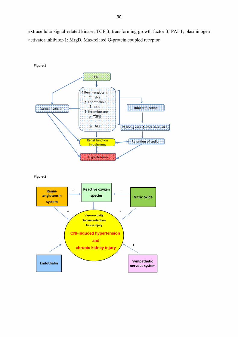
mechanism in the development of hypertension and renal damage (Figure 1). The major
mechanisms that have been proposed are: activation of the renin-angiotensin system (Lee
1997, Mervaala et al. 1999) and the sympathetic nervous system (Murray et al. 1985),
increased production of endothelin-1 (Kohan 1997, Lanese and Conger 1993), induction of
oxidative stress (Nishiyama et al. 2003) and alteration of the NO system (Curtis 2002,
Naesens et al. 2009). Together these changes cause systemic and renal vasoconstriction
leading to the development of endothelial dysfunction, hypertension and kidney damage
(Figure 2). Besides the major mechanisms, the effect of CNIs induces other processes, such as
reduced production of prostacyclin and increased synthesis of thromboxane and TGF ß
(Campistol et al. 2001). Some studies have indicated altered regulation of intracellular
calcium (Lo Russo et al. 1996), magnesium deficiency (Mervaala et al. 1997) and decreased
dopamine production in the kidney (Pestana et al. 1997). Finally, disruption in sodium
excretion has been linked to the effect of CNI on tubular transport (Hoorn et al. 2011). CNI-
induced oxidative stress and changes in HDL cholesterol may also affect both the
cardiovascular system and kidney function (Nishiyama et al. 2003; Singh et al. 2014).
The renin-angiotensin system
The renin-angiotensin system (RAS) is a signalling pathway responsible for regulating
electrolytes of the body as well as water homeostasis and blood pressure. The complete
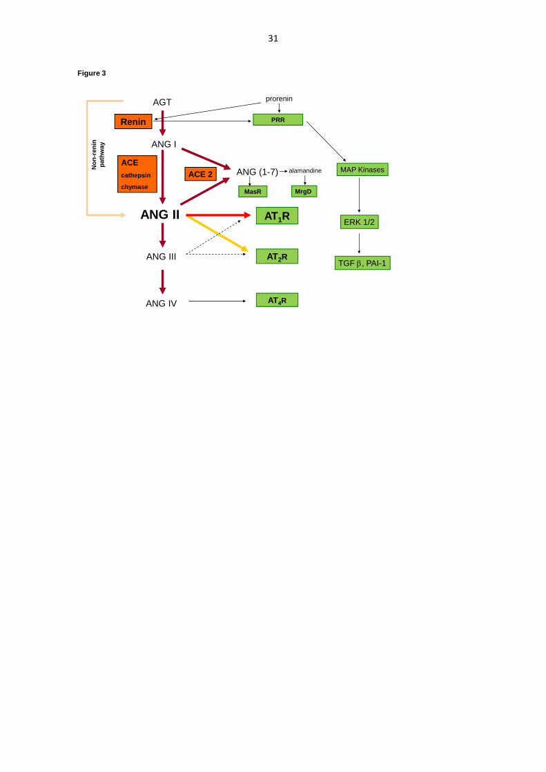
scheme of the RAS cascade is demonstrated in Figure 3. The main cascade indicates that
9
renin is an important enzyme that cleaves plasma angiotensinogen (AGT) to angiotensin I
(ANG I). Renin secretion is controlled by three following mechanisms: 1) the intrarenal
baroreceptor mechanism, 2) the macula densa mechanism and 3) the activity of the
sympathetic nervous system (SNS). The next step is the conversion of ANG I to ANG II by
angiotensin-converting enzyme (ACE) which catalyzes this reaction (Navar et al. 2002).
ANG II is a potent vasoconstrictor agent, which potentiates aldosterone release and
stimulates the release of catecholamine, increases the release of antidiuretic hormone in the
brain and stimulates thirst. It also increases myocardial contractility, and reduces urinary
losses of water. ANG II may also cause pressure-induced renal injury via its ability to induce
systemic and glomerular hypertension (Siragy 2006). It also exhibits hypertrophic and
proliferative effects and contributes to the progression of renal damage, thus driving the
recruitment and inflammatory activation of immune cells and fibrosis (Neilson et al. 1993).
Most of the hypertensive effects of ANG II (vasoconstriction, sodium retention) are mediated
via specific receptors for angiotensin II type 1 receptor (AT1R) (Cervenka et al. 1999, Navar
2013, Carey 2015), which also support cell growth. Besides the kidney, AT1R are found in
many other organs, including the heart (positive chronotropic and ionotropic effects of ANG
II on cardiomyocytes) and the brain (release of vasopressin, thirst, sympathetic activation,
regulation of blood pressure). Thus, selective AT1R antagonist (ARB) drugs are used to
efficiently reduce blood pressure (Navar et al. 2002, Carey and Siragy 2003, Carey 2015).
The vasodilatative arm partially antagonises the vasoconstrictive arm. In contrast to
AT1R, ANG II type 2 receptor (AT2R) inhibits cell growth and proliferation and increases the
excretion of sodium. Stimulation of AT2R causes significant vasodilation. Recent studies have
investigated the role of nitric oxide (NO) in the AT2-mediated vasodilatory response to ANG
II in various vessels (Dinh et al. 2001, Mehta and Griendling 2007). AT2R also inhibit renin
synthesis (Siragy et al. 2005).
10
Angiotensin-converting enzyme 2 (ACE2) forms angiotensin 1-7 (ANG 1-7), which
exerts anti-proliferative and anti-hypertensive effects. The potential imbalance between these
two arms can contribute to hypertension or hypotension (Navar 2013). Stimulation of MasR
by the ANG 1-7 pathway leads to activation of endothelial NO synthase. The consequent
release of NO induces vasodilation (Carey 2015). The diuretic and natriuretic effects of ANG
1-7 are partially due to renal vasodilation, but may also be caused by the inhibition of sodium
and water reabsorption along the nephron. An additional new RAS pathway is the
alamandine/Mas-related G-protein coupled receptor (MrgD). Alamandine also induces NO-
dependent vasorelaxation and its anti-fibrotic activity has been demonstrated (Gembardt et al.
2008).
In addition, activation of the receptor prorenin (PRR) increases plasminogen activator
inhibitor 1 (PAI-1) and collagen formation, and potentially contributes to tissue fibrosis
through the generation of TGF-β (Krebs et al. 2007; Sihn et al. 2010).
The role of intrarenal RAS in the regulation of renal function
Intrarenal RAS has been described in some studies as an independent functional tissue
system and characterised on the basis in vivo experiments. In these studies, intrarenal
inhibition of RAS resulted in significant increases in renal hemodynamics and tubular
excretion of sodium and water, after which all RAS components were ascertained in the
kidney (Navar et al. 2002; Carey and Siragy 2003). Renin is not only produced by cells of the
juxtaglomerular apparatus but also by proximal tubular cells. AGT is also secreted by
proximal tubular cells into tubules or into the interstitial space as a source of ANG I and
further converted to ANG II by ACE on the brush border of the proximal tubule (Carey and
Siragy 2003). Local expression of AGT is stimulated by ANG II and this positive feedback
can lead to an increase in intrarenal ANG II concentration (Navar et al. 2002). Furthermore,
11
intrarenal ANG II concentration is also enhanced by internalisation via AT1 receptor-
mediated endocytosis (Navar et al., 2002). Altogether, these mechanisms exceed ANG II
concentration in the kidney over plasma level (Nishiyama et al. 2002).
Activation of the renin-angiotensin system by CNIs
Some interesting data of the interaction between CNIs and RAS have been presented
over recent years (Lee 1997, Mervaala et al. 1999; Stilman et al. 1995, Hošková et al. 2014).
The activation of RAS is initiated by direct induction of renin production and release in
juxtaglomerular cells (Hoorn et al. 2012). A study by Prokai showed that CNIs induce renin
production in the collecting duct and correspond with increased vascular endothelial growth
factor (VEGF) production, which results in disproportional vessel growth (Prokai et al. 2016).
A study by Nishyiama and colleagues demonstrated that ANG II levels are increased in both
plasma and kidney tissues of CsA-induced hypertensive rats (Nishiyama et al. 2003).
However, the mechanisms by which CNIs induce ANG II production remain to be elucidated.
Increased renin secretion may also be induced secondarily by the hemodynamic effect of CNI
(Hansen et al. 1997). CsA causes intrarenal vasoconstriction, leading to a severe reduction in
blood supply to nephrons. In addition, cyclosporine stimulates platelet-derived growth factor
(PDGF) (Shehata et al. 1995) and TGF-β in juxtaglomerular cells (Matl et al. 2005).
Therefore, these growth factors may also mediate the renin secretory effect of CsA (Lee
1997). Taking these findings together, CNI may cause a vicious circle in which RAS
activation by CNI reduces renal hemodynamics, induces local ischaemia in the kidneys,
further increases renin release and potentiates nephrotoxicity (Figure 1).
Recent findings indicate that CNIs enhance intrarenal RAS activity, which in turn may
increase blood pressure and further contribute to the progression of renal damage (Nishyiama
12
et al. 2003; Hošková et al. 2014). These studies suggest that inappropriate intrarenal
activation of RAS by CNI is a crucial pathophysiological mechanism underlying chronic CNI
nephrotoxicity. It also indicated that RAS blockade efficiently attenuates Tac-induced arterial
hypertension and nephrotoxicity. Therefore, inhibition of RAS seems to be essential therapy
for alleviation of nephrotoxic effects of chronic CNI medication. Treatment regimen should
optimally involve RAS inhibition by ACEI and ARB, and combination may potentiate
renoprotective effects of these antihypertensive agents (Hošková at al. 2014). Besides
lowering of blood pressure, it seems important to substantially reduce the intrarenal
enhancement of RAS activity. We aware that the current recommendation of the dual RAS
blockade in human remains questionable particularly in diabetic patients for their uncertainty
concerning adverse effects and outcomes that may limit applicability to clinical practice
(Mancia et al. 2013).
The sympathetic nervous system
The SNS plays an important role in the regulation of arterial pressure. Not
surprisingly, increased sympathetic nervous system activity has been implicated as a primary
precursor of hypertension in both humans and animal models (Esler et al. 2003; Ameer et al.
2014). Elevated sympathetic activity accelerates the development of hypertension through
sympathetically-mediated vasoconstriction and increased tubular sodium reabsorption.
Cardiac sympathetic stimulation also contributes to the development of left ventricular
hypertrophy. The baroreceptor system opposes increases and decreases in arterial pressure.
The primary purpose of the arterial baroreflex is to keep blood pressure close to a particular
set point over a relatively short period of time. Overactivity of sympathetic nerves plays an
important role in the pathogenesis of hypertension, heart failure and the development of
chronic kidney disease (DiBona 2004, Grisk et al. 2004, Kobuchi et al. 2014). Sympathetic
13
nerve fibres entering the kidneys terminate in the vascular wall of the juxtaglomerular
apparatus and in the renal tubules. Activation of sympathetic nerves in the kidneys increases
tubular sodium reabsorption, renin release and renal vascular resistance (DiBona 2004),
leading to a subsequent reduction in renal haemodynamics associated with the impairment of
renal function.
Activation of the sympathetic nervous system by CNIs
Increased sympathetic nervous activity is also involved in the pathogenesis of CNI-
induced arterial hypertension and nephrotoxicity (Scherrer et al. 1990). CNIs modulate
glutaminergic neurotransmission in rat cortical neurons through a presynaptic mechanism
(Victor et al. 1995). Based on available evidence, CsA increases sympathetic nervous activity
either by direct central postsynaptic excitation mediated by the modulation of glutaminergic
neurotransmission (Grassi 2009) or by increased renal efferent signalling through
cyclosporine-induced vasoconstriction of the renal vascular bed, thereby leading to increased
central sympathetic outflow (Zhang et al. 2000). The sympathetic renal nerves also richly
innervate the proximal tubule and thus, play an important role in the regulation of proximal
tubule sodium reabsorption (Quan and Baum 2001). Accordingly, enhanced sodium retention
and increased plasma volume contribute to development of hypertension. Therefore beta-
blockers display the efficient attenuation of the enhanced sympathetic activity during CNI
medication.
The endothelin system
Endothelins (ETs) and their receptors are closely involved in physiological regulation
of blood pressure and sodium homeostasis. ETs directly regulate cardiac output, the
14
peripheral and central nervous systems, renal sodium and water excretion, as well as systemic
vascular resistance. Endothelin-1 (ET-1), a peptide derived from endothelial cells with high
vasoconstrictor activity, was discovered in 1988 (Yanagisawa et al. 1988). Release of ET-1 is
stimulated by a variety of endogenous substances, such as ANG II, antidiuretic hormone,
thrombin and cytokines. The production of ET-1 is inhibited by the release of prostacyclin,
atrial natriuretic peptide and NO. Thus, ET-1 plays a major role in the functional and
structural changes observed in arterial and pulmonary hypertension, glomerulosclerosis,
atherosclerosis and heart failure. ET-1 interacts with the specific endothelin receptor types A
and B. In the vasculature, ETA receptors are found predominantly in smooth muscle cells,
whereas ETB receptors are localised in endothelial cells and, to some extent, in smooth muscle
cells, macrophages and renal tubules (Kohan et al. 2011).
There are opposing actions to the ETA and ETB receptors. Activation of ETA induces
long-lasting vasoconstriction as well as cell proliferation in different tissues and fibrosis. In
contrast, activation of endothelial ETB receptor stimulates the release of NO and prostacyclin
and, at the beginning, leads to vasodilatation. ETB receptor also prevents apoptosis and
mediates pulmonary clearance of circulating ET-1 and the reuptake of ET-1 by endothelial
cells (Lüscher et al. 2000).
The role of endothelin in renal physiology
Increased expression of ET-1 has also been observed in the kidneys (Kitamura et al.
1989). ET-1 regulates renal vascular resistance through direct interaction with the agonists,
the ETA and ETB receptors, leading to modulation of afferent and efferent arteriolar
resistance. In addition, ET-1 increases intracellular Ca2+ in pre-glomerular smooth muscle
cells, leading to renal microvascular vasoconstriction. ET-1 is also an important regulator of
renal sodium and water excretion. Finally, ET-1 plays an important role in the development of
15
proteinuria, fibrosis, cell proliferation and inflammation. An interesting aspect of ETs activity
is that can be considered a hallmark of proteinuric renal diseases as it precedes the
development of glomerulosclerosis (Barton et al. 2012). Podocyte cells synthetise ET-1 and
podocytes express both ETA and ETB receptors. Production of renal ET-1 is increased in
diabetes mellitus, obesity, autoimmune diseases, oxidative stress and NO deficiency (Barton
and Yanagisawa 2008). Another study demonstrated that local ET-1 levels increase in the
vascular wall during hypertension (Dhaun et al. 2008). The nature of this pathophysiological
regulation is complex and probably depends on the degree of renal damage. It appears to be
associated with increased ET-1 in the kidneys (Kohan 1997).
Activation of the endothelin system by CNIs
Enhanced ET-1 expression may be a natural consequence of heart transplantation.
Furthermore, CNIs can also induce the production of endogenous ET-1. Preclinical trials have
shown that CNI treatment induces ET-1 synthesis by both endothelial cells and proximal
tubular cells in cultures (Bunchman et al. 1991). Indeed, one human study has also
demonstrated that CNI induces plasma ET-1 which is associated with higher blood pressure,
renal dysfunction and the progression of renal injury (Cauduro et al. 2005). Endothelial
damage leads to activation of the coagulation cascade and stimulates platelet-derived growth
factor and local production of cytokines. ET-1 induces the expression of TGF-β, an important
cytokine of fibrogenesis (Hutchinson 1999). TGF-β1 is a key profibrogenic cytokine
associated with chronic allograft nephropathy (Viklický et al. 2003). However, the therapeutic
use of ET receptors inhibition remains uncertain.
16
The nitric oxide system
Nitric oxide (NO) is the most important mediator of vasodilation. It is released during
the conversion of L-arginine to L-citrulline by NO synthases (NOS) (Wink and Mitchell
1998). As a radical, NO diffuses through cell membranes and activates intracellular guanylyl
cyclase. In the smooth muscle cells, production of cyclic guanosine monophosphate (cGMP)
lowers cellular calcium levels, leading to vasodilation (Loscalzo 1995). NO also inhibits
apoptotic signals (programmed cell death) via cGMP and G kinase. In immune reactions, NO
plays a part in non-specific defence mechanisms, induces interferon production and regulates
phagocytosis and inflammation reactions.
In the cardiovascular system, NO is significantly involved in the regulation of vascular
tonus and blood pressure, affects renal hemodynamics and tubular function (Wilcox 2005),
and plays a role in the gastrointestinal tract by relaxing smooth muscle and functions of the
mucosal immune system. NO-induced vasodilation in the corpus cavernosum causes erection.
Furthermore, it inhibits the adhesion and aggregation of thrombocytes and leucocyte
activation and proliferation. In CNS, NO modulates structural morphogenesis of the brain and
synapsis and neurotransmitter release. It also plays a role as a mediator of pain.
The role of NO in the regulation of blood pressure and renal function
The important role of the NO system has been shown in the regulation of
cardiovascular and renal function (Majid and Navar 2001, Modlinger et al. 2004). Besides its
potent vasodilatory effects, NO also influences sodium tubular transport and thus also affects
blood pressure. Furthermore, its properties antagonize many endogenous vasoconstrictors,
particularly ANG II. In the kidneys, all NOS isoforms are present, but their role in NO
17
production remains unclear. Pharmacological inhibition of particular NOS has revealed some
of their specific roles in the regulation of renal function. Non-selective systemic NOS
inhibition leads to increases in blood pressure and vascular resistance. It also alters sodium
reabsorption, most likely due to enhanced sympathetic activity (Liu and Barajas 1998). On the
other hand, selective NOS inhibition in the kidneys suggests that NOS isoforms may be
differently involved in many pathophysiological processes. In general, lacking NO production
and reduced NO bioavailability markedly impairs renal function and increases the
susceptibility to organ damage induced by enhanced activity of vasoconstrictors.
Inhibition of the NO system by CNIs
Another pathophysiological mechanism of CNI-induced nephrotoxicity appears to be
direct vascular effect which leads to endothelial dysfunction. However, the direct link is still
unknown. It is likely that CNIs alter the functions of all NOS isoforms by several different
mechanisms (Naesens et al. 2009) and thus, reducing NO production. This decreased NO
bioavailability could lead to an inadequate vasodilation to unopposed vasoconstriction, which
is a main mechanism of CNI-induced alteration of organ hemodynamics, including the
kidney. Other possibility could be a lower NO availability due to other radicals during
oxidative stress and tissue damage processes, such as ischemic-reperfusion injury in renal
tubules leading to local apoptosis (Hortelano et al. 2000). It has been shown that
supplementation of NO donors displays some protective effects against CNI-induced
nephrotoxicity (Lopau et al. 2000, Mansour et al. 2002). Taken together NO system is
significantly involved in the renoprotection and its impairment markedly contributes to the
progression of CNI-induced hypertension and nephrotoxicity. Therefore antihypertensive
treatment that positively affects NO system, particularly by increases of NO bioavailability,
may exhibit better protection against negative effects of CNIs.
18
Oxidative stress
Living cells constantly generate free radicals throughout many metabolic pathways.
Radicals can be classified as reactive oxygen species (ROS) and reactive nitrogen species
(RNS). They form a part of the natural defence system and have important functions,
especially in phagocytosis and apoptosis. The most important radicals are superoxide (O2-)
and the hydroxyl radical (OH.). Hydrogen peroxide (H2O2) is not radical but quickly oxidizes
to radicals. Importantly, NO is also present in living cells as a radical and can be transformed
to other RNS. Production of radicals needs to be controlled by several antioxidative systems
to maintain balance and protect other cells. Oxidative stress has been implicated in the
pathophysiology of many disorders and diseases, such as cardiovascular disorders, including
arterial hypertension (Wilcox 2005; Pacher et al. 2007) and atherosclerosis. It is also involved
in damaging cells and is associated with ageing, inflammatory processes, cancer growth and
the development of neurodegenerative diseases. An important role of ROS has also been
demonstrated in reperfusion, where mitochondria display enhanced oxygen consumption. This
active metabolism can cause an accumulation of ROS and thus a risk of irreversible oxidative
injury.
Overall, the role of ROS as an omnipresent product of metabolic processes has been
studied extensively. There is mounting evidence that ROS can directly influence organ
function. In the kidneys, ROS exhibit direct vasoconstriction and also alter sodium
reabsorption in the tubules. Therefore, they may alter blood pressure regulation and enhance
the accumulation of O2- in the kidneys. In this way, ROS appear to contribute to development
of hypertension (Kopkan and Červenka 2009). These mechanisms have been described in
many studies that indicate important interactions between ANG II, NO and O2- (Wilcox
2005). Enhanced O2- activity diminishes NO bioavailability in tissues, leading increased
19
vascular resistance, lowering of GFR and sodium retention. All these processes contribute to
development of arterial hypertension.
Activation of ROS production by CNIs
Zhong et al. (1998) showed that CsA administration led to the enhanced activity of
ROS. Another clinical study also indicates that plasma levels of H2O2 in kidney transplant
patients treated with CNIs significantly increase (Calo et al. 2002). In a subsequent study by
Nishiyama et al. (2003), clear evidence was provided to indicate that activation of RAS and
ROS is involved in CsA-induced hypertension. Both RAS inhibition by the AT1 receptor
blocker and administration of the antioxidant 4-hydroxy-TEMPOL reduced oxidative stress
and prevented development of hypertension in this model. The positive effect of RAS
inhibition on reducing ROS production in transplant patients was suggested by Kidokoro et
al. (2012). This study also indicated that overexpression of AT1R in the kidneys may
contribute to CNI-induced nephrotoxicity. Thus lowering of oxidative stress induced by CNI
should be also one of the main targets of antihypertensive treatment to provide substantial
organoprotection.
Renal sodium transport systems
One of the major kidney functions is continuous regulation of ion and water
homeostasis. Sodium transport and its excretion in all parts of the nephron are especially
important for maintenance of extracellular fluid volume and thus, for blood pressure
regulation (Hoorn et al. 2012). The main sodium transport mechanisms are as follows: 1) in
the proximal tubule, a high electrochemical gradient is maintained by the sodium-potassium
pump (Na+-K+-ATP ase) that transports sodium from tubular fluid to the cells; 2) another
20
transporter, called the Na+/H+ exchanger type 3 (NHE3), is responsible for the electroneutral
exchange of sodium and hydrogen ions; 3) in the ascending limb of the loop of Henle, sodium
is transported by the Na+-K+-2Cl¯ co-transporter (NKCC2) to the site of action for loop
diuretics; 4) in the distal tubule, the thiazide-sensitive Na+/Cl¯ co-transporter (NCC), is the
main mechanism of sodium handling; 5) in the collecting duct, sodium channels (ENaC)
regulate sodium concentrations under the control of aldosterone and vasopressin.
Alterations of sodium transport mechanisms by long-term CNI administration
Another important mechanism that contributes to development of arterial hypertension
during long-term CNI administration in heart transplant patients is retention of sodium and
water (Ciresi et al. 1992). As the kidneys play a key role in the regulation of sodium and
water balance, the effects of CNIs on renal function have been shown during the early stages
of rat experiments to cause vasoconstriction, particularly of the afferent arteriole. This process
results in lowering of perfusion pressure and sodium filtration, and terminates with an
increase in blood pressure (Kaskel et al. 1987). CsA has been suggested as being responsible
for reducing the reabsorption of sodium in the proximal tubule most likely via alteration of
NHE3 activity (Lorenz et al. 1999). Esteva-Font et al. (2007) found that cyclosporine induces
exaggerated NKCC2 expression in the proximal tubules when compared with an untreated
control group. Recent studies indicate enhanced activity of the thiazide-sensitive co-
transporter NCC as another possible explanation for sodium retention during CNI
administration (Hoorn et al. 2011). Therefore, thiazide diuretics could be another option in
hypertensive patients treated with CNI (Colussi et al. 2007). Other studies have demonstrated
that CNIs may also influence Na+- K+-ATPase; however, the results remain inconclusive
(Hoorn et al. 2012). Therefore, all the above mechanisms can significantly alter sodium
excretion and thus, contribute to the pathogenesis of hypertension induced by CNIs. Base on
21
that, diuretics are usually combined with other antihypertensives in treatment of chronic CNI-
induced hypertension.
Conclusions
Heart transplantation is now an established treatment modality in patients with end-
stage heart failure when all other therapeutic options fail. The long-term outcomes of heart
transplant recipients have improved considerably since the introduction of CNIs. In this
review, we have outlined the pathophysiology of CNI-induced nephrotoxicity and
hypertension. We have shown that CNIs lead to activation of the renin-angiotensin and
endothelin systems and to increase of sympathetic nerve activity. In addition, CNIs are known
to inhibit NO synthesis and NO-mediated vasodilation, and also increase free radicals and
superoxide production through vasoconstriction-associated hypoxia. Increased levels of
intrarenal renin and angiotensin II induced by CNI are recognized as an important mechanism
that contributes to nephrotoxicity. The direct effect of CNI on the tubular epithelial cells plays
a major role in the development of interstitial fibrosis. Activation of RAS is not only
important in terms of its hemodynamic contribution, but it also directly promotes renal
interstitial fibrosis through profibrotic effect of TGF β. Finally, the effect of CNI on tubular
function may explain sodium retention. Continuing research of side effects of CNI and their
mechanisms remains is an important prerequisite for development of novel therapeutic
strategies to reduce nephrotoxicity and to manage arterial hypertension.
Acknowledgements
The authors gratefully thank Dr. Josef Zicha, Institute of Physiology of the ASCR, for his
valuable comments and suggestions during preparation of this manuscript.
22
References
AMEER OZ, HILDRETH CM, PHILLIPS JK. Sympathetic overactivity prevails over the
vascular amplifier phenomena in a chronic kidney disease rat model of hypertension. Physiol
Rep, 2: e12205, 2014.
ANDOH TF, BURDMANN EA, BENNETT WM: Nephrotoxicity of immunosuppressive
drugs: experimental and clinical observations. Semin Nephrol 17: 34-45, 1997.
BARTON M, YANAGISAWA M: Endothelin: 20 years from discovery to therapy. Can J
Physiol Pharmacol 86: 485-498, 2008.
BARTON M, THARAUX PL: Endothelin and the podocyte. Clin Kidney J. 5:17-27, 2012.
BUNCHMAN TE, BROOKSHIRE CA: Cyclosporine-induced synthesis of endothelin by
cultured human endothelial cells. J Clin Invest 88: 310-3144, 1991.
BOLMAN RM, CANCE C, SPRAY T, GENTON R, WEISS C, SAFFITZ J, EISEN H: The
changing face of cardiac transplantation: The Washington University Program 1985-1987.
Ann Thorac Surg 45: 192-197, 1987.
CALNE RY, ROLLES K, WHITE DJ, THIRU S, EVANS DB, MCMASTER P, DUNN DC,
CRADDOCK GN, HENDERSON RG, AZIZ S, LEWIS P: Cyclosporin A initially as the only
immunosuppressant in 34 recipients of cadaveric organs: 32 kidneys, 2 pancreases, and 2
livers. Lancet 2: 1033-1036, 1979.
CALÒ LA, DAVIS PA, GIACON B, PAGNIN E, SARTORI M, RIEGLER P,
ANTONELLO A, HUBER W, SEMPLICINI A: Oxidative stress in kidney transplant patients
with calcineurin inhibitor-induced hypertension: effect of ramipril. J Cardiovasc Pharmacol
40: 625-631, 2002.
CAMPISTOL JM, IÑIGO P, LARIOS S, BESCOS M, OPPENHEIMER F: Role of
transforming growth factor-beta1 in the progression of chronic allograft nephropathy. Nephrol
Dial Transplant 16 Suppl 1: 114-116, 2001.
CAREY RM, SIRAGY HM. The intrarenal renin-angiotensin system and diabetic
nephropathy. Trends Endocrinol Metab 14: 274-281, 2003.
CAREY RM. The intrarenal renin-angiotensin system in hypertension. Adv Chronic Kidney
Dis 22: 204-210, 2015.
CAUDURO RL, COSTA C, LHULIER F, GARCIA RG, CABRAL RD, GONÇALVES LF,
MANFRO RC: Endothelin-1 plasma levels and hypertension in cyclosporine-treated renal
transplant patients. Clin Transplant 19: 470-474, 2005.
CERVENKA L, WANG CT, MITCHELL KD, NAVAR LG. Proximal tubular angiotensin II
levels and renal functional responses to AT1 receptor blockade in nonclipped kidneys of
Goldblatt hypertensive rats. Hypertension 33: 102-107, 1999.
CIRESI DL1, LLOYD MA, SANDBERG SM, HEUBLEIN DM, EDWARDS BS: The
sodium retaining effects of cyclosporine. Kidney Int 41: 1599-1605, 1992.
23
COLUSSI G, BETTINELLI A, TEDESCHI S, DE FERRARI ME, SYRÉN ML, BORSA N,
MATTIELLO C, CASARI G, BIANCHETTI MG: A thiazide test for the diagnosis of renal
tubular hypokalemic disorders. Clin J Am Soc Nephrol 2: 454-460, 2007.
CURTIS JJ: Hypertensinogenic mechanism of the calcineurin inhibitors. Curr Hypertens Rep
4: 377-380, 2002.
DHAUN N, GODDARD J, KOHAN DE, POLLOCK DM, SCHIFFRIN EL, WEBB DJ: Role
of endothelin-1 in clinical hypertension: 20 years on. Hypertension 52: 452-459, 2008.
DIBONA GF: The sympathetic nervous system and hypertension: recent developments.
Hypertension 43: 147-150, 2004.
DINH DT, FRAUMAN AG, JOHNSTON CI, FABIANI ME. Angiotensin receptors:
distribution, signalling and function. Clin Sci (Lond) 100: 481-492, 2001.
DUNLAY SM, ROGER VL: Understanding the epidemic of heart failure: past, present, and
future. Curr Heart Fail Rep. 11: 404-415, 2014.
ESLER M, LAMBERT G, BRUNNER-LA ROCCA HP, VADDADI G, KAYE D:
Sympathetic nerve activity and neurotransmitter release in humans: translation from
pathophysiology into clinical practice. Acta Physiol Scand 177: 275-284, 2003.
ESTEVA-FONT C, ARS E, GUILLEN-GOMEZ E, CAMPISTOL JM, SANZ L, JIMÉNEZ
W, KNEPPER MA, TORRES F, TORRA R, BALLARÍN JA, FERNÁNDEZ-LLAMA P:
Cyclosporin induced hypertension is associated with increased sodium transporter of the loop
of Henle (NKCC2). Nephrol Dial Transplant 22: 2810-2816, 2007.
FAUL C, DONNELLY M, MERSCHER-GOMEZ S, CHANG YH, FRANZ S,
DELFGAAUW J, CHANG JM, CHOI HY, CAMPBELL KN, KIM K, REISER J, MUNDEL
P: The actin cytoskeleton of kidney podocytes is a direct target of the antiproteinuric effect of
cyclosporine A. Nat Med 14: 931-938.
FRIMAT L, VILLEMOT JP, CORMIER L, CAO-HUU T, RENOULT E, HESTIN D,
DOPFF C, MATTÉI S, HUBERT J, KESSLER M: Treatment of end-stage renal failure after
heart transplantation. Nephrol Dial Transplant 13: 2905-2908, 1998.
GEMBARDT F, GRAJEWSKI S, VAHL M, SCHULTHEISS HP, WALTHER T:
Angiotensin metabolites can stimulate receptors of the Mas-related genes family. Mol Cell
Biochem 319: 115-123, 2008.
GRASSI G: Assessment of sympathetic cardiovascular drive in human hypertension:
achievements and perspectives. Hypertension 54: 690-697, 2009.
GRISK O, RETTIG R: Interactions between the sympathetic nervous system and the kidneys
in arterial hypertension. Cardiovasc Res 61: 238-246, 2004.
HANSEN JM, FOGH-ANDERSEN N, CHRISTENSEN NJ, STRANDGAARD S:
Cyclosporine-induced hypertension and decline in renal function in healthy volunteers. J
Hypertens 15: 319-26, 1997.
24
HOORN EJ, WALSH SB, MCCORMICK JA, FÜRSTENBERG A, YANG CL, ROESCHEL
T, PALIEGE A, HOWIE AJ, CONLEY J, BACHMANN S, UNWIN RJ, ELLISON DH: The
calcineurin inhibitor tacrolimus activates the renal sodium chloride cotransporter to cause
hypertension. Nat Med 17: 1304-1309, 2011.
HOORN EJ, WALSH SB, MCCORMICK JA, ZIETSE R, UNWIN RJ, ELLISON DH:
Pathogenesis of calcineurin inhibitor-induced hypertension. J Nephrol 25: 269-275, 2012.
HORTELANO S, CASTILLA M, TORRES AM, TEJEDOR A, BOSCÁ L: Potentiation by
nitric oxide of cyclosporin A and FK506-induced apoptosis in renal proximal tubule cells. J
Am Soc Nephrol 11: 2315-2323, 2000.
HOSKOVÁ L, VIKLICKÝ O, MÁLEK I, PODZIMKOVÁ M, HEGAROVÁ M, PIRK J,
VÍTKO S, KAUTZNER J: Ischaemic heart disease is a risk factor for renal failure after heart
transplantation Int J Cardiol 123: 358-360, 2008.
HOŠKOVÁ L, MÁLEK I, KAUTZNER J, HONSOVÁ E, VAN DOKKUM RP, HUSKOVÁ
Z, VOJTÍŠKOVÁ A, VARCABOVÁ S, CERVENKA L, KOPKAN L: Tacrolimus-induced
hypertension and nephrotoxicity in Fawn-Hooded rats are attenuated by dual inhibition of
renin-angiotensin system. Hypertens Res 37:724-732, 2014.
HUTCHINSON IV. The role of transforming growth factor-beta in transplant rejection.
Transplant Proc 31: 9S-13S, 1999.
IDEMA RN, VAN DEN MEIRACKER AH, BALK AH, BOS E, SCHALEKAMP MA,
MAN IN 'T VELD AJ: Abnormal diurnal variation of blood pressure, cardiac output, and
vascular resistance in cardiac transplant recipients. Circulation 6: 2797-2803, 1994.
KIDOKORO K, SATOH M, NAGASU H, SAKUTA T, KUWABARA A, YORIMITSU D,
NISHI Y, TOMITA N, SASAKI T, KASHIHARA N: Tacrolimus induces glomerular injury
via endothelial dysfunction caused by reactive oxygen species and inflammatory change.
Kidney Blood Press Res 35: 549-557, 2012.
KASKEL FJ, DEVARAJAN P, ARBEIT LA, PARTIN JS, MOORE LC: Cyclosporine
nephrotoxicity: sodium excretion, autoregulation, and angiotensin II. Am J Physiol 252: F733-
F742, 1987.
KINO T, HATANAKA H, HASHIMOTO M, NISHIYAMA M, GOTO T, OKUHARA M,
KOHSAKA M, AOKI H, IMANAKA H: FK-506, a novel immunosuppressant isolated from
a Streptomyces. I. Fermentation, isolation, and physico-chemical and biological
characteristics. J Antibiot 40: 1249-1255, 1987.
KITAMURA K, TANAKA T, KATO J, ETO T, TANAKA K: Regional distribution of
immunoreactive endothelin in porcine tissue: abundance in inner medulla of kidney. Biochem
Biophys Res Commun 161: 348–352, 1989.
KOBUCHI S, TANAKA R, FUNAI A, SUZUKI R, YAZAWA M, TSUTSUI H, OHKITA
M, AYAJIKI K, MATSUMURA Y: Involvement of renal sympathetic nerve overactivation in
the progression of chronic kidney disease in rats. J Cardiovasc Pharmacol 63: 9-15, 2014.
KOHAN D. Endothelins in the normal and diseased kidney. Am J Kidney Dis 29: 2-26, 1997.
25
KOHAN DE, ROSSI NF, INSCHO EW, POLLOCK DM: Regulation of blood pressure and
salt homeostasis by endothelin. Physiol Rev 91:1-77, 2011.
KOPKAN L, CERVENKA L. Renal interactions of renin-angiotensin system, nitric oxide and
superoxide anion: implications in the pathophysiology of salt-sensitivity and hypertension.
Physiol Res 58 (Suppl 2): S55-S67, 2009.
KREBS C, HAMMING I, SADAGHIANI S, STEINMETZ OM, MEYER-SCHWESINGER
C, FEHR S, STAHL RA, GARRELDS IM, DANSER AH, VAN GOOR H, CONTREPAS A,
NGUYEN G, WENZEL U: Antihypertensive therapy upregulates renin and (pro)renin
receptor in the clipped kidney of Goldblatt hypertensive rats. Kidney Int 72: 725-730, 2007.
LANESE DM, CONGER JD: Effects of endothelin receptor antagonist on cyclosporine-
induced vasoconstriction in isolated rat renal arterioles. J Clin Invest 91: 2144-2149, 1993.
LEE DB: Cyclosporine and the renin-angiotensin axis. Kidney Int 52:248-260, 1997.
LIAO R, LIU Q, ZHENG Z, FAN J, PENG W, KONG Q, HE H, YANG S, CHEN W, TANG
X, YU X: Tacrolimus protects podocytes from injury in lupus nephritis partly by stabilizing
the cytoskeleton and inhibiting podocyte apoptosis. PLoS One 10(7): e0132724.
doi:10.1371/journal.pone.0132724
LIU L, BARAJAS L: Evidence for NOS-containing renal neuronal somata transiently
expressing a catecholaminergic phenotype during development in the rat. Neurosci Lett 251:
161-164, 1998.
LO RUSSO A, PASSAQUIN AC, ANDRÉ P, SKUTELLA M, RÜEGG UT: Effect of
cyclosporin A and analogues on cytosolic calcium and vasoconstriction: possible lack of
relationship to immunosuppressive activity. Br J Pharmacol 118: 885-892, 1996.
LOPAU K, KLEINERT D, ERLER J, SCHRAMM L, HEIDBREDER E, WANNER C:
Tacrolimus in acute renal failure: does L-arginine-infusion prevent changes in renal
hemodynamics? Transpl Int 13: 436-442, 2000.
LORENZ JN, SCHULTHEIS PJ, TRAYNOR T, SHULL GE, SCHNERMANN J:
Micropuncture analysis of single nephron function in NHE3-deficient mice. Am J Physiol
277: F447-F453, 1999.
LOSCALZO J, WELCH G: Nitric oxide and its role in the cardiovascular system. Prog
Cardiovasc Dis 38: 87-104, 1995.
LUND LH, EDWARDS LB, KUCHERYAVAYA AY, BENDEN C, DIPCHAND AI,
GOLDFARB S, LEVVEY BJ, MEISER B, ROSSANO JW, YUSEN RD, STEHLIK J: The
Registry of the International Society for Heart and Lung Transplantation: Thirty-second
Official Adult Heart Transplantation Report-2015; Focus Theme: Early Graft Failure. J Heart
Lung Transplant 34: 1244-1254, 2015.
LÜSCHER TF, BARTON M: Endothelins and endothelin receptor antagonists
therapeutic considerations for a novel class of cardiovascular drugs. Circulation
102: 2434-2440, 2000.
26
MAJID DS, NAVAR LG: Nitric oxide in the control of renal hemodynamics and excretory function. Am J Hypertens 14: 74S-82S, 2001. MANCIA G, FAGARD R, NARKIEWICZ K, REDÓN J, ZANCHETTI A, BÖHM M, CHRISTIAENS T, CIFKOVA R, DE BACKER G, DOMINICZAK A, GALDERISI M, GROBBEE DE, JAARSMA T, KIRCHHOF P, KJELDSEN SE, LAURENT S, MANOLIS AJ, NILSSON PM, RUILOPE LM, SCHMIEDER RE, SIRNES PA, SLEIGHT P, VIIGIMAA M, WAEBER B, ZANNAD F; TASK FORCE MEMBERS: 2013 ESH/ESC Guidelines for the management of arterial hypertension: the Task Force for the management of arterial hypertension of the European Society of Hypertension (ESH) and of the European Society of Cardiology (ESC). J Hypertens 31:1281-1357, 2013. MANSOUR M, DABA MH, GADO A, AL-RIKABI A, AL-MAJED A: Protective effect of L-arginine against nephrotoxicity induced by cyclosporine in normal rats. Pharmacol Res 45: 441-446, 2002. MATL I, VIKLICKÝ O, VOSKA L, LODEREROVÁ A, VÍTKO S: The effect of different immunosuppressive regimens on TGF-beta 1 expression in kidney transplant patients. Transplant Int 18: 668-667, 2005. MCCAULEY J: The nephrotoxicity of FK-506 as compared with cyclosporine. Curr Opin
Nephrol Hypertens 2: 662-669, 1993.
MEHTA PK, GRIENDLING KK. Angiotensin II cell signaling: physiological and
pathological effects in the cardiovascular system. Am J Physiol Cell Physiol 292: C82-97,
2007.
MERVAALA E, LASSILA M, VASKONEN T, KROGERUS L, LÄHTEENMÄKI T,
VAPAATALO H, KARPPANEN H: Effects of ACE inhibition on cyclosporine A-induced
hypertension and nephrotoxicity in spontaneously hypertensive rats on a high-sodium diet.
Blood Press 8: 49-56, 1999.
MERVAALA EM, PERE AK, LINDGREN L, LAAKSO J, TERÄVÄINEN TL, KARJALA
K, VAPAATALO H, AHONEN J, KARPPANEN H: Effects of dietary sodium and
magnesium on cyclosporin A-induced hypertension and nephrotoxicity in spontaneously
hypertensive rats. Hypertension 29: 822-827, 1997.
MIGITA K, ORIGUCHI T, KAWABE Y, TOMINAGA M, IDA H, KAWAKAMI A,
EGUCHI K: FK506 markedly enhances apoptosis of antigen-stimulated peripheral T cells by
down-regulation of Bcl-xL. Transplantation 68: 1018-1023, 1999.
MODLINGER PS, WILCOX CS, ASLAM S: Nitric oxide, oxidative stress, and progression
of chronic renal failure. Semin Nephrol 24:354-365, 2004.
MURRAY BM, PALLER MS, FERRIS TF: Effect of cyclosporine administration on renal
hemodynamics in conscious rats. Kidney Int 28:767-774, 1985.
MYERS BD, ROSS J, NEWTON L, LUETSCHER J, PERLROTH M: Cyclosporine-
associated chronic nephropathy. N Engl J Med 311: 699-705, 1984.
NAESENS M, KUYPERS DR, SARWAL M: Calcineurin inhibitor nephrotoxicity. Clin J Am
Soc Nephrol 4:481-508, 2009.
27
NAKATA Y, YOSHIBAYASHI M, YONEMURA T, UEMOTO S, INOMATA Y,
TANAKA K, FURUSHO K: Tacrolimus and myocardial hypertrophy. Transplantation 69:
1960-1962, 2000.
NAVAR LG, HARRISON-BERNARD LM, NISHIYAMA A, KOBORI H. Regulation of
intrarenal angiotensin II in hypertension. Hypertension 39: 316-322, 2002.
NAVAR LG: Translational studies on augmentation of intratubular renin-angiotensin system
in hypertension. Kidney Int Suppl 3: 321-325, 2013.
NEILSON JA, WEST MJ: Structural influences on resistance in the hindlimb vascular bed of
the renal hypertensive rabbit. Clin Exp Pharmacol Physiol 20: 377-379, 1993.
NISHIYAMA A, SETH DM, NAVAR LG: Renal interstitial fluid concentrations of
angiotensins I and II in anesthetized rats. Hypertension 39:129-134, 2002.
NISHIYAMA A, KOBORI H, FUKUI T, ZHANG GX, YAO L, RAHMAN M, HITOMI H,
KIYOMOTO H, SHOKOJI T, KIMURA S, KOHNO M, ABE Y: Role of angiotensin II and
reactive oxygen species in cyclosporine A-dependent hypertension. Hypertension 42: 754-
760, 2003.
PACHER P, BECKMAN JS, LIAUDET L: Nitric oxide and peroxynitrite in health and
disease. Physiol Rev 87: 315-424, 2007.
PARRY G, MEISER B, RÁBAGO G: The clinical impact of cyclosporine nephrotoxicity in
heart transplantation. Transplantation 69: S23-S26, 2000.
PESTANA M, FARIA MS, OLIVEIRA JG, BALDAIA J, SANTOS A, GUERRA LE,
SOARES-DA-SILVA P: Assessment of renal dopaminergic system activity during the
recovery of renal function in human kidney transplant recipients. Nephrol Dial Transplant 12:
2667-2672, 1997.
PETERS DH, FITTON A, PLOSKER GL, FAULDS D: Tacrolimus: a review of its
pharmacology, and therapeutic potential in hepatic and renal transplantation. Drugs 46: 746-
796, 1993.
PHAM PT, EDLING KL, CHAKKERA HA, PHAM PC, PHAM PM: Screening strategies
and predictive diagnostic tools for the development of new-onset diabetes mellitus after
transplantation: an overview. Diabetes Metab Synd Obes 5: 379-387, 2012.
PIRSCH JD, MILLER J, DEIERHOI MH, VINCENTI F, FILO RS: A comparison of
tacrolimus (FK 506) and cyclosporine for immunosuppression after cadaveric renal
transplantation. Transplantation 63: 977-983, 1997.
PRÓKAI Á, CSOHÁNY R, SZIKSZ E, PAP D, BALICZA-HIMER L, BOROS S, MAGDA
B, VANNAY Á, KIS-PETIK K, FEKETE A, PETI-PETERDI J, SZABÓ AJ: Calcineurin-
inhibition results in upregulation of local renin and subsequent vascular endothelial growth
factor production in renal collecting ducts. Transplantation [In print] 2016.
QUAN A, BAUM M: The renal nerve is required for regulation of proximal tubule transport
by intraluminally produced ANG II. Am J Physiol Renal Physiol 280: F524-529, 2001.
28
SHEHATA M, EL NAHAS M, BARKWORTH E, COPE GH, RAFTERY AT: Localization
of PDGF-BB in the juxtaglomerular cells of cyclosporin-treated rats. ExpNephrol 3:173-179,
1995.
SCHERRER U, VISSING SF, MORGAN BJ, ROLLINS JA, TINDALL RS, RING S,
HANSON P, MOHANTY PK, VICTOR RG: Cyclosporine-induced sympathetic activation
and hypertension after heart transplantation. N Engl J Med 323:693–639, 1990.
SIHN G, ROUSSELLE A, VILIANOVITCH L, BURCKLE C, BADER M: Physiology of the
(pro)renin receptor: Wnt of change? Kidney Int 78:246-256, 2010.
SINGH N, JACOBS F, RADER DJ, VANHAECKE J, VAN CLEEMPUT J, DE GEEST B:
Impaired cholesterol efflux capacity and vasculoprotective function of high-density
lipoprotein in heart transplant recipients. J Heart Lung Transplant 33: 499-506, 2014.
SIRAGY HM, XUE C, ABADIR P, CAREY RM: Angiotensin subtype-2 receptors inhibit
renin biosynthesis and angiotensin II formation. Hypertension 45: 133-137, 2005.
SIRAGY HM: Angiotensin II compartmentalization within the kidney: effects of salt diet and
blood pressure alterations. Curr Opin Nephrol Hypertens 15: 50-53, 2006.
STILLMAN IE, ANDOH TF, BURDMANN EA, BENNETT WM, ROSEN S: FK506
nephrotoxicity: morphologic and physiologic characterization of a rat model. Lab Invest 73:
794-803, 1995.
SUTHANTHIRAN M: Molecular analysis of human renal allografts: differential intragraft
gene expression during rejection. Kidney Int 58: S15-21, 1997.
VICTOR RG, THOMAS GD, MARBAN E, O'ROURKE B: Presynaptic modulation of
cortical synaptic activity by calcineurin. Proc Natl Acad Sci USA 92: 6269–673, 1995.
VIKLICKÝ O, PODZIMKOVÁ M, MATL I, MÁLEK I, VÍTKO Š, LÁCHA J: Renal
function in cyclosporine-treated heart transplant recipients long term follow-up. Cor Vasa 41:
131-134, 1999.
VIKLICKÝ O, MATL I, VOSKA L, BÖHMOVÁ R, JARESOVÁ M, LÁCHA J,
LODEREROVÁ A, STRÍZ I, TEPLAN V, VÍTKO S: TGF-beta1 expression and chronic
allograft nephropathy in protocol kidney graft biopsy. Physiol Res 52: 353-360, 2003.
WESTHOFF TH, SCHMIDT S, ZIDEK W, BEIGE J, VAN DER GIET M: Tacrolimus in
steroid-resistant and steroid-dependent nephrotic syndrome. Clin Nephrol J 65: 393-400,
2006.
WILCOX CS. Oxidative stress and nitric oxide deficiency in the kidney: a critical link to
hypertension? Am J Physiol 289: R913-R935, 2005.
WILLIAMS D, HARAGSIM L: Calcineurin nephrotoxicity. Adv Chronic Kidney Dis 13: 47-
55, 2006.
WINK DA, MITCHELL JB. Chemical biology of nitric oxide: Insights into regulatory,
cytotoxic, and cytoprotective mechanisms of nitric oxide. Free Radic Biol Med 25: 434-456,
1998.
29
WOO J, PROPPER DJ, THOMSON AW: Antigen presentation and HLA-DR expression by
FK-506-treated human monocytes. Immunology 71: 551-555, 1990.
YANAGISAWA M, KURIHARA H, KIMURA S, TOMOBE Y, KOBAYASHI M, MITSUI
Y, YAZAKI Y, GOTO K, MASAKI T: A novel potent vasoconstrictor peptide produced by
vascular endothelial cells. Nature 332: 411-415, 1988.
ZHANG W, LI JL, HOSAKA M, JANZ R, SHELTON JM, ALBRIGHT GM,
RICHARDSON JA, SÜDHOF TC, VICTOR RG: Cyclosporine A-induced hypertension
involves synapsin in renal sensory nerve endings. Proc Natl Acad Sci USA 97: 9765-9700,
2000.
Legends of figures
Figure 1
Pathophysiological mechanisms of CNI-induced hypertension and chronic
nephrotoxicity
CNI, calcineurin inhibitor; NO, nitric oxide; ROS, reactive oxygen species; SNS, sympathetic
nerve activity; NCC, renal sodium chloride co-transporter; NHE3, proximal sodium hydrogen
exchanger type 3; NKCC2, sodium potassium chloride co-transporter type 2; Na+-K+-ATPase,
sodium-potassium pump.
Figure 2
The major vasoactive systems and their interactions in the development of CNI-induced
endothelial dysfunction, hypertension and kidney damage
Figure 3
Schematic illustration of the renin-angiotensin system cascade
ANG I, angiotensin I; ACE, angiotensin-converting enzyme; ANG II, angiotensin II; ANG
III, angiotensin III; ANG IV, angiotensin IV; ACE 2, angiotensin-converting enzyme 2;
ANG-(1-7), angiotensin 1-7; AT1R, angiotensin type 1 receptor; AT2R, angiotensin type 2
receptor; AT4R, angiotensin type 4 receptor; MasR, Mas receptor (receptor for angiotensin 1-
7); PRR, (pro)renin receptor; MAP kinases, mitogen-activated protein kinases; ERK,
30
extracellular signal-related kinase; TGF , transforming growth factor ; PAI-1, plasminogen
activator inhibitor-1; MrgD, Mas-related G-protein coupled receptor
CNI
Renal functionimpairment
Tubular functionVasoconstriction
Renin‐angiotensin
SNS
Endothelin‐1
ROS
Thromboxane
TGF
NO
Hypertension
Retention of sodium
NCC NHE3 NKCC2 Na+K+‐ATP?
Figure 1
Renin‐angiotensin
systemNitric oxide
Reactive oxygen
species
Endothelin
Vasoreactivity
Sodium retention
Tissue injury
Sympatheticnervous system
+
+
‐
‐
++
+
CNI-induced hypertension
and
chronic kidney injury
Figure 2
31
ANG I
ANG II
Renin
AT1R
AGTN
on
-ren
in
pat
hw
ay
ACE
cathepsin
chymase
AT2R
ANG (1-7)
ANG III
ANG IV
ACE 2
AT4R
MasR
PRR
MAP Kinases
prorenin
ERK 1/2
TGF , PAI-1
alamandine
MrgD
Figure 3