PATHOLOGY OF THE RESPIRATORY SYSTEM Part Twopeople.upei.ca/lopez/respirweb-5.pdf · •...
29
Transcript of PATHOLOGY OF THE RESPIRATORY SYSTEM Part Twopeople.upei.ca/lopez/respirweb-5.pdf · •...
Thoracic Cavity
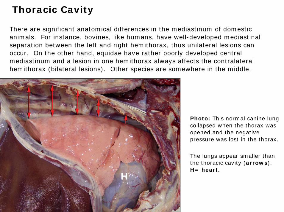
There are significant anatomical differences in the mediastinum of domestic animals. For instance, bovines, like humans, have well-developed mediastinal separation between the left and right hemithorax, thus unilateral lesions can occur. On the other hand, equidae have rather poorly developed central mediastinum and a lesion in one hemithorax always affects the contralateral hemithorax (bilateral lesions). Other species are somewhere in the middle.
Photo: This normal canine lung collapsed when the thorax was opened and the negative pressure was lost in the thorax.
The lungs appear smaller than the thoracic cavity (arrows). H= heart.
HH
Thoracic Cavity
The most abnormalities in the thoracic cavity are:
• Pneumothorax (air)
• Hydrothorax (fluid)
• Hemothorax (blood)
• Chylothorax (chyle)
• Pyothorax (pus)
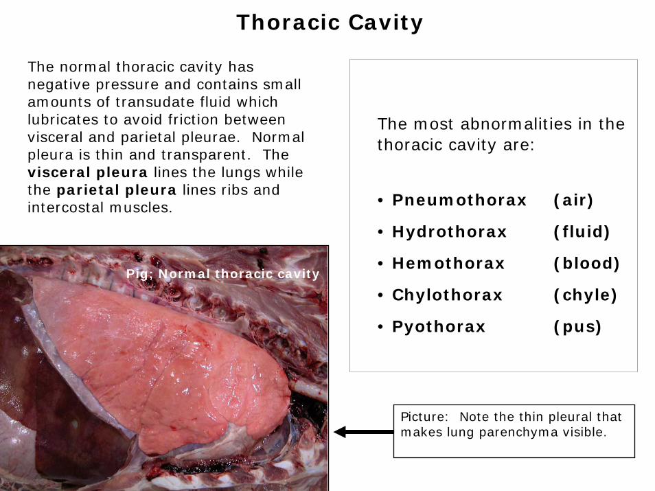
The normal thoracic cavity has negative pressure and contains small amounts of transudate fluid which lubricates to avoid friction between visceral and parietal pleurae. Normal pleura is thin and transparent. The visceral pleura lines the lungs while the parietal pleura lines ribs and intercostal muscles.
Pig; Normal thoracic cavity
Picture: Note the thin pleural that makes lung parenchyma visible.
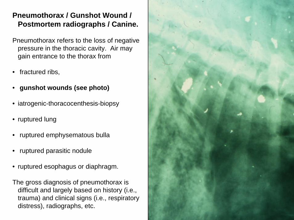
Pneumothorax / Gunshot Wound / Postmortem radiographs / Canine.
Pneumothorax refers to the loss of negative pressure in the thoracic cavity. Air may gain entrance to the thorax from
• fractured ribs,
• gunshot wounds (see photo)
• iatrogenic-thoracocenthesis-biopsy
• ruptured lung
• ruptured emphysematous bulla
• ruptured parasitic nodule
• ruptured esophagus or diaphragm.
The gross diagnosis of pneumothorax is difficult and largely based on history (i.e., trauma) and clinical signs (i.e., respiratory distress), radiographs, etc.
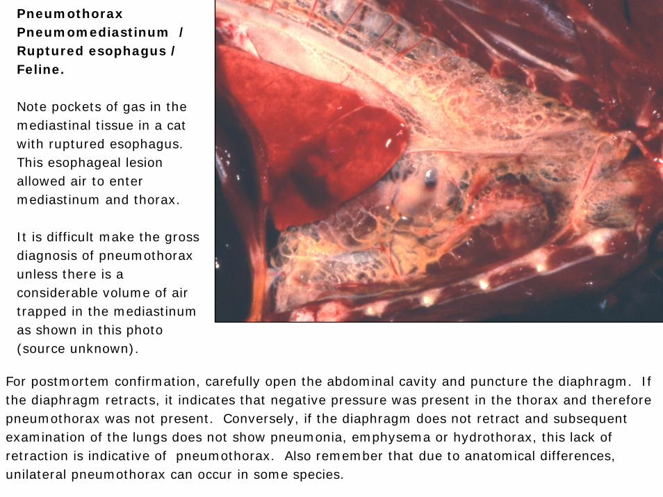
Pneumothorax Pneumomediastinum / Ruptured esophagus / Feline.
Note pockets of gas in the mediastinal tissue in a cat with ruptured esophagus. This esophageal lesion allowed air to enter mediastinum and thorax.
It is difficult make the gross diagnosis of pneumothorax unless there is a considerable volume of air trapped in the mediastinum as shown in this photo (source unknown).
For postmortem confirmation, carefully open the abdominal cavity and puncture the diaphragm. If the diaphragm retracts, it indicates that negative pressure was present in the thorax and therefore pneumothorax was not present. Conversely, if the diaphragm does not retract and subsequent examination of the lungs does not show pneumonia, emphysema or hydrothorax, this lack of retraction is indicative of pneumothorax. Also remember that due to anatomical differences, unilateral pneumothorax can occur in some species.

Hydrothorax• Transudate in thorax• Etiology:
• Congestive heart failure• Hypoproteinemia:
• Starvation• Renal disease• Intestinal disease• Lymphatic obstruction
(neoplasia)
• Sequels:• Atelectasis• Chronic pleural irritation
Photo: Note thoracic cavity and syringe filled with transudate. In severe cases, hydrothorax results in compressive atelectasis and respiratory distress. Note that the lungs of this cat are collapsed (dark) because of the compression exerted by the thoracic fluid.
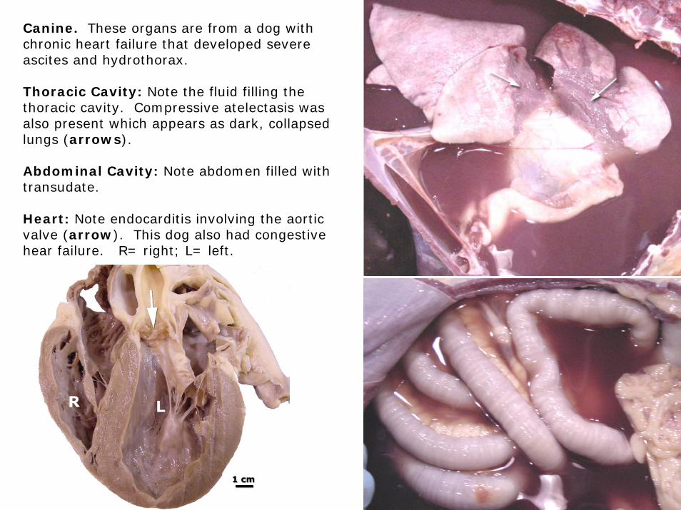
Canine. These organs are from a dog with chronic heart failure that developed severe ascites and hydrothorax.
Thoracic Cavity: Note the fluid filling the thoracic cavity. Compressive atelectasis was also present which appears as dark, collapsed lungs (arrows).
Abdominal Cavity: Note abdomen filled with transudate.
Heart: Note endocarditis involving the aortic valve (arrow). This dog also had congestive hear failure. R= right; L= left.
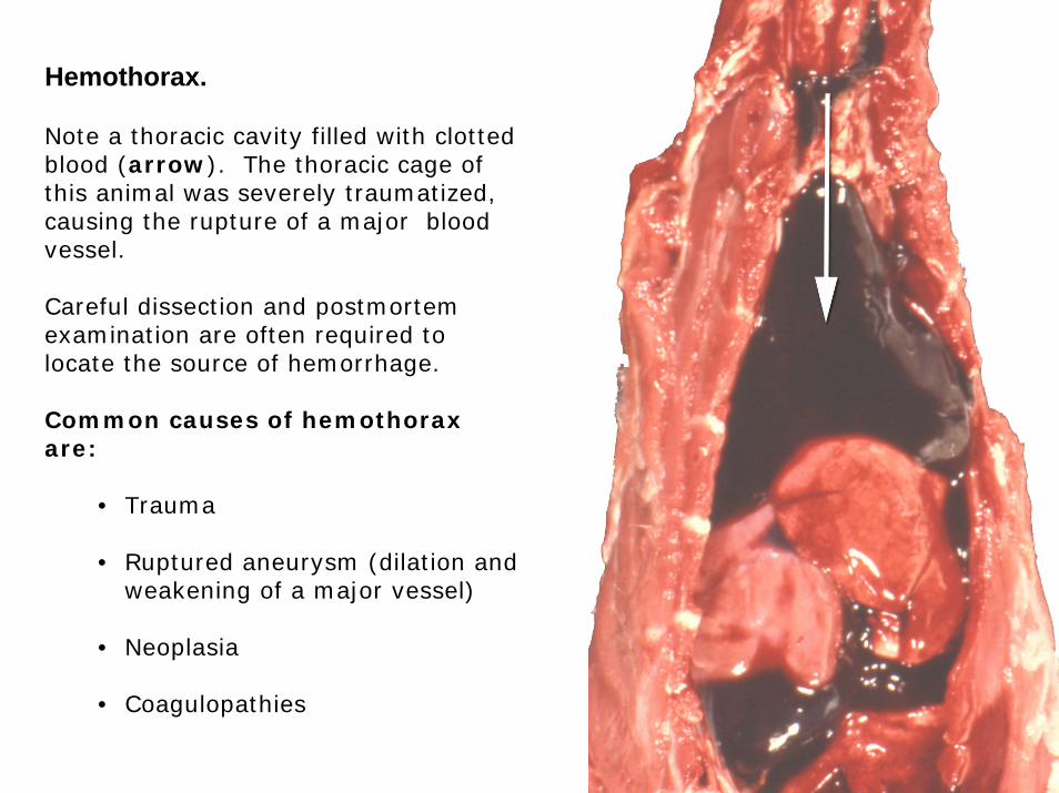
Hemothorax.
Note a thoracic cavity filled with clotted blood (arrow). The thoracic cage of this animal was severely traumatized, causing the rupture of a major blood vessel.
Careful dissection and postmortem examination are often required to locate the source of hemorrhage.
Common causes of hemothoraxare:
• Trauma
• Ruptured aneurysm (dilation and weakening of a major vessel)
• Neoplasia
• Coagulopathies
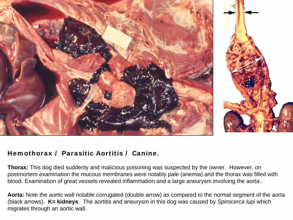
Hemothorax / Parasitic Aortitis / Canine.
Thorax: This dog died suddenly and malicious poisoning was suspected by the owner. However, on postmortem examination the mucous membranes were notably pale (anemia) and the thorax was filled with blood. Examination of great vessels revealed inflammation and a large aneurysm involving the aorta.
Aorta: Note the aortic wall notable corrugated (double arrow) as compared to the normal segment of the aorta (black arrows). K= kidneys. The aortitis and aneurysm in this dog was caused by Spirocerca lupi which migrates through an aortic wall.
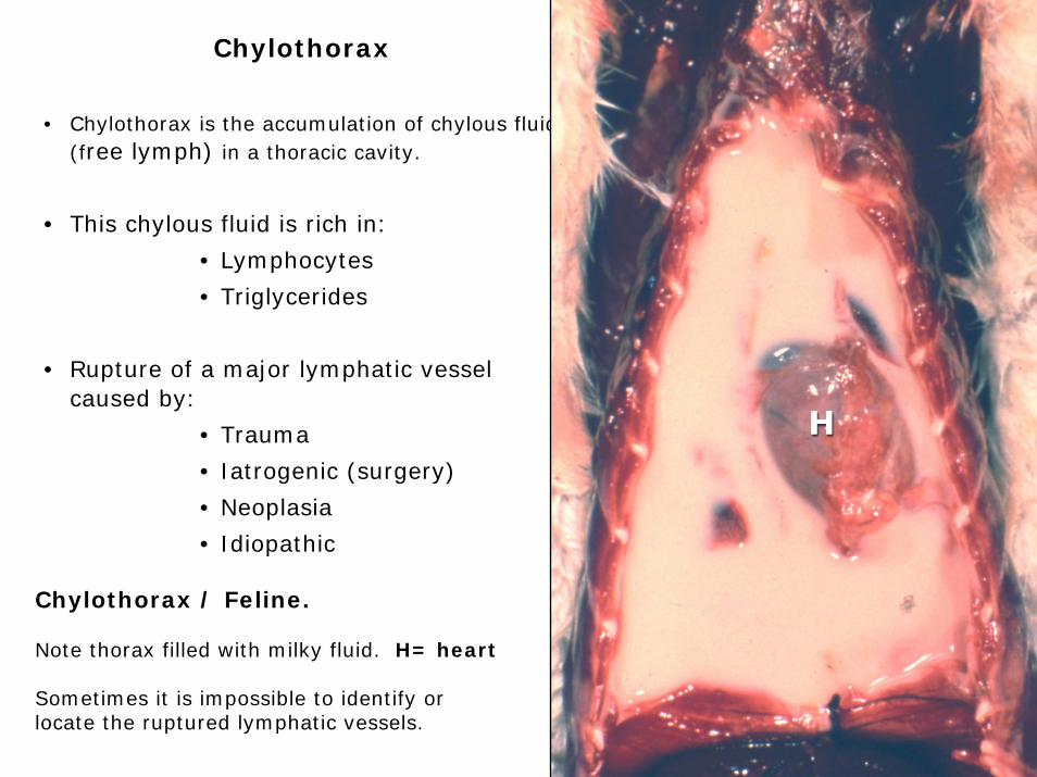
Chylothorax
• Chylothorax is the accumulation of chylous fluid (free lymph) in a thoracic cavity.
• This chylous fluid is rich in:
• Lymphocytes
• Triglycerides
• Rupture of a major lymphatic vessel caused by:
• Trauma
• Iatrogenic (surgery)
• Neoplasia
• Idiopathic
Chylothorax / Feline.
Note thorax filled with milky fluid. H= heart
Sometimes it is impossible to identify or locate the ruptured lymphatic vessels.
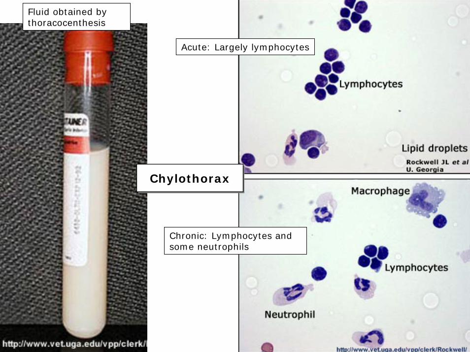
ChylothoraxChylothorax
Acute: Largely lymphocytes
Chronic: Lymphocytes and some neutrophils
Fluid obtained by thoracocenthesis
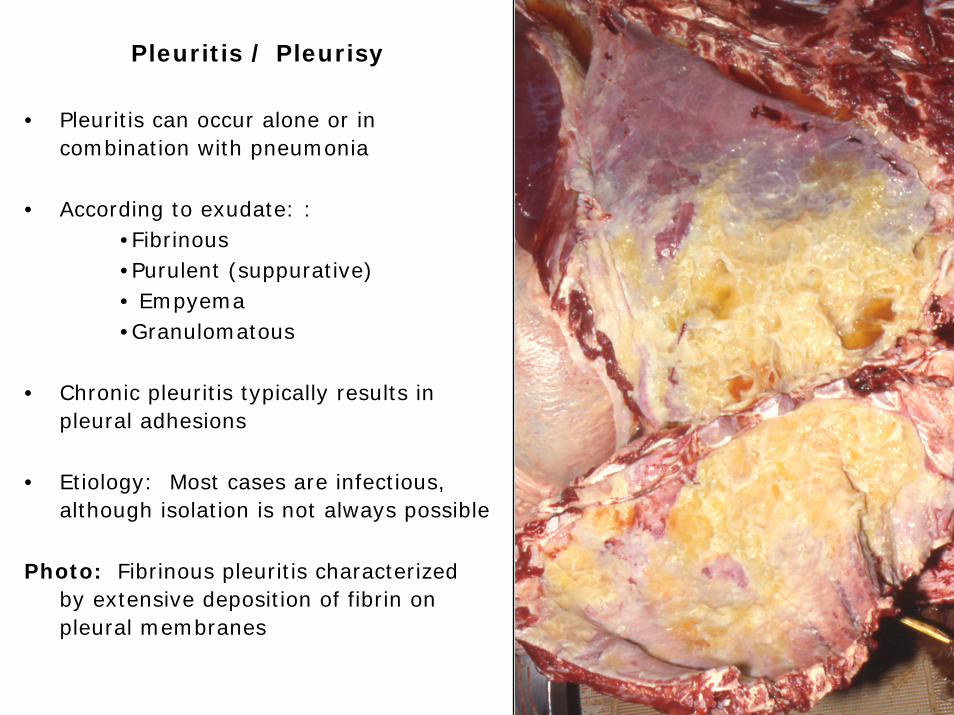
Pleuritis / Pleurisy
• Pleuritis can occur alone or in combination with pneumonia
• According to exudate: :•Fibrinous•Purulent (suppurative)• Empyema•Granulomatous
• Chronic pleuritis typically results in pleural adhesions
• Etiology: Most cases are infectious, although isolation is not always possible
Photo: Fibrinous pleuritis characterized by extensive deposition of fibrin on pleural membranes
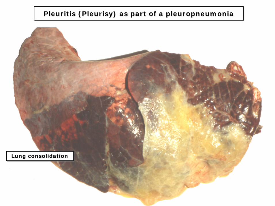
Pleuritis (Pleurisy) as part of a pleuropneumoniaPleuritis (Pleurisy) as part of a pleuropneumoniaPleuritis (Pleurisy) as part of a pleuropneumonia
Lung consolidationLung consolidationLung consolidation
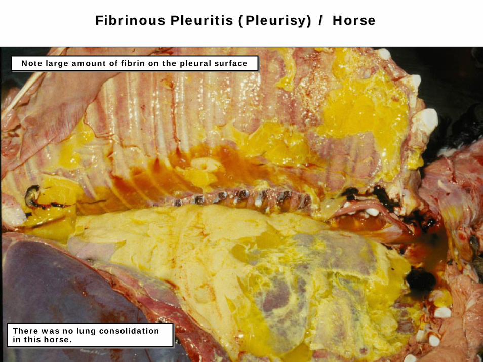
Fibrinous Pleuritis (Pleurisy) / HorseFibrinous Pleuritis (Pleurisy) / Horse
There was no lung consolidation in this horse.There was no lung consolidation in this horse.
Note large amount of fibrin on the pleural surfaceNote large amount of fibrin on the pleural surface
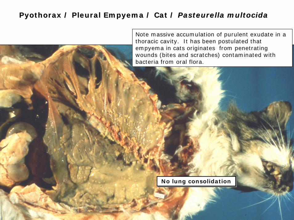
Pyothorax / Pleural Empyema / Cat / Pyothorax / Pleural Empyema / Cat / Pasteurella multocidaPasteurella multocida
No lung consolidationNo lung consolidation
Note massive accumulation of purulent exudate in a thoracic cavity. It has been postulated that empyema in cats originates from penetrating wounds (bites and scratches) contaminated with bacteria from oral flora.
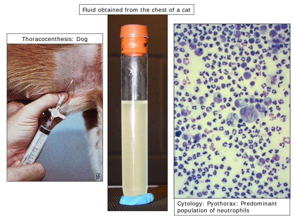
Thoracocenthesis: Dog
Fluid obtained from the chest of a cat
Cytology: Pyothorax: Predominant population of neutrophils
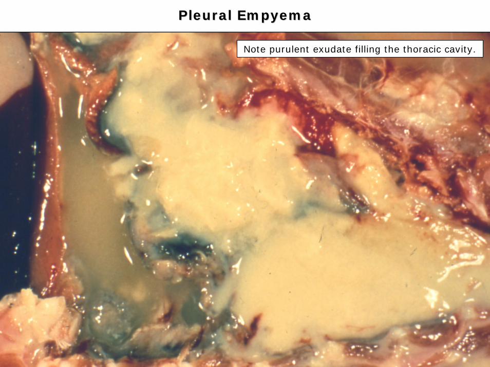
Pleural EmpyemaPleural Empyema
Note purulent exudate filling the thoracic cavity.
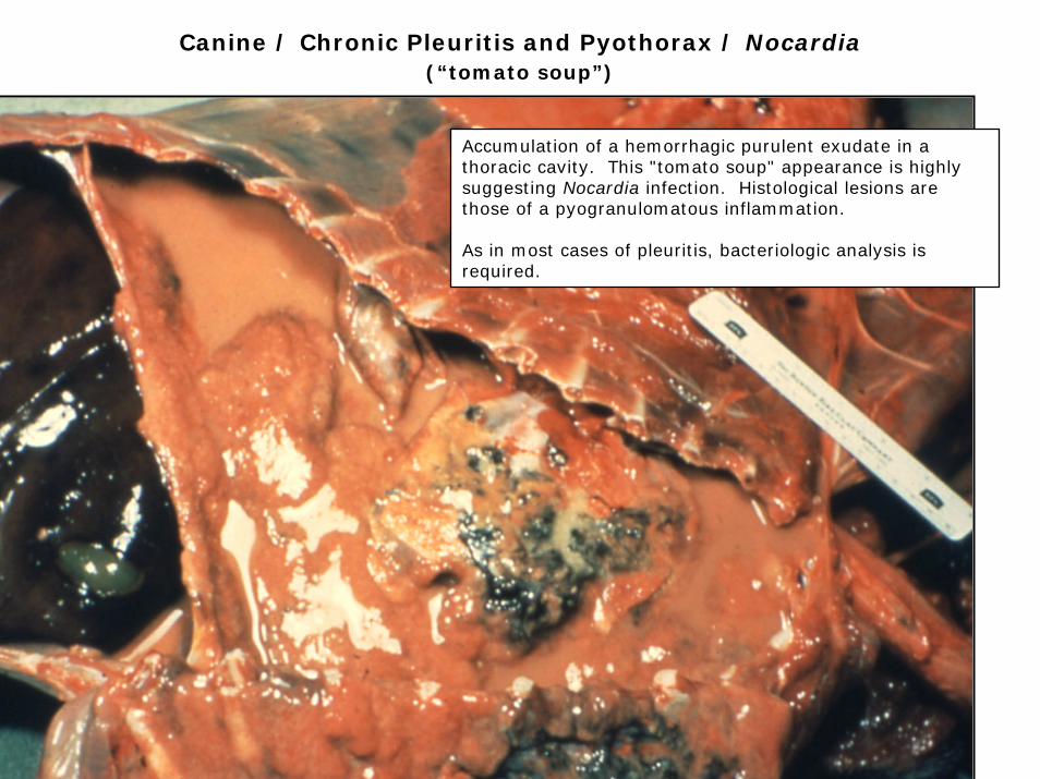
Canine / Chronic Pleuritis and Pyothorax / Nocardia(“tomato soup”)
Accumulation of a hemorrhagic purulent exudate in a thoracic cavity. This "tomato soup" appearance is highly suggesting Nocardia infection. Histological lesions are those of a pyogranulomatous inflammation.
As in most cases of pleuritis, bacteriologic analysis is required.
Lung TumorsLung Tumors
• Relatively rare in animals compared to human beings.
• More common in dogs and cats.
• According to cell line:
• Epithelial (adenoma or carcinoma).
• Mesenchymal (fibroma or fibrosarcoma / hemangioma or hemangiosarcoma).
• Most common epithelial tumors in domestic animals:
• Adenocarcinoma.
• Bronchiolo-alveolar carcinoma.
• Mesothelioma (pleura).
• Histopathology: Lung biopsy is the last diagnostic resource.
• Secondary tumors (metastatic) are common.
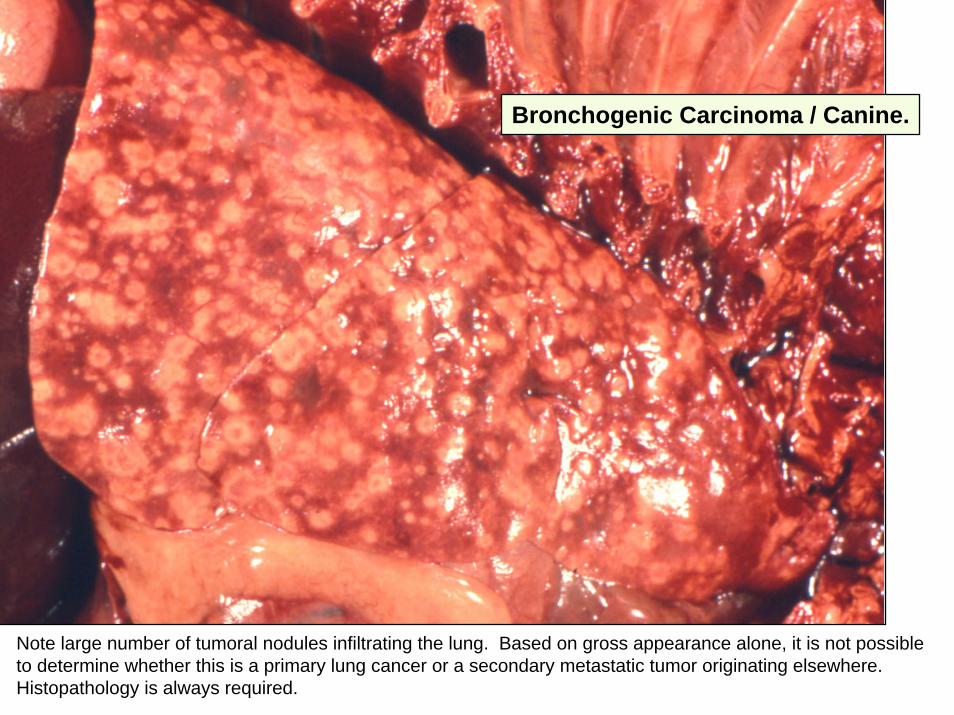
Note large number of tumoral nodules infiltrating the lung. Based on gross appearance alone, it is not possible to determine whether this is a primary lung cancer or a secondary metastatic tumor originating elsewhere. Histopathology is always required.
Bronchogenic Carcinoma / Canine.
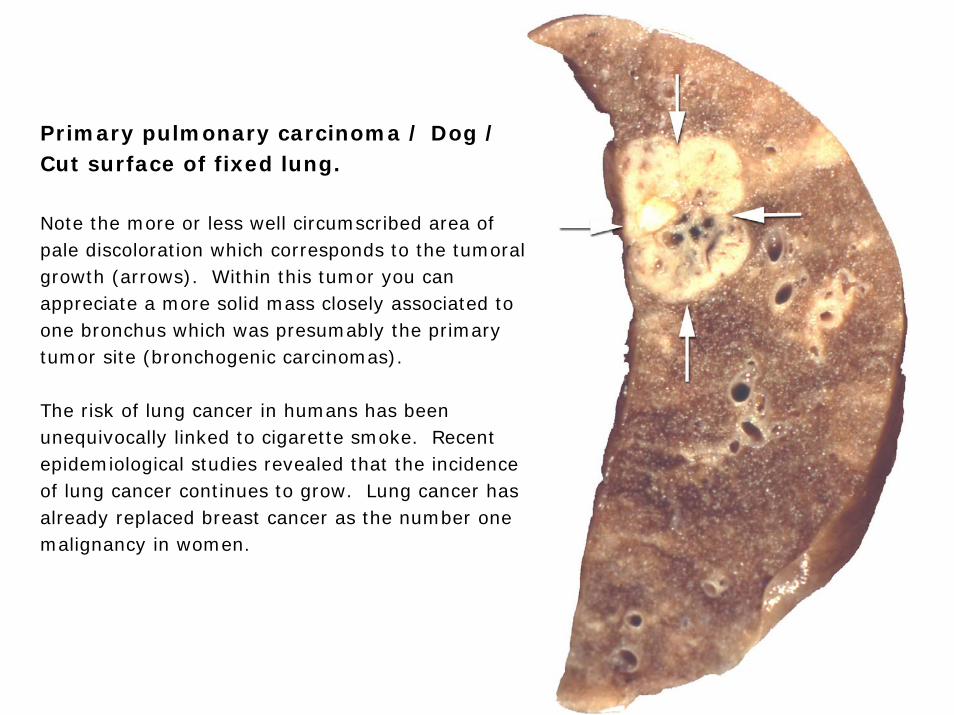
Primary pulmonary carcinoma / Dog / Cut surface of fixed lung.
Note the more or less well circumscribed area of pale discoloration which corresponds to the tumoral growth (arrows). Within this tumor you can appreciate a more solid mass closely associated to one bronchus which was presumably the primary tumor site (bronchogenic carcinomas).
The risk of lung cancer in humans has been unequivocally linked to cigarette smoke. Recent epidemiological studies revealed that the incidence of lung cancer continues to grow. Lung cancer has already replaced breast cancer as the number one malignancy in women.
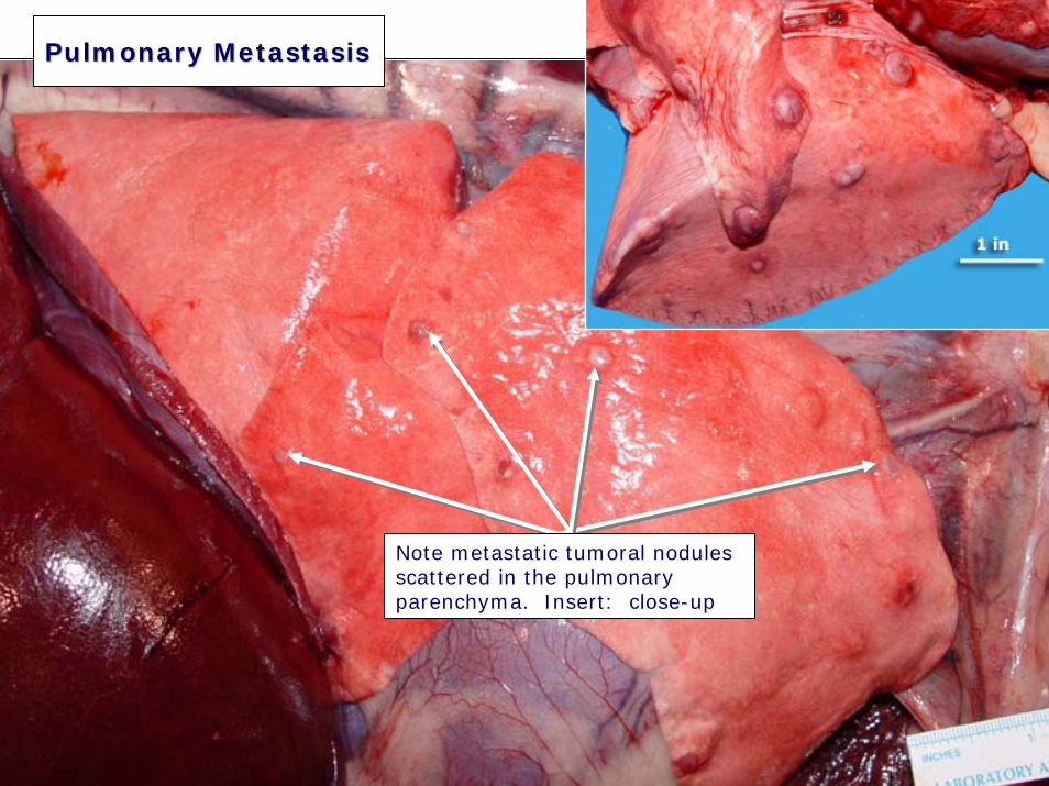
PulmonaryPulmonary MetastasisMetastasis
Note metastatic tumoral nodules scattered in the pulmonary parenchyma. Insert: close-up
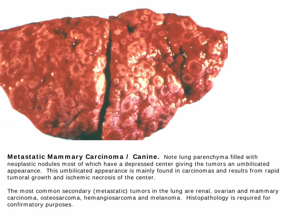
Metastatic Mammary Carcinoma / Canine. Note lung parenchyma filled with neoplastic nodules most of which have a depressed center giving the tumors an umbilicated appearance. This umbilicated appearance is mainly found in carcinomas and results from rapid tumoral growth and ischemic necrosis of the center.
The most common secondary (metastatic) tumors in the lung are renal, ovarian and mammary carcinoma, osteosarcoma, hemangiosarcoma and melanoma. Histopathology is required for confirmatory purposes.
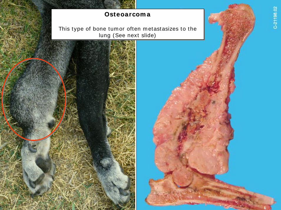
Osteoarcoma
This type of bone tumor often metastasizes to the lung (See next slide)
OsteoarcomaOsteoarcoma
This type of bone tumor often metastasizes to the lung (See next slide)
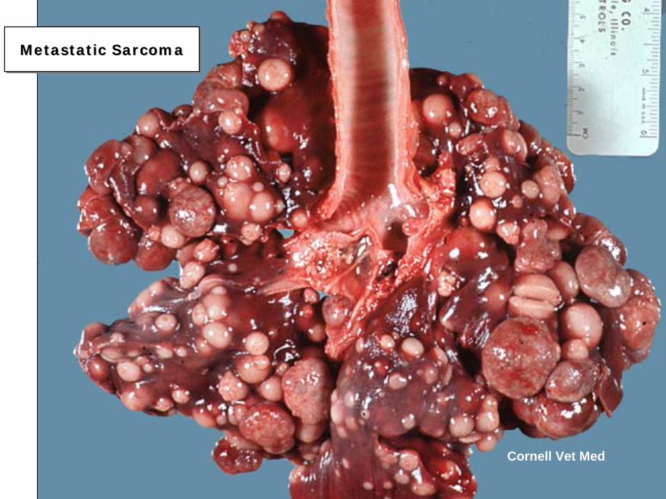
Metastatic SarcomaMetastaticMetastatic SarcomaSarcoma
Cornell Vet Med
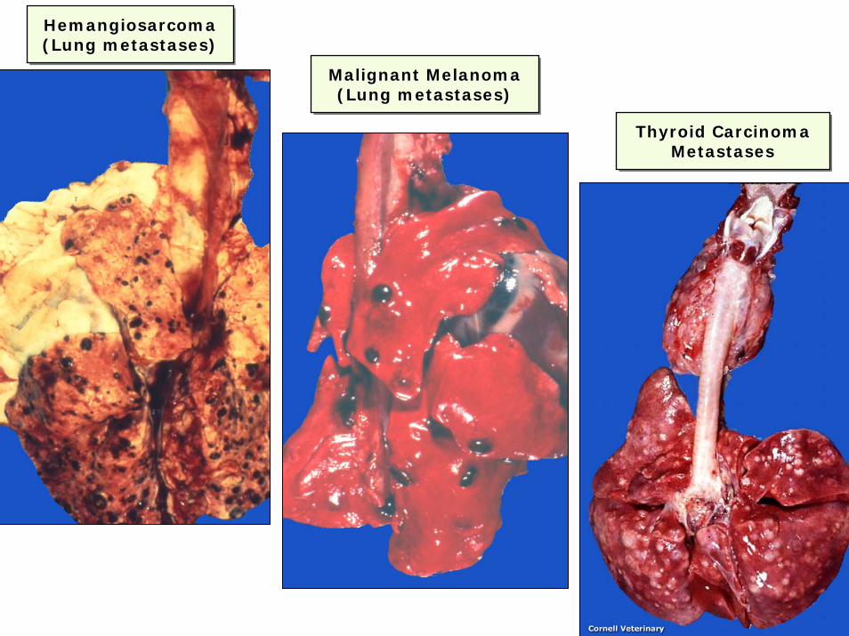
Malignant Melanoma (Lung metastases)
Malignant Melanoma Malignant Melanoma (Lung metastases)(Lung metastases)
Thyroid Carcinoma Metastases
Thyroid Carcinoma Thyroid Carcinoma MetastasesMetastases
Hemangiosarcoma(Lung metastases)HemangiosarcomaHemangiosarcoma(Lung metastases)(Lung metastases)
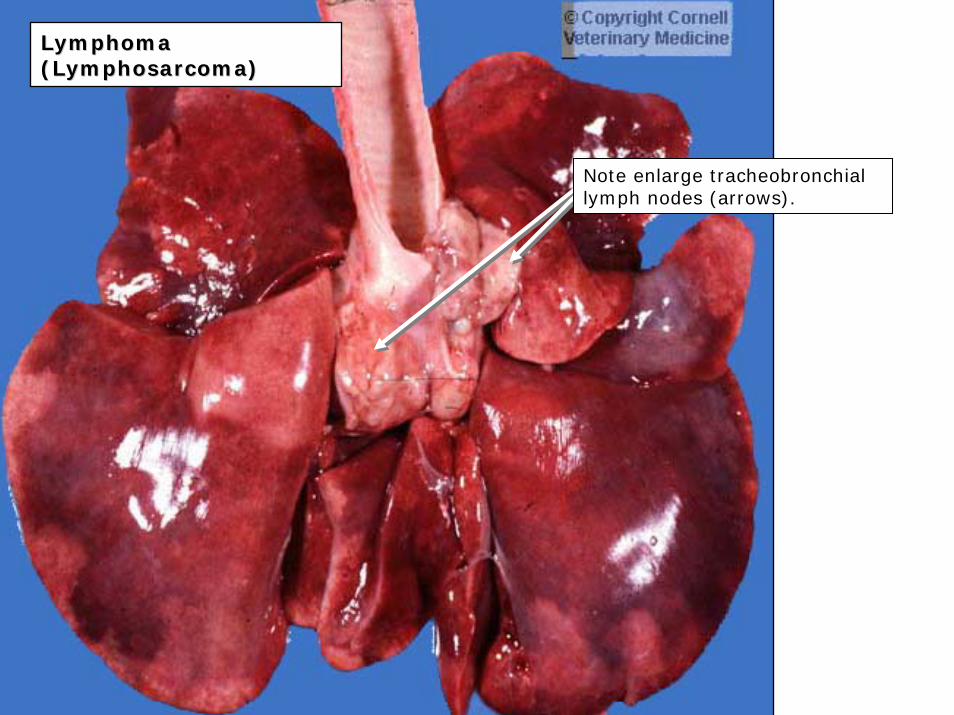
LymphomaLymphoma((LymphosarcomaLymphosarcoma))
Note enlarge tracheobronchiallymph nodes (arrows).
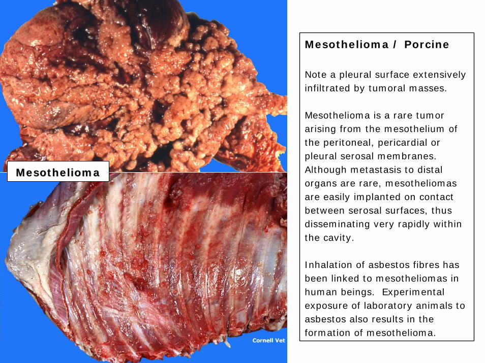
Mesothelioma / Porcine
Note a pleural surface extensively infiltrated by tumoral masses.
Mesothelioma is a rare tumor arising from the mesothelium of the peritoneal, pericardial or pleural serosal membranes. Although metastasis to distal organs are rare, mesotheliomas are easily implanted on contact between serosal surfaces, thus disseminating very rapidly within the cavity.
Inhalation of asbestos fibres has been linked to mesotheliomas in human beings. Experimental exposure of laboratory animals to asbestos also results in the formation of mesothelioma.
MesotheliomaMesothelioma

Irrational thoughts commonly observed in students undergoing exam-induced stress.
Following graduation in the summer of the rapidly approaching year 2007, these uncontrollable thoughts tend to progressively change into a more positive view of Pathology. Contrary to what has been said by some contemporary philosophers, you do not have to be insane to become a Pathologist.
Have a nice day!!
I hate PathologyI hate Pathology