
Pathology of the Alimentary Tract - University of Prince...
34
Pathology of the Alimentary Tract Shannon Martinson: http://people.upei.ca/smartinson VPM 221: November, 2011 Lab 2: Lower alimentary tract – SI, LI, cecum, and peritoneum “GIST” in the cecum of a dog
Transcript of Pathology of the Alimentary Tract - University of Prince...

Pathology of the Alimentary Tract
Shannon Martinson: http://people.upei.ca/smartinson
VPM 221: November, 2011
Lab 2: Lower alimentary tract – SI, LI, cecum, and peritoneum
“GIST” in the cecum of a dog
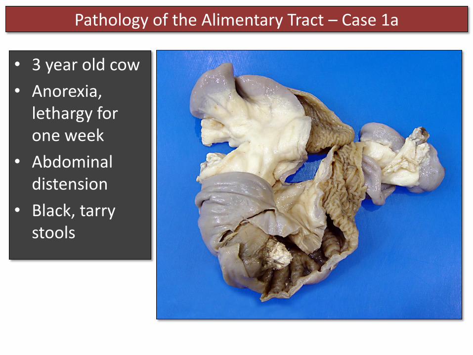
• 3 year old cow
• Anorexia, lethargy for one week
• Abdominal distension
• Black, tarry stools
Pathology of the Alimentary Tract – Case 1a

Pathology of the Alimentary Tract – Case 1a
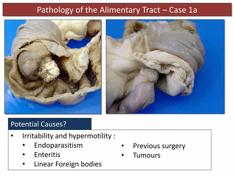
Description A ~ 5 cm segment of jejunum and the associated mesentery has telescoped into the distal segment of intestine. The segments are firmly attached to one another and the entrapped portion is necrotic with friable white material adhered to the surface.

Pathology of the Alimentary Tract – Case 1a
Morphologic Diagnosis Intestinal intussusception, segmental, subacute to chronic, severe, with necrosis
One portion of the intestine ( = the intussusceptum) invaginates into another (the intussuscipiens)
Pathology of the Alimentary Tract – Case 1a
Potential Causes?
• Irritability and hypermotility : • Endoparasitism • Enteritis • Linear Foreign bodies
• Previous surgery • Tumours

Pathology of the Alimentary Tract – Case 1a
Potential Outcome?
• Intestinal obstruction • Necrosis of the entrapped segment →endotoxemia /sepsis

Pathology of the Alimentary Tract – Case 1b
• Female lamb
• Caudal abdominal distension

Pathology of the Alimentary Tract – Case 1b
Description? A large segment of small intestine (and the associated peritoneum) has passed into the subcutis through a defect in the left caudal abdominal body wall.

Pathology of the Alimentary Tract – Case 1b
Morphologic Diagnosis Intestinal herniation through the abdominal wall (external abdominal hernia)
Possible sequelae Ileus, Intestinal incarceration, infarction

Pathology of the Alimentary Tract – Case 2
• 3 gilts in the past 2 weeks have developed black, bloody diarrhea followed by death

Pathology of the Alimentary Tract – Case 2
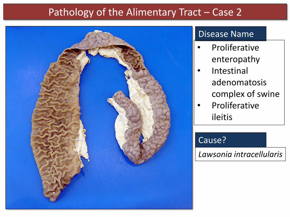
Description
There is marked thickening of the wall of the small intestine imparting a cerebriform appearance on the serosal surface and a corrugated appearance of the mucosal surface.
Morphologic Diagnosis
Proliferative enteritis (ileitis), segmental, subacute, severe
Pathology of the Alimentary Tract – Case 2
Cause?
Lawsonia intracellularis
Disease Name
• Proliferative enteropathy
• Intestinal adenomatosis complex of swine
• Proliferative ileitis
Pathology of the Alimentary Tract – Case 2
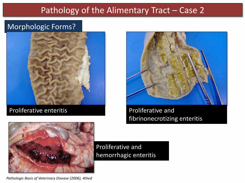
Morphologic Forms?
Proliferative enteritis Proliferative and fibrinonecrotizing enteritis
Proliferative and hemorrhagic enteritis
Pathologic Basis of Veterinary Disease (2006), 4thed

Pathology of the Alimentary Tract – Case 2b
Morphologic Diagnosis
Enteritis, granulomatous, segmental, chronic, severe
Disease Name and Etiology?
Johne’s Disease: Mycobacterium avium subsp paratuberculosis
• Adult cow with chronic diarrhea and weight loss
Pathology of the Alimentary Tract – Case 3
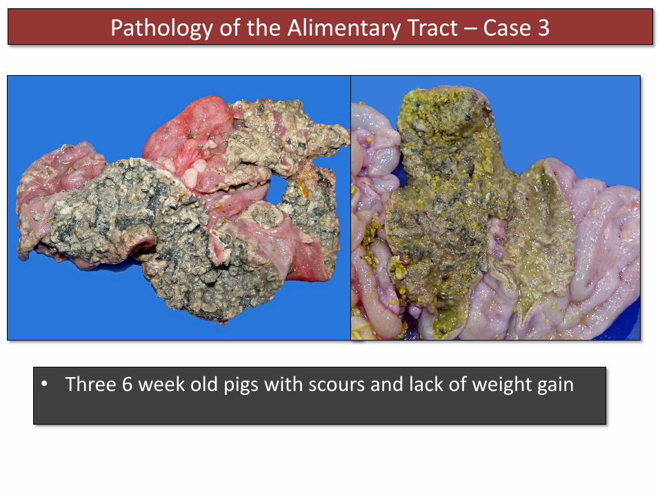
• Three 6 week old pigs with scours and lack of weight gain

Pathology of the Alimentary Tract – Case 3
Thick coalescing plaques of grey-yellow friable material are attached to the mucosal surface of the colon and cecum.
Description

Pathology of the Alimentary Tract – Case 3
Fibrinonecrotizing typhlocolitis, multifocally extensive, acute to subacute, severe
Morphological Diagnosis
Pathology of the Alimentary Tract – Case 3
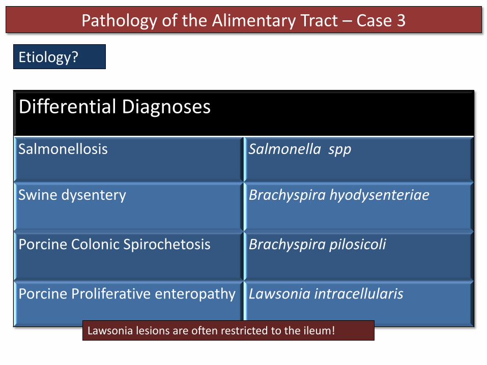
Etiology?
Differential Diagnoses
Salmonellosis Salmonella spp
Swine dysentery Brachyspira hyodysenteriae
Porcine Colonic Spirochetosis Brachyspira pilosicoli
Porcine Proliferative enteropathy Lawsonia intracellularis
Lawsonia lesions are often restricted to the ileum!

Pathology of the Alimentary Tract – Case 4
Rumen
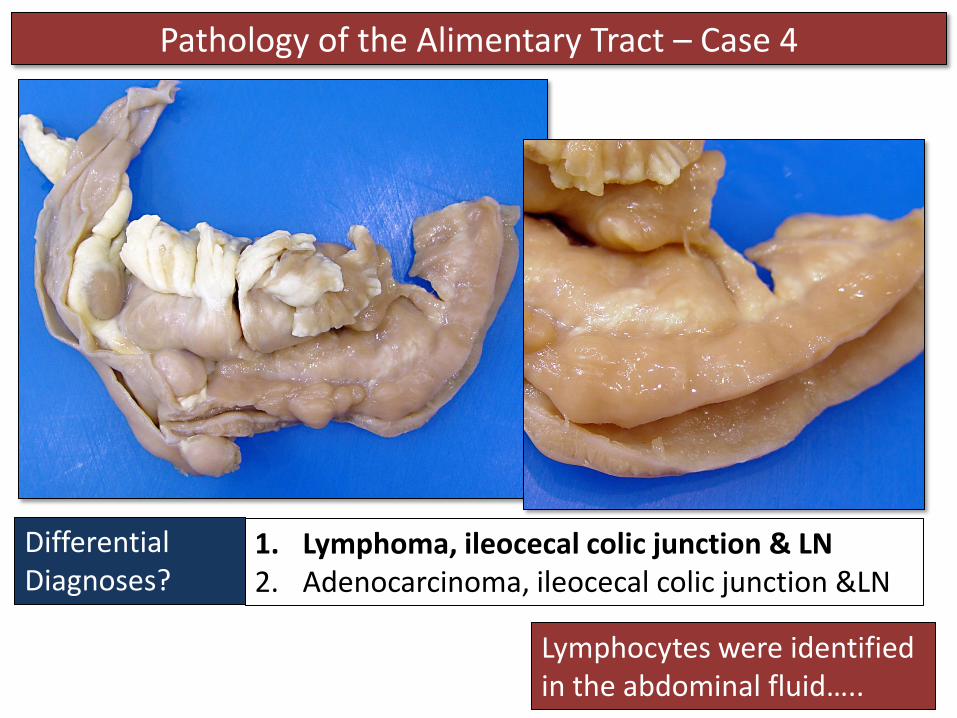
• Adult cat
• Found in ditch
• Abdominal mass found via radiographs
• Ascites developed
• Abdominocentesis and FNA performed: lymphocytes identified in abdominal fluid

Pathology of the Alimentary Tract – Case 4
Rumen
Tan homogenous tissue infiltrates and expands the wall of the intestine at the ileocecal-colic junction. Similar tissue invades the mesentery and enlarges/replaces the adjacent lymph nodes.
Description?
Pathology of the Alimentary Tract – Case 4
Rumen
1. Lymphoma, ileocecal colic junction & LN 2. Adenocarcinoma, ileocecal colic junction &LN
Differential Diagnoses?
Lymphocytes were identified in the abdominal fluid…..

Pathology of the Alimentary Tract – Case 4
1. Lymphoma, ileocecal colic junction & LN 2. Adenocarcinoma, ileocecal colic junction & LN
Differential Diagnoses?
3. Pyogranulomatous enterotyphlocolitis and lymphadenitis!!
Feline Infectious Peritonitis (FIP) : Feline coronavirus

Pathology of the Alimentary Tract – Case 5
• 6 year old Jersey cow
• Abdominal distension
• The cow had abundant abdominal adipose stores
• 17 year old FS cat
• Abdominal pain, ↑amylase/lipase
• Abdominal US : suggestive of peritonitis or carcinomatosis

Pathology of the Alimentary Tract – Case 5
Description?
Abundant fat surrounds the small intestinal loops adhering the loops to one another. The fat is diffusely firm with white chalky foci.
Scattered multifocally throughout the mesenteric fat are small white chalky foci
Description?

Pathology of the Alimentary Tract – Case 5
Morphologic Diagnosis Mesenteric fat necrosis, diffuse and marked (cow) / multifocal and mild to moderate (cat)
Why are the lesions ‘chalky’ and white?
Saponification of fat – when fatty acids are released from necrotic fat they combine with calcium deposits to form soaps

Pathology of the Alimentary Tract – Case 5
Cause? In cattle with pathogenesis is obscure. Often occurs in Jersey cattle with abundant adipose stores. It may be related to diet and ↑saturated fatty acid production in the rumen. Fat forms which is solid at body temperature → this fat may be more prone to trauma / ischemic necrosis

Pathology of the Alimentary Tract – Case 5
Cause? In cats, fat necrosis more commonly results from enzymatic damage, usually resulting from acute pancreatitis → release of proteolytic and lipolytic enzymes.

Pathology of the Alimentary Tract – Case 6
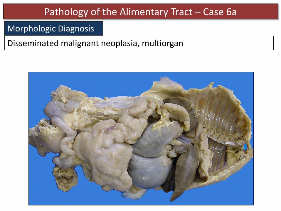
• 7 year old female rabbit with a history GI stasis

Pathology of the Alimentary Tract – Case 6a
Multifocal to coalescing , firm, tan, homogenous masses ranging in size from 0.5 cm to 10 x 15 cm are scattered throughout the abdominal cavity affecting most of the viscera. Smaller masses are also present in the thoracic body wall and diaphragm.
Description?
Pathology of the Alimentary Tract – Case 6a
Disseminated malignant neoplasia, multiorgan
Morphologic Diagnosis
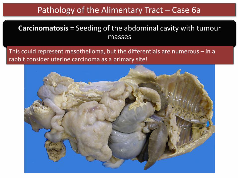
Pathology of the Alimentary Tract – Case 6a
Carcinomatosis = Seeding of the abdominal cavity with tumour masses
This could represent mesothelioma, but the differentials are numerous – in a rabbit consider uterine carcinoma as a primary site!

Pathology of the Alimentary Tract – Case 6b
• Two chicks from a farm with increased chick mortality

Pathology of the Alimentary Tract – Case 6b
Morphologic Diagnosis
Strands and sheets of tan, friable material are loosely adhered to the serosa of the abdominal viscera and body wall
Description
Fibrinous peritonitis (coelomitis), diffuse, acute, severe
Possible causes
1. Bacterial septicemia 2. Penetrating injury through the
body wall or ascending from umbilicus
3. Rupture of the GI tract E. coli septicemia is a common cause in chicks

Questions??


